
Combined Metabolic and Functional Tumor Volumes on [18F]FDG-PET/MRI in Neuroblastoma Using Voxel-Wise Analysis

 ,
,  ,
,  , , , ,
Abstract
1. Introduction
2. Methods
2.1. Patients
2.2. Image Acquisition
2.3. Data Processing
2.4. Data Analysis
2.5. Statistical Evaluation
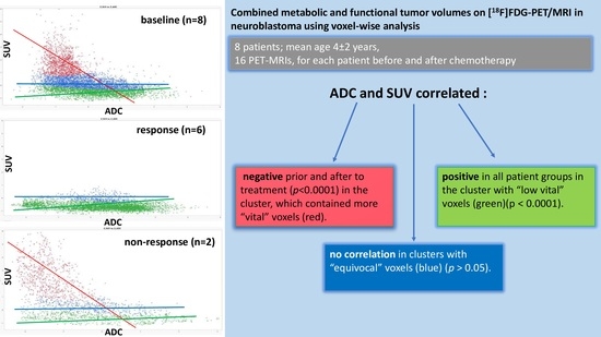
3. Results
4. Discussion
Author Contributions
Funding
Institutional Review Board Statement
Informed Consent Statement
Data Availability Statement
Conflicts of Interest
References
- Cohn, S.L.; Pearson, A.D.; London, W.B.; Monclair, T.; Ambros, P.F.; Brodeur, G.M.; Faldum, A.; Hero, B.; Iehara, T.; Machin, D.; et al. The International Neuroblastoma Risk Group (INRG) classification system: An INRG Task Force report. J. Clin. Oncol. 2009, 27, 289–297. [Google Scholar] [CrossRef] [PubMed]
- Regier, M.; Derlin, T.; Schwarz, D.; Laqmani, A.; Henes, F.O.; Groth, M.; Buhk, J.H.; Kooijman, H.; Adam, G. Diffusion weighted MRI and 18F-FDG PET/CT in non-small cell lung cancer (NSCLC): Does the apparent diffusion coefficient (ADC) correlate with tracer uptake (SUV)? Eur. J. Radiol. 2012, 81, 2913–2918. [Google Scholar] [CrossRef] [PubMed]
- Simon, T.; Hero, B.; Schulte, J.H.; Deubzer, H.; Hundsdoerfer, P.; von Schweinitz, D.; Fuchs, J.; Schmidt, M.; Prasad, V.; Krug, B.; et al. 2017 GPOH Guidelines for Diagnosis and Treatment of Patients with Neuroblastic Tumors. Klin. Padiatr. 2017, 229, 147–167. [Google Scholar] [CrossRef]
- Monclair, T.; Mosseri, V.; Cecchetto, G.; De Bernardi, B.; Michon, J.; Holmes, K. Influence of image-defined risk factors on the outcome of patients with localised neuroblastoma. A report from the LNESG1 study of the European International Society of Paediatric Oncology Neuroblastoma Group. Pediatr. Blood Cancer 2015, 62, 1536–1542. [Google Scholar] [CrossRef] [PubMed]
- Kwee, T.C.; Basu, S.; Saboury, B.; Ambrosini, V.; Torigian, D.A.; Alavi, A. A new dimension of FDG-PET interpretation: Assessment of tumor biology. Eur. J. Nucl. Med. Mol. Imaging 2011, 38, 1158–1170. [Google Scholar] [CrossRef]
- Li, C.; Zhang, J.; Chen, S.; Huang, S.; Wu, S.; Zhang, L.; Zhang, F.; Wang, H. Prognostic value of metabolic indices and bone marrow uptake pattern on preoperative 18F-FDG PET/CT in pediatric patients with neuroblastoma. Eur. J. Nucl. Med. Mol. Imaging 2018, 45, 306–315. [Google Scholar] [CrossRef]
- Schäfer, J.F.; Herrmann, J.; Kammer, B.; Koerber, F.; Tsiflikas, I.; von Kalle, T.; Mentzel, H.J. Fortschrittliche radiologische Diagnostik bei soliden Tumoren im Kindes- und Jugendalter. Onkologe 2021, 27, 410–426. [Google Scholar] [CrossRef]
- Gassenmaier, S.; Tsiflikas, I.; Fuchs, J.; Grimm, R.; Urla, C.; Esser, M.; Maennlin, S.; Ebinger, M.; Warmann, S.W.; Schafer, J.F. Feasibility and possible value of quantitative semi-automated diffusion weighted imaging volumetry of neuroblastic tumors. Cancer Imaging 2020, 20, 89. [Google Scholar] [CrossRef]
- Peschmann, A.L.; Beer, M.; Ammann, B.; Dreyhaupt, J.; Kneer, K.; Beer, A.J.; Beltinger, C.; Steinbach, D.; Cario, H.; Neubauer, H. Quantitative DWI predicts event-free survival in children with neuroblastic tumours: Preliminary findings from a retrospective cohort study. Eur. Radiol. Exp. 2019, 3, 6. [Google Scholar] [CrossRef]
- Neubauer, H.; Li, M.; Muller, V.R.; Pabst, T.; Beer, M. Diagnostic Value of Diffusion-Weighted MRI for Tumor Characterization, Differentiation and Monitoring in Pediatric Patients with Neuroblastic Tumors. Rofo 2017, 189, 640–650. [Google Scholar] [CrossRef]
- Divine, M.R.; Katiyar, P.; Kohlhofer, U.; Quintanilla-Martinez, L.; Pichler, B.J.; Disselhorst, J.A. A Population-Based Gaussian Mixture Model Incorporating 18F-FDG PET and Diffusion-Weighted MRI Quantifies Tumor Tissue Classes. J. Nucl. Med. 2016, 57, 473–479. [Google Scholar] [CrossRef] [PubMed]
- Heusch, P.; Buchbender, C.; Kohler, J.; Nensa, F.; Beiderwellen, K.; Kuhl, H.; Lanzman, R.S.; Wittsack, H.J.; Gomez, B.; Gauler, T.; et al. Correlation of the apparent diffusion coefficient (ADC) with the standardized uptake value (SUV) in hybrid 18F-FDG PET/MRI in non-small cell lung cancer (NSCLC) lesions: Initial results. Rofo 2013, 185, 1056–1062. [Google Scholar] [CrossRef] [PubMed]
- Ho, K.C.; Lin, G.; Wang, J.J.; Lai, C.H.; Chang, C.J.; Yen, T.C. Correlation of apparent diffusion coefficients measured by 3T diffusion-weighted MRI and SUV from FDG PET/CT in primary cervical cancer. Eur. J. Nucl. Med. Mol. Imaging 2009, 36, 200–208. [Google Scholar] [CrossRef]
- Rakheja, R.; Chandarana, H.; DeMello, L.; Jackson, K.; Geppert, C.; Faul, D.; Glielmi, C.; Friedman, K.P. Correlation between standardized uptake value and apparent diffusion coefficient of neoplastic lesions evaluated with whole-body simultaneous hybrid PET/MRI. AJR Am. J. Roentgenol. 2013, 201, 1115–1119. [Google Scholar] [CrossRef] [PubMed]
- Shimada, H.; Ikegaki, N. Genetic and Histopathological Heterogeneity of Neuroblastoma and Precision Therapeutic Approaches for Extremely Unfavorable Histology Subgroups. Biomolecules 2022, 12, 79. [Google Scholar] [CrossRef]
- Schmidt, H.; Brendle, C.; Schraml, C.; Martirosian, P.; Bezrukov, I.; Hetzel, J.; Muller, M.; Sauter, A.; Claussen, C.D.; Pfannenberg, C.; et al. Correlation of simultaneously acquired diffusion-weighted imaging and 2-deoxy-[18F] fluoro-2-D-glucose positron emission tomography of pulmonary lesions in a dedicated whole-body magnetic resonance/positron emission tomography system. Investig. Radiol. 2013, 48, 247–255. [Google Scholar] [CrossRef]
- Gatidis, S.; la Fougere, C.; Schaefer, J.F. Pediatric Oncologic Imaging: A Key Application of Combined PET/MRI. Rofo 2016, 188, 359–364. [Google Scholar] [CrossRef]
- Delso, G.; Furst, S.; Jakoby, B.; Ladebeck, R.; Ganter, C.; Nekolla, S.G.; Schwaiger, M.; Ziegler, S.I. Performance measurements of the Siemens mMR integrated whole-body PET/MR scanner. J. Nucl. Med. 2011, 52, 1914–1922. [Google Scholar] [CrossRef]
- Patz, E.F., Jr.; Lowe, V.J.; Hoffman, J.M.; Paine, S.S.; Burrowes, P.; Coleman, R.E.; Goodman, P.C. Focal pulmonary abnormalities: Evaluation with F-18 fluorodeoxyglucose PET scanning. Radiology 1993, 188, 487–490. [Google Scholar] [CrossRef]
- Hellwig, D.; Graeter, T.P.; Ukena, D.; Groeschel, A.; Sybrecht, G.W.; Schaefers, H.J.; Kirsch, C.M. 18F-FDG PET for mediastinal staging of lung cancer: Which SUV threshold makes sense? J. Nucl. Med. 2007, 48, 1761–1766. [Google Scholar] [CrossRef]
- Schmitz, J.; Schwab, J.; Schwenck, J.; Chen, Q.; Quintanilla-Martinez, L.; Hahn, M.; Wietek, B.; Schwenzer, N.; Staebler, A.; Kohlhofer, U.; et al. Decoding Intratumoral Heterogeneity of Breast Cancer by Multiparametric In Vivo Imaging: A Translational Study. Cancer Res. 2016, 76, 5512–5522. [Google Scholar] [CrossRef] [PubMed]
- McLachlan, G.J.; Lee, S.X.; Rathnayake, S.I. Finite Mixture Models; John Wiley & Sons Ltd.: New York, NY, USA, 2004. [Google Scholar]
- Maennlin, S.; Chaika, M.; Gassenmaier, S.; Grimm, R.; Sparber-Sauer, M.; Fuchs, J.; Schmidt, A.; Ebinger, M.; Hettmer, S.; Gatidids, S.; et al. Evaluation of functional and metabolic tumor volume using voxel-wise analysis in childhood rhabdomyosarcoma. Pediatr. Radiol. 2022, 53, 438–449. [Google Scholar] [CrossRef] [PubMed]
- Ghosh, A.; Yekeler, E.; Dalal, D.; Holroyd, A.; States, L. Whole-tumour apparent diffusion coefficient (ADC) histogram analysis to identify MYCN-amplification in neuroblastomas: Preliminary results. Eur. Radiol. 2022, 32, 8453–8462. [Google Scholar] [CrossRef]
- Uhl, M.; Altehoefer, C.; Kontny, U.; Il’yasov, K.; Buchert, M.; Langer, M. MRI-diffusion imaging of neuroblastomas: First results and correlation to histology. Eur. Radiol. 2002, 12, 2335–2338. [Google Scholar] [CrossRef] [PubMed]
- Surov, A.; Meyer, H.J.; Schob, S.; Hohn, A.K.; Bremicker, K.; Exner, M.; Stumpp, P.; Purz, S. Parameters of simultaneous 18F-FDG-PET/MRI predict tumor stage and several histopathological features in uterine cervical cancer. Oncotarget 2017, 8, 28285–28296. [Google Scholar] [CrossRef] [PubMed][Green Version]
- Pedersen, C.; Aboian, M.; McConathy, J.E.; Daldrup-Link, H.; Franceschi, A.M. PET/MRI in Pediatric Neuroimaging: Primer for Clinical Practice. AJNR Am. J. Neuroradiol. 2022, 43, 938–943. [Google Scholar] [CrossRef]
- Padma, M.V.; Said, S.; Jacobs, M.; Hwang, D.R.; Dunigan, K.; Satter, M.; Christian, B.; Ruppert, J.; Bernstein, T.; Kraus, G.; et al. Prediction of pathology and survival by FDG PET in gliomas. J. Neurooncol. 2003, 64, 227–237. [Google Scholar] [CrossRef]
- Man, S.; Yan, J.; Li, J.; Cao, Y.; Hu, J.; Ma, W.; Liu, J.; Zhao, Q. Value of pretreatment 18F-FDG PET/CT in prognosis and the reflection of tumor burden: A study in pediatric patients with newly diagnosed neuroblastoma. Int. J. Med. Sci. 2021, 18, 1857–1865. [Google Scholar] [CrossRef]
- Schaarschmidt, B.M.; Buchbender, C.; Nensa, F.; Grueneisen, J.; Gomez, B.; Kohler, J.; Reis, H.; Ruhlmann, V.; Umutlu, L.; Heusch, P. Correlation of the apparent diffusion coefficient (ADC) with the standardized uptake value (SUV) in lymph node metastases of non-small cell lung cancer (NSCLC) patients using hybrid 18F-FDG PET/MRI. PLoS ONE 2015, 10, e0116277. [Google Scholar] [CrossRef]
- Surov, A.; Stumpp, P.; Meyer, H.J.; Gawlitza, M.; Hohn, A.K.; Boehm, A.; Sabri, O.; Kahn, T.; Purz, S. Simultaneous (18)F-FDG-PET/MRI: Associations between diffusion, glucose metabolism and histopathological parameters in patients with head and neck squamous cell carcinoma. Oral. Oncol. 2016, 58, 14–20. [Google Scholar] [CrossRef]
- Sun, H.; Xin, J.; Zhang, S.; Guo, Q.; Lu, Y.; Zhai, W.; Zhao, L.; Peng, W.; Wang, B. Anatomical and functional volume concordance between FDG PET, and T2 and diffusion-weighted MRI for cervical cancer: A hybrid PET/MR study. Eur. J. Nucl. Med. Mol. Imaging 2014, 41, 898–905. [Google Scholar] [CrossRef] [PubMed]
- Le Bihan, D.J. Differentiation of benign versus pathologic compression fractures with diffusion-weighted MR imaging: A closer step toward the “holy grail” of tissue characterization? Radiology 1998, 207, 305–307. [Google Scholar] [CrossRef] [PubMed]
- Ahangari, S.; Littrup Andersen, F.; Liv Hansen, N.; Jakobi Nottrup, T.; Berthelsen, A.K.; Folsted Kallehauge, J.; Richter Vogelius, I.; Kjaer, A.; Espe Hansen, A.; Fischer, B.M. Multi-parametric PET/MRI for enhanced tumor characterization of patients with cervical cancer. Eur. J. Hybrid. Imaging 2022, 6, 7. [Google Scholar] [CrossRef] [PubMed]


| Dixon | STIRcor | T2-TSE | STIRax | DWI | |
|---|---|---|---|---|---|
| TE (echo time) [ms] | 1.23/2.46 | 78 | 100 | 81 | 60 |
| TR (repetition time) [ms] | 3.6 | 6400 | 3500 | 4500 | 6000 |
| bandwidth [Hz/px] | 965 | 383 | 260 | 220 | 1860 |
| matrix size [px] | 79 × 192 | 256 × 256 | 256 × 300 | 197 × 384 | 108 × 192 |
| resolution [mm3] | 4.1 × 2.6 × 2.6 | 1.5 × 1.5 × 4 | 1.25 × 1.25 × 5 | 1.2 × 0.83 × 5 | 2.6 × 2.6 × 5 |
| excitation angle [°] | 10 | 120 | 90 | 120 | 90 |
| inversion time [ms] | 200 | 220 | |||
| b-values [mm2/s] | 50 and 800 |
| Patients | 8 |
| Sex | 5 male, 3 female |
| Age: | |
| Mean age ± SD | 4 ± 2 years |
| Range | 1–10 years |
| Histology: | |
| MYNC-positive | n = 4 |
| MiBG-positive | n = 3 |
| ALK-amplification-positive | n = 2 |
| Risk group stratification: | |
| High risk | 5 |
| Intermediate risk | 2 |
| Low risk | 1 |
| Patient | Sex | Age | Stage/Risk | Genetic, EFS/OAS | Baseline | Post-Chemo |
|---|---|---|---|---|---|---|
| 1 | male | 3 | IV./high | - |  |  |
| 2 | female | 10 | IV./high | ALK-positive |  |  |
| 3 | male | 4 | IV./high | ALK-positive |  |  |
| 4 | female | 3 | IV./high | N-MYC-positive |  |  |
| 5 | male | 1 | III./low | ALK-positive |  |  |
| 6 | male | 4 | IV./high | N-MYC-positive |  |  |
| 7 | male | 5 | IV./high | - |  |  |
| 8 | female | 5 | IV./high | N-MY-Cpositive |  |  |
| Before Treatment | Response (n = 6) | Progress (n = 2) | |
|---|---|---|---|
| ADC mean ± SD | 1159 ± 417 | 1402 ± 511 | 928 ± 338 |
| SUV mean ± SD | 1.75 ± 0.92 | 0.97 ± 0.28 | 2.26 ± 1.52 |
| Median Volume (total) [mL] | 1605 | 318 | 190 |
| vital | 26.3% | 0.03% | 41.8% |
| low vital | 35.8% | 65.7% | 24.9% |
| equivocal | 37.9% | 34.3% | 33.3% |
Disclaimer/Publisher’s Note: The statements, opinions and data contained in all publications are solely those of the individual author(s) and contributor(s) and not of MDPI and/or the editor(s). MDPI and/or the editor(s) disclaim responsibility for any injury to people or property resulting from any ideas, methods, instructions or products referred to in the content. |
© 2023 by the authors. Licensee MDPI, Basel, Switzerland. This article is an open access article distributed under the terms and conditions of the Creative Commons Attribution (CC BY) license (https://creativecommons.org/licenses/by/4.0/).
Share and Cite
Chaika, M.; Männlin, S.; Gassenmaier, S.; Tsiflikas, I.; Dittmann, H.; Flaadt, T.; Warmann, S.; Gückel, B.; Schäfer, J.F. Combined Metabolic and Functional Tumor Volumes on [18F]FDG-PET/MRI in Neuroblastoma Using Voxel-Wise Analysis. J. Clin. Med. 2023, 12, 5976. https://doi.org/10.3390/jcm12185976
Chaika M, Männlin S, Gassenmaier S, Tsiflikas I, Dittmann H, Flaadt T, Warmann S, Gückel B, Schäfer JF. Combined Metabolic and Functional Tumor Volumes on [18F]FDG-PET/MRI in Neuroblastoma Using Voxel-Wise Analysis. Journal of Clinical Medicine. 2023; 12(18):5976. https://doi.org/10.3390/jcm12185976
Chicago/Turabian StyleChaika, Maryanna, Simon Männlin, Sebastian Gassenmaier, Ilias Tsiflikas, Helmut Dittmann, Tim Flaadt, Steven Warmann, Brigitte Gückel, and Jürgen Frank Schäfer. 2023. "Combined Metabolic and Functional Tumor Volumes on [18F]FDG-PET/MRI in Neuroblastoma Using Voxel-Wise Analysis" Journal of Clinical Medicine 12, no. 18: 5976. https://doi.org/10.3390/jcm12185976
APA StyleChaika, M., Männlin, S., Gassenmaier, S., Tsiflikas, I., Dittmann, H., Flaadt, T., Warmann, S., Gückel, B., & Schäfer, J. F. (2023). Combined Metabolic and Functional Tumor Volumes on [18F]FDG-PET/MRI in Neuroblastoma Using Voxel-Wise Analysis. Journal of Clinical Medicine, 12(18), 5976. https://doi.org/10.3390/jcm12185976