Review

Impairment of Mesenteric Perfusion as a Marker of Major Bleeding in Trauma Patients

1
Department of Traumatology, University of Szeged, H-6725 Szeged, Hungary
2
Institute of Surgical Research, University of Szeged, H-6724 Szeged, Hungary
3
Department of Sports Medicine, University of Szeged, H-6725 Szeged, Hungary
*
Author to whom correspondence should be addressed.
These authors contributed equally to this work.
J. Clin. Med. 2023, 12(10), 3571; https://doi.org/10.3390/jcm12103571
Submission received: 21 April 2023 / Revised: 17 May 2023 / Accepted: 18 May 2023 / Published: 20 May 2023
(This article belongs to the Special Issue Advances in Trauma Treatment)

Abstract

:
The majority of potentially preventable mortality in trauma patients is related to bleeding; therefore, early recognition and effective treatment of hemorrhagic shock impose a cardinal challenge for trauma teams worldwide. The reduction in mesenteric perfusion (MP) is among the first compensatory responses to blood loss; however, there is no adequate tool for splanchnic hemodynamic monitoring in emergency patient care. In this narrative review, (i) methods based on flowmetry, CT imaging, video microscopy (VM), measurement of laboratory markers, spectroscopy, and tissue capnometry were critically analyzed with respect to their accessibility, and applicability, sensitivity, and specificity. (ii) Then, we demonstrated that derangement of MP is a promising diagnostic indicator of blood loss. (iii) Finally, we discussed a new diagnostic method for the evaluation of hemorrhage based on exhaled methane (CH4) measurement. Conclusions: Monitoring the MP is a feasible option for the evaluation of blood loss. There are a wide range of experimentally used methodologies; however, due to their practical limitations, only a fraction of them could be integrated into routine emergency trauma care. According to our comprehensive review, breath analysis, including exhaled CH4 measurement, would provide the possibility for continuous, non-invasive monitoring of blood loss.

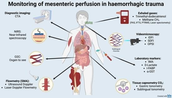
Graphical Abstract

1. Introduction

Despite the development of trauma care in the past few decades, roughly 25% of post-injury mortality may be potentially preventable by early detection and proper treatment of life-threatening deteriorations. The further development in the management of hemorrhagic shock (HS) is of utmost importance, since HS can be referred as the main cause of potentially preventable trauma mortality [1,2,3].
Regarding the recognition of HS, the challenge is to identify its impending presence in the pre-shock state. To date, the initial hemodynamic assessment of the injured relies on vital signs (VS) such as heart rate, and metabolic markers such as base deficit (BD) and lactate [4,5,6]. However, the specificity of VS and metabolic markers for hypovolemia remained questionable, since several factors such as medication, alcohol intoxication, administration of crystalloids (lactated ringer or saline), or even advanced age can diminish their reliability [7,8,9,10,11]. Furthermore, VS, BD, and lactate are global markers of shock that are maintained at near-normal levels until the compensatory mechanisms of the individual patient become fully exhausted. Consequently, derangements of these indicators during blood loss may remain subtle in the pre-shock state and become apparent when the changes are already non-reversible. In contrast, hemorrhage induces early compensatory mechanisms and temporospatial differences in regional perfusion hallmarked by a redistribution of blood flow from non-vital organs (e.g., the gut and the skin) towards vital vascular beds (i.e., the coronary and cerebral areas) [12,13].
Additionally, the evaluation of the efficacy of treatment is often challenging. Increasing urinary output is a reasonably sensitive marker of improving hemodynamic status; nevertheless, underlying kidney injury, hyperglycemia, or diuretic agents can limit its accuracy [6]. Invasive monitoring methods such as pulmonary artery catheterization offer substantial benefits; however, they are hardly applicable during the initial phase of therapy due to patient positioning and time factor [14,15].
In addition to VS and metabolic markers, hemoglobin (Hb) and hematocrit (Hct) levels are the most frequently used indicators of blood loss due to their several advantages including easy accessibility either with standard laboratory or minimally invasive point of care (POC) testing. However, their diagnostic values in the initial management of trauma patients remains controversial [16]. Initial Hb and Hct levels are influenced by many factors that are not associated with bleeding, such as the patient’s age, gender, weight, and underlying conditions including anemia [17,18]. Furthermore, the on-site Hb values are often lower due to the almost immediate fluid refilling from the interstitium to restore the intravascular volume, early after sustaining trauma. Then, prehospital fluid resuscitation induces further hemodilution and fall in Hct and Hb. Therefore, serial measurements are recommended for the evaluation of trauma-related hemorrhage [18,19], but the results are still controversial [17,20].
Imaging modalities are important adjuncts to the initial hemodynamic assessment in trauma care. Computer tomography (CT) is a reliable method for detecting internal hemorrhage; however, it requires transportation out of the emergency department, resulting in unfavorable time delays. As compared to CT, ultrasound has notable advantages including bedside availability, lack of radiation, reproducibility, and low costs [21]. The focused assessment with sonography in trauma (FAST) and extended FAST (eFAST) protocols can be performed in less than 5 min and display high sensitivity and specificity for hemoperitoneum, hemopericardium, and hemothorax. [22]. Nonetheless, eFAST is hampered by several limitations. Most importantly, the reliability of POC ultrasound depends on the experience of the user and the patient’s body composition. Additionally, visualization of retroperitoneal hemorrhage and differentiation between blood and urine are hardly feasible with ultrasound [21].
Ultimately, no gold standard technique exists for diagnosing and assessing hemorrhage in severe trauma; thus, decision-making is commonly based on a combination of tests, which all have their strengths and limitations. Frequently used tests for the initial hemodynamic assessment of trauma patients are presented in Figure 1, with respect to their accessibility, applicability, sensitivity, and specificity for blood loss scored from 1 to 3 with an arbitrary scaling (Figure 1).
Accessibility refers to the availability of the requirements for implementing the technique, such as machinery, proper instruments, and specially trained personnel. Promptly available vital signs and blood gas parameters obtained the highest accessibility (3). Although urinary catheterization is mostly easy to perform, the hourly diuresis can hardly be determined promptly. Furthermore, catheterization is contraindicated if urethral injury is suspected (e.g., in case of perineal or scrotal hematoma or blood at the meatus) [6]. Due to these limitations, urinary output was judged to carry a moderate level of accessibility (2) in emergency trauma. CT angiography and measurement of pulmonary artery pressure are widely available; nevertheless, they require specific equipment and staff, entailing a medium accessibility (2) for the method. Microcirculatory measurements and monitoring mesenterial blood flow are not routinely utilized techniques and they need a more sophisticated instrumental background. Consequently, their accessibility was ranked the lowest (1).
Applicability is confined to feasibility for continuous monitoring. The parameters and techniques allowing real time monitoring (e.g., pulse oximetry, urinary output, etc.) received the highest applicability (3). The applicability was judged as moderate (2) if monitoring is not a feasible option, but repeated measurements can be performed easily (e.g., blood gas parameters). Although CT angiography can be repeated, executing several CTs during the early phase of patient management is unpractical and potentially dangerous, as it is relatively time consuming and it requires the transportation of the patient to the radiology unit. Therefore, CT angiography obtained the lowest applicability score (1).
Sensitivity refers to the capability used in terms of prompt indication of blood loss and changes in the hemodynamic status of patients. CT angiography, pulmonary artery pressure monitoring, mesenterial blood flow, and microcirculatory changes of gastrointestinal vascular beds are considered as highly sensitive indicators of blood loss [23,24,25]. Additionally, a high sensitivity score (3) was associated with pulse oximetry, as it indubitably reacts to hypoperfusion, although this reaction may be an absent or inaccurate reading. Being more reliable than conventional metabolic markers and vital signs, urinary output, eFAST, Hb, and Hct received medium sensitivity scores (2) [6,21,26,27]. Despite being useful markers in the early assessment of trauma patients, HR, blood pressure, lactate, and BD obtained a sensitivity score of 1 due to the compensatory mechanisms of the body and external influencing factors potentially keeping these values in normal range in the early phases of hemorrhage [6,12].
Specificity refers to the selectivity of the technique for volume depletion and bleeding. Methods directly examining macro- or microvascular systems received the highest specificity value (3). Hb and Hct also obtained a high specificity score (3) for bleeding [16,28,29]. Metabolic markers and cardiovascular vital parameters such as HR and SBP were judged to hold medium specificity for bleeding, since several other factors can influence their values as discussed above. Pulse oximetry is routinely performed to assess blood oxygenation in most emergencies and critical care settings. However, since low perfusion degrades the performance of pulse oximetry, it aims primarily to draw attention to respiratory insufficiencies [30]. For this reason, pulse oximetry obtained the lowest specificity score (1).
Ultimately, it is important to emphasize that the above-described scoring is arbitrary, even though its foundation relies on scientific data. The main goal of this scoring is to illustrate trends and highlight the lack of an easily accessible, highly applicable test with high sensitivity and specificity, calling for further research in the diagnostics of acute blood loss.
Commonly used tests for the initial hemodynamic assessment of trauma patients are presented by highlighting their accessibility, applicability, sensitivity, and specificity for blood loss, based on arbitrary scoring. Mesenterial blood flow refers to the superior mesenteric artery perfusion, while mesenteric mucosal microcirculation concerns specifically microperfusion. Darker colors and higher numbers indicate higher value (easier accessibility, better applicability, higher sensitivity and specificity). Here, accessibility refers to the availability of the requirements for implementing the technique, such as machinery, proper instruments, and specially trained personnel, while applicability is confined to feasibility for continuous monitoring. Sensitivity is capability used in terms of prompt indication of blood loss and changes in the hemodynamic status of patients. Specificity refers to the selectivity of the technique for bleeding; thus, highly specific methods are characterized by reliability for indicating bleeding without being influenced by other factors such as medication, pain, or anxiety. Although vital signs such as heart rate and blood pressure are easily accessible and applicable, they display poor sensitivity and specificity for blood loss. Blood gas parameters, laboratory markers, and imaging modalities provide substantial benefits; however, they do not allow continuous monitoring. Monitoring pulmonary artery pressure and superior mesenteric artery flow blood flow have only the lack of accessibility as a major disadvantage. Making these parameters more accessible in emergency situations by developing prompt and non-invasive techniques to measure them may significantly improve the quality of care.
The reduction in mesenteric perfusion (MP) is among the first compensatory reactions to blood loss, thereby being a potential early clinical indicator of hemorrhage [23,31]. This review offers an insight into the currently available techniques for the evaluation of MP and discusses the possibility of a promising new method that may lead to future quality improvement in emergency trauma care.

2. Materials and Methods

2.1. Setting the Aim

Some of the authors face the hardships of emergency trauma care day by day. Quality improvement consultations are scheduled on a regular basis in order to designate areas of development in patient care. We agreed that the early recognition of internal hemorrhage is a critical issue that requires improvement in the future. Based on previous studies of our research group, we believed that utilizing the rapid alterations of mesenteric perfusion in circulatory volume depletion has the potential to facilitate this process. For this reason, we decided to provide a comprehensive review of the currently available techniques for the assessment of mesenteric perfusion in the context of emergency trauma.

2.2. Data Collection Methods

As a narrative review, data collection was performed from multiple sources, not exclusively from one systematic search. As a first step, the authors listed all methods that were theoretically applicable for the assessment of mesenteric perfusion in the clinical setting. The literature was reviewed by two authors independently, based on a search in MEDLINE (via PubMed) database with the following search terms: “monitoring” AND (“mesenteric perfusion” OR “splanchnic perfusion” OR “intestinal perfusion” OR “mesenteric circulation” OR “splanchnic circulation” OR “intestinal circulation”) AND (“bleeding” OR “haemorrhage” OR “hemorrhage” OR “haemodynamic” OR “hemodynamic”). This search yielded 106 papers which were assessed based on title and abstract. Ultimately, 34 full texts were reviewed. The information obtained from these studies was critically evaluated and extended with data extracted from further papers. Fifty-seven manuscripts were reached via the reference lists of the 34 papers accessed through the above-mentioned MEDLINE search. Forty-nine studies related to the topic were already known by the authors. Nine articles were suggested by other experts the authors briefly consulted with. Twelve studies were accessed through non-systematic use of online search engines. In total, the authors reviewed 161 full-text articles.

3. Results

The rapid response of MP is regulated by finely tuned physiological reflexes and neurohumoral processes. As an initial response to a hemorrhage, when the circulating blood volume decreases, the reduction in arterial baroreceptor filling leads to an increased efferent sympathetic activation.
The increased sympathetic output is associated with reflex tachycardia, which, together with the fluid retention via aldosterone and vasopressin, aim to maintain blood pressure. Apart from the cardiac effects, released sympathetic mediators stimulate the α-adrenergic receptors on both the afferent and efferent sides of the microcirculation. Selective vasoconstriction of the afferent arterioles serves to sustain the vascular resistance, while the stimulation of α-adrenergic receptors on postcapillary venules and veins results in autotransfusion by increasing vascular and ultimately the cardiac filling [32].
Arteriolar responses depend on the distribution of the vasoconstrictor α-adrenergic and the vasodilator β2-adrenergic receptor subtypes, which vary within the different tissues. Accordingly, the visceral perfusion is partly sacrificed through the vasoconstrictive response that is mediated by the sympathetic nervous system. However, the abdominal organs are affected unequally by redistribution; for example, intestinal, gastric, and pancreatic blood supplies are more susceptible to the effects of hemorrhage compared to the liver due to the hepatic arterial buffer response [33,34,35]. Intestines are affected by ischemia, particularly adversely and rapidly, due to their unique microanatomy, where the artery and vein within the villi run parallel to each other, which results in low oxygenation in the most luminal areas of the intestine, even under optimal conditions [36,37]. The particular sensitivity of MP to blood loss demonstrated by studies on large animal models, where the superior mesenteric artery (SMA) flow displays a significant drop already at 5% loss of total blood volume; and continues to diminish in parallel with ongoing hemorrhage [23]. Considering the total circulating blood volume as 5 L for an adult, 5% loss means 250 mL of blood, which can hardly be detected with the currently used routine diagnostic tools. This conceptual framework provides the rationale for using biomarkers of the integrity of MP to assess the amount of blood loss in trauma patients.
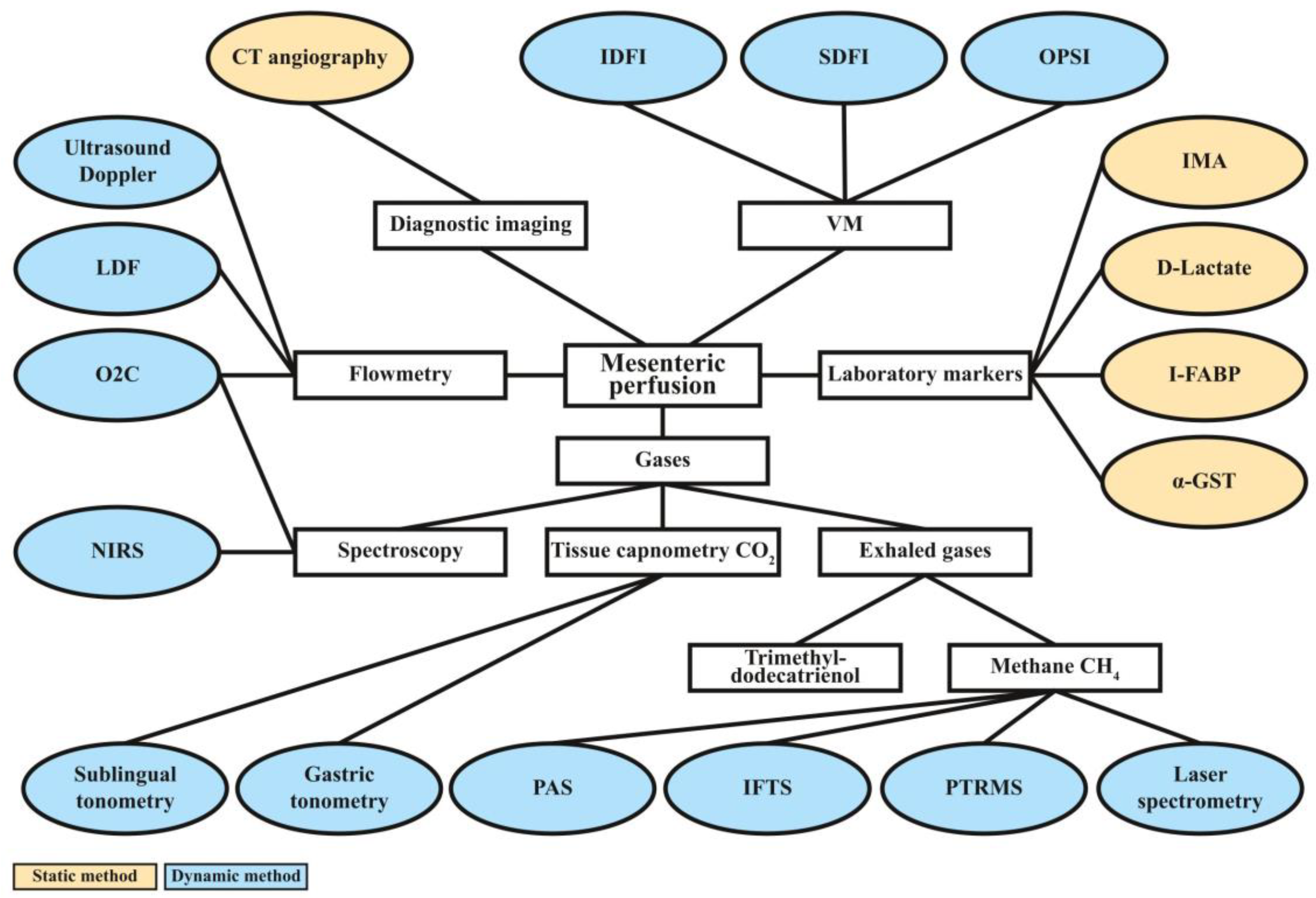
The almost immediate circulatory redistribution detailed above makes monitoring MP a promising approach in the initial assessment of bleeding trauma patients [23]. Theoretically, there are a wide range of experimentally used methodologies for the evaluation of intestinal macro-and micro perfusion; however, only a fraction of them were integrated into routine emergency trauma care. Methods based on flowmetry, CT imaging, videomicroscopy (VM), measurement of laboratory markers, spectroscopy, tissue capnometry, and breath analysis can all provide valuable information on MP; nevertheless, each technique has its limitations. Figure 2 provides an overview of the currently available methods for assessing MP. In the following, the strengths and limitations of each technique are discussed.
Mesenteric perfusion can be investigated with a variety of techniques. The principles of the methods (diagnostic imaging, flowmetry, VM, laboratory tests, analysis of dissolved and exhaled gases) are shown in white rectangles. The specific techniques or markers are presented in oval text boxes. Based on the capability for the real-time monitoring of MP, techniques can have a static or dynamic nature, which is represented by orange and blue colors. In general, static imaging techniques and laboratory tests reflect the clinical condition of only one moment; thus, they have limited ability in patient monitoring, as bleeding and trauma-related HS are often dynamically progressing conditions. CT = computer tomography, VM = videomicroscopy, OPSI = orthogonal polarization spectral imaging, SDFI = side stream dark field imaging, IDFI = incident dark field imaging, I-FABP = intestinal fatty acid binding protein, IMA = ischemia modified albumin, α-GST = α-glutathione S-transferase, PTRMS = proton transfer reaction mass spectrometry, IFTS = interfacial tensions measurement, PAS = photoacoustic spectroscopy, NIRS = near-infrared spectroscopy, O2C = oxygen-to-see, LDF = laser Doppler flowmetry, CH4 = methane.

3.1. Diagnostic Imaging

Despite the indubitable value of conventional radiological imaging in trauma care, methods such as extended focused assessment with sonography in trauma (eFAST) and CT cannot be used to monitor the hemodynamic state or MP of trauma patients. It is also important to note that although CT angiography (CTA) is a gold standard for the detection of occlusive mesenteric ischemia, it is hardly capable of providing information on circulatory redistribution during ongoing hemorrhage [37,38,39]. Furthermore, CT is time consuming and requires the transportation of the unstable patient to the radiology unit [39,40]. For these reasons, conventional diagnostic imaging only has limited utility in assessing MP.

3.2. Doppler Ultrasound and Laser Doppler Flowmetry

In contrast to conventional diagnostic imaging, Doppler ultrasound (DU) and laser Doppler flowmetry (LDF) are suitable methods for the dynamic visualization of perfusion. Duplex ultrasound combines B-mode and Doppler functions to visualize vessels and their blood flow. Volume flow can be calculated after measuring the cross-sectional area or circumference of the vessel at 90° to the angle of insonation [41]. The LDF technique is based on the Doppler-shift of the reflecting laser beam from moving particles (such as red blood cells (RBCs)) [42]. Both techniques are non-invasive and inexpensive; however, they require superficial targets or artery exposure for precise measurements [43]. Although the assessment of SMA blood flow is, in principle, possible with both DU and LDF; in clinical reality, only newborn patients or intraoperative use can improve their reliability to an acceptable level [44,45,46,47]. As a further limitation of LDF, signals from neighboring large vessels can influence the measurement, resulting in false results [48,49,50].

3.3. Videomicroscopic Approaches

Compared to LDF, VM utilizes a different approach by targeting the direct visualization of peripheral microcirculatory networks. There are abundant data supporting the profound disruption of microcirculation in shock, especially in those of septic origin. Therefore, VM is used most commonly as a guide for resuscitation in critical care [51,52,53,54,55,56,57,58,59]. Videomicroscopy allows the bedside assessment of microcirculation by using handheld microscopes to visualize red blood cells in the capillaries of mucosal surfaces [52,54,55]. The first generation of handheld microscopes utilizes orthogonal polarization spectral imaging (OPSI), i.e., polarized light in the wavelength of the spectrum absorption of Hb to detect red blood cells [52]. Subsequently, technological development resulted in the elaboration of side stream dark field imaging (SDFI), a stroboscopic light-emitting diode (LED) ring-based technique allowing better capillary contrast. The second generation of handheld microscopes are SDFI devices (e.g., Microscan (Micro vision Medical B.V., Amsterdam, the Netherlands), CapiScope HVCS (KK Technology, Honiton, UK)) [60,61]. Further improvements were implemented in the third generation of handheld microscopes (incident dark field imaging (IDFI) devices, e.g., CytoCam-IDF (Braedius Medical B.V), which uses a system of 12 high-intensity, short-pulsed LEDs designed to direct the illumination toward the optical axis; and provides greater sensor pixel density [62].
Theoretically, VM is a suitable method for assessing mesenterial microperfusion; however, intestinal mucosa is difficult to access. Nonetheless, if hemodynamic coherence is presumed between the microcirculatory systems of the gut and the sublingual mucosa, sublingual VM is a reasonable approach. Although the association between sublingual and gut microcirculatory networks is supported by evidence [56,57], the reaction of the sublingual area to hemodynamic changes seems to be significantly slower than the response of more distal gastrointestinal regions [23]. Time factor poses an important obstacle to the clinical use of VM in emergency medicine, as it is time-consuming to analyze the records. Furthermore, it may be technically difficult to make the recordings in patients with facial injuries, and results depend on the experience of the examiner.

3.4. Laboratory Markers

Contrary to LDF and VM investigating the blood flow directly, laboratory tests aim to detect indirect markers of diminished perfusion. One of the main advantages of laboratory tests is that they provide user-independent, quantitative results with clear cut-off values. Although a variety of MP-specific markers were identified in the past decades, their use in clinical practice is rather uncommon due to several controversies. In the following, the most promising markers including D-lactate, intestinal fatty acid binding protein (I-FABP), ischemia modified albumin (IMA), and α-glutathione S-transferase (α-GST) are discussed.
Mesenteric ischemia is accompanied by the impairment of the gut barrier and possible bacterial translocation [63]. D-Lactate and I-FABP are markers of the integrity and barrier function of the intestinal mucosa [64,65]. D-Lactate is produced mainly by gut bacteria, and its plasma concentration is normally maintained at a concentration of only about 0.01 mm A minor increase in plasma concentration may already indicate intestinal ischemia with enteric bacterial translocation, and a value of 3 mM or higher is known as D-lactic acidosis syndrome [66,67,68]. Intestinal fatty acid binding protein is a cytosolic enzyme present exclusively in enterocytes, and its presence in serum is considered to indicate mucosal injury [67,68]. To date, the evidence supporting the reliability of D-lactate and I-FABP biomarkers in the early diagnosis of HS is scarce; however, promising results with small sample sizes warrant further investigation. A recent study on 26 patients with HS demonstrated significantly elevated I-FABP levels independently from the presence of abdominal injury, compared to a control group of severely injured patients with no HS. The measured I-FABP levels also correlated with clinical parameters for HS such as BD [69]. Nevertheless, it is important to note that despite the potential benefits, I-FABP and D-lactate do not allow continuous monitoring of MP, as they require repeated sampling and laboratory analysis.
Intestinal fatty acid binding protein is not the only cytosolic enzyme with a potential value in assessing intestinal perfusion. α-glutathione S-transferase is an enzyme that is highly active both in the liver and the small intestine mucosa. In addition to its good sensitivity for detecting hepatic ischemia and injury, studies suggest that α-GST may also be useful for diagnosing mesenteric ischemia [70,71,72]. According to a meta-analysis from 2017, the sensitivity and specificity of α-GST for diagnosing acute intestinal ischemia reach 0.68 and 0.84 [73], highlighting moderate potential for providing extra benefit in clinical practice. However, just as other serum biomarkers, α-GST cannot be monitored continuously; moreover, its measurement requires ELISA kits, thus being hardly suitable for emergency situations [74].
Another promising candidate for detecting diminished MP is IMA, an easily accessible marker indicating hypoxic conditions such as pulmonary embolism, acute myocardial infarction, or mesenteric ischemia [74]. In line with the onset of hypoxia, the level of IMA rises rapidly, and then displays a slower, continuous increase for hours [75]. Theoretically, these kinetics makes IMA favorable for the early detection of circulatory redistribution-induced mesenteric ischemia. Nonetheless, the results of animal and clinical studies on IMA were inconsistent. A recent experiment using a hemorrhagic rat model found that IMA and IMA/albumin ratio values followed a similar course to those of lactate; and suggested its use for the early diagnosis of HS under conditions affecting lactate levels [76]. In contrast, other studies found no association between mesenteric ischemia and IMA levels [77]. Based on the currently available literature, IMA may be a useful parameter in the early hemodynamic assessment of trauma patients; however, it is not specific for mesenteric ischemia, and ultimately, it may not provide additional value to current clinical practice.

3.5. Measurements of Gas Tensions in Tissues and Exhaled Air

In contrast to laboratory markers, gaseous elements of the human body are suitable subjects for continuous monitoring. Detecting and measuring dissolved or exhaled gases is a well-established, yet rapidly evolving diagnostic field. Within the wide range of techniques, there is a remarkable heterogeneity regarding the target gas, the approach (direct/indirect), and the area of clinical use. In addition to aiding the diagnostics of acute and chronic respiratory illnesses, gastrointestinal ulcers, and lactose intolerance, some gases can provide information also on MP. In the following, the relation between intestinal blood flow and near-infrared spectroscopy (NIRS), micro-lightguide spectrophotometry (O2C), tissue capnometry, and measurement of exhaled methane (CH4) is discussed.

3.5.1. Near-Infrared Spectroscopy

Near-infrared spectroscopy (NIRS) is a non-invasive method that is suitable for the continuous, in vivo monitoring of regional tissue oxygenation [78,79]. Similarly, to pulse oximetry, NIRS is based on the modified Beer–Lambert law which relates the attenuation of light to the characteristics of the material through which the light passes [79,80,81]. Since Hb displays different absorption of near-infrared light in response to changes in oxygen levels, changes in tissue oxygenation can be detected with NIRS [82]. Most clinical analyses monitor two different wavelengths utilizing the differential absorption properties of oxygenated and deoxygenated Hb. As a result, an index of oxygenated/deoxygenated Hb can be obtained [83]. In addition to Hb, other biologically important molecules such as albumin [84,85,86], and cholesterol [87,88] can also be investigated using near-infrared light.
In the 1970s, NIRS was originally developed to evaluate cerebral oxygenation; however, it was used for a much wider range of clinical and research purposes in the past two decades [89]. In addition to brain function tests, NIRS is a suitable technique for the quantitative assessment of exercise intolerance in patients suffering from congestive heart failure; furthermore, for evaluating peripheral artery diseases [90], cytochrome c oxidase deficiency [91], metabolic myopathy [92], Friedreich’s ataxia [93], and mitochondrial myopathy [94]. Moreover, apart from the medical field, NIRS has several applications in agriculture as it can provide information on many chemical and physical parameters in crops, fruit, soil, and processed food [95].
Attempts were made for the early recognition of mesenteric ischemia with NIRS, although mainly on animal models and preterm infants at risk of necrotizing enterocolitis [96,97,98,99,100]. The ability of NIRS to detect low mesenteric oxygenation was confirmed; however, the technique is hampered with a number of limitations [101]. It is important to note that Hb and myoglobin have similar optical properties; thus, the extent of the contribution of myoglobin in the measurement sparks controversy. Furthermore, intestines are not located superficial enough in adults to provide a reliable measurement site for a transcutaneous method such as NIRS. Consequently, most studies investigating the potential benefits of NIRS for trauma patients assessed the oxygenation of peripheral musculature instead of intestines [102]. However, skeletal muscles are main stores of myoglobin in the human body [103,104], making the results of these studies even more debatable. Additionally, single-use patient sensors make NIRS monitoring relatively expensive, and comprehensive cost/benefit assessments were not performed for most clinical applications yet [101].

3.5.2. Micro-Lightguide Spectrophotometry (“Oxygen-to-See”/O2C)

Micro-lightguide spectrophotometry is a non-invasive, rapid, and painless method for assessing microvascular circulation [105]. The O2C technique unites backscattering spectroscopy and laser-Doppler flowmetry for measuring oxygen saturation, relative Hb, erythrocyte velocity, and relative blood flow in tissues [105,106,107]. Theoretically, intestinal microcirculation and MP could be assessed with O2C; however, the method shares the main limitation of NIRS as the measurements with the O2C probe are possible only up to a depth of few millimeters [108].

3.5.3. Tissue Capnometry

The measurement of the partial pressure of carbon dioxide (pCO2) in tissues is a potentially feasible method for the indirect evaluation of microcirculation [109,110]. Tonometry utilizes the principle that at equilibrium the partial pressure of a diffusible gas such as CO2 is equal in the mucosa and in the lumen of a viscus. Thus, gastric tonometry was originally designed to assess splanchnic perfusion in critically ill patients [111], as the stomach is easy to access and is known to be highly sensitive to tissue hypoperfusion [112]. The technique requires the placement of a modified nasogastric tube with a silicone balloon. After luminal pCO2 equilibrates the fluid or air in the balloon, CO2 is measured via an infra-red CO2 analyzer. Thereafter, the discrepancy between gastric pCO2 and arterial or end-tidal pCO2, the so-called pCO2 gap can be calculated. The pCO2 gap is suggested to be highly predictive for poor outcome in critically ill patients and patients undergoing major surgery [113,114]. Moreover, a study on six volunteers demonstrated that pCO2 gap can indicate hypovolemia before blood pressure, heart rate, lactate, BD, and stroke volume could display any alteration during progressive hemorrhage [115]. Nevertheless, despite being the focus of numerous studies in the 1990s and 2000s, gastric tonometry did not become a routine diagnostic tool in clinical practice [116]. This may be partly a consequence of that gastric tonometry was made commercially available before all of its early methodological issues were resolved and this may have resulted in negative perception [116]. Nonetheless, beyond equivocal reputation, the technique has some practical disadvantages that can hardly be bypassed. Most importantly, the time interval needed for gases to reach equilibrium can be a major hurdle in the emergency setting [117,118]. As well as being time consuming, tube placement can also be an issue of concern. Nasogastric tube insertion can hardly be performed safely on patients with head injuries and potential basilar skull fracture, while orogastric tubes carry additional risks in case of atlanto-occipital dislocation, the most common cervical spine injury related to motor vehicle accidents [119]. Although fiber optic-guided tube insertion may eliminate these risks [120], it would further complicate the method.
The stomach is not the only suitable site of the gastrointestinal tract for tissue capnometry. As gastric microcirculation corresponds to the microcirculation of the sublingual region, measuring pCO2 in the sublingual mucosa appears to be a reasonable alternative to gastric tonometry [121,122,123,124]. The difference between pCO2 in the sublingual mucosa and arterial pCO2 is considered to be predictive of mortality in acute circulatory failure, especially with a cutoff level of 70 mmHg [112,125]. Moreover, the sublingual area is easier to access and free of some limitations of gastric tonometry, such as potential interference of gastric acid [110,126]. The benefits of sublingual capnometry for the management of critically ill or severely injured patients were studied for decades with promising results [12,125,127,128,129,130,131]; however, its clinical use did not become widespread [112]. This may be a consequence of some unelucidated limitations of the method such as the blood-flow-enhancing effect of the device itself through tactile stimuli under the tongue, long equilibration time, and the interference of the CO2 production of the oral bacterial flora [132]. Furthermore, prospective, clinical validation studies on large patient populations are also lacking [110,128].

3.5.4. Detection of Exhaled Gases

Breath analysis is a constantly evolving, promising scientific domain being already used routinely for diagnosing pathologies such as lactose intolerance, uremia, or peptic ulcer disease [133,134]. The history of breath testing goes back in time all the way to Hippocrates [135], although its real potential started to unfold with Linus Pauling’s discovery of 250 unique substances present in exhaled breath [136]. The analysis of exhaled gas can be performed on people of all ages and conditions without posing a risk to the patients. Although the potential of breath analysis for the detection and monitoring of mesenteric ischemia is still elusive, attempts were made to test the applicability of the method. A pilot study on rat model aimed to identify volatile markers specific to intestinal ischemia in exhaled breath, and found significantly elevated levels of trimethyldodecatrienol (Z,Z-farnesol-C15H260, 222.37 g/mol MW) during ischemic and reperfusion phases, compared to control measurements [137]. In addition to Z,Z-farnesol, the literature suggests other candidates for extending the list of diagnostic tools for reduced mesenteric blood flow, of which CH4 may be the most promising one [23,138]. CH4 is an intrinsically non-toxic, combustible gas produced by anaerobic bacterial fermentation [139,140,141]. According to the literature, CH4 in the human body originates mainly from methanogenic intestinal microorganisms [66,142]. Due to its physicochemical attributes, CH4 can enter freely to the intestinal microcirculation and systemic circulation, and as a gas with low solubility in blood, it becomes rapidly excreted by the lungs [143].
For the measurement of exhaled CH4, gas chromatography mass spectrometry is considered as the gold standard technique; however, it does not allow continuous monitoring. Real-time monitoring can be conducted with selected ion flow tube-mass spectrometry, proton transfer reaction mass spectrometry, laser spectrometry, or with photoacoustic spectroscopy (PAS)-based sensors [144,145].
According to the literature, exhaled CH4 concentrations correspond to the changes in the blood flow of the SMA [39]. Since SMA perfusion drops significantly already at 5% loss of total blood volume and continues to diminish in parallel with the severity of bleeding [23,138], measuring exhaled CH4 levels may offer a new method for the early detection and monitoring of hemorrhage. However, to the best of our knowledge, the validity of this theory was only investigated in animal models so far. A recent study using Vietnamese minipigs (n = 6) tested the sensitivity of exhaled CH4 for changes in mesenteric macro-and microperfusion during controlled, graded hemorrhage and subsequent fluid resuscitation. Additionally, the performance of this new diagnostic method was compared with sublingual microcirculatory monitoring. The SMAs of the anesthetized, intubated, ventilated animals were accessed from median laparotomy to record blood flow. To provide access to the ileal mucosa for microcirculatory measurements, a 5 cm incision was performed with diathermy 15 cm orally from the ileo–cecal junction. The open mucosal and serosal surfaces were rinsed constantly with saline. Vital signs were monitored continuously during the procedure. CH4 concentrations were obtained by attaching a near-infrared laser technique-based PAS apparatus to the exhalation outlet of the ventilator. Hemorrhage was induced and divided into seven phases, followed by gradual fluid resuscitation in five steps, until 80% of the baseline mean arterial pressure value was reached. Each bleeding and resuscitation interval was started with microcirculatory recordings at the ileal mucosal and serosal surfaces and at the sublingual area with IDFI technique (using CytoCam Video Microscope System; Braedius Medical, Huizen, The Netherlands). To quantitatively characterize microcirculation, De Backer score, microvascular flow index, and microvascular heterogeneity index were calculated. The researchers found that diminution in SMA flow and ileal microperfusion were followed rigorously by changes of exhaled CH4 levels, and they developed earlier than systemic hemodynamic responses. In contrast, sublingual microcirculation was unable to follow the alterations of MP [23]. These results raise the possibility of a future non-invasive diagnostic and monitoring method in the management of severely injured patients; however, many questions need to be addressed, warranting further research. Since breath analysis does not pose a risk to patients, it is feasible and necessary to conduct human studies. Although swine is considered as the most appropriate animal species for cardiovascular research due to their cardiac anatomy and hemodynamic resemblance to humans [146], it is important to emphasize that the intestinal vascular anatomy and MP of pigs is considerably different [147]. Furthermore, as the above-discussed paper also stated, an important limitation of the method was that some situations did not allow clinicians to obtain baseline CH4 values. Consequently, only the alterations of exhaled CH4 levels could indicate bleeding, not exact values. Beyond these issues, the influence of thoracal injuries, differences in gut microbiome, and prehospital treatment also need to be elucidated.

4. Conclusions

The present review highlighted major difficulties of the initial management of bleeding trauma patients, including the early recognition of HS and the monitoring of therapeutic responses during hemodynamic resuscitation. Ideally, bleeding is identified in the compensatory phase prior to shock; however, the prompt detection of circulatory redistribution often poses a challenge for clinicians. Based on the fact that intestines are affected by hypovolemia particularly adversely and rapidly, we put the diminution of MP, one of the first compensatory reactions to blood loss, into the focus of our study. Theoretically, there are a wide range of experimentally used methodologies for the evaluation of intestinal macro-and microperfusion; however, due to their practical limitations, only a fraction of them were integrated into routine emergency trauma care. The present paper provided an overview on methods based on flowmetry, CT imaging, videomicroscopy (VM), measurement of laboratory markers, spectroscopy, tissue capnometry, and breath analysis, highlighting their strengths and drawbacks.
In the search for a solution to the shortcomings of the currently available methods for assessing MP, we presented a promising new technique, the real-time monitoring of exhaled CH4 levels. Studies on animal models demonstrated that exhaled CH4 concentrations correspond to the blood flow of the SMA [39], an early indicator of circulatory redistribution. Although animal experiments showed encouraging results, human studies are needed to clarify the relevance and feasibility of this method in clinical practice. A prospective observational study investigating the clinical value of measuring exhaled CH4 concentrations in trauma patients is already in progress in our trauma center. Upon completion of the research, the results will be shared with the scientific community through publication in a peer-reviewed journal. In case of a significant association between exhaled CH4 and bloodloss, a national multi-center study will be initiated. Additionally, as a near-infrared laser technique-based PAS apparatus can easily be placed in an ambulance car, we also intend to test the method in the prehospital setting. Ultimately, other specialties such as gastroenterology and obstetrics may also benefit from a promptly available non-invasive method indicating circulatory redistribution; thus, the expected benefits clearly justify the thorough investigation of the technique.

Author Contributions

Conceptualization: P.H., P.J. and T.D.; writing—original draft preparation, P.H. and P.J.; writing—review and editing, P.H., A.S. and L.S.; figure preparation, T.D. and T.H.; supervision, P.H. and L.T.; funding acquisition, P.H. All authors have read and agreed to the published version of the manuscript.

Funding

The study was funded by the National Research Development and Innovation Office OTKA Research Grant FK138839. Further funding was provided by the Hungarian Academy of Sciences: János Bolyai Research Grant BO/00605/21/5. Funders did not influence the design and scientific content of our paper in any way.

Institutional Review Board Statement

Not applicable.

Informed Consent Statement

Not applicable.

Data Availability Statement

All the data are available within the article.

Conflicts of Interest

The authors declare no conflict of interest.

References

  1. Eastridge, B.J.; Holcomb, J.B.; Shackelford, S. Outcomes of Traumatic Hemorrhagic Shock and the Epidemiology of Preventable Death from Injury. Transfusion 2019, 59, 1423–1428. [Google Scholar] [CrossRef]
  2. Chang, R.; Eastridge, B.J.; Holcomb, J.B. Remote Damage Control Resuscitation in Austere Environments. Wilderness Environ. Med. 2017, 28, S124–S134. [Google Scholar] [CrossRef]
  3. Jávor, P.; Hanák, L.; Hegyi, P.; Csonka, E.; Butt, E.; Horváth, T.; Góg, I.; Lukacs, A.; Soós, A.; Rumbus, Z.; et al. Predictive Value of Tachycardia for Mortality in Trauma-Related Haemorrhagic Shock: A Systematic Review and Meta-Regression. BMJ Open 2022, 12, e059271. [Google Scholar] [CrossRef] [PubMed]
  4. Brooke, M.; Yeung, L.; Miraflor, E.; Garcia, A.; Victorino, G.P. Lactate Predicts Massive Transfusion in Hemodynamically Normal Patients. J. Surg. Res. 2016, 204, 139–144. [Google Scholar] [CrossRef]
  5. Mutschler, M.; Nienaber, U.; Brockamp, T.; Wafaisade, A.; Fabian, T.; Paffrath, T.; Bouillon, B.; Maegele, M. Renaissance of Base Deficit for the Initial Assessment of Trauma Patients: A Base Deficit-Based Classification for Hypovolemic Shock Developed on Data from 16,305 Patients Derived from the TraumaRegister DGU®. Crit. Care 2013, 17, R42. [Google Scholar] [CrossRef]
  6. American College of Surgeons Shock. Advanced Trauma Life Support: Student Course Manual; American College of Surgeons: Chicago, IL, USA, 2018; pp. 42–61. [Google Scholar]
  7. Jávor, P.; Csonka, E.; Butt, E.; Rárosi, F.; Babik, B.; Török, L.; Varga, E.; Hartmann, P. Comparison of the Previous and Current Trauma-Related Shock Classifications: A Retrospective Cohort Study from a Level I Trauma Center. Eur. Surg. Res. 2021, 62, 229–237. [Google Scholar] [CrossRef] [PubMed]
  8. Raux, M.; Le Manach, Y.; Gauss, T.; Baumgarten, R.; Hamada, S.; Harrois, A.; Riou, B.; Duranteau, J.; Langeron, O.; Mantz, J.; et al. Comparison of the Prognostic Significance of Initial Blood Lactate and Base Deficit in Trauma Patients. Anesthesiology 2017, 126, 522–533. [Google Scholar] [CrossRef] [PubMed]
  9. Gustafson, M.L.; Hollosi, S.; Chumbe, J.T.; Samanta, D.; Modak, A.; Bethea, A. The Effect of Ethanol on Lactate and Base Deficit as Predictors of Morbidity and Mortality in Trauma. Am. J. Emerg. Med. 2015, 33, 607–613. [Google Scholar] [CrossRef] [PubMed]
  10. Herbert, H.K.; Dechert, T.A.; Wolfe, L.; Aboutanos, M.B.; Malhotra, A.K.; Ivatury, R.R.; Duane, T.M. Lactate in Trauma: A Poor Predictor of Mortality in the Setting of Alcohol Ingestion. Am. Surg. 2011, 77, 1576–1579. [Google Scholar] [CrossRef]
  11. Davis, J.W.; Kaups, K.L. Base Deficit in the Elderly: A Marker of Severe Injury and Death. J. Trauma Acute Care Surg. 1998, 45, 873–877. [Google Scholar] [CrossRef]
  12. Chung, K.K.; Ryan, K.L.; Rickards, C.A.; Hinojosa-Laborde, C.; Pamplin, J.C.; Patel, S.S.; Herold, T.S.; Convertino, V.A. Progressive Reduction in Central Blood Volume Is Not Detected by Sublingual Capnography. Shock 2012, 37, 586–591. [Google Scholar] [CrossRef] [PubMed]
  13. Clavijo-Alvarez, J.A.; Sims, C.A.; Pinsky, M.R.; Puyana, J.C. Monitoring Skeletal Muscle and Subcutaneous Tissue Acid-Base Status and Oxygenation during Hemorrhagic Shock and Resuscitation. Shock 2005, 24, 270–275. [Google Scholar] [CrossRef] [PubMed]
  14. Kuster, M.; Exadaktylos, A.; Schnüriger, B. Non-Invasive Hemodynamic Monitoring in Trauma Patients. World J. Emerg. Surg. 2015, 10, 11. [Google Scholar] [CrossRef] [PubMed]
  15. McCann, U.G.; Schiller, H.J.; Carney, D.E.; Kilpatrick, J.; Gatto, L.A.; Paskanik, A.M.; Nieman, G.F. Invasive Arterial BP Monitoring in Trauma and Critical Care: Effect of Variable Transducer Level, Catheter Access, and Patient Position. Chest 2001, 120, 1322–1326. [Google Scholar] [CrossRef] [PubMed]
  16. Figueiredo, S.; Taconet, C.; Harrois, A.; Hamada, S.; Gauss, T.; Raux, M.; Duranteau, J.; Attias, A.; Ausset, S.; Boutonnet, M.; et al. How Useful Are Hemoglobin Concentration and Its Variations to Predict Significant Hemorrhage in the Early Phase of Trauma? A Multicentric Cohort Study. Ann. Intensive Care 2018, 8, 76. [Google Scholar] [CrossRef]
  17. Acker, S.N.; Petrun, B.; Partrick, D.A.; Roosevelt, G.E.; Bensard, D.D. Lack of Utility of Repeat Monitoring of Hemoglobin and Hematocrit Following Blunt Solid Organ Injury in Children. J. Trauma Acute Care Surg. 2015, 79, 991–994. [Google Scholar] [CrossRef]
  18. Zehtabchi, S.; Sinert, R.; Goldman, M.; Kapitanyan, R.; Ballas, J. Diagnostic Performance of Serial Haematocrit Measurements in Identifying Major Injury in Adult Trauma Patients. Injury 2006, 37, 46–52. [Google Scholar] [CrossRef]
  19. Shahi, V.; Shahi, V.; Mower, W.R. Using Serial Hemoglobin Levels to Detect Occult Blood Loss in the Early Evaluation of Blunt Trauma Patients. J. Emerg. Med. 2018, 55, 307–312. [Google Scholar] [CrossRef]
  20. Opreanu, R.C.; Arrangoiz, R.; Stevens, P.; Morrison, C.; Mosher, B.D.; Kepros, J.P. Hematocrit, Systolic Blood Pressure and Heart Rate Are Not Accurate Predictors for Surgery to Control Hemorrhage in Injured Patients. Am. Surg. 2010, 76, 296–301. [Google Scholar] [CrossRef]
  21. Wang, P.H.; Lin, H.Y.; Chang, P.Y.; Lien, W.C. Focused Assessment with Sonography for Trauma. J. Med. Ultrasound 2023, 29, 151–153. [Google Scholar] [PubMed]
  22. Richards, J.R.; McGahan, J.P. Focused Assessment with Sonography in Trauma (FAST) in 2017: What Radiologists Can Learn. Radiology 2017, 283, 30–48. [Google Scholar] [CrossRef] [PubMed]
  23. Bársony, A.; Vida, N.; Gajda, Á.; Rutai, A.; Mohácsi, Á.; Szabó, A.; Boros, M.; Varga, G.; Érces, D. Methane Exhalation Can Monitor the Microcirculatory Changes of the Intestinal Mucosa in a Large Animal Model of Hemorrhage and Fluid Resuscitation. Front. Med. 2020, 7, 567260. [Google Scholar] [CrossRef] [PubMed]
  24. Dubin, A.; Pozo, M.O.; Ferrara, G.; Murias, G.; Martins, E.; Canullán, C.; Canales, H.S.; Kanoore Edul, V.S.; Estenssoro, E.; Ince, C. Systemic and Microcirculatory Responses to Progressive Hemorrhage. Intensive Care Med. 2009, 35, 556–564. [Google Scholar] [CrossRef] [PubMed]
  25. Hadian, M.; Pinsky, M.R. Evidence-Based Review of the Use of the Pulmonary Artery Catheter: Impact Data and Complications. Crit. Care 2006, 10 (Suppl. 3), S8. [Google Scholar] [CrossRef]
  26. Thorson, C.M.; Van Haren, R.M.; Ryan, M.L.; Pereira, R.; Olloqui, J.; Guarch, G.A.; Barrera, J.M.; Busko, A.M.; Livingstone, A.S.; Proctor, K.G. Admission Hematocrit and Transfusion Requirements after Trauma. J. Am. Coll. Surg. 2013, 216, 65–73. [Google Scholar] [CrossRef]
  27. Nijboer, J.M.M.; Van Der Horst, I.C.C.; Hendriks, H.G.D.; Ten Duis, H.J.; Nijsten, M.W.N. Myth or Reality: Hematocrit and Hemoglobin Differ in Trauma. J. Trauma Acute Care Surg. 2007, 62, 1310–1312. [Google Scholar] [CrossRef]
  28. Tomizawa, M.; Shinozaki, F.; Hasegawa, R.; Shirai, Y.; Motoyoshi, Y.; Sugiyama, T.; Yamamoto, S.; Ishige, N. Low Hemoglobin Levels Are Associated with Upper Gastrointestinal Bleeding. Biomed. Rep. 2016, 5, 349. [Google Scholar] [CrossRef]
  29. Bruns, B.; Lindsey, M.; Rowe, K.; Brown, S.; Minei, J.P.; Gentilello, L.M.; Shafi, S. Hemoglobin Drops within Minutes of Injuries and Predicts Need for an Intervention to Stop Hemorrhage. J. Trauma Acute Care Surg. 2007, 63, 312–315. [Google Scholar] [CrossRef]
  30. Jubran, A. Pulse Oximetry. Crit. Care 2015, 19, 272. [Google Scholar] [CrossRef]
  31. Jakob, S.M.; Tenhunen, J.J.; Laitinen, S.; Heino, A.; Alhava, E.; Takala, J. Effects of Systemic Arterial Hypoperfusion on Splanchnic Hemodynamics and Hepatic Arterial Buffer Response in Pigs. Am. J. Physiol. Gastrointest. Liver Physiol. 2001, 280, G819–G827. [Google Scholar] [CrossRef]
  32. Jakob, S.M. Clinical Review: Splanchnic Ischaemia. Crit. Care 2002, 6, 306. [Google Scholar] [CrossRef] [PubMed]
  33. Wu, C.Y.; Chan, K.C.; Cheng, Y.J.; Yeh, Y.C.; Chien, C.T. Effects of Different Types of Fluid Resuscitation for Hemorrhagic Shock on Splanchnic Organ Microcirculation and Renal Reactive Oxygen Species Formation. Crit. Care 2015, 19, 434. [Google Scholar] [CrossRef] [PubMed]
  34. Eipel, C.; Abshagen, K.; Vollmar, B. Regulation of Hepatic Blood Flow: The Hepatic Arterial Buffer Response Revisited. World J. Gastroenterol. WJG 2010, 16, 6046. [Google Scholar] [CrossRef] [PubMed]
  35. Dickson, K.; Malitan, H.; Lehmann, C. Imaging of the Intestinal Microcirculation during Acute and Chronic Inflammation. Biology 2020, 9, 418. [Google Scholar] [CrossRef]
  36. Zheng, L.; Kelly, C.J.; Colgan, S.P. Physiologic Hypoxia and Oxygen Homeostasis in the Healthy Intestine. A Review in the Theme: Cellular Responses to Hypoxia. Am. J. Physiol. Cell Physiol. 2015, 309, C350–C360. [Google Scholar] [CrossRef] [PubMed]
  37. Oliva, I.B.; Davarpanah, A.H.; Rybicki, F.J.; Desjardins, B.; Flamm, S.D.; Francois, C.J.; Gerhard-Herman, M.D.; Kalva, S.P.; Ashraf Mansour, M.; Mohler, E.R.; et al. ACR Appropriateness Criteria® Imaging of Mesenteric Ischemia. Abdom. Imaging 2013, 38, 714–719. [Google Scholar] [CrossRef]
  38. Cangemi, J.R.; Picco, M.F. Intestinal Ischemia in the Elderly. Gastroenterol. Clin. N. Am. 2009, 38, 527–540. [Google Scholar] [CrossRef]
  39. Szucs, S.; Bari, G.; Ugocsai, M.; Lashkarivand, R.A.; Lajkó, N.; Mohácsi, Á.; Szabó, A.; Kaszaki, J.; Boros, M.; Érces, D.; et al. Detection of Intestinal Tissue Perfusion by Real-Time Breath Methane Analysis in Rat and Pig Models of Mesenteric Circulatory Distress. Crit. Care Med. 2019, 47, e403–e411. [Google Scholar] [CrossRef]
  40. Trompeter, M.; Brazda, T.; Remy, C.T.; Vestring, T.; Reimer, P. Non-Occlusive Mesenteric Ischemia: Etiology, Diagnosis, and Interventional Therapy. Eur. Radiol. 2002, 12, 1179–1187. [Google Scholar] [CrossRef]
  41. Perko, M.J. Duplex Ultrasound for Assessment of Superior Mesenteric Artery Blood Flow. Eur. J. Vasc. Endovasc. Surg. 2001, 21, 106–117. [Google Scholar] [CrossRef]
  42. Jarabin, J.; Bere, Z.; Hartmann, P.; Tóth, F.; Kiss, J.G.; Rovó, L. Laser-Doppler Microvascular Measurements in the Peri-Implant Areas of Different Osseointegrated Bone Conductor Implant Systems. Eur. Arch. Otorhinolaryngol. 2015, 272, 3655–3662. [Google Scholar] [CrossRef] [PubMed]
  43. Limjeerajarus, C. Laser Doppler Flowmetry: Basic Principle, Current Clinical and Research Applications in Dentistry. CU Dent. J. 2014, 37, 123–136. [Google Scholar]
  44. Corbett, E.J.; Barry, B.N.; Pollard, S.G.; Lodge, J.P.A.; Bellamy, M.C. Laser Doppler Flowmetry Is Useful in the Clinical Management of Small Bowel Transplantation. The Liver Transplant Group. Gut 2000, 47, 580–583. [Google Scholar] [CrossRef]
  45. Urboniene, A.; Palepsaitis, A.; Uktveris, R.; Barauskas, V. Doppler Flowmetry of the Superior Mesenteric Artery and Portal Vein: Impact for the Early Prediction of Necrotizing Enterocolitis in Neonates. Pediatr. Surg. Int. 2015, 31, 1061–1066. [Google Scholar] [CrossRef]
  46. Yanagisawa, T.; Kuzuhara, K.; Nishimori, S.; Kurooka, Y.; Yamada, A.; Harihara, Y.; Ogura, Y.; Otsubo, O.; Inou, T. Evaluation of Laser Doppler Flowmetry in Renal Transplantation. J. Clin. Laser Med. Surg. 1994, 12, 231–232. [Google Scholar] [CrossRef]
  47. Seifalian, A.M.; Mallet, S.V.; Rolles, K.; Davidson, B.R. Hepatic Microcirculation during Human Orthotopic Liver Transplantation. Br. J. Surg. 2005, 84, 1391–1395. [Google Scholar] [CrossRef]
  48. Urbanavičius, L.; Pattyn, P.; Van de Putte, D.; Venskutonis, D. How to Assess Intestinal Viability during Surgery: A Review of Techniques. World J. Gastrointest. Surg. 2011, 3, 59. [Google Scholar] [CrossRef]
  49. Karliczek, A.; Benaron, D.A.; Baas, P.C.; Zeebregts, C.J.; Wiggers, T.; van Dam, G.M. Intraoperative Assessment of Microperfusion with Visible Light Spectroscopy for Prediction of Anastomotic Leakage in Colorectal Anastomoses. Color. Dis. 2010, 12, 1018–1025. [Google Scholar] [CrossRef]
  50. Greksa, F.; Butt, E.; Csonka, E.; Jávor, P.; Tuboly, E.; Török, L.; Szabo, A.; Varga, E.; Hartmann, P. Periosteal and Endosteal Microcirculatory Injury Following Excessive Osteosynthesis. Injury 2021, 52 (Suppl. 1), S3–S6. [Google Scholar] [CrossRef]
  51. Trzeciak, S.; Cinel, I.; Dellinger, R.P.; Shapiro, N.I.; Arnold, R.C.; Parrillo, J.E.; Hollenberg, S.M. Resuscitating the Microcirculation in Sepsis: The Central Role of Nitric Oxide, Emerging Concepts for Novel Therapies, and Challenges for Clinical Trials. Acad. Emerg. Med. 2008, 15, 399–413. [Google Scholar] [CrossRef]
  52. Dubin, A.; Edul, V.S.K.; Eguillor, J.F.C.; Ferrara, G. Monitoring Microcirculation: Utility and Barriers—A Point-of-View Review. Vasc. Health Risk Manag. 2020, 16, 577. [Google Scholar] [CrossRef] [PubMed]
  53. Bezemer, R.; Bartels, S.A.; Bakker, J.; Ince, C. Clinical Review: Clinical Imaging of the Sublingual Microcirculation in the Critically Ill--Where Do We Stand? Crit. Care 2012, 16, 224. [Google Scholar] [CrossRef] [PubMed]
  54. Hilty, M.P.; Guerci, P.; Ince, Y.; Toraman, F.; Ince, C. MicroTools Enables Automated Quantification of Capillary Density and Red Blood Cell Velocity in Handheld Vital Microscopy. Commun. Biol. 2019, 2, 217. [Google Scholar] [CrossRef] [PubMed]
  55. Uz, Z.; Ince, C.; Guerci, P.; Ince, Y.; Araujo, R.P.; Ergin, B.; Hilty, M.P.; van Gulik, T.M.; de Mol, B.A. Recruitment of Sublingual Microcirculation Using Handheld Incident Dark Field Imaging as a Routine Measurement Tool during the Postoperative De-Escalation Phase—A Pilot Study in Post ICU Cardiac Surgery Patients. Perioper. Med. 2018, 7, 18. [Google Scholar] [CrossRef]
  56. Verdant, C.L.; De Backer, D.; Bruhn, A.; Clausi, C.M.; Su, F.; Wang, Z.; Rodriguez, H.; Pries, A.R.; Vincent, J.L. Evaluation of Sublingual and Gut Mucosal Microcirculation in Sepsis: A Quantitative Analysis. Crit. Care Med. 2009, 37, 2875–2881. [Google Scholar] [CrossRef]
  57. Ince, C. Hemodynamic Coherence and the Rationale for Monitoring the Microcirculation. Crit. Care 2015, 19, S8. [Google Scholar] [CrossRef]
  58. De Backer, D.; Durand, A. Monitoring the Microcirculation in Critically Ill Patients. Best Pract. Res. Clin. Anaesthesiol. 2014, 28, 441–451. [Google Scholar] [CrossRef]
  59. Naumann, D.N.; Mellis, C.; Smith, I.M.; Mamuza, J.; Skene, I.; Harris, T.; Midwinter, M.J.; Hutchings, S.D. Safety and Feasibility of Sublingual Microcirculation Assessment in the Emergency Department for Civilian and Military Patients with Traumatic Haemorrhagic Shock: A Prospective Cohort Study. BMJ Open 2016, 6, e014162. [Google Scholar] [CrossRef]
  60. Bezemer, R.; Goedhart, P.; Khalilzada, M.; Ince, C. Sidestream Dark-Field Imaging versus Orthogonal Polarization Spectroscopic Imaging: A Comparative Study. Crit. Care 2008, 12, P63. [Google Scholar] [CrossRef]
  61. Aykut, G.; Veenstra, G.; Scorcella, C.; Ince, C.; Boerma, C. Cytocam-IDF (Incident Dark Field Illumination) Imaging for Bedside Monitoring of the Microcirculation. Intensive Care Med. Exp. 2015, 3, 4. [Google Scholar] [CrossRef]
  62. Massey, M.J.; Shapiro, N.I. A Guide to Human in Vivo Microcirculatory Flow Image Analysis. Crit. Care 2016, 20, 35. [Google Scholar] [CrossRef]
  63. Rosero, O.; Ónody, P.; Kovács, T.; Molnár, D.; Lotz, G.; Tóth, S.; Turóczi, Z.; Fülöp, A.; Garbaisz, D.; Harsányi, L.; et al. Impaired Intestinal Mucosal Barrier upon Ischemia-Reperfusion: “Patching Holes in the Shield with a Simple Surgical Method”. Biomed. Res. Int. 2014, 2014, 210901. [Google Scholar] [CrossRef] [PubMed]
  64. Camara-Lemarroy, C.R.; Escobedo-Zúñiga, N.; Guzmán-de la Garza, F.J.; Castro-Garza, J.; Vargas-Villarreal, J.; Góngora-Rivera, F. D-Lactate and Intestinal Fatty Acid-Binding Protein Are Elevated in Serum in Patients with Acute Ischemic Stroke. Acta Neurol. Belg. 2021, 121, 87–93. [Google Scholar] [CrossRef] [PubMed]
  65. Guo, Y.; Liu, M.; He, X.; Jiang, C.; Liu, R. Functional Changes of Intestinal Mucosal Barrier in Surgically Critical Patients. World J. Emerg. Med. 2010, 1, 205. [Google Scholar] [PubMed]
  66. Levitt, M.D.; Levitt, D.G. Quantitative Evaluation of D-Lactate Pathophysiology: New Insights into the Mechanisms Involved and the Many Areas in Need of Further Investigation. Clin. Exp. Gastroenterol. 2020, 13, 321–337. [Google Scholar] [CrossRef] [PubMed]
  67. Acosta, S.; Nilsson, T. Current Status on Plasma Biomarkers for Acute Mesenteric Ischemia. J. Thromb. Thrombolysis 2012, 33, 355–361. [Google Scholar] [CrossRef]
  68. Demir, I.E.; Ceyhan, G.O.; Friess, H. Beyond Lactate: Is There a Role for Serum Lactate Measurement in Diagnosing Acute Mesenteric Ischemia? Dig. Surg. 2012, 29, 226–235. [Google Scholar] [CrossRef]
  69. Voth, M.; Lustenberger, T.; Relja, B.; Marzi, I. Is I-FABP Not Only a Marker for the Detection Abdominal Injury but Also of Hemorrhagic Shock in Severely Injured Trauma Patients? World J. Emerg. Surg. 2019, 14, 49. [Google Scholar] [CrossRef]
  70. van den Heijkant, T.C.; Aerts, B.A.C.; Teijink, J.A.; Buurman, W.A.; Luyer, M.D.P. Challenges in Diagnosing Mesenteric Ischemia. World J. Gastroenterol. 2013, 19, 1338–1341. [Google Scholar] [CrossRef]
  71. McMonagle, M.P.; Halpenny, M.; McCarthy, A.; Mortell, A.; Manning, F.; Kilty, C.; Mannion, D.; Wood, A.E.; Corbally, M.T. Alpha Glutathione S-Transferase: A Potential Marker of Ischemia-Reperfusion Injury of the Intestine after Cardiac Surgery? J. Pediatr. Surg. 2006, 41, 1526–1531. [Google Scholar] [CrossRef]
  72. Khurana, S.; Corbally, M.T.; Manning, F.; Armenise, T.; Kierce, B.; Kilty, C. Glutathione S-Transferase: A Potential New Marker of Intestinal Ischemia. J. Pediatr. Surg. 2002, 37, 1543–1548. [Google Scholar] [CrossRef]
  73. Treskes, N.; Persoon, A.M.; van Zanten, A.R.H. Diagnostic Accuracy of Novel Serological Biomarkers to Detect Acute Mesenteric Ischemia: A Systematic Review and Meta-Analysis. Intern. Emerg. Med. 2017, 12, 821–836. [Google Scholar] [CrossRef] [PubMed]
  74. Montagnana, M.; Danese, E.; Lippi, G. Biochemical Markers of Acute Intestinal Ischemia: Possibilities and Limitations. Ann. Transl. Med. 2018, 6, 341. [Google Scholar] [CrossRef]
  75. Gunduz, A.; Turedi, S.; Mentese, A.; Altunayoglu, V.; Turan, I.; Karahan, S.C.; Topbas, M.; Aydin, M.; Eraydin, I.; Akcan, B. Ischemia-Modified Albumin Levels in Cerebrovascular Accidents. Am. J. Emerg. Med. 2008, 26, 874–878. [Google Scholar] [CrossRef] [PubMed]
  76. Türedi, S.; Şahin, A.; Akça, M.; Demir, S.; Köse, G.D.R.; Çekiç, A.B.; Yıldırım, M.; Yuluğ, E.; Menteşe, A.; Türkmen, S.; et al. Ischemia-Modified Albumin and the IMA/Albumin Ratio in the Diagnosis and Staging of Hemorrhagic Shock: A Randomized Controlled Experimental Study. Ulus Travma Acil Cerrahi Derg. 2020, 26, 153–162. [Google Scholar] [CrossRef] [PubMed]
  77. Uygun, M.; Yilmaz, S.; Pekdemir, M.; Duman, C.; Gürbüz, Y.S. The Diagnostic Value of Ischemia-Modified Albumin in a Rat Model of Acute Mesenteric Ischemia. Acad. Emerg. Med. 2011, 18, 355–359. [Google Scholar] [CrossRef]
  78. Moerman, A.; Wouters, P. Near-Infrared Spectroscopy (NIRS) Monitoring in Contemporary Anesthesia and Critical Care. Acta Anaesthesiol. Belg. 2010, 61, 185–194. [Google Scholar] [PubMed]
  79. Gruszecka, A.; Gruszecki, M.; Neary, J.P.; Singh, J.; Teckchandani, T.; Waskow, M.; Wszedybyl-Winklewska, M.; Guminski, W.; Frydrychowski, A.F.; Rumiński, J.; et al. Comparison of near Infrared Spectroscopy (NIRS) and near-Infrared Transillumination-Backscattering Sounding (NIR-T/BSS) Methods. Sci. Rep. 2020, 10, 18668. [Google Scholar] [CrossRef]
  80. Maikala, R.V. Modified Beer’s Law-Historical Perspectives and Relevance in near-Infrared Monitoring of Optical Properties of Human Tissue. Int. J. Ind. Ergon. 2010, 40, 125–134. [Google Scholar] [CrossRef]
  81. Watkin, S.L.; Spencer, S.A.; Dimmock, P.W.; Wickramasinghe, Y.A.B.D.; Rolfe, P. A Comparison of Pulse Oximetry and near Infrared Spectroscopy (NIRS) in the Detection of Hypoxaemia Occurring with Pauses in Nasal Airflow in Neonates. J. Clin. Monit. Comput. 1999, 15, 441–447. [Google Scholar] [CrossRef]
  82. Taylor, D.E.; Simonson, S.G. Use of Near-Infrared Spectroscopy to Monitor Tissue Oxygenation. New Horiz. 1996, 4, 420–425. [Google Scholar]
  83. Sakudo, A. Near-Infrared Spectroscopy for Medical Applications: Current Status and Future Perspectives. Clin. Chim. Acta 2016, 455, 181–188. [Google Scholar] [CrossRef]
  84. Murayama, K.; Yamada, K.; Tsenkova, R.; Wang, Y.; Ozaki, Y. Near-Infrared Spectra of Serum Albumin and γ-Globulin and Determination of Their Concentrations in Phosphate Buffer Solutions by Partial Least Squares Regression. Vib. Spectrosc. 1998, 18, 33–40. [Google Scholar] [CrossRef]
  85. Hall, J.W.; Pollard, A. Near-Infrared Spectroscopic Determination of Serum Total Proteins, Albumin, Globulins, and Urea. Clin. Biochem. 1993, 26, 483–490. [Google Scholar] [CrossRef]
  86. Hall, J.W.; Pollard, A. Near-Infrared Spectrophotometry: A New Dimension in Clinical Chemistry. Clin. Chem. 1992, 38, 1623–1631. [Google Scholar] [CrossRef]
  87. Hirosawa, N.; Sakamoto, Y.; Katayama, H.; Tonooka, S.; Yano, K. In Vivo Investigation of Progressive Alterations in Rat Mammary Gland Tumors by Near-Infrared Spectroscopy. Anal. Biochem. 2002, 305, 156–165. [Google Scholar] [CrossRef]
  88. Peuchant, E.; Salles, C.; Jensen, R. Determination of Serum Cholesterol by Near-Infrared Reflectance Spectrometry. Anal. Chem. 1987, 59, 1816–1819. [Google Scholar] [CrossRef]
  89. Chakravarti, S.; Srivastava, S.; Mittnacht, A.J.C. Near Infrared Spectroscopy (NIRS) in Children. Semin. Cardiothorac. Vasc. Anesth. 2008, 12, 70–79. [Google Scholar] [CrossRef] [PubMed]
  90. Mohler, E.R.; Lech, G.; Supple, G.E.; Wang, H.; Chance, B. Impaired Exercise-Induced Blood Volume in Type 2 Diabetes with or without Peripheral Arterial Disease Measured by Continuous-Wave near-Infrared Spectroscopy. Diabetes Care 2006, 29, 1856–1859. [Google Scholar] [CrossRef] [PubMed]
  91. Bank, W.; Chance, B. An Oxidative Defect in Metabolic Myopathies: Diagnosis by Noninvasive Tissue Oximetry. Ann. Neurol. 1994, 36, 830–837. [Google Scholar] [CrossRef] [PubMed]
  92. Grassi, B.; Porcelli, S.; Marzorati, M.; Lanfranconi, F.; Vago, P.; Marconi, C.; Morandi, L. Metabolic Myopathies: Functional Evaluation by Analysis of Oxygen Uptake Kinetics. Med. Sci. Sports Exerc. 2009, 41, 2120–2127. [Google Scholar] [CrossRef] [PubMed]
  93. Lynch Gwen Lech, D.R.; Farmer, J.M.; Balcer, L.J.; Bank, W.; Chance, B.; Wilson, R.B. Near Infrared Muscle Spectroscopy in Patients with Friedreich’s Ataxia. Muscle Nerve 2002, 25, 664–673. [Google Scholar] [CrossRef]
  94. Grassi, B.; Marzorati, M.; Lanfranconi, F.; Ferri, A.; Longaretti, M.; Stucchi, A.; Vago, P.; Marconi, C.; Morandi, L. Impaired Oxygen Extraction in Metabolic Myopathies: Detection and Quantification by near-Infrared Spectroscopy. Muscle Nerve 2007, 35, 510–520. [Google Scholar] [CrossRef] [PubMed]
  95. García-Sánchez, F.; Galvez-Sola, L.; Martínez-Nicolás, J.J.; Muelas-Domingo, R.; Nieves, M.; García-Sánchez, F.; Galvez-Sola, L.; Martínez-Nicolás, J.J.; Muelas-Domingo, R.; Nieves, M. Using Near-Infrared Spectroscopy in Agricultural Systems. In Developments in Near-Infrared Spectroscopy; IntechOpen: London, UK, 2017. [Google Scholar] [CrossRef]
  96. Varela, J.E.; Cohn, S.M.; Giannotti, G.D.; Dolich, M.O.; Ramon, H.; Wiseberg, J.A.; McKenney, M. Near-Infrared Spectroscopy Reflects Changes in Mesenteric and Systemic Perfusion during Abdominal Compartment Syndrome. Surgery 2001, 129, 363–370. [Google Scholar] [CrossRef]
  97. Cohn, S.M.; Varela, J.E.; Giannotti, G.; Dolich, M.O.; Brown, M.; Feinstein, A.; McKenney, M.G.; Spalding, P. Splanchnic Perfusion Evaluation during Hemorrhage and Resuscitation with Gastric Near-Infrared Spectroscopy. J. Trauma Acute Care Surg. 2001, 50, 629–635. [Google Scholar] [CrossRef]
  98. Varela, J.E.; Cohn, S.M.; Diaz, I.; Giannotti, G.D.; Proctor, K.G. Splanchnic Perfusion during Delayed, Hypotensive, or Aggressive Fluid Resuscitation from Uncontrolled Hemorrhage. Shock 2003, 20, 476–480. [Google Scholar] [CrossRef]
  99. Gay, A.N.; Lazar, D.A.; Stoll, B.; Naik-Mathuria, B.; Mushin, O.P.; Rodriguez, M.A.; Burrin, D.G.; Olutoye, O.O. Near-Infrared Spectroscopy Measurement of Abdominal Tissue Oxygenation Is a Useful Indicator of Intestinal Blood Flow and Necrotizing Enterocolitis in Premature Piglets. J. Pediatr. Surg. 2011, 46, 1034–1040. [Google Scholar] [CrossRef]
  100. Ledo, A.; Aguar, M.; Núñez-Ramiro, A.; Saénz, P.; Vento, M. Abdominal Near-Infrared Spectroscopy Detects Low Mesenteric Perfusion Early in Preterm Infants with Hemodynamic Significant Ductus Arteriosus. Neonatology 2017, 112, 238–245. [Google Scholar] [CrossRef]
  101. Scheeren, T.W.L.; Schober, P.; Schwarte, L.A. Monitoring Tissue Oxygenation by near Infrared Spectroscopy (NIRS): Background and Current Applications. J. Clin. Monit. Comput. 2012, 26, 279–287. [Google Scholar] [CrossRef] [PubMed]
  102. Crookes, B.A.; Cohn, S.M.; Bloch, S.; Amortegui, J.; Manning, R.; Li, P.; Proctor, M.S.; Hallal, A.; Blackbourne, L.H.; Benjamin, R.; et al. Can Near-Infrared Spectroscopy Identify the Severity of Shock in Trauma Patients? J. Trauma Acute Care Surg. 2005, 58, 806–816. [Google Scholar] [CrossRef]
  103. Kanatous, S.B.; Mammen, P.P.A. Regulation of Myoglobin Expression. J. Exp. Biol. 2010, 213, 2741–2747. [Google Scholar] [CrossRef]
  104. Gros, G.; Wittenberg, B.A.; Jue, T. Myoglobin’s Old and New Clothes: From Molecular Structure to Function in Living Cells. J. Exp. Biol. 2010, 213, 2713. [Google Scholar] [CrossRef] [PubMed]
  105. Gyldenlove, T.; Jorgensen, L.P.; Schroeder, T.V. Micro-Lightguide Spectrophotometry (O2C) for Lower Limb Perfusion: Effects of Exercise Walking in Claudicants. Int. J. Angiol. 2019, 28, 161–166. [Google Scholar] [CrossRef] [PubMed]
  106. Sommer, B.; Kreuzer, M.; Bischoff, B.; Wolf, D.; Schmitt, H.; Eyupoglu, I.Y.; Rössler, K.; Buchfelder, M.; Ganslandt, O.; Wiendieck, K. Combined Laser-Doppler Flowmetry and Spectrophotometry: Feasibility Study of a Novel Device for Monitoring Local Cortical Microcirculation during Aneurysm Surgery. J. Neurol. Surg. A Cent. Eur. Neurosurg. 2017, 78, 1–11. [Google Scholar] [CrossRef] [PubMed]
  107. Beckert, S.; Witte, M.B.; Königsrainer, A.; Coerper, S. The Impact of the Micro-Lightguide O2C for the Quantification of Tissue Ischemia in Diabetic Foot Ulcers. Diabetes Care 2004, 27, 2863–2867. [Google Scholar] [CrossRef]
  108. Mirle, E.; Wogatzki, A.; Kunzmann, R.; Schoenfelder, A.M.; Litzke, L.F. Correlation between Capillary Oxygen Saturation and Small Intestinal Wall Thickness in the Equine Colic Patient. Vet. Rec. Open 2017, 4, e000197. [Google Scholar] [CrossRef] [PubMed]
  109. Tang, W.; Weil, M.H.; Sun, S.; Noc, M.; Gazmuri, R.J.; Bisera, J. Gastric Intramural PCO2 as Monitor of Perfusion Failure during Hemorrhagic and Anaphylactic Shock. J. Appl. Physiol. 1994, 76, 572–577. [Google Scholar] [CrossRef] [PubMed]
  110. Palágyi, P.; Kaszaki, J.; Rostás, A.; Érces, D.; Németh, M.; Boros, M.; Molnár, Z. Monitoring Microcirculatory Blood Flow with a New Sublingual Tonometer in a Porcine Model of Hemorrhagic Shock. Biomed. Res. Int. 2015, 2015, 847152. [Google Scholar] [CrossRef] [PubMed]
  111. Carlesso, E.; Taccone, P.; Gattinoni, L. Gastric Tonometry. Minerva Anestesiol. 2006, 72, 529–532. [Google Scholar]
  112. Bar, S.; Fischer, M.O. Regional Capnometry to Evaluate the Adequacy of Tissue Perfusion. J. Thorac. Dis. 2019, 11, S1568. [Google Scholar] [CrossRef]
  113. Bennett-Guerrero, E.; Panah, M.H.; Bodian, C.A.; Methikalam, B.J.; Alfarone, J.R.; DePerio, M.; Mythen, M.G. Automated Detection of Gastric Luminal Partial Pressure of Carbon Dioxide during Cardiovascular Surgery Using the Tonocap. Anesthesiology 2000, 92, 38–45. [Google Scholar] [CrossRef] [PubMed]
  114. Zhang, X.; Xuan, W.; Yin, P.; Wang, L.; Wu, X.; Wu, Q. Gastric Tonometry Guided Therapy in Critical Care Patients: A Systematic Review and Meta-Analysis. Crit. Care 2015, 19, 22. [Google Scholar] [CrossRef] [PubMed]
  115. Hamilton-Davies, C.; Mythen, M.G.; Salmon, J.B.; Jacobson, D.; Shukla, A.; Webb, A.R. Comparison of Commonly Used Clinical Indicators of Hypovolaemia with Gastrointestinal Tonometry. Intensive Care Med. 1997, 23, 276–281. [Google Scholar] [CrossRef]
  116. Mythen, M.G. Does Gastric Tonometry-Guided Therapy Reduce Total Mortality in Critically Ill Patients? Crit. Care 2015, 19, 172. [Google Scholar] [CrossRef] [PubMed]
  117. Creteur, J.; De Backer, D.; Vincent, J.L. Monitoring Gastric Mucosal Carbon Dioxide Pressure Using Gas Tonometry: In Vitro and In Vivo Validation Studies. Anesthesiology 1997, 87, 504–510. [Google Scholar] [CrossRef]
  118. Öz, B.; Akyüz, M.; Emek, E.; Sözüer, E.; Akyildiz, H.; Akcan, A.; Ok, E. The Effectiveness of Gastric Tonometry in the Diagnosis of Acute Mesenteric Ischemia in Cases Where a Contrast-Enhanced Computed Tomography Cannot Be Obtained. Turk. J. Surg./Ulus. Cerrahi Derg. 2015, 31, 26. [Google Scholar] [CrossRef]
  119. Baker, R.A.; Baker, S. Orogastric Tube Placement during Trauma Arrest. J. Am. Coll. Emerg. Physicians Open 2020, 1, 642–644. [Google Scholar] [CrossRef]
  120. Lipov, E.G.; Sosis, M.B. Safe Nasogastric Tube Placement in a Patient with a Basal Skull Fracture. Anesthesiology 1993, 78, 989. [Google Scholar] [CrossRef]
  121. Nakagawa, Y.; Weil, M.H.; Tang, W.; Sun, S.; Yamaguchi, H.; Jin, X.; Bisera, J. Sublingual Capnometry for Diagnosis and Quantitation of Circulatory Shock. Am. J. Respir. Crit. Care Med. 1998, 157, 1838–1843. [Google Scholar] [CrossRef]
  122. Creteur, J.; De Backer, D.; Sakr, Y.; Koch, M.; Vincent, J.L. Sublingual Capnometry Tracks Microcirculatory Changes in Septic Patients. Intensive Care Med. 2006, 32, 516–523. [Google Scholar] [CrossRef]
  123. Rózsavölgyi, Z.; Boda, D.; Hajnal, A.; Boda, K.; Somfay, A. A Newly Developed Sublingual Tonometric Method for the Evaluation of Tissue Perfusion and Its Validation In Vitro and in Healthy Persons In Vivo and the Results of the Measurements in COPD Patients. Crit. Care Res. Pract. 2014, 2014, 534130. [Google Scholar] [CrossRef]
  124. Povoas, H.P.; Weil, M.H.; Tang, W.; Moran, B.; Kamohara, T.; Bisera, J. Comparisons between Sublingual and Gastric Tonometry during Hemorrhagic Shock. Chest 2000, 118, 1127–1132. [Google Scholar] [CrossRef]
  125. Weil, M.H.; Nakagawa, Y.; Tang, W.; Sato, Y.; Ercoli, F.; Finegan, R.; Grayman, G.; Bisera, J. Sublingual Capnometry: A New Noninvasive Measurement for Diagnosis and Quantitation of Severity of Circulatory Shock. Crit. Care Med. 1999, 27, 1225–1229. [Google Scholar] [CrossRef]
  126. Boswell, S.A.; Scalea, T.M. Sublingual Capnometry: An Alternative to Gastric Tonometry for the Management of Shock Resuscitation. AACN Clin. Issues 2003, 14, 176–184. [Google Scholar] [CrossRef]
  127. Jin, X.; Weil, M.H.; Sun, S.; Tang, W.; Bisera, J.; Mason, E.J. Decreases in Organ Blood Flows Associated with Increases in Sublingual PCO2 during Hemorrhagic Shock. J. Appl. Physiol. (1985) 1998, 85, 2360–2364. [Google Scholar] [CrossRef]
  128. Gattas, D.; Ayer, R.; Suntharalingam, G.; Chapman, M. Carbon Dioxide Monitoring and Evidence-Based Practice-Now You See It, Now You Don’t. Crit. Care 2004, 8, 219–221. [Google Scholar] [CrossRef] [PubMed]
  129. Marik, P.E. Sublingual Capnography: A Clinical Validation Study. Chest 2001, 120, 923–927. [Google Scholar] [CrossRef]
  130. Marik, P.E.; Bankov, A. Sublingual Capnometry versus Traditional Markers of Tissue Oxygenation in Critically Ill Patients. Crit. Care Med. 2003, 31, 818–822. [Google Scholar] [CrossRef]
  131. Rackow, E.C.; O’Neil, P.; Astiz, M.E.; Carpati, C.M. Sublingual Capnometry and Indexes of Tissue Perfusion in Patients with Circulatory Failure. Chest 2001, 120, 1633–1638. [Google Scholar] [CrossRef] [PubMed]
  132. Maciel, A.T.; Creteur, J.; Vincent, J.L. Tissue Capnometry: Does the Answer Lie under the Tongue? Intensive Care Med. 2004, 30, 2157–2165. [Google Scholar] [CrossRef] [PubMed]
  133. Calvet, X.; SáDelgado, J.; Montserrat, A.; Lario, S.; Ramírez-Lázaro, M.J.; Quesada, M.; Casalots, A.; Suárez, D.; Campo, R.; Brullet, E.; et al. Accuracy of Diagnostic Tests for Helicobacter Pylori: A Reappraisal. Clin. Infect. Dis. 2009, 48, 1385–1391. [Google Scholar] [CrossRef]
  134. Law, D.; Conklin, J.; Pimentel, M. Lactose Intolerance and the Role of the Lactose Breath Test. Am. J. Gastroenterol. 2010, 105, 1726–1728. [Google Scholar] [CrossRef]
  135. Rana, S.V.; Malik, A. Hydrogen Breath Tests in Gastrointestinal Diseases. Indian J. Clin. Biochem. 2014, 29, 398–405. [Google Scholar] [CrossRef] [PubMed]
  136. Pauling, L.; Robinson, A.B.; Teranishi, R.; Cary, P. Quantitative Analysis of Urine Vapor and Breath by Gas-Liquid Partition Chromatography. Proc. Natl. Acad. Sci. USA 1971, 68, 2374–2376. [Google Scholar] [CrossRef]
  137. Jimenez, J.C.; Delano, F.; Wilson, J.M.; Kokubun, B.A.; Bennion, R.S.; Thompson, J.E.; Schmid-Schonbein, G.; Saltzman, D.J. Analysis of Exhaled Volatile Compounds Following Acute Superior Mesenteric Artery Occlusion in a Pilot Rat Study. Ann. Vasc. Surg. 2011, 25, 1113–1117. [Google Scholar] [CrossRef]
  138. Jávor, P.; Rárosi, F.; Horváth, T.; Török, L.; Varga, E.; Hartmann, P. Detection of Exhaled Methane Levels for Monitoring Trauma-Related Haemorrhage Following Blunt Trauma: Study Protocol for a Prospective Observational Study. BMJ Open 2022, 12, e057872. [Google Scholar] [CrossRef]
  139. Jia, Y.; Li, Z.; Liu, C.; Zhang, J. Methane Medicine: A Rising Star Gas with Powerful Anti-Inflammation, Antioxidant, and Antiapoptosis Properties. Oxid. Med. Cell. Longev. 2018, 2018, 1912746. [Google Scholar] [CrossRef]
  140. Poles, M.Z.; Juhász, L.; Boros, M. Methane and Inflammation-A Review (Fight Fire with Fire). Intensive Care Med. Exp. 2019, 7, 68. [Google Scholar] [CrossRef]
  141. Mészáros, A.T.; Szilágyi, Á.L.; Juhász, L.; Tuboly, E.; Érces, D.; Varga, G.; Hartmann, P. Mitochondria as Sources and Targets of Methane. Front. Med. 2017, 4, 195. [Google Scholar] [CrossRef] [PubMed]
  142. Boros, M.; Ghyczy, M.; Irces, D.; Varga, G.; Tokés, T.; Kupai, K.; Torday, C.; Kaszaki, J. The Anti-Inflammatory Effects of Methane. Crit. Care Med. 2012, 40, 1269–1278. [Google Scholar] [CrossRef] [PubMed]
  143. Szabó, A.; Unterkofler, K.; Mochalski, P.; Jandacka, M.; Ruzsanyi, V.; Szabó, G.; Mohácsi, Á.; Teschl, S.; Teschl, G.; King, J. Modeling of Breath Methane Concentration Profiles during Exercise on an Ergometer. J. Breath Res. 2016, 10, 017105. [Google Scholar] [CrossRef] [PubMed]
  144. Szabó, A.; Ruzsanyi, V.; Unterkofler, K.; Mohácsi; Tuboly, E.; Boros, M.; Szabó, G.; Hinterhuber, H.; Amann, A. Exhaled Methane Concentration Profiles during Exercise on an Ergometer. J. Breath Res. 2015, 9, 016009. [Google Scholar] [CrossRef]
  145. Tuboly, E.; Molnár, R.; Tokés, T.; Turányi, R.N.; Hartmann, P.; Mészáros, A.T.; Strifler, G.; Földesi, I.; Siska, A.; Szabó, A.; et al. Excessive Alcohol Consumption Induces Methane Production in Humans and Rats. Sci. Rep. 2017, 7, 7329. [Google Scholar] [CrossRef] [PubMed]
  146. Lelovas, P.P.; Kostomitsopoulos, N.G.; Xanthos, T.T. A Comparative Anatomic and Physiologic Overview of the Porcine Heart. J. Am. Assoc. Lab. Anim. Sci. 2014, 53, 432–438. [Google Scholar] [PubMed]
  147. von Trotha, K.T.; Butz, N.; Grommes, J.; Binnebösel, M.; Charalambakis, N.; Mühlenbruch, G.; Schumpelick, V.; Klinge, U.; Neumann, U.P.; Prescher, A.; et al. Vascular Anatomy of the Small Intestine-a Comparative Anatomic Study on Humans and Pigs. Int. J. Colorectal. Dis. 2015, 30, 683–690. [Google Scholar] [CrossRef]
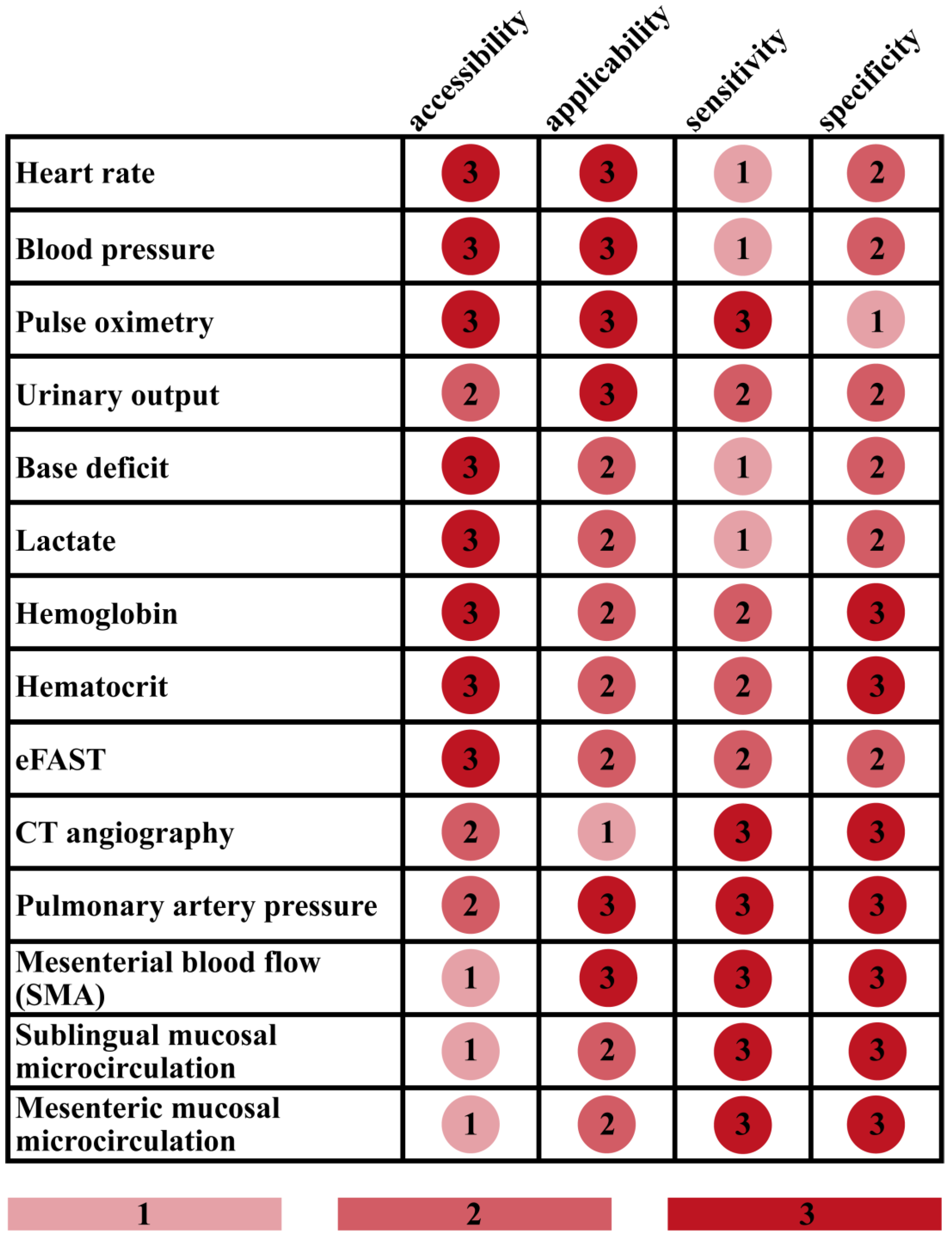
Figure 1. Characterization of frequently used tests for assessing blood loss. eFAST = extended focused assessment with sonography in trauma, CT = computer tomography, SMA = superior mesenteric artery.
Jcm 12 03571 g001
Figure 2. Techniques for the assessment of mesenteric perfusion.
Jcm 12 03571 g002
Disclaimer/Publisher’s Note: The statements, opinions and data contained in all publications are solely those of the individual author(s) and contributor(s) and not of MDPI and/or the editor(s). MDPI and/or the editor(s) disclaim responsibility for any injury to people or property resulting from any ideas, methods, instructions or products referred to in the content.