Case Report

Novel SPEF2 Variant in a Japanese Patient with Primary Ciliary Dyskinesia: A Case Report and Literature Review

1
Department of Respiratory Medicine, Graduate School of Biomedical Sciences, Nagasaki University, Nagasaki 852-8501, Japan
2
Department of Pathology, Graduate School of Biomedical Sciences, Nagasaki University, Nagasaki 852-8520, Japan
3
J-One, Nagasaki 852-8505, Japan
4
Central Electron Microscope Laboratory, Graduate School of Biomedical Sciences, Nagasaki University, Nagasaki 852-8523, Japan
5
Center for Medical Innovation, Nagasaki University, Nagasaki 852-8521, Japan
6
Department of Infection Control and Education Center, Nagasaki University Hospital, Nagasaki 852-8501, Japan
7
Department of Otorhinolaryngology-Head and Neck Surgery, Graduate School of Medicine, Mie University, Tsu 514-8507, Japan
*
Author to whom correspondence should be addressed.
J. Clin. Med. 2023, 12(1), 317; https://doi.org/10.3390/jcm12010317
Submission received: 24 November 2022 / Revised: 23 December 2022 / Accepted: 29 December 2022 / Published: 31 December 2022
(This article belongs to the Special Issue Rare Respiratory Diseases: A Personal and a Public Health Problem 2.0)

Abstract

:
Primary ciliary dyskinesia (PCD) is a genetic and congenital disease associated with an abnormal ciliary ultrastructure and function and is estimated to affect 1 in 15,000–20,000 individuals. A PCD diagnosis can be achieved by genotyping. Here, we performed whole-exome analysis for the diagnosis of PCD and described the detailed clinical characteristics of the case. A 39-year-old Japanese woman with sinusitis and bronchiectasis without situs inversus had had upper and lower respiratory symptoms since childhood and had received long-term macrolide therapy without an accurate diagnosis. A moderate deterioration of cilia function was observed by high-speed video microscopy analysis; additionally, the number of cells with moving cilia was fewer than that in patients without PCD. Electron microscopy revealed no apparent structural abnormalities. We performed whole-exome analysis and identified novel biallelic variants of SPEF2 in the homozygous state (c.1860_1861insCT). We confirmed the absence of SPEF2 protein expression in the cilia of the nasal mucosa using fluorescent immunostaining. Accordingly, she was diagnosed as having PCD with the SPEF2 variant. The present case suggests that the deterioration of cilia function is moderate, the number of respiratory cells with moving cilia might be reduced, and the respiratory condition could be severe in patients with PCD with the SPEF2 variant.

1. Introduction

Primary ciliary dyskinesia (PCD) is a genetic and congenital disease associated with abnormal ciliary function and the overall minimum global prevalence is calculated to be at least 1 in 7554 individuals [1,2,3]. Repeated lower respiratory tract infections due to ciliary dysfunction result in the progressive destruction of the lung, which can lead to severe and fatal pulmonary dysfunction. Diagnosis is difficult, and the majority of patients visit a physician more than 30–50 times before diagnosis [4,5]. All diagnostic tests, including nasal nitric oxide measurements, analyses of ciliary beats, ultrastructural analyses of cilia, and molecular testing for mutations in PCD genes, require specific instruments and specialists, and all the tests have limitations and are not perfect for diagnosis [6]. New PCD causative genes have been discovered each year, and genotyping may identify a genetic cause in 50–75% of cases. In addition, each mutation is associated with distinct clinical characteristics of PCD, such as disease severity, ciliary beat patterns, and ultrastructural abnormalities of cilia [7,8,9].
Here, we performed whole-exome analysis for the diagnosis of PCD, identified the novel biallelic variants of Sperm Flagellar 2 (SPEF2) in the homozygous state, c.1860_1861insCT, which has not previously been reported, and described the detailed clinical characteristics of the case.

2. Case Presentation

A 39-year-old Japanese woman with sinusitis and bronchiectasis had had upper and lower respiratory symptoms, such as a wet cough, since childhood. At 13 years of age, the patient was diagnosed with sinobronchial syndrome and received long-term macrolide therapy. At the age of 25 years, the patient was referred to our hospital, and a nasal biopsy for the diagnosis of PCD was performed; however, no cilia were found in the tissue, and the result did not reach the level of diagnosis. Pseudomonas aeruginosa was detected in the patient’s sputum at the age of 25 years. Repeats of acute bronchiectasis exacerbations and a gradual decline in respiratory function were also shown. At 39 years of age, home oxygen therapy was initiated, and registration on a waiting list for lung transplants from brain-dead donors was applied for. The pulmonary function test results at the age of 39 years were as follows: forced vital capacity, 2.27 L (78.5% of the predicted value); forced expiratory volume in 1 s, 1.18 L (43.4% of the predicted value); and carbon monoxide diffusion capacity of the lung, 16.5 mL/min/mmHg (83.0% of the predicted value). The fraction of exhaled nitric oxide levels was low (5 ppb; the mean levels of Japanese individuals are approximately 15 ppb). Chest radiography (Figure 1) revealed diffuse nodules, consolidation, and bronchial wall thickening, predominantly in the lower lung fields.
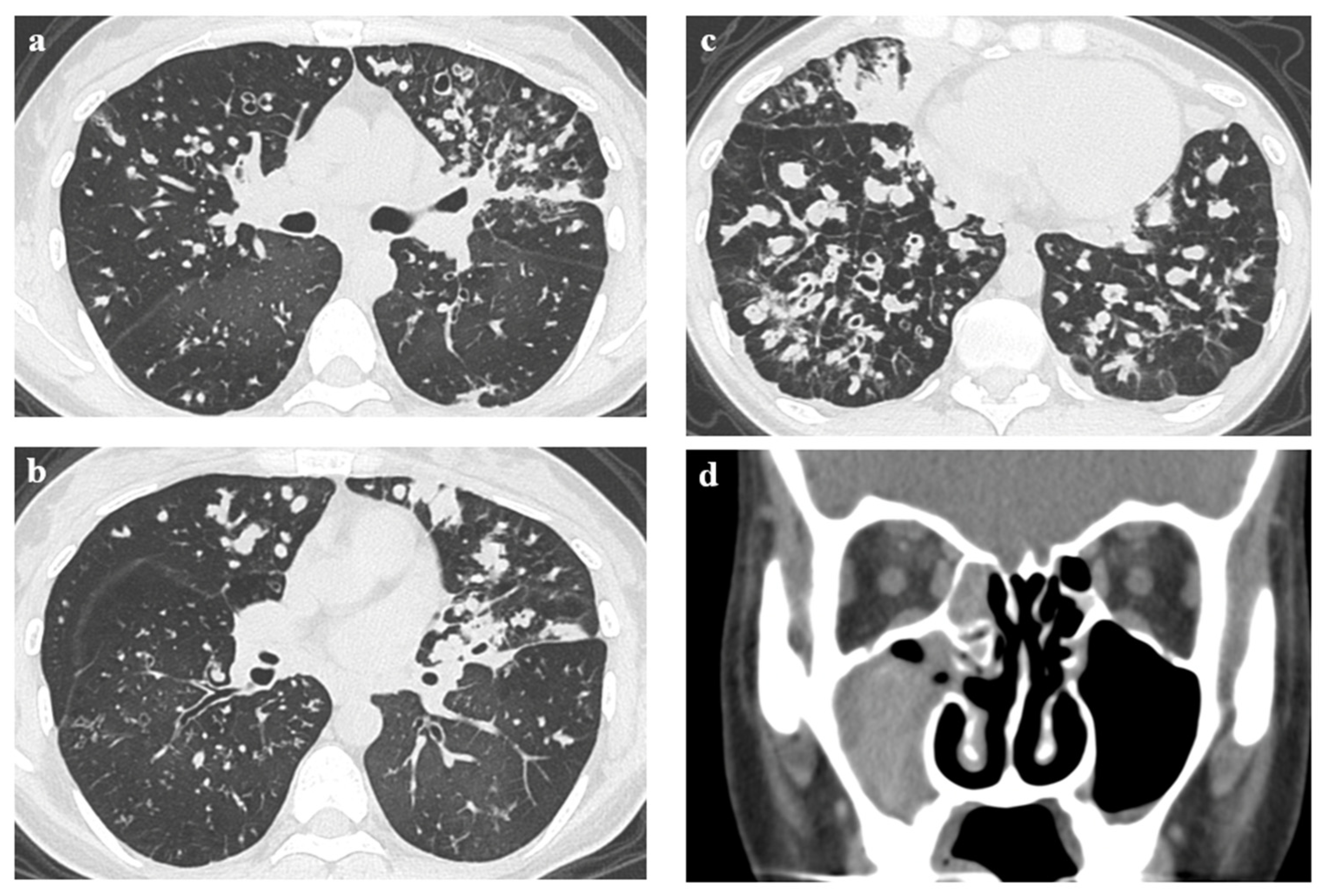
Chest high-resolution computed tomography (HRCT) revealed diffuse bronchial wall thickening, bronchial dilatation, and mucus plugs with predominant bilateral middle and lower lung fields (Figure 2a–c). A sinus computed tomography (CT) scan showed fluid retention and mucosal thickening occupying the right maxillary sinus, suggesting chronic sinusitis (Figure 2d).
The ciliary beat frequency (CBF), ciliary beat amplitude (CBA) (assessed by protractor, Figure S1), and ciliary coordination were immediately (within 10–30 min) assessed using cells obtained by nasal mucosal brushing with a high-speed video camera (HAS-L1, DITECT, Tokyo, Japan). One cell in five movies, five cells in total, of each right and left nostril was assessed in the medium (1:1 bronchial epithelial cell growth medium and Dulbecco’s modified eagle medium) [10]. Videos attached to a microscope were recorded at high speeds (more than 100 fps) and were replayed more slowly (5–30 fps) for evaluation using software (HAS-X viewer Ver.1.3.5.0., DITECT). The right median CBF and CBA were 4.63 Hz and 80 degrees, respectively, and the left median CBF and CBA were 2.75 Hz and 50 degrees, respectively, suggesting moderate deterioration of cilia function at the levels of our laboratory data. In patients without PCD, the median CBF and CBA are approximately 10 Hz and 100–150 degrees, respectively, in our laboratory data. In addition, ciliary coordination was moderately poorer than that of healthy participants and better than that of a patient with outer dynein arm defects in our laboratory data. Although five cells in the right and left nostrils were assessed, fewer cells with moving cilia were observed than in patients without PCD (Figure 3, Video S1).
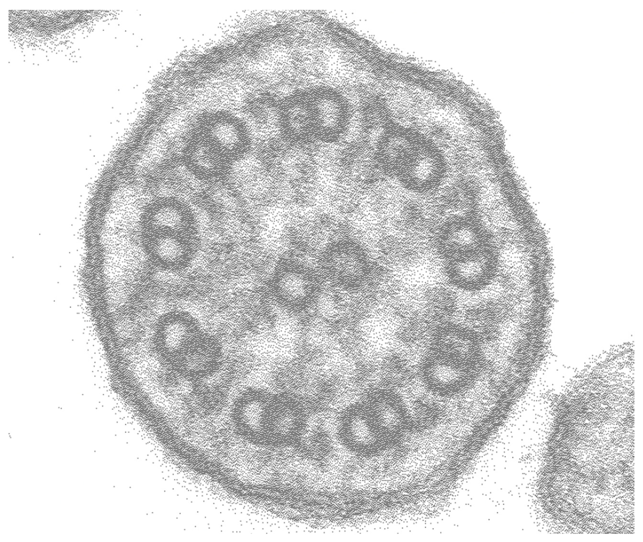
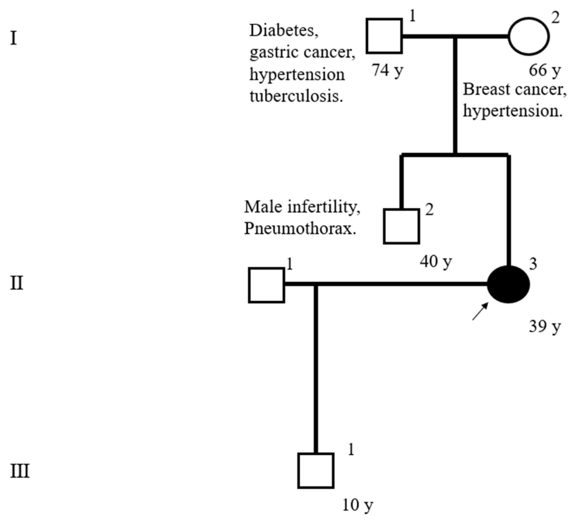
Electron microscopy did not reveal any obvious structural abnormalities in cilia (Figure 4), and genetic testing showed no variants in cystic fibrosis-related genes. The pedigree is shown in Figure 5.
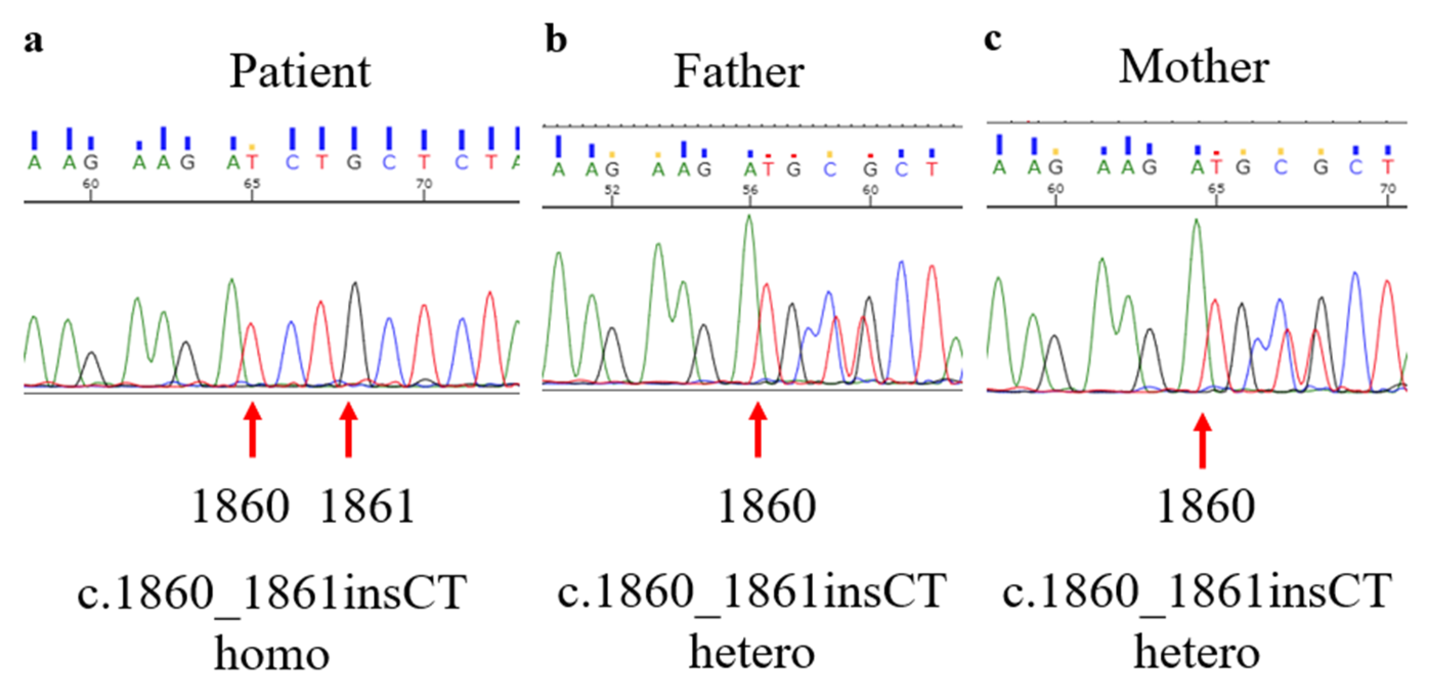
We performed a PCD gene panel, whole-exome sequencing, and Sanger sequencing as described previously [11], and identified the frameshift mutation c.1860_1861insCT (p.Ala621LeufsTer59) in SPEF2 in a homozygous state (Figure 6).
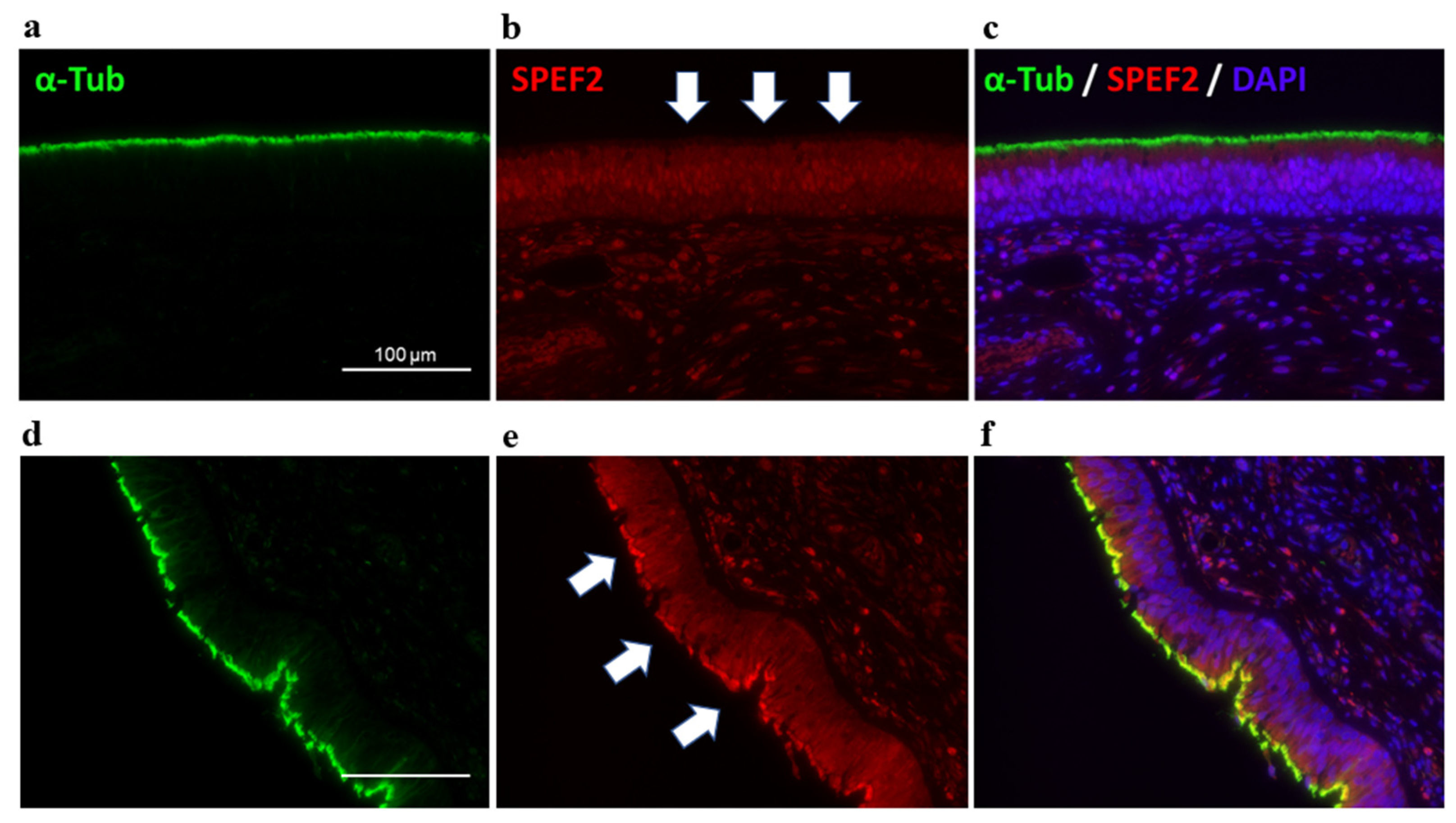
The parents had heterozygous variants, suggesting a compound heterozygous inheritance trait. We also confirmed the absence of SPEF2 protein expression by fluorescent immunostaining in the cilia of the sinus tissues that were obtained by surgery for chronic sinusitis (Figure 7) [12]. Accordingly, she was diagnosed as having PCD with the SPEF2 variant.

3. Discussion

Herein, we present an SPEF2 variant in a 39-year-old Japanese woman with progressive respiratory dysfunction. Currently, only a 31-year-old woman from Germany and six men (ages unknown) with infertility from China were reported in 2020 as cases of PCD with the SPEF2 variant [12,13]. In addition, some SPEF2 variants in patients with male infertility have been reported in China [14]. In the present case, we identified novel variants of SPEF2 in the homozygous state (c.1860_1861insCT), which have not been reported previously. In patients with PCD, homozygous variants of c.1639C>T from Germany as well as homozygous variants of c.2507 + 5delG, c.2649dupA, and c.C4096T and heterozygous variants of c.3400delA and c.3922dupA from China have been shown.
SPEF2 is widely expressed in cilia-related organs, such as the lungs, spleen, trachea, brain, and testis [15,16]. SPEF2 is a component of the central pair complex (CPC) of cilia, and SPEF2 mutations cause symptoms associated with ciliary dysfunction, such as male infertility and/or PCD [12,13,14]. In ultrastructural analyses using transmission electron microscopy, morphological abnormalities of the sperm flagellum have been observed in patients with SPEF2 mutations, but no visible ultrastructural abnormalities of the respiratory cilia have been observed [12,13]. Thus, we confirmed the diagnosis via the lack of SPEF2 by fluorescent immunostaining, in addition to gene analyses, similar to the cases from Germany and China.
Although little is known regarding the ciliary beat motion of patients with PCD with SPEF2 variants, the present case showed the moderate deterioration of the CBF (4.63 and 2.75 Hz) and CBA (80 and 50 degrees) (median, right and left nostrils, respectively), and moderately poorer ciliary coordination. Only one case from Germany showed the deterioration of the CBF (5 to 2.5 Hz) without information on the CBA and ciliary coordination [12]. Ciliary function is closely associated with specific ultrastructural defects, and the CBF of minor or no obvious ultrastructural abnormalities is relatively mildly reduced compared with the CBF of “classic” PCD with outer dynein arm defects or no dynein arms [1,17]. As SPEF2 is a component of the CPC and is not related to the dynein arms of cilia, the moderate deterioration of the CBF in patients with SPEF2 variants is reasonable. The CBA and ciliary coordination were also moderately poor in the present case. Furthermore, fewer cells with moving cilia were observed in the present case than in healthy participants. It was also shown that the total number of cilia was greatly reduced, and the apoptotic signal from the nasopharynx increased in epithelial cells, which might be caused by chronic inflammation or partly attributed to the ciliogenesis defect triggered by SPEF2 deficiency [13]. We believe that these results are reasonable and very important, but the evidence is very limited, and the accumulation of knowledge is expected. To avoid the effects of secondary damage by sampling and infection, it is recommended to confirm with repeated evaluation and/or cell culture samples [8]. Though we evaluated twice (right and left nasal brushing), it might be better to evaluate with further samples, such as bronchial cilia and/or cell culture samples.
The present case and a case from Germany were reported in female patients, and six cases from China were reported in male patients with infertility [12,13]. All eight patients had upper and lower airway symptoms without a situs inversus. The situs inversus is not observed in patients with PCD with CPC-related gene variants, including SPEF2 [12]. The predicted value of the forced expiratory volume in 1 s (%FEV1) was only 43.4% at 39 years of age and required home oxygen therapy and registration for lung transplant. Furthermore, %FEV1 was 50% in a 31-year-old woman from Germany. These two cases from Japan and Germany suggest that reduced lung function in patients with PCD with SPEF2 variants was relatively severe, although the deterioration of the CBF in these two cases was relatively moderate. Persistent Pseudomonas aeruginosa infection, known as a poor prognosis factor, seems to be one of the causes of progression in the present case [1,18,19]. The association of severity, prognosis, and persistent Pseudomonas aeruginosa infection in patients with PCD with SPEF2 variants remains unclear, and an accumulation of evidence is expected.
In conclusion, we present a rare case of PCD with the SPEF2 variant as a third report, preceded by reports from Germany and China, although the variant (c.1860_1861insCT) has never been reported. The present case suggests that the deterioration of cilia function is moderate, the number of respiratory cells with moving cilia is highly reduced, and the respiratory condition could be severe in patients with PCD with SPEF2 variants. The number of reported cases is limited, and the accumulation of evidence is expected.

Supplementary Materials

The following supporting information can be downloaded at: https://www.mdpi.com/article/10.3390/jcm12010317/s1, Figure S1: An illustrative diagram of measuring CBA. Video S1a: Video of cilia of the patient. Video S1b: Video of cilia of healthy participants.

Author Contributions

M.M., T.K. and N.S. conceived, designed, and drafted the manuscript. M.O. contributed to the pathological diagnosis and fluorescent immunostaining. K.K., Y.N. and Y.T. performed the high-speed video microscopy analysis. T.S. contributed to the electron microscopy analyses. K.T. contributed to whole-exome sequencing and Sanger sequencing for gene analysis and made critical revisions. T.T., D.O., H.Y., A.H., H.I., Y.O., K.I. and H.M. made critical revisions. All authors have read and agreed to the published version of the manuscript.

Funding

Non-Profit Organization aimed at supporting community medicine research in Nagasaki: NA; JSPS Grants-in-Aid for Scientific Research (C): 19K09886 and 22K09665.

Institutional Review Board Statement

Gene analyses for the diagnosis of PCD were approved by the Nagasaki University Hospital Clinical Research Ethics Committee (approval numbers: 21091323) and the Ethics Committee of Mie University (1363). Written informed consent was obtained from the patient and her parents.

Informed Consent Statement

Written informed consent has been obtained from the patient to publish this paper.

Data Availability Statement

The datasets for the current case are available from the corresponding author upon reasonable request.

Acknowledgments

We gratefully acknowledge Masakazu Takemoto and Hiroki Yamada for their support in evaluating ciliary function. The authors also thank Konami Abe and Yasushi Miyauchi at Kamakura Techno-Science for supporting fluorescent immunostaining, Yoshiko Ayabe and Yoshiko Akiyama for supporting the office and laboratory work.

Conflicts of Interest

The authors declare no conflict of interest.

Abbreviations

CBAciliary beat amplitude
CBFciliary beat frequency
CPCcentral pair complex
PCDprimary ciliary dyskinesia
HRCThigh-resolution computed tomography
%FEV1predicted value of forced expiratory volume in 1 s

References

  1. Noone, P.G.; Leigh, M.W.; Sannuti, A.; Minnix, S.L.; Carson, J.L.; Hazucha, M.; Zariwala, M.A.; Knowles, M.R. Primary ciliary dyskinesia: Diagnostic and phenotypic features. Am. J. Respir. Crit. Care Med. 2004, 169, 459–467. [Google Scholar] [CrossRef] [PubMed] [Green Version]
  2. Hogg, C. Primary ciliary dyskinesia: When to suspect the diagnosis and how to confirm it. Paediatr. Respir. Rev. 2009, 10, 244–250. [Google Scholar] [CrossRef] [PubMed]
  3. Hannah, W.B.; WB Seifert, B.A.; BA Truty, R.; Zariwala, M.A.; Ameel, K.M.; Zhao, Y.; Nykamp, K.; Gaston, B. The global prevalence and ethnic heterogeneity of primary ciliary dyskinesia gene variants: A genetic database analysis. Lancet Respir. Med. 2022, 10, 459–468. [Google Scholar] [CrossRef] [PubMed]
  4. Sommer, J.U.; Schäfer, K.; Omran, H.; Olbrich, H.; Wallmeier, J.; Blum, A.; Hörmann, K.; Stuck, B.A. ENT manifestations in patients with primary ciliary dyskinesia: Prevalence and significance of otorhinolaryngologic co-morbidities. Eur. Arch. Otorhinolaryngol. 2011, 268, 383–388. [Google Scholar] [CrossRef] [PubMed]
  5. Behan, L.; Dunn Galvin, A.; Rubbo, B.; Masefield, S.; Copeland, F.; Manion, M.; Rindlisbacher, B.; Redfern, B.; Lucas, J.S. Diagnosing primary ciliary dyskinesia: An international patient perspective. Eur. Respir. J. 2016, 48, 1096–1107. [Google Scholar] [CrossRef] [PubMed] [Green Version]
  6. Leigh, M.W.; Ferkol, T.W.; Davis, S.D.; Lee, H.S.; Rosenfeld, M.; Dell, S.D.; Sagel, S.D.; Milla, C.; Olivier, K.N.; Sullivan, K.; et al. Clinical Features and Associated Likelihood of Primary Ciliary Dyskinesia in Children and Adolescents. Ann. Am. Thorac. Soc. 2016, 13, 1305–1313. [Google Scholar] [CrossRef] [PubMed] [Green Version]
  7. Davis, S.D.; Ferkol, T.W.; Rosenfeld, M.; Lee, H.S.; Dell, S.D.; Sagel, S.D.; Milla, C.; Zariwala, M.A.; Pittman, J.E.; Shapiro, A.J.; et al. Clinical features of childhood primary ciliary dyskinesia by genotype and ultrastructural phenotype. Am. J. Respir. Crit. Care Med. 2015, 191, 316–324. [Google Scholar] [CrossRef] [PubMed] [Green Version]
  8. Lucas, J.S.; Barbato, A.; Collins, S.A.; Goutaki, M.; Behan, L.; Caudri, D.; Dell, S.; Eber, E.; Escudier, E.; Hirst, R.A.; et al. European Respiratory Society guidelines for the diagnosis of primary ciliary dyskinesia. Eur. Respir. J. 2017, 49, 1601090. [Google Scholar] [CrossRef] [PubMed] [Green Version]
  9. Raidt, J.; Wallmeier, J.; Hjeij, R.; Onnebrink, J.G.; Pennekamp, P.; Loges, N.T.; Olbrich, H.; Häffner, K.; Dougherty, G.W.; Omran, H.; et al. Ciliary beat pattern and frequency in genetic variants of primary ciliary dyskinesia. Eur. Respir. J. 2014, 44, 1579–1588. [Google Scholar] [CrossRef] [PubMed] [Green Version]
  10. Hirst, R.A.; Jackson, C.L.; Coles, J.L.; Williams, G.; Rutman, A.; Goggin, P.M.; Adam, E.C.; Page, A.; Evans, H.J.; Lackie, P.M.; et al. Culture of primary ciliary dyskinesia epithelial cells at air-liquid interface can alter ciliary phenotype but remains a robust and informative diagnostic aid. PLoS ONE 2014, 9, e89675. [Google Scholar] [CrossRef] [PubMed]
  11. Takeuchi, K.; Xu, Y.; Kitano, M.; Chiyonobu, K.; Abo, M.; Ikegami, K.; Ogawa, S.; Ikejiri, M.; Kondo, M.; Gotoh, S.; et al. Copy number variation in DRC1 is the major cause of primary ciliary dyskinesia in the Japanese population. Mol. Genet. Genomic Med. 2020, 8, e1137. [Google Scholar] [CrossRef] [PubMed] [Green Version]
  12. Cindrić, S.; Dougherty, G.W.; Olbrich, H.; Hjeij, R.; Loges, N.T.; Amirav, I.; Philipsen, M.C.; Marthin, J.K.; Nielsen, K.G.; Sutharsan, S.; et al. SPEF2- and HYDIN-mutant cilia lack the central pair-associated protein SPEF2, aiding primary ciliary dyskinesia diagnostics. Am. J. Respir. Cell Mol. Biol. 2020, 62, 382–396. [Google Scholar] [CrossRef] [PubMed]
  13. Tu, C.; Nie, H.; Meng, L.; Wang, W.; Li, H.; Yuan, S.; Cheng, D.; He, W.; Liu, G.; Du, J.; et al. Novel mutations in SPEF2 causing different defects between flagella and cilia bridge: The phenotypic link between MMAF and PCD. Hum. Genet. 2020, 139, 257–271. [Google Scholar] [CrossRef] [PubMed]
  14. Liu, C.; Lv, M.; He, X.; Zhu, Y.; Amiri-Yekta, A.; Li, W.; Wu, H.; Kherraf, Z.E.; Liu, W.; Zhang, J.; et al. Homozygous mutations in SPEF2 induce multiple morphological abnormalities of the sperm flagella and male infertility. J. Med. Genet. 2020, 57, 31–37. [Google Scholar] [CrossRef] [PubMed]
  15. Ostrowski, L.E.; Andrews, K.; Potdar, P.; Matsuura, H.; Jetten, A.; Nettesheim, P. Cloning and characterization of KPL2, a novel gene induced during ciliogenesis of tracheal epithelial cells. Am. J. Respir. Cell Mol. Biol. 1999, 20, 675–683. [Google Scholar] [CrossRef] [PubMed]
  16. Li, D.Y.; Yang, X.X.; Tu, C.F.; Wang, W.L.; Meng, L.L.; Lu, G.X.; Tan, Y.Q.; Zhang, Q.J.; Du, J. Sperm flagellar 2 (SPEF2) is essential for sperm flagellar assembly in humans. Asian J. Andro. 2022, 24, 359–366. [Google Scholar] [CrossRef]
  17. Chilvers, M.A.; Rutman, A.; O’Callaghan, C. Ciliary beat pattern is associated with specific ultrastructural defects in primary ciliary dyskinesia. J. Allergy Clin. Immunol. 2003, 112, 518–524. [Google Scholar] [CrossRef] [PubMed]
  18. Kennedy, M.P.; Noone, P.G.; Leigh, M.W.; Zariwala, M.A.; Minnix, S.L.; Knowles, M.R.; Molina, P.L. High-resolution CT of patients with primary ciliary dyskinesia. AJR Am. J. Roentgenol. 2007, 188, 1232–1238. [Google Scholar] [CrossRef] [PubMed]
  19. Kido, T.; Yatera, K.; Yamasaki, K.; Nagata, S.; Choujin, Y.; Yamaga, C.; Hara, K.; Ishimoto, H.; Hisaoka, M.; Mukae, H. Two cases of primary ciliary dyskinesia with different responses to macrolide treatment. Intern Med. 2012, 51, 1093–1098. [Google Scholar] [CrossRef] [PubMed]
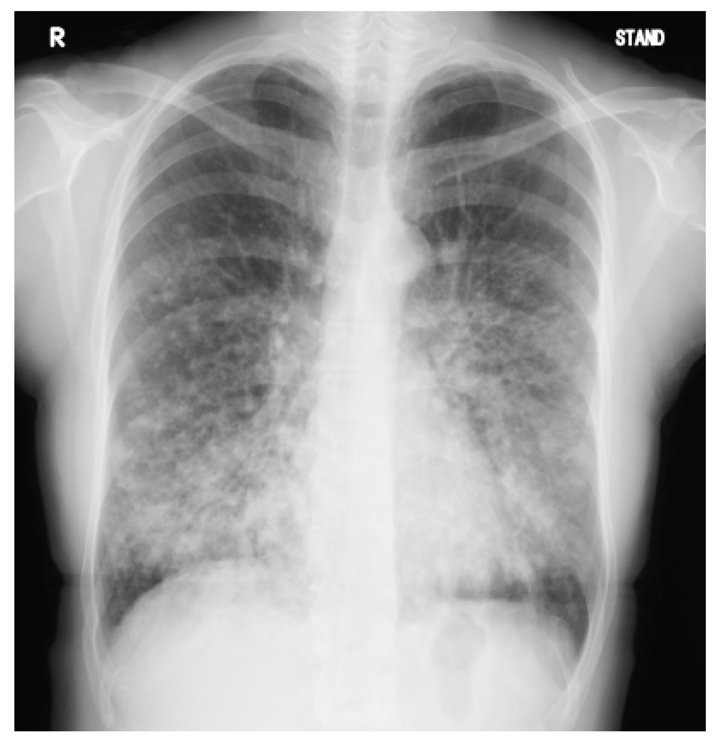
Figure 1. A chest radiograph of the patient at 39 years of age. Diffuse nodules, consolidation, and bronchial wall thickening predominantly in the lower lung fields are observed.
Jcm 12 00317 g001
Figure 2. (ac) Representative images of CT scan. Chest CT scan shows diffuse bronchial wall thickening, bronchial dilatation, and mucus plugs with bilateral middle and lower lung fields predominant; (d) A sinus CT scan shows fluid retention and mucosal thickening occupying the right maxillary sinus.
Jcm 12 00317 g002
Figure 3. Captured picture (a,b) of videos (Video S1a,b) during the assessment of the ciliary beat. Ciliary beats for the present case (a and Video S1a) and healthy participants (b and Video S1b) were assessed using cells obtained by nasal mucosal brushing. Videos were recorded with a high-speed video camera (HAS-L1, DITECT). Compared with healthy participants, the velocity amplitude, angle, and coordination of ciliary beats of the patient were slower, lower, and poorer. Although only one cell in the center of the picture (white arrow) can be observed in the present movie of this case (a and Video S1a), many cells with moving cilia were observed in healthy participants (b and Video S1b). The original magnification of the optical microscope is 400.
Jcm 12 00317 g003
Figure 4. Representative image of electron microscopy. There are no obvious structural abnormalities in nasal cilia.
Jcm 12 00317 g004
Figure 5. The patient’s pedigree. The patient’s brother was diagnosed male infertility.
Jcm 12 00317 g005
Figure 6. Genetic analysis using Sanger sequencing of (a) the patient, (b) the patient’s father, and (c) the patient’s mother. Stop mutation c.1860_1861insCT (p.Ala621LeufsTer59) in SPEF2 in a homozygous state was observed in (a) the patient and the heterozygous mutations were found in (b,c) the patient’s father and mother. SPEF2 = sperm flagellar 2. Green: adenine (A). Black: guanine (G). Red: thymine (T). Blue: cytosine (C).
Jcm 12 00317 g006
Figure 7. Immunofluorescence analysis. (ac) Images of sinus tissues obtained by surgery for chronic sinusitis of the present case; (df) the other case of PCD used as a positive control. Anti-SPEF2 (HPA039606, Atlas antibodies, Bromma, Sweden) (red) is stained with anti-acetylated tubulin (T7451, Sigma-Aldrich, St Louis, MO, USA) (green) to visualize the entire ciliary axonemes, and with 4′,6-diamidino-2-phenylindole (17507, AAT Bioquest, Pleasanton, MO, USA) (blue) to show the nucleus as well. The white arrows and yellow color, indicate the presence of SPEF2 in the cilia in the image of the control (e,f). In (b,c), a lack of SPEF2 expression can be confirmed. Scale bar: 100 μm.
Jcm 12 00317 g007
Disclaimer/Publisher’s Note: The statements, opinions and data contained in all publications are solely those of the individual author(s) and contributor(s) and not of MDPI and/or the editor(s). MDPI and/or the editor(s) disclaim responsibility for any injury to people or property resulting from any ideas, methods, instructions or products referred to in the content.