Article

Marginal versus Standard Donors in Heart Transplantation: Proper Selection Means Heart Transplant Benefit

1
Department of Cardiac, Thoracic, Vascular Sciences and Public Health, University of Padua, 35128 Padua, Italy
2
Cardiothoracovascular Department, Niguarda Hospital, 20162 Milan, Italy
3
Vascular Unit, Portogruaro Hospital, 30026 Portogruaro, Italy
4
Unit of Biostatistics, Epidemiology and Public Health, Department of Cardiac, Thoracic, Vascular Sciences and Public Health, University of Padua, 35128 Padua, Italy
5
Italian National Transplant Centre, National Institute of Health, 00162 Rome, Italy
6
Regional Transplant Centre, 35128 Padua, Italy
*
Author to whom correspondence should be addressed.
These authors contributed equally to this work.
J. Clin. Med. 2022, 11(9), 2665; https://doi.org/10.3390/jcm11092665
Submission received: 14 April 2022 / Revised: 1 May 2022 / Accepted: 7 May 2022 / Published: 9 May 2022
(This article belongs to the Special Issue Surgical Treatment of End Stage Heart Failure)

Abstract

:
Background: In this study, we assessed the mid-term outcomes of patients who received a heart donation from a marginal donor (MD), and compared them with those who received an organ from a standard donor (SD). Methods: All patients who underwent HTx between January 2012 and December 2020 were enrolled at a single institution. The primary endpoints were early and long-term survival of MD recipients. Risk factors for primary graft failure (PGF) and mortality in MD recipients were also analyzed. The secondary endpoint was the comparison of survival of MD versus SD recipients. Results: In total, 238 patients underwent HTx, 64 (26.9%) of whom received an organ from an MD. Hospital mortality in the MD recipient cohort was 23%, with an estimated 1 and 5-year survival of 70% (59.2–82.7) and 68.1% (57.1–81), respectively. A multivariate analysis in MD recipients showed that decreased renal function and increased inotropic support of recipients were associated with higher mortality (p = 0.04 and p = 0.03). Cold ischemic time (p = 0.03) and increased donor inotropic support (p = 0.04) were independent risk factors for PGF. Overall survival was higher in SD than MD (85% vs. 68% at 5 years, log-rank = 0.008). However, risk-adjusted mortality (p = 0.2) and 5-year conditional survival (log-rank = 0.6) were comparable. Conclusions: Selected MDs are a valuable resource for expanding the cardiac donor pool, showing promising results. The use of MDs after prolonged ischemic times, increased inotropic support of the MD or the recipient and decreased renal function are associated with worse outcomes.

1. Introduction

Heart transplantation (HTx) is the gold standard therapy in end-stage heart failure, although it is limited by the shortage of available donor organs [1]. In 2020, a death rate of 3–5% on the waiting list for heart transplant has been estimated with an average waiting time of about 4 years [2].
Considering the growing demand for cardiac organs, despite recent advances in mechanical circulatory support, the inclusion of selected marginal donors (MDs) has been proposed [3,4,5,6,7,8,9]. Controversial results have been reported on posttransplant outcomes following an MD donation, with relatively short-term data [7]. The main concerns are related to increased susceptibility to primary graft failure (PGF), coronary graft vasculopathy and high perioperative mortality [7].
The aim of this study (retrospective, observational) was to analyze the mid-term outcomes of recipients from MDs in a single-center experience, comparing them to those of patients receiving a standard donation (SD), and identifying the risk factors for an improper matching. In particular, we investigated early and mid-term survival of MD recipients and compared the survival of MD versus SD recipients.

2. Materials and Methods

2.1. Study Population

This was a retrospective, observational, single-center study of all consecutive patients who received an HTx at the University of Padua (Italy) since the start of the MD program, between January 2012 and December 2020. Patients who received organs from SDs and MDs were included (Table 1). All pediatric patients (<18 years of age), multiorgan donors and cardiac retransplants were excluded.
The medical records of all patients were reviewed. Anonymity and professional confidentiality were respected. Every reasonable effort was made to obtain written informed consent to participate in this study. In particular, the use of data for scientific and research purposes was already included in the written informed consent used. The local Institutional Review Board (Azienda Ospedaliera Università, Padua, Italy) approved the study design, the consent, and the review of the data (IBR number 48421; 23 September 2021). The uses of collected information and statistical analysis exclusively for scientific purposes were also granted by the Scientific Committee of the Italian National Transplant Network on 11 February 2021.

2.2. Outcomes and Definitions

The primary endpoints were early and mid-term survival of MD recipients. The secondary endpoint was the comparison of survival of MD versus SD recipients. Risk factors for primary graft failure (PGF) and mortality (30-day mortality, in-hospital mortality) in MD recipients were also analyzed.
MDs were defined with the following criteria: age over 60 years; reduced left ventricular performance (ejection fraction between 40–50%); left ventricular hypertrophy (septal thickness > 14 mm on echocardiographic evaluation); focal lesion of the coronary artery; significant valvular heart disease [10]. Primary graft failure (PGF) was defined according to the recent ISHLT consensus statement [11]. Acute cellular and antibody-mediated rejections were classified according to the guidelines of the International Society for Heart and Lung Transplantation (ISHLT) and treated in cases of grade ≥ 2 [12]. Coronary graft vasculopathy (CAV) was defined according to the ISHLT guidelines [13].
During the study period, the HTx waiting list in our Organ Procurement Organization was structured as follows.
  • Status 1:
    a.
    mechanical circulatory support (MCS) due to acute haemodynamic deterioration; RVAD or biventricular assist device (BiVAD); LVAD with device-related complications; total artificial heart (TAH); intra-aortic balloon pump (IABP); extracorporeal membrane oxygenation (ECMO);
    b.
    mechanical ventilation.
  • Status 2:
    a.
    uncomplicated LVAD; continuous inotrope infusion; patients with implantable cardioverter defibrillator (ICD) and malignant relapsing ventricular arrhythmias;
    b.
    outpatients, not included in the categories listed above.
  • Status 3: temporarily inactive.
Patients in Status 1 were eligible to enter into the national High-Urgency Program, where they were prioritized at a national level.

2.3. Cardiac Transplantation Protocol

All grafts were retrieved from brain-dead beating heart donors. Laboratory tests, transthoracic echocardiography and chest X-rays were used for the evaluation of the donors. A coronary angiography was performed whenever possible. Cardioplegic arrest was achieved with antegrade crystalloid solution at 4 °C (Celsior®). During transport, the graft was protected with topical hypothermia (ice-cold Ringer’s solution). All heart transplants were orthotopic, with bicaval anastomosis.
After transplantation, the induction of immunosuppressive therapy with intravenous antithymocyte globulin (0.1 mg/kg/h for three days) and methylprednisolone (1 g intraoperative, then 125 mg/8 h for two days) was initiated. Routine maintenance immunosuppressive drugs consisted of calcineurin inhibitors, mainly cyclosporine (1–2 mg/kg/12 h aiming 150–250 ng/mL), prednisone (2.5–7.5 mg per day, usually discontinued after the first year) and mycophenolic acid (180–720 mg/8–12 h).
Right ventricular endomyocardial (BEM) biopsies were performed weekly in the first month, every two weeks through the fourth month and monthly through the end of the first year. Thereafter, patients were followed up regularly with a transthoracic echocardiography and an annual coronary angiography.
In the case of severe forms of postoperative dysfunction, initial signs of severe PGF and difficult weaning from cardiopulmonary bypass, the implantation of extracorporeal membrane oxygenators (ECMO) was performed [14,15,16]. As soon as the recovery of cardiac function was observed, a weaning test was completed, and weaning was achieved whenever possible.

2.4. Statistical Analysis

Continuous variables are expressed as median and interquartile ranges. Categorical variables are presented as absolute numbers and percentage. The characteristics of SD vs. MD were compared with a Student’s t test for continuous variables and with Pearson’s chi-square test or with Fisher’s exact test for categorical values. The significance of the covariates in the univariate analysis of hospital mortality and PGF was also assessed in the same fashion. A multivariable analysis of hospital mortality and PGF was performed by forward conditional logistic regression, including covariates with a univariate p-value ≤ 0.20. Univariate and multivariable analyses of mid-term mortality were performed with a Cox regression model (covariates with a univariate p-value ≤ 0.20 entered the multivariable analysis). The proportional hazard assumption was verified with the graphical Schoenfeld residuals method. Results are reported as the odds ratio (OR), 95% confidence interval and p-value. Survival curves were plotted with the Kaplan–Meier method. Significance was set at p < 0.05. Analyses were performed using the R System (R Development Core Team. R: A language and environment for statistical computing. Vienna, Austria: R Foundation for Statistical Computing; 2015).

3. Results

A total of 238 patients who underwent orthotopic HTx between January 2012 and December 2020 at the University of Padua were included in the study. Based on the characteristics of the donor, the recipients were divided into two groups: SD recipients (174 patients, 73.1%) and MD recipients (64 patients, 26.9%) (Table 1 and Table 2).
The mean follow-up time was 3.2 ± 2.6 years. The overall Kaplan–Maier (KM) survival rates were 81.2% (76.3%–86.5%) and 79.6% (74.5%–85.1%), respectively, at 1 and 5 years (Figure 1).

3.1. MD Recipients

Of 64 MDs, 61 were considered marginal for age over 60 years-old, 2 for focal lesion of the coronary artery, needing coronary artery bypass graft at the time of HTx and 1 for reduced left ventricular performance. Thirty-day and hospital mortality were 14% and 23%, respectively (Table 3). Most of the hospital mortality (73%) was related to multiorgan failure (MOF), mainly as a complication of severe PGF. Acute renal failure requiring continuous venovenous hemofiltration (CVVH) was the most frequent postoperative adverse event (50% of MDs). Severe PGF occurred in 28% of patients. All of these were assisted by ECMO support with a mean time of 4 ± 3 days. In 45% of the cases, weaning from ECMO was achieved. Clinical acute rejection and CAV occurred, respectively, in 24 (37.5%) and 14 (21.9%) recipients during the follow-up (Table 3).
During the univariate analysis for follow-up mortality, a higher preoperative bilirubin level (p = 0.08), higher rate of CVVH (p = 0.001) and cold ischemic time (p = 0.12) resulted as significant risk factors. By the multivariable analysis, only recipients’ characteristics were identified as risk factors [renal impairment (0.95–0.99; p = 0.037) and inotropic support (1.17–13.44; p = 0.027)]. No donors’ characteristics were significantly associated (Table 4).
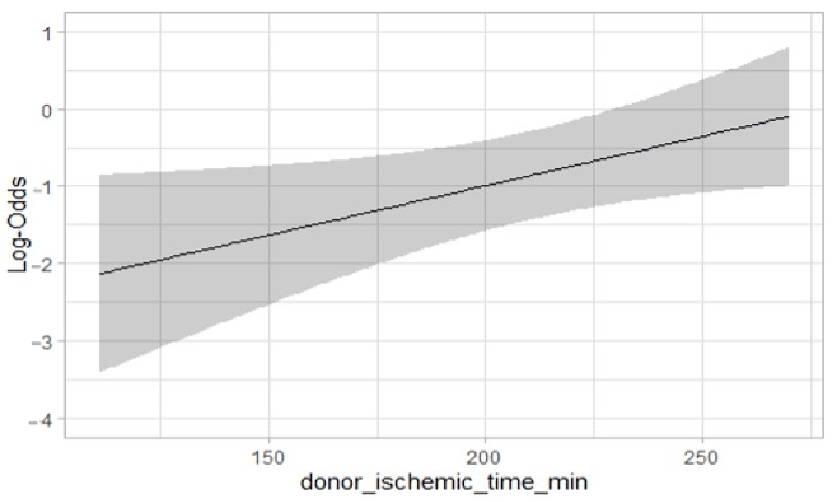
During the univariate and multivariable analyses for PGF, cold ischemic time and donor inotropic support were independent risk factors (1.00–1.02; p < 0.03 and 1.06–39.23; p = 0.04) (Table 5). A logistic regression model showed a linear association between cold ischemic time and PGF (OR 1.01; IC 95% 1.00–1.02; p = 0.03) (Figure 2).

3.2. SD Recipients versus MDs

The basic characteristics of the SD and MD donors and recipients are summarized in Table 1 and Table 2. MD recipients were older (p < 0.001), suffering from a higher incidence of arterial hypertension (p = 0.04), chronic obstructive pulmonary disease (p = 0.002) and lower glomerular filtration rate (p = 0.03). The diagnoses of ischemic cardiomyopathy, primary dilated cardiomyopathy, previous cardiac surgery and left ventricular assist device support were comparable (Table 1). The emergent and urgent states (according to the definitions of the Italian national centers) were also similar (p = 0.5), as well as the pretransplant provisional mechanical support (p = 0.2).
Marginal donors were older (p < 0.001) and had a higher incidence of diabetes (p = 0.02). Although not significant, arterial hypertension (p = 0.08), hyperlipidemia (p = 0.09) and coronary heart disease (p = 0.08) were also more frequent in MDs. Smoking as a risk factor was recorded in 28.1% of MDs. The percentage of inotropic support (74% vs. 72%; p = 0.6) and the mean cold ischemic time of the graft (207 ± 60 min. vs. 197 ± 58 min.; p = 0.2) were similar.
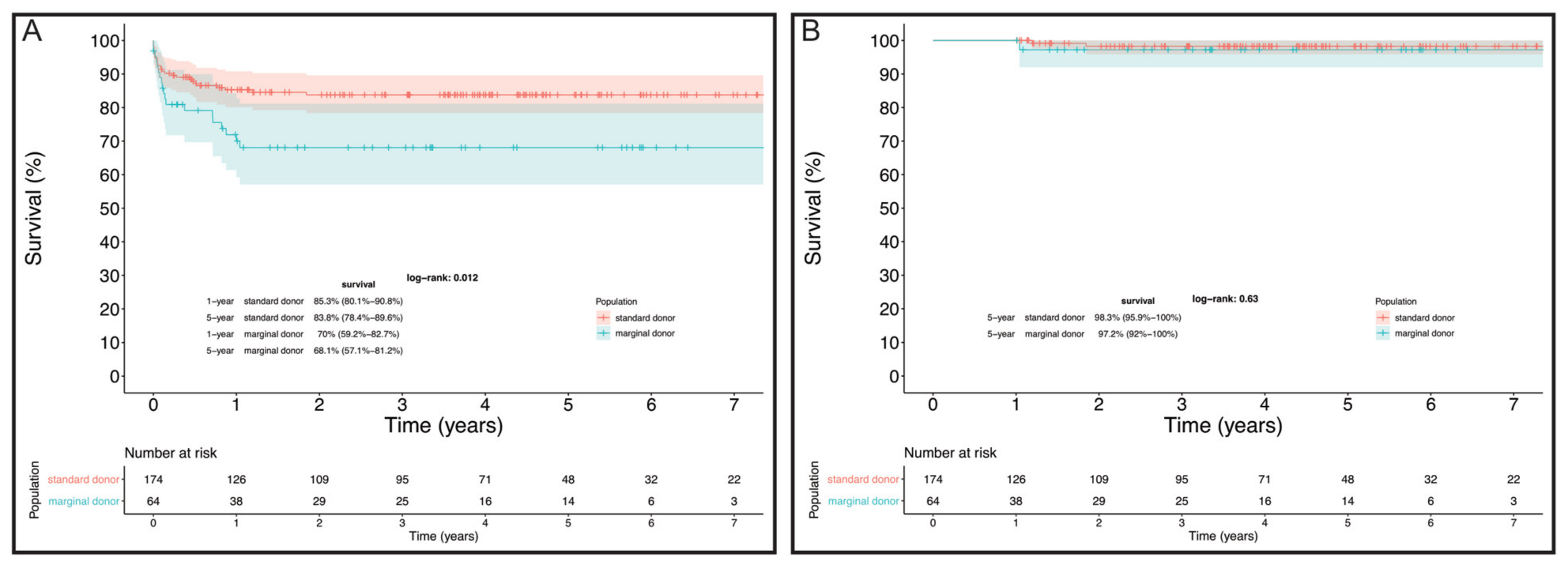
Considering the differences at baseline between the two groups, overall survival was significantly higher in SD than in MD recipients (83.8% vs. 68.1% at 5 years, log-rank = 0.01, Figure 3A). This finding was mainly associated with a higher early mortality rate in MD recipients. However, in the multivariate Cox regression, risk-adjusted mortality for baseline characteristics of MD versus SD recipients [HR 1.8 (0.8–4.1); p = 0.2] and 5-year conditional survival (log-rank = 0.6) were comparable (Figure 3A,B).

4. Comment

The use of organs from MDs, beyond the controversial opinions, is slowly spreading, to overcome the cardiac donors’ undersupply. In the absence of a universal consensus on the definition of the cardiac risk profiles for the donors and guidelines on the use of organs from high-risk or MDs, the main challenge for the transplant team is to avoid complications on the waiting list and establish the correct timing of HTx [17,18]. As reported by Lietz et al. [19], despite an increase in postoperative mortality and the risk of transplant-related coronary heart disease, “older grafts” bring more benefits than staying on the waiting list. However, the models for risk stratification in heart failure patients awaiting HTx and the mid long-term outcomes of MDs still appear to be limited [20,21].
In the current study, a cohort of MDs was first analyzed at a 5-year interval. Early and late survival were satisfactory, as well as the rate of adverse events. Hospital mortality in the MD recipient cohort was 23%, with an estimated 1 and 5-year survival of 70% and 69%, respectively. A multivariate analysis of MD recipients showed that decreased renal function and increased inotropic support of recipients were associated with higher hospital mortality. Univariate and multivariable analyses of overall mortality did not provide further significant results. Inotropic support and end-stage renal disease have been already reported as independent predictors of mortality [22]. Indeed, increased inotropic exposure may affect HTx survival in terms of myocardial dysfunction, poor outcome and long-term complications [23]. As described by Trivedi et al. [24], risk factors of the recipients are more relevant in predicting post-transplant survival than donor factors. Our analysis confirmed these results. We suggest for appropriate matching, in the case of long-distance organ procurement, to not use recipients with high inotropic support and end-stage renal disease. However, there is no objective scoring system that independently uses recipient and donor risk factors to predict post-HTx survival [25].
The most common cause of hospital mortality of MD recipients is PGF, although several causes can be considered responsible [25]. This occurs in up to 30% of MD recipients [26]. In our study, inotropic donors and prolonged ischemic time were found to be the main risk factors. A statistically significant and linear association was observed between cold ischemic time and the development of PGF. An option to overcome the limitations of cold ischemic storage is ex-vivo normothermic perfusion (e.g., organ care system (OCS) (Transmedics, Andover, MA, USA), which allows one to monitor the function of the graft, addressing the time constraints crucial for geographic allocation [27]. In fact, the evaluation of the lactate trend can be useful in evaluating myocardial protection of the graft and in the recognition of unsuitable organs. The largest study on the use of OCS in the preservation and evaluation of non-standard donor hearts is the EXPAND-Heart-Trial [28]. The results of this study showed excellent short-term posttransplant results, particularly a low PGF rate, supporting ex-vivo perfusion as a procedure to be used for MD procurement. However, the results observed with OCS were not superior to the standard cold storage procedure, concluding that further investigation was mandatory. Our current experience with OCS and MDs is limited to five cases, so we cannot draw any conclusions from this. However, the results of our study on MDs can be a driving force in the more frequent OCS use.
In the second part of our study, we then compared the mid-term outcome of recipients from MDs versus SDs. Although the perioperative mortality of MD recipients appeared to be higher than that of SDs, mid-term conditional survival was comparable, emphasizing adequate graft function in the long term. Several reports showed no inferiority of marginal donors, even though the criteria for defining MDs were less selective than ours (age > 55 years, cold ischemic time > 4 h, high dose of noradrenaline) [8,9,18].
However, in selected scenarios, HTx was performed using hearts affected by coronary arteries and valvular disease, or congenital heart defects. This group of organs can be successfully transplanted with a concomitant procedure at the time of HTx [29].
A low incidence of acute and chronic rejection events in MD recipients was found in our population, and no significant differences were observed when comparing them to SD recipients. Conversely, we observed a higher risk of infection as reported by Sugimara et al. They retrospectively analyzed the clinical outcome of HTx recipients focusing on the donor’s left ventricular function. Although prolonged posttransplant recovery was observed, no difference in terms of 1-year survival was shown [30].
Commonly, the development of CAV is considered a non-negligible complication in the use of MDs [9]. However, this was not found to be significantly different in our cohorts.

Study Limitations

This is a single-center retrospective study. Although the number of our study population can be considered relatively small, our marginal donor pool represents one-third of the total MDs assigned in our country during the study period. A multi-center effort is undoubtedly needed to expand the population and further validate our preliminary results. Unfortunately, we could not perform a comparison between the cold storage procurement strategy and the ex-vivo perfusion procedure. Therefore, further investigation is needed on this aspect. Finally, it was not possible to standardize and include in the analysis the biological age of donors and recipients. Therefore, only chronological age was included in the analysis [31].

5. Conclusions

The use of marginal donors could be considered a valid resource in an era of cardiac donor organ shortages. Provided that careful donor–recipient matching occurs, satisfactory early and long-term results are achievable.
The use of MDs after prolonged ischemic times, increased inotropic support of the MD or the recipient and decreased renal function are associated with worse outcomes in terms of both mortality and incidence of PGF.

Author Contributions

O.B.: Conceptualization, Writing; R.C.: Conceptualization, Writing; M.C. (Masimiliano Carrozzini): Conceptualization, Writing; A.G.: Critical Revision; J.B.: Data Collection; A.A.: Critical Revision, Approval of Article, Supervision; M.F.: Data Collection; C.C.: Data Collection; G.T.: Data Collection; G.L.: Statistical Analysis; V.T.: Data Collection; D.G.: Statistical Analysis; M.C. (Massimo Cardillo): Validation; F.P.: Validation; G.F.: Validation; G.G.: Critical Revision, Approval of Article, Supervision; T.B.: Critical Revision, Supervision. All authors have read and agreed to the published version of the manuscript.

Funding

This research received no external funding.

Institutional Review Board Statement

The study was conducted in accordance with the Declaration of Helsinki, and approved by the Institutional Review Board of Azienda Ospedaliera, Univesity of Padua, Italy (Clinical registration number: IBR number 48421; 23 September 2021). The uses of collected information and statistical analysis exclusively for scientific purposes were also granted by the Scientific Committee of the Italian National Transplant Network on 11 February 2021.

Informed Consent Statement

Informed consent was obtained from all subjects involved in the study. Written informed consent has been obtained from the patient(s) to publish this paper.

Data Availability Statement

The data that support the findings of this study are available on request from the corresponding author, [TB]. The data are not publicly available due to restrictions, and their information could compromise the privacy of research participants.

Conflicts of Interest

All authors have no conflict of interest to declare.

Abbreviation and Acronymous

BEMEndomyocardial biopsies
CAVCoronary graft vasculopathy
COPDChronic obstructive pulmonary disease
CVVHContinuous venovenous hemofiltration
ECMOExtracorporeal membrane oxygenator
ESRDEnd-stage renal disease
HTxHeart transplantation
ICUIntensive care unit
ISHLTInternational Society for Heart and Lung Transplantation
K-MKaplan–Meier
LVADLeft ventricle assist device
MDsMarginal donors
OCSOrgan care support
PGFPrimary graft failure
SDsStandard donors

References

  1. Mehra, M.R.; Canter, C.E.; Hannan, M.M.; Semigran, M.J.; Uber, P.A.; Baran, D.A.; Danziger-Isakov, L.; Kirklin, J.K.; Kirk, R.; Kushwaha, S.S.; et al. International Society for Heart Lung Transplantation (ISHLT) Infectious Diseases, Pediatric and Heart Failure and Transplantation Councils. The 2016 International Society for Heart Lung Transplantation listing criteria for heart transplantation: A 10-year update. J. Heart Lung Transplant. 2016, 35, 1–23. [Google Scholar] [CrossRef] [PubMed]
  2. Available online: https://www.trapianti.salute.gov.it/imgs/C_17_cntPubblicazioni_415_allegato.pdf (accessed on 21 August 2021).
  3. Carrozzini, M.; Bejko, J.; Guariento, A.; Rubino, M.; Bianco, R.; Tarzia, V. Minimally Invasive Implantation of Continuous Flow Left Ventricular Assist Devices: The Evolution of Surgical Techniques in a Single-Center Experience. Artif. Organs 2019, 43, E41–E52. [Google Scholar] [CrossRef] [PubMed]
  4. Apostolo, A.; Paolillo, S.; Contini, M.; Vignati, C.; Tarzia, V.; Campodonico, J.; Mapelli, M.; Massetti, M.; Bejko, J.; Righini, F.; et al. Comprehensive effects of left ventricular assist device speed changes on alveolar gas exchange, sleep ventilatory pattern, and exercise performance. J. Heart Lung Transplant. 2018, 37, 1361–1371. [Google Scholar] [CrossRef] [PubMed]
  5. Bejko, J.; Toto, F.; Gregori, D.; Gerosa, G.; Bottio, T. Left ventricle assist devices and driveline’s infection incidence: A single-centre experience. J. Artif. Organs 2017, 21, 52–60. [Google Scholar] [CrossRef] [PubMed]
  6. Bottio, T.; Bagozzi, L.; Fiocco, A.; Nadali, M.; Caraffa, R.; Bifulco, O. COVID-19 in Heart Transplant Recipients: A Multicenter Analysis of the Northern Italian Outbreak. JACC Heart Fail. 2021, 9, 52–61. [Google Scholar] [CrossRef] [PubMed]
  7. Guihaire, J.; Noly, P.E.; Martin, A.; Rojo, M.; Aymami, M.; Ingels, A.; Lelong, B.; Chabanne, C.; Verhoye, J.-P.; Flécher, E. Impact of donor comorbidities on heart transplant outcomes in the modern era. Interact. Cardiovasc. Thorac. Surg. 2017, 24, 898–904. [Google Scholar] [CrossRef] [Green Version]
  8. Galeone, A.; Lebreton, G.; Coutance, G.; Demondion, P.; Schmidt, M.; Amour, J.; Varnous, S.; Leprince, P. A single-center long-term experience with marginal donor utilization for heart transplantation. Clin. Transplant. 2020, 34, e14057. [Google Scholar] [CrossRef]
  9. Prieto, D.; Correia, P.; Baptista, M.; Antunes, M.J. Outcome after heart transplantation from older donor age: Expanding the donor pool. Eur. J. Cardio-Thoracic Surg. 2014, 47, 672–678. [Google Scholar] [CrossRef] [Green Version]
  10. Available online: https://www.trapianti.salute.gov.it/imgs/C_17_cntPubblicazioni_394_allegato.pdf (accessed on 5 November 2020).
  11. Kobashigawa, J.; Zuckermann, A.; Macdonald, P.; Leprince, P.; Esmailian, F.; Luu, M.; Mancini, D.; Patel, J.; Razi, R.; Reichenspurner, H.; et al. Consensus Conference participants. Report from a consensus conference on primary graft dysfunction after cardiac transplantation. J. Heart Lung Transplant. 2014, 33, 327–340. [Google Scholar] [CrossRef]
  12. Stewart, S.; Winters, G.L.; Fishbein, M.C.; Tazelaar, H.D.; Kobashigawa, J.; Abrams, J.; Andersen, C.B.; Angelini, A.; Berry, G.J.; Burke, M.M.; et al. Revision of the 1990 Working Formulation for the Standardization of Nomenclature in the Diagnosis of Heart Rejection. J. Heart Lung Transplant. 2005, 24, 1710–1720. [Google Scholar] [CrossRef]
  13. Mehra, M.R.; Crespo-Leiro, M.G.; Dipchand, A.; Ensminger, S.M.; Hiemann, N.E.; Kobashigawa, J.A. International Society for Heart and Lung Transplantation working formulation of a standardized nomenclature for cardiac graft vasculopathy-2010. J. Heart Lung Transplant. 2010, 29, 717–727. [Google Scholar] [CrossRef]
  14. Listijono, D.R.; Watson, A.; Pye, R.; Keogh, A.M.; Kotlyar, E.; Spratt, P. Usefulness of extracorporeal membrane oxygenation for early cardiac graft dysfunction. J. Heart Lung Transplant. 2011, 30, 783–789. [Google Scholar] [CrossRef]
  15. Mastroianni, C.; Nenna, A.; Lebreton, G.; D’Alessandro, C.; Greco, S.M.; Lusini, M.; Leprince, P.; Chello, M. Extracorporeal membrane oxygenation as treatment of graft failure after heart transplantation. Ann. Cardiothorac. Surg. 2019, 8, 99–108. [Google Scholar] [CrossRef] [Green Version]
  16. Pozzi, M.; Bottin, C.; Armoiry, X.; Sebbag, L.; Boissonnat, P.; Hugon-Vallet, E.; Koffel, C.; Flamens, C.; Paulus, S.; Fellahi, J.L.; et al. Extracorporeal life support for primary graft dysfunction after heart transplantation. Interact. Cardiovasc. Thorac. Surg. 2018, 27, 778–784. [Google Scholar] [CrossRef]
  17. Wittwer, T.; Wahlers, T. Marginal donor grafts in heart transplantation: Lessons learned from 25 years of experience. Transplant. Int. 2007, 21, 113–125. [Google Scholar] [CrossRef]
  18. Piperata, A.; Caraffa, R.; Bifulco, O.; Avesani, M.; Apostolo, A.; Gerosa, G. Marginal donors and organ shortness: Concomitant surgical procedures during heart transplantation: A literature review. J. Cardiovasc. Med. 2021, 23, 167–175. [Google Scholar] [CrossRef]
  19. Lietz, K.; John, R.; Mancini, D.M.; Edwards, N.M. Outcomes in cardiac transplant recipients using grafts from older donors versus mortality on the transplant waiting list; Implications for donor selection criteria. J. Am. Coll. Cardiol. 2004, 43, 1553–1561. [Google Scholar] [CrossRef] [Green Version]
  20. Goda, A.; Williams, P.; Mancini, D.; Lund, L.H. Selecting patients for heart transplantation: Comparison of the Heart Failure Survival Score (HFSS) and the Seattle Heart Failure Model (SHFM). J. Heart Lung Transplant. 2011, 30, 1236–1243. [Google Scholar] [CrossRef]
  21. AbouEzzeddine, O.F.; Redfield, M.M. Who Has Advanced Heart Failure? Definition and Epidemiology. Congest. Heart Fail. 2011, 17, 160–168. [Google Scholar] [CrossRef]
  22. Pouteil-Noble, C.; Hemdawy, A.; Villar, E.; Boissonnat, P.; Sebbag, L. Chronic Renal Failure and End-Stage Renal Disease Are Associated With a High Rate of Mortality After Heart Transplantation. Transplant. Proc. 2005, 37, 1352–1354. [Google Scholar] [CrossRef]
  23. Shahin, J.; Devarennes, B.; Tse, C.W.; Amarica, D.-A.; Dial, S. The relationship between inotrope exposure, six-hour postoperative physiological variables, hospital mortality and renal dysfunction in patients undergoing cardiac surgery. Crit. Care 2011, 15, R162. [Google Scholar] [CrossRef] [PubMed] [Green Version]
  24. Trivedi, J.R.; Cheng, A.; Ising, M.; Lenneman, A.; Birks, E.; Slaughter, M.S. Heart Transplant Survival Based on Recipient and Donor Risk Scoring: A UNOS Database Analysis. ASAIO J. 2016, 62, 297–301. [Google Scholar] [CrossRef] [PubMed]
  25. Amarelli, C.; De Santo, L.S.; Marra, C.; Maiello, C.; Bancone, C.; Della Corte, A.; Nappi, G.; Romano, G. Early graft failure after heart transplant: Risk factors and implications for improved donor-recipient matching. Interact. Cardiovasc. Thorac. Surg. 2012, 15, 57–62. [Google Scholar] [CrossRef] [PubMed] [Green Version]
  26. Isaac, D. Primary cardiac graft failure-defining, predicting, preventing. J. Heart Lung Transplant. 2013, 32, 1168–1169. [Google Scholar] [CrossRef]
  27. Collins, M.J.; Moainie, S.L.; Griffith, B.P.; Poston, R.S. Preserving and evaluating hearts with ex vivo machine perfusion: An avenue to improve early graft performance and expand the donor pool. Eur. J. Cardio-Thorac. Surg. 2008, 34, 318–325. [Google Scholar] [CrossRef] [Green Version]
  28. Schroder, J.; D’Alessandro, D.; Esmailian, F.; Boeve, T.; Tang, P.; Liao, K.; Wang, I.; Anyanwu, A.; Shah, A.; Mudy, K.; et al. Successful Utilization of Extended Criteria Donor (ECD) Hearts for Transplantation-Results of the OCS™ Heart EXPAND Trial to Evaluate the Effectiveness and Safety of the OCS Heart System to Preserve and Assess ECD Hearts for Transplantation. J. Heart Lung Transplant. 2019, 38, S42. [Google Scholar] [CrossRef]
  29. Piperata, A.; Caraffa, R.; Bifulco, O.; Avesani, M.; Gerosa, G.; Bottio, T. Heart transplantation in the new era of extended donor criteria. J. Card. Surg. 2021, 36, 4828–4829. [Google Scholar] [CrossRef]
  30. Sugimura, Y.; Immohr, M.B.; Aubin, H.; Mehdiani, A.; Rellecke, P.; Tudorache, I.; Lichtenberg, A.; Boeken, U.; Akhyari, P. Impact of Reported Donor Ejection Fraction on Outcome after Heart Transplantation. Thorac. Cardiovasc. Surg. 2021, 69, 490–496. [Google Scholar] [CrossRef]
  31. Pavanello, S.; Campisi, M.; Fabozzo, A.; Cibin, G.; Tarzia, V.; Toscano, G.; Gerosa, G. The biological age of the heart is consistently younger than chronological age. Sci. Rep. 2020, 10, 10752. [Google Scholar] [CrossRef]
Figure 1. Overall survival in HTx population. Survival at 1 year was 81.2% (76.3–86.5%), at 8 years 79.6% (74.5–85.1%).
Jcm 11 02665 g001
Figure 2. Graphical representation of linear and significant association between cold ischemic time in MDs and primary graft failure onset. (Logistic regression model).
Jcm 11 02665 g002
Figure 3. (A) Overall survival in SDs compared to MDs; (B) 5-year conditional survival of MDs compared to SDs.
Jcm 11 02665 g003
Table 1. (a,b) Basic characteristics of organ recipients from standard versus marginal donors.
SDs (n = 174)MDs (n = 64)p
n (%) or Median (IQR)n (%) or Median (IQR)
(a)
Gender (Female)39 (22.4%)17 (26.6%)0.5
Age (years)56.4 (46.8–63.3)63.6 (55.3–66.8)<0.001
Cardiac diagnosis 0.5
Dilatative67 (38.5%)18 (28.1%)
Ischemic71 (40.8%)33 (51.6%)
Congenital12 (6.9%)3 (4.7%)
Valvular3 (1.7%)2 (3.1%)
Hypertrophic10 (5.7%)2 (3.1%)
Other11 (6.3%)6 (9.4%)
BSA (m2)1.7 (1.7–2.0)1.8 (1.7–1.9)0.6
Dyslipidemia63 (36.2%)25 (39.1%)0.8
Hypertension62 (35.6%)33 (51.6%)0.04
Cancer9 (5.2%)2 (3.1%)0.7
Diabetes31 (17.8%)16 (25.0%)0.3
PVD12 (6.9%)7 (10.9%)0.2
COPD10 (5.7%)13 (20.3%)0.002
ICD129 (74.1%)54 (84.4%)0.1
Cerebral event28 (16.1%)16 (25.0%)0.1
Smoker63 (36.2%)23 (35.9%)0.6
Genetic syndrome11 (6.3%)3 (4.7%)0.8
Previous cardiac surgery67 (38.5%)32 (50.0%)0.1
Bilirubin (µmol/L)14.6 (9.3–22.6)15.0 (7.2–22.7)0.3
GFR (mL/min/mq)71.0 (49.0–89.0)51.0 (44.0–68.0)0.03
(b)
Intracorporeal LVAD49 (28.2%)19 (29.7%)
Length of LVAD support (months)13.1 (4.8–31.6)19.0 (13.7–28.0)0.4
List status 0.02
2B74 (42.5%)28 (43.8%)
2A51 (29.3%)21 (32.8%)
15 (2.9%)7 (10.9%)
HU44 (25.3%)8 (12.5%)
Status 1 + HU49 (28.2%)15 (23.4%)0.5
Waiting list time (months)4.8 (1.1–18.1)6.5 (2.2–21.6)0.5
ICU-stay62 (35.6%)15 (23.4%)0.2
Inotropic support47 (27.0%)15 (23.4%)0.6
Mechanical ventilation7 (4.0%)4 (6.3%)0.5
CVVH8 (4.6%)3 (4.7%)0.9
Temporary-MCS42 (24.14%)8 (12.5%)0.2
IABP1 (0.6%)0 (0.0%)0.9
ECMO18 (10.3%)3 (4.7%)0.2
Paracorporeal-LVAD12 (6.9%)3 (4.7%)0.8
Paracorporeal-RVAD3 (1.7%)1 (1.6%)0.8
Paracorporeal-BiVAD8 (4.6%)1 (1.6%)0.5
Abbreviations: BSA, body surface area; COPD, chronic obstructive pulmonary disease; CVVH, continuous venovenous hemofiltration; ECMO, extracorporeal membrane oxygenator; GFR, glomerular filtration rate; HU, high urgency; IABP, intra-aortic balloon pump; ICD, implantable cardioverter defibrillator; ICU, intensive care unit; LVAD, left ventricular assistance device; MCS, mechanical circulatory support; PVD, peripheral vascular disease, RVAD, right ventricular assist device.
Table 2. Basic characteristics of standard versus marginal donors.
Standard (n = 174)Marginal (n = 64)p
n (%) or Median (IQR)n (%) or Median (IQR)
Age (years)45.0 (29.5–53.0)64.0 (62.0–66.0)<0.001
Match age−10.0 (−23.3–1.2)2.8 (−1.2–10.1)<0.001
Gender (female)71 (40.8%)33 (51.6%)0.2
Mismatch gender55 (31.6%)20 (31.3%)0.9
Inotropic support130 (74.7%)46 (71.9%)0.6
Cardiac arrest33 (19.0%)9 (14.1%)0.4
Cath abnormalities1 (0.6%)4 (6.3%)0.08
Hypertension17 (9.8%)12 (18.8%)0.08
Smoker51 (29.3%)18 (28.1%)0.9
Dyslipidemia3 (1.7%)4 (6.3%)0.1
Diabetes1 (0.6%)4 (6.3%)0.02
Cold ischemic time (minutes)220.0 (160.0–250.0)200.0 (156.3–234.3)0.2
Table 3. Outcomes and complications of organ recipients from standard versus marginal donors.
SDs (n = 174)MDs (n = 64)p
n (%) or Median (IQR)n (%) or Median (IQR)
Severe PGF36 (20.7%)18 (28.1%)0.2
Postoperative ECMO support (days)5.0 (3.0–6.5)3.5 (2.0–6.8)0.2
CVVH54 (31.0%)32 (50.0%)0.01
Intrahospital infection61 (35.1%)30 (46.9%)0.1
Clinical cellular rejection62 (35.6%)24 (37.5%)0.9
CAV28 (16.1%)14 (21.9%)0.6
Cerebral event17 (9.8%)12 (18.8%)0.07
30-day mortality13 (7.5%)9 (14.1%)0.1
Hospital mortality21 (12.1%)15 (23.4%)0.04
Cause of hospital mortality
MOF10 (47.6%)11 (73.3%)
Neurologic2 (9.5%)1 (6.7%)
Infection4 (19.1%)3 (20.0%)
Other5 (23.8%)0 (0.0%)
Abbreviations: CAV, cardiac graft vasculopathy; CVVH, continuous venovenous hemofiltration; ECMO, extracorporeal membrane oxygenator; MOF: multiorgan failure; PGF, primary graft failure.
Table 4. Univariate and multivariate analysis of organ recipients from marginal donors for mortality.
Recipient CharacteristicsMDs Alive (n = 44)MDs Mortality (n = 19)pMultivariate Analysis
n (%) or Median (IQR)n (%) or Median (IQR)
Listing status 0.12
2B24 (55%)3 (16%)
2A12 (27%)9 (47%)
13 (7%)4 (21%)
HU5 (11%)3 (16%)
Peripheral vascular disease2 (5%)4 (21%)0.01
Platelets count (103/mm3)187 (167–249)189 (128–255)0.13
Bilirubin (µmol/L)13 (7–21)20 (10–44)0.08
C-reactive Protein (mg/L)3.8 (2.9–15.1)26 (4–99)0.12
eGFR (mL/min/m2)60 (49–84)47 (42–53)0.02HR 0.98 (0.95–0.99; p = 0.04)
Inotropic support5 (11%)10 (53%)<0.01HR 3.96 (1.17–13.44; p = 0.03)
CVVH0 (0%)3 (16%)<0.01
Mechanical ventilation1 (2%)3 (16%)0.08
ICU stay8 (18%)7 (37%)0.13
Paracorporeal-LVAD1 (2%)2 (11%)0.17
Donors CharacteristicsMDs Alive (n = 44)MDs Mortality (n = 19)pMultivariate Analysis
n (%) or Median (IQR)n (%) or Median (IQR)
Cold ischemic time (min)200 (149–234)216 (169–240)0.12
Hypertension6 (14%)6 (32%)0.13
Donor/Recipient BSA0.0 (−0.2–0.1)0.1 (0.0–0.2)0.12
Abbreviations: CVVH, continuous venovenous hemofiltration; GFR, glomerular filtration rate; HU, high urgency; BSA, body surface area; ICU, intensive care unit; LVAD, left ventricular assistance device.
Table 5. Univariate and multivariate analysis of organ recipients from marginal donors for primary graft failure.
Recipients’ CharacteristicsMarginal Donors w/o PGF (n = 46)Marginal Donors with PGF (n = 18)pMultivariate Analysis
n (%) or Median (IQR)n (%) or Median (IQR)
List Status 0.02
2B23 (50.0%)5 (27.8%)
2A12 (26.1%)9 (50.0%)
13 (6.5%)4 (22.2%)
HU8 (17.4%)0 (0.0%)
Status 1 + HU11 (23.9%)4 (22.2%)0.9
Donor CharacteristicsMarginal Donors w/o PGF (n = 46)Marginal Donors with PGF (n = 18)pMultivariate Analysis
n (%) or Median (IQR)n (%) or Median (IQR)
Inotropic support30 (65.2%)16 (88.9%)0.06OR 6.50 (1.06–39.22; p = 0.04)
Cold ischemic time (minutes)193.0 (147.3–228.5)227.5 (180.0–246.0)0.02OR 1.01 (1.00–1.02; p = 0.03
PGF, primary graft failure; HU, high urgency.
Publisher’s Note: MDPI stays neutral with regard to jurisdictional claims in published maps and institutional affiliations.