The Prognostic Value of Natriuretic Peptides in Stable Patients with Suspected Acute Myocarditis: A Retrospective Study


Abstract
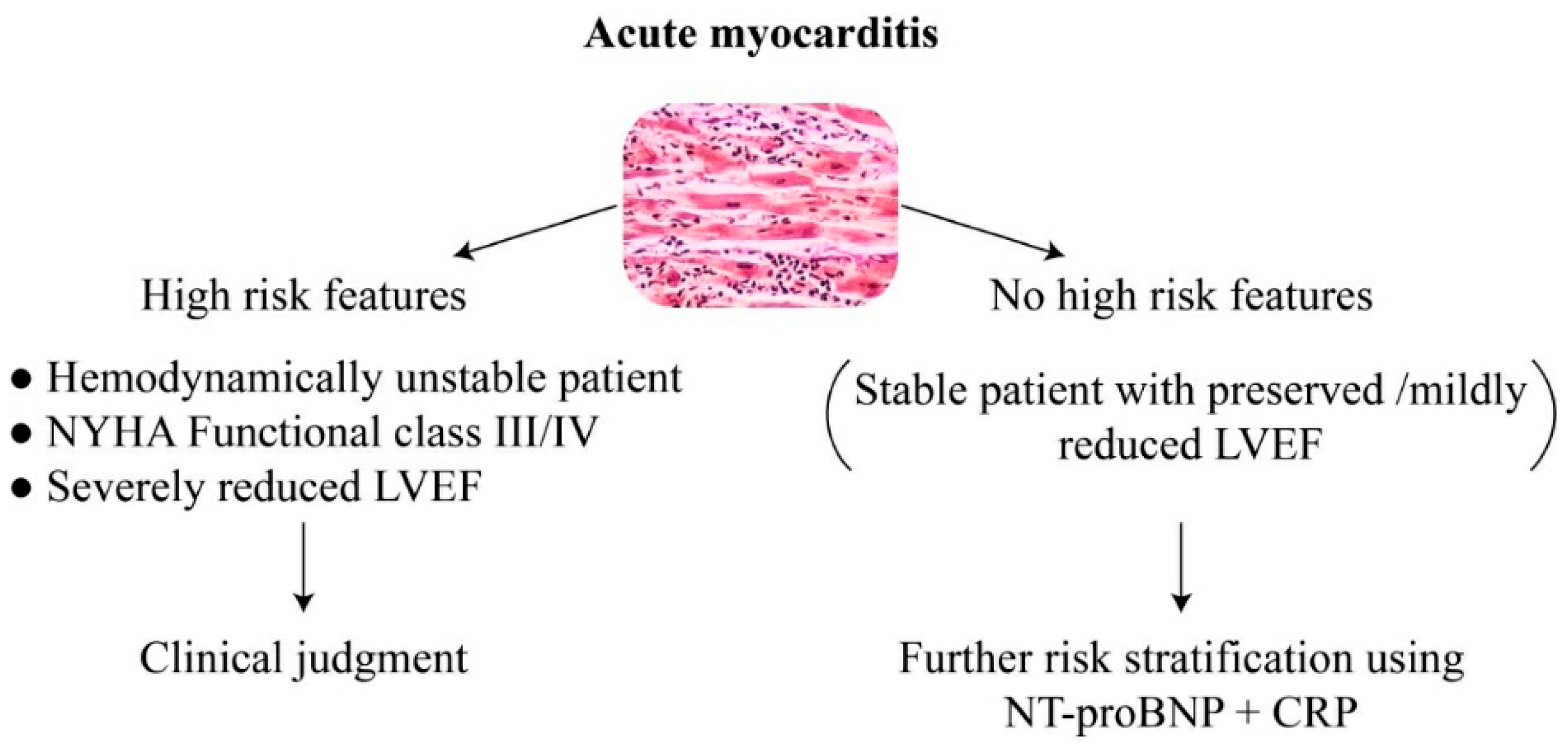
:1. Introduction
2. Materials and Methods
2.1. Study Population
2.2. Echocardiographic Data
2.3. Laboratory Data
2.4. Statistical Analysis
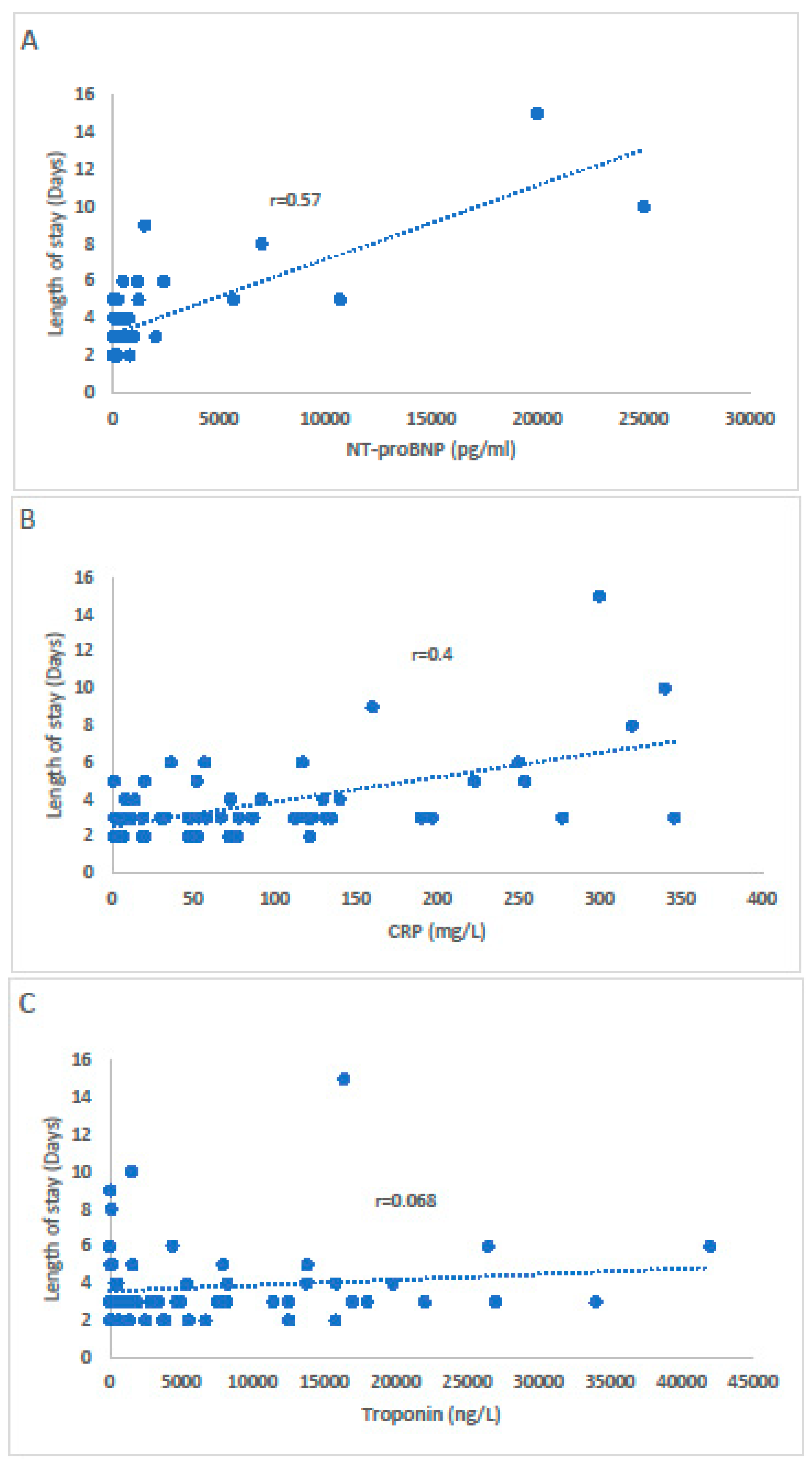
3. Results
4. Discussion
5. Limitations
6. Conclusions
Author Contributions
Funding
Institutional Review Board Statement
Informed Consent Statement
Conflicts of Interest
References
- Caforio, A.L.; Pankuweit, S.; Arbustini, E.; Basso, C.; Gimeno-Blanes, J.; Felix, S.B.; Fu, M.; Heliö, T.; Heymans, S.; Jahns, R.; et al. European Society of Cardiology Working Group on Myocardial and Pericardial Diseases. Current state of knowledge on aetiology, diagnosis, management, and therapy of myocarditis: A position statement of the European Society of Cardiology Working Group on Myocardial and Pericardial Diseases. Eur. Heart J. 2013, 34, 2636–2648. [Google Scholar] [PubMed]
- Fung, G.; Luo, H.; Qiu, Y.; Yang, D.; McManus, B. Myocarditis. Circ. Res. 2016, 118, 496–514. [Google Scholar] [CrossRef] [PubMed]
- Global Burden of Disease Study 2013 Collaborators. Global, regional, and national incidence, prevalence, and years lived with disability for 301 acute and chronic diseases and injuries in 188 countries, 1990–2013: A systematic analysis for the Global Burden of Disease Study 2013. Lancet 2015, 386, 743–800. [Google Scholar] [CrossRef] [Green Version]
- Sagar, S.; Liu, P.P.; Cooper, L.T., Jr. Myocarditis. Lancet 2012, 75, 1046–1057. [Google Scholar] [CrossRef] [Green Version]
- Mahrholdt, H.; Wagner, A.; Deluigi, C.C.; Kispert, E.; Hager, S.; Meinhardt, G.; Vogelsberg, H.; Fritz, P.; Dippon, J.; Bock, C.T.; et al. Presentation, patterns of myocardial damage, and clinical course of viral myocarditis. Circulation 2006, 114, 1581–1590. [Google Scholar] [CrossRef] [Green Version]
- Ammirati, E.; Cipriani, M.; Moro, C.; Raineri, C.; Pini, D.; Sormani, P.; Mantovani, R.; Varrenti, M.; Pedrotti, P.; Conca, C.; et al. Clinical Presentation and Outcome in a Contemporary Cohort of Patients with Acute Myocarditis: Multicenter Lombardy Registry. Circulation 2018, 138, 1088–1099. [Google Scholar] [CrossRef]
- Sharma, A.N.; Stultz, J.R.; Bellamkonda, N.; Amsterdam, E.A. Fulminant Myocarditis: Epidemiology, Pathogenesis, Diagnosis, and Management. Am. J. Cardiol. 2019, 124, 1954–1960. [Google Scholar] [CrossRef]
- Peretto, G.; Sala, S.; Rizzo, S.; Palmisano, A.; Esposito, A.; De Cobelli, F.; Campochiaro, C.; De Luca, G.; Foppoli, L.; Dagna, L.; et al. Ventricular Arrhythmias in Myocarditis: Characterization and Relationships with Myocardial Inflammation. J. Am. Coll. Cardiol. 2020, 75, 1046–1057. [Google Scholar] [CrossRef]
- Calabrese, F.; Thiene, G. Myocarditis and inflammatory cardiomyopathy: Microbiological and molecular biological aspects. Cardiovasc. Res. 2003, 60, 11–25. [Google Scholar] [CrossRef]
- Friedrich, M.G.; Sechtem, U.; Schulz-Menger, J.; Holmvang, G.; Alakija, P.; Cooper, L.T.; White, J.A.; Abdel-Aty, H.; Gutberlet, M.; Prasad, S.; et al. International Consensus Group on Cardiovascular Magnetic Resonance in Myocarditis. Cardiovascular magnetic resonance in myocarditis: A JACC White Paper. J. Am. Coll. Cardiol. 2009, 53, 1475–1487. [Google Scholar] [CrossRef] [Green Version]
- Ferreira, V.M.; Schulz-Menger, J.; Holmvang, G.; Kramer, C.M.; Carbone, I.; Sechtem, U.; Kindermann, I.; Gutberlet, M.; Cooper, L.T.; Liu, P.; et al. Cardiovascular Magnetic Resonance in Nonischemic Myocardial Inflammation: Expert Recommendations. J. Am. Coll. Cardiol. 2018, 72, 3158–3176. [Google Scholar] [CrossRef] [PubMed]
- Lurz, P.; Luecke, C.; Eitel, I.; Föhrenbach, F.; Frank, C.; Grothoff, M.; de Waha, S.; Rommel, K.P.; Lurz, J.A.; Klingel, K.; et al. Comprehensive Cardiac Magnetic Resonance Imaging in Patients with Suspected Myocarditis: The MyoRacer-Trial. J. Am. Coll. Cardiol. 2016, 19, 1800–1811. [Google Scholar] [CrossRef] [PubMed]
- Smith, S.C.; Ladenson, J.H.; Mason, J.W.; Jaffe, A.S. Elevations of cardiac troponin I associated with myocarditis. Experimental and clinical correlates. Circulation 1997, 95, 163–168. [Google Scholar] [CrossRef] [PubMed]
- Galvani, M.; Ottani, F.; Oltrona, L.; Ardissino, D.; Gensini, G.F.; Maggioni, A.P.; Mannucci, P.M.; Mininni, N.; Prando, M.D.; Tubaro, M.; et al. Italian Working Group on Atherosclerosis, Thrombosis, and Vascular Biology and the Associazione Nazionale Medici Cardiologi Ospedalieri (ANMCO). N-terminal pro-brain natriuretic peptide on admission has prognostic value across the whole spectrum of acute coronary syndromes. Circulation 2004, 110, 128–134. [Google Scholar] [PubMed] [Green Version]
- ten Wolde, M.; Tulevski, I.I.; Mulder, J.W.; Söhne, M.; Boomsma, F.; Mulder, B.J.; Büller, H.R. Brain natriuretic peptide as a predictor of adverse outcome in patients with pulmonary embolism. Circulation 2003, 107, 2082–2084. [Google Scholar] [CrossRef] [PubMed] [Green Version]
- Januzzi, J.L., Jr.; Camargo, C.A.; Anwaruddin, S.; Baggish, A.L.; Chen, A.A.; Krauser, D.G.; Tung, R.; Cameron, R.; Nagurney, J.T.; Chae, C.U.; et al. The N-terminal Pro-BNP investigation of dyspnea in the emergency department (PRIDE) study. Am. J. Cardiol. 2005, 95, 948–954. [Google Scholar] [CrossRef]
- Januzzi, J.L.; van Kimmenade, R.; Lainchbury, J.; Bayes-Genis, A.; Ordonez-Llanos, J.; Santalo-Bel, M.; Pinto, Y.M.; Richards, M. NT-proBNP testing for diagnosis and short-term prognosis in acute destabilized heart failure: An international pooled analysis of 1256 patients: The International Collaborative of NT-proBNP Study. Eur. Heart J. 2006, 27, 330–337. [Google Scholar] [CrossRef] [Green Version]
- Ali-Ahmed, F.; Dalgaard, F.; Al-Khatib, S.M. Sudden cardiac death in patients with myocarditis: Evaluation, risk stratification, and management. Am. Heart J. 2020, 220, 29–40. [Google Scholar] [CrossRef]
- Kindermann, I.; Kindermann, M.; Kandolf, R.; Klingel, K.; Bültmann, B.; Müller, T.; Lindinger, A.; Böhm, M. Predictors of outcome in patients with suspected myocarditis. Circulation 2008, 118, 639–648. [Google Scholar] [CrossRef] [Green Version]
- Gulati, A.; Jabbour, A.; Ismail, T.F.; Guha, K.; Khwaja, J.; Raza, S.; Morarji, K.; Brown, T.D.; Ismail, N.A.; Dweck, M.R.; et al. Association of fibrosis with mortality and sudden cardiac death in patients with nonischemic dilated cardiomyopathy. JAMA 2013, 309, 896–908. [Google Scholar] [CrossRef] [Green Version]
- Gräni, C.; Eichhorn, C.; Bière, L.; Murthy, V.L.; Agarwal, V.; Kaneko, K.; Cuddy, S.; Aghayev, A.; Steigner, M.; Blankstein, R.; et al. Prognostic Value of Cardiac Magnetic Resonance Tissue Characterization in Risk Stratifying Patients with Suspected Myocarditis. J. Am. Coll. Cardiol. 2017, 70, 1964–1976. [Google Scholar] [CrossRef] [PubMed]
- Vodovar, N.; Séronde, M.F.; Laribi, S.; Gayat, E.; Lassus, J.; Boukef, R.; Nouira, S.; Manivet, P.; Samuel, J.L.; Logeart, D.; et al. Post-translational modifications enhance NT-proBNP and BNP production in acute decompensated heart failure. Eur. Heart J. 2014, 35, 3434–3441. [Google Scholar] [CrossRef] [PubMed] [Green Version]
- Maeda, K.; Tsutamoto, T.; Wada, A.; Hisanaga, T.; Kinoshita, M. Plasma brain natriuretic peptide as a biochemical marker of high left ventricular end-diastolic pressure in patients with symptomatic left ventricular dysfunction. Am. Heart J. 1998, 135, 825–832. [Google Scholar] [CrossRef]
- Semenov, A.G.; Tamm, N.N.; Seferian, K.R.; Postnikov, A.B.; Karpova, N.S.; Serebryanaya, D.V.; Koshkina, E.V.; Krasnoselsky, M.I.; Katrukha, A.G. Processing of pro-B-type natriuretic peptide: Furin and corin as candidate convertases. Clin. Chem. 2010, 56, 1166–1176. [Google Scholar] [CrossRef] [Green Version]
- Ichiki, T.; Burnett, J.C., Jr. Post-transcriptional modification of pro-BNP in heart failure: Is glycosylation and circulating furin key for cardiovascular homeostasis? Eur. Heart J. 2014, 35, 3001–3003. [Google Scholar] [CrossRef] [Green Version]
- Coronado, M.J.; Bruno, K.A.; Blauwet, L.A.; Tschöpe, C.; Cunningham, M.W.; Pankuweit, S.; van Linthout, S.; Jeon, E.S.; McNamara, D.M.; Krejčí, J.; et al. Elevated Sera sST2 Is Associated with Heart Failure in Men ≤ 50 Years Old With Myocarditis. J. Am. Heart Assoc. 2019, 8, e008968. [Google Scholar] [CrossRef] [Green Version]
- Tymińska, A.; Ozierański, K.; Skwarek, A.; Kapłon-Cieślicka, A.; Baritussio, A.; Grabowski, M.; Marcolongo, R.; Caforio, A.L. Personalized Management of Myocarditis and Inflammatory Cardiomyopathy in Clinical Practice. J. Pers. Med. 2022, 12, 183. [Google Scholar] [CrossRef]
- Ukena, C.; Kindermann, M.; Mahfoud, F.; Geisel, J.; Lepper, P.M.; Kandolf, R.; Böhm, M.; Kindermann, I. Diagnostic and prognostic validity of different biomarkers in patients with suspected myocarditis. Clin. Res. Cardiol. 2014, 103, 743–751. [Google Scholar] [CrossRef]
- Sara, B.; Monteiro, J.J.; Carvalho, P.; Ribeiro Carvalho, C.; Chemba, J.; Ferreira, C.; Moreira, J.I. Are high NT-proBNP levels more related to inflammation than to left ventricular systolic dysfunction in acute myocarditis? Eur. Heart J. Acute Cardiovasc. Care 2021, 10, zuab020.189. [Google Scholar] [CrossRef]
- Blanco-Domínguez, R.; Sánchez-Díaz, R.; de la Fuente, H.; Jiménez-Borreguero, L.J.; Matesanz-Marín, A.; Relaño, M.; Jiménez-Alejandre, R.; Linillos-Pradillo, B.; Tsilingiri, K.; Martín-Mariscal, M.L.; et al. A Novel Circulating MicroRNA for the Detection of Acute Myocarditis. N. Engl. J. Med. 2021, 384, 2014–2027. [Google Scholar] [CrossRef]
- Yu, S.R.; Zhang, C.Y.; Xiong, W.J.; Chen, J.T.; Song, J.X.; Chen, H. An Hypothesis: Disproportion between Cardiac Troponin and B-Type Natriuretic Peptide Levels-A High Risk and Poor Prognostic Biomarker in Patients with Fulminant Myocarditis? Heart Lung Circ. 2021, 30, 837–842. [Google Scholar] [CrossRef] [PubMed]
- Imazio, M.; Brucato, A.; Barbieri, A.; Ferroni, F.; Maestroni, S.; Ligabue, G.; Chinaglia, A.; Cumetti, D.; Della Casa, G.; Bonomi, F.; et al. Good prognosis for pericarditis with and without myocardial involvement: Results from a multicenter, prospective cohort study. Circulation 2013, 128, 42–49. [Google Scholar] [CrossRef] [PubMed] [Green Version]
- Kobayashi, D.; Aggarwal, S.; Kheiwa, A.; Shah, N. Myopericarditis in children: Elevated troponin I level does not predict outcome. Pediatr. Cardiol. 2012, 33, 1040–1045. [Google Scholar] [CrossRef] [PubMed]
- Goitein, O.; Sabag, A.; Koperstein, R.; Hamdan, A.; Di Segni, E.; Konen, E.; Matetzky, S. Role of C reactive protein in evaluating the extent of myocardial inflammation in acute myocarditis. J. Cardiovasc. Magn. Reson. 2015, 17 (Suppl. 1), P291. [Google Scholar] [CrossRef] [Green Version]
- Bachmann, K.N.; Gupta, D.K.; Xu, M.; Brittain, E.; Farber-Eger, E.; Arora, P.; Collins, S.; Wells, Q.S.; Wang, T.J. Unexpectedly Low Natriuretic Peptide Levels in Patients with Heart Failure. Heart Fail. 2021, 9, 192–200. [Google Scholar] [CrossRef] [PubMed]

| Age (Years) | 28 (20–43) |
|---|---|
| Male | 45 (76.3%) |
| BMI (Kg/m2) | 26.1 (18.1–27) |
| Current smoker | 17 (28.8%) |
| Diabetes mellitus | 6 (10.2%) |
| Hyperlipidemia | 12 (20.3%) |
| Hypertension | 6 (10.2%) |
| Coronary artery disease | 1 (1.7%) |
| Family history of coronary artery disease | 15 (25.4%) |
| Chronic kidney disease | 3 (5.1%) |
| Chronic obstructive pulmonary disease | 2 (3.4%) |
| Chronic medications | |
| ACE inhibitors/ARBs | 5 (8.5%) |
| Beta blockers | 6 (10.2%) |
| Statins | 10 (16.9%) |
| Aspirin | 3 (5.1%) |
| Eltroxin | 5 (8.5%) |
| Clinical presentation | |
| Chest pain | 52 (88.1%) |
| Dyspnea | 12 (20.3%) |
| Palpitations | 7 (11.9%) |
| Viral prodrome | 50 (84.9%) |
| LVEF (%) | 48% (54–62) |
|---|---|
| LVEDD (mm) | 47 (44–50) |
| LAVI (mL/m2) | 23 (20–26) |
| E/e’ | 8.5 (7.2–9.3) |
| Pericardial effusion | 12 (20.3%) |
| Mitral valve regurgitation | 3 (5.1%) |
| Tricuspid valve regurgitation | 4 (6.8%) |
| Estimated SPAP (mmHg) | 22 (16–28) |
| TAPSE (cm) | 1.8 (1.7–2.0) |
| Pulse (BPM) | 76 (70–88) |
|---|---|
| SBP (mmHg) | 116 (110–125) |
| DBP (mmHg) | 73 (64–80) |
| Spo2 (%) | 96 (94–98) |
| Respiratory rate | 14 (12–18) |
| Temp (°C) | 37 (36–37.8) |
| ECG changes | 35 (59.3%) |
| NT-proBNP pg/mL | 300 (59–830) |
| Hs-TnI (ng/L) | 3435 (373–12,500) |
| CRP (mg/L) | 67 (20–130) |
| WBC (×109/L) | 10 (8–13.7) |
| Hemoglobin (g/dl) | 14 (13–15) |
| Creatinine (mg/dl) | 0.83 (0.7–0.99) |
| eGFR (CKD-EPI) (mL/min/1.73 m2) | 117 (98–123) |
| Potassium (mmol/L) | 4.0 (3.7–5.1) |
| Sodium (mEq/L) | 139 (130–138) |
| Use of analgesia | 50 (84.7%) |
| Invasive coronary angiography | 11 (18.6%) |
| Cardiac MRI | 10 (16.9%) |
| Complications | 14 (23.7%) |
| Atrial fibrillation | 3 (5.1%) |
| Ventricular tachycardia | 6 (10.2%) |
| Syncope | 2 (3.4%) |
| Pulmonary congestion | 3 (5.1%) |
| Length of stay (days) | 3 (2–5) |
Publisher’s Note: MDPI stays neutral with regard to jurisdictional claims in published maps and institutional affiliations. |
© 2022 by the authors. Licensee MDPI, Basel, Switzerland. This article is an open access article distributed under the terms and conditions of the Creative Commons Attribution (CC BY) license (https://creativecommons.org/licenses/by/4.0/).
Share and Cite
Moady, G.; Perlmutter, S.; Atar, S. The Prognostic Value of Natriuretic Peptides in Stable Patients with Suspected Acute Myocarditis: A Retrospective Study. J. Clin. Med. 2022, 11, 2472. https://doi.org/10.3390/jcm11092472
Moady G, Perlmutter S, Atar S. The Prognostic Value of Natriuretic Peptides in Stable Patients with Suspected Acute Myocarditis: A Retrospective Study. Journal of Clinical Medicine. 2022; 11(9):2472. https://doi.org/10.3390/jcm11092472
Chicago/Turabian StyleMoady, Gassan, Shahar Perlmutter, and Shaul Atar. 2022. "The Prognostic Value of Natriuretic Peptides in Stable Patients with Suspected Acute Myocarditis: A Retrospective Study" Journal of Clinical Medicine 11, no. 9: 2472. https://doi.org/10.3390/jcm11092472
APA StyleMoady, G., Perlmutter, S., & Atar, S. (2022). The Prognostic Value of Natriuretic Peptides in Stable Patients with Suspected Acute Myocarditis: A Retrospective Study. Journal of Clinical Medicine, 11(9), 2472. https://doi.org/10.3390/jcm11092472