Dermoscopy, Reflectance Confocal Microscopy and Optical Coherence Tomography Features of Acne: A Systematic Review
Abstract
1. Introduction
2. Methods
2.1. Study Selection Criteria
2.2. Data Search
2.3. Study Selection and Data Collection
2.4. Study Outcomes
3. Results
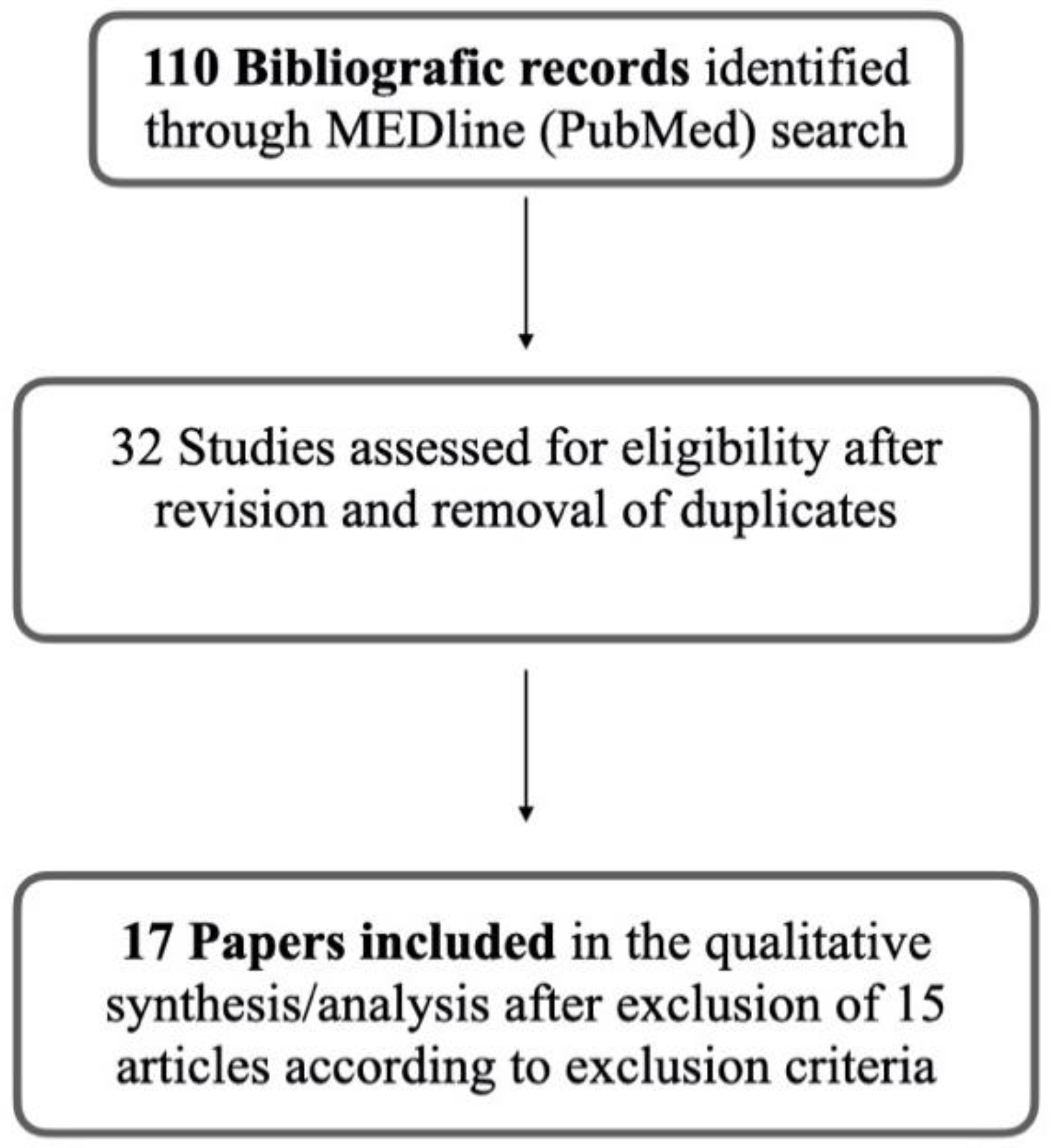
Study Selection
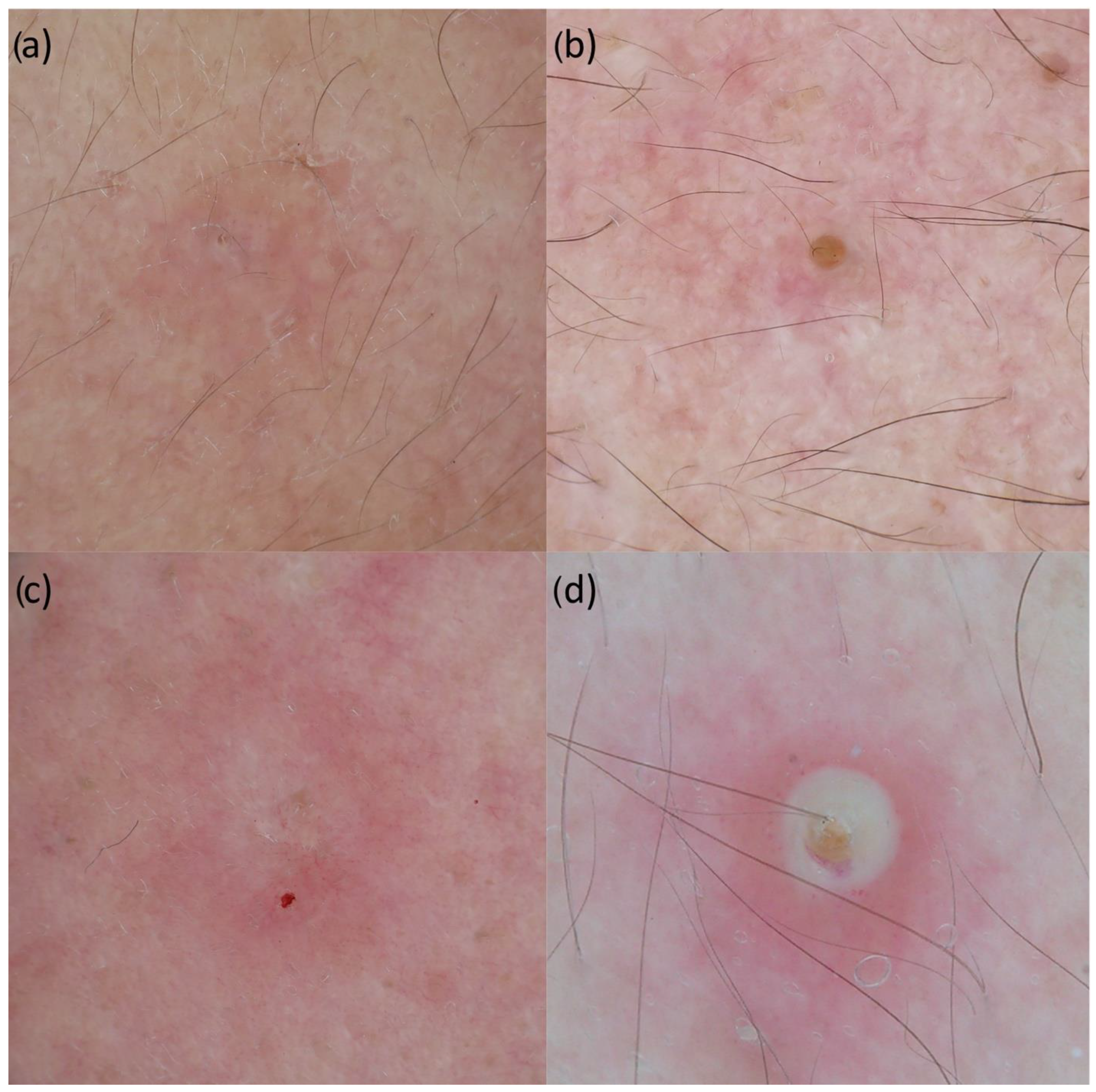
4. Dermoscopy in Acne Lesions
5. Rcm in Acne Lesions and Perilesional Skin
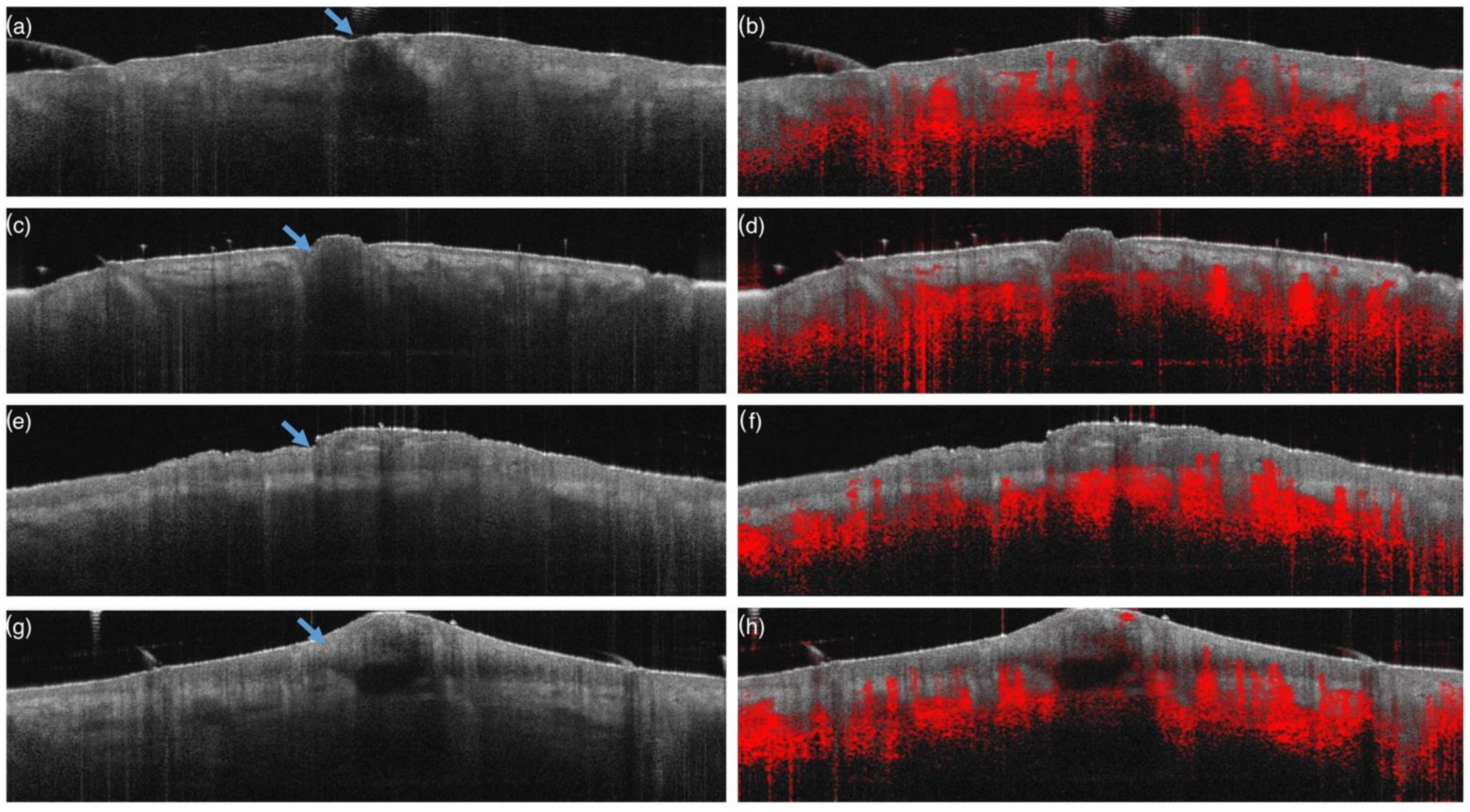
6. Oct in Acne Lesions
7. Acne Pathophysiology
- (1)
- Initially, acne skin is characterized by an increased number of small bright follicles (microcomedos).
- (2)
- Subsequently, several large bright follicles appear in the area of interest, associated with increased hyperkeratinization of the infundibula, leading to its occlusion (comedos).
- (3)
- Abruptly, several inflammatory phenomena occur with the appearance of papules, pustules, or nodular lesions with increased vessel density, exocytosis, and organized inflammatory infiltrate and an increase of adjacent small bright follicles.
- (4)
- Finally, resolution of the inflammatory lesion is characterized by the absence of inflammatory phenomena and the persistence of an increased number of small and large bright follicles associated with dilated sebaceous gland morphology.
8. Noninvasive Acne Therapy Monitoring
9. Conclusions
Funding
Institutional Review Board Statement
Informed Consent Statement
Data Availability Statement
Conflicts of Interest
References
- Ulrich, M.; Themstrup, L.; De Carvalho, N.; Manfredi, M.; Grana, C.; Ciardo, S.; Kästle, R.; Holmes, J.; Whitehead, R.; Jemec, G.B.E.; et al. Dynamic Optical Coherence Tomography in Dermatology. Dermatology 2016, 232, 298–311. [Google Scholar] [CrossRef] [PubMed]
- Srivastava, R.; Manfredini, M.; Rao, B.K. Noninvasive imaging tools in dermatology. Cutis 2019, 104, 108–113. [Google Scholar] [PubMed]
- Rajadhyaksha, M.; González, S.; Zavislan, J.M.; Anderson, R.R.; Webb, R.H. In Vivo Confocal Scanning Laser Microscopy of Human Skin II: Advances in Instrumentation and Comparison with Histology. J. Investig. Dermatol. 1999, 113, 293–303. [Google Scholar] [CrossRef] [PubMed]
- Sattler, E.; Kästle, R.; Welzel, J. Optical coherence tomography in dermatology. J. Biomed. Opt. 2013, 18, 061224. [Google Scholar] [CrossRef]
- Ruini, C.; Schuh, S.; Sattler, E.; Welzel, J. Line-field confocal optical coherence tomography—Practical applications in dermatology and comparison with established imaging methods. Ski. Res. Technol. 2020, 27, 340–352. [Google Scholar] [CrossRef] [PubMed]
- Page, M.J.; McKenzie, J.E.; Bossuyt, P.M.; Boutron, I.; Hoffmann, T.C.; Mulrow, C.D.; Shamseer, L.; Tetzlaff, J.M.; Akl, E.A.; Brennan, S.E.; et al. The PRISMA 2020 statement: An updated guideline for reporting systematic reviews. Int. J. Surg. 2021, 88, 105906. [Google Scholar] [CrossRef] [PubMed]
- Lacarrubba, F.; Ardigò, M.; Di Stefani, A.; Verzì, A.E.; Micali, G. Dermatoscopy and Reflectance Confocal Microscopy Correlations in Nonmelanocytic Disorders. Dermatol. Clin. 2018, 36, 487–501. [Google Scholar] [CrossRef]
- Guida, S.; Longo, C.; Casari, A.; Ciardo, S.; Manfredini, M.; Reggiani, C.; Pellacani, G.; Farnetani, F. Update on the use of confocal microscopy in melanoma and non-melanoma skin cancer. G. Ital. Dermatol. Venereol. 2015, 150, 547–563. [Google Scholar]
- Alfaro-Castellón, P.; Mejía-Rodríguez, S.A.; Valencia-Herrera, A.; Ramírez, S.; Mena-Cedillos, C. Dermoscopy Distinction of Eruptive Vellus Hair Cysts with Molluscum Contagiosum and Acne Lesions. Pediatr. Dermatol. 2012, 29, 772–773. [Google Scholar] [CrossRef]
- Pellacani, G.; Longo, C.; Malvehy, J.; Puig, S.; Carrera, C.; Segura, S.; Bassoli, S.; Seidenari, S. In Vivo Confocal Microscopic and Histopathologic Correlations of Dermoscopic Features in 202 Melanocytic Lesions. Arch. Dermatol. 2008, 144, 1597–1608. [Google Scholar] [CrossRef][Green Version]
- Farnetani, F.; Manfredini, M.; Chester, J.; Ciardo, S.; Gonzalez, S.; Pellacani, G. Reflectance confocal microscopy in the diagnosis of pigmented macules of the face: Differential diagnosis and margin definition. Photochem. Photobiol. Sci. 2019, 18, 963–969. [Google Scholar] [CrossRef] [PubMed]
- González, S.; Tannous, Z. Real-time, in vivo confocal reflectance microscopy of basal cell carcinoma. J. Am. Acad. Dermatol. 2002, 47, 869–874. [Google Scholar] [CrossRef]
- Boone, M.; Jemec, G.B.E.; Del Marmol, V. High-definition optical coherence tomography enables visualization of individual cells in healthy skin: Comparison to reflectance confocal microscopy. Exp. Dermatol. 2012, 21, 740–744. [Google Scholar] [CrossRef] [PubMed]
- Ahlgrimm-Siess, V.; Horn, M.; Koller, S.; Ludwig, R.; Gerger, A.; Hofmann-Wellenhof, R. Monitoring efficacy of cryotherapy for superficial basal cell carcinomas with in vivo reflectance confocal microscopy: A preliminary study. J. Dermatol. Sci. 2009, 53, 60–64. [Google Scholar] [CrossRef] [PubMed]
- Fuchs, C.S.K.; Andersen, A.J.B.; Ardigo, M.; Philipsen, P.A.; Haedersdal, M.; Mogensen, M. Acne vulgaris severity graded by in vivo reflectance confocal microscopy and optical coherence tomography: In vivo rcm and oct imaging of acne morphology. Lasers Surg. Med. 2018, 51, 104–113. [Google Scholar] [CrossRef]
- Manfredini, M.; Mazzaglia, G.; Ciardo, S.; Farnetani, F.; Mandel, V.D.; Longo, C.; Zauli, S.; Bettoli, V.; Virgili, A.; Pellacani, G. Acne: In vivo morphologic study of lesions and surrounding skin by means of reflectance confocal microscopy. J. Eur. Acad. Dermatol. Venereol. 2014, 29, 933–939. [Google Scholar] [CrossRef]
- Manfredini, M.; Greco, M.; Farnetani, F.; Mazzaglia, G.; Ciardo, S.; Bettoli, V.; Virgili, A.; Pellacani, G. In vivo monitoring of topical therapy for acne with reflectance confocal microscopy. Ski. Res. Technol. 2016, 23, 36–40. [Google Scholar] [CrossRef]
- Guenot, L.M.; Jourdain, M.V.; Saint-Jean, M.; Corvec, S.; Gaultier, A.; Khammari, A.; Le Moigne, M.; Boisrobert, A.; Paugam, C.; Dréno, B. Confocal microscopy in adult women with acne. Int. J. Dermatol. 2018, 57, 278–283. [Google Scholar] [CrossRef]
- Manfredini, M.; Bettoli, V.; Sacripanti, G.; Farnetani, F.; Bigi, L.; Puviani, M.; Corazza, M.; Pellacani, G. The evolution of healthy skin to acne lesions: A longitudinal, in vivo evaluation with reflectance confocal microscopy and optical coherence tomography. J. Eur. Acad. Dermatol. Venereol. 2019, 33, 1768–1774. [Google Scholar] [CrossRef]
- Mogensen, M.; Thrane, L.; Jørgensen, T.M.; Andersen, P.E.; Jemec, G.B.E. OCT imaging of skin cancer and other dermatological diseases. J. Biophotonics 2009, 2, 442–451. [Google Scholar] [CrossRef]
- Sattler, E.C.E.; Poloczek, K.; Kästle, R.; Welzel, J. Confocal laser scanning microscopy and optical coherence tomography for the evaluation of the kinetics and quantification of wound healing after fractional laser therapy. J. Am. Acad. Dermatol. 2013, 69, e165–e173. [Google Scholar] [CrossRef] [PubMed]
- Manfredini, M.; Chello, C.; Ciardo, S.; Guida, S.; Chester, J.; Lasagni, C.; Bigi, L.; Farnetani, F.; Bettoli, V.; Pellacani, G. Hidradenitis Suppurativa: Morphologic and vascular study of nodular inflammatory lesions by means of optical coherence tomography. Exp. Dermatol. 2022. [Google Scholar] [CrossRef]
- Manfredini, M.; Greco, M.; Farnetani, F.; Ciardo, S.; De Carvalho, N.; Mandel, V.D.; Starace, M.; Pellacani, G. Acne: Morphologic and vascular study of lesions and surrounding skin by means of optical coherence tomography. J. Eur. Acad. Dermatol. Venereol. 2017, 31, 1541–1546. [Google Scholar] [CrossRef] [PubMed]
- Hermsmeier, M.; Sawant, T.; Chowdhury, K.; Nagavarapu, U.; Chan, K.F. First Use of Optical Coherence Tomography on In Vivo Inflammatory Acne-Like Lesions: A Murine Model. Lasers Surg. Med. 2019, 52, 207–217. [Google Scholar] [CrossRef] [PubMed]
- Manfredini, M.; Liberati, S.; Ciardo, S.; Bonzano, L.; Guanti, M.; Chester, J.; Kaleci, S.; Pellacani, G. Microscopic and functional changes observed with dynamic optical coherence tomography for severe refractory atopic dermatitis treated with dupilumab. Ski. Res. Technol. 2020, 26, 779–787. [Google Scholar] [CrossRef]
- Fuchs, C.S.K.; Ortner, V.K.; Mogensen, M.; Philipsen, P.A.; Haedersdal, M. Transfollicular delivery of gold microparticles in healthy skin and acne vulgaris, assessed by in vivo reflectance confocal microscopy and optical coherence tomography. Lasers Surg. Med. 2019, 51, 430–438. [Google Scholar] [CrossRef]
- Rossi, E.; Mandel, V.D.; Paganelli, A.; Farnetani, F.; Pellacani, G. Plasma exeresis for active acne vulgaris: Clinical and in vivo microscopic documentation of treatment efficacy by means of reflectance confocal microscopy. Ski. Res. Technol. 2018, 24, 522–524. [Google Scholar] [CrossRef]
- Garofalo, V.; Cannizzaro, M.V.; Mazzilli, S.; Bianchi, L.; Campione, E. Clinical evidence on the efficacy and tolerability of a topical medical device containing benzoylperoxide 4%, retinol 0.5%, mandelic acid 1% and lactobionic acid 1% in the treatment of mild facial acne: An open label pilot study. Clin. Cosmet. Investig. Dermatol. 2019, 12, 363–369. [Google Scholar] [CrossRef]
- Villani, A.; Annunziata, M.C.; Cinelli, E.; Donnarumma, M.; Milani, M.; Fabbrocini, G. Efficacy and Safety of a New Topical Gel Formulation Containing Retinol Encapsulated in Glycospheres and Hydroxypinacolone Retinoate, an Antimicrobial Pep-tide, Salicylic Acid, Glycolic Acid and Niacinamide for the Treatment of Mild Acne: Preliminary Results of a 2-Month Prospective Study. G. Ital. Dermatol. Venereol. 2020, 155, 676–679. [Google Scholar] [CrossRef]
- Capitanio, B.; Lora, V.; Ludovici, M.; Sinagra, J.L.M.; Ottaviani, M.; Mastrofrancesco, A.; Ardigo, M.; Camera, E. Modulation of sebum oxidation and interleukin-1? levels associates with clinical improvement of mild comedonal acne. J. Eur. Acad. Dermatol. Venereol. 2014, 28, 1792–1797. [Google Scholar] [CrossRef]
- Fuchs, C.S.K.; Ortner, V.K.; Hansen, F.S.; Philipsen, P.A.; Haedersdal, M. Subclinical effects of adapalene-benzoyl peroxide: A prospective in vivo imaging study on acne micromorphology and transfollicular delivery. J. Eur. Acad. Dermatol. Venereol. 2021, 35, 1377–1385. [Google Scholar] [CrossRef] [PubMed]


| Elementary Acne Lesion Features | Dermoscopy Features | RCM Features | OCT Features | Histopathology Features |
|---|---|---|---|---|
| Comedos | Dilated pilosebaceous units filled with a white-yellow circle or a brown plug | Enlarged infundibula with a hyperkeratotic bright border | Enlarged infundibula with hyperkeratinized borders and a variable darker appearance depending on the amount of keratin. | A comedo is an altered pilo-sebaceous unit characterized by the presence of hyperkeratinization at the infundibulum and the istmus. The histopathologic appearance of comedos is characterized by the presence of a cyst-like cavity with a keratinous mass, colonized by bacteria. |
| Papules | Erythematous roundish lesions | Poorly defined lesions with intact or excoriated epidermis and an abundant inflammatory infiltrate in the epidermis or in the dermis. | Dome-shaped lesions with an either intact or thin and uneven epidermal surface and abundant inflammatory phenomena. | Papules are dome-shaped skin lesions characterized by the accumulation of inflammatory cells in dermis in a circumscribed area around one or more pilosebaceous units |
| Pustules | Inflammatory lesions centered by a white-yellowish area | Inflammatory lesions characterized by the accumulation of an organized inflammatory infiltrate and increased vascularity | Dome- shaped lesions characterized by abundant inflammatory phenomena associated to the presence of purulent aggregates inside the pustular cavity. | Pustules are dome-shaped skin lesions characterized by the accumulation of neutrophils within comedones, usually leading to the rupture of the original cystic cavity in the dermis. |
| Author | Number of Patients | Study Design | Acne Severity Grade | Technique | Number of Acquired Skin Areas | Aim of the Study | Type of Treatment |
|---|---|---|---|---|---|---|---|
| Manfredini 2015 [5] | 35 | Observational study | Absent to moderate | RCM | 55 | Acne characterization | n.s. |
| Manfredini 2019 [6] | 10 | Observational pilot study | Mild to moderate | RCM and OCT | 70 RCM and 70 OCT | Acne characterization | n.s. |
| Fuchs 2018 [7] | 21 | Explorative | Absent to moderate | RCM and OCT | 108 RCM and 54 OCT | Acne characterization | n.s. |
| Guenot 2018 [8] | 42 | Prospective | n.s. | RCM | 42 | Acne characterization in adult women | n.s. |
| Manfredini 2017 [9] | 19 | Observational study | Mild to moderate | RCM | 76 | Treatment monitoring | Hydroypinacolone retinoate/BIOPEP |
| Garofalo 2019 [10] | 20 | Pilot study | Mild | RCM | 60 | Treatment monitoring | Benzoylperoxide 4%, retinol 0.5%, mandelic acid 1%, and lactobionic acid 1% |
| Fuchs 2021 [11] | 15 | Prospective study | Mild to moderate | RCM and OCT | 60 RCM and 60 OCT | Treatment monitoring | Adapalen-benzoyl peroxide |
| Manfredini 2017 [12] | 31 | Prospective | Mild to moderate | OCT | 132 | Treatment monitoring | Oral antibiotic tratment |
| Capitanio 2014 [13] | 28 | Prospective | Absent to mild | RCM | 8 | Treatment monitoring | Mixed RetinSphere- vitamin E formulation |
| Fuchs 2019 [14] | 12 | n.s. | Absent to moderate | RCM and OCT | 109 RCM and 120 OCT | Treatment monitoring | Gold microparticles |
| Rossi 2018 [15] | 2 | n.s. | n.s. | RCM | 4 | Treatment monitoring | Plasma exeresis |
Publisher’s Note: MDPI stays neutral with regard to jurisdictional claims in published maps and institutional affiliations. |
© 2022 by the authors. Licensee MDPI, Basel, Switzerland. This article is an open access article distributed under the terms and conditions of the Creative Commons Attribution (CC BY) license (https://creativecommons.org/licenses/by/4.0/).
Share and Cite
Alma, A.; Sticchi, A.; Chello, C.; Guida, S.; Farnetani, F.; Chester, J.; Bettoli, V.; Pellacani, G.; Manfredini, M. Dermoscopy, Reflectance Confocal Microscopy and Optical Coherence Tomography Features of Acne: A Systematic Review. J. Clin. Med. 2022, 11, 1783. https://doi.org/10.3390/jcm11071783
Alma A, Sticchi A, Chello C, Guida S, Farnetani F, Chester J, Bettoli V, Pellacani G, Manfredini M. Dermoscopy, Reflectance Confocal Microscopy and Optical Coherence Tomography Features of Acne: A Systematic Review. Journal of Clinical Medicine. 2022; 11(7):1783. https://doi.org/10.3390/jcm11071783
Chicago/Turabian StyleAlma, Antonio, Alberto Sticchi, Camilla Chello, Stefania Guida, Francesca Farnetani, Johanna Chester, Vincenzo Bettoli, Giovanni Pellacani, and Marco Manfredini. 2022. "Dermoscopy, Reflectance Confocal Microscopy and Optical Coherence Tomography Features of Acne: A Systematic Review" Journal of Clinical Medicine 11, no. 7: 1783. https://doi.org/10.3390/jcm11071783
APA StyleAlma, A., Sticchi, A., Chello, C., Guida, S., Farnetani, F., Chester, J., Bettoli, V., Pellacani, G., & Manfredini, M. (2022). Dermoscopy, Reflectance Confocal Microscopy and Optical Coherence Tomography Features of Acne: A Systematic Review. Journal of Clinical Medicine, 11(7), 1783. https://doi.org/10.3390/jcm11071783


