Article

Clinical Outcome, Cognition, and Cerebrovascular Reactivity after Surgical Treatment for Moyamoya Vasculopathy: A Dutch Prospective, Single-Center Cohort Study

by
Pieter Thomas Deckers
1,2,*,†,
Annick Kronenburg
1,†,
Esther van den Berg
3,
Monique M. van Schooneveld
4,
Evert-Jan P. A. Vonken
5,
Willem M. Otte
1,
Bart N. M. van Berckel
6,
Maqsood Yaqub
6,
Catharina J. M. Klijn
1,7,
Albert van der Zwan
1 and
Kees P. J. Braun
1
1
Department of Neurology and Neurosurgery, UMC Utrecht Brain Center, 3584 CG Utrecht, The Netherlands
2
Department of Radiology and Nuclear Medicine, Meander Medisch Centrum, 3813 TZ Amersfoort, The Netherlands
3
Department of Neurology, Erasmus University Medical Center, 3015 GD Rotterdam, The Netherlands
4
Department of Pediatric Psychology, Wilhelmina Children’s Hospital, 3584 EA Utrecht, The Netherlands
5
Department of Radiology, UMC Utrecht Imaging Division, 3508 GA Utrecht, The Netherlands
6
Department of Nuclear Medicine & PET Research, Amsterdam UMC, 1081 HV Amsterdam, The Netherlands
7
Department of Neurology, Donders Institute for Brain, Cognition and Behavior, Center for Neuroscience, Radboud University Medical Center, 6525 GA Nijmegen, The Netherlands
*
Author to whom correspondence should be addressed.
These authors contributed equally to this work.
J. Clin. Med. 2022, 11(24), 7427; https://doi.org/10.3390/jcm11247427
Submission received: 5 November 2022 / Revised: 29 November 2022 / Accepted: 2 December 2022 / Published: 14 December 2022
(This article belongs to the Special Issue Diagnosis and Treatment of Moyamoya Disease)

Abstract

:
Background: It remains unclear whether revascularization of moyamoya vasculopathy (MMV) has a positive effect on cognitive function. In this prospective, single-center study, we investigated the effect of revascularization on cognitive function in patients with MMV. We report clinical and radiological outcome parameters and the associations between clinical determinants and change in neurocognitive functioning. Methods: We consecutively included all MMV patients at a Dutch tertiary referral hospital who underwent pre- and postoperative standardized neuropsychological evaluation, [15O]H2O-PET (including cerebrovascular reactivity (CVR)), MRI, cerebral angiography, and completed standardized questionnaires on clinical outcome and quality of life (QOL). To explore the association between patient characteristics, imaging findings, and change in the z-scores of the cognitive domains, we used multivariable linear- and Bayesian regression analysis. Results: We included 40 patients of whom 35 (27 females, 21 children) were treated surgically. One patient died after surgery, and two withdrew from the study. TIA- and headache frequency and modified Rankin scale (mRS) improved (resp. p = 0.001, 0.019, 0.039). Eleven patients (seven children) developed a new infarct during follow-up (31%), five of which were symptomatic. CVR-scores improved significantly (p < 0.0005). The language domain improved (p = 0.029); other domains remained stable. In adults, there was an improvement in QOL. We could not find an association between change in imaging and cognitive scores. Conclusion: In this cohort of Western MMV patients, TIA frequency, headache, CVR, and mRS improved significantly after revascularization. The language domain significantly improved, while others remained stable. We could not find an association between changes in CVR and cognitive scores.

1. Introduction

Moyamoya vasculopathy (MMV) is a cerebrovascular disorder of largely unknown etiology, characterized by progressive stenosis or occlusion of the supraclinoid internal carotid arteries and their proximal branches [1,2]. Most patients present with transient ischemic attacks (TIAs) or ischemic or hemorrhagic stroke and others with cognitive impairment [3]. Idiopathic MMV is referred to as moyamoya disease (MMD); MMV associated with another predisposing condition, e.g., neurofibromatosis, is referred to as moyamoya syndrome (MMS) [1]. Revascularization surgery is recommended for patients with ischemic symptoms or disturbed cerebrovascular reactivity (CVR) [2,4,5]. To identify brain areas at risk of ischemia, [15O]H2O-positron emission tomography (PET) is commonly used and enables the assessment of CVR after acetazolamide challenge [2].
We recently studied the cognitive profile of 40 MMV patients and found that 73% had cognitive impairments in at least one domain; children performed better in processing speed, and adults had higher scores in visuospatial functioning [6]. Little is known about the effect of revascularization on cognition in MMV, especially in the Western world [3], and on quality of life (QOL) [7].
In this prospective cohort study, we investigated the effect of revascularization on clinical outcome and cognition in patients with MMV and to what extent cognitive functions are related to CVR.

2. Materials and Methods

2.1. Patient Selection

The Medical Ethics Review Committee UMC Utrecht confirmed that the Medical Research Involving Human Subjects Act (WMO) did not apply. All patients or representatives gave written informed consent. We prospectively included forty consecutive MMV patients (children up to 18 years old and adults) who presented between October 2012 and September 2017 in our center, were not previously treated with revascularization surgery, could understand Dutch, were available for follow-up, and had a confirmed diagnosis of MMV by digital subtraction angiogram (DSA) or magnetic resonance angiography (MRA) [4]. Their baseline characteristics have been reported previously [6]. There were no eligible patients who refused participation.
Thirty-five patients (21 children) were treated operatively and five conservatively. Only the surgically treated group is included in the analysis. Since the treatment was tailored and not randomized, outcomes of conservatively treated patients are reported in the appendix (Appendix I and Appendix J) and were not directly compared to surgical patients.

2.2. Treatment

All patients took acetylsalicylic acid (38 mg for children, 100 mg for adults). Timing, type, and location of revascularization was decided in a multidisciplinary meeting by the treating neurologist and neurosurgeon based on symptoms, [15O]H2O-PET findings, and shared decision making with the patients, parents, or caregivers. The preferred initial surgical treatment was a single-staged combined direct and indirect revascularization. In children, an additional bifrontal revascularization was performed if indicated (Appendix A for details) [6,8].

2.3. Clinical Follow-Up

All patients were seen for follow-up at least one year after the last operation or at least one year after the baseline visit for patients treated conservatively. Patients were neurologically examined and interviewed using standardized questionnaires and a predefined case record form (for characteristics see Appendix B).
Any peri-operative complication or adverse event < 30 days after surgery was carefully noted and reviewed at the routine 6-week postoperative outpatient evaluation. Any complications resulting in permanent deficits or requiring additional surgery were classified as “serious”, while others were classified as “transient”. In case of withdrawal or death of patients during follow-up, all available endpoints were used (e.g., death, new infarction), but these patients were excluded from the final cognitive analysis.
To assess stroke severity, we applied the Pediatric National Institutes of Health Stroke scale (pedNIHSS) [9] in children and the NIHSS [10] in adults. The modified Rankin scale (mRS) [11] was applied in both groups to assess functional status; additionally, for children, the Pediatric Stroke Outcome Measure (PSOM) [12] was used. To measure QOL, we applied the Pediatric Quality of Life Inventory in children, including the parent-proxy questionnaire (PedsQL) [13]. Adults completed the Short Form—36 (SF-36) [14] and the EuroQol EQ-5D-3L (EQ-5D) [15].

2.4. Imaging

Patients were evaluated by MRI, digital subtraction angiography (DSA), and [15O]H2O-PET in combination with an acetazolamide challenge to determine CVR, as described previously (see also Appendix C for further details) [6].
MRI and [15O]H2O-PET images were scored in three global regions of interest (ROIs) of comparable size in each hemisphere (labeled as “frontal”, “middle”, and “posterior”) by two reviewers (P.T.D. and A.K.), blinded for all patient characteristics [6,16].
The ROIs in the MRI-FLAIR were scored on the presence of infarcts using the following scoring: 0 = none; 1 = small; 2 = intermediate; and 3 = large infarct. Furthermore, we scored periventricular (WMDp) and deep white matter disease (WMDd), applying an adapted Fazekas score per ROI: 0 = none; 1 = small or subtle; 2 = intermediate; and 3 = extensive [6,17]. When the differentiation between a new white matter lesion and a small lacunar infarction was ambiguous, diffusion-weighted imaging (DWI) and T1 images were additionally reviewed.
The [15O]H2O-PET CVR was qualitatively scored per ROI using the following score: 0 = CVR normal; 1 = minimal CVR present; 2 = CVR absent; and 3 = a steal phenomenon is present in any region within the ROI (i.e., the reduction of CBF after acetazolamide administration). If the entire ROI could not be scored due to infarction, this was noted as a missing value. Furthermore, the CVR was visually compared between baseline and follow-up to rate the CVR qualitatively as “improved”, “stable”, or “deteriorated”. For the calculation of the WMD, infarctions, and CVR scores, we averaged the valid ROI scores from the six regions.
DSA images were reviewed by an experienced neuroradiologist (E.J.V.) blinded for other data. The involvement of the ACA, MCA, and PCA was assessed according to the following categories: 0 = no evidence of disease; 1 = mild to moderate stenosis with absent or slightly developed MMD collaterals; 2 = severe stenosis with well-developed MMD collaterals; 3 = occlusion with well-developed MMD collaterals; and 4 = occlusion with absent or slightly developed MMD collaterals. To categorize the overall severity of MMV, the modified Suzuki score (mSS) was determined based on the highest hemispheric score [18]. Furthermore, the change relative to the baseline DSA was noted, including increase of collaterals, bypass patency, and change in mSS.

2.5. Cognitive Evaluation

All patients underwent a standardized neuropsychological evaluation test battery specified for MMV (Appendix D, Table A1) at baseline and at follow-up [6]. Neurocognitive tests were planned just before the follow-up visit, after a median of sixty weeks following last surgery (range: 40–108).
Tests were specifically chosen for children or adults but covered the same predefined cognitive test domains: general functioning; memory; working memory; language; attention and executive functioning; processing speed; and visuospatial functioning [19]. We assessed cognitive domains of all patients combined and for children and adults separately. As is common in clinical studies, patients were tested according to their age and capabilities. Not all patients performed the complete neuropsychological test battery. All available data were used as an estimation of individual cognitive domains. Raw test scores were corrected for age and education level using their respective manuals. All available adjusted scores were then converted to z-scores and averaged per domain. Cognitive impairment was defined as 1.5 SD or more below the population mean (i.e., z-score of <−1.5) in one or more domains [6]. Two children in whom we could not establish any reliable test score due to a low developmental age or insufficient understanding of the test were assumed to have a cognitive impairment but could not be quantitatively analyzed. When deemed necessary by the treating physicians, patients received speech therapy during the follow-up.

2.6. Data-Analysis and Statistics

All questionnaires—(ped)NIHSS, PSOM, mRS, PedsQL, SF-36, and EQ-5D-3L—were analyzed according to their respective manuals [6]. Changes in continuous variables (neurocognitive z-scores, PedsQL, SF-36, and EQ-5D-3L) were analyzed using Student’s t-tests after visually checking normality using histograms and q-q plots and formally with Kolmogorov–Smirnov tests. Changes between baseline and follow-up for ordinal variables (mRS, stratified TIA and headache frequency, (ped)NIHSS, and PSOM) were analyzed using paired-samples sign tests. A p-value < 0.05 was considered significant.
To investigate the effect of possibly relevant determinants on postoperative change in neurocognitive functioning, we used both multivariate linear regression and Bayesian regression. These determinants included: age categories (adult versus child); change in mSS, infarction, WMD, and CVR-score; and the presence of an associated predisposing condition that could have led to cognitive deficits in children with MMS (Down or Noonan syndrome; microcephalic osteodysplastic primordial dwarfism—II (MOPD-II); neurofibromatosis(NF)-1; and posterior fossa brain malformations, hemangioma, arterial lesions, cardiac abnormalities, and eye abnormalities syndrome (PHACES)) [6]. For the Bayesian regression, the Bayes factor (BF) was determined. A commonly used list divides the evidence in favor of an association into four strength ranges: BFs 1–3.2: “not worth more than a bare mention”; 3.2–10: “substantial”; 10–100: “strong”; and >100: “decisive” evidence [20]. Furthermore, we used univariate linear regression analysis and Bayesian regression to correlate postoperative change in cognitive domain scores with baseline CVR scores and change in CVR scores for each of the three ROIs separately. Statistics were performed using SPSS Statistics version 26 (IBM Corp, Armonk, NY, USA) and Bayesian and linear regression with JASP version 0.14.0 (JASP Team (2020)).

3. Results

3.1. Patient Characteristics and Treatment

Of the 40 included patients at baseline, 35 were operated (27 females; 21 children). We excluded three adults from the analysis of cognitive outcomes (one died two days postoperatively; two withdrew from follow-up) but included their available clinical end-points (e.g., complications, MRS). The remaining 32 patients underwent MRI and [15O]H2O-PET at follow-up; 30 received a DSA (one MOPD-II child was too small; in one child, it was postponed due to perioperative infarction). Thirty-one underwent neuropsychological evaluation (the parents of one child refused follow-up evaluation), and median time of neuropsychological evaluation was 60 weeks (range 40–108).
Twenty-one patients were treated bilaterally and fourteen unilaterally (Table 1). Two of the bilaterally treated patients had a single-stage procedure: one child with an indirect bilateral fronto-parietal bypass and another child with a unilateral combined bypass and bifrontal indirect procedure. The other bilateral procedures were two-staged. Twenty-nine patients (fifteen children) received a direct bypass in at least one hemisphere.

3.2. Surgical Complications

Four patients (11% of 35 patients; 7.4% of 54 operations) had serious complications resulting in permanent deficits (three patients) or death (one patient) within 30 days after operation. One patient died due to an intraparenchymal hemorrhage, most probably caused by hemorrhagic transformation of an infarcted area caused by hyperperfusion. The deceased patient was in poor clinical condition pre-operatively (see Appendix E). Three patients had new permanent deficits due to infarction (two adults). One of these adults also developed a bone flap infection requiring additional surgery. The infarction led to a deterioration in mRS at follow-up one year after operation from 1 to 2 in the child. The two adults withdrew from follow-up so their follow-up mRS is unavailable. Five patients had transient deficits due to hyperperfusion syndrome, and three patients had TIAs during the first 30 days following surgery. Further details are provided in Appendix E.

3.3. Clinical Follow-Up

There were 26 patients who had preoperative TIAs. Of those 26 patients the TIA frequency improved after surgery in 19 (73%); 16 became completely TIA-free (62%, 11 children, Figure 1A); and in 4, frequency remained unchanged. Overall TIA frequency improved significantly for the total group (p = 0.001) and for children (p < 0.0005), while there was no significant change for adults (p = 0.727).

3.4. Headache

Twenty-seven patients presented with headache (fifteen children, Figure 1C), of whom twelve had weekly complaints. Postoperatively, frequency improved in fifteen patients (43%; eight children), remained stable in thirteen (37%; nine children) and deteriorated in four patients (11%; all children). Frequency improved significantly in the total group (p = 0.019) and in adults (p = 0.016), not in children (p = 0.398).

3.5. Other Clinical Outcomes

Three patients (two children) presented with seizures: two became asymptomatic, and one remained stable. One child presented with chorea, which completely resolved postoperatively.

3.6. mRS, (ped)NIHSS, PSOM

MRS (Figure 1B) at follow-up improved in ten patients (29%, seven children) and deteriorated in three (9%; one child). Overall scores improved significantly for the total group (p = 0.039) and for children (p = 0.039; Appendix F, Table A2). Median pedNIHSS was 0 (range 0–3) at baseline and 0 (0–2) at follow-up, NIHSS was 1 (0–7) at baseline and 1 (0–2) at follow-up. Median PSOM at baseline was 0 (0–3) and 0 (0–2) at follow-up. These changes were non-significant.

3.7. Imaging

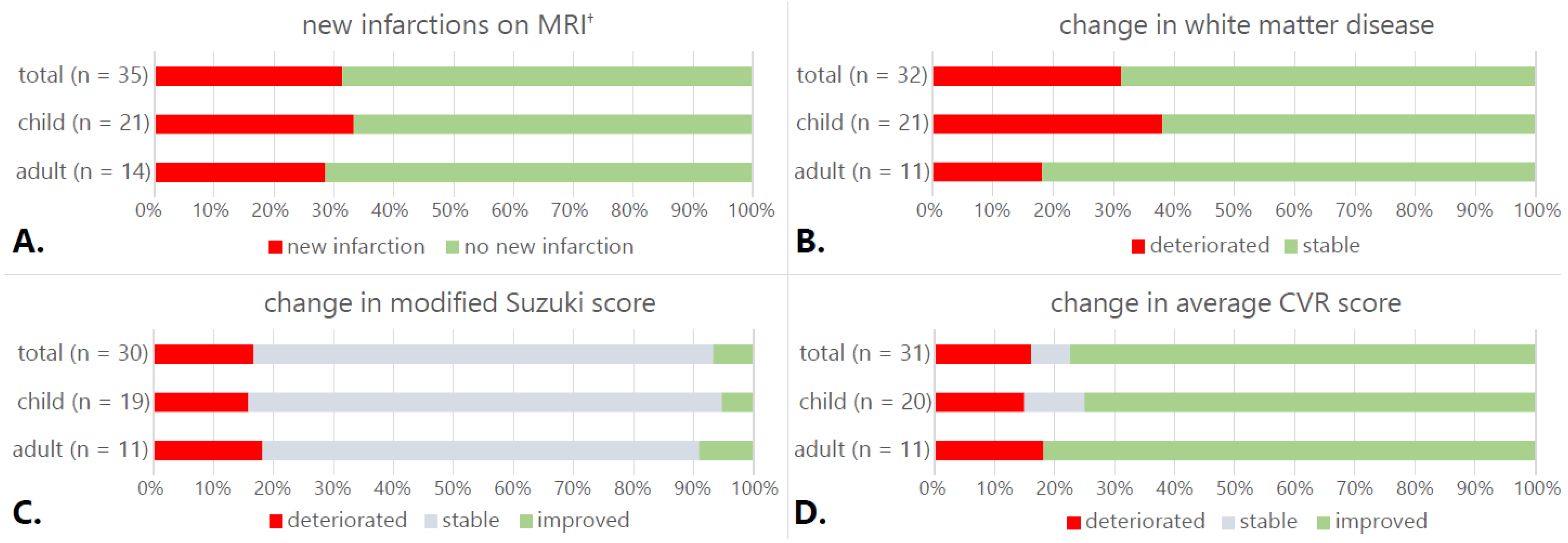
Eleven patients (31%, seven children) developed new infarctions on MRI (Figure 2A). Five (two children) were symptomatic, and in three patients, infarcts occurred peri-operatively. The average infarction score for the total group at follow-up increased from 0.68 to 0.82 (95%CI −0.27–−0.018, p = 0.027). Ten patients had an increase in WMD score (29%, eight children, Figure 2B); the average WMD score for the total group increased from 0.61 to 0.66 (95%CI −0.103–0.013, p = 0.127).
DSA showed an increase of extracranial-to-intracranial collaterals in all operated hemispheres. In patients who underwent direct bypass surgery (n = 29), all bypasses were open. The mSS improved in two patients (one child, Figure 2C) and deteriorated in five (three children). Mean mSS remained stable (3.0 at baseline, 3.1 at follow-up (95%CI: −0.279–0.079, p = 0.264)).
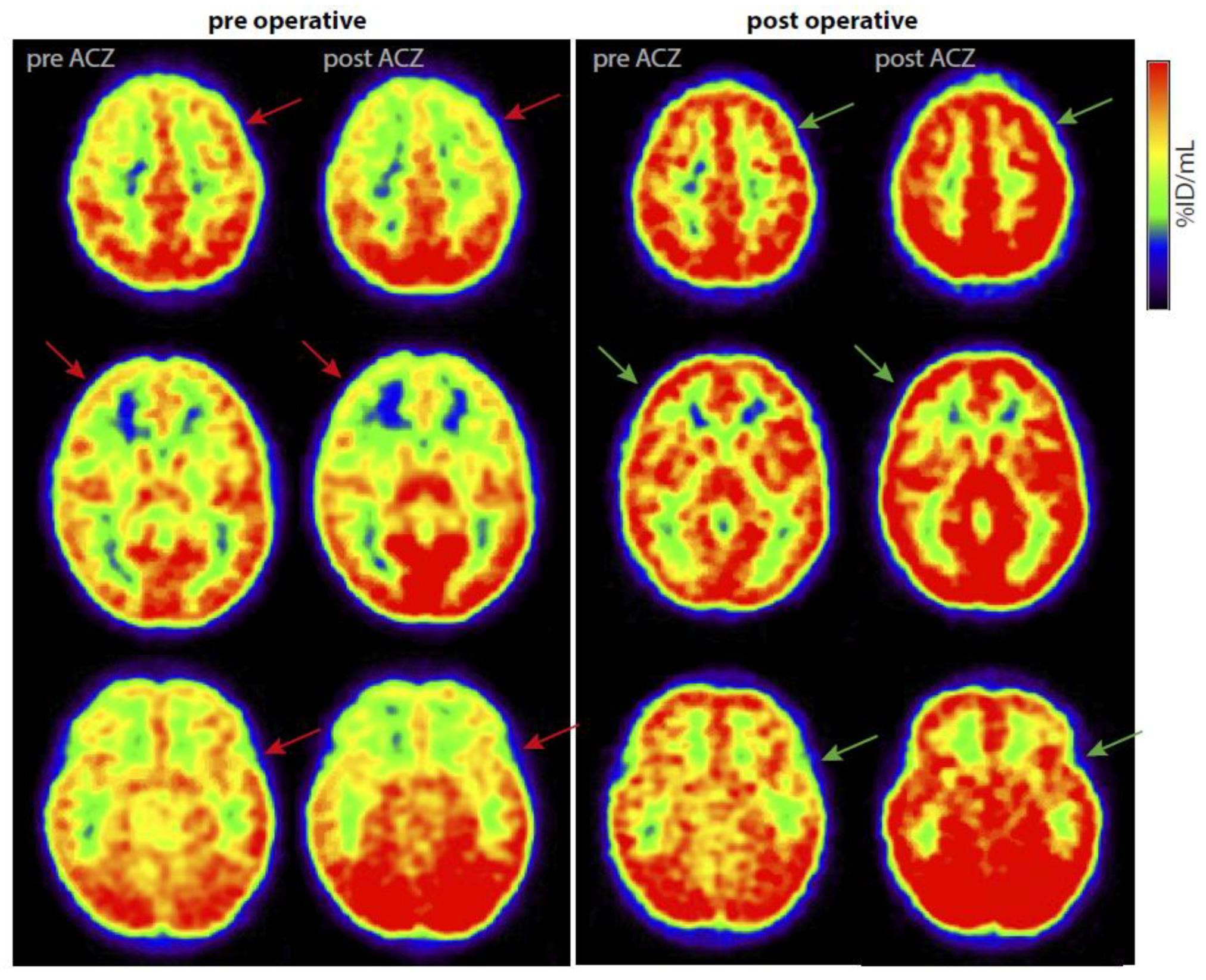
Average CVR scores improved in 24 patients (15 children, Figure 2D and Figure 3), deteriorated in 5 (three children), and remained stable in 2 (both children). Group CVR scores improved from 1.93 at baseline to 0.82 at follow-up (95%CI: 0.668–1.567, p ≤ 0.0005).

3.8. Neuropsychological Evaluation

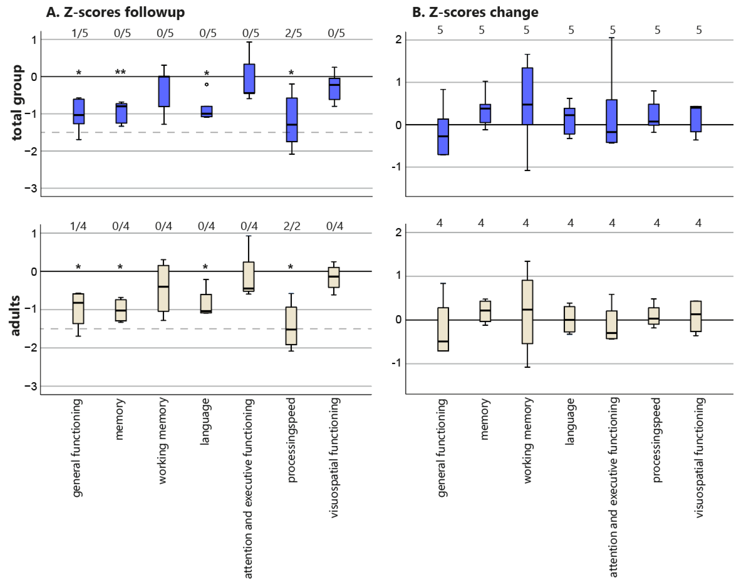
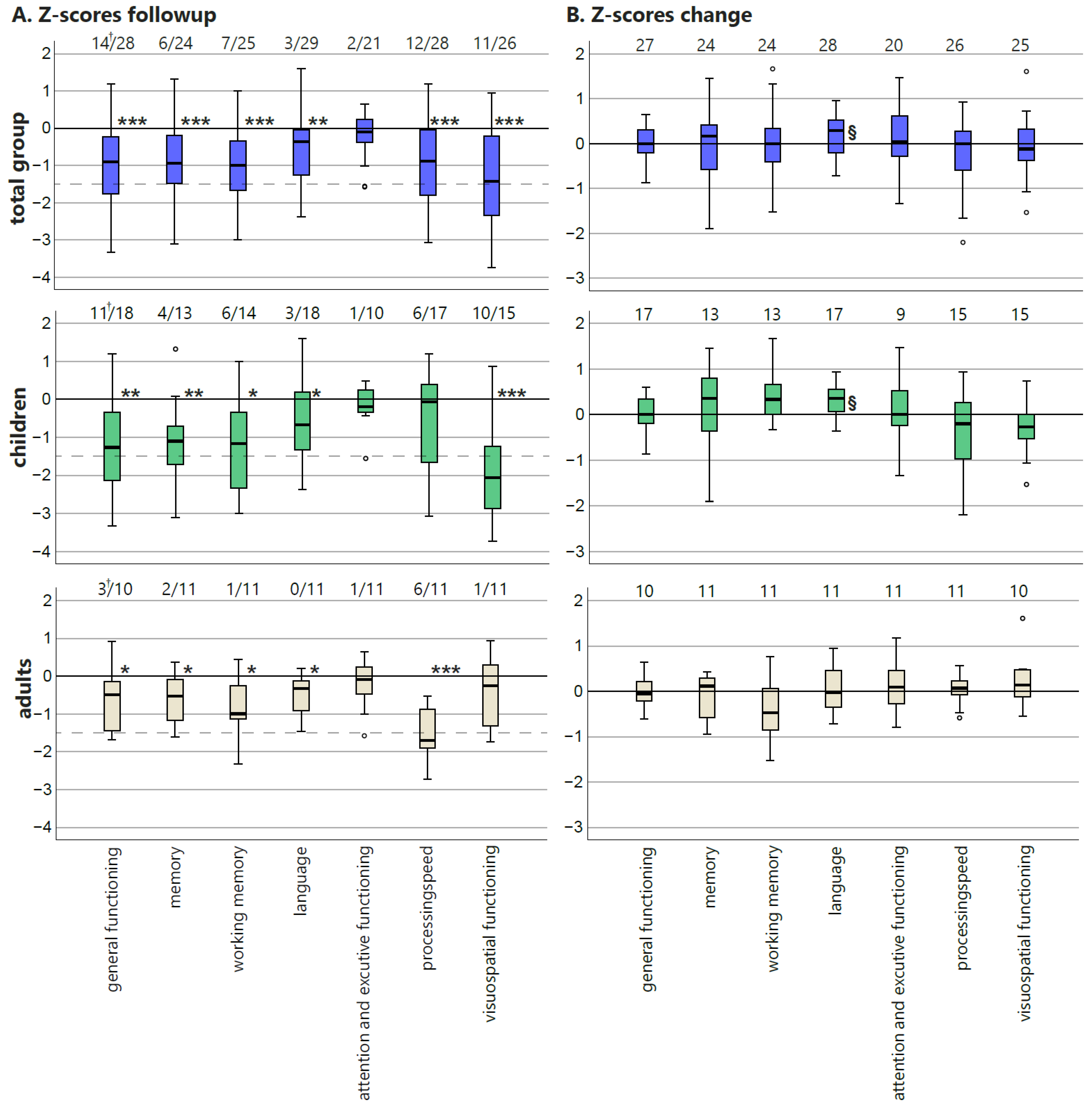
Cognitive functioning on a group level at follow-up was comparable with baseline [6]: postoperative test scores of all domains were significantly lower than the population mean in the total group and in children except for attention and executive functioning (Figure 4A) and visuospatial functioning in adults.
When looking at the change in z-scores in the total group, language domain scores improved significantly (p = 0.029), while the other mean z-scores remained stable (Figure 4B). In adults, none of the domain scores significantly changed after surgery, while children showed a significant improvement in language domain functioning (p = 0.006). Since the z-scores are corrected for age, this improvement exceeds the expected normal development.
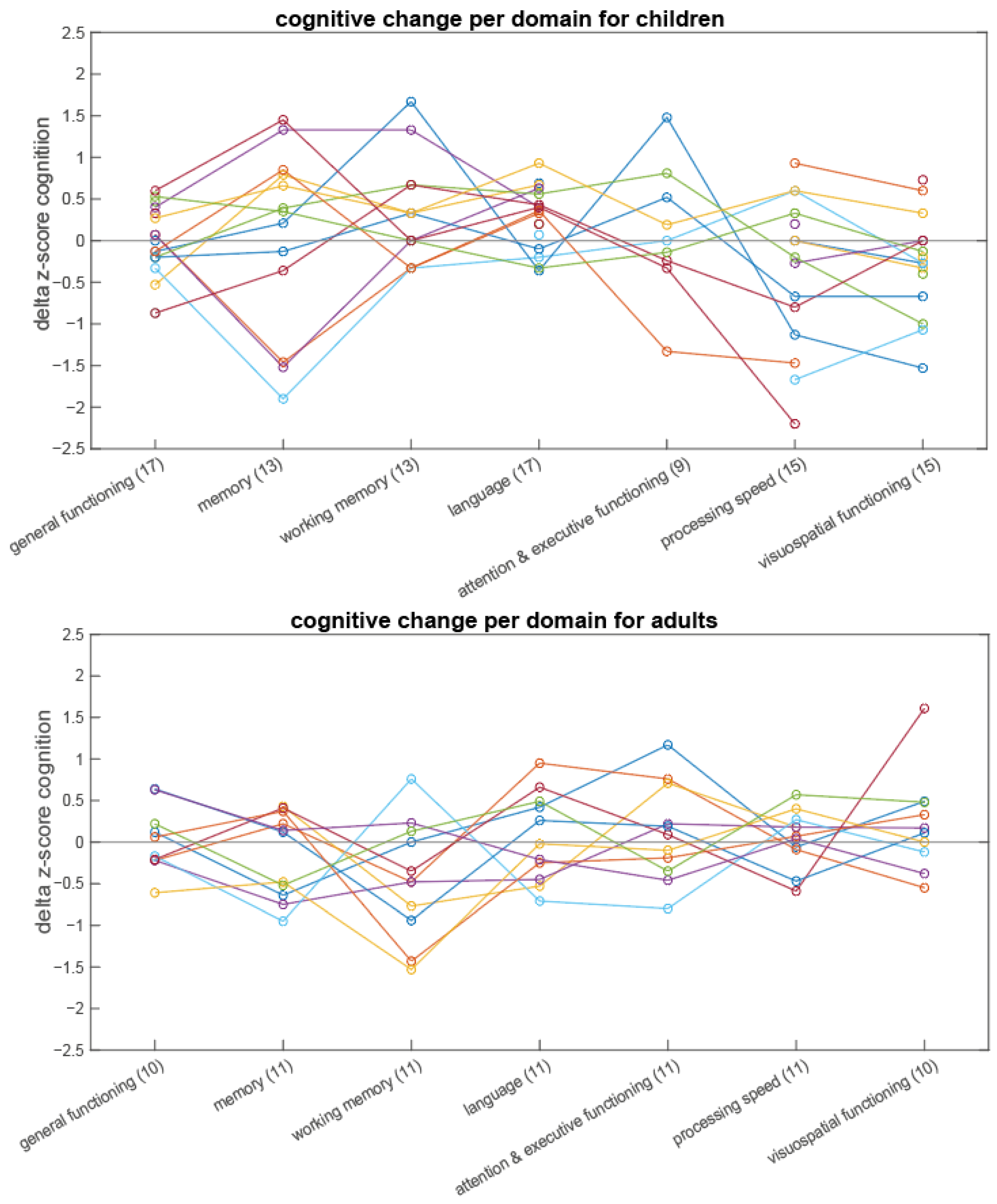
There were no significant changes in the number of patients with cognitive deficits after surgery. Seven patients (six children) had one or more deficits in a domain at baseline that resolved after follow-up (Figure 4A). However, in four patients (three children), a new deficit developed in another domain. One child improved from having cognitive impairment to none at follow-up; one adult developed cognitive impairment postoperatively; the others remained stable. To show the variability of changes between different cognitive domain scores in single patients, individual changes are graphically depicted in Appendix G, Figure A1.

3.9. Quality of Life

In children, QOL scores remained stable: the summary, PedsQl scores went from 74.1 to 76.7 (95%CI: −18.8–13.3, p = 0.73) and the parent-proxy score from 70.2 to 69.4 (95%CI: −7.0–8.4, p = 0.85). In adults, the mean of the physical component summary of the SF-36 remained stable from 47.0 to 50.1 (95%CI: −8.3–1.99, p = 0.19), and the mental component summary improved from 44.4 to 50.3 (95%CI: −10.7–−1.1, p = 0.022). The visual analogue scale of the EQ-5D improved from 73.9 to 82.0 (95%CI: −14.2–−2.99, p = 0.015), and the index remained stable (0.880 to 0.862 (95%CI: −0.090–0.1262, p = 0.715)).

3.10. Correlation of Imaging Changes to Neurocognitive Changes

We tested the hypothesis that changes in imaging parameters and CVR were associated with a change in cognitive scores (Table 2). Improvement in mSS was significantly associated with improvement in memory (p = 0.019). Children improved significantly more than adults in the working memory domain (p = 0.027). Finally, a worse score of WMD correlated to an improved visuospatial functioning score. We found no significant association between changes in clinical and radiological parameters (especially CVR) and changes in the other cognitive domains.
When comparing baseline CVR in each of the three ROIs (frontal, middle, and posterior) separately to postoperative changes in cognition in a univariable regression analysis, we found a significant correlation between worse baseline CVR scores in the middle and posterior regions and improvement in the cognitive domain visuo-spatial functioning (B = −0.378 (95%CI: −0.634–−0.122, p = 0.006); B = −0.314 (95%CI: −0.569–−0.059, p = 0.018), respectively; Appendix H, Table A3). All other associations were non-significant.

4. Discussion

In this prospective, single-center Dutch cohort study of 35 operatively treated MMV patients, we showed a significant improvement in frequency of TIAs and headache and mRS after revascularization. CVR improved significantly in both children and adults. Neuropsychological evaluation showed that patients performed significantly beneath population mean on all domains except for attention and executive functioning on a group level. After revascularization, language improved significantly—in the total group and in children—whereas other domains remained stable. While the improvement was corrected for age, it might be influenced by the speech therapy some of the patients received. QOL remained stable in children, while we found significant improvement in adults. We found no statistically significant associations between changes in clinical, radiological, and hemodynamic variables and cognitive domain scores.
Cognitive outcome following surgery may be expected to differ between children and adults. Although cognitive scores take age-specific reference values into account, and the use of z-scores allows for pooling of test results, inherent differences between age groups justified the analysis and presentation of results for children and adults separately. The eventual postoperative cognitive domain z-scores showed a relative vulnerability for visuospatial functioning in children and for processing speed in adults. Multivariable change of cognitive scores was not significantly determinant by age group except for the working memory domain. The single domain that showed postoperative significant improvement—and only so in children—was language. Possibly, the younger brain of children with a cerebrovascular compromise has a higher potential of functional recovery and improved language development than that of adults.
We saw eight new infarctions not associated with the surgical treatment between baseline and follow-up (median follow-up time: 21 months, Table 1). Only two of these led to clear clinical symptoms. In the conservative group, we saw no new ischemic lesions on follow-up MRI (Appendix I). Since the treatment was not randomized but specifically tailored to the patient, this group is inherently different from the surgical patients, so they cannot be directly compared. Furthermore, follow-up duration was shorter in the conservative group, making an ischemic event less likely to occur.
The compromised cerebrovascular hemodynamics and fragile MMV vessels may lead to a high surgical risk [21]. Therefore, patients need to be carefully selected for surgical treatment, and maintaining adequate blood pressure during anesthesia is of great importance. Even with precautionary measures, the risk of complications in our cohort was high, with four patients (11%) experiencing severe complications, of whom one, who was in a poor preoperative condition, died.
Since prospectively performed studies are rare, the overall mortality rate of surgical treatment of MMV remains unclear.
Previous studies reported mortality ranges between 0.86% [22] and 1.86% [23]. Overall adverse postoperative events (mainly ischemic and hemorrhagic stroke) are reported to be between 5–14% [22,23,24] in MMV, consistent with our results. Remarkably, the only randomized controlled surgical trial in MMD comparing STA-MCA bypass surgery to conservative treatment in adults who presented with hemorrhage reported not a single perioperative adverse event after 84 operations [25]. In another study, postoperative routine DWI revealed new ischemic lesions in 9% of 140 procedures [26]. In the subgroup of twenty-four procedures in patients who were considered to have “unstable MMD” (defined as rapid stenosis progression or recurrent stroke), 33% had postoperative DWI lesions, suggesting that postoperative ischemia is not uncommon [26]. In our study, imaging was not routinely performed directly postoperatively. Therefore, it remains unknown what proportion of silent infarctions was associated with surgery.
Our finding that revascularization surgery reduces TIA frequency in children is consistent with other studies in adults [27] and children [28]. The beneficial effect of revascularization on headache has also been reported before in children [29] and adults [30] although headache as a primary outcome is probably underreported.
Revascularization surgery has been shown to improve CVR [31]. This is confirmed by our study. Impaired CVR has previously been linked to cognitive decline [32,33]. We could not demonstrate a direct correlation between improvement of CVR and cognitive improvement. Several studies have investigated the relationship between cerebral hemodynamics and cognition—as described below—but consistent associations were not found. Asian and Western MMV populations appear to differ, with Asian MMV patients tending to be younger, presenting more often with hemorrhages, and being often more severely affected. Therefore, associations between CVR and cognition are possibly not directly comparable between populations [34].
One Western study showed that—although there was no improvement in CVR—there was some improvement in executive functioning [24]. The difference in CVR change could be explained by the burr-hole technique used, possibly leading to a slower increase in hemodynamic functioning than the combined direct–indirect technique we used. Another study showed that postoperative cognitive function remained stable in 75% of patients, significantly deteriorated in 14%, and improved in 11% of the patients [35]. These results are in line with ours. Although improvement of cognitive functioning would ideally be aimed for, in a progressive disease such as MMV, stabilization of cognition may still be considered a positive outcome. Furthermore, cognitive decline in conservatively treated MMS patients has been previously shown [36]. We hypothesize that revascularization prevents this decline in selected patients.
A Japanese prospective study showed improvement in cognitive functioning in adults after bilateral direct revascularization, with some cognitive tests correlating to preoperative CBF [37]. However, the increase was only visible two years after treatment, which could explain the difference with our results. In addition, IQ scores in those studies were—in contrast to ours—within normal range at baseline. Another retrospective cohort study found no significant difference between pre- and postoperative IQ, while the cerebral perfusion improved in several regions [38]. Change in perfusion did not correlate with change in cognitive tests. However, a high baseline oxygen ejection fraction (OEF) correlated with improvement in performance IQ, and improvement of cerebral metabolic rate of oxygen correlated with improvement in verbal IQ. Another prospective study in conservatively treated MMD patients without misery perfusion on PET showed that the CBF increased significantly although no significant cognitive changes were observed [39]. This study, however, entailed a selected group since only mildly affected MMD patients show no misery perfusion. Overall, the results of these studies are similar to ours although we used different hemodynamic parameters.
Only a few retrospective Asian studies have assessed the correlation between cognitive and cerebrovascular changes exclusively in children. One showed that CBF increased in all hemispheres, and in some ROIs, this correlated with change in cognition [40]. However, there were also territories with a negative correlation between CBF and cognitive test results, making it more difficult to draw definite conclusions. Two other studies suggested some improvement of cognition after treatment, but this was not correlated to CVR change [41,42].
QOL in MMV is underreported [7]. We showed that the PedsQL remained stable after treatment in children. Our results align with norm scores for Dutch children with a chronic health condition [43] and with another study of surgically treated children with MMD [7]. A study from the U.K. using only the parent-proxy of the PedsQl reported a lower score of 66.0 compared to the children’s scores in our cohort [44]. In adults, we found a statistically significant improvement for the QOL as measured by the VAS from the EQ-5D and the MCS of the SF36 but not for the EQ-5D index nor for the PCS. After treatment, the VAS scores were close to the population norms [45].
An important limitation of our study—and one of the possible reasons for not being able to demonstrate a correlation between surgery, change in hemodynamic measures, and cognitive outcome—is the relatively short follow-up time. It might take longer to show quantifiable cognitive improvement than just one year [37]. Furthermore, our cohort included the full range of MMS/MMD patients, making it a heterogeneous group possibly distorting the outcome. Next, the cohort was too small for further subgroup analysis. Heterogeneity of the cohort also implied that some patients had such low cognitive performance that they could not complete the complete test battery, making the reported z-scores an overestimation of the groups’ cognitive abilities. Furthermore, the [15O]H2O-PET was not fully quantitated, and the ROI-based analysis was not accurate enough to specifically look into specific anatomical regions, possibly affecting the sensitivity of our analysis. One of the strengths of this study is its prospective design, resulting in a detailed diagnostic follow up evaluation. We specifically choose to include pediatric, adult, and MMS and MMD patients, reflecting standard clinical practice. MMV is a rare disease, and this is one of the largest Western MMV cohorts with detailed cognitive test results available to date.
Despite our research and all previous efforts, there are still unanswered questions regarding MMV treatment. It remains unknown if the patients we chose to operate would have improved as much without surgery, nor do we know the best revascularization method if surgery is indeed indicated. More research is needed to understand how QOL in MMV can be improved. For tailored treatment strategies in different subgroups (e.g., children/adults, MMD/MMS, primary presentation of hemorrhage or ischemia) with the optimal revascularization method, future research should focus on standardized, multicenter prospective studies to improve knowledge on treatment of MMV.

5. Conclusions

In this prospective, single-center cohort study of MMV patients, we showed that one year after revascularization, CVR improved, and cognition remained stable in most domains and significantly improved in the language domain, specifically in children. Furthermore, TIA frequency and mRS improved significantly in children, while we found improvements in headache and QOL in adults. We could not find a relationship between change in CVR and change in neurocognitive parameters. We report a rate of serious complication of 11%, which is high but comparable to what was previously reported, stressing the importance of carefully counseling the patients of risk involved before surgery.

Author Contributions

Conceptualization, P.T.D., A.K., E.v.d.B., M.M.v.S., A.v.d.Z., C.J.M.K. and K.P.J.B.; methodology, P.T.D., A.K., E.v.d.B., M.M.v.S., A.v.d.Z., W.M.O., C.J.M.K. and K.P.J.B.; software, W.M.O.; validation, P.T.D. and A.K.; formal analysis, P.T.D.; investigation, P.T.D., A.K., E.v.d.B., M.M.v.S., E.-J.P.A.V., B.N.M.v.B., M.Y., A.v.d.Z. and K.P.J.B.; data curation, P.T.D.; writing—original draft, P.T.D. and A.K.; writing—review and editing, all authors; visualization, P.T.D.; supervision, A.v.d.Z. and K.P.J.B.; project administration, P.T.D., A.K., C.J.M.K., A.v.d.Z. and K.P.J.B.; funding adquisition, P.T.D., A.K., C.J.M.K., A.v.d.Z. and K.P.J.B. All authors have read and agreed to the published version of the manuscript.

Funding

This work was supported by the Dutch Brain Foundation ((2012(1)-179); the Christine Bader Fund Irene Children’s Hospital); the Tutein Nolthenius Oldenhof Fund, the Johanna Children Fund; Friends of the Wilhelmina Children’s Hospital; and the Brain Technology Institute Foundation.

Institutional Review Board Statement

The study was conducted in accordance with the Declaration of Helsinki. The Medical Ethics Review Committee of the UMC Utrecht confirmed that the Medical Research Involving Human Subjects Act (known as WMO in the Netherland) did not apply.

Informed Consent Statement

Written informed consent was obtained from all subjects (or their representatives) involved in the study.

Data Availability Statement

The data presented in this study are available on request from the corresponding author.

Conflicts of Interest

The authors declare no conflict of interest.

Appendix A. Surgical Treatment

Preferred initial surgical treatment was a single-staged combined procedure consisting of a unilateral direct superficial temporal artery (STA) to middle cerebral artery (MCA) bypass, combined with an indirect encephalo-duro-myo-synangiosis (EDMS) [6]. In children, depending on the [15O]H2O-PET results, an additional (bi-)frontal indirect encephalo-duro-periosteal-synangiosis (EDPS) was performed in the same surgical session [8]. If a direct procedure was not possible due to the size of the donor or recipient artery, we performed an indirect bypass (encephalo-duro-arterio-myo-synangiosis EDAMS). Apart from the bifrontal indirect procedure in children and one indirect bilateral procedure in the MCA area in one child, initial surgical treatment never consisted of a bilateral direct bypass in the MCA territory. Contralateral revascularization with a second operation, performed at least five weeks after the initial procedure, was only considered if clinical symptoms and [15O]H2O-PET results clearly showed bilateral involvement before treatment or, if during follow-up, clinical symptoms or CVR measurements suggested an ongoing contralateral cerebrovascular compromise.

Appendix B. Collected Characteristics

At baseline, we collected the following characteristics: age (at onset of first symptoms and at presentation); sex; associated disorders; type of first and recurrent symptoms: acute ischemic stroke (AIS, with or without transient ischemic attacks (TIAs), recurrent TIAs only (defined as symptoms < 24 h without permanent deficits or new ischemic brain lesions)), hemorrhage, seizures, headache, or other complaints (or if none, whether asymptomatic patients were diagnosed with MMV after screening in the context of predisposing disorders); duration of symptoms; level of education and occupation; and previous medical treatment. The CRF at follow-up detailed all (new) symptoms and covered the possibly changed variables as detailed at baseline. Occurrence of clinical complaints such as headaches or TIAs were stratified to “daily”, “weekly”, “monthly”, or “<12x/year”.

Appendix C. Additional Imaging Methods

Appendix C.1. MRI

The MRI consisted of a standardized protocol on a 3T system (Philips, Best, The Netherlands) with at least a T1, T2 fluid attenuation inversion recovery (FLAIR) and diffusion-weighted imaging (DWI).

Appendix C.2. Digital Subtraction Angiography (DSA)

DSA was performed using standard fluoroscopic DSA techniques on a bi-plane Allura Xper FD20 (Philips, Best, The Netherlands) system. The internal and external carotid arteries and both vertebral arteries were selectively catheterized in all patients. Anterior-posterior and lateral images after manual injection of contrast were acquired.

Appendix C.3. [15O]H2O-PET

PET scans were acquired using a Siemens ECAT EXACT HR+, Philips Gemini PET-conventional tomography (CT), or Philips Ingenuity TF PET-CT scanner. All patients received a venous cannula for tracer injection. Patients were scanned under general anesthesia when deemed appropriate due to their age or mental disability. The patients were asked to close their eyes and were scanned with dimmed lights and without music. Head movement was restricted by a head holder with headband and regularly checked during scanning to minimize patient motion during the study and in between scans. Each study consisted of two consecutive 10 min [15O]H2O-PET emission scans: one at baseline and one after intravenous administration of 20 mg/kg acetazolamide (maximal 1g). Using the HR+ scanner, a 10 min transmission scan was acquired prior to the first emission scan for attenuation and scatter correction purposes. Using the PET-CT scanners, the protocol started with a low-dose CT for attenuation correction. Then, a bolus (~5 s) of approximately 550 MBq [15O]H2O was administered intravenously, simultaneously starting a 3D dynamic emission scan. HR+ scans were reconstructed using FORE + 2D FBP reconstruction with a Hanning filter at Nyquist frequency. A matrix size of 256 × 256 and a zoom of 2.1 were applied, resulting in a voxel size of 1.2 × 1.2 × 2.4 mm3 and a final image resolution of ~7 mm full width at half maximum. PET-CT scans were reconstructed using LOR row action maximum likelihood algorithm (RAMLA) with a matrix size of 128 × 128 × 90 and a final voxel size of 2 × 2 × 2 mm3, with a final image resolution of ~5 mm full width at half maximum. All standard corrections for dead time, decay, attenuation, randoms, and scatter were performed. Standardized uptake (SUV) images, averaged over 15–105 s post injection, of both baseline and post acetazolamide perfusions scans were created by correcting the raw images for injected dose. After coregistration of scans from both timepoints, subtraction images were created by subtracting the baseline SUV image from the post acetazolamide SUV image.
All patients were securely monitored by measuring vital signs as well as clinical chemistry laboratory investigations before and after the PET scan. If there were neither clinical signs of cerebral ischemia nor laboratory findings suggestive of metabolic acidosis, patients were discharged the next day.

Appendix D

Table A1. Neuropsychological tests applied in adults and children and the grouping of tests in cognitive domains according to Lezak, as described previously [6,19].
DomainsAdultsChildren
General IntelligenceNational Adult Reading TestDevelopmental age
Raven’s Advanced Progressive MatricesVerbal IQ
Similarities (WAIS-III)Performance IQ
Full-Scale IQ
Verbal Comprehension Index (VCI)
Perceptual Organization Index (POI)
MemoryRey Auditory Verbal Learning Task (RAVLT)RAVLT
Rey Complex Figure Test (RCFT) delayed recallRCFT
Rivermead Behavioral Memory Test (RBMT) A/BWord list recall
Paragraph recall
Working memoryDigit span (WAIS-III)Number sequences
LanguageVerbal FluencyPassive Word comprehension (PPVT-III)
Vocabulary (WAIS-III)Confrontation Naming (CELF-4) Active Vocabulary
Boston Naming TestCELF-4 Word Associations
Attention and executive functioningZoo and rule-shifting task Behavioral Assessment of the Dysexecutive Syndrome (BADS) Test of Everyday Attention for Children (TEA-Ch)
Stroop Color Word Test Part I and IIZoo and rule-shifting task Behavioral Assessment of the Dysexecutive Syndrome (BADS-C)
Brixton Spatial Anticipation TestQuestionnaire for Executive Functioning (BRIEF) for caregivers
Processing speedSymbol Digit Modalities TestProcessing Speed index (VSI)
Stroop Color Word Test Part I and II
Visuospatial functioningFacial Recognition TestBeery Visual Motor Integration (VMI)
Judgement Of Line Orientation (JLO)
RCFT copy
For intelligence in children, the following tests were applied depending on age: Bayley scales of Infant Development, second edition, Dutch version (BSID-II-NL); Snijders-Oomennon-verbal intelligence test (SON-R) 2, 5–7, and 6–40 years; Wechsler Intelligence Scales for Children, third edition, Dutch version (WISC-IIINL); Wechsler Preschool and Primary Scale of Intelligence (WPPSI-III-NL) 2, 6–7, and 11 years; Wechsler Non-Verbal-NL—4, 0–7, and 8–21 years.

Appendix E. Clinical Follow-Up of Patients with Complications of Operative Treatment

There were four patients with serious complications (causing permanent damage or additional surgery), one of whom died, and eight patients with transient deficits.
The patient who died was a 53-year-old woman who presented with multiple infarctions, mostly in the left hemisphere, which led to a hemiparesis and motoric dysphasia. Other relevant medical history was immune thrombocytopenia and depression with a recent suicide attempt. Because vasculitis was suspected as a cause of recurring ischemia, a biopsy of the dura mater, cortex, and ischemic lesion was performed, which ruled out vasculitis but showed atypical ischemic lesions. Afterwards, she was diagnosed with idiopatic bilateral MMD. Because the perfusion and reactivity was inadequate, she was indicated for a left-sided direct bypass. This was planned three months after the last ischemic event. Before treatment, she received human normal immunoglobulin (1 g/kg IV for two days) because of thrombocytopenia (32 × 109/L). Perioperative the acetylic acid was paused. The operation (STA-MCA bypass, occlusion time of recipient vessel of 18 min) was uneventful, and after surgery, she was neurologically the same as preoperative. One day after the operation, she developed high blood pressure in combination with a bradycardia and a drop in GCS scores. A CT revealed a left-sided temporal intracerebral hemorrhage, most probably because of hemorrhagic transformation of an infarction, caused by hyperperfusion. Because of the poor preclinical condition and the suspected poor outcome, no further surgical intervention was performed, and the patient died one day later.
There were three patients with symptomatic postoperative infarctions: two adults and one pediatric. The first adult was a 49-year-old patient diagnosed with bilateral MMD along with hypertension and hypercholesterolemia. She previously suffered from two infarctions in the left hemisphere, and suffered preoperatively mostly of TIAs and concentration problems. She was first operated on the left hemisphere with an STA-MCA bypass, which was uneventful. One day after surgery, she collapsed, which lead to a dysarthria and a left-sided paresis, which gradually improved. A CT scan three days after surgery revealed no signs of new ischemia, but five days after surgery, the scan was repeated, and right-frontal ischemia (contralateral from the surgical site) and a smaller area left-frontally were found. The right-sided infarct was most probably a watershed infarction. After a rehabilitation period, she was operated on the right side with an STA-MCA bypass. She had poor concentration and complained frequently of fatigue and did not want to complete the follow-up imaging and neuropsychological examination, so she withdrew from the study after the second operation.
The second adult was a 42-year-old women who presented with recurrent ischemia caused by bilateral MMD. A left-sided STA-MCA bypass was performed, which was uneventful. One day after surgery, she developed dysphasia and started vomiting. A CT revealed a subdural hematoma under the boneflap and new ischemia left-frontally, which was not treated with an extra operation. After rehabilitation, and around nine months after the operation, she was readmitted with wound dehiscence and a wound infection, for which first the infected boneflap was removed. The wound still did not heal properly, so a month later, the wound was revised again, after which it stayed closed. The bone defect was not reconstructed. This patient also refused further investigations and withdrew from the study.
The pediatric patient was an 11-year-old girl suffering from bilateral MMS (combined with palatoschisis, congenital heart disease) who presented with recurring TIAs and infarction. Based on the TIAs originating from both hemisphere and steal seen bilaterally and bifrontally, the initial plan was to operate both sides, starting with the left side. During the operation, a left-sided STA-MCA bypass was made, combined with a bifrontal EDPS. Directly postoperatively, the patient was aphatic and had a hemiparesis on the right side, and a CT showed bifrontal infarctions. After months of rehabilitating, most motor functions on the right side recovered. The PET scans were repeated and showed bilateral improvement of CBF and CVR. Since the patient did not suffer from recurrent TIAs, she was monitored closely for four years (until now) and remained stable, without needing additional surgery.
There were eight patients with transient complaints after the operation. In five patients (three adults), these complains were suspected to be caused by hyperperfusion syndrome (although hyperperfusion was not proven with direct postoperative perfusion imaging), and one adult had these symptoms after both operations. Typically, complaints arose 1–3 days after operations; consisted of dysphasia, dysarthria, or fine motor function deficits; and lasted a couple of days, but eventually, complaints disappeared fully. Imaging was conducted in four of the patients to rule out new ischemia, which was not shown.
Three patients suffered from postoperative TIAs on day 1–4 after the operation. These typically lasted less than one hour and were comparable to TIAs experienced before the operation.

Appendix F

Table A2. Clinical outcome scores.
Total Group (n = 35)Baseline Follow-Up
Mean (SD)Median (Range)Valid nMean (SD)Median (Range)Valid n
NIHSS 1 (1.48)0.5 (0–7)320.46 (0.72)0 (0–2)24
n (percentage) n (percentage)
mRS baseline06 (17.1%) 13 (37.1%)
111 (31.4%) 8 (22.9%)
216 (45.7%) 11 (31.4%)
32 (5.7%) 0 (0%)
Children(n = 21)Baseline Follow-Up
Mean (SD)Median (Range)ValidnMean (SD)Median (Range)Validn
Ped. Quality of Life72.6 (19.1)71.7 (43.5–97.8)1574.2 (18.5)79.4 (39.1–96.3)16
Ped. Quality of Life parents 68.6 (18.8)76.1 (33.7–97.8)2169.4 (24.6)78.3 (17–98.8)19
Ped. NIHSS 0.7 (1.1)0 (0–3)180.3 (0.6)0 (0–2)15
PSOM 1.5 (1.7)1 (0–6)200.9 (1.2)0.3 (0–4)16
n (percentage) n (percentage)
mRS baseline05 (23.8%) 11 (52.3%)
17 (33.3%) 5 (23.8%)
28 (38.1%) 5 (23.8%)
31 (4.7%) 0 (0%)
Adults(n = 14)Baseline Follow-Up
Mean (SD)Median (Range)ValidnMean (SD)Median (Range)Validn
SF-36 Physical Component scale44.49 (9.31)43.68 (30.46–57.73)1550.14 (7.73)51.6 (35.9–59.2)16
SF-36 Mental Component scale39.85 (14.05)42.5 (16.79–61.3)2150.33 (11.88)53.6 (22.5–59.1)19
EQ5D VAS 69.29 (16.71)65.5 (40–98)1882 (12.26)82.5 (60–99)15
EQ5D index 0.77 (0.27)0.8 (0–1)200.86 (0.17)0.93 (0.5–1)16
NIHSS 1.36 (1.86)1 (0–7)210.78 (0.83)1 (0–2)17
n (percentage) n (percentage)
mRS baseline01 (7.1%) 2 (14.3%)
14 (28.6%) 3 (21.4%)
28 (57.1%) 6 (42.9%)
31 (7.1%) 0 (0%)
Clinical outcome scores for total surgical group, both children and adults. Abbreviations: EQ5D, EuroQol 5 Dimension; mRS, modified Rankin scale; NIHSS, National Institutes of Health Stroke Scale; Ped., pediatric, PSOM, Pediatric Stroke Outcome Measure; SD, standard deviation; SF-36, Short Form—36; VAS, visual analogue scale.

Appendix G

Figure A1. Cognitive change per domain. Change in cognitive domain per operatively treated patient. This graph shows the variability in cognitive scores between domains and between patients. Each color represents a single patient. The lines are used to connect the dots for easier comparison; consequently, the directional coefficient of the lines does not hold meaningful value. The numbers represent the number of valid test scores per domain.
Jcm 11 07427 g0a1

Appendix H

Table A3. Baseline CVR versus change in cognitive domains (univariable linear regression and Bayesian regression).
General FunctioningB (95%CI)pBF
Frontal baseline CVR8.898 × 10−4 (−0.168–0.17)0.9910.358
Middle baseline CVR−0.085 (−0.280–0.109)0.3750.484
Posterior baseline CVR−0.123 (−0.304–0.057)0.1720.738
MemoryB (95%CI)pBF
Frontal baseline CVR−0.065 (−0.441–0.311)0.7230.391
Middle baseline CVR−0.117 (−0.550–0.317)0.5820.418
Posterior baseline CVR−0.043 (−0.430–0.344)0.8190.38
Working MemoryB (95%CI)pBF
Frontal baseline CVR0.055 (−0.275–0.386)0.7310.39
Middle baseline CVR0.301 (−0.059–0.661)0.0971.073
Posterior baseline CVR−0.026 (−0.367–0.314)0.8730.376
LanguageB (95%CI)pBF
Frontal baseline CVR0.072 (−0.261–0.404)0.6620.357
Middle baseline CVR0.013 (−0.189–0.215)0.8990.355
Posterior baseline CVR0.008 (−0.184–0.2)0.9320.354
Attention and Executive FunctioningB (95%CI)pBF
Frontal baseline CVR0.116 (−0.177–0.408)0.4190.715
Middle baseline CVR−0.068 (−0.457–0.321)0.7160.417
Posterior baseline CVR−0.053 (−0.390–0.283)0.7420.413
Processing SpeedB (95%CI)pBF
Frontal baseline CVR−0.225 (−0.533–0.083)0.1450.825
Middle baseline CVR−0.293 (−0.640–0.054)0.0951.074
Posterior baseline CVR−0.124 (−0.443–0.194)0.4280.461
Visuo-spatial functioningB (95%CI)pBF
Frontal baseline CVR−0.228 (−0.474–0.018)0.0681.341
Middle baseline CVR−0.378 (−0.634–−0.122)0.0067.985
Posterior baseline CVR−0.314 (−0.569–−0.059)0.0183.355
To investigate the relationship between baseline CVR in three regions (frontal, middle, and posterior) and postoperative change in the measured z-scores of the seven different cognitive domains, we used univariable linear regression analysis, reported with the unstandardized regression coefficient (B) with 95% confidence interval (CI) and p-value, and Bayesian regression analysis, reported with the Bayes factor (BF). A common way to interpret the BF is dividing the evidence into four strength ranges: BFs between 1–3.2 are “not worth more than a bare mention”, between 3.2–10 are “substantial”, between 10–100 are “strong”, and >100 are “decisive” evidence [20]. Abbreviations: CVR, cerebrovascular reactivity.

Appendix I. Outcome of Conservatively Treated Patients

There were five patients conservatively treated in our cohort: four adults and one child. We advised revascularization for the child, but patient and parents refused. The adults were all stable, and in combination with the relatively good PET results, conservatively treatment was indicated. One of the adults deteriorated after the initial conservative treatment (increase of TIA frequency in combination with deteriorated CVR) and was eventually bilaterally operated. The follow-up of that operation was not included in this paper.

Appendix I.1. TIAs

Three of the five patients (one child, two adults) had TIAs at baseline, all in a low frequency: sometimes a year. Between baseline and follow-up, there were no new TIAs seen in two patients, but in one adult, the frequency increased to monthly. This patient was operatively treated after the initial conservative treatment (follow-up is not included).

Appendix I.2. Headache

There were four patients who suffered from headaches at baseline: one child and three adults. This improved in all; three did not have any complains of headache at follow-up, while one adult only suffered from headache a couple of times a year, while at baseline, it was almost daily. The patient who did not suffer from any headaches at baseline deteriorated and had mild daily headaches at follow-up.

Appendix I.3. Clinical Scores and Functional Deficits

PedNIHSS score and PSOM score were zero at baseline, and follow-up scores were missing. NIHSS changed from 0.5 (0–10; median, range) to 0.0 (0–7; median, range). In adults, there was one patient with an mRS of 1, two patients with mRS of 2, and one patient with mRS 3. This remained stable between baseline and follow-up. The mRS in the child went from 1 at baseline to 0 at follow-up.

Appendix I.4. Other Clinical Outcome

At baseline, one patient suffered from seizures several times a year. She had four seizures between baseline and follow-up, which was stable in frequency compared to before inclusion. There were no patients in the conservative group with other complaints (e.g., chorea, dystonia, limb-shaking) at baseline, while at follow-up, one adult complained of an almost daily occurring slight involuntary tremor in her left arm.

Appendix I.5. Radiological Findings at Follow-Up

There were no patients with new infarctions or hemorrhages. There were no patients with a deterioration of WMD. The mSS remained stable in the three patients (all adults) who received a DSA at follow-up. In one of these, new collaterals were seen (showing signs of improvement of vascularization), while in the other two, there was no difference. The CVR improved for two patients (one adult, one child) and deteriorated in two patients. For one patient, the CVR remained stable.

Appendix I.6. Cognitive Follow-Up

At follow-up, the z-scores of the neurocognitive test of the total group were significantly below population mean for the domains general functioning, memory, language, and processing speed (Appendix J, Figure A2A). There were no significant changes in z-scores after conservative treatment (Appendix J, Figure A2B).

Appendix I.7. QOL

The child did not complete the follow-up QOL assessment. In the four adults, the EQ-5D VAS went from 63.8 (12.5; mean (SD)) to 65.0 (10.80), and the EQ-5D index went from 0.70 (0.0) to 0.63 (0.22). The SF-36 physical component summary went from 39.3 (8.8) to 42.3 (9.9) and the mental component summary from 44.7 (9.0) to 40.6 (4.6). All these changes in QOL were not statistically significant.

Appendix J

Figure A2. Z-scores at follow-up. (A) z-scores of neurocognitive tests on follow-up (conservative treatment) for the total group and for adults only for all seven cognitive domains. The numbers represent the amount of patients with a z-score below −1.5 SD compared to the total number of patients with a valid domain score per group. Scores significantly different from the population mean (one sample t-test) are noted with * (p < 0.05); ** (p < 0.005). (B) change in z-score between baseline and follow-up. A higher score indicates an improvement. The numbers represent the amount of valid test scores per cognitive domain. There were no significant changes after follow-up.
Jcm 11 07427 g0a2

References

  1. Scott, R.M.; Smith, E.R. Moyamoya Disease and Moyamoya Syndrome. N. Engl. J. Med. 2009, 360, 1226–1237. [Google Scholar] [CrossRef] [PubMed] [Green Version]
  2. Kronenburg, A.; Braun, K.P.J.; Van Der Zwan, A.; Klijn, C.J.M. Recent Advances in Moyamoya Disease: Pathophysiology and Treatment. Curr. Neurol. Neurosci. Rep. 2014, 14, 423. [Google Scholar] [CrossRef] [PubMed]
  3. Kronenburg, A.; van den Berg, E.; van Schooneveld, M.M.; Braun, K.P.J.; Calviere, L.; van der Zwan, A.; Klijn, C.J.M. Cognitive Functions in Children and Adults with Moyamoya Vasculopathy: A Systematic Review and Meta-Analysis. J. Stroke 2018, 20, 332–341. [Google Scholar] [CrossRef] [PubMed] [Green Version]
  4. Fukui, M. Guidelines for the Diagnosis and Treatment of Spontaneous Occlusion of the Circle of Willis (“Moyamoya” Disease). Clin. Neurol. Neurosurg. 1997, 99, S238–S240. [Google Scholar] [CrossRef]
  5. Ferriero, D.M.; Fullerton, H.J.; Bernard, T.J.; Billinghurst, L.; Daniels, S.R.; Debaun, M.R.; Deveber, G.; Ichord, R.N.; Jordan, L.C.; Massicotte, P.; et al. Management of Stroke in Neonates and Children: A Scientific Statement from the American Heart Association/American Stroke Association. Stroke 2019, 50, e51–e96. [Google Scholar] [CrossRef] [Green Version]
  6. Kronenburg, A.; Deckers, P.T.; van den Berg, E.; van Schooneveld, M.M.; Vonken, E.-J.; van der Zwan, A.; van Berckel, B.N.M.; Yaqub, M.; Otte, W.; Klijn, C.J.M.; et al. The Profile of Cognitive Impairment and Hemodynamic Compromise in Moyamoya: A Single-Center Prospective Cohort Study. J. Neurosurg. 2022, 1, 1–12. [Google Scholar] [CrossRef]
  7. Ball, A.J.; Steinberg, G.K.; Elbers, J. Pediatric Quality of Life in Children with Moyamoya Disease and Stroke. Ann. Neurol. 2015, 78, S214–S215. [Google Scholar] [CrossRef] [Green Version]
  8. Kronenburg, A.; Esposito, G.; Fierstra, J.; Braun, K.P.; Regli, L. Combined Bypass Technique for Contemporary Revascularization of Unilateral MCA and Bilateral Frontal Territories in Moyamoya Vasculopathy. Trends Neurovascular Interv. 2014, 119, 65–70. [Google Scholar] [CrossRef] [Green Version]
  9. Ichord, R.N.; Bastian, R.; Abraham, L.; Askalan, R.; Benedict, S.; Bernard, T.J.; Beslow, L.; Dowling, M.; Friedman, N.; Jordan, L.; et al. Inter-Rater Reliability of the Pediatric NIH Stroke Scale (PedNIHSS) in a Multicenter Study. Stroke 2011, 42, 613–617. [Google Scholar] [CrossRef] [Green Version]
  10. Brott, T.; Adams, H.P.; Olinger, C.P.; Marle, J.R.; Barsan, W.G.; Biller, J.; Spilker, J.; Holleran, R.; Eberle, R.; Hertzberg, V.; et al. Measurements of Acute Cerebral Infarction: A Clinical Examination Scale. Stroke 1989, 20, 864–870. [Google Scholar] [CrossRef]
  11. van Swieten, J.C.; Koudstaal, P.J.; Visser, M.C.; Schouten, H.J.; van Gijn, J. Interobserver Agreement for the Assessment of Handicap in Stroke Patients. Stroke 1988, 19, 604–607. [Google Scholar] [CrossRef] [PubMed] [Green Version]
  12. DeVeber, G.A.; MacGregor, D.; Curtis, R.; Mayank, S. Neurologic Outcome in Survivors of Childhood Arterial Ischemic Stroke and Sinovenous Thrombosis. J. Child Neurol. 2000, 15, 316–324. [Google Scholar] [CrossRef] [PubMed]
  13. Varni, J.W.; Planning, U. Pediatric Quality of Life InventoryTM PedsQL. Available online: http://www.pedsql.org/ (accessed on 1 December 2022).
  14. Brazier, J.; Roberts, J.; Deverill, M. The Estimation of a Preference-Based Measure of Health from the SF-36. J. Health Econ. 2002, 21, 271–292. [Google Scholar] [CrossRef] [PubMed] [Green Version]
  15. Langsdale, T. Eq-5D. Pharmacoecon. Outcomes News 1997, 109, 3–4. [Google Scholar] [CrossRef]
  16. Deckers, P.T.; van Hoek, W.; Kronenburg, A.; Yaqub, M.; Siero, J.C.W.; Bhogal, A.A.; van Berckel, B.N.M.; van der Zwan, A.; Braun, K.P.J. Contralateral Improvement of Cerebrovascular Reactivity and TIA Frequency after Unilateral Revascularization Surgery in Moyamoya Vasculopathy. NeuroImage Clin. 2021, 30, 102684. [Google Scholar] [CrossRef] [PubMed]
  17. Fazekas, F.; Kleinert, R.; Offenbacher, H.; Schmidt, R.; Kleinert, G.; Payer, F.; Radner, H.; Lechner, H. Pathologic Correlates of Incidental MRI White Matter Signal Hyperintensities. Neurology 1993, 43, 1683. [Google Scholar] [CrossRef]
  18. Kang, K.H.; Kim, H.S.; Kim, S.Y. Quantitative Cerebrovascular Reserve Measured by Acetazolamide-Challenged Dynamic CT Perfusion in Ischemic Adult Moyamoya Disease: Initial Experience with Angiographic Correlation. Am. J. Neuroradiol. 2008, 29, 1487–1493. [Google Scholar] [CrossRef] [Green Version]
  19. Lezak, M.D.; Howieson, D.B.; Loring, D.W.; Fischer, J.S. Neuropsychological Assessment; Oxford University Press: Cary, NC, USA, 2004; ISBN 0195111214. [Google Scholar]
  20. Kass, R.E.; Raftery, A.E. Bayes Factors. J. Am. Stat. Assoc. 1995, 90, 773–795. [Google Scholar] [CrossRef]
  21. Yu, J.; Shi, L.; Guo, Y.; Xu, B.; Xu, K. Progress on Complications of Direct Bypass for Moyamoya Disease. Int. J. Med. Sci. 2016, 13, 578–587. [Google Scholar] [CrossRef] [Green Version]
  22. Guzman, R.; Lee, M.; Achrol, A.; Bell-Stephens, T.; Kelly, M.; Do, H.M.; Marks, M.P.; Steinberg, G.K. Clinical Outcome after 450 Revascularization Procedures for Moyamoya Disease: Clinical Article. J. Neurosurg. 2009, 111, 927–935. [Google Scholar] [CrossRef]
  23. Khan, N.; Achrol, A.S.; Guzman, R.; Burns, T.C.; Dodd, R.; Bell-Stephens, T.; Steinberg, G.K. Sex Differences in Clinical Presentation and Treatment Outcomes in Moyamoya Disease. Neurosurgery 2012, 71, 587–593. [Google Scholar] [CrossRef] [PubMed]
  24. Calviere, L.; Loubiere, P.; Planton, M.; Cazzola, V.; Catalaa, I.; Mirabel, H.; Sol, J.C.; Bonneville, F. Decreased Frontal White-Matter Diffusion and Improved Cognitive Flexibility after Burr-Hole Surgery in Moyamoya Angiopathy. BMC Neurol. 2020, 20, 30. [Google Scholar] [CrossRef] [PubMed] [Green Version]
  25. Miyamoto, S.; Yoshimoto, T.; Hashimoto, N.; Okada, Y.; Tsuji, I.; Tominaga, T.; Nakagawara, J.; Takahashi, J.C.; Yamada, K.; Tomata, Y.; et al. Effects of Extracranial-Intracranial Bypass for Patients with Hemorrhagic Moyamoya Disease: Results of the Japan Adult Moyamoya Trial. Stroke 2014, 45, 1415–1421. [Google Scholar] [CrossRef] [PubMed]
  26. Funaki, T.; Takahashi, J.C.; Takagi, Y.; Kikuchi, T.; Yoshida, K.; Mitsuhara, T.; Kataoka, H.; Okada, T.; Fushimi, Y.; Miyamoto, S. Unstable Moyamoya Disease: Clinical Features and Impact on Perioperative Ischemic Complications. J. Neurosurg. 2015, 122, 400–407. [Google Scholar] [CrossRef] [Green Version]
  27. Kim, S.H.; Lee, H.; Yoo, M.; Jin, S.; Lee, S.; Choi, B.S.; Kim, H.Y.; Jin, S.C. Angiographic and Clinical Outcomes of Non-Patent Anastomosis after Bypass Surgery in Adult Moyamoya Disease. Acta Neurochir. 2019, 161, 379–384. [Google Scholar] [CrossRef]
  28. Suzuki, Y.; Negoro, M.; Shibuya, M.; Yoshida, J.; Negoro, T.; Watanabe, K. Surgical Treatment for Pediatric Moyamoya Disease: Use of the Superficial Temporal Artery for Both Areas Supplied by the Anterior and Middle Cerebral Arteries. Neurosurgery 1997, 40, 324–330. [Google Scholar] [CrossRef]
  29. Blauwblomme, T.; Mathon, B.; Naggara, O.; Kossorottof, M.; Bourgeois, M.; Puget, S.; Meyer, P.; Brousse, V.; De Montalembert, M.; Brunelle, F.; et al. Long-Term Outcome after Multiple Burr Hole Surgery in Children with Moyamoya Angiopathy: A Single-Center Experience in 108 Hemispheres. Neurosurgery 2017, 80, 950–956. [Google Scholar] [CrossRef]
  30. Gao, B.; Kang, K.; Zhang, J.; Zhang, D.; Zhao, X. Clinical Characteristics and Long-Term Outcome of Headaches Associated with Moyamoya Disease in the Chinese Population—A Cohort Study. Front. Neurol. 2020, 11, 605636. [Google Scholar] [CrossRef]
  31. Han, J.S.; Abou-Hamden, A.; Mandell, D.M.; Poublanc, J.; Crawley, A.P.; Fisher, J.A.; Mikulis, D.J.; Tymianski, M. Impact of Extracranial-Intracranial Bypass on Cerebrovascular Reactivity and Clinical Outcome in Patients with Symptomatic Moyamoya Vasculopathy. Stroke 2011, 42, 3047–3054. [Google Scholar] [CrossRef] [Green Version]
  32. Roder, C.; Haas, P.; Fudali, M.; Milian, M.; Ernemann, U.; Meyer, P.T.; Tatagiba, M.; Khan, N. Neuropsychological Impairment in Adults with Moyamoya Angiopathy: Preoperative Assessment and Correlation to MRI and H215O PET. Neurosurg. Rev. 2020, 43, 1615–1622. [Google Scholar] [CrossRef]
  33. Silvestrini, M.; Pasqualetti, P.; Baruffaldi, R.; Bartolini, M.; Handouk, Y.; Matteis, M.; Moffa, F.; Provinciali, L.; Vernieri, F. Cerebrovascular Reactivity and Cognitive Decline in Patients with Alzheimer Disease. Stroke 2006, 37, 1010–1015. [Google Scholar] [CrossRef] [PubMed] [Green Version]
  34. Hever, P.; Alamri, A.; Tolias, C. Moyamoya Angiopathy—Is There a Western Phenotype? Br. J. Neurosurg. 2015, 29, 765–771. [Google Scholar] [CrossRef] [PubMed]
  35. Zeifert, P.D.; Karzmark, P.; Bell-Stephens, T.E.; Steinberg, G.K.; Dorfman, L.J. Neurocognitive Performance after Cerebral Revascularization in Adult Moyamoya Disease. Stroke 2017, 48, 1514–1517. [Google Scholar] [CrossRef] [PubMed]
  36. Hogan, A.M.; Kirkham, F.J.; Isaacs, E.B.; Wade, A.M.; Vargha-Khadem, F. Intellectual Decline in Children with Moyamoya and Sickle Cell Anaemia. Dev. Med. Child Neurol. 2005, 47, 824–829. [Google Scholar] [CrossRef]
  37. Kazumata, K.; Tha, K.K.; Tokairin, K.; Ito, M.; Uchino, H.; Kawabori, M.; Sugiyama, T. Brain Structure, Connectivity, and Cognitive Changes Following Revascularization Surgery in Adult Moyamoya Disease. Clin. Neurosurg. 2019, 85, E943–E952. [Google Scholar] [CrossRef]
  38. Hara, S.; Kudo, T.; Hayashi, S.; Inaji, M.; Tanaka, Y.; Maehara, T.; Ishii, K.; Nariai, T. Improvement in Cognitive Decline after Indirect Bypass Surgery in Adult Moyamoya Disease: Implication of 15O-Gas Positron Emission Tomography. Ann. Nucl. Med. 2020, 34, 467–475. [Google Scholar] [CrossRef] [PubMed]
  39. Miyoshi, K.; Chida, K.; Kobayashi, M.; Kubo, Y.; Yoshida, K.; Terasaki, K.; Ogasawara, K. Two-Year Clinical, Cerebral Hemodynamic, and Cognitive Outcomes of Adult Patients Undergoing Medication Alone for Symptomatically Ischemic Moyamoya Disease without Cerebral Misery Perfusion: A Prospective Cohort Study. Neurosurgery 2019, 84, 1233–1241. [Google Scholar] [CrossRef]
  40. Hsu, Y.H.; Chen, Y.F.; Yang, S.H.; Yang, C.C.; Kuo, M.F. Postoperative Change of Neuropsychological Function after Indirect Revascularization in Childhood Moyamoya Disease: A Correlation with Cerebral Perfusion Study. Child’s Nerv. Syst. 2020, 36, 1245–1253. [Google Scholar] [CrossRef]
  41. Lee, J.Y.; Phi, J.H.; Wang, K.C.; Cho, B.K.; Shin, M.S.; Kim, S.K. Neurocognitive Profiles of Children with Moyamoya Disease before and after Surgical Intervention. Cerebrovasc. Dis. 2011, 31, 230–237. [Google Scholar] [CrossRef] [PubMed]
  42. Kim, W.; Lee, E.-Y.; Park, S.-E.; Park, E.-K.; Kim, J.-S.; Kim, D.-S.; Shim, K.-W. Neuropsychological Impacts of Indirect Revascularization for Pediatric Moyamoya Disease. Child’s Nerv. Syst. 2018, 34, 1199–1206. [Google Scholar] [CrossRef]
  43. Engelen, V.; Haentjens, M.M.; Detmar, S.B.; Koopman, H.M.; Grootenhuis, M.A. Health Related Quality of Life of Dutch Children: Psychometric Properties of the PedsQL in the Netherlands. BMC Pediatr. 2009, 9, 68. [Google Scholar] [CrossRef] [PubMed] [Green Version]
  44. Kalra, N.; Bautista, M.; McCullagh, H.; Tyagi, A. PedsQL Score Post Encephalo-Duro-Arterio-Myo-Synangiosis Procedure for Moyamoya Disease: A Single Center Experience. World Neurosurg. 2020, 144, e674–e678. [Google Scholar] [CrossRef] [PubMed]
  45. König, H.-H.; Bernert, S.; Angermeyer, M.C.; Matschinger, H.; Martinez, M.; Vilagut, G.; Haro, J.M.; de Girolamo, G.; de Graaf, R.; Kovess, V.; et al. Comparison of Population Health Status in Six European Countries. Med. Care 2009, 47, 255–261. [Google Scholar] [CrossRef] [PubMed]
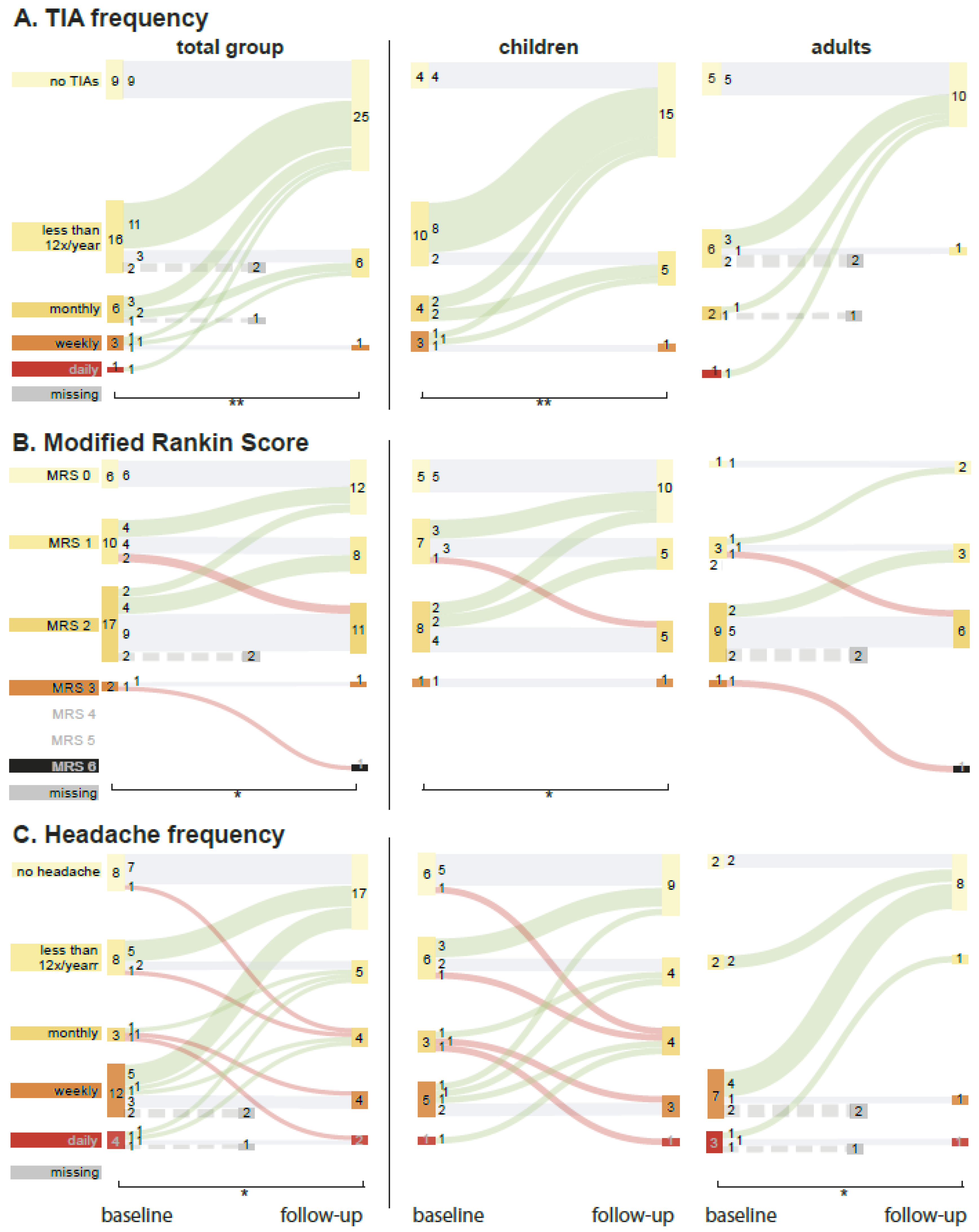
Figure 1. Clinical outcome of treated patients for the total group and for children and adults separately. The width of the bars represents the number of patients and is described by the value within. Green upward line = improvement; horizontal grey line = stable; downward red line = deterioration. Dotted line represents a patient with missing outcome data. (A) Effect of operative treatment on TIA frequency. (B) Change in postoperative modified Rankin scale (mRS). (C). Headache frequency before and after treatment. Significant differences between baseline and follow-up are denoted with a * (p-value of <0.05) and ** (p < 0.005).
Jcm 11 07427 g001
Figure 2. Change in imaging scores. (A) New infarctions on the MRI one year after the last operation. (†) Three adults did not receive a scan at follow-up but received an MRI shortly after the operation and are included in this graph. (B) Change in average white matter disease. (C) Change in modified Suzuki score (mSS). (D) Change in average cerebrovascular reactivity (CVR) score.
Jcm 11 07427 g002
Figure 3. Example of improvement of cerebrovascular reactivity (CVR) on [15O]H2O-PET in an 11-year-old girl with moyamoya syndrome. The left two columns are pre-operative and before and after acetazolamide (ACZ) challenge. The red arrows show examples of vascular steal. She was first operated with a right-sided combined direct/indirect bypass with additionally a bifrontal EDAMS, followed by a left-sided indirect bypass during a second surgery. The right two columns show the same patient approximately at follow-up. The green arrows show the same areas with improved CVR.
Jcm 11 07427 g003
Figure 4. (A) Z-scores of neurocognitive tests at follow-up for the total group for children and adults separately for all seven cognitive domains. The numbers represent the amount of patients with a z-score below −1.5 SD (dashed line) compared to the total number of patients with a valid domain score per group. Scores significantly different from the population mean (one-sample t-test) are noted with * (p < 0.05), ** (p < 0.005), and *** (p < 0.0005). (†) Two pediatric patients could not understand the tests due to insufficient cognitive functioning. These patients were given a “deficit” score for general functioning but could not be included in the box plots. (B) Change in z-score between follow-up and baseline. A higher score indicates an improvement. The numbers represent the amount of valid test scores per cognitive domain; (§) significant improvements in z-score (p ≤ 0.05).
Jcm 11 07427 g004
Table 1. Patient characteristics and treatment.
Total Group (n = 35)Children (n = 21)Adults (n = 14)
SexFemale2777.10%1571.40%1285.70%
Moyamoya diagnosisMMD unilateral12.90%00.00%17.10%
MMD bilateral2262.90%1152.40%1178.60%
MMS unilateral38.60%314.30%00.00%
MMS bilateral925.70%733.30%214.30%
Age at follow-up in years (mean (SD))20.6 (15.5)11.0 (4.1)38.8 (12.4)
Time between baseline and follow-up in months (median (min-max))21 (14–72)21 (14–72)20 (14–24)
Time between last OR and follow-up in months (median (min–max))15 (10–27)16 (10–27)12 (10–24)
Treatment typeBilateral treatment2160.00%1676.20%535.70%
Unilateral treatment1440.00%523.80%964.30%
Total patients with direct bypass2982.90%1571.40%14100.00%
Total patients with frontal procedure1337.10%1361.90%00.00%
All operated patients are included in this table. MMD, moyamoya disease; MMS, moyamoya syndrome; SD, standard deviation; OR, operation.
Table 2. Multivariable regression analysis of possible determinants of change in cognitive domain scores.
General FunctioningB (95%CI)pBF
Adult vs. child0.14 (−0.345–0.624)0.5520.325
Infarct score change−0.103 (−0.996–0.791)0.8110.316
MSS change−0.045 (−0.504–0.414)0.8370.295
WMD score change0.942 (−0.641–2.525)0.2260.469
CVR score change−0.053 (−0.267–0.161)0.6100.314
Other reason for cognitive defect0.307 (−0.399–1.013)0.3710.361
MemoryB (95%CI)pBF
Adult vs. child−0.606 (−1.469–0.258)0.1550.813
Infarct score change1.326 (−0.583–3.236)0.1580.854
mSS change−0.963 (−1.741–−0.185)0.0192.474
WMD score change−0.972 (−4.942–2.997)0.6080.608
CVR score change−0.020 (−0.368–0.329)0.9050.571
Other reason for cognitive defect0.144 (−1.821–2.11)0.8770.630
Working MemoryB (95%CI)pBF
Adult vs. child−1.057 (−1.974–−0.140)0.0271.965
Infarct score change0.294 (−1.733–2.322)0.7600.51
mSS change0.465 (−0.360–1.291)0.2470.599
WMD score change−0.609 (−4.823–3.605)0.7610.505
CVR score change−0.191 (−0.561–0.179)0.2860.585
Other reason for cognitive defect−0.652 (−2.739–1.435)0.5140.483
LanguageB (95%CI)pBF
Adult vs. child−0.330 (−0.788–0.129)0.1480.682
Infarct score change0.11 (−0.753–0.974)0.7910.360
mSS change−0.347 (−0.791–0.096)0.1170.686
WMD score change−0.110 (−1.640–1.421)0.8820.356
CVR score change0.035 (−0.172–0.242)0.7270.375
Other reason for cognitive defect−0.095 (−0.778–0.587)0.7720.366
Attention and Executive FunctioningB (95%CI)pBF
Adult vs. child0.172 (−0.710–1.055)0.6760.528
Infarct score change−1.706 (−3.722–0.31)0.0901.214
mSS change−0.186 (−1.044–0.671)0.6420.526
WMD score change0.678 (−3.322–4.678)0.7160.655
CVR score change−0.144 (−0.496–0.208)0.3870.658
Other reason for cognitive defect1.338 (−0.519–3.196)0.1410.716
Processing SpeedB (95%CI)pBF
Adult vs. child0.308 (−0.416–1.031)0.3810.445
Infarct score change−0.781 (−2.154–0.592)0.2450.424
mSS change0.234 (−0.513–0.98)0.5170.366
WMD score change1.931 (−0.496–4.359)0.1110.610
CVR score change−0.036 (−0.371–0.298)0.8210.365
Other reason for cognitive defect−0.474 (−2.260–1.312)0.5820.400
Visuo-spatial functioningB (95%CI)pBF
Adult vs. child0.321 (−0.298–0.94)0.2881.326
Infarct score change0.317 (−0.820–1.454)0.5630.854
mSS change−0.020 (−0.595–0.554)0.9410.668
WMD score change2.11 (0.119–4.101)0.0392.205
CVR score change−0.256 (−0.530–0.017)0.0641.632
Other reason for cognitive defect−0.097 (−0.992–0.799)0.8220.677
B, unstandardized regression coefficient; BF, Bayes factor; CI, confidence interval; CVR, cerebrovascular reactivity; mSS, modified Suzuki score; WMD, white matter disease. Other reason for cognitive defects: patients with moyamoya syndrome with another condition influencing their cognition (e.g., Down’s syndrome).
Publisher’s Note: MDPI stays neutral with regard to jurisdictional claims in published maps and institutional affiliations.