Article

Association between Pulmonary Function and Body Composition in Children and Adolescents with and without Obesity

by
Mariana Simões Ferreira
1,2,3,*,
Fernando Augusto Lima Marson
1,2,4,
Vaneza Lira Waldow Wolf
1,2,
Mariana Porto Zambon
1,
Maria Ângela Reis de Góes Monteiro Antonio
1,
José Dirceu Ribeiro
1,2 and
Roberto Teixeira Mendes
1
1
Department of Pediatrics, Faculty of Medical Sciences, University of Campinas, Campinas 13083-887, Brazil
2
Laboratory of Lung Function, Center of Investigation in Pediatrics, Faculty of Medical Sciences, University of Campinas, Campinas 13083-887, Brazil
3
Teaching and Research Center of Rede Mário Gatti, Campinas 13036-902, Brazil
4
Laboratory of Human and Medical Genetics, Post-Graduation Program in Health Sciences, São Francisco University, Bragança Paulista 12916-900, Brazil
*
Author to whom correspondence should be addressed.
J. Clin. Med. 2022, 11(24), 7410; https://doi.org/10.3390/jcm11247410
Submission received: 7 November 2022 / Revised: 28 November 2022 / Accepted: 1 December 2022 / Published: 14 December 2022
(This article belongs to the Special Issue Recent Studies on Childhood Obesity)

Abstract

:
Lung function in children and adolescents with obesity must consider the coexistence of two complex and related phenomena: obesity and growth. The assessment of body composition can identify changes in respiratory dynamics arising, exclusively or jointly, from adiposity and lean body mass. This study aimed to compare pulmonary function and the dysanapsis indices of children and adolescents without asthma, with and without obesity, considering body composition, pubertal development, and physical activity practice. We performed a cross-sectional study with 69 participants, 41 (59.42%) of whom have obesity. All participants carried out spirometry and the assessment of, respectively, body composition by dual-energy X-ray absorptiometry, vital signs, pubertal development, and physical activity practice. In our data, the group with obesity had higher values of forced vital capacity (FVC) and lower values of the ratio between forced expiratory volume in one second and FVC (FEV1/FVC). Analyzing the entire sample, we found a positive correlation between FVC and a negative correlation between FEV1/FVC with fat mass markers. At the same time, inspiratory capacity, expiratory reserve volume, and peak expiratory flow were correlated with lean body mass markers. In addition, participants with obesity presented a lower dysanapsis index. In conclusion, children and adolescents with obesity showed increased FVC and reduced FEV1/FVC. Our findings are possibly related to the increase in fat mass, not to lean body mass. We hypothesize that these findings are associated with the dysanaptic growth pattern, which is higher in obesity, evidenced by the reduction of the dysanapsis index.

1. Introduction

Obesity is a chronic dysfunction that interferes with multiple systems of the human body, including the respiratory system and pulmonary function [1,2,3]. The literature, unlike for adults [4,5,6,7], has no consensus on how obesity modulates the pulmonary function of children and adolescents or about when excess weight starts to impair the respiratory system [8,9,10,11,12,13,14,15,16,17]. Thus, the critical analysis of this topic should consider the coexistence of two complex and related phenomena: obesity and growth.
Both obesity and growth act in the development of systems. For this reason, in the analysis of the pulmonary function of individuals with and without obesity, one must consider the transformations of the body during childhood and puberty [17]. Recent studies have returned to the concept of airway dysanapsis, which may portray the interference of obesity in respiratory system growth [18,19].
The interpretation of pulmonary function and growth in obesity is even more complex in the presence of diseases. The literature frequently associates the pulmonary function of children and adolescents with obesity and asthma since they present prevalence with parallel trajectories over the years and inflammatory and immunological aspects, and their causality relationship is not yet well defined [20,21,22,23].
Thus, this study aimed to compare the pulmonary function and dysanapsis indices (DIs) of children and adolescents without asthma, with and without obesity, considering their body composition, pubertal development, and physical activity practice.

2. Materials and Methods

2.1. Characterization of the Study Participants

Non-smoking participants without asthma, according to the International Study of Asthma and Allergies in Childhood (ISAAC) questionnaire [24], of both sexes, aged between four and 19 years old, were included. Initially, the sample consisted of 83 participants. Fourteen participants were excluded, and the final sample comprised 69 participants, 41 (59.42%) of whom had obesity.
Participants with obesity do follow-up in the outpatient clinic of obesity in children and adolescents of the Clinical Hospital of the University of Campinas (Unicamp) and receive multidisciplinary guidelines regarding the practice of physical activity and healthy eating. Participants of the control group volunteered to take part in the assessments, seeking the service after disclosure in social networks.

2.2. Evaluated Markers—Demographic, Clinical, and Laboratory

The participants and those responsible for them received prior guidance for participants to attend the study location with light clothes, without metals (such as buttons or zippers), and having only a light meal six hours before the evaluations. Participants were told not to perform strenuous physical activity in the 24 h prior to the assessments. In the study, sex and ethnicity (race) were obtained by interview.

2.2.1. Anthropometric Markers and Vital Signs

On the assessment day, participants remained resting for at least ten minutes. Subsequently, the anthropometric measurements of body weight and standing height were carried out, and the vital signs of heart rate (HR), respiratory rate (RR), peripheral oxygen saturation (SpO2), and systolic (SBP) and diastolic (DBP) blood pressure were measured. The anthropometric data were used to calculate the body mass index (BMI)—weight/height2—and its distribution in the percentiles and z-score, following the criteria of the World Health Organization (WHO) for individuals up to 19 years [25]. The data were estimated in WHO AnthroPlus software (https://www.who.int/growthref/tools/en/; Geneva, Switzerland—accessed on 6 November 2022). The normal value was a BMI z-score ranging from −2 to +1. Individuals with obesity were considered those with a BMI z-score > +2. Mean blood pressure (MBP) was calculated from SBP and DBP by the formula: MBP = [SBP + (DBP × 2)]/3 [26].

2.2.2. Pulmonary Function—Spirometry and Dysanapsis Index

Pulmonary function was assessed in the spirometer MasterScreenTM Pneumo (Jaeger; Wüzburg, Germany), integrated with JLab version 5.20 (Erich Jaeger, Inc., Wüzburg, Germany), following the specifications of the European Respiratory Society (ERS) and American Thoracic Society (ATS) [27]. The participants underwent spirometry standing, keeping their heads upright, and using a nose clip. The following markers were analyzed: (i) forced vital capacity (FVC); (ii) forced expiratory volume in one second (FEV1); (iii) FEV1/FVC; (iv) expiratory reserve volume (ERV); (v) average expiratory flow (FEF25–75%); (vi) peak expiratory flow (PEF); and (vii) inspiratory capacity (IC). FVC, FEV1, FEV1/FVC, and FEF25–75% were analyzed by z-score and percentile, using the references from Quanjer et al. (2012) from the software of the Global Lung Initiative.
The absolute values of pulmonary function markers were used in the calculation of the DI, proposed by Mead (DI1) (1980), to quantify the ratio between lung and airway size. DI was calculated by the ratio between forced expiratory flow at 50% (FEF50%) of the FVC, FVC, and elastic recoil pressure at 50% of the FVC (Pst 50%) by the formula: DI1 = FEF50%/(FVC × Pst 50%) [28]. The Pst 50% was obtained by the formula: 6.3038 − (0.056 × age). In this study, the calculation of the DI, proposed by Tager et al. (DI2) (1986), was also measured by the formula: DI2 = FEF25–75%/FVC [29].

2.2.3. Dual-Energy X-ray Absorptiometry

Body composition was measured by dual-energy X-ray absorptiometry (DXA) in the equipment iDXA (GE Healthcare Lunar, Madison, WI, USA) with fan beam detectors. The data were processed and analyzed in enCoreTM (2011), version 13.6 (GE Healthcare Lunar), with the inclusion of the absolute value and normality adjustment for age, sex, and ethnicity (race). Some of the participants with obesity exceeded the area of assessment of DXA; thus, the whole-body technique with mirroring of the left arm was used to standardize the measures without a bias between the techniques. The DXA markers assessed were: (i) fat mass and lean body mass (trunk, android, gynoid, and total); (ii) total body mass; (iii) total fat percentage; and (iv) fat-free mass.

2.2.4. Physical Activity and Pubertal Development

This study collected data considering the practice of scheduled and unscheduled physical activities by applying the international physical activity questionnaire (IPAQ) [30]. According to the responses, participants were classified as: very active, active, irregularly active, or sedentary. Later, for homogenization of the groups, the classifications were divided into very active and active or irregularly active and sedentary. Additionally, pubertal development was also assessed by Marshall and Tanner’s criteria of pubic hair and genitalia for boys and pubic hair and breasts for girls [31,32].

2.3. Statistical Analysis

The descriptive analysis is presented by relative and absolute frequencies for categorical data and mean ± standard deviation and median (25th percentile and 75th percentile) for the data with the numerical distribution. The following techniques evaluated the normality of the numerical data: (i) analysis of descriptive measures for central trend; (ii) graphic method (normal Q-Q plot, Q-Q plot without trend, and boxplot); and (iii) method by statistical testing (normality tests): Kolmogorov–Smirnov and Shapiro–Wilk tests.
The association between the groups with and without obesity—the control group (independent variable) and the numeric markers (dependent variable) for the two groups—was examined by the Mann–Whitney U test for independent samples or by a t-test for independent samples, depending on the data distribution. The same statistical tests were applied to compare the distribution of the numerical data according to sex in each group (obesity versus control group). Pearson correlation and Spearman correlation tests were used in the correlation analysis, depending on the data distribution. In the comparison between categorical variables, Fisher’s exact and chi-square tests were applied.
The statistical analysis was performed in the Statistical Package for the Social Sciences (IBM Corp. Released 2017. IBM SPSS Statistics for Windows, Version 25.0. IBM Corp., Armonk, NY, USA). The analyses considered the alpha value of 0.05. No technique was used to handle the adjustments for “missing data”. All the data were collected from all participants for the measures of pulmonary function and body composition.

3. Results

Table 1 presents the distribution of groups for the anthropometric data. This study showed no differences between the groups with and without obesity (control group) for height, age, and sex. As expected, weight, BMI, and BMI z-score (BMI-z) were higher in the group with obesity (p < 0.05). In addition, ethnic (race) differences between the groups were found (p = 0.038). There were no significant differences in DI2 (p = 0.065) between groups; however, participants from the obesity group presented lower DI1 (p = 0.031).
Table 2 presents the distribution of participants regarding physical activity practice and shows no difference between the groups according to obesity. However, differences between the groups for some markers were found for clinical signs. The group with obesity showed the highest values of RR, SBP, DBP, and MBP compared to the group without obesity (control group) (Table 3). HR and SpO2 were equal between the groups.
Figure 1 shows data concerning pubertal development according to genitalia and breasts (p = 0.138)/pubic hair (p = 0.089), and no difference was found between the groups with and without obesity.
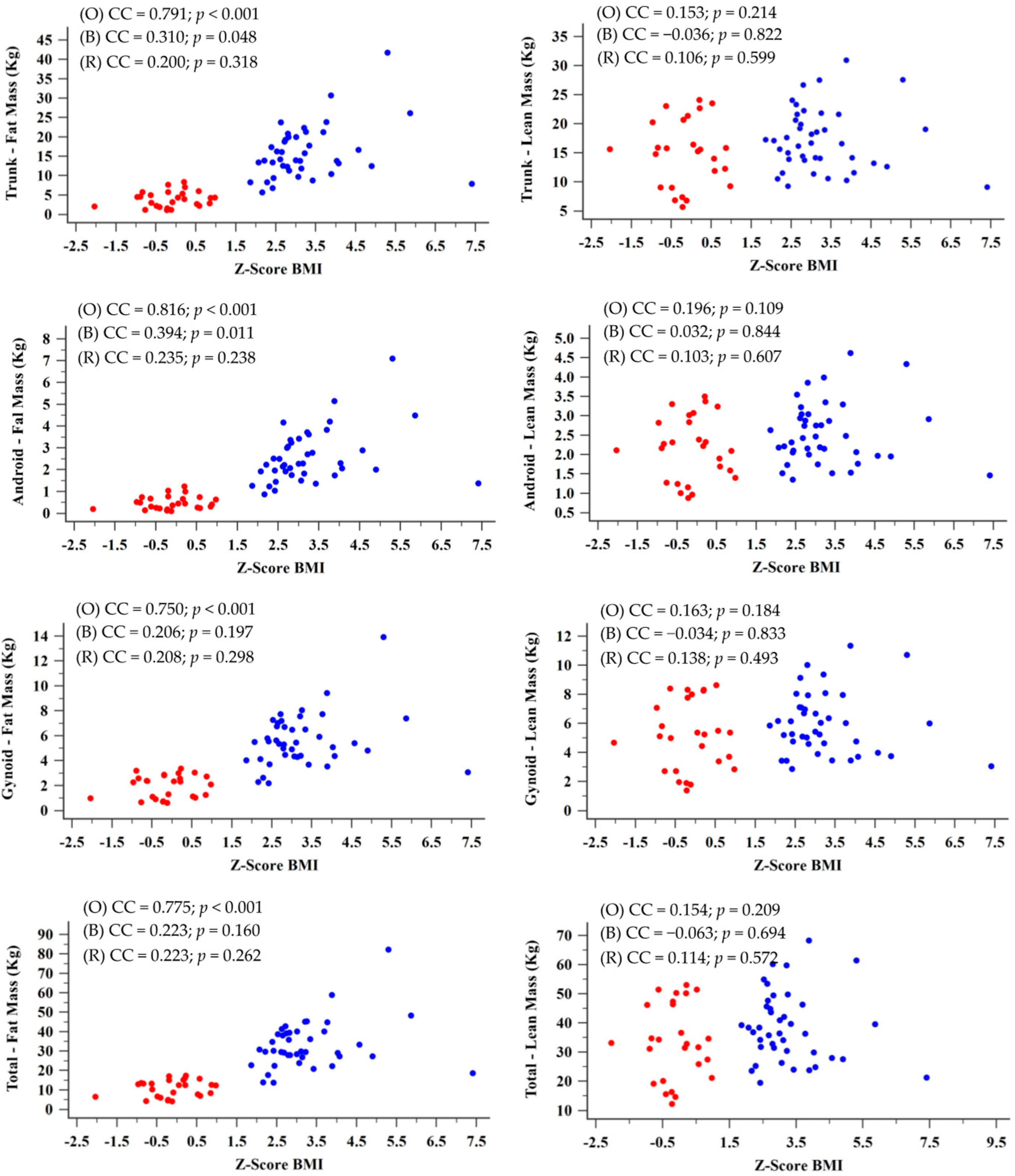
Considering the entire sample, we found a strong/moderate positive correlation between BMI-z and fat mass markers (Figure 2). However, a significative correlation was not observed for lean body mass markers (p > 0.05). Trunk fat mass and BMI-z had a CC = 0.791 (p < 0.001), while trunk lean mass and BMI-z had a CC = 0.153 (p = 0.214). Android fat mass and BMI-z had a CC = 0.816 (p < 0.01), while android lean mass and BMI-z had a CC = 0.196 (p = 0.109). Gynoid fat mass and BMI-z had a CC = 0.750 (p < 0.01), while gynoid lean mass and BMI-z had a CC = 0.163 (p = 0.184). Finally, total fat mass and BMI-z had a CC = 0.775 (p < 0.001), while total lean mass and BMI-z had a CC = 0.154 (p = 0.209). When we analyzed correlation data considering the division by groups with and without obesity (control group), we found only a weak correlation between trunk fat mass and BMI-z (CC = 0.310; p = 0.048) and between android fat mass and BMI-z (CC = 0.394; p = 0.011), both in the group of participants with obesity.
Table 4 presents the correlation between pulmonary function markers and body composition variables. When we considered the entire sample, we observed significant correlations between fat mass markers and FVC z-score (trunk (CC = 0.329; p = 0.006), android (CC = 0.338; p = 0.005), gynoid (CC = 0.301; p = 0.012), total (CC = 0.315; p = 0.008), and fat percentage (CC = 0.359; p = 0.002)), FVC percentile (trunk (CC = 0.330; p = 0.006), android (CC = 0.339; p = 0.004), gynoid (CC = 0.302; p = 0.012), total (CC = 0.317; p = 0.008), and fat percentage (CC = 0.358; p = 0.002)), and FEV1/FVC z-score (trunk (CC = −0.256; p = 0.034), android (CC = −0.260; p = 0.031), total (CC = −0.244; p = 0.043), and fat percentage (CC = −0.263; p = 0.029)).
Additionally, we found significant correlations between lean mass markers and IC (trunk (CC = 0.341; p = 0.006), android (CC = 0.338; p = 0.007), gynoid (CC = 0.334; p = 0.007), total (CC = 0.322; p = 0.010), and fat-free mass (CC = 0.340; p = 0.006)), ERV (trunk (CC = −0.262; p = 0.036), android (CC = −0.253; p = 0.044), gynoid (CC = −0.271; p = 0.030), and fat-free mass (CC = −0.266; p = 0.034)), and PEF (trunk (CC = 0.287; p = 0.018), android (CC = 0.258; p = 0.033), gynoid (CC = 0.268; p = 0.027), total (CC = 0.291; p = 0.016), and fat-free mass (CC = 0.276; p = 0.023)) (Table 4).
When analyzing the correlations considering groups with and without obesity (control group), we found different results. In the group with obesity, the significant correlations with fat mass markers were with IC (trunk (CC = 0.352; p = 0.033)) and PEF (trunk (0.472; p = 0.002), android (CC = 0.457; p = 0.003), gynoid (CC = 0.458; p = 0.003), and total (CC = 0.467; p = 0.002)). At the same time, we found significant correlations between lean mass and IC (trunk (CC = 0.401; p = 0.014), android (CC = 0.390; p = 0.017), gynoid (CC = 0.369; p = 0.025), total (CC = 0.383; p = 0.019), and fat-free mass (CC = 0.411; p = 0.011)), and PEF (trunk (CC = 0.519; p = 0.001), android (CC = 0.497; p = 0.001), gynoid (CC = 0.529; p < 0.001), total (CC = 0.510; p = 0.001), and fat-free mass (CC = 0.478; p = 0.002)) (Table 4). In the group without obesity (control group), we found a significant correlation between trunk fat mass and PEF (trunk (CC = −0.389; p = 0.045)), and we also found significant correlations between lean mass markers and FVC z-score (gynoid (CC = −0.406; p = 0.032), total (−0.381; p = 0.046), and fat-free mass (−0.424; p = 0.024)), FVC percentile (gynoid (CC = −0.414; p = 0.029), total (CC = −0.386; p = 0.042), and fat-free mass (CC = −0.431; p = 0.022)), and ERV (gynoid (CC = −0.458; p = 0.018)) (Table 4).
In the evaluation of pulmonary function, participants with obesity presented higher FVC (z-score (p = 0.002) and percentile (p = 0.03)) and lowered FEV1/FVC (z-score (p = 0.02) and percentile (p = 0.049)) (Table 5).
Table 6 presents the differences in fat mass markers between groups with and without obesity, considering a subdivision of groups by sex. All variables were significantly higher in the group with obesity for both males and females (p < 0.001).
Nevertheless, when considering the differences in lean mass markers between groups, the differences remain significant only in the female group, as is shown in Table 7. Besides the fat markers, girls with obesity also had significantly higher values in the mean of lean mass markers (trunk: p = 0.05; android: p = 0.033; gynoid: p = 0.044; total: p = 0.028; and fat-free mass: p = 0.023).

4. Discussion

In the analysis of anthropometric data, it is interesting to note that BMI-z was an accurate variable to assess the adiposity of participants. The literature points to numerous limitations regarding the use of BMI since it quantifies mass and not fat and may overestimate and classify as overweight or obese individuals with high lean mass [33,34]. However, in the population of children and adolescents evaluated in our study, BMI-z presented a strong correlation with the markers of fat mass (trunk, android, gynoid, and fat percentage) and no correlation with lean body mass markers. Therefore, the classification of the groups with and without obesity was faithful to body composition, which confirms the difference between the groups for fat mass and not for lean body mass.
Although the relationship between obesity and physical inactivity is clear, no difference was found between participants with and without obesity for physical activity practice. We believe this finding is related to three possibilities: (i) a small sample size with a high probability of type II error (false negative result); (ii) a group with obesity receiving multidisciplinary outpatient follow-up with physical educators who emphasize the importance of physical activities—individuals with obesity are in the process of transition of life habits, and possibly for this reason there was no difference in the practice of physical activity among children and adolescents with and without obesity; and (iii) high prevalence of physical inactivity or irregular activity among children and adolescents, whether obese (56.1%) or not (42.9%), a fact already discussed in the literature [35,36,37,38]. The prevalence of physical inactivity was high, noting that the IPAQ assessment addresses scheduled and unscheduled activities and time sitting. The widespread use of screens [39,40], the short time of physical activity at school, the lack of adequate and safe spaces and equipment (e.g., sports courts and swimming pools) in Brazil, and inadequate habits from parents, which culturally follow this inactive model, relegate children to their house and the sedentary lifestyle, without a satisfactory energy expenditure [41,42].
The increase in SBP, DBP, and MBP between the groups, even though these are statistically equal regarding height and age, shows a possible overload of the cardiovascular system in obesity. Excess adipose tissue increases metabolic demands; for this reason, the body requires more blood supply, with a concomitant increase in cardiac activity [43]. The described excessive demand increases the risk of cardiovascular comorbidity and death [44].
Obesity also presented a greater RR value. The increased metabolic demand, mentioned earlier, implies a greater need for oxygen, with the consequent need for greater lung volume. However, the increased fat deposition, especially around the thorax and abdomen, generates a mechanical barrier that hinders thoracic expandability, and this could explain why an individual with obesity requires more respiratory incursions per minute [20,45].
This study conducted a comparative analysis of pulmonary function and body composition by DXA, which allowed us to verify more accurately the respiratory variables related to obesity. When comparing groups, we found that individuals with obesity had significantly higher values in FVC and, consequently, significantly lower values in FEV1/FVC. Analyzing the entire sample, we have found that FVC was positively correlated with fat mass (total, trunk, android, gynoid, and fat percentage) and that FEV1/FVC was negatively correlated with the mentioned markers, except gynoid fat mass. Other variables correlated with muscular strength and were assessed by lean body mass markers (trunk, android, gynoid, and total) with IC and PEF.
However, when we analyzed the correlations grouping the sample with and without obesity, these differences did not hold. We believe that the sample size, after the group division, could interfere with the power of statistical analysis.
Another fact that stood out was the positive correlation between FVC and fat mass markers; in the comparison between the groups, this variable was higher in participants with obesity. This group also presented the lowest FEV1/FVC and a negative correlation with fat mass measures. According to the literature, this finding was present in studies with children and adolescents and was not found in adults [5,8,9,13,14,15,22,46]. Thus, this fact guides us to associate it with the growth period.
Within this perspective, some previous studies justified findings similar to dysanapsis of the airways, which is the disproportionate growth between the pulmonary parenchyma and the airways, with an increase in lung volume different from the increase in the caliber of the airways [22]. This growth pattern is physiologically influenced by sex [47]; however, the relationship of obesity with dysanaptic growth has been studied [18,19,48]. This study found differences between the DIs, assessed by one method [28,29]. Besides that, in our view, the lower difference is justified by the careful selection/exclusion of participants with respiratory symptoms. Even without a great change in DI, the increased FVC, reduced FEV1/FVC, and FEV1 with no difference in participants with obesity directs the findings to the dysanaptic pattern in this group of individuals.
One of the differences of our study was the exclusion of participants with asthma, differing from previous studies associating asthma and obesity, even if independent. In addition, the body composition analysis has allowed us to confirm that the increase in FVC and reduction in FEV1/FVC is related to adiposity and not to an increase in strength of participants with obesity, because of early maturation, as was discussed in the literature [46]. This analysis has shown that the stress-dependent variables—thus positively influenced by increasing lean body mass—are PEF, IC, and ERV.
The body composition analysis was a differential to understand the pulmonary function of children and adolescents with and without obesity, basing its clinical applicability in the pediatric area, especially in the care of individuals with obesity, since obesity and growth are complex phenomena that can occur concurrently. It is important to note that differences in body composition can result in lung function implications. As we observed, both boys and girls with obesity had significantly higher fat markers, as expected. However, girls with obesity also had significantly higher lean mass markers when compared to girls without obesity. Studies show that obesity can contribute to the early onset of puberty in girls [49,50,51,52]. The earlier pubertal development may interfere with the growth and development of muscular mass, which can explain higher values of lean mass in girls with obesity.
To the best of our knowledge, this is the only study that established a correlation between lean body mass and fat mass variables with the pulmonary function of children and adolescents without asthma.
The present study had some limitations. The number of participants may have been insufficient to identify the differences between both DIs. In addition, we did not analyze the correlations between groups with and without obesity considering sex differences because the subgroups would be too small, compromising the relevance of the statistical analysis.

5. Conclusions

We conclude that the relationship between obesity and pulmonary function in children and adolescents is complex and that it is essential to reduce the confounding biases, to understand which changes are obesity-related and which are growth-related. Participants with obesity showed increased FVC and reduced FEV1/FVC. The findings are related to the increase in fat mass, with no relation to lean body mass. We hypothesize that these findings are associated with the dysanaptic growth pattern, which is higher in obesity, evidenced by the reduction of dysanapsis index.

Author Contributions

Conceptualization, M.S.F., F.A.L.M., J.D.R. and R.T.M.; methodology, M.S.F.; J.D.R. and R.T.M.; formal analysis, M.S.F. and F.A.L.M.; investigation, M.S.F., V.L.W.W., M.P.Z. and M.Â.R.d.G.M.A.; resources, M.S.F., F.A.L.M., V.L.W.W., M.P.Z. and M.Â.R.d.G.M.A.; data curation, M.S.F. and F.A.L.M.; writing—original draft preparation, M.S.F. and F.A.L.M.; writing—review and editing, M.S.F., F.A.L.M., J.D.R. and R.T.M.; visualization, M.S.F., F.A.L.M., J.D.R. and R.T.M.; supervision, M.S.F., F.A.L.M., J.D.R. and R.T.M.; project administration, M.S.F., F.A.L.M., J.D.R. and R.T.M. All authors have read and agreed to the published version of the manuscript.

Funding

This research received no external funding.

Institutional Review Board Statement

The study was conducted in accordance with the Declaration of Helsinki and approved by the Ethics Committee of Unicamp (protocol code CAAE: #42633515.0.0000.5404—6 December 2016) for studies involving humans.

Informed Consent Statement

Participants over 18 years and those responsible for minors signed the informed consent form. Participants younger than 18 years old with reading ability and understanding of the research project have signed the informed agreement form.

Data Availability Statement

The crude data presented in this study will be available on request.

Conflicts of Interest

The authors declare no conflict of interest.

References

  1. Güngör, N.K. Overweight and Obesity in Children and Adolescents. J. Clin. Res. Pediatr. Endocrinol. 2014, 6, 129–143. [Google Scholar] [CrossRef]
  2. Ebbeling, C.B.; Pawlak, D.B.; Ludwig, D.S. Childhood obesity: Public-health crisis, common sense cure. Lancet 2002, 360, 473–482. [Google Scholar] [CrossRef]
  3. Bhadoria, A.; Sahoo, K.; Sahoo, B.; Choudhury, A.; Sufi, N.; Kumar, R. Childhood obesity: Causes and consequences. J. Fam. Med. Prim. Care 2015, 4, 187. [Google Scholar] [CrossRef]
  4. Littleton, S.W. Impact of obesity on respiratory function. Respirology 2012, 17, 43–49. [Google Scholar] [CrossRef]
  5. Melo, L.C.; Silva, M.A.M.; Calles, A.C.N. Obesity and lung function: A systematic review. Einstein 2014, 12, 120–125. [Google Scholar] [CrossRef]
  6. Salome, C.M.; King, G.G.; Berend, N. Physiology of obesity and effects on lung function. J. Appl. Physiol. 2010, 64, 206–211. [Google Scholar] [CrossRef] [Green Version]
  7. Mancuso, P. Obesity and Lung Inflammation. J. Appl. Physiol. 2009, 108, 722–728. [Google Scholar] [CrossRef] [Green Version]
  8. Yao, T.C.; Tsai, H.J.; Chang, S.W.; Chung, R.H.; Hsu, J.Y.; Tsai, M.H.; Liao, S.L.; Hua, M.C.; Lai, S.H.; Chen, L.C.; et al. Obesity disproportionately impacts lung volumes, airflow and exhaled nitric oxide in children. PLoS ONE 2017, 12, e0174691. [Google Scholar] [CrossRef] [Green Version]
  9. Peng, R.; Li, S.; Zhang, H.; Zeng, H.; Jiang, B.; Liu, Y.; Yi, X.; Xu, M.; Zhu, L.; Zhang, Z. Weight Status Is Associated with Blood Pressure, Vital Capacity, Dental Decay, and Visual Acuity among School-Age Children in Chengdu, China. Ann. Nutr. Metab. 2016, 69, 237–245. [Google Scholar] [CrossRef]
  10. Khan, S.; Little, J.; Chen, Y. Relationship Between Adiposity and Pulmonary Function in School-Aged Canadian Children. Pediatr. Allergy Immunol. Pulmonol. 2014, 27, 126–132. [Google Scholar] [CrossRef]
  11. Faria, A.G.; Ribeiro, M.A.G.O.; Marson, F.A.L.; Schivinski, C.I.S.; Severino, S.D.; Ribeiro, J.D.; Barros Filho, A.A. Effect of exercise test on pulmonary function of obese adolescents. J. Pediatr. 2014, 90, 242–249. [Google Scholar] [CrossRef] [Green Version]
  12. LoMauro, A.; Cesareo, A.; Agosti, F.; Tringali, G.; Salvadego, D.; Grassi, B.; Sartorio, A.; Aliverti, A. Effects of a multidisciplinary body weight reduction program on static and dynamic thoraco-abdominal volumes in obese adolescents. Appl. Physiol. Nutr. Metab. 2016, 41, 649–658. [Google Scholar] [CrossRef] [Green Version]
  13. Chen, Y.; Rennie, D.; Cormier, Y.; Dosman, J.A. Waist circumference associated with pulmonary function in children. Pediatr. Pulmonol. 2009, 44, 216–221. [Google Scholar] [CrossRef]
  14. Ferreira, M.S.; Mendes, R.T.; Marson, F.A.L.; Zambon, M.P.; Antonio, M.A.R.G.M.; Paschoal, I.A.; Toro, A.A.; Severino, S.D.; Ribeiro, M.A.; Ribeiro, J.D. Espirometria e capnografia volumétrica na avaliação da função pulmonar de indivíduos obesos e eutróficos sem asma. J. Pediatr. 2017, 93, 398–405. [Google Scholar] [CrossRef]
  15. Davidson, W.J.; Msc, K.A.M.-R.; Witmans, M.B.; Montgomery, M.D.; Ball, G.D.; Egbogah, S.; Eves, N.D. Negatively impacts lung function in children and adolescents. Pediatr. Pulmonol. 2014, 49, 1003–1010. [Google Scholar] [CrossRef]
  16. Spathopoulos, D.; Paraskakis, E.; Trypsianis, G.; Tsalkidis, A.; Arvanitidou, V.; Emporiadou, M.; Bouros, D.; Chatzimichael, A. The effect of obesity on pulmonary lung function of school aged children in Greece. Pediatr. Pulmonol. 2009, 44, 273–280. [Google Scholar] [CrossRef]
  17. Dunger, D.B.; Ahmed, M.L.; Ong, K.K. Effects of obesity on growth and puberty. Best Pract. Res. Clin. Endocrinol. Metab. 2005, 19, 375–390. [Google Scholar] [CrossRef]
  18. Forno, E.; Weiner, D.J.; Mullen, J.; Sawicki, G.; Kurland, G.; Han, Y.Y.; Cloutier, M.M.; Canino, G.; Weiss, S.T.; Litonjua, A.A.; et al. Obesity and Airway Dysanapsis in Children with and without Asthma. Am. J. Respir. Crit. Care Med. 2017, 195, 314–323. [Google Scholar] [CrossRef] [Green Version]
  19. Dixon, A.E.; Peters, U. The effect of obesity on lung function. Expert Rev. Respir. Med. 2018, 12, 755–767. [Google Scholar] [CrossRef]
  20. Farah, C.S.; Salome, C.M. Asthma and obesity: A known association but unknown mechanism. Respirology 2012, 17, 412–421. [Google Scholar] [CrossRef]
  21. Camilo, D.F.; Ribeiro, J.D.; Toro, A.D.C.; Baracat, E.C.E.; Filho, A.A.B. Obesity and asthma: Association or coincidence? J. Pediatr. 2010, 86, 6–14. [Google Scholar] [CrossRef] [PubMed]
  22. Forno, E.; Han, Y.Y.; Mullen, J.; Celedón, J.C. Overweight, obesity, and lung function in children and adults-A meta-analysis. J. Allergy Clin. Immunol. Pract. 2018, 6, 570–581. [Google Scholar] [CrossRef] [PubMed]
  23. Mafort, T.T.; Rufino, R.; Costa, C.H.; Lopes, A.J. Obesity: Systemic and pulmonary complications, biochemical abnormalities, and impairment of lung function. Multidiscip. Respir. Med. 2016, 11, 28. [Google Scholar] [CrossRef] [PubMed] [Green Version]
  24. Sole, D.; Vanna, A.T.; Yamada, E.; Rizzo, M.C.; Naspitz, C.K. International Study of Asthma and Allergies in Childhood (ISAAC) written questionnaire: Validation of the asthma component among Brazilian children. J. Investig. Allergol. Clin. Immunol. 1998, 8, 376–382. [Google Scholar] [PubMed]
  25. Onis, M.A.; Onyango, A.W.; Borghi, E.; Siyam, A.; Nishida, C.; Siekmann, J. Development of a WHO growth reference for school-aged children and adolescents. Bull. World Health Organ. 2007, 85, 660–667. [Google Scholar] [CrossRef] [PubMed]
  26. Malachias, M.V.B.; Souza, W.K.S.B.; Plavinik, F.L.; Rodrigues, C.I.S.; Brandão, A.A.; Neves, M.F.T. 7a diretriz brasileira de hipertensão arterial. Arq. Bras. Cardilogia 2016, 107, 83–89. [Google Scholar]
  27. Miller, M.R.; Hankinson, J.; Brusasco, V.; Burgos, F.; Casaburi, R.; Coates, A.; Crapo, R.; Enright, P.; Van Der Grinten, C.P.M.; Gustafsson, P.; et al. Standardisation of spirometry. Eur. Respir. J. 2005, 26, 319–338. [Google Scholar] [CrossRef] [Green Version]
  28. Mead, J. Dysanapsis in normal lungs assessed by the relationship between maximal flow, static recoil, and vital capacity. Am. Rev. Respir. Dis. 1980, 121, 339–342. [Google Scholar]
  29. Tager, I.B.; Weiss, S.T.; Munoz, A.; Welty, C.; Speizer, F.E. Determinants of response to eucapneic hyperventilation with cold air in a population-based study. Am. Rev. Respir. Dis. 1986, 134, 502–508. [Google Scholar]
  30. Matsudo, S.; Araujo, T.; Matsudo, V.; Andrade, D.; Andrade, E.; Oliveira, L.C.; Braggion, G. Questionário Internacional de atividade física (IPAQ): Estudo de validade e reprodutibilidade no Brasil. Rev. Bras. Ativ. Fís. Saúde 2001, 6, 5–18. [Google Scholar]
  31. Marshall, W.A.; Tanner, J.M. Variations in the pattern of pubertal changes in Boys. Arch. Dis. Child. 1970, 45, 13–23. [Google Scholar] [CrossRef] [Green Version]
  32. Marshall, W.A.; Tanner, J.M. Variations in Pattern of Pubertal Changes in Girls. Arch. Dis. Child. 1969, 44, 291–303. [Google Scholar] [CrossRef] [Green Version]
  33. Nuttall, F.Q. Body mass index: Obesity, BMI, and health: A critical review. Nutr. Today 2015, 50, 117–128. [Google Scholar] [CrossRef] [Green Version]
  34. Centers of Disease Control and Prevention. Body Mass Index: Considerations for Practitioners. 2011; pp. 1–4. Available online: https://stacks.cdc.gov/view/cdc/25368 (accessed on 6 November 2022).
  35. Nelson, M.C.; Neumark-Stzainer, D.; Hannan, P.J.; Sirard, J.R.; Story, M. Longitudinal and Secular Trends in Physical Activity and Sedentary Behavior During Adolescence. Pediatrics 2006, 118, e1627–e1634. [Google Scholar] [CrossRef]
  36. Hidding, L.M.; Altenburg, T.M.; van Ekris, E.; Chinapaw, M.J.M. Why do children engage in sedentary behavior? Child- and parent-perceived determinants. Int. J. Environ. Res. Public Health 2017, 14, 671. [Google Scholar] [CrossRef] [Green Version]
  37. Coombs, N.; Shelton, N.; Rowlands, A.; Stamatakis, E. Children’s and adolescents’ sedentary behaviour in relation to socioeconomic position. J. Epidemiol. Community Health 2013, 67, 868–874. [Google Scholar] [CrossRef] [Green Version]
  38. Guerra, P.H.; Farias Júnior, J.C.d.; Florindo, A.A. Sedentary behavior in Brazilian children and adolescents: A systematic review. Rev. Saude Publica 2016, 50, 1–15. [Google Scholar] [CrossRef] [Green Version]
  39. LeBlanc, A.G.; Katzmarzyk, P.T.; Barreira, T.V.; Broyles, S.T.; Chaput, J.P.; Church, T.S.; Fogelholm, M.; Harrington, D.M.; Hu, G.; Kuriyan, R.; et al. Correlates of total sedentary time and screen time in 9–11 year-old children around the world: The international study of childhood obesity, lifestyle and the environment. PLoS ONE 2015, 10, e0129622. [Google Scholar] [CrossRef] [Green Version]
  40. Tandon, P.S.; Zhou, C.; Sallis, J.F.; Cain, K.L.; Frank, L.D.; Saelens, B.E. Home environment relationships with children’s physical activity, sedentary time, and screen time by socioeconomic status. Int. J. Behav. Nutr. Phys. Act. 2012, 9, 88. [Google Scholar] [CrossRef] [Green Version]
  41. Farley, T.A.; Meriwether, R.A.; Baker, E.T.; Watkins, L.T.; Johnson, C.C.; Webber, L.S. Safe play spaces to promote physical activity in inner-city children: Results from a pilot study of an environmental intervention. Am. J. Public Health 2007, 97, 1625–1631. [Google Scholar] [CrossRef]
  42. Singh, G.K.; Yu, S.M.; Siahpush, M.; Kogan, M.D. High levels of physical inactivity and sedentary behaviors among US immigrant children and adolescents. Arch. Pediatr. Adolesc. Med. 2008, 162, 756–763. [Google Scholar] [CrossRef] [PubMed] [Green Version]
  43. Cote, A.T.; Harris, K.C.; Panagiotopoulos, C.; Sandor, G.G.S.; Devlin, A.M. Childhood obesity and cardiovascular dysfunction. J. Am. Coll. Cardiol. 2013, 62, 1309–1319. [Google Scholar] [CrossRef] [PubMed] [Green Version]
  44. Franks, P.W.; Hanson, R.L.; Knowler, W.C.; Sievers, M.L.; Bennett, P.H.; Looker, H.C. Childhood obesity, other cardiovascular risk factors, and premature death paul. N. Engl. J. Med. 2010, 362, 485–493. [Google Scholar] [CrossRef] [PubMed]
  45. Parameswaran, K.; Todd, D.C.; Soth, M. Altered respiratory physiology in obesity. Can. Respir. J. 2006, 13, 203–210. [Google Scholar] [CrossRef]
  46. He, Q.-Q.; Wong, T.-W.; Du, L.; Jiang, Z.-Q.; Qiu, H.; Gao, Y.; Liu, J.-W.; Wu, J.-G.; Yu, I.T.-S. Respiratory health in overweight and obese Chinese children. Pediatr. Pulmonol. 2009, 44, 997–1002. [Google Scholar] [CrossRef]
  47. Lomauro, A.; Aliverti, A. Sex differences in respiratory function. Breathe 2018, 14, 131–140. [Google Scholar] [CrossRef] [Green Version]
  48. Jones, M.H.; Roncada, C.; Fernandes, M.T.C.; Filho, J.P.H.; Icaza, E.E.S.; Mattiello, R.; Pitrez, P.M.C.; Pinto, L.A.; Stein, R. Asthma and Obesity in Children Are Independently Associated with Airway Dysanapsis. Front. Pediatr. 2017, 18, 270. [Google Scholar] [CrossRef] [Green Version]
  49. Brown, K.A.; Patel, D.R.; Darmawan, D. Participation in sports in relation to adolescent growth and development. Transl. Pediatr. 2017, 6, 150–159. [Google Scholar] [CrossRef] [Green Version]
  50. Costa, T.; Murara, P.; Vancini, R.L.; de Lira, C.A.B.; Andrade, M.S. Influence of Biological Maturity on the Muscular Strength of Young Male and Female Swimmers. J. Hum. Kinet. 2021, 78, 67–77. [Google Scholar] [CrossRef]
  51. Li, W.; Liu, Q.; Deng, X.; Chen, Y.; Liu, S.; Story, M. Association between Obesity and Puberty Timing: A Systematic Review and Meta-Analysis. Int. J. Environ. Res. Public Health 2017, 14, 1266. [Google Scholar] [CrossRef]
  52. Chung, S. Growth and Puberty in Obese Children and Implications of Body Composition. J. Obes. Metab. Syndr. 2017, 26, 243–250. [Google Scholar] [CrossRef] [PubMed]
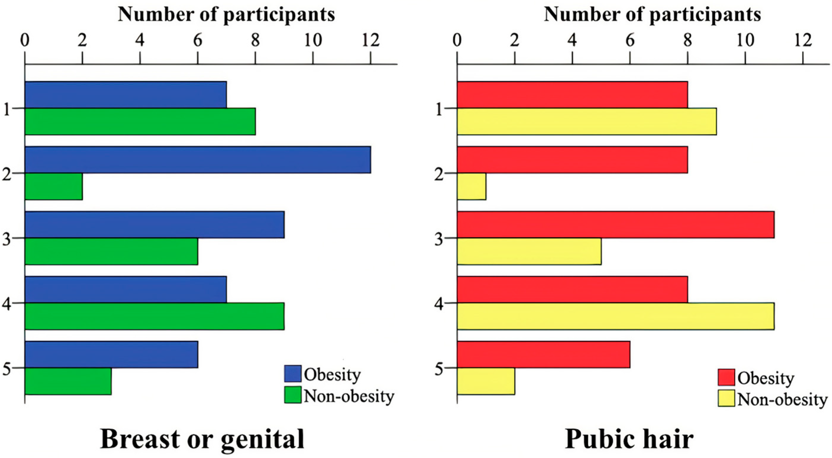
Figure 1. Distribution of pubertal development according to genitalia and breasts (p = 0.138) and pubic hair (p = 0.089) between the groups with and without obesity.
Jcm 11 07410 g001
Figure 2. Correlation between the body mass index (BMI) z-score and the variables of fat mass and lean body mass. Blue (B) indicates participants with obesity, and red (R) indicates participants without obesity (control group). CC, correlation coefficient. The statistical analyses were carried out based on Spearman’s rank correlation coefficient. Alpha = 0.05.
Jcm 11 07410 g002
Table 1. Distribution and comparison of sex, ethnicity (race), anthropometric markers, and dysanapsis indexes between groups with and without obesity.
MarkerGroupObesity (n = 41)Control (n = 28)p
Sex 1Male14 (34.1%)14 (50%)0.188
Female27 (65.9%)14 (50%)
Ethnicity (race) 2White25 (61%)25 (85.7%)0.038
Black8 (19.5%)1 (3.6%)
Mixed race *8 (19.5%)2 (7.1%)
Asian1 (3.6%)
Height 3 (cm) 153.27 ± 13.86;
155.50 (142.70 to 163.25)
152.65 ± 22.75;
161.90 (134.50 to 171.90)
0.460
Weight 4 (kg) 74.04 ± 23.67;
69.10 (58.35 to 89.45)
44.98 ± 17.22;
49.30 (29.27 to 61.53)
<0.001
BMI 3 (kg/m2) 30.85 ± 6.63;
29.92 (27.24 to 33.44)
18.30 ± 2.54;
18.84 (15.57 to 20.3)
<0.001
BMI Z-score 3 3.25 ± 1.09;
3 (2.57 to 3.73)
−0.16 ± 0.68;
−0.20 (−0.63 to 0.22)
<0.001
Age 3 (years) 11.98 ± 3.62;
11.74 (8.99 to 14.94)
12.97 ± 4.91;
14.18 (8.72 to 18.05)
0.213
DI1 4 0.20 ± 0.40;
0.19 (0.16 to 0.23)
0.22 ± 0.50;
0.19 to 0.26)
0.031
DI2 4 0.98 ± 0.24;
0.95 (0.81 to 1.14)
1.09 ± 0.27;
1.13 (0.87 to 1.25)
0.065
n, number of participants; cm, centimeters; BMI, body mass index; kg, kilograms; kg/m2, kilograms/square meter; DI1, dysanapsis index 1; DI2, dysanapsis index 2. Categorical data are presented in absolute frequency (relative frequency). Numerical data are presented by mean ± standard deviation; median (25th percentile to 75th percentile). In the statistical analysis of the data, the following tests were used: 1, chi-square test; 2, Fisher’s exact test; 3, Mann–Whitney test for independent samples; 4, t-test for independent samples. *, Pardos (multiracial background). Alpha = 0.05. Data with significant p (≤0.05) are presented in bold.
Table 2. Classification and comparison of the groups regarding the practice of physical activities, according to the IPAQ, considering the complete (A) and synthesized (B) classifications.
A 1ObesityControlp
Sedentary1 (2.4%)0.777
Irregularly active A8 (19.5%)3 (10.7%)
Irregularly active B14 (34.1%)9 (32.1%)
Active6 (14.6%)6 (21.4%)
Very active12 (29.3%)10 (35.7%)
B 2ObesityControlp
Sedentary or irregularly active23 (56.1%)12 (42.90%)0.280
Active or very active18 (43.9%)16 (57.1%)
In the statistical analysis of the data, the following tests were used: 1, Fisher’s exact test; 2, chi-square test. IPAQ, international physical activity questionnaire. Alpha = 0.05. Categorical data are presented in absolute frequency (relative frequency).
Table 3. Comparison between the vital signs of participants with and without obesity.
VariableObesityControlp
Heart rate 185.83 ± 15.65; 87 (73 to 97.50)81.11 ± 13.70; 82 (70.25 to 90.50)0.200
Respiratory rate 121.44 ± 3.97; 22 (18 to 23.50)18.43 ± 4.51; 18 (15.25 to 22.75)0.005
SpO2 297.44 ± 1.21; 98 (97 to 98)97.50 ± 0.92; 97 (97 to 98)0.798
Blood pressureObesityControlp
Systolic 1118.39 ± 14.03; 120 (87 to 147)102.50 ± 12.23; 103 (86 to 124)<0.001
Diastolic 278.12 ± 10.54; 78 (71 to 80)67.89 ± 7.36; 70 (60 to 70)<0.001
Mean 291.54 ± 10.96; 92.33 (85 to 96.50)79.43 ± 8.28; 80.17 (70.17 to 86)<0.001
SpO2, peripheral oxygen saturation. Numerical data are presented by mean ± standard deviation; median (25th percentile to 75th percentile). In the statistical analysis of the data, the following tests were used: 1, t-test for independent samples; 2, Mann–Whitney test. Alpha = 0.05. Data with significant p (≤0.05) are presented in bold.
Table 4. Correlation between pulmonary function markers and body composition, respectively, evaluated by spirometry and dual-energy X-ray absorptiometry.
Markers Fat MassLean Body Mass
Overall TrunkAndroidGynoidTotalFat PercentageTrunkAndroidGynoidTotalFat-Free Mass
FVC z-scoreCC0.3290.3380.3010.3150.359−0.0330.011−0.074−0.042−0.073
p0.0060.0050.0120.0080.0020.7910.9260.5480.7310.551
FVC—percentileCC0.3300.3390.3020.3170.358−0.0310.014−0.072−0.040−0.071
p0.0060.0040.0120.0080.0020.8020.9110.5560.7440.561
FEV1/FVC z-scoreCC−0.256−0.260−0.236−0.244−0.2630.029−0.0100.0220.0250.030
p0.0340.0310.0510.0430.0290.8110.9370.8550.8400.805
Inspiratory capacityCC0.1620.1330.1520.144−0.0630.3410.3380.3340.3220.340
p0.2060.2980.2350.2590.6240.0060.0070.0070.0100.006
ERVCC−0.090−0.063−0.066−0.0640.091−0.262−0.253−0.271−0.241−0.266
p0.4810.6210.6050.6160.4770.0360.0440.0300.0550.034
PEFCC0.0650.0670.0810.072−0.0920.2870.2580.2680.2910.276
p0.5990.5870.5090.5590.4570.0180.0330.0270.0160.023
Obesity
FVC z-scoreCC0.2100.2090.2330.184−0.0190.1830.1940.1660.1600.142
p0.1880.1890.1430.2490.9070.2530.2250.3000.3000.377
FVC—percentileCC0.2160.2150.2390.190−0.0190.1870.1980.1700.1640.145
p0.1760.1770.1330.2340.9060.2420.2140.2890.3060.365
FEV1/FVC z-scoreCC0.0230.0020.0490.0610.0650.008−0.0320.0130.0050.022
p0.8880.9880.7600.7030.6860.9610.8420.9370.9770.890
Inspiratory capacityCC0.3520.2950.2650.290−0.2130.4010.3900.3690.3830.411
p0.0330.0760.1140.0810.2050.0140.0170.0250.0190.011
ERVCC−0.175−0.126−0.077−0.1150.175−0.171−0.165−0.133−0.149−0.184
p0.2940.4500.6450.4910.2920.3030.3230.4270.3720.270
PEFCC0.4720.4570.4580.4670.0170.5190.4970.5290.5100.478
p0.0020.0030.0030.0020.9140.0010.001<0.0010.0010.002
Without obesity
FVC z-scoreCC−0.184−0.167−0.208−0.2080.285−0.355−0.287−0.406−0.381−0.424
p0.3490.3960.2880.2890.1410.0640.1380.0320.0460.024
Continue
FVC—percentileCC−0.187−0.169−0.209−0.2080.286−0.361−0.292−0.414−0.386−0.431
p0.3420.3910.2860.287−0.1400.0590.1310.0290.0420.022
FEV1/FVC z-scoreCC−0.113−0.111−0.061−0.079−0.2510.1710.1360.1300.1740.168
p0.5680.5730.7570.6900.1980.3850.4890.5090.3750.394
Inspiratory capacityCC0.0950.0500.1910.127−0.0920.2720.2940.3640.3160.309
p0.6450.8100.3500.5360.6550.1790.1440.0670.1160.124
ERVCC−0.257−0.218−0.274−0.2470.028−0.355−0.359−0.458−0.379−0.377
p0.2050.2840.1760.2230.8930.0750.0720.0180.0560.058
PEFCC−0.389−0.351−0.279−0.320−0.3670.003−0.049−0.0030.0320.054
p0.0450.0730.1580.1030.0600.9880.8060.9880.8730.789
CC, correlation coefficient; FVC, forced vital capacity; FEV1/FVC, ratio between forced expiratory volume in one second and forced vital capacity; ERV, expiratory reserve volume; PEF, peak expiratory flow. Spearman’s rank correlation coefficient was used in the statistical analysis of the data. Alpha = 0.05. Data with significant p (≤0.05) are presented in bold.
Table 5. Differences between pulmonary function markers in groups of participants with and without obesity.
MarkersObesityControlp
FVC z-score0.43 ± 0.62;
0.41 (0.025 to 0.82)
−0.20 ± 0.92;
−0.23 (−0.98 to 0.49)
0.002 1
FVC—percentile64.32 ± 19.77;
66 (51 to 79)
43.46 ± 28.89;
41 (16.50 to 68.50)
0.003 2
FEV1 z-score0.39 ± 0.69;
0.36 (0.01 to 0.79)
0.06 ± 0.96;
0.05 (−0.66 to 0.77)
0.125 1
FEV1—percentile63.24 ± 21.54;
64 (50.50 to 78.50)
51.86 ± 29.16;
51.50 (25.75 to 77.75)
0.124 2
FEV1/FVC z-score−0.10 ± 1.01;
−0.33 (−0.76 to 0.49)
0.54 ± 1.14;
0.75 (−0.50 to 1.45)
0.020 1
FEV1/FVC percentile45.90 ± 29.79;
37 (22.50 to 69)
61.37 ± 33.48;
71 (27.25 to 92.75)
0.049 2
FEF25–75% z-score−0.13 ± 0.97;
−0.15 (−0.73 to 0.63)
−0.09 ± 1.08;
−0.02 (−0.97 to 0.64)
0.885 1
FEF25–75% percentile45.95 ± 28.43;
44 (23.50 to 73.50)
47.64 ± 31.14;
49.50 (16.75 to 73.75)
0.869 2
Inspiratory capacity103.34 ± 38.77;
97.70 (71.75 to 140.45)
101.65 ± 31.79;
100.25 (80.73 to 124.10)
0.791 2
Expiratory reserve volume117.44 ± 84.25;
103.65 (49.13 to 167.73)
123.87 ± 113.40;
111.45 (53.48 to 149.03)
0.796 1
Peak expiratory flow87.64 ± 15.72;
88 (76.05 to 96.85)
89.28 ± 14.36;
90.40 (79.80 to 100.90)
0.665 1
FVC, forced vital capacity; FEV1/FVC, ratio between forced expiratory volume in one second and forced vital capacity. Numerical data are presented by mean ± standard deviation; median (25th percentile to 75th percentile). In the statistical analysis of the data, the following tests were used: 1, t-test for independent samples; 2, Mann–Whitney test for independent samples. Alpha = 0.05. Data with significant p (≤0.05) are presented in bold.
Table 6. Association between body composition (fat mass) evaluated by dual-energy X-ray absorptiometry in groups of participants with and without obesity and according to sex.
Trunk Fat MassObesityControlp
Male16.96 ± 5.80;
18.30 (11.20 to 21.56)
3.79 ± 2.22;
3.00 (2.17 to 5.19)
<0.001
Female15.47 ± 7.56;
13.80 (11.26 to 17.77)
4.17 ± 2.19;
4.32 (1.77 to 5.85)
<0.001
p0.2040.635
Android Fat MassObesityControlp
Male2.84 ± 1.06;
3.07 (1.73 to 3.75)
0.47 ± 0.34;
0.34 (0.23 to 0.70)
<0.001
Female2.48 ± 1.32;
2.15 (1.75 to 2.78)
0.49 ± 0.29;
0.47 (0.20 to 0.74)
<0.001
p0.1760.874
Gynoid Fat MassObesityControlp
Male5.76 ± 1.63;
5.66 (4.22 to 7.34)
1.68 ± 0.80;
1.28 (1.02 to 2.42)
<0.001
Female5.55 ± 2.38;
5.36 (4.34 to 6.51)
2.26 ± 1.11;
2.47 (0.86 to 3.09)
<0.001
p0.4880.246
Total Fat MassObesityControlp
Male34.50 ± 9.19;
37.23 (25.82 to 42.26)
10.09 ± 4.26;
8.56 (6.57 to 13.75)
<0.001
Female32.63 ± 13.95;
29.60 (27.24 to 38.02)
11.56 ± 5.06;
12.60 (5.65 to 15.28)
<0.001
p0.3200.667
Fat PercentageObesityControlp
Male43.64 ± 5.74;
43.07 (39.51 to 46.61)
20.87 ± 4.45;
21.20 (17.61 to 23.86)
<0.001
Female46.03 ± 4.21;
46.12 (44.62 to 48.17)
28.00 ± 5.33;
27.10 (24.86 to 32.39)
<0.001
p0.1080.001
Numerical data are presented by mean ± standard deviation; median (25th percentile to 75th percentile). The Mann–Whitney test for independent samples was applied in the statistical analysis of the data. Alpha = 0.05. Data with significant p (≤0.05) are presented in bold.
Table 7. Association between body composition (lean body mass) evaluated by dual-energy X-ray absorptiometry in groups of participants with and without obesity and according to sex.
Trunk Lean Body MassObesityControlp
Male19.47 ± 5.34;
20.10 (15.90 to 23.49)
17.01 ± 6.53;
20.48 (11.15 to 22.78)
0.286
Female16.15 ± 5.16;
14.99 (11.59 to 19.04)
12.89 ± 4.27;
15.02 (8.50 to 15.87)
0.050
p0.0600.061
Android Lean Body MassObesityControlp
Male2.87 ± 0.73;
2.90 (2.37 to 3.40)
2.41 ± 0.93;
2.83 (1.51 to 3.26)
0.165
Continue
Female2.37 ± 0.78;
2.18 (1.76 to 2.87)
1.84 ± 0.61;
2.13 (1.21 to 2.32)
0.033
p0.0540.069
Gynoid Lean Body MassObesityControlp
Male6.83 ± 2.12;
6.99 (5.27 to 8.34)
5.90 ± 2.76;
7.42 (3.22 to 8.31)
0.323
Female5.52 ± 2.05;
5.20 (3.75 to 6.67)
4.21 ± 1.64;
5.05 (2.52 to 5.41)
0.044
p0.0610.062
Total Lean Body MassObesityControlp
Male43.51 ± 11.95; 44.20 (35.18 to 53.85)37.98 ± 14.81; 46.32 (24.50 to 50.61)0.287
Female35.89 ± 11.35; 34.18 (26.33 to 40.92)27.89 ± 9.04; 31.57 (18.24 to 34.66)0.028
p0.0520.041
Fat-Free MassObesityControlp
Male44.86 ± 12.59; 45.19 (35.61 to 55.29)39.52 ± 15.54; 48.87 (25.67 to 51.34)0.327
Female37.19 ± 11.70;35.95 (27.56 to 41.48)28.59 ± 9.58; 32.15 (18.24 to 36.67)0.023
p0.0600.036
Numerical data are presented by mean ± standard deviation; median (25th percentile to 75th percentile). The t-test for independent samples was applied in the statistical analysis of the data. Alpha = 0.05. Data with significant p (≤0.05) are presented in bold.
Publisher’s Note: MDPI stays neutral with regard to jurisdictional claims in published maps and institutional affiliations.