Review

Risk of Contrast-Associated Acute Kidney Injury in Patients Undergoing Peripheral Angiography with Carbon Dioxide Compared to Iodine-Containing Contrast Agents: A Systematic Review and Meta-Analysis

1
Department for Evidence-Based Medicine and Evaluation, University for Continuing Education Krems, Dr.-Karl-Dorrek-Strasse 30, 3500 Krems, Austria
2
RTI International, 3040 East Cornwallis Road, Research Triangle Park, NC 27709-2194, USA
3
Karl Landsteiner University of Health Sciences, Dr.-Karl-Dorrek-Strasse 30, 3500 Krems, Austria
4
Department of Internal Medicine, University Hospital St. Poelten, Dunant-Platz 1, 3100 St. Poelten, Austria
5
Department of Internal Medicine, Neurology and Dermatology, Division of Angiology, University Hospital Leipzig, Liebigstrasse 20, 04103 Leipzig, Germany
6
Helmholtz Institute for Metabolic, Obesity and Vascular Research (HI-MAG) of the Helmholtz Zentrum Muenchen, University Hospital Leipzig and University of Leipzig, Liebigstrasse 20, 04103 Leipzig, Germany
*
Author to whom correspondence should be addressed.
J. Clin. Med. 2022, 11(23), 7203; https://doi.org/10.3390/jcm11237203
Submission received: 21 October 2022 / Revised: 26 November 2022 / Accepted: 28 November 2022 / Published: 4 December 2022
(This article belongs to the Section Vascular Medicine)

Abstract

:
The aim of this systematic review was to summarise the comparative evidence on the risk of contrast-associated acute kidney injury (CA-AKI) with CO2 or iodinated contrast medium (ICM) for peripheral vascular interventions. We searched Ovid MEDLINE, Cochrane Library, Embase, Epistemonikos, PubMed-similar-articles, clinical trial registries, journal websites, and reference lists up to February 2022. We included studies comparing the risk of CA-AKI in patients who received CO2 or ICM for peripheral angiography with or without endovascular intervention. Two reviewers screened the references and assessed the risk of bias of the included studies. We extracted data on study population, interventions and outcomes. For the risk of CA-AKI as our primary outcome of interest, we calculated risk ratios (RRs) with a 95% confidence interval (CI) and performed random-effects meta-analyses. We identified three RCTs and five cohort studies that fully met our eligibility criteria. Based on a random-effects meta-analysis, the risk of CA-AKI was lower with CO2 compared to ICM (8.6% vs. 15.2%; RR, 0.59; 95% CI 0.33–1.04). Only limited results from a few studies were available on procedure and fluoroscopy time, radiation dose and CO2-related adverse events. The evidence suggests that the use of CO2 for peripheral vascular interventions reduces the risk of CA-AKI compared to ICM. However, due to the relevant residual risk of CA-AKI with the use of CO2, other AKI risk factors must be considered in patients undergoing peripheral vascular interventions.

1. Introduction

Peripheral vascular interventions (PVIs) are increasingly performed in patients with peripheral arterial disease (PAD) [1] and, in parallel, the complexity of the procedures is on the rise, as more patients with advanced age and multiple co-morbidities are treated.
During PVIs, the administration of an iodinated contrast medium (ICM) has been described as a risk factor for both acute kidney injury (AKI) and subsequent clinically relevant major adverse kidney events, such as persistently impaired renal function, need for new haemodialysis and death [2]. Although contrast-associated AKI (CA-AKI) is usually mild and creatinine levels typically return to baseline within two weeks [3,4,5], it is associated with an increased risk of serious adverse in-hospital and long-term outcomes, including all-cause mortality [6,7]. While the underlying pathophysiology of CA-AKI has not been fully elucidated, the proposed mechanisms include the direct cytotoxic effects of ICM on tubular cells as well as perturbed renal haemodynamics [5].
The reported risk of CA-AKI varies between 7% and 11% [2,4,8,9,10,11]. Substantially higher and lower rates were attributed to the use of different AKI definitions, different ICM administration modes (intravenous vs. intraarterial), ICM choice and dose, as well as the considerable heterogeneity in patient populations with respect to co-morbidities and the severity of underlying renal disease [2]. Pre-existing individual risk factors, particularly chronic kidney disease and the patient’s hydration status, are considered major risk determinants for the development of CA-AKI [10,12]. In previous randomised controlled trials (RCTs), no preventive measures, including hydration, sodium bicarbonate and acetylcysteine, could convincingly demonstrate a reduction in CA-AKI [13,14].
Importantly, it cannot be excluded that other factors beyond ICM contribute to the observed renal impairment and some researchers have even questioned whether ICM plays a significant role at all for the observed deterioration in kidney function [8,15], claiming that the risk of CA-AKI for the patient is overstated in the literature and overestimated by physicians [8,16]. Such overestimation of CA-AKI risk could even be harmful to patients if needed imaging procedures are delayed for unfounded fear of CA-AKI. Thus, it is of great scientific interest and clinical relevance to study the potential role of contrast-saving or contrast-avoiding strategies for the reduction of CA-AKI.
For PVI, it is possible to reduce the quantity of conventional ICM or even completely avoid it using carbon dioxide (CO2) as an alternative contrast agent [12]. CO2 acting as a negative contrast agent has been used for a variety of vascular procedures since the introduction of digital subtraction angiography. Because of its high solubility rate and rapid diffusibility via the lungs, CO2 is safe for peripheral intravascular use, but should not be used above the diaphragm to avoid the possibility of causing a cerebral air embolism, associated with stroke or death [17].
Current research on the clinical benefits and risks of CO2 angiography is limited and based on observational data and small RCTs. Therefore, we conducted a systematic review to summarise the comparative evidence on the risk of CA-AKI with CO2 or conventional ICM for peripheral angiography with or without endovascular intervention.

2. Materials and Methods

We followed the Preferred Reporting Items for Systematic Reviews and Meta-Analyses (PRISMA) guidelines in reporting the results of this systematic review [18].

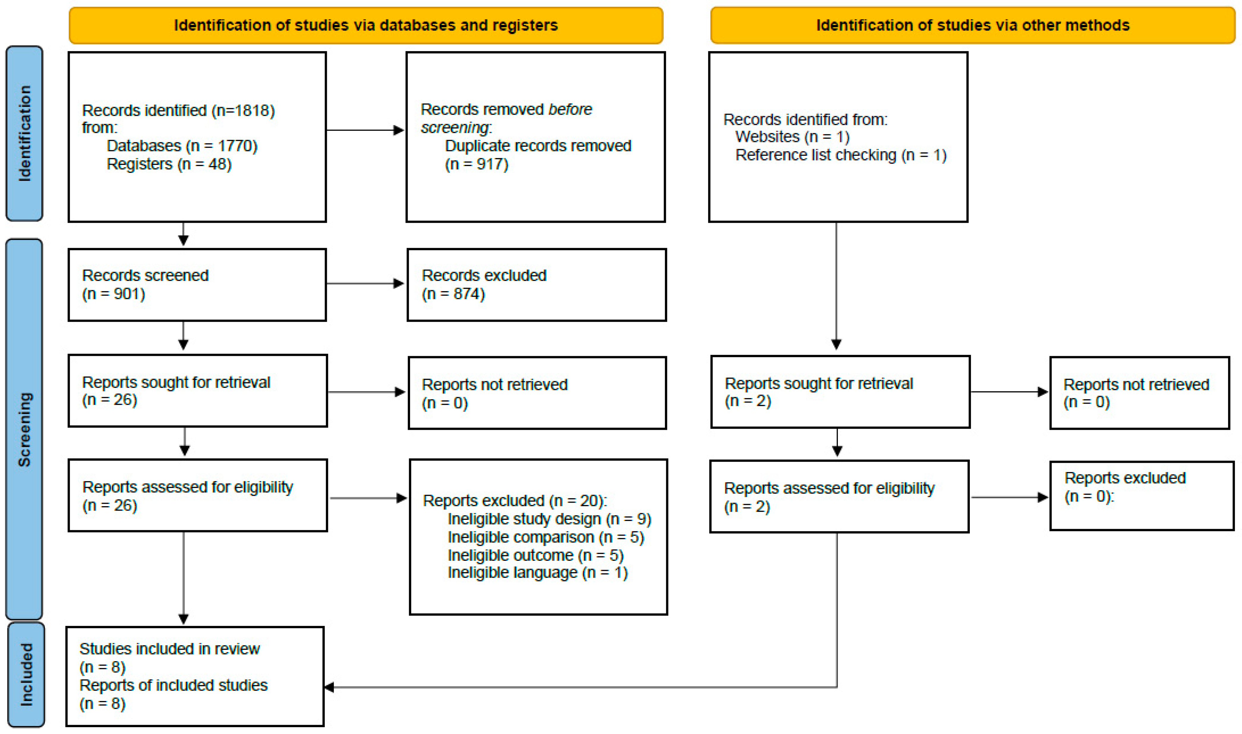
2.1. Literature Search

An experienced information specialist (I.K.) conducted the following database searches from inception to February 2022: Ovid MEDLINE (accessed on 14 February 2022), Cochrane Library (Wiley) (accessed on 15 February 2022) and Embase.com (Elsevier, Netherlands) (accessed on 15 February 2022). In addition, we searched the following trial registries to identify unpublished and ongoing studies: ClinicalTrials.gov and the World Health Organization International Clinical Trials Registry Platform (ICTRP) (both accessed on 15 February 2022). Two sources were searched from inception to May 2021 but not updated because they had not identified any relevant studies: Epistemonikos.org (accessed on 19 May 2021) and PubMed-similar-articles (publications identified as potentially relevant in the preliminary search served as source references) (accessed on 18 May 2021). When possible, we combined controlled vocabulary (e.g., Medical Subject Headings (MeSH)) and free-text terms in the search strategies. We provide our full search strategies as Supplementary Table S1. In addition, we checked the reference lists of the included studies, published reviews and trial registry entries as well as the websites of journals in the vascular medicine field not indexed in the searched databases (e.g., Journal of Critical Limb Ischemia).

2.2. Eligibility Criteria and Study Selection

We included studies that compared the risk of CA-AKI injury in patients who received CO2 or ICM for angiography of the lower limb arteries, kidney arteries or infrarenal aorta with or without endovascular intervention (angioplasty, stent, endograft). We determined CA-AKI as our primary outcome of interest and included only studies that defined and reported CA-AKI as a binary outcome. Thus, studies reporting only continuous outcomes such as changes in creatinine or in the glomerular filtration rate (GFR) were excluded. For consistency, we will use the more current term CA-AKI throughout this manuscript instead of contrast-induced nephropathy, which was often used in the included studies. Studies that include patients receiving haemodialysis prior to the intervention were excluded. Appendix Table A1 shows the inclusion and exclusion criteria applied during literature screening in detail.
All references identified by our literature search were organised with Endnote X9.3 (Clarivate, PA, USA). We used the Covidence (Veritas Health Innovation, Melbourne, Australia) [19] online systematic review tool to screen references against our eligibility criteria. Two reviewers (G.W., A.G.) independently screened the references in two subsequent steps. First, they screened the references yielded by the systematic search based on title and abstract. Second, for those references considered relevant by both reviewers, full-text articles were retrieved and screened to ascertain whether the study met the eligibility criteria. At each step, conflicts were discussed and resolved between the two reviewers. If necessary, a third reviewer with extensive clinical expertise in the interventional angiology field (S.S.) was involved.

2.3. Risk of Bias and Certainty of Evidence

To assess the risk of bias, we used the Cochrane Risk of Bias 2 (RoB 2) tool [20] for RCTs and the Risk Of Bias In Non-Randomized Studies of Interventions (ROBINS-I) tool [21] for non-randomised studies. Different pairs of reviewers (A.G., E.P., G.W.) independently rated the risk of bias. Consensus was obtained through discussion. We employed the Grading of Recommendations Assessment, Development and Evaluation (GRADE) [22] approach to assess the certainty of evidence for our primary outcome of interest. We used the GRADEpro (McMaster University and Evidence Prime, Hamilton, ON, Canada) [23] online tool to create a GRADE evidence profile and summary of findings table.

2.4. Data Collection

We used electronic extraction tables to collect the following data items from the included studies: first author, year, country, study design, follow-up duration, recruitment period, inclusion and exclusion criteria, patient characteristics, intervention description including amount of CO2 and ICM, type of ICM, procedure and fluoroscopy time, radiation dose-area product, definition and risk of CA-AKI, additional adverse events and CO2-related side effects, and procedural outcome. One reviewer (G.W.) extracted the data into tables that a second reviewer (E.P.) checked for completeness and correctness.

2.5. Data Analysis

We conducted meta-analyses if the clinical heterogeneity among studies was reasonable. Based on the number of patients with CA-AKI and the number of patients at risk in each group, we calculated risk ratios (RRs) with 95% confidence intervals (CIs). We performed random-effects meta-analyses using the Paule–Mandel estimator of tau² [24]. To assess the statistical heterogeneity across studies, we visually inspected the forest plots and calculated the I2 statistics [25]. For CA-AKI, our primary outcome of interest, the number of events, patients at risk and effect estimates with 95% CI were presented as forest plots. The results of other outcomes were summarised and presented in tables. We conducted a sensitivity analysis with (1) studies rated as low and moderate/some risk of bias, (2) studies predominantly including patients with impaired renal function and (3) studies including more than 40% of patients with diabetes at baseline. We conducted a subgroup analysis according to the study design. If we had identified more than 10 studies, we intended to assess publication bias using a visual assessment of the funnel plots. For all analyses, we used the meta package [26] in RStudio (RStudio, PBC, Boston, MA, USA) [27] within the R environment (R Foundation for Statistical Computing, Vienna, Austria) [28].

3. Results

3.1. Study Characteristics

After deduplication, our searches yielded 901 references. Of those, we included eight studies that fully met our eligibility criteria: three RCTs [29,30,31] and five cohort studies [32,33,34,35,36] (three retrospective and two prospective). The studies were published between the years 2001 and 2021. Participants were recruited between the years 1996 and 2018 and followed up for up to 6 months. The studies took place in Europe (Sweden, England, Germany), the United States, Egypt, Iran and Saudi Arabia. Figure 1 provides details of the study selection process. Notably, two of the included studies were published in journals not indexed in the searched databases. One study was identified through a trial registry entry and the other by hand searching the journal website. In Supplementary Table S2, we provide a list of references that were excluded based on a full-text assessment, including the reasons for exclusion.

3.2. Study Population

The included studies’ sample sizes ranged from 64 to 313 participants, and the reported mean age was between 54 and 78 years, with a majority being male. The proportion of participants with diabetes mellitus varied considerably in the individual studies, ranging from 17% to 65%. Most studies included participants with impaired renal function. Two studies only included patients with chronic kidney disease (CKD) stage 3 or higher (i.e., GFR < 60 mL/min) [33,34]. In four studies [31,32,35,36], participants’ serum creatinine at baseline was significantly higher in the CO2 group than in the ICM group.
Study participants received CO2 or ICM for angiography alone or combined with subsequent endovascular interventions. Most studies included patients with PAD as the underlying vascular condition [29,31,33,34,35]. One study each compared CO2 and ICM for endovascular infrarenal aortic aneurysm repair (EVAR) [32] or angiography of the renal artery with or without endovascular intervention [30]. One study included patients with diagnostic and therapeutic vascular procedures at different sites [36]. In patients with PAD, two studies reported a mean amount of 115 and 171 mL of CO2. Four publications did not provide this information. A small amount of ICM was applied in a variable number of patients in the CO2 groups in all studies. The control groups received ICM only.
The included studies’ authors used various definitions for CA-AKI. Commonly, an increase in serum creatinine of >25% or >0.5 mg/dL within 48 h, 72 h or even longer from baseline was considered CA-AKI. Notably, one study considered changes in creatinine up to 1 month. Table 1 and Table A2 summarise the characteristics of the included studies. Supplementary Table S3 presents the inclusion and exclusion criteria, recruitment periods and primary and secondary outcomes of each study.

3.3. Risk of Bias and Certainty of Evidence
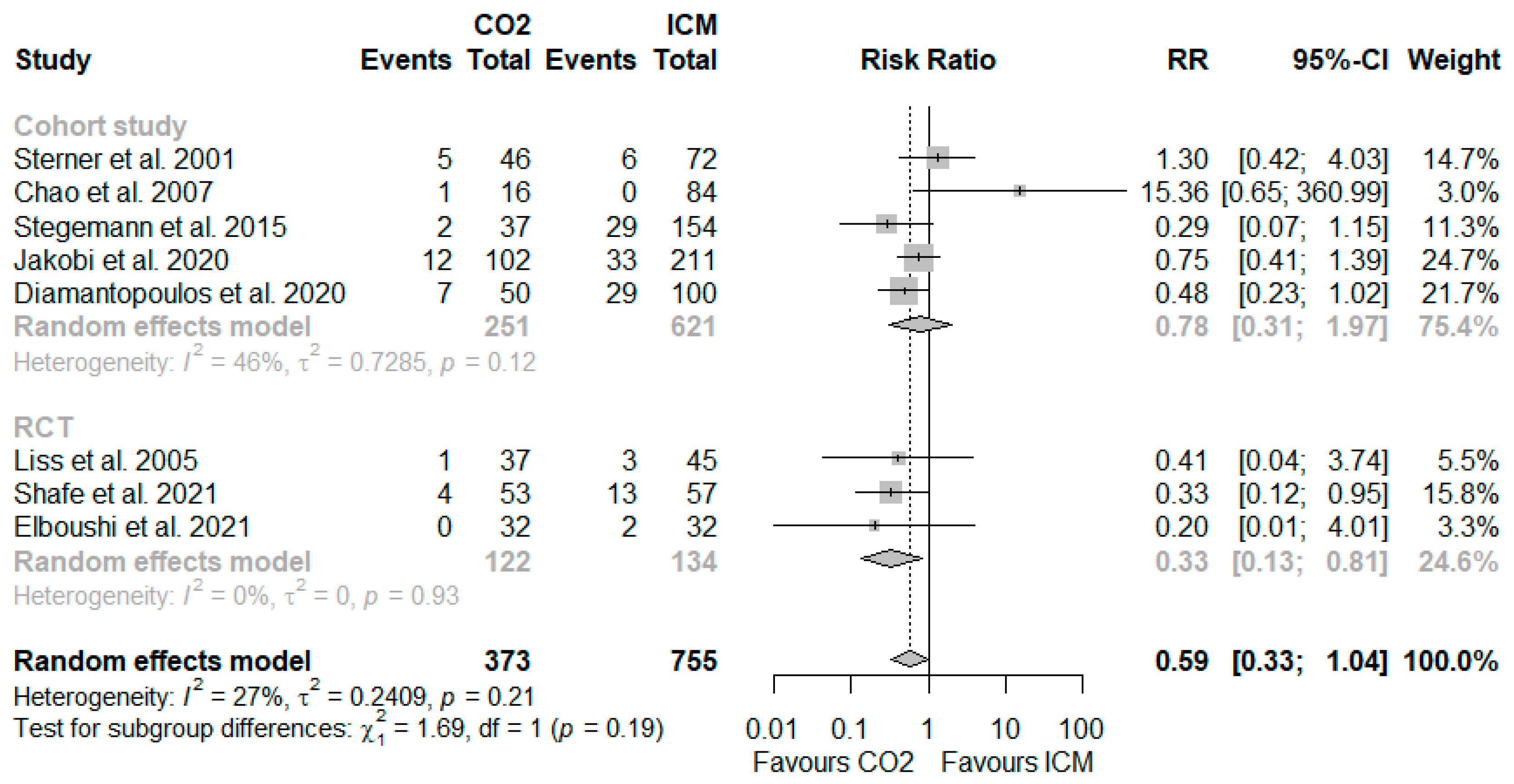
For the RCTs, we assessed the risk of bias as low for one trial [29] and as some risk of bias for two trials [30,31]. We rated two cohort studies [33,34] as moderate and three [32,35,36] as serious risk of bias. Supplementary Figures S1 and S2 show the risk of bias ratings for each individual domain of the respective assessment tools. We graded the certainty of evidence as low for the RCTs’ findings and as very low for the cohort studies’. Supplementary Table S4 presents a detailed GRADE evidence assessment with a summary of findings.

3.4. Risk of CA-AKI

Among the eight included studies with a total of 1128 participants, the risk of CA-AKI varied considerably and ranged up to 14% in the CO2 group and up to 29% in the ICM group (see Table 1). Based on a random-effects meta-analysis of eight studies, five rated as low or moderate/some risk of bias and three as serious risk of bias, the CA-AKI event rates were lower in participants receiving CO2 as a contrast agent compared to those exposed to ICM alone (8.6% vs. 15.2%; RR, 0.59; 95% CI 0.33–1.04; I2 = 27%; see Figure 2). The risk reduction was greater in a meta-analysis based on the RCTs (4.1% vs. 13.4%; RR, 0.33; 95% CI 0.13–0.81; I2 = 0%) as compared to those based on the cohort studies (10.8% vs. 15.6%; RR, 0.78; 95% CI 0.31–1.97; I2 = 46%). No patient required haemodialysis following the procedure in any of the studies reporting this outcome.
The largest study we identified was a recent single-centre prospective cohort study, including 313 participants with PAD and renal insufficiency (CKD stage 3 or higher) [34]. Overall, 102 participants who underwent PVI with CO2 were compared to 211 matched patients who received standard ICM [34]. The percentage of participants who experienced CA-AKI was lower in those who received less than 50 mL of ICM (6.8%) compared to those who received 51 to 100 mL (18.2%) or >100 mL (16.7%).
A sensitivity meta-analysis, including only low and moderate/some risk of bias studies, yielded a statistically significant lower risk of CA-AKI in participants receiving CO2 compared to those who received ICM (8.8% vs. 18.0%; RR, 0.55; 95% CI 0.36–0.83; I2 = 0%; see Supplementary Figure S3). A sensitivity analysis of the studies that predominantly included participants with impaired renal function (i.e., GFR < 60 mL/min) (RR, 0.69; 95% CI 0.45–1.06; I2 = 0%; see Supplementary Figure S4) or more than 40% patients with diabetes mellitus (RR, 0.52; 95% CI 0.35–0.79; I2 = 0%; see Supplementary Figure S5) rendered a lower risk of CA-AKI with CO2 than ICM. Supplementary Figures S6 and S7 show risk ratios in individual studies according to the type of intervention and amount of iodinated contrast medium administered in the control group.

3.5. Procedural Variables and Outcomes

In general, the technical success rate was 97% to 100% with CO2 and 95% to 100% with ICM [29,31,34,35]. Four studies reported on the procedure duration [29,32,34,35]. In the three studies comprising PAD patients undergoing PVI, the mean procedure duration with CO2 was in a similar range to that with ICM: 87 vs. 77 min [29], 83 vs. 79 min [35] and 92 vs. 102 min [34]. In a study of patients who received EVAR, the authors reported a significant difference of 3.0 vs. 2.3 h between the groups [32]. The radiation dose-area product differed substantially between studies and treatment arms [32,34,35].

3.6. Additional Adverse Events and CO2-Related Side Effects

The reporting of additional adverse events and CO2-related side effects differed considerably between studies. The number of vascular complications, however, was very low and the risk was similar in both groups. Four studies reported CO2-related side effects, such as nausea, vomiting and transient limb pain with variable frequency [30,33,34,35]. No deaths were reported during the follow-up of up to 6 months. Appendix Table A2 shows the procedural variables, additional adverse events and CO2-related side effects in detail.

4. Discussion

Based on RCTs and observational studies, this systematic review provides evidence that the use of CO2 for peripheral angiography with or without endovascular intervention reduces the risk of CA-AKI compared to conventional ICM. However, the certainty of evidence from RCTs is low because of imprecision due to the small number of events and small sample size of the included studies. The most common CO2-related adverse events included nausea, vomiting and transient leg pain. These were reported in only about half of the studies and the frequency varied widely between studies. Data on fluoroscopy time and radiation dose are also inconclusive because they were only reported in a few studies.
The findings of our review support the general concept of a renoprotective role of CO2 as an alternative contrast agent. Our results confirm the findings from a previous review and meta-analysis on the benefit of CO2 angiography, which involved substantially fewer patients [37]. In this review, the use of CO2 compared to ICM was also associated with a reduced risk of CA-AKI (4.3% vs. 11.1%, odds ratio (OR), 0.47; 95% CI 0.22–0.99) and this effect remained of the same magnitude in a relatively small subgroup analysis of four studies that included patients with CKD. However, the authors also included studies in which both the intervention and control groups received CO2 angiography with different doses of additional ICM. We used more stringent inclusion criteria comparing CO2 angiography with bailout ICM administration vs. ICM only, thus reflecting clinically relevant strategies.
The potential mechanistic pathway of the association between CA-AKI and adverse clinical events remains to be elucidated and it is still under dispute to what extent CA-AKI represents a mediator or risk marker, especially in mild AKI cases [38]. It cannot be excluded that other factors beyond ICM contribute to the observed renal impairment, and some researchers have even questioned whether ICM plays a significant role at all in the observed deterioration in kidney function [8,15]. Therefore, the term CA-AKI, rather than the previously used contrast-induced acute kidney injury, has been adopted. Importantly, CA-AKI occurred in over 8.8% of patients in the CO2 group in our meta-analysis, highlighting the role of additional harmful factors beyond the administration of ICM during vascular interventions.
Across the included studies, we observed a major difference regarding the risk of CA-AKI that could be explained by different causes. First, the different sites of peripheral angiographies (aortic, renal, infrainguinal) as diagnostic or interventional procedures required variable amounts of ICM. In addition, the complexity of the interventions has an impact on the amount of contrast agent. Second, the proportion of patients with diabetes mellitus and impaired renal function at baseline varied. Third, no uniform definition was used for CA-AKI. In particular, the time points and periods of post-procedural creatinine measurements were different. While our review supports a potential benefit of CO2 with respect to renal outcomes, we cannot draw conclusions about the frequency and severity of CO2-related side effects, as reporting was not sufficient in the included trials. Importantly, data on image quality were limited, which has been reported to be inadequate for clinical decision-making with CO2 angiography, especially for infrapopliteal interventions [39]. More research, ideally based on RCTs, is needed to describe the extent of renal protection and intraprocedural adverse events with CO2 angiography for vascular interventions.
This systematic review has the following limitations. First, we restricted our eligibility criteria to English and German publications. Second, publication bias could affect our findings since we could only rely on published data. Third, we observed heterogeneity regarding the baseline characteristics of the study participants, types of interventions and applied definition of CA-AKI.
Since most published data on the benefits of CO2 angiography focus on a small number of patients, further evidence from larger perspective RCTs is needed. There is also a lack of data regarding the association between ICM administration during PVI and clinically relevant composite outcomes, such as major adverse kidney events, consisting of persistently impaired renal function, need for new haemodialysis and death. The studies we identified rarely reported adverse events other than CA-AKI. If reported, the observation period was often very short.
In conclusion, the application of CO2 for PVI may reduce the risk of CA-AKI compared to ICM. However, a relevant residual risk of CA-AKI has been described with the use of CO2, indicating the influence of other risk factors. This should be considered when patients undergo PVI with CO2 for renal protection.

Supplementary Materials

The following supporting information can be downloaded at: https://www.mdpi.com/article/10.3390/jcm11237203/s1, Table S1: Search strategies; Table S2: Reasons for exclusion based on full-text assessment; Table S3. Inclusion and exclusion criteria as well as primary and secondary outcomes of included studies; Table S4. GRADE evidence profile and summary of findings for the comparison of CO2 and conventional ICM for peripheral angiography with or without vascular intervention; Figure S1: Risk-of-bias assessment for RCTs; Figure S2: Risk-of-bias assessment for cohort studies; Figure S3: Forest plot of the sensitivity analysis including only low and moderate/some risk of bias studies; Figure S4: Forest plot of the sensitivity analysis with studies predominantly including patients with impaired renal function (GFR < 60 mL/min); Figure S5: Forest plot of the sensitivity analysis with studies including more than 40% patients with diabetes; Figure S6: Forest plot with subgroups according to the type of intervention; Figure S7: Forest plot with subgroups according to the mean or median amount of iodinated contrast medium administered in the control group.

Author Contributions

Conceptualization, S.S.; methodology, A.G., G.W., S.S. and G.G.; literature search, I.K.; literature screening, A.G. and G.W.; study quality assessment, A.G., E.P. and G.W.; data extraction, E.P. and G.W.; meta-analysis, G.W.; writing—original draft preparation, S.S. and G.W.; writing—review and editing, A.G., D.M., E.P., G.G. and I.K.; supervision, D.M., G.G. and S.S. All authors have read and agreed to the published version of the manuscript.

Funding

This research was supported by internal funds from the Department of Evidence-based Medicine and Evaluation, University for Continuing Education Krems, Austria.

Institutional Review Board Statement

Not applicable.

Informed Consent Statement

Not applicable.

Data Availability Statement

All data analysed during this study are included in this published article and its Supplementary Materials.

Acknowledgments

We thank Edith Kertesz from the University for Continuing Education Krems for administrative support. Open Access Funding by the University for Continuing Education Krems.

Conflicts of Interest

S.S. received consulting fees from Bayer, Boston Scientific and Cook Medical as well as research funding from C.R. Bard. D.M. received speaker honoraria and consulting fees from AstraZeneca, Bayer, Boston Scientific, Novartis and Vifor. A.G., I.K., E.P, G.G. and G.W. have no conflict of interest.

Appendix A

Table A1. Study eligibility criteria.
IncludedExcluded
PopulationsAdult patients undergoing angiography of the lower limb arteries, kidney arteries or infrarenal aorta with or without endovascular interventionChildren
Angiography of other arteries (e.g., coronary angiography)
Patients who required haemodialysis prior to the intervention
InterventionApplication of CO2 with or without supplemental use of a small amount of iodinated contrast mediumAny other intervention
ComparatorApplication of conventional iodinated contrast medium onlyApplication of CO2
Any other comparator
OutcomesPrimary outcome:
Risk of contrast-associated acute kidney injury as defined by study authors
Secondary outcomes:
Need for haemodialysis after the intervention
Additional adverse events (e.g., vascular complications)
CO2-related side effects (e.g., nausea, vomiting, limb pain)
Procedural outcomes (e.g., technical success)
Post-procedural increase in creatinine/decrease in GFR without distinct definition or classification of contrast-associated acute kidney injury
Other surrogate outcomes
Study designsRCT
Non-randomised controlled trials
Controlled cohort studies
Cohort studies without control group
Narrative and systematic reviews
Case reports and case series
Publication typeFull publicationAbstracts only
Letters and editorials
Publication languageEnglish, GermanAll other languages
Abbreviations: CO2, carbon dioxide; GFR, glomerular filtration rate; RCT, randomised controlled trial.
Table A2. Additional characteristics and outcome of the included studies.
Author, Year, CountryPre-Intervention,
GFR,
Serum Creatinine
(Mean ± SD or Median [IQR])
Procedure Time
(Mean ± SD or Median [IQR])
Fluoroscopy Time,
Radiation Dose-Area
Product
(Mean ± SD or
Median [IQR])
Definition of Contrast-Induced NephropathyAdditional Adverse Events,
CO2-Related Side Effects and Procedural Outcome
n/N (%) a
Elboushi et al. 2021 [29]
Saudi Arabia and Egypt
GFR, mL/min:
CO2: NR
ICM: NR
Creatinine, mg/dL:
CO2: 0.92 ± 0.16
ICM: 0.94 ± 0.2
Procedure time, minutes:
CO2: 87 ± 22
ICM: 77 ± 28
Fluoroscopy time:
NR
Radiation dose-area product:
NR
Increase in serum creatinine exceeding 25% or more than or equal to 0.5 mg/dL within 1 month. bTechnical success rate:
CO2: 32/32 (100%)
ICM: 32/32 (100%)
No cardiac death, myocardial infarction, stroke and/or death within 3 months
Groin hematoma:
CO2: 2/31c (6.5%)
ICM: 2/32 (6.3%)
Pseudoaneurysm:
CO2: 1/31 c (3.2%)
ICM: 0/32
Major amputation:
CO2: 2/31 c (6.5%)
ICM: 3/32 (9.4%)
Shafe et al. 2021 [31]
Iran
GFR, mL/min:
CO2: 60.9 ± 22.0
ICM: 74.7 ± 23.6
Creatinine, mg/dL:
CO2: 1.46 ± 0.45
ICM: 1.13 ± 0.28
Procedure time:
NR
Fluoroscopy time:
NR
Radiation dose-area product:
NR
Increase in serum creatinine exceeding 25% or 0.5 mg/dL within 72 h after the procedureTechnical success rate:
CO2: 53/53 (100%)
ICM: 57/57 (100%)
Lower-limb pain:
CO2: 12/53 (22.6%)
ICM: 0/57
Major vascular complications:
CO2: 0/53
ICM: 0/57
Death:
CO2: 0/53
ICM: 0/57
Liss et al. 2005 [30]
Sweden
GFR, mL/min:
CO2: 54 ± 22
ICM: 59 ± 29
Creatinine, mg/dL: d
CO2: 1.45 ± 0.43
ICM: 1.36 ± 0.42
Procedure time:
NR
Fluoroscopy time:
NR
Radiation dose-area product:
NR
Increase in serum creatinine by >25% within one week after the procedureVomiting:
CO2: 1/37 (27.0%)
ICM: 0/45
Nausea:
CO2: 8/37 (21.6%)
ICM: 1/45 (2.2%)
Sterner et al. 2001 [36]
Sweden
GFR, mL/min:
CO2: NR
ICM: NR
Creatinine, mg/dL e,f
CO2: 2.57 [NR]; 3.71 [NR]
ICM: 1.98 [NR]
Procedure time:
NR
Fluoroscopy time:
NR
Radiation dose-area product:
NR
Increase in serum creatinine by >25% within two weeks after the procedure NR
Chao et al. 2007 [32]
USA
GFR, mL/min:
CO2: 36 ± NR
ICM: 81 ± NR
Creatinine, mg/dL:
CO2: 1.8 ± NR g
ICM: 1.0 ± NR
Procedure time, hours:
CO2: 3.0 ± 0.3
ICM: 2.3 ± 0.2
Fluoroscopy time, minutes:
CO2: 46 ± 7
ICM: 24 ± 1.5
Radiation dose-area product, cGy.cm2:
CO2: 92,500 ± 13,800
ICM: 52,900 ± 4400
Increase in serum creatinine by >20% within 24 h after the procedureMorbidity:
CO2: 2/16 (12%)
ICM: 5/84 (6%)
Death:
CO2: 0/16
ICM: 0/84
Stegemann et al. 2015 [35]
Germany
GFR, mL/min:
CO2: 22 ± 34
ICM: 76 ± 28
Creatinine, mg/dL:
CO2: 2.1 ± 1.3
ICM: 1.1 ± 0.6
Procedure time, minutes:
CO2: 83 + 32
ICM: 79 + 37
Fluoroscopy time, minutes:
CO2: 22 + 14
ICM: 23 + 17
Radiation dose-area product, cGy.cm2:
CO2: 8054 + 12,764
ICM: 9359 + 11,474
Increase in serum creatinine by >25% or >0.5 mg/dL within 48 h after the procedureTechnical success rate:
CO2: 37/37 (100%)
ICM: 148/154 (96%)
Nausea:
CO2: 1/37 (2.7%)
ICM: 0/154
Several patients
described temporary acute ischaemic lower leg pain following both ICM and CO2 injection.
Diamantopoulus et al. 2020 [33]
England
GFR, mL/min:
CO2: 38.6 ± 13.2
ICM: 43.3 ± 12.2
Creatinine, mg/dL:f
CO2: 1.7 ± 0.55
ICM: 1.54 ± 0.52
Procedure time:
NR
Fluoroscopy time:
NR
Radiation dose-area product:
NR
Increase in serum creatinine by >25% or >0.5 mg/dL within 72 h after the procedureThere were no major complications associated with the use of CO2. Most of the CLI cases reported transient discomfort (seconds) at the level of the symptomatic foot.Major complications:
CO2: 0/50
ICM: NR
Jakobi et al. 2021 [34]
Germany
GFR, mL/min:
CO2: 32.4 ± 11.8
ICM: 33.1 ± 15.6
Creatinine, mg/dL:
CO2: NR
ICM: NR
Procedure time, minutes:
CO2: 92.3 ± 35
ICM: 101.8 ± 47.2
Fluoroscopy time:
NR
Radiation dose-area product, cGy.cm2:
CO2: 6025 ± 6926
ICM: 4281 ± 4722
Increase in serum creatinine by a factor of 1.5 to 1.9 or ≥0.3 mg/dL within 48 h after the procedureTechnical success rate:
CO2: 99/102 (97%)
ICM: 201/211 (95.3%)
Severe procedure-related complications:
CO2: 0/102
ICM: 0/211
Transient leg pain:
CO2: 15/102 (14.7%)
ICM: 0/211
Abbreviations: CLI, critical limb ischaemia; CO2, carbon dioxide; cGy, centigray; GFR, glomerular filtration rate; ICM, iodinated contrast medium; IQR, interquartile range; NR, not reported; n, number of patients with events; N, number of patients at risk; mg, milligram; PAD, peripheral arterial disease; SD, standard deviation. a: Percentages or number of events were self-calculated if not reported in the publication. b: Timeframe not included in the definition, but creatinine was measured up to 1 month after the procedure. c: One patient from the CO2 group was lost to follow-up. d: Only patients with a serum creatinine lower than 2.3 mg/dL (200 µmol/L) were included. e: Only patients with a serum creatinine of 1.7 mg/dL (150 µmol/L) or higher were included. f: Creatinine was reported in µmol/L and converted to mg/dL. g: Only patients with a serum creatinine of 1.5 mg/dL or higher were included in the CO2 group.

References

  1. Malyar, N.; Furstenberg, T.; Wellmann, J.; Meyborg, M.; Luders, F.; Gebauer, K.; Bunzemeier, H.; Roeder, N.; Reinecke, H. Recent trends in morbidity and in-hospital outcomes of in-patients with peripheral arterial disease: A nationwide population-based analysis. Eur. Heart J. 2013, 34, 2706–2714. [Google Scholar] [CrossRef] [PubMed] [Green Version]
  2. Latus, J.; Schwenger, V.; Schlieper, G.; Reinecke, H.; Hoyer, J.; Persson, P.B.; Remppis, B.A.; Mahfoud, F. Contrast medium-induced acute kidney injury-Consensus paper of the working group “Heart and Kidney” of the German Cardiac Society and the German Society of Nephrology. Internist 2021, 62, 111–120. [Google Scholar] [CrossRef] [PubMed]
  3. Lee, S.R.; Zhuo, H.; Zhang, Y.; Dahl, N.; Dardik, A.; Ochoa Chaar, C.I. Risk factors and safe contrast volume thresholds for postcontrast acute kidney injury after peripheral vascular interventions. J. Vasc. Surg. 2020, 72, 603–610.e601. [Google Scholar] [CrossRef] [PubMed]
  4. Prasad, A.; Hughston, H.; Michalek, J.; Trevino, A.; Gupta, K.; Martinez, J.P.; Hoang, D.T.; Wu, P.B.; Banerjee, S.; Masoomi, R. Acute kidney injury in patients undergoing endovascular therapy for critical limb ischemia. Catheter. Cardiovasc. Interv. 2019, 94, 636–641. [Google Scholar] [CrossRef]
  5. Mehran, R.; Dangas, G.D.; Weisbord, S.D. Contrast-Associated Acute Kidney Injury. N. Engl. J. Med. 2019, 380, 2146–2155. [Google Scholar] [CrossRef]
  6. Coca, S.G.; Yusuf, B.; Shlipak, M.G.; Garg, A.X.; Parikh, C.R. Long-term risk of mortality and other adverse outcomes after acute kidney injury: A systematic review and meta-analysis. Am. J. Kidney Dis. 2009, 53, 961–973. [Google Scholar] [CrossRef] [Green Version]
  7. See, E.J.; Jayasinghe, K.; Glassford, N.; Bailey, M.; Johnson, D.W.; Polkinghorne, K.R.; Toussaint, N.D.; Bellomo, R. Long-term risk of adverse outcomes after acute kidney injury: A systematic review and meta-analysis of cohort studies using consensus definitions of exposure. Kidney Int. 2019, 95, 160–172. [Google Scholar] [CrossRef]
  8. Katsogridakis, E.; Lea, T.; Yap, T.; Batchelder, A.; Saha, P.; Diamantopoulos, A.; Saratzis, N.; Davies, R.; Zayed, H.; Bown, M.J.; et al. Acute kidney injury following endovascular intervention for peripheral artery disease. Br. J. Surg. 2021, 108, 152–159. [Google Scholar] [CrossRef]
  9. Prasad, A.; Ortiz-Lopez, C.; Khan, A.; Levin, D.; Kaye, D.M. Acute kidney injury following peripheral angiography and endovascular therapy: A systematic review of the literature. Catheter. Cardiovasc. Interv. 2016, 88, 264–273. [Google Scholar] [CrossRef]
  10. Safley, D.M.; Salisbury, A.C.; Tsai, T.T.; Secemsky, E.A.; Kennedy, K.F.; Rogers, R.K.; Latif, F.; Shammas, N.W.; Garcia, L.; Cavender, M.A.; et al. Acute Kidney Injury Following In-Patient Lower Extremity Vascular Intervention: From the National Cardiovascular Data Registry. JACC Cardiovasc. Interv. 2021, 14, 333–341. [Google Scholar] [CrossRef]
  11. Tsai, T.T.; Patel, U.D.; Chang, T.I.; Kennedy, K.F.; Masoudi, F.A.; Matheny, M.E.; Kosiborod, M.; Amin, A.P.; Messenger, J.C.; Rumsfeld, J.S.; et al. Contemporary incidence, predictors, and outcomes of acute kidney injury in patients undergoing percutaneous coronary interventions: Insights from the NCDR Cath-PCI registry. JACC Cardiovasc. Interv. 2014, 7, 1–9. [Google Scholar] [CrossRef] [PubMed] [Green Version]
  12. Fähling, M.; Seeliger, E.; Patzak, A.; Persson, P.B. Understanding and preventing contrast-induced acute kidney injury. Nat. Rev. Nephrol. 2017, 13, 169–180. [Google Scholar] [CrossRef] [PubMed]
  13. Nijssen, E.C.; Rennenberg, R.J.; Nelemans, P.J.; Essers, B.A.; Janssen, M.M.; Vermeeren, M.A.; Ommen, V.V.; Wildberger, J.E. Prophylactic hydration to protect renal function from intravascular iodinated contrast material in patients at high risk of contrast-induced nephropathy (AMACING): A prospective, randomised, phase 3, controlled, open-label, non-inferiority trial. Lancet 2017, 389, 1312–1322. [Google Scholar] [CrossRef]
  14. Weisbord, S.D.; Gallagher, M.; Jneid, H.; Garcia, S.; Cass, A.; Thwin, S.S.; Conner, T.A.; Chertow, G.M.; Bhatt, D.L.; Shunk, K.; et al. Outcomes after Angiography with Sodium Bicarbonate and Acetylcysteine. N. Engl. J. Med. 2018, 378, 603–614. [Google Scholar] [CrossRef] [PubMed]
  15. McDonald, J.S.; McDonald, R.J.; Comin, J.; Williamson, E.E.; Katzberg, R.W.; Murad, M.H.; Kallmes, D.F. Frequency of acute kidney injury following intravenous contrast medium administration: A systematic review and meta-analysis. Radiology 2013, 267, 119–128. [Google Scholar] [CrossRef]
  16. Wilhelm-Leen, E.; Montez-Rath, M.E.; Chertow, G. Estimating the Risk of Radiocontrast-Associated Nephropathy. J. Am. Soc. Nephrol. 2017, 28, 653–659. [Google Scholar] [CrossRef] [Green Version]
  17. Sharafuddin, M.J.; Marjan, A.E. Current status of carbon dioxide angiography. J. Vasc. Surg. 2017, 66, 618–637. [Google Scholar] [CrossRef]
  18. Page, M.J.; McKenzie, J.E.; Bossuyt, P.M.; Boutron, I.; Hoffmann, T.C.; Mulrow, C.D.; Shamseer, L.; Tetzlaff, J.M.; Akl, E.A.; Brennan, S.E.; et al. The PRISMA 2020 statement: An updated guideline for reporting systematic reviews. BMJ 2021, 372, n71. [Google Scholar] [CrossRef]
  19. Veritas Health Innovation. Covidence Systematic Review Software; Veritas Health Innovation: Melbourne, Australia, 2021; Available online: www.covidence.org (accessed on 20 October 2020).
  20. Sterne, J.A.C.; Savović, J.; Page, M.J.; Elbers, R.G.; Blencowe, N.S.; Boutron, I.; Cates, C.J.; Cheng, H.Y.; Corbett, M.S.; Eldridge, S.M.; et al. RoB 2: A revised tool for assessing risk of bias in randomised trials. BMJ 2019, 366, l4898. [Google Scholar] [CrossRef] [Green Version]
  21. Sterne, J.A.; Hernán, M.A.; Reeves, B.C.; Savović, J.; Berkman, N.D.; Viswanathan, M.; Henry, D.; Altman, D.G.; Ansari, M.T.; Boutron, I.; et al. ROBINS-I: A tool for assessing risk of bias in non-randomised studies of interventions. BMJ 2016, 355, i4919. [Google Scholar] [CrossRef]
  22. Guyatt, G.H.; Oxman, A.D.; Vist, G.E.; Kunz, R.; Falck-Ytter, Y.; Alonso-Coello, P.; Schünemann, H.J. GRADE: An emerging consensus on rating quality of evidence and strength of recommendations. BMJ 2008, 336, 924–926. [Google Scholar] [CrossRef] [PubMed] [Green Version]
  23. McMaster University and Evidence Prime. GRADEpro GDT: GRADEpro Guideline Development Tool; McMaster University and Evidence Prime: Hamilton, ON, CAN, 2022; Available online: gradepro.org (accessed on 20 October 2020).
  24. Paule, R.C.; Mandel, J. Consensus Values and Weighting Factors. J. Res. Natl. Bur. Stand. 1982, 87, 377–385. [Google Scholar] [CrossRef] [PubMed]
  25. Higgins, J.P.; Thompson, S.G. Quantifying heterogeneity in a meta-analysis. Stat. Med. 2002, 21, 1539–1558. [Google Scholar] [CrossRef] [PubMed]
  26. Balduzzi, S.; Rücker, G.; Schwarzer, G. How to perform a meta-analysis with R: A practical tutorial. Evid. Based Ment. Health 2019, 22, 153–160. [Google Scholar] [CrossRef] [PubMed] [Green Version]
  27. RStudio Team. RStudio: Integrated Development Environment for R; RStudio, PBC: Boston, MA, USA, 2021. [Google Scholar]
  28. R Core Team. R: A Language and Environment for Statistical Computing; R Foundation for Statistical Computing: Vienna, Austria, 2021. [Google Scholar]
  29. Elboushi, A.; Tawfik, A.; Abouissa, A.; Harraz, M.; El-Laboudy, M. Carbon dioxide versus iodine contrast medium for endovascular revascularization of aortoiliac occlusive disease: A two-center randomized controlled trial. Egypt. J. Surg. 2021, 40, 264–271. [Google Scholar] [CrossRef]
  30. Liss, P.; Eklöf, H.; Hellberg, O.; Hägg, A.; Boström-Ardin, A.; Löfberg, A.M.; Olsson, U.; Orndahl, P.; Nilsson, H.; Hansell, P.; et al. Renal effects of CO2 and iodinated contrast media in patients undergoing renovascular intervention: A prospective, randomized study. J. Vasc. Interv. Radiol. 2005, 16, 57–65. [Google Scholar] [CrossRef]
  31. Shafe, O.; Samiee, E.; Moosavi, J.; Mohebbi, B.; Fakhrabadi, A.; Bakhshandeh, H.; Naghavi, B.; Tahivili, R.; Cheraghi, S.; Sadeghipour, P. Comparison Between Intra-Arterial Carbon Dioxide and Iodinated Contrast Agent Injections in Patients with Lower-Limb Peripheral Arterial Diseases and Mild-to-Moderate Renal Dysfunction: A Randomized Controlled Trial. J. Crit. Limb. Ischem. 2021, 1, E104–E109. [Google Scholar]
  32. Chao, A.; Major, K.; Kumar, S.R.; Patel, K.; Trujillo, I.; Hood, D.B.; Rowe, V.L.; Weaver, F.A. Carbon dioxide digital subtraction angiography-assisted endovascular aortic aneurys.sm.m repair in the azotemic patient. J. Vasc. Surg. 2007, 45, 451–458; discussion 458–460. [Google Scholar] [CrossRef] [Green Version]
  33. Diamantopoulos, A.; Patrone, L.; Santonocito, S.; Theodoulou, I.; Ilyas, S.; Dourado, R.; Game, D.; Karunanithy, N.; Patel, S.; Zayed, H.; et al. Carbon dioxide angiography during peripheral angioplasty procedures significantly reduces the risk of contrast-induced nephropathy in patients with chronic kidney disease. CVIR Endovasc. 2020, 3, 9. [Google Scholar] [CrossRef] [Green Version]
  34. Jakobi, T.; Meyborg, M.; Freisinger, E.; Gebauer, K.; Stella, J.; Engelbertz, C.; Reinecke, H.; Malyar, N.M. Feasibility and impact of carbon dioxide angiography on acute kidney injury following endovascular interventions in patients with peripheral artery disease and renal impairment. J. Nephrol. 2021, 8, 8. [Google Scholar] [CrossRef]
  35. Stegemann, E.; Tegtmeier, C.; Bimpong-Buta, N.Y.; Sansone, R.; Uhlenbruch, M.; Richter, A.; Stegemann, B.; Roden, M.; Westenfeld, R.; Kelm, M.; et al. Carbondioxide-Aided Angiography Decreases Contrast Volume and Preserves Kidney Function in Peripheral Vascular Interventions. Angiology 2016, 67, 875–881. [Google Scholar] [CrossRef] [PubMed]
  36. Sterner, G.; Nyman, U.; Valdes, T. Low risk of contrast-medium-induced nephropathy with modern angiographic technique. J. Intern. Med. 2001, 250, 429–434. [Google Scholar] [CrossRef] [PubMed] [Green Version]
  37. Ghumman, S.S.; Weinerman, J.; Khan, A.; Cheema, M.S.; Garcia, M.; Levin, D.; Suri, R.; Prasad, A. Contrast induced-acute kidney injury following peripheral angiography with carbon dioxide versus iodinated contrast media: A meta-analysis and systematic review of current literature. Catheter. Cardiovasc. Interv. 2017, 90, 437–448. [Google Scholar] [CrossRef]
  38. Weisbord, S.D.; Palevsky, P.M.; Kaufman, J.S.; Wu, H.; Androsenko, M.; Ferguson, R.E.; Parikh, C.R.; Bhatt, D.L.; Gallagher, M. Contrast-Associated Acute Kidney Injury and Serious Adverse Outcomes Following Angiography. J. Am. Coll. Cardiol. 2020, 75, 1311–1320. [Google Scholar] [CrossRef] [PubMed]
  39. Gupta, A.; Dosekun, A.K.; Kumar, V. Carbon dioxide-angiography for patients with peripheral arterial disease at risk of contrast-induced nephropathy. World J. Cardiol. 2020, 12, 76–90. [Google Scholar] [CrossRef] [PubMed]
Figure 1. Preferred Reporting Items for Systematic Reviews and Meta-Analyses (PRISMA) flow-chart adapted from Page et al. 2021 [18].
Jcm 11 07203 g001
Figure 2. Forest plot for the risk of CA-AKI with CO2 compared to ICM [29,30,31,32,33,34,35,36]. Abbreviations: CA-AKI, contrast-associated acute kidney injury; CI, confidence interval; CO2, carbon dioxide; ICM, iodinated contrast medium; RCT, randomised controlled trial; RR, risk ratio.
Jcm 11 07203 g002
Table 1. Key characteristics and outcome of the included studies.
Author, Year, CountryStudy Design
Risk of Bias
Follow-Up
NAge, Years,
Mean (SD)
WomenDiabetes MellitusCKD
Stage 3–5
Condition/Site and InterventionCO2 and ICM Amount,
ICM Type,
(Mean ± SD or
Median [IQR])
CA-AKI,
Haemodialysis
n/N (%) a
Elboushi et al. 2021 [29]
Saudi Arabia and Egypt
RCT
Low
3 months b
Total: 64
CO2: 32
ICM: 32
Age:
CO2: 54.3 ± 9.8
ICM: 56.3 ± 9.7
Women:
CO2: 29.0%
ICM: 53.1%
Diabetes mellitus:
All: 45.3%
CO2: 55.0%
ICM: 37.5%
CKD stage 3–5:
CO2: 0
ICM: 0
PAD
Angiography with aortoiliac endovascular intervention
CO2 amount, mL:
CO2: 171 [NR]
ICM: 0
ICM amount, mL:
CO2: 10 [NR] (N = 3)
ICM: 78 [NR]
ICM type:
Iohexol
CA-AKI:
CO2: 0/32
ICM: 2/32 (6.3%)
Haemodialysis:
NR
Shafe et al. 2021 [31]
Iran
RCT
Some
concerns
1 month
Total: 110
CO2: 53
ICM: 57
Age:
CO2: 62.5 ± 8.4
ICM: 63.3 ± 11.7
Women:
CO2: 24.5%
ICM: 19.3%
Diabetes mellitus:
All: 45%
CO2: 51%
ICM: 40%
CKD stage 3–5:
CO2: NR
ICM: NR
PAD
Angiography without (29%) or with aortoiliac, femoropopliteal or infrapopliteal endovascular intervention (71%)
CO2 amount, mL:
CO2: NR
ICM: 0
ICM amount, mL:
CO2: 11.4 ± 6.1
ICM: 93.2 ± 43.0
ICM type:
NR
CA-AKI:
CO2: 4/53 (7.5%)
ICM: 13/57 (22.8%)
Haemodialysis:
CO2: 0/53
ICM: 0/57
Liss et al. 2005 [30]
Sweden
RCT
Some
concerns
3 weeks
Total: 82
CO2: 37
ICM: 45
Age:
CO2: 67 ± 8
ICM: 63 ± 11
Women:
CO2: NR
ICM: NR
Diabetes mellitus:
All: 17%
CO2: 27%
ICM: 9%
CKD stage 3–5:
CO2: NR
ICM: NR
Renal arteries
Angiography with or without endovascular intervention
CO2 amount, mL:
CO2: 191 ± 118
ICM: 0
ICM amount, mL:
CO2: 35.1 ± 6.4
ICM: 88.4 ± 42.9
ICM type:
Ioxaglate
CA-AKI:
CO2: 1/37 (2.7%)
ICM: 3/45 (6.7%)
Haemodialysis:
CO2: 0/37
ICM: 1/45 (2.2%)
Sterner et al. 2001 [36]
Sweden
Cohort study
Serious
2 weeks
Total: 118
CO2: 46 c
ICM: 72 d
Age:
CO2: 71 ± NR
ICM: 72 ± NR
Women:
CO2: 15%
ICM: 33%
Diabetes mellitus:
All: 21%
CO2: 20%
ICM: 22%
CKD stage 3–5:
CO2: NR
ICM: NR
PAD,
Renal and mesenteric arteries d
Angiography with or without endovascular intervention
CO2 amount, mL:
CO2: NR
ICM: 0
ICM amount, mL:
CO2: 5 [NR]
ICM: 22 [NR]
ICM type:
Iohexol
CA-AKI:
CO2: 5/46 (10.9%)
ICM: 6/72 (8.3%)
Haemodialysis:
NR
Chao et al. 2007 [32]
USA
Cohort study
Serious
6 months
Total: 100
CO2: 16
ICM: 84
Age:
CO2: 77 ± NR
ICM: 76 ± NR
Women:
CO2: 6%
ICM: 18%
Diabetes mellitus:
All: 13%
CO2: 20%
ICM: 12%
CKD stage 3–5:
CO2: 88%
ICM: 34%
Abdominal aortic aneurysm
EVAR
CO2 amount, mL:
CO2: 50 ± NR
ICM: 0
ICM amount, mL:
CO2: 27 ± 5
ICM: 148 ± 20
ICM type:
Iopamidol
CA-AKI:
CO2: 1/16 (6.3%)
ICM: 0/84
Haemodialysis:
CO2: 0/16
ICM: 0/84
Stegemann et al. 2015 [35]
Germany
Cohort study
Serious
NR
Total: 191
CO2: 37
ICM: 154
Age:
CO2: 70 ± 10
ICM: 73 ± 12
Women:
CO2: 38%
ICM: 23%
Diabetes mellitus:
All: 51%
CO2: 51%
ICM: 51%
CKD stage 3–5:
CO2: 86%
ICM: 29%
PAD
Endovascular intervention (aortoiliac, femoropopliteal, below-the-knee)
CO2 amount, mL:
CO2: NR
ICM: 0
ICM amount, mL:
CO2: 34 ± 41
ICM: 112 ± 76
ICM type:
Iodixanol
CA-AKI:
CO2: 2/37 (5%)
ICM: 29/154 (19%)
Haemodialysis:
CO2: 0/37
ICM: 0/154
Diamantopoulus et al. 2020 [33]
England
Cohort study
Moderate
30 days
Total: 150
CO2: 50
ICM: 100
Age:
CO2: 77.5 ± 10.4
ICM: 76.5 ± 10.5
Women:
CO2: NR
ICM: NR
Diabetes mellitus:
All: 65%
CO2: 66%
ICM: 64%
CKD stage 3–5:
CO2: 100%
ICM: 100%
PAD
Endovascular intervention
(aortoiliac, femoropopliteal, below-the-knee)
CO2 amount, mL:
CO2: NR
ICM: 0
ICM amount, mL:
CO2: 15.1 ± 14.0
ICM: 115.6 ± 58.1
ICM type:
Iodixanol
CA-AKI:
CO2: 7/50 (14%)
ICM: 29/100 (29%)
Haemodialysis:
NR
Jakobi et al. 2021 [34]
Germany
Cohort study
Moderate
48 h
Total: 313
CO2: 102
ICM: 211
Age:
CO2: 74.8 ± 8.7
ICM: 72.4 ± 9.3
Women:
CO2: 36.3%
ICM: 24.6%
Diabetes mellitus:
All: 52%
CO2: 51%
ICM: 54%
CKD stage 3–5:
CO2: 100%
ICM: 100%
PAD
Endovascular intervention (aortoiliac, femoropopliteal, below-the-knee)
CO2 amount, mL:
CO2: 114.5 ± 53.4
ICM: 0
ICM amount, mL:
CO2: 41.9 ± 31.6
(N = 86)
ICM: 118.9 ± 51.1
ICM type:
NR
CA-AKI:
CO2: 12/102 (11.8%)
ICM: 33/211 (15.6%)
Haemodialysis:
NR
Abbreviations: CA-AKI, contrast-associated acute kidney injury; CKD, chronic kidney disease; CO2, carbon dioxide; EVAR, endovascular aortic aneurysm repair; ICM, iodinated contrast medium; IQR, interquartile range; NR, not reported; n, number of patients with events; N, number of patients at risk; mg, milligram; PAD, peripheral arterial disease; SD, standard deviation. a: Percentages or number of events were self-calculated if not reported in the publication. b: Based on the definition of outcomes. c: Patients from group B (iodine + CO2) and group C (CO2) were added. d: Patients in the control group underwent coronary (n = 25), pulmonary (n = 6), renal (n = 7), aortofemoral (n = 29), aortocervical (n = 4) and mesenteric. (n = 1) percutaneous endoluminal diagnosis and therapy procedures.
Publisher’s Note: MDPI stays neutral with regard to jurisdictional claims in published maps and institutional affiliations.

