
Human Primary Odontoblast-like Cell Cultures—A Focused Review Regarding Cell Characterization


Abstract
:1. Introduction
2. Methods
2.1. Search Strategy
2.2. Inclusion and Exclusion Criteria
2.3. Data Extraction
3. Results
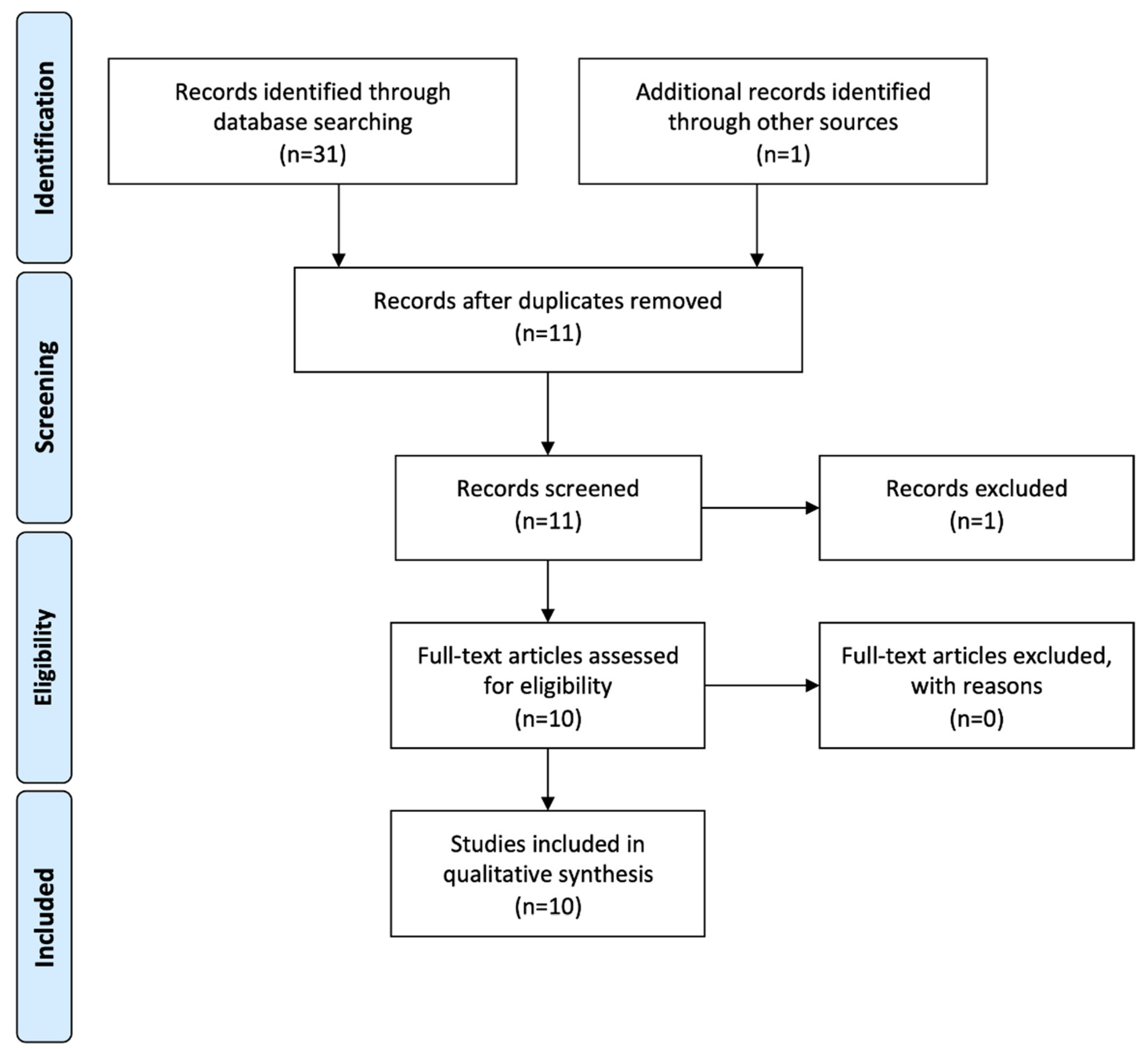
3.1. Literature Search and Screening Process
3.2. Cell Culture
3.3. Mesenchymal Stem Cell Screening
3.4. Induction Medium
3.5. Mineralization
3.6. Characterization
3.7. Immunohistochemistry and Western Blotting
3.8. RT-PCR
3.9. RT-(q)PCR Parameters
4. Discussion
4.1. Cell Characterization
4.2. Strengths and Limitations of This Review
5. Conclusions and Recommendations
- When extracting third molars, the apex should still be open and root growth should not yet be completed, regardless of the age of the donor. This ensures that the odontoblast layer still forms primary dentin [77] and that a significant expression profile can be identified.
- Total RNA for reference purposes should be extracted from both the odontoblast layer and the pulp tissue of each tooth from which a cell culture is established.
- RT-qPCR of all SIBLINGS, BGLAP, SPARC, ALPL and COL1A1 as target genes should be performed not only for the differentiated and undifferentiated cell cultures but also for the odontoblast layer and pulp tissue of the respective tooth and reported according to MIQE guidelines [19].
- For undifferentiated cell cultures, at least the expression of the surface antigens should be demonstrated from the criteria for mesenchymal stem cells of the International Society for Cellular Therapy [34].
- The mineralization of the differentiated cell cultures should be documented by a suitable staining method.
- The number of biological replicates (i.e., donors) should be reported unambiguously.
- For better reader comprehension and to ensure repeatability of the experiments, the complete methodology of the cell culture and its characterization, including the quality-assurance measures, should be given separately and as a summary for each cell culture/donor, without references to other publications and their results.
- To ensure the comparability of results, international societies (e.g., European Society of Endodontology, American Association of Endodontists) should define minimum standards for the establishment of odontoblast-like cell cultures and their characterization.
Author Contributions
Funding
Institutional Review Board Statement
Informed Consent Statement
Data Availability Statement
Conflicts of Interest
References
- Gstraunthaler, G.; Lindl, T. Zell- und Gewebekultur, 7th ed.; Springer Spektrum: Berlin/Heidelberg, Germany, 2013; p. 354. [Google Scholar] [CrossRef]
- Nino-Barrera, J.L.; Gutierrez, M.L.; Garzon-Alvarado, D.A. A theoretical model of dentinogenesis: Dentin and dentinal tubule formation. Comput. Methods Programs Biomed. 2013, 112, 219–227. [Google Scholar] [CrossRef] [PubMed]
- Freshney, R.I. Subculture and Cell Lines. In Culture of Animal Cells, 6th ed.; Wiley-Blackwell: Hoboken, NJ, USA, 2011; Chapter 12; pp. 187–206. [Google Scholar] [CrossRef]
- Freshney, R.I. Characterization. In Culture of Animal Cells, 6th ed.; Wiley-Blackwell: Hoboken, NJ, USA, 2011; Chapter 15; pp. 239–268. [Google Scholar] [CrossRef]
- Widbiller, M.; Bucchi, C.; Rosendahl, A.; Spanier, G.; Buchalla, W.; Galler, K.M. Isolation of primary odontoblasts: Expectations and limitations. Aust. Endod. J. 2019, 45, 378–387. [Google Scholar] [CrossRef] [PubMed]
- Cuffaro, H.M.; Paakkonen, V.; Tjaderhane, L. Enzymatic isolation of viable human odontoblasts. Int. Endod. J. 2016, 49, 454–461. [Google Scholar] [CrossRef] [PubMed]
- Sun, S.; Wang, G.-L.; Huang, Y.; Diwu, H.-L.; Luo, Y.-C.; Su, J.; Xiao, Y.-H. The effects of 2-hydroxyethyl methacrylate on matrix metalloproteinases 2 and 9 in human pulp cells and odontoblast-like cells in vitro. Int. Endod. J. 2018, 51 (Suppl. 2), e157–e166. [Google Scholar] [CrossRef] [PubMed]
- Baldion, P.A.; Cortes, C.C.; Castellanos, J.E.; Betancourt, D.E. Effect of myricetin on odontoblast-like cells and its potential to preserve resin-dentin Bonds. J. Mech. Behav. Biomed. Mater. 2021, 117, 104392. [Google Scholar] [CrossRef]
- Baldion, P.A.; Velandia-Romero, M.L.; Castellanos, J.E. Dental resin monomers induce early and potent oxidative damage on human odontoblast-like cells. Chem. Biol. Interact. 2021, 333, 109336. [Google Scholar] [CrossRef]
- Latorre, K.L.; Baldion, P.A. Polymodal Activation and Desensitization of TRPV1 Receptor in Human Odontoblasts-Like Cells with Eugenol. Int. J. Dent. 2020, 2020, 8813979. [Google Scholar] [CrossRef]
- Sabandal, M.M.I.; Schäfer, E.; Imper, J.; Jung, S.; Kleinheinz, J.; Sielker, S. Simvastatin Induces In Vitro Mineralization Effects of Primary Human Odontoblast-Like Cells. Materials 2020, 13, 4679. [Google Scholar] [CrossRef]
- Wen, W.; Que, K.; Zang, C.; Wen, J.; Sun, G.; Zhao, Z.; Li, Y. Expression and distribution of three transient receptor potential vanilloid (TRPV) channel proteins in human odontoblast-like cells. J. Mol. Histol. 2017, 48, 367–377. [Google Scholar] [CrossRef]
- Meng, R.; Li, D.; Feng, Z.; Xu, Q. MyD88 hypermethylation mediated by DNMT1 is associated with LTA-induced inflammatory response in human odontoblast-like cells. Cell Tissue Res. 2019, 376, 413–423. [Google Scholar] [CrossRef]
- Liu, J.; Que, K.; Liu, Y.; Zang, C.; Wen, J. Tumor Necrosis Factor-α Regulates the TRPA1 Expression in Human Odontoblast-Like Cells. J. Pain Res. 2020, 13, 1655–1664. [Google Scholar] [CrossRef] [PubMed]
- Feng, Z.; Meng, R.; Li, Q.; Li, D.; Xu, Q. 5-aza-2′-deoxycytidine may regulate the inflammatory response of human odontoblast-like cells through the NF-κB pathway. Int. Endod. J. 2021, 54, 1105–1117. [Google Scholar] [CrossRef] [PubMed]
- Liu, Y.; Wang, Y.; Lou, Y.; Tian, W.; Que, K. Functional expression of TRPA1 channel, TRPV1 channel and TMEM100 in human odontoblasts. J. Mol. Histol. 2021, 52, 1105–1114. [Google Scholar] [CrossRef] [PubMed]
- Liberati, A.; Altman, D.G.; Tetzlaff, J.; Mulrow, C.; Gotzsche, P.C.; Ioannidis, J.P.; Clarke, M.; Devereaux, P.J.; Kleijnen, J.; Moher, D. The PRISMA statement for reporting systematic reviews and meta-analyses of studies that evaluate health care interventions: Explanation and elaboration. PLoS Med. 2009, 6, e1000100. [Google Scholar] [CrossRef] [PubMed]
- Tjäderhane, L.; Salo, T.; Larjava, H.; Larmas, M.; Overall, C.M. A novel organ culture method to study the function of human odontoblasts in vitro: Gelatinase expression by odontoblasts is differentially regulated by TGF-β1. J. Dent. Res. 1998, 77, 1486–1496. [Google Scholar] [CrossRef] [PubMed]
- Bustin, S.A.; Benes, V.; Garson, J.A.; Hellemans, J.; Huggett, J.; Kubista, M.; Mueller, R.; Nolan, T.; Pfaffl, M.W.; Shipley, G.L.; et al. The MIQE guidelines: Minimum information for publication of quantitative real-time PCR experiments. Clin. Chem. 2009, 55, 611–622. [Google Scholar] [CrossRef] [PubMed]
- Xiong, H.; Chen, K. Multipotent stem cells from apical pulp of human deciduous teeth with immature apex. Tissue Cell 2021, 71, 101556. [Google Scholar] [CrossRef]
- Baldion, P.A.; Velandia-Romero, M.L.; Castellanos, J.E. Odontoblast-Like Cells Differentiated from Dental Pulp Stem Cells Retain Their Phenotype after Subcultivation. Int. J. Cell Biol. 2018, 2018, 6853189. [Google Scholar] [CrossRef]
- Couble, M.L.; Farges, J.C.; Bleicher, F.; Perrat-Mabillon, B.; Boudeulle, M.; Magloire, H. Odontoblast differentiation of human dental pulp cells in explant cultures. Calcif. Tissue Int. 2000, 66, 129–138. [Google Scholar] [CrossRef]
- Zhang, D.; Li, Q.; Rao, L.; Yi, B.; Xu, Q. Effect of 5-Aza-2′-deoxycytidine on odontogenic differentiation of human dental pulp cells. J. Endod. 2015, 41, 640–645. [Google Scholar] [CrossRef]
- About, I.; Bottero, M.J.; de Denato, P.; Camps, J.; Franquin, J.C.; Mitsiadis, T.A. Human dentin production in vitro. Exp. Cell Res. 2000, 258, 33–41. [Google Scholar] [CrossRef] [PubMed]
- Jung, S.; Sielker, S.; Hanisch, M.R.; Libricht, V.; Schäfer, E.; Dammaschke, T. Cytotoxic effects of four different root canal sealers on human osteoblasts. PLoS ONE 2018, 13, e0194467. [Google Scholar] [CrossRef] [PubMed]
- Sielker, S.; Jung, S.; Kleinheinz, J. Isolation and cultivation of human mandibular osteoblasts. Preprint 2002. [Google Scholar] [CrossRef]
- Lee, H.-K.; Park, J.-W.; Seo, Y.-M.; Kim, H.-H.; Lee, G.; Bae, H.-S.; Park, J.-C. Odontoblastic inductive potential of epithelial cells derived from human deciduous dental pulp. J. Mol. Histol. 2016, 47, 345–351. [Google Scholar] [CrossRef]
- Ramakers, C.; Ruijter, J.M.; Deprez, R.H.; Moorman, A.F. Assumption-free analysis of quantitative real-time polymerase chain reaction (PCR) data. Neurosci. Lett. 2003, 339, 62–66. [Google Scholar] [CrossRef]
- Pfaffl, M.W.; Horgan, G.W.; Dempfle, L. Relative expression software tool (REST) for group-wise comparison and statistical analysis of relative expression results in real-time PCR. Nucleic Acids Res. 2002, 30, e36. [Google Scholar] [CrossRef]
- Andrews, P.; ten Cate, A.J.; Davies, J.E. Mineralized matrix synthesis by isolated mouse odontoblast-like cells in vitro. Cells Mater. 1993, 3, 67–82. [Google Scholar]
- Kasugai, S.; Shibata, S.; Suzuki, S.; Susami, T.; Ogura, H. Characterization of a system of mineralized-tissue formation by rat dental pulp cells in culture. Arch. Oral Biol. 1993, 38, 769–777. [Google Scholar] [CrossRef]
- About, I.; Laurent-Maquin, D.; Lendahl, U.; Mitsiadis, T.A. Nestin expression in embryonic and adult human teeth under normal and pathological conditions. Am. J. Pathol. 2000, 157, 287–295. [Google Scholar] [CrossRef]
- Linde, A.; Goldberg, M. Dentinogenesis. Crit. Rev. Oral Biol. Med. 1993, 4, 679–728. [Google Scholar] [CrossRef]
- Dominici, M.; Le Blanc, K.; Mueller, I.; Slaper-Cortenbach, I.; Marini, F.; Krause, D.; Deans, R.; Keating, A.; Prockop, D.; Horwitz, E. Minimal criteria for defining multipotent mesenchymal stromal cells. The International Society for Cellular Therapy position statement. Cytotherapy 2006, 8, 315–317. [Google Scholar] [CrossRef] [PubMed]
- Yamamoto, R.; Oida, S.; Yamakoshi, Y. Dentin Sialophosphoprotein-derived Proteins in the Dental Pulp. J. Dent. Res. 2015, 94, 1120–1127. [Google Scholar] [CrossRef] [PubMed]
- Goldberg, M.; Kulkarni, A.B.; Young, M.; Boskey, A. Dentin: Structure, composition and mineralization. Front. Biosci. (Elite Ed.) 2011, 3, 711–735. [Google Scholar] [CrossRef] [PubMed]
- Livak, K.J.; Schmittgen, T.D. Analysis of relative gene expression data using real-time quantitative PCR and the 2−ΔΔCT Method. Methods 2001, 25, 402–408. [Google Scholar] [CrossRef]
- Buchaille, R.; Couble, M.L.; Magloire, H.; Bleicher, F. A substractive PCR-based cDNA library from human odontoblast cells: Identification of novel genes expressed in tooth forming cells. Matrix Biol. 2000, 19, 421–430. [Google Scholar] [CrossRef]
- Klein, C.; Alexander, D.; Wedenig, G.; Vogel, A.; ElAyouti, A.; Löst, C. Gene expression of alkaline phosphatase (ALPL) is not sufficient to characterise ‘pulp derived cells’ as ‘odontoblast-like cells’. Abstract No. R36. Int. Endod. J. 2009, 42, 1136. [Google Scholar]
- Vandesompele, J.; De Preter, K.; Pattyn, F.; Poppe, B.; Van Roy, N.; De Paepe, A.; Speleman, F. Accurate normalization of real-time quantitative RT-PCR data by geometric averaging of multiple internal control genes. Genome Biol. 2002, 3, research0034.1. [Google Scholar] [CrossRef]
- Gronthos, S.; Mankani, M.; Brahim, J.; Robey, P.G.; Shi, S. Postnatal human dental pulp stem cells (DPSCs) in vitro and in vivo. Proc. Natl. Acad. Sci. USA 2000, 97, 13625–13630. [Google Scholar] [CrossRef]
- Mori, G.; Brunetti, G.; Oranger, A.; Carbone, C.; Ballini, A.; Lo Muzio, L.; Colucci, S.; Mori, C.; Grassi, F.R.; Grano, M. Dental pulp stem cells: Osteogenic differentiation and gene expression. Ann. N. Y. Acad. Sci. 2011, 1237, 47–52. [Google Scholar] [CrossRef]
- Okajcekova, T.; Strnadel, J.; Pokusa, M.; Zahumenska, R.; Janickova, M.; Halasova, E.; Skovierova, H. A Comparative In Vitro Analysis of the Osteogenic Potential of Human Dental Pulp Stem Cells Using Various Differentiation Conditions. Int. J. Mol. Sci. 2020, 21, 2280. [Google Scholar] [CrossRef]
- Otaki, S.; Ueshima, S.; Shiraishi, K.; Sugiyama, K.; Hamada, S.; Yorimoto, M.; Matsuo, O. Mesenchymal progenitor cells in adult human dental pulp and their ability to form bone when transplanted into immunocompromised mice. Cell Biol. Int. 2007, 31, 1191–1197. [Google Scholar] [CrossRef] [PubMed]
- Bellows, C.G.; Aubin, J.E.; Heersche, J.N.; Antosz, M.E. Mineralized bone nodules formed in vitro from enzymatically released rat calvaria cell populations. Calcif. Tissue Int. 1986, 38, 143–154. [Google Scholar] [CrossRef] [PubMed]
- Ecarot-Charrier, B.; Shepard, N.; Charette, G.; Grynpas, M.; Glorieux, F.H. Mineralization in osteoblast cultures: A light and electron microscopic study. Bone 1988, 9, 147–154. [Google Scholar] [CrossRef]
- Maniatopoulos, C.; Sodek, J.; Melcher, A.H. Bone formation in vitro by stromal cells obtained from bone marrow of young adult rats. Cell Tissue Res. 1988, 254, 317–330. [Google Scholar] [CrossRef]
- Nefussi, J.R.; Boy-Lefevre, M.L.; Boulekbache, H.; Forest, N. Mineralization in vitro of matrix formed by osteoblasts isolated by collagenase digestion. Differentiation 1985, 29, 160–168. [Google Scholar] [CrossRef]
- Lowenstam, H.A. Minerals formed by organisms. Science 1981, 211, 1126–1131. [Google Scholar] [CrossRef]
- Veis, A. Mineralization in Organic Matrix Frameworks. Rev. Mineral. Geochem. 2003, 54, 249–289. [Google Scholar] [CrossRef]
- Kawasaki, K.; Weiss, K.M. Mineralized tissue and vertebrate evolution: The secretory calcium-binding phosphoprotein gene cluster. Proc. Natl. Acad. Sci. USA 2003, 100, 4060–4065. [Google Scholar] [CrossRef]
- Fisher, L.W.; Fedarko, N.S. Six genes expressed in bones and teeth encode the current members of the SIBLING family of proteins. Connect. Tissue Res. 2003, 44 (Suppl. 1), 33–40. [Google Scholar] [CrossRef]
- Huang, B.; Sun, Y.; Maciejewska, I.; Qin, D.; Peng, T.; McIntyre, B.; Wygant, J.; Butler, W.T.; Qin, C. Distribution of SIBLING proteins in the organic and inorganic phases of rat dentin and bone. Eur. J. Oral Sci. 2008, 116, 104–112. [Google Scholar] [CrossRef]
- Kawasaki, K.; Suzuki, T.; Weiss, K.M. Phenogenetic drift in evolution: The changing genetic basis of vertebrate teeth. Proc. Natl. Acad. Sci. USA 2005, 102, 18063–18068. [Google Scholar] [CrossRef] [PubMed]
- Huq, N.L.; Cross, K.J.; Ung, M.; Reynolds, E.C. A review of protein structure and gene organisation for proteins associated with mineralised tissue and calcium phosphate stabilisation encoded on human chromosome 4. Arch. Oral Biol. 2005, 50, 599–609. [Google Scholar] [CrossRef] [PubMed]
- Sire, J.Y.; Kawasaki, K. Origin and evolution of bone and dentin, and of their phosphorylated, acid-rich matrix proteins. In Phosphorylated Extracellular Matrix Proteins of Bone and Dentin; Goldberg, M., Ed.; Bentham Science Publishers Ltd: Dubai, United Arab Emirates, 2012; pp. 3–58. [Google Scholar] [CrossRef]
- Qin, C.; Brunn, J.C.; Jones, J.; George, A.; Ramachandran, A.; Gorski, J.P.; Butler, W.T. A comparative study of sialic acid-rich proteins in rat bone and dentin. Eur. J. Oral Sci. 2001, 109, 133–141. [Google Scholar] [CrossRef] [PubMed]
- Butler, W.T.; Brunn, J.C.; Qin, C. Dentin extracellular matrix (ECM) proteins: Comparison to bone ECM and contribution to dynamics of dentinogenesis. Connect. Tissue Res. 2003, 44 (Suppl. 1), 171–178. [Google Scholar] [CrossRef]
- Qin, C.; Baba, O.; Butler, W.T. Post-translational modifications of sibling proteins and their roles in osteogenesis and dentinogenesis. Crit. Rev. Oral Biol. Med. 2004, 15, 126–136. [Google Scholar] [CrossRef]
- Hall, B.K.; Witten, P.E. Plasticity and Variation of Skeletal Cells and Tissues and the Evolutionary Development of Actinopterygian Fishes. In Evolution and Development of Fishes; Johanson, C., Underwood, C., Richter, M., Eds.; Cambridge University Press: Cambridge, MA, USA, 2018; pp. 126–143. [Google Scholar] [CrossRef]
- Qin, C.; Brunn, J.C.; Cadena, E.; Ridall, A.; Tsujigiwa, H.; Nagatsuka, H.; Nagai, N.; Butler, W.T. The expression of dentin sialophosphoprotein gene in bone. J. Dent. Res. 2002, 81, 392–394. [Google Scholar] [CrossRef] [PubMed]
- Lendahl, U.; Zimmerman, L.B.; McKay, R.D. CNS stem cells express a new class of intermediate filament protein. Cell 1990, 60, 585–595. [Google Scholar] [CrossRef]
- Kachinsky, A.M.; Dominov, J.A.; Miller, J.B. Intermediate filaments in cardiac myogenesis: Nestin in the developing mouse heart. J. Histochem. Cytochem. 1995, 43, 843–847. [Google Scholar] [CrossRef]
- Fröjdman, K.; Pelliniemi, L.J.; Lendahl, U.; Virtanen, I.; Eriksson, J.E. The intermediate filament protein nestin occurs transiently in differentiating testis of rat and mouse. Differentiation 1997, 61, 243–249. [Google Scholar] [CrossRef]
- Chen, J.; Boyle, S.; Zhao, M.; Su, W.; Takahashi, K.; Davis, L.; Decaestecker, M.; Takahashi, T.; Breyer, M.D.; Hao, C.-M. Differential expression of the intermediate filament protein nestin during renal development and its localization in adult podocytes. J. Am. Soc. Nephrol. 2006, 17, 1283–1291. [Google Scholar] [CrossRef]
- Zulewski, H.; Abraham, E.J.; Gerlach, M.J.; Daniel, P.B.; Moritz, W.; Muller, B.; Vallejo, M.; Thomas, M.K.; Habener, J.F. Multipotential nestin-positive stem cells isolated from adult pancreatic islets differentiate ex vivo into pancreatic endocrine, exocrine, and hepatic phenotypes. Diabetes 2001, 50, 521–533. [Google Scholar] [CrossRef] [PubMed]
- Mayer, E.J.; Hughes, E.H.; Carter, D.A.; Dick, A.D. Nestin positive cells in adult human retina and in epiretinal membranes. Br. J. Ophthalmol. 2003, 87, 1154–1158. [Google Scholar] [CrossRef] [PubMed]
- El-Helou, V.; Dupuis, J.; Proulx, C.; Drapeau, J.; Clement, R.; Gosselin, H.; Villeneuve, L.; Manganas, L.; Calderone, A. Resident nestin+ neural-like cells and fibers are detected in normal and damaged rat myocardium. Hypertension 2005, 46, 1219–1225. [Google Scholar] [CrossRef] [PubMed]
- Li, H.; Cherukuri, P.; Li, N.; Cowling, V.; Spinella, M.; Cole, M.; Godwin, A.K.; Wells, W.; DiRenzo, J. Nestin is expressed in the basal/myoepithelial layer of the mammary gland and is a selective marker of basal epithelial breast tumors. Cancer Res. 2007, 67, 501–510. [Google Scholar] [CrossRef]
- Martens, W.; Wolfs, E.; Struys, T.; Politis, C.; Bronckaers, A.; Lambrichts, I. Expression pattern of basal markers in human dental pulp stem cells and tissue. Cells Tissues Organs 2012, 196, 490–500. [Google Scholar] [CrossRef]
- Frisen, J.; Johansson, C.B.; Torok, C.; Risling, M.; Lendahl, U. Rapid, widespread, and longlasting induction of nestin contributes to the generation of glial scar tissue after CNS injury. J. Cell Biol. 1995, 131, 453–464. [Google Scholar] [CrossRef]
- Wong, A.; Ghassemi, E.; Yellowley, C.E. Nestin expression in mesenchymal stromal cells: Regulation by hypoxia and osteogenesis. BMC Vet. Res. 2014, 10, 173. [Google Scholar] [CrossRef] [Green Version]
- Schneider, M.R. Von Kossa and his staining technique. Histochem. Cell Biol. 2021, 156, 523–526. [Google Scholar] [CrossRef]
- Puchtler, H.; Meloan, S.N.; Terry, M.S. On the history and mechanism of alizarin and alizarin red S stains for calcium. J. Histochem. Cytochem. 1969, 17, 110–124. [Google Scholar] [CrossRef]
- Bonewald, L.F.; Harris, S.E.; Rosser, J.; Dallas, M.R.; Dallas, S.L.; Camacho, N.P.; Boyan, B.; Boskey, A. von Kossa staining alone is not sufficient to confirm that mineralization in vitro represents bone formation. Calcif. Tissue Int. 2003, 72, 537–547. [Google Scholar] [CrossRef]
- Kok, Z.Y.; Alaidaroos, N.Y.A.; Alraies, A.; Colombo, J.S.; Davies, L.C.; Waddington, R.J.; Sloan, A.J.; Moseley, R. Dental Pulp Stem Cell Heterogeneity: Finding Superior Quality “Needles” in a Dental Pulpal “Haystack” for Regenerative Medicine-Based Applications. Stem Cells Int. 2022, 2022, 9127074. [Google Scholar] [CrossRef] [PubMed]
- Simon, S.; Smith, A.J.; Lumley, P.J.; Berdal, A.; Smith, G.; Finney, S.; Cooper, P.R. Molecular characterization of young and mature odontoblasts. Bone 2009, 45, 693–703. [Google Scholar] [CrossRef] [PubMed]

| Criterion | Inclusion | Exclusion |
|---|---|---|
| Organism | human | animals |
| Cell culture | primary cell cultures | organ cultures immortalized cell lines spheroid cell cultures |
| Publication date | 2016–2022 | published before 2016 |
| Access | Full-text access | no full-text access |
| Language | English | others |
| Article type | original article | review, data brief, etc. |
| Review | journals with peer review | journals without peer review |
| Publication | Cited Publication | Source | Digestion Process | Medium | FBS | Antibiotics | Antifungal Configuration |
|---|---|---|---|---|---|---|---|
| Baldion et al. (2021) [8] | Baldion et al. (2018) [21] | third molars (14–18 years) | 3 mg/mL collagenase I + 4 mg/mL dispase (overnight) | DMEM | 10% | penicillin + streptomycin | none |
| Feng et al. (2021) [15] | Couble et al. (2000) [22]/Zhang et al. (2015) [23] | third molars (17(18)–25 years) | 3 mg/mL collagenase I (20 min) | DMEM | 10% | 100 µg/mL penicillin + 100 mg/mL streptomycin | none |
| Baldion et al. (2021) [9] | Baldion et al. (2018) [21] | third molars (young patients) | 3 mg/mL collagenase I + 4 mg/mL dispase (overnight) | DMEM (lg) | 10% | 100 µg/mL penicillin + 100 µg/mL streptomycin | none |
| Liu et al. (2021) [16] | About et al. (2000) [24] | third molars (age not stated) | no digestion | DMEM | 10% | 100 U/mL penicillin + 100 mg/mL streptomycin | none |
| Sabandal et al. (2020) [11] | Jung et al. (2018) [25]/Sielker et al. (2020) [26] | third molars (18–35 years) | no digestion | DMEM (lg) | 10% | 10.000 U/mL penicillin + 10.000 g/mL streptomycin | 250 mg/mL amphotericin B |
| Liu et al. (2020) [14] | About et al. (2000) [24]/Lee et al. (2016) [27] | third molars (16–18 years) | no digestion | MEM | 10% | 100 U/mL penicillin + 100 mg/mL streptomycin | 0.25 mg/mL amphotericin B |
| Latorre et al. (2020) [10] | Baldion et al. (2018) [21] | third molars (14–18 years) | 3 mg/mL collagenase I + 4 mg/mL dispase (16 h) | DMEM (lg) | 10% | 100 U/mL penicillin + 100 µg/mL streptomycin | none |
| Meng et al. (2019) [13] | Couble et al. (2000) [22] | third molars (17–25 years) | no digestion | DMEM | 10% | 100 µg/mL penicillin + 100 mg/mL streptomycin | none |
| Wen et al. (2017) [12] | About et al. (2000) [24]/Lee et al. (2016) [27] | third molars (16–18 years) | no digestion | MEM | 10% | 100 U/mL penicillin + 100 mg/mL streptomycin | 0.25 mg/mL amphotericin B |
| Sun et al. (2018) [7] | - | teeth not specified (12–25 years) | no digestion | DMEM | 10% | 0.2% penicillin/streptomycin | none |
| Publication | Cited Publication | Dexamethasone | β-glycerophosphate | Ascorbic Acid | TGF-β1 | Application Period | Passages Used |
|---|---|---|---|---|---|---|---|
| Baldion et al. (2021) [8] | Baldion et al. (2018) [21] | 0.1 µM | 5 mM | 50 µg/mL | 10 ng/mL | 21 days | not stated |
| Feng et al. (2021) [15] | Couble et al. (2000) [22]/Zhang et al. (2015) [23] | 0.1 µM | 10 mM | 50 µg/mL | none | 2–14 days | 2nd–3rd |
| Baldion et al. (2021) [9] | Baldion et al. (2018) [21] | 0.1 µM | 5 mM | 50 µg/mL | 10 ng/mL | 21 days | not stated |
| Liu et al. (2021) [16] | About et al. (2000) [24] | 10-7 mM (0.1 nM) | 10 mM | 50 µg/mL | none | 21 days | 3rd–6th |
| Sabandal et al. (2020) [11] | Jung et al. (2018) [25]/Sielker et al. (2020) [26] | 16 ng/mL (0.04 µM) | 10 mM | 1.4 mM (2465 µg/mL) | none | not stated | not stated |
| Liu et al. (2020) [14] | About et al. (2000) [24]/Lee et al. (2016) [27] | none | 2 mM | none | none | not stated | 4th–6th |
| Latorre et al. (2020) [10] | Baldion et al. (2018) [21] | 0.1 µM | 5 mM | 50 µg/mL | 10 ng/mL | 7, 14 and 21 days | not stated |
| Meng et al. (2019) [13] | Couble et al. (2000) [22] | 0.1 µM | 10 mM | 50 µg/mL | none | not stated | 2nd–3rd |
| Wen et al. (2017) [12] | About et al. (2000) [24]/Lee et al. (2016) [27] | none | 2 mM | none | none | not stated | 3rd |
| Sun et al. (2018) [7] | - | none | 10 mM | 5 mg/mL | none | 21–28 days | 3rd–5th |
| Publication | Cited Publication | PCR (ALPL) | Histology (ALPL) | Wetern Blot (ALPL) | Activity (ALPL) | Von Kossa Staining | Alizarin Red Staining |
|---|---|---|---|---|---|---|---|
| Baldion et al. (2021) [8] | Baldion et al. (2018) [21] | qPCR (primers specified) | Alkaline Phosphatase Assay Kit (Abcam) | yes | yes | ||
| Feng et al. (2021) [15] | Couble et al. (2000) [22]/Zhang et al. (2015) [23] | ||||||
| Baldion et al. (2021) [9] | Baldion et al. (2018) [21] | Alkaline Phosphatase Assay Kit (Abcam) | yes | yes | |||
| Liu et al. (2021) [16] | About et al. (2000) [24] | yes | |||||
| Sabandal et al. (2020) [11] | Jung et al. (2018) [25]/Sielker et al. (2020) [26] | Alkaline Phosphatase Assay Kit (Abcam) | Alizarin Red S Staining Quantification Assay (ScienCell) | ||||
| Liu et al. (2020) [14] | About et al. (2000) [24]/Lee et al. (2016) [27] | ||||||
| Latorre et al. (2020) [10] | Baldion et al. (2018) [21] | yes | |||||
| Meng et al. (2019) [13] | Couble et al. (2000) [22] | ||||||
| Wen et al. (2017) [12] | About et al. (2000) [24]/Lee et al. (2016) [27] | ||||||
| Sun et al. (2018) [7] | - | PCR + electrophoresis (primers specified) |
| Publication | Cited Publication | PCR (DSPP) | Histology (DSPP) | Western Blot (DSPP) | PCR (DMP1) | Histology (DMP1) | Western Blot (DMP1) | PCR (Nestin) | Histology (Nestin) | Western Blot (Nestin) |
|---|---|---|---|---|---|---|---|---|---|---|
| Baldion et al. (2021) [8] | Baldion et al. (2018) [21] | qPCR (primers reported) | rabbit polyclonal anti-DSP antibody (Abcam) | qPCR (primers reported) | rabbit polyclonal antibody (Sigma-Aldrich) | |||||
| Feng et al. (2021) [15] | Couble et al. (2000) [22]/Zhang et al. (2015) [23] | qPCR (primers reported) | mouse polyclonal antibody (Santa Cruz) | mouse polyclonal antibody (Santa Cruz) | qPCR (primers reported) | rabbit monoclonal antibody (Abcam) | rabbit monoclonal antibody (Abcam) | rabbit monoclonal antibody (Abcam) | rabbit monoclonal antibody (Abcam) | |
| Baldion et al. (2021) [9] | Baldion et al. (2018) [21] | qPCR (primers reported) | rabbit polyclonal anti-DSP antibody (Abcam) | qPCR (primers reported) | rabbit polyclonal antibody (Sigma Aldrich) | |||||
| Liu et al. (2021) [16] | About et al. (2000) [24] | rabit polyclonal anti-DSPP antibody (Bioss) | rabit polyclonal anti-DSPP antibody (Bioss) | mouse polyclonal antibody (Proteintech Group) | mouse polyclonal antibody (Proteintech Group) | |||||
| Sabandal et al. (2020) [11] | Jung et al. (2018) [25]/Sielker et al. (2020) [26] | PCR (primers reported) | ||||||||
| Liu et al. (2020) [14] | About et al. (2000) [24]/Lee et al. (2016) [27] | mouse polyclonal antibody (Santa Cruz) | rabbit polyclonal antibody (Proteintech Group) | |||||||
| Latorre et al. (2020) [10] | Baldion et al. (2018) [21] | rabbit polyclonal anti-DSP antibody (Abcam) | rabbit polyclonal antibody (Sigma-Aldrich) | |||||||
| Meng et al. (2019) [13] | Couble et al. (2000) [22] | |||||||||
| Wen et al. (2017) [12] | About et al. (2000) [24]/Lee et al. (2016) [27] | qPCR (primers reported) | mouse polyclonal antibody (Santa Cruz) | qPCR (primers reported) | rabbit polyclonal antibody (Proteintech Group) | |||||
| Sun et al. (2018) [7] | - | PCR + electrophoresis (primers reported) |
| Publication | Cited Publication | PCR | Forward Primer Published | Reverse Primer Published | Product Size Published | Sequence Found (Forward Primer) | Start Position | Sequence Found (Reverse-Primer) | End Position | Product Size Found | Amplicon Position |
|---|---|---|---|---|---|---|---|---|---|---|---|
| Baldion et al. (2021) [8,9] | Baldion et al. (2018) [21] | qPCR | GGCAGTGCATCAAAAGGAGC | TGCTGTCACTGTCACTGCTG | not stated | ggcagtgactcaaaaggagc | 10,644 | tgctgtcactgtcactgctg (cagcagtgacagtgacagca) | 10,848 | 205 bp | Exon 5 |
| Feng et al. (2021) [15] | Couble et al. (2000) [22]/Zhang et al. (2015) [23] | qPCR | GGCTGAGATGAGGCAAAAAG | ACCAACTCGGTACAGGATGC | not stated | not found | |||||
| Sabandal et al. (2020) [11] | Jung et al. (2018) [25]/Sielker et al. (2020) [26] | PCR | GTCGCTGTTGTCCAAGAAGA | ATCCTCATCTGCTCCATTCC | 239 bp | gtcgctgttgtccaagaaga | 9184 | atcctcatctgctccattcc (ggaatggagcagatgaggat) | 9420 | 237 bp | Exon 4 |
| Wen et al. (2017) [12] | About et al. (2000) [24]/Lee et al. (2016) [27] | qPCR | CAGTACAGGATGAGTTAAATGCAAGTG | CCATCCCTTCTCCCTTGTGACC | 118 bp | not found | |||||
| Sun et al. (2018) [7] | PCR + electrophoresis | GGTGTCCTGGTGCATGAAGGT | CCTCGTCTTCATCCTCATCTG | 601 bp | ggtgtcctggtgcatgaaggt | 8830 | cctcgtcttcatcctcatctg (cagatgaggatgaagacgagg) | 9430 | 601 bp | Exon 4 | |
| Publication Parameter | Baldion et al. (2021) [8] | Feng et al. (2021) [15] | Baldion et al. (2021) [9] | Sabandal et al. (2020) [11] | Liu et al. (2020) [14] | Meng et al. (2019) [13] | Wen et al. (2017) [12] | Sun et al. (2018) [12] |
|---|---|---|---|---|---|---|---|---|
| RT-PCR | ✓ | ✓ | ✓ | ✓ | ✓ | ✓ | ✓ | ✓ |
| RNA quality | ✗ | ✗ | ✗ | ✗ | ✗ | ✗ | ✗ | ✗ |
| cDNA priming | ✗ | ✗ | oligo-dT or random primers | ✗ | ✗ | ✗ | ✗ | ✗ |
| PCR primers sequence | ✓ | ✓ | ✓ | ✓ | ✓ | ✓ | ✓ | ✓ |
| Accession number provided | (✗) | ✗ | ✓ | ✗ | ✗ | ✗ | ✗ | ✗ |
| Temperature/volume/time | ✓/✓/✓ | ✗/✗/✗ | ✓/✗/✓ | ✓/✗/✗ | ✓/✗/✓ | ✗/✗/✗ | ✓/✗/✓ | ✓/✗/✓ |
| RNA/water control | ✗/✗ | ✗/✗ | ✗/✗ | ✗/✗ | ✗/✗ | ✗/✗ | ✗/✗ | ✗/✗ |
| PCR efficiency | LinRegPCR | ✗ | LinRegPCR | no qPCR | ✗ | ✗ | REST 2005 | no qPCR |
| Normalization | single, validated RG | single, unvalidated RG | single, validated RG | single, unvalidated RG | single, unvalidated RG | single, validated RG | ||
| Biological replicates | ✗ | ✗ | ✗ | ✗ | ✗ | ✗ | ✗ | ✗ |
Publisher’s Note: MDPI stays neutral with regard to jurisdictional claims in published maps and institutional affiliations. |
© 2022 by the authors. Licensee MDPI, Basel, Switzerland. This article is an open access article distributed under the terms and conditions of the Creative Commons Attribution (CC BY) license (https://creativecommons.org/licenses/by/4.0/).
Share and Cite
Klein, C.; Meller, C.; Schäfer, E. Human Primary Odontoblast-like Cell Cultures—A Focused Review Regarding Cell Characterization. J. Clin. Med. 2022, 11, 5296. https://doi.org/10.3390/jcm11185296
Klein C, Meller C, Schäfer E. Human Primary Odontoblast-like Cell Cultures—A Focused Review Regarding Cell Characterization. Journal of Clinical Medicine. 2022; 11(18):5296. https://doi.org/10.3390/jcm11185296
Chicago/Turabian StyleKlein, Christian, Christian Meller, and Edgar Schäfer. 2022. "Human Primary Odontoblast-like Cell Cultures—A Focused Review Regarding Cell Characterization" Journal of Clinical Medicine 11, no. 18: 5296. https://doi.org/10.3390/jcm11185296
APA StyleKlein, C., Meller, C., & Schäfer, E. (2022). Human Primary Odontoblast-like Cell Cultures—A Focused Review Regarding Cell Characterization. Journal of Clinical Medicine, 11(18), 5296. https://doi.org/10.3390/jcm11185296