Article

Inodilators May Improve the In-Hospital Mortality of Patients with Cardiogenic Shock Undergoing Veno-Arterial Extracorporeal Membrane Oxygenation

1
Division of Cardiology, Department of Internal Medicine, Chungnam National University Hospital, Daejeon 35015, Korea
2
Division of Cardiology, Department of Medicine, Heart Vascular Stroke Institute, Samsung Medical Center, Sungkyunkwan University School of Medicine, Seoul 06351, Korea
3
Division of Cardiology, Severance Cardiovascular Hospital, Yonsei University College of Medicine, Seoul 03722, Korea
4
Division of Cardiology, Department of Internal Medicine, Korea University Anam Hospital, Seoul 02841, Korea
5
Department of Cardiology, Samsung Changwon Hospital, Sungkyunkwan University School of Medicine, Changwon 51353, Korea
6
Department of Cardiology, Ewha Woman’s University Seoul Hospital, Ehwa Woman’s University School of Medicine, Seoul 07804, Korea
7
Division of Cardiology, Department of Medicine, Konkuk University Medical Center, Seoul 05030, Korea
8
Department of Internal Medicine, Chungbuk National University College of Medicine, Cheongju 28644, Korea
9
Division of Cardiology, Department of Internal Medicine, Ilsan Paik Hospital, University of Inje College of Medicine, Seoul 48108, Korea
10
Division of Cardiology, Department of Medicine, Sejong General Hospital, Bucheon 14754, Korea
11
Division of Cardiology, Department of Medicine, Chung-Ang University Hospital, Seoul 06973, Korea
12
Division of Cardiology, Department of Medicine, Inha University Hospital, Incheon 22332, Korea
13
Division of Cardiovascular Medicine, Department of Internal Medicine, Dankook University Hospital, Dankook University College of Medicine, Cheonan 31116, Korea
*
Author to whom correspondence should be addressed.
J. Clin. Med. 2022, 11(17), 4958; https://doi.org/10.3390/jcm11174958
Submission received: 11 July 2022 / Revised: 22 August 2022 / Accepted: 23 August 2022 / Published: 24 August 2022

Abstract

:
Although inodilators (dobutamine and milrinone) are widely used empirically for cardiogenic shock (CS), the efficacy of inodilators for patients with CS undergoing veno-arterial extracorporeal membrane oxygenation (VA-ECMO) is controversial. We evaluated the effects of inodilators on clinical outcomes using the RESCUE (REtrospective and prospective observational Study to investigate Clinical oUtcomes and Efficacy of left ventricular assist device for Korean patients with cardiogenic shock; NCT02985008) registry. We selected and analyzed the clinical outcomes of 496 patients who underwent VA-ECMO and did or did not receive inodilators. Of the 496 patients, 257 (51.8%) died during hospitalization. We selected 191 matched pairs to adjust for baseline clinical characteristics after 1:1 propensity score matching (PSM). The univariate and multivariate analyses showed that the inodilator group had significantly lower in-hospital mortality than the no-inodilator group (unadjusted hazard ratio [HR], 0.768; 95% confidence interval [CI], 0.579–1.018; p = 0.066, adjusted HR, 0.702; 95% CI, 0.552–0.944; p = 0.019). For patients with CS undergoing VA-ECMO, inodilators may improve clinical outcomes.

1. Introduction

Cardiogenic shock (CS) is circulatory failure related to systemic hypoperfusion and end-organ dysfunction resulting from a low cardiac output state. Early correction of hemodynamic instability may prevent the evolution of multiple organ failure and reduce mortality [1]. Although advances in pharmacological and mechanical circulatory support have reduced the in-hospital mortality of patients with CS, the mortality rate remains high [2,3]. Specifically, refractory CS, which is persistent CS despite volume resuscitation or the use of vasopressors or inotropes, has a case fatality rate of more than 50% [4].
Veno-arterial-extracorporeal membrane oxygenation (VA-ECMO) is a therapeutic option for stabilizing patients with refractory CS. Furthermore, VA-ECMO is an alternative to conventional medical therapy and is expected to improve patient survival. The number of patients treated with VA-ECMO has increased exponentially over the past decade [5], and several studies have shown its beneficial effects on the prognosis of patients with CS [6,7].
Currently, inotropes or vasopressors are used empirically for many patients with refractory CS undergoing VA-ECMO. Dobutamine and milrinone are among the most common inodilators used for patients with CS. Inodilators increase the stroke volume and cardiac output by directly stimulating myocardial contractility and reducing afterload through systemic vasodilation. Using inodilators in conjunction with VA-ECMO can increase left ventricular ejection, thereby reducing the risk of intracardiac stasis. However, the clinical effects of inodilators on these patients are controversial. This is mainly because of the lack of alternative therapies and prospective randomized controlled trials. There is uncertainty about whether inodilators may benefit patients with CS treated with VA-ECMO. Therefore, we analyzed the in-hospital outcomes of patients with refractory CS treated using VA-ECMO to determine the beneficial effects of inodilators (dobutamine and milrinone).

2. Materials and Methods

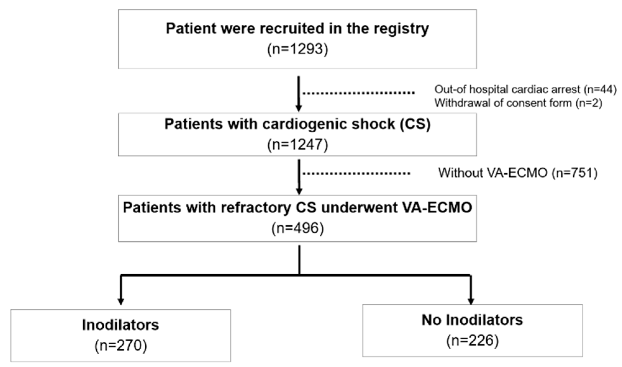
2.1. Study Population

The RESCUE (REtrospective and prospective observational Study to investigate Clinical oUtcomes and Efficacy of left ventricular assist device for Korean patients with cardiogenic shock; NCT02985008 at www.clinicaltrials.gov, assessed on 5 December 2016) registry is a multicenter registry. This study was conducted retrospectively and prospectively and included patients with CS aged > 19 years old from January 2014 to December 2018. In this registry, 1247 patients were enrolled from 12 tertiary centers in the Republic of Korea (954 retrospectively and 293 prospectively). According to the IABP-shock II trial definition, we included patients who had a systolic blood pressure (SBP) < 90 mmHg for 30 min or needed inotrope or vasopressor support to achieve an SBP > 90 mmHg and the presence of pulmonary congestion and signs of impaired organ perfusion (altered mental status, cold skin and extremities, urine output <0.5 mL/kg/h for the previous 6 h, or serum lactate > 2 mmol/L). The major exclusion criteria were out-of-hospital cardiac arrest, shock caused by factors other than CS (hypovolemic shock, septic shock, or post-cardiotomy shock), and refusal of active treatment.

2.2. Data Collection and Outcomes

The Institutional Review Board (IRB) of each hospital approved the study protocol. This study was conducted in accordance with the latest principles of the Declaration of Helsinki. The IRBs of the participating hospitals waived the requirement for informed consent from the retrospectively enrolled patients. However, we obtained written informed consent from all prospectively enrolled patients before enrollment. Independent clinical research coordinators collected patient data from web-based case report forms. Additional clinical information was obtained from medical records if necessary. The primary outcome was the in-hospital mortality rate. The secondary outcomes included in-hospital cardiac mortality and complications related to VA-ECMO, such as bleeding, limb ischemia, cerebrovascular accidents, and sepsis. All-cause mortality was defined as death from any cause, and all deaths were considered attributable to cardiac causes unless a non-cardiac cause was certain. The vasoactive-inotropic score was calculated as the maximal administration rate of dopamine, dobutamine, epinephrine, norepinephrine, milrinone, and vasopressin during the 48-h period of the shock event.

2.3. ECMO Management

VA-ECMO was initiated for patients with prolonged and recurrent arrest or with severe therapy-resistant CS [8]. The VA-ECMO device was inserted by percutaneous cannulation using Seldinger’s technique or surgical cannulation using the cut-down method at the femoral vessels. For patients without life-threatening bleeding, anticoagulation was provided intravenously using unfractionated heparin with an activated clotting time of 180 to 200 s during ECMO support. Management of the volume and vasopressors or inotropes was based on the clinical judgment of the attending physician in charge. Additionally, the decision to administer inodilators (dobutamine and milrinone) was based on the clinical judgment of the attending physician.

2.4. Statistical Analysis

Continuous variables are presented as the mean ± standard deviation and were compared using Student’s t-test. Categorical variables are presented as numbers and percentages and were compared using the χ2 test. We calculated the propensity score to match the baseline characteristics between the groups with and without inodilators use. Patients were matched 1:1. Univariate and multivariate time-dependent Cox-proportional hazard analyses were performed to predict the in-hospital mortality of the entire cohort and the propensity score-matched cohort. Covariates that were statistically significant in the univariate analysis (p < 0.1) or considered clinically important were included in the multivariate models. Hazard ratios (HRs) were calculated as an estimate of the risk associated with a particular variable based on binomial distributions and presented with 95% confidence intervals (CIs). All probability values were two-tailed, and p < 0.05 was considered statistically significant. Statistical analyses were performed using SPSS software version 23 (IBM, Armonk, NY, USA).

3. Results

3.1. Baseline Characteristics According to the Use of Inodilators

This cohort included 1247 patients with CS, with 496 of them treated with VA-ECMO during the course of the study (Figure 1).
The mean age of the study population was 61.8 ± 14.2 years, and 69.2% were men. Of the 496 patients, 257 (51.8%) died during hospitalization, and 270 (54.4%) were treated with inodilators. The baseline characteristics of the total cohort and propensity-matched cohort are shown in Table 1.
Compared to the no-inodilator group, the inodilator group had a significantly lower left ventricular ejection fraction (26.2 ± 12.4% versus 29.0 ± 15.0%, p = 0.034) and a higher inotropic score (34.1 ± 41.5 versus 17.6 ± 40.9, p < 0.001). The total bilirubin levels were higher in the inodilator group than in the no-inodilator group (1.4 ± 3.3 versus 0.8 ± 0.9, p = 0.010). There were no other significant differences in baseline characteristics according to the use of inodilators, except for the low prevalence of diabetes mellitus in the inodilator group (83 (30.7%) versus 96 (42.5%), p = 0.007).

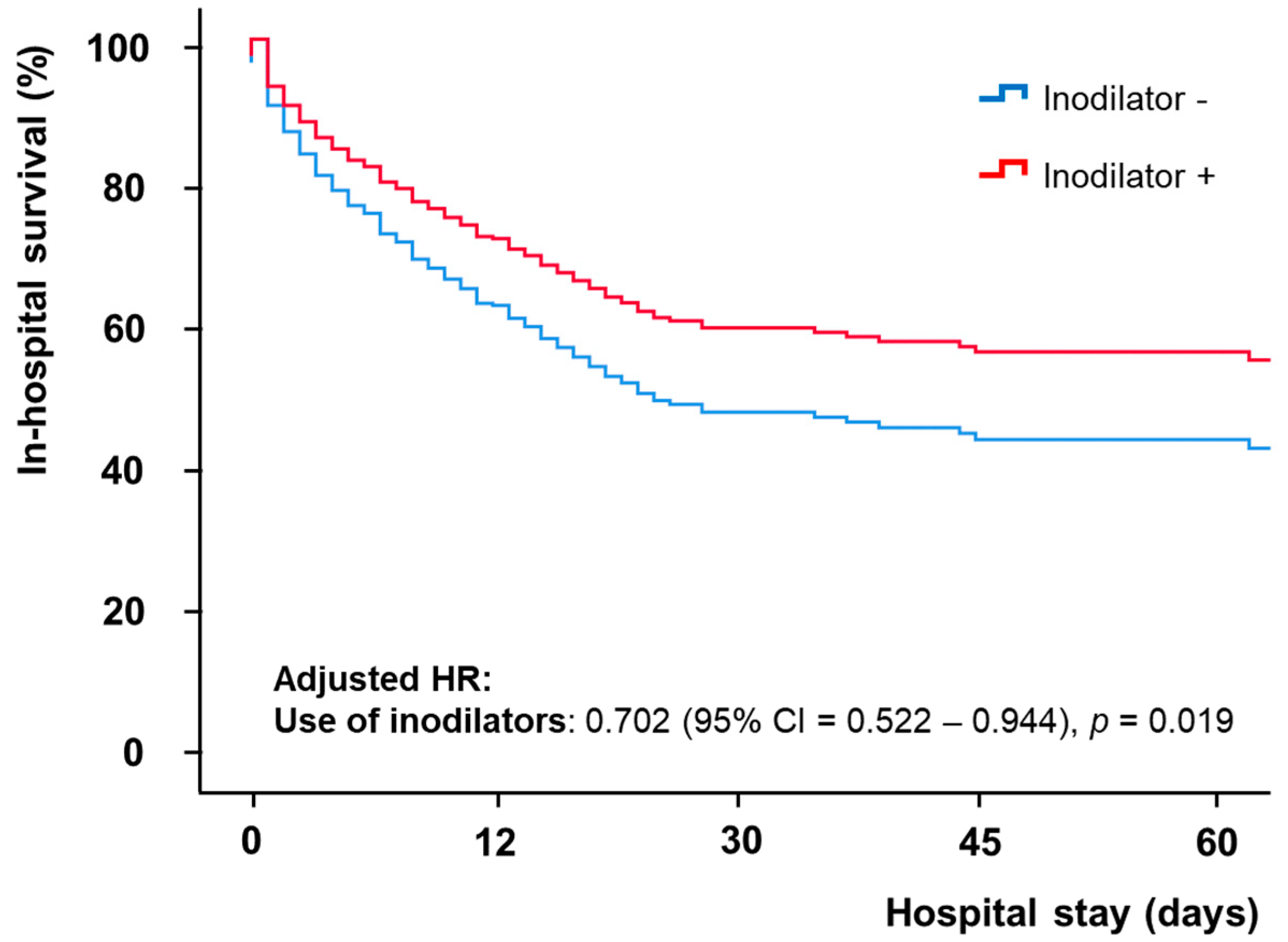
3.2. In-Hospital Outcomes of the Propensity Score Matched Analysis

After propensity score matching, 191 matched pairs (191 patients from the inodilator group and 191 patients from the no-inodilator group) were obtained. There were no significant differences in the baseline clinical characteristics, except for previous cerebrovascular accidents (Table 1). In the matched cohort, 194 (50.8%) patients died during hospitalization. The clinical outcomes are summarized in Table 2. There were no significant differences in in-hospital mortality, in-hospital cardiac mortality, ECMO cannula insertion site bleeding, limb ischemia, gastrointestinal bleeding, cerebrovascular accidents, or sepsis.
The results of the univariate and multivariate analyses of the in-hospital mortality in propensity score matched cohort are listed in Table 3. The univariate analysis revealed that age, heart rate, hypertension, previous chronic kidney disease, ischemic cardiomyopathy, creatinine clearance rate, lactic acid level, peak creatine kinase-myocardial band level, left ventricular ejection fraction, cardiopulmonary resuscitation, shock to ECMO insertion time, continuous renal replacement therapy, mechanical ventilation, ECMO initial pump flow, distal perfusion, and vasoactive inotropic score were significant determinants of in-hospital mortality. In the multivariate analysis, the use of inodilators was significantly associated with lower in-hospital mortality after adjustment (adjusted HR = 0.702, 95% CI = 0.552–0.944, p = 0.019) (Figure 2). Furthermore, age, lactic acid level, left ventricular ejection fraction, continuous renal replacement therapy, mechanical ventilation, distal perfusion, and vasoactive inotropic score remained statistically significant after adjustment.

4. Discussion

We showed that the use of inodilators was beneficial for patients with CS treated with VA-ECMO.
Inotropic agents have been fundamental for resuscitation cases of CS for several decades. In patients with CS, the blood pressure may fail to increase after the initial resuscitation, prompting the use of inotropic agents, as proposed by the current international guidelines [9,10]. Inotropic agents can be classified as inopressors and inodilators based on their effects on systemic vascular resistance. Dobutamine and milrinone are two inodilators approved for use in the United States [11]. Dobutamine is a synthetic catecholamine that has a direct agonistic effect on β1- and β2-adrenergic receptors, resulting in improved myocardial inotropy, weak chronotropy, and systemic vasodilation. Milrinone is a phosphodiesterase III inhibitor that increases intracellular cyclic adenosine monophosphate levels, resulting in improved cardiac contractility as well as systemic and pulmonary circulatory vasodilation. Therefore, both dobutamine and milrinone lead to increased cardiac output and decreased left ventricular filling pressure [12,13]. Previous studies have shown changes in hemodynamic parameters with increased cardiac index and decreased pulmonary capillary wedge pressure when inodilators were used [14,15,16].
VA-ECMO leads to an increase in the afterload [17,18]. As a result, it can increase left ventricular and atrial filling pressures, pulmonary edema, left ventricular distension, and stasis in the pulmonary circulation [19,20]. Inodilators can increase left ventricular performance; therefore, they can be used to treat stasis induced by VA-ECMO and reduce systemic and pulmonary vascular resistance. Therefore, the use of inodilators can be beneficial for patients undergoing VA-ECMO.
Pirracchio et al. reported that a combination of inodilators and inopressors was associated with a significant improvement in the short-term survival of patients with severe CS without ECMO compared with the use of inopressors alone [21]. These findings suggest that either excessive vasoconstriction can be detrimental to patients with severe CS or vasodilation may be beneficial.
Zotzmann et al. reported no statistically significant difference in the short-term survival of patients who received inodilators and those who did not receive inotropes. However, the survival rate of the group that received inodilators was 52.2%, which was higher than that of the group that did not receive inotropes (47.9%) [22].
Previous studies have compared the effects of dobutamine and milrinone. Mathew et al. reported that no significant differences were observed between milrinone and dobutamine in terms of the clinical outcomes, including in-hospital death, of patients with CS [23]. Lewis et al. also indicated that there were no significant differences in the efficacy and safety of milrinone and dobutamine [24]. Therefore, the efficacy and safety of both drugs are similar, and milrinone or dobutamine can be used as inodilators for CS, depending on the condition of the patient.
There are no convincing data supporting the use of inodilators to reduce the mortality of patients with refractory CS undergoing VA-ECMO. However, by using a large-scale multicenter registry, this study provided evidence that inodilators may be beneficial for these patients.
This study had several limitations. First, the RESCUE registry contains observational data; therefore, residual confounding variables may have affected our findings, even after multiple adjustments. Second, the in-hospital treatment reflects the preferences and discretion of individual physicians, which may have introduced confounding selection bias. Third, the corresponding action and adverse effects may differ depending on the dose, duration of use, and method of administration of dobutamine and milrinone [11,25]. However, this has not been considered in detail, and the adverse effects have not been properly evaluated. Additionally, hemodynamic parameters, such as cardiac output, cardiac index, central venous pressure, and pulmonary capillary wedge pressure, were not included. If hemodynamic changes attributable to the use of inodilators had been included in the data, concrete evidence for the clinical effect could have been presented. Furthermore, the study was restricted to in-hospital outcomes only; however, mid-term as well as long-term outcome assessments are required to confirm the results. Finally, fewer patients received milrinone and more patients received dobutamine; therefore, caution is advised when interpreting these results.

5. Conclusions

In-hospital mortality remains high for patients undergoing VA-ECMO for refractory CS. Although the use of inodilators should be considered based on clinical judgment, their use may improve the clinical outcomes of these patients. To confirm the favorable effects of inodilators on patients with refractory CS treated with VA-ECMO, future studies involving well-designed randomized controlled trials are required.

Author Contributions

Conceptualization, P.S.S.; methodology, S.-D.P. and S.S.C.; software, S.U.K. and W.S.L.; validation, C.W.Y., H.-J.K. and Y.-G.K.; formal analysis, M.K. and S.-W.S.; investigation, J.H.Y. and W.J.J.; resources, W.J.C. and J.-W.B.; data curation, S.-W.S. and H.-J.L.; writing—original draft preparation, M.K.; writing—review and editing, J.-H.P.; visualization, J.-H.P.; supervision, J.-H.P. and P.S.S.; project administration J.-O.J. and H.-C.G. All authors have read and agreed to the published version of the manuscript.

Funding

This research received no external funding.

Institutional Review Board Statement

The study was conducted in accordance with the Declaration of Helsinki and approved by the Institutional Review Board (or Ethics Committee) of Samsung Medical Center (protocol code 2016-03-130, 6 April 2016) and the local ethics committee of all the study centers.

Informed Consent Statement

Informed consent was obtained from all subjects involved in the study. Informed consent was allowed to be waived by the ethics committee based on retrospective review of medical records.

Data Availability Statement

The data used during the current study are available from the corresponding author on reasonable request.

Conflicts of Interest

The authors declare no conflict of interest.

References

  1. Thiele, H.; Ohman, E.M.; Desch, S.; Eitel, I.; de Waha, S. Management of cardiogenic shock. Eur. Heart J. 2015, 36, 1223–1230. [Google Scholar] [CrossRef] [Green Version]
  2. Kolte, D.; Khera, S.; Aronow, W.S.; Mujib, M.; Palaniswamy, C.; Sule, S.; Jain, D.; Gotsis, W.; Ahmed, A.; Frishman, W.H. Trends in Incidence, Management, and Outcomes of Cardiogenic Shock Complicating ST-Elevation Myocardial Infarction in the United States. J. Am. Heart Assoc. 2014, 3, e000590. [Google Scholar] [CrossRef] [Green Version]
  3. Reynolds, H.R.; Hochman, J.S. Cardiogenic shock: Current concepts and improving outcomes. Circulation 2008, 117, 686–697. [Google Scholar] [CrossRef]
  4. Takayama, H.; Truby, L.; Koekort, M.; Uriel, N.; Colombo, P.; Mancini, D.M.; Jorde, U.P.; Naka, Y. Clinical outcome of mechanical circulatory support for refractory cardiogenic shock in the current era. J. Heart Lung Transplant. 2013, 32, 106–111. [Google Scholar] [CrossRef]
  5. Schrage, B.; Becher, P.M.; Goßling, A.; Savarese, G.; Dabboura, S.; Yan, I.; Beer, B.; Söffker, G.; Seiffert, M.; Kluge, S. Temporal trends in incidence, causes, use of mechanical circulatory support and mortality in cardiogenic shock. ESC Heart Fail. 2021, 8, 1295–1303. [Google Scholar] [CrossRef]
  6. Patricio, D.; Peluso, L.; Brasseur, A.; Lheureux, O.; Belliato, M.; Vincent, J.-L.; Creteur, J.; Taccone, F.S. Comparison of extracorporeal and conventional cardiopulmonary resuscitation: A retrospective propensity score matched study. Crit. Care 2019, 23, 27. [Google Scholar] [CrossRef] [Green Version]
  7. Ouweneel, D.M.; Schotborgh, J.V.; Limpens, J.; Sjauw, K.D.; Engström, A.; Lagrand, W.K.; Cherpanath, T.G.; Driessen, A.H.; de Mol, B.A.; Henriques, J.P. Extracorporeal life support during cardiac arrest and cardiogenic shock: A systematic review and meta-analysis. Intensive Care Med. 2016, 42, 1922–1934. [Google Scholar] [CrossRef] [Green Version]
  8. Peura, J.L.; Colvin-Adams, M.; Francis, G.S.; Grady, K.L.; Hoffman, T.M.; Jessup, M.; John, R.; Kiernan, M.S.; Mitchell, J.E.; O’Connell, J.B. Recommendations for the use of mechanical circulatory support: Device strategies and patient selection: A scientific statement from the American Heart Association. Circulation 2012, 126, 2648–2667. [Google Scholar] [CrossRef] [Green Version]
  9. McDonagh, T.A.; Metra, M.; Adamo, M.; Gardner, R.S.; Baumbach, A.; Böhm, M.; Burri, H.; Butler, J.; Čelutkienė, J.; Chioncel, O. 2021 ESC Guidelines for the diagnosis and treatment of acute and chronic heart failure: Developed by the Task Force for the diagnosis and treatment of acute and chronic heart failure of the European Society of Cardiology (ESC) with the special contribution of the Heart Failure Association (HFA) of the ESC. Eur. Heart J. 2021, 42, 3599–3726. [Google Scholar]
  10. Yancy, C.W.; Jessup, M.; Bozkurt, B.; Butler, J.; Casey, D.E., Jr.; Drazner, M.H.; Fonarow, G.C.; Geraci, S.A.; Horwich, T.; Januzzi, J.L. 2013 ACCF/AHA guideline for the management of heart failure: Executive summary: A report of the American College of Cardiology Foundation/American Heart Association Task Force on practice guidelines. Circulation 2013, 128, 1810–1852. [Google Scholar] [CrossRef]
  11. Jentzer, J.C.; Coons, J.C.; Link, C.B.; Schmidhofer, M. Pharmacotherapy update on the use of vasopressors and inotropes in the intensive care unit. J. Cardiovasc. Pharmacol. Ther. 2015, 20, 249–260. [Google Scholar] [CrossRef] [PubMed]
  12. Francis, G.S.; Bartos, J.A.; Adatya, S. Inotropes. J. Am. Coll. Cardiol. 2014, 63, 2069–2078. [Google Scholar] [CrossRef] [Green Version]
  13. Harjola, V.P.; Mebazaa, A.; Čelutkienė, J.; Bettex, D.; Bueno, H.; Chioncel, O.; Crespo-Leiro, M.G.; Falk, V.; Filippatos, G.; Gibbs, S. Contemporary management of acute right ventricular failure: A statement from the Heart Failure Association and the Working Group on Pulmonary Circulation and Right Ventricular Function of the European Society of Cardiology. Eur. J. Heart Fail. 2016, 18, 226–241. [Google Scholar] [CrossRef] [PubMed]
  14. Feneck, R.O.; Sherry, K.M.; Withington, P.S.; Oduro-Dominah, A.; European Milrinone Multicenter Trial Group. Comparison of the hemodynamic effects of milrinone with dobutamine in patients after cardiac surgery. J. Cardiothorac. Vasc. Anesth. 2001, 15, 306–315. [Google Scholar] [CrossRef] [PubMed]
  15. Carmona, M.J.C.; Martins, L.M.; Vane, M.F.; Longo, B.A.; Paredes, L.S.; Malbouisson, L.M.S. Comparison of the effects of dobutamine and milrinone on hemodynamic parameters and oxygen supply in patients undergoing cardiac surgery with low cardiac output after anesthetic induction. Rev. Bras. Anestesiol. 2010, 60, 237–246. [Google Scholar] [CrossRef] [Green Version]
  16. Karlsberg, R.P.; DeWood, M.A.; DeMaria, A.N.; Berk, M.R.; Lasher, K.P.; Group, M.D.S. Comparative efficacy of short-term intravenous infusions of milrinone and dobutamine in acute congestive heart failure following acute myocardial infarction. Clin. Cardiol. 1996, 19, 21–30. [Google Scholar] [CrossRef]
  17. Burkhoff, D.; Sayer, G.; Doshi, D.; Uriel, N. Hemodynamics of mechanical circulatory support. J. Am. Coll. Cardiol. 2015, 66, 2663–2674. [Google Scholar] [CrossRef] [Green Version]
  18. Donker, D.W.; Brodie, D.; Henriques, J.P.; Broomé, M. Left ventricular unloading during veno-arterial ECMO: A simulation study. ASAIO J. 2019, 65, 11. [Google Scholar] [CrossRef]
  19. Rupprecht, L.; Flörchinger, B.; Schopka, S.; Schmid, C.; Philipp, A.; Lunz, D.; Müller, T.; Camboni, D. Cardiac decompression on extracorporeal life support: A review and discussion of the literature. ASAIO J. 2013, 59, 547–553. [Google Scholar] [CrossRef]
  20. Ostadal, P.; Mlcek, M.; Kruger, A.; Hala, P.; Lacko, S.; Mates, M.; Vondrakova, D.; Svoboda, T.; Hrachovina, M.; Janotka, M.; et al. Increasing venoarterial extracorporeal membrane oxygenation flow negatively affects left ventricular performance in a porcine model of cardiogenic shock. J. Transl. Med. 2015, 13, 266. [Google Scholar] [CrossRef] [Green Version]
  21. Pirracchio, R.; Parenica, J.; Resche Rigon, M.; Chevret, S.; Spinar, J.; Jarkovsky, J.; Zannad, F.; Alla, F.; Mebazaa, A.; GREAT network. The effectiveness of inodilators in reducing short term mortality among patient with severe cardiogenic shock: A propensity-based analysis. PLoS ONE 2013, 8, e71659. [Google Scholar] [CrossRef]
  22. Zotzmann, V.; Rilinger, J.; Lang, C.N.; Kaier, K.; Benk, C.; Duerschmied, D.; Biever, P.M.; Bode, C.; Wengenmayer, T.; Staudacher, D.L. Epinephrine, inodilator, or no inotrope in venoarterial extracorporeal membrane oxygenation implantation: A single-center experience. Crit. Care 2019, 23, 320. [Google Scholar] [CrossRef] [PubMed] [Green Version]
  23. Mathew, R.; Di Santo, P.; Jung, R.G.; Marbach, J.A.; Hutson, J.; Simard, T.; Ramirez, F.D.; Harnett, D.T.; Merdad, A.; Almufleh, A.; et al. Milrinone as compared with dobutamine in the treatment of cardiogenic shock. N. Engl. J. Med. 2021, 385, 516–525. [Google Scholar] [CrossRef] [PubMed]
  24. Lewis, T.C.; Aberle, C.; Altshuler, D.; Piper, G.L.; Papadopoulos, J. Comparative effectiveness and safety between milrinone or dobutamine as initial inotrope therapy in cardiogenic shock. J. Cardiovasc. Pharmacol. Ther. 2019, 24, 130–138. [Google Scholar] [CrossRef]
  25. Bayram, M.; De Luca, L.; Massie, M.B.; Gheorghiade, M. Reassessment of dobutamine, dopamine, and milrinone in the management of acute heart failure syndromes. Am. J. Cardiol. 2005, 96, 47G–58G. [Google Scholar] [CrossRef]
Figure 1. Study flow.
Jcm 11 04958 g001
Figure 2. Comparison of in-hospital mortality according to the use of inodilators. The adjusted hazard ratio (HR) was calculated using the Cox-proportional hazard analysis to rule out the effects of other significant variables in the univariate analysis.
Jcm 11 04958 g002
Table 1. Comparison of clinical characteristics according to the use of inodilators.
Total Cohort (n = 596)Propensity Score Matched Cohort (n = 382)
Inodilators
(n = 270)
No Inodilators
(n = 226)
p ValueInodilators
(n = 191)
No Inodilators
(n = 191)
p Value
Age (year) 60.9 ± 14.862.9 ± 13.70.13460.9 ± 14.461.7 ± 14.00.134
Gender (male)178 (65.8)165 (73.0)0.089130 (68.1)135 (70.7)0.579
Body mass index (kg/m2)23.2 ± 3.423.4 ± 3.40.50523.4 ± 3.423.3 ± 3.20.079
Hypertension122 (45.2)112 (49.6)0.33184 (44.0)88 (46.1)0.681
Diabetes mellitus83 (30.7)96 (42.5)0.00763 (33.0)72 (37.7)0.335
Dyslipidemia47 (17.4)56 (24.8)0.04438 (19.9)42 (22.0)0.615
Current smoking67 (24.8)68 (30.1)0.18947 (24.6)60 (31.4)0.139
Chronic kidney disease18 (6.7)20 (8.8)0.36315 (7.9)16 (8.4)0.851
Previous myocardial infarction32 (11.9)30 (13.3)0.63323 (12.0)23 (12.0)1.000
Previous coronary revascularization38 (14.1)40 (17.7)0.26929 (15.2)33 (17.3)0.579
Previous cerebrovascular accident25 (9.3)14 (6.2)0.20722 (11.5)8 (4.2)0.008
Systolic blood pressure (mmHg)65.0 ± 27.868.0 ± 35.00.29666.4 ± 24.865.3 ± 32.50.709
Diastolic blood pressure (mmHg)43.8 ± 23.543.8 ± 23.50.99144.4 ± 19.942.0 ± 22.00.278
Heart rate (beat/min)83.8 ± 37.282.8 ± 39.80.77183.2 ± 35.881.0 ± 40.60.573
Hemoglobin (mg/dL)12.3 ± 2.712.5 ± 2.80.35912.4 ± 2.612.5 ± 2.80.595
Total bilirubin (mg/dL)1.4 ± 3.30.8 ± 0.90.0100.9 ± 0.80.9 ± 0.90.594
Creatinine clearance rate (mL/min/1.73 m2)53.4 ± 35.356.1 ± 30.30.35754.6 ± 39.957.4 ± 31.20.448
Serum glucose (mg/dL)241.4 ± 131.9236.2 ± 117.90.658246.4 ± 133.0233.8 ± 118.80.348
NT–proBNP (pg/mL)10,598.1 ± 11,784.38321.0 ± 14,234.50.14711,104.2 ± 12,349.38735.5 ± 14,800.70.192
Lactic acid (mmol/L)7.4 ± 3.77.6 ± 3.90.6847.4 ± 3.77.6 ± 3.90.670
Peak CK-MB (ng/mL)221.3 ± 558.0212.9 ± 222.40.821243.7 ± 650.9221.5 ± 227.50.657
Cardiopulmonary resuscitation129 (47.8)116 (51.3)0.43190 (47.1)105 (55.0)0.125
Shock to ECMO insertion time (min) 434.7 ± 869.9358.7 ± 787.00.309389.1 ± 713.8312.3 ± 673.90.280
Initial pump flow (L/min)3.0 ± 0.82.8 ± 0.90.1322.9 ± 0.82.8 ± 0.90.255
Distal perfusion97 (35.9)90 (39.8)0.37273 (38.2)78 (40.8)0.601
Unloading of left ventricle55 (20.4)32 (14.2)0.07041 (21.5)28 (14.7)0.084
Left ventricular ejection fraction (%)26.2 ± 12.429.0 ± 15.80.03427.0 ± 12.227.7 ± 15.00.596
Dopamine161 (59.6)126 (55.8)0.384105 (55.0)119 (62.3)0.146
Norepinephrine170 (63.0)159 (70.4)0.083121 (63.4)133 (69.6)0.193
Epinephrine44 (16.3)25 (11.1)0.09321 (11.0)24 (12.6)0.634
Vasopressin41 (15.2)21 (9.3)0.04828 (14.7)16 (8.4)0.054
Vasoactive inotropic score110.3 ± 176.790.4 ± 134.80.15694.3 ± 182.493.9 ± 134.10.977
Inotropic score34.1 ± 41.517.6 ± 40.9<0.00123.6 ± 21.120.5 ± 43.80.387
Ischemic cardiomyopathy177 (65.8)165 (73.0)0.074124 (64.9)141 (73.8)0.059
Continuous renal replacement therapy115 (42.6)80 (35.4)0.10285 (44.5)69 (36.1)0.095
Mechanical ventilation226 (83.7)183 (81.0)0.426154 (80.6)156 (81.7)0.794
CK-MB = creatine kinase-myocardial band, ECMO = extracorporeal membrane oxygenation, NT–proBNP: N terminal pro B type natriuretic peptide. Values are expressed as n (%) or mean ± standard deviation.
Table 2. In-hospital outcomes according to the use of inodilators in the propensity score-matched cohort.
Overall
(n = 382)
Inodilators
(n = 191)
No Inodilators
(n = 191)
p Value
In-hospital cardiac mortality194 (50.8%)91 (47.6%)103 (53.9%)0.220
In-hospital mortality168 (44.0%)79 (41.4%)89 (46.6%)0.304
ECMO site bleeding54 (14.1%)29 (15.2%)25 (13.1%)0.558
Limb ischemia32 (8.4%)16 (8.4%)16 (8.4%)1.000
Stroke16 (4.2%)9 (4.7%)7 (3.7%)0.611
GI bleeding24 (6.3%)11 (5.8%)13 (6.8%)0.674
Sepsis18 (4.7%)9 (4.7%)9 (4.7%)1.000
ECMO: extracorporeal membrane oxygenation; GI: gastrointestinal.
Table 3. Univariate and multivariate analysis of the prediction of in-hospital mortality.
Univariate AnalysisMultivariate Analysis
HR (95% CI)p ValueHR (95% CI)p Value
Use of inodilators0.768 (0.579–1.018)0.0660.702 (0.522–0.944)0.019
Age (year)1.024 (1.013–1.034)<0.0011.019 (1.005–1.033)0.007
Gender (male)0.865 (0.633–1.182)0.363
Body mass index (kg/m2)1.035 (0.991–1.080)0.118
Systolic blood pressure (mmHg)0.999 (0.993–1.004)0.595
Heart rate0.995 (0.991–0.999)0.0070.998 (0.995–1.002)0.392
Hypertension1.504 (1.133–1.996)0.0051.196 (0.868–1.646)0.274
Diabetes mellitus1.161 (0.871–1.549)0.309
Previous chronic kidney disease1.515 (0.987–2.323)0.0570.928 (0.575–1.495)0.758
Ischemic cardiomyopathy2.191 (1.546–3.107)<0.0011.131 (0.753–1.697)0.553
Hemoglobin (g/dL)1.036 (0.981–1.094)0.204
Creatinine (mg/dL)1.083 (0.993–1.180)0.070
Creatinine clearance rate (mL/min/1.73 m2)0.994 (0.989–0.999)0.0291.001 (0.996–1.006)0.773
Lactic acid (mmol/L)1.090 (1.052–1.129)<0.0011.077 (1.037–1.119)<0.001
Peak CK-MB (ng/mL)1.000 (1.000–1.000)0.0021.000 (1.000–1.000)0.076
Left ventricular ejection fraction (%)0.984 (0.972–0.995)0.0070.986 (0.974–0.999)0.032
Cardiopulmonary resuscitation2.229 (1.659–2.995)<0.0011.389 (0.993–1.945)0.055
Shock to ECMO insertion time (min) 1.000 (1.000–1.000)0.0871.000 (1.000–1.000)0.070
Continuous renal replacement therapy1.910 (1.436–2.540)<0.0011.558 (1.138–2.134)0.006
Mechanical ventilation6.412 (3.280–12.534)<0.0013.266 (1.612–6.616)0.001
Initial pump flow (L/min)0.804 (0.663–0.973)0.0250.842 (0.689–1.029)0.094
Distal perfusion0.644 (0.478–0.866)0.0040.692 (0.506–0.946)0.021
Unloading of left ventricle1.229 (0.873–1.730)0.238
Vasoactive inotropic score1.001 (1.000–1.001)0.0011.001 (1.000–1.002)0.001
Inotropic score1.006 (1.004–1.009)<0.001
Dopamine2.033 (1.499–2.756)<0.001
Norepinephrine1.791 (1.292–2.484)<0.001
Epinephrine1.401 (0.949–2.068)0.090
Vasopressin1.531 (1.037–2.261)0.032
CI: confidence interval; CK-MB: creatine kinase-myocardial band; ECMO: extracorporeal membrane oxygenation; HR: hazard ratio.
Publisher’s Note: MDPI stays neutral with regard to jurisdictional claims in published maps and institutional affiliations.

