Review

Association between Immediate Postoperative Radiographic Findings and Failed Internal Fixation for Trochanteric Fractures: Systematic Review and Meta-Analysis

1
Department of Orthopedic Surgery, Miyamoto Orthopedic Hospital, Okayama 773-8236, Japan
2
Department of Epidemiology, Graduate School of Medicine, Dentistry and Pharmaceutical Sciences, Okayama University, Okayama 700-8530, Japan
3
Scientific Research Works Peer Support Group (SRWS-PSG), Osaka 541-0043, Japan
4
Departments of Health Promotion and Human Behavior, Kyoto University Graduate School of Medicine/School of Public Health, Kyoto 606-8501, Japan
5
Oku Medical Clinic, Osaka 573-0164, Japan
6
Cochrane Japan, Tokyo 104-0044, Japan
7
Department of Orthopedic Surgery, National Hospital Organization Okayama Medical Center, Okayama 701-1192, Japan
8
Department of Orthopedic Surgery, Tsuyama Chuo Hospital, Okayama 708-0841, Japan
9
Department of Orthopedic Surgery and Traumatology, Kawasaki Medical School General Medical Center, Okayama 700-8505, Japan
10
Department of Orthopedic Surgery, Okayama University Graduate School of Medicine, Dentistry and Pharmaceutical Science, Okayama 700-8530, Japan
*
Author to whom correspondence should be addressed.
J. Clin. Med. 2022, 11(16), 4879; https://doi.org/10.3390/jcm11164879
Submission received: 7 July 2022 / Revised: 11 August 2022 / Accepted: 17 August 2022 / Published: 19 August 2022
(This article belongs to the Special Issue Clinical Management of Osteoporotic Fractures)

Abstract

:
Failed internal fixations for trochanteric fractures have a strong negative impact owing to increased postoperative mortality and high medical costs. However, evidence on the prognostic value of postoperative radiographic findings for failed internal fixations is limited. We aimed to clarify the association between comprehensive immediate postoperative radiographic findings and failed internal fixation using relative and absolute risk measures. We followed the meta-analysis of observational studies in epidemiology guidelines and the Cochrane handbook. We searched specific databases in November 2021. The outcomes of interest were failed internal fixation and cut-out. We pooled the odds ratios and 95% confidence intervals using a random-effects model and calculated the number needed to harm for each outcome. Thirty-six studies involving 8938 patients were included. The certainty of evidence in the association between postoperative radiographic findings and failed internal fixation or cut-out was mainly low or very low except for the association between intramedullary malreduction on the anteromedial cortex and failed internal fixation. Moderate certainty of evidence supported that intramedullary malreduction on the anteromedial cortex was associated with failed internal fixation. Most postoperative radiographic findings on immediate postoperative radiographs for trochanteric fractures were uncertain as prognostic factors for failed internal fixations.

1. Introduction

The incidence of geriatric trochanteric fractures has increased with the aging population [1]. Postoperative failed internal fixations leading to reoperation, such as cut-out, nonunion, and implant failure, have a strong negative impact owing to increased postoperative mortality and high medical costs [2]. In 1980, the five major factors related to the treatment outcomes following internal fixation for trochanteric fractures were: bone quality, fragment geometry, implant selection, reduction quality, and screw placement in the femoral head [3]. Among them, surgically modifiable factors are reduction quality and screw placement in the femoral head, which can be evaluated using postoperative radiographic findings.
However, evidence on the prognostic value of postoperative radiographic findings for failed internal fixations is scarce. Some postoperative radiographic findings, such as the Baumgaertner reduction criteria and a high tip–apex distance (TAD), have been investigated for their association with failed internal fixation; however, no comprehensive summary is available [4,5,6]. Therefore, the factor that has the greatest impact on failed internal fixations remains undetermined.
The aim of this systematic review and meta-analysis was to clarify the association of between all immediate postoperative radiographic findings and failed internal fixation for internally fixed trochanteric fractures in terms of relative and absolute risk measures. Identifying the relevant surgical factors and recognizing the degree of association will facilitate intraoperative decisions and postoperative follow-up planning and improve clinical outcomes.

2. Materials and Methods

We followed the meta-analysis of observational studies in epidemiology (MOOSE) guidelines, the Cochrane handbook [7,8], and the grading of recommendations, assessment, development, and evaluation (GRADE) criteria [9]. Prior to study initiation, we made our study protocol available in the open science framework [10].

2.1. Eligibility Criteria

2.1.1. Study Type

We included all observational studies and secondary analyses of randomized trials investigating the association between immediate postoperative radiographic findings and failed internal fixation in trochanteric fractures. We did not apply restrictions on publication date and status (full publication, conference abstract, and unpublished data) and languages. We excluded case reports and cadaveric studies.

2.1.2. Study Participants

We included patients with trochanteric fractures that were treated using internal fixation (sliding hip screw (SHS) or cephalomedullary nail (CMN)). Inclusion criteria involved the following patients: (1) aged >50 years, (2) with fracture type classified under the Arbeitsgemeinschaft für Osteosynthesefragen/Orthopedic Trauma Association (AO/OTA) classification 31A [11], and (3) with follow-up periods of more than 3 months postoperatively. The exclusion criteria were pathological fractures caused by specific pathologies other than osteoporosis, open fractures with history of operation at the ipsilateral proximal femur, and additional augmentation in fixation (cement augmentation, additional screw insertion separately from the original implant).

2.2. Exposures

The exposures of interest were the following five immediate postoperative radiographic findings using plain radiography: Baumgaertner reduction criteria, overall alignment in the anteroposterior (AP) radiograph, local alignment on the anteromedial cortex, TAD, and screw placement in the femoral head. We selected these radiographic findings based on earlier reviews [4,5,6]. These were evaluated using AP and/or lateral plain radiography.

2.3. Reduction Quality

1.
The Baumgaertner reduction criteria: The reduction quality is classified as good, acceptable, or poor [12]. Poor reduction indicates varus on the AP radiograph and angulation greater than 20 degrees on the lateral radiograph and displacement more than 4 mm on both views. Here, we classified it into two categories: not poor (good or acceptable) or poor.
2.
Overall alignment using AP radiography: We classified it as either varus or nonvarus (adequate reduction quality). When the femoral neck angle of the operated hip was reduced compared with that of the contralateral hip, we defined it as varus alignment. The original authors have established a detailed definition of varus.
3.
Local alignment on the anteromedial cortex: We classified it as either intramedullary type or nonintramedullary type (adequate reduction quality) [13,14]. The intramedullary type was defined as a head–neck fragment located laterally or posteriorly to the cortex of the femoral shaft [14].

2.4. Screw Placement in the Femoral Head

4.
TAD [12]. It was classified as TAD ≥ 25 or not TAD ≥ 25 (adequate screw placement). If the original authors used continuous values or the other TAD cut-off values, we requested a reanalysis with a cut off value of 25 mm from them [4,12].
5.
Screw placement in the femoral head [15]. It was classified as adequate or inadequate screw placement. Adequate screw placement was defined as center–center or inferior–center placement [16]. If the original authors used the Parker’s ratio [17], we requested them for a reanalysis.

2.5. Outcomes

The primary outcome of this study was failed internal fixation, which indicated complications with fracture healing following internal fixation. Failed internal fixation included cut-out, cut-through, nonunion, implant failure, and over sliding distance, all of which required reoperation. We considered any of these as a failed internal fixation. Cut-out indicated the screw cut-out in any direction from the femoral head.

2.6. Search Method

We searched the Cochrane central register of controlled trials, MEDLINE, Embase, and study registries including clinicaltrials.gov and international clinical trials registry platform on 27 November 2021, using the search strategy (Supplementary Appendix A);. We also manually searched the reference lists of the included studies and international guidelines [18,19].

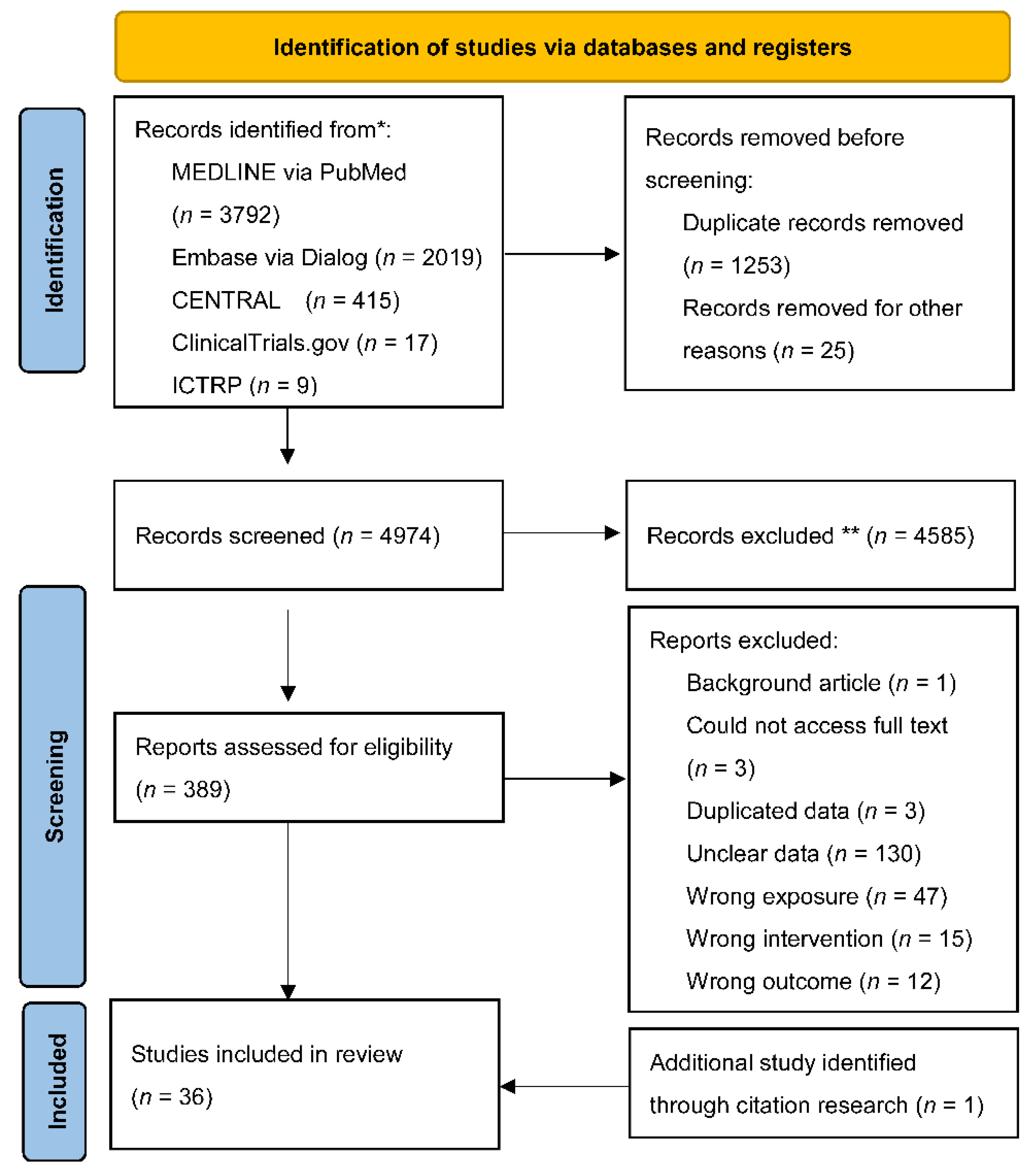
2.7. Study Selection and Data Extraction

Three pairs of reviewers independently screened every title and abstract of the articles based on the inclusion and exclusion criteria. Any conflicts between the two reviewers were resolved through discussion or with the help of a third reviewer.
Two reviewers on each trial independently performed data extraction of the included trials. For the effective measurement of the outcomes, we extracted both crude and multivariable estimates. We predefined the following possible confounders: age, sex, and fracture type (stable or unstable type based on AO/OTA classification (31A1, 2, 3)) [11,20]. If the possible confounders were not adjusted for in the original studies, we requested a re-analysis from the original authors.

2.8. Risk of Bias (Quality) Assessment

Two reviewers on each trial independently assessed the risk of bias using the quality in prognosis studies (QUIPS) tool [21]. If the possible confounders were adjusted for in the original studies, we rated the study as “low risk bias” for confounding factors. The overall risk of bias was rated low if all the QUIPS domains were rated low, high if two or more domains were with high risk of bias, and moderate in the other ratings.

2.9. Statistical Analysis

We pooled the crude and adjusted odds ratios (ORs) with 95% confidence intervals (CIs) for all outcomes using a random-effects meta-analysis model weighted by the inverse variance estimate. When both crude and adjusted ORs were used in the original papers, adjusted ORs were selected. We calculated the number needed to harm (NNH) for each outcome using the pooled relative risk and the median event rate of the control group.
We evaluated the statistical heterogeneity by visual inspection of the forest plots and calculation of the I2 statistic. We assessed the reason for heterogeneity when it was moderate or high (I2 > 50%). If more than 10 studies were included in each outcome, we performed a funnel plot analysis and the Egger’s test to assess reporting bias.
We judged the GRADE criteria for each association [9] (Table 1). We used the informative statements corresponding to the GRADE criteria [22].
A prespecified subgroup analysis was performed for the fracture type (stable or unstable) and the implant type used for internal fixation (SHS or CMN). The stable type was defined as A1 and A2.1 in the 2007 classification [20] and A1 in the 2018 classification [11] according to the AO/OTA guidelines.
We used crude ORs in the sensitivity analysis. As a post hoc analysis, we used adjusted and pooled ORs in studies or excluded studies with a high risk of bias in the overall risk of bias domain.
All analyses were performed using RevMan 5.4 (Cochrane Collaboration, London, UK) and STATA 17.0 (Stata-Corp LP, College Station, TX, USD). Values of p < 0.05 were considered significant.

3. Results

After screening 4974 records, we included 36 studies (8938 participants) in the final analysis (Figure 1) [14,23,24,25,26,27,28,29,30,31,32,33,34,35,36,37,38,39,40,41,42,43,44,45,46,47,48,49,50,51,52,53,54]. Supplementary Appendix B Table S1 shows the excluded studies with reasons for their exclusion. By contacting the authors, we obtained unpublished data from 11 studies (Supplementary Appendix B Table S2) [38,39,40,41,42,44,45,46,47,48]. The characteristics of the included studies are summarized in Supplementary Appendix B Table S2.
In the 36 included studies, 31 were retrospective cohort studies, three were prospective cohort studies, and two were case-control studies. Twenty-four (66.7%) studies included participants from Asian countries. Eighteen studies (50.0%) reported both TAD and cut-out values.
Seven studies on failed internal fixation and 12 studies on cut-out had zero outcome events in at least one arm. Studies with no outcome events in both arms were excluded from the meta-analysis. A few studies had assessed the association with adjusted ORs. Supplementary Appendix B Table S3 presents the quality assessment results of the included studies using the QUIPS tool. The overall risk of bias in most of the included studies was moderate or high, mainly because of insufficient adjustment for confounders. The prevalence (median, interquartile range) of each postoperative radiographic findings was as follows: poor reduction based on the Baumgaertner criteria (5.9%, 4.0–10.9%), varus malreduction (3.3%, 3.1–17.1%), intramedullary malreduction on the anteromedial cortex (22.4%, 11.4–28.6%), TAD ≥ 25 (17.5%, 6.8–25.5%), and inadequate screw placement in the femoral head (44.3%, 30.0–56.0%). The incidence rate (median, interquartile range) of each outcome was as follows: failed internal fixation (2.8%, 0–7.0%) and cut-out (5.6%, 0.5–8.1%).

3.1. Association between Immediate Postoperative Radiographic Findings and Failed Internal Fixations

The findings of this review are summarized in Table 1 (Supplementary Appendix C Figure S1–S10). Moderate certainty of evidence supported that intramedullary malreduction on the anteromedial cortex (pooled OR 7.23, 95% CI 2.49–21.01; NNH 21, 95% CI 7–83) (Supplementary Appendix C Figure S5) was associated with failed internal fixation.
The low certainty of evidence supported that the association with failed internal fixation was ranked using pooled ORs as follows: inadequate screw placement in the femoral head (pooled OR 6.11, 95% CI 1.00–37.27; NNH 6, 95% CI 2–12151) (Supplementary Appendix C Figure S9) and varus malreduction (pooled OR 4.48; 95% CI 1.03–19.46; NNH 8, 95% CI 2–1000) (Supplementary Appendix C Figure S3) were associated with failed internal fixation. The evidence on whether TAD ≥ 25 (pooled OR 1.39, 95% CI 0.65–2.98; NNH 200, 95% CI −200–37) (Supplementary Appendix C Figure S7) and poor reduction based on the Baumgaertner criteria is associated with failed internal fixation (pooled OR 1.17, 95% CI 0.01–92.75; NNH 500, 95% CI −71–2) (Supplementary Appendix C Figure S1) is uncertain.

3.2. Association between Immediate Postoperative Radiographic Findings and Cut-Outs

Low certainty of evidence supported that the association with cut-out was ranked using pooled ORs as follows: TAD ≥ 25 (pooled OR 7.92, 95% CI 3.33–18.82; NNH 14, 95% CI 6–40) (Supplementary Appendix C Figure S8) and poor reduction by Baumgaertner criteria (pooled OR 7.78, 95% CI 3.41–17.76; NNH 5, 95% CI 3–11) (Supplementary Appendix C Figure S2), poor reduction by intramedullary malreduction on the anteromedial cortex (pooled OR 7.35, 95% CI 2.83–19.10; NNH 53, 95% CI 20–200) (Supplementary Appendix C Figure S6) , varus malreduction (pooled OR 4.54, 95% CI 2.17–9.49; NNH 28, 95% CI 11–200) (Supplementary Appendix C Figure S4). The evidence on whether inadequate screw placement in the femoral head (pooled OR 2.75, 95% CI 1.03–7.33; NNH 16, 95% CI 5–1000) (Supplementary Appendix C Figure S10) is associated with cut-out is uncertain.
Supplementary Appendix B Figures S11–S13 depicts study estimates in a funnel plot. The Egger’s test results for the funnel plot asymmetry were insignificant; however, the plot for the association between poor reduction by Baumgaertner criteria or inadequate screw placement in the femoral head and cut-out was asymmetrical (p value = 0.15, 0.17, respectively).

3.3. Subgroup and Sensitivity Analyses

No study had assessed the association in only participants with stable fracture types (Supplementary Appendix C Table S4). The subgroup analysis indicated a significant difference between implant types (SHS and CMN) in the association between poor reduction based on the Baumgaertner criteria and cut-out (p = 0.008 for the interaction), between TAD ≥ 25 and cut-out (p = 0.012 for the interaction), and between inadequate screw placement in the femoral head and cut-out (p < 0.001 for the interaction) (Table 2, Supplementary Appendix C Figures S14–S19).
The sensitivity analyses performed by repeating the main analysis with crude ORs and adjusted ORs or excluding studies with a high risk of bias indicated a consistent association between intramedullary malreduction on the anteromedial cortex and failed internal fixation and between TAD ≥ 25 and cut-out (Table 3).

4. Discussion

The main findings of this systematic review and meta-analysis were that the evidence for most immediate postoperative radiographic findings as the prognostic factors for failed internal fixation is uncertain. Intramedullary malreduction on the anteromedial cortex is probably associated with an increased risk of failed internal fixation. TAD ≥ 25 may be associated with an increased risk of cut-out. The results were consistent with those of the sensitivity analyses; however, the robustness was uncertain owing to limited data in the subgroup analyses, including implant and fracture types.
In terms of pooled ORs, the intramedullary malreduction on the anteromedial cortex had the greatest impact on failed internal fixation. Based on the NNH 21 of intramedullary malreduction and its relatively high prevalence (22.4%) among postoperative radiographic findings, surgeons may first consider it as a prognostic factor for failed internal fixation. The findings are consistent with those of a previous review that discovered a link between intramedullary malreduction on the anteromedial cortex and nonunion [6]. We confirmed this association by supplementing the comprehensive review and GRADE assessment.
The usefulness of other postoperative radiographic findings as prognostic factors for failed internal fixation and cut-out is debatable. TAD ≥ 25 is recognized as the predictive factor based on consensus [4,5]; however, we demonstrated that the prognostic value of TAD ≥ 25 was uncertain based on the serious risk of bias and inconsistency in the results from the included studies. Moreover, other postoperative radiographic findings had a serious risk of bias and concerns about publication bias or imprecision, which had not been sufficiently assessed in earlier reviews [4,5]. Hence, routine assessment of these postoperative radiographic findings may be unnecessary because of lack of evidence. In the future, larger studies with adjustments for important confounders are warranted to determine which radiographic findings are clinically useful as prognostic factors.
Our subgroup analysis revealed that the strength of the association between postoperative radiographic findings and prognosis may vary with implant types. However, these results should be interpreted with caution because of the small number of studies in each subgroup.

4.1. Limitations

This review has several limitations. First, despite the comprehensive search, the number of included original studies for each association was few (median, 5; interquartile range 4–6) (Table 1). In addition, we could not perform several prespecified subgroup and sensitivity analyses due to the limited data.
Second, we included some studies with zero outcome events in either arm owing to the small sample size. Hence, the incidence of failed internal fixation was lower than that of a previous report [55]. Third, this review only indicated the certainty of evidence on prognostic factor estimates and not causal relationships. Further prospective studies should be performed to investigate the effectiveness of modifying these prognostic factors to determine the causal relationships. Finally, the results may not be generalizable to other races because most of the included studies were conducted in Asia.

4.2. Strengths

Despite the limitations mentioned above, this is the first review to provide comprehensive evidence on the association between postoperative findings and failed internal fixation, which compensates for the scarcity of evidence in earlier systematic reviews [4,5,6]. Our findings are useful for establishing future clinical guidelines, intraoperative decision-making, postoperative follow-up management, and constructing clinical risk prediction models for failed internal fixation. Moreover, this review was based on rigorous methodology such as the MOOSE guidelines, Cochrane handbook, and GRADE recommendations [7,8,9]. Finally, this review includes unpublished data and adjusted ORs following a reanalysis to adjust for possible confounders.

5. Conclusions

Most postoperative radiographic findings on immediate postoperative radiographs for trochanteric fractures were uncertain as prognostic factors for failed internal fixations. For internally fixed trochanteric fractures, surgeons may consider the intramedullary malreduction on the anteromedial cortex to estimate the risk of failed internal fixation. However, we may need to reassess whether postoperative radiographic findings are a reliable prognostic factor with further rigid research because the other postoperative radiographic findings have little or no evidence. Further well-designed studies considering a larger sample size and adjustment for confounders would improve the certainty of the evidence.

Supplementary Materials

The following supporting information can be downloaded at: https://www.mdpi.com/article/10.3390/jcm11164879/s1, Appendix A. Search strategies. Appendix B Table S1. List of studies excluded from this review and reasons for exclusion. Appendix B Table S2. Characteristics of included studies. Appendix B Table S3. Risk of bias judgment in the included studies. Appendix C Figure S1. Forest plots showing the association between poor reduction by Baumgaertner criteria and failed internal fixation. Appendix C Figure S2. Forest plots showing the association between poor reduction by Baumgaertner criteria and cut-out. Appendix C Figure S3. Forest plots showing the association between varus malreduction and failed internal fixation. Appendix C Figure S4. Forest plots showing the association between varus malreduction and cut-out. Appendix C Figure S5. Forest plots showing the association between intramedullary malreduction on anteromedial cortex and failed internal fixation. Appendix C Figure S6. Forest plots showing the association between intramedullary malreduction on anteromedial cortex and cut-out. Appendix C Figure S7. Forest plots showing the association between TAD ≥ 25 and failed internal fixation. Appendix C Figure S8. Forest plots showing the association between TAD ≥ 25 and cut-out. Appendix C Figure S9. Forest plots showing the association between inadequate screw placement in the femoral head and failed internal fixation. Appendix C Figure S10. Forest plots showing the association between inadequate screw placement in the femoral head and cut-out. Appendix C Figure S11. Funnel plot of estimates from the included studies on the association between poor reduction by Baumgaertner criteria and cut-out. Appendix C Figure S12. Funnel plot of estimates from the included studies on the association between TAD ≥ 25 and cut-out. Appendix C Figure S13. Funnel plot of estimates from the included studies on the association between inadequate screw placement in the femoral head and cut-out. Appendix C Figure S14. Subgroup analysis of implant types for the association between poor reduction by Baumgaertner criteria and cut-out. Appendix C Figure S15. Subgroup analysis of implant types for the association between varus malreduction and cut-out. Appendix C Figure S16. Subgroup analysis of implant types for the association between TAD ≥ 25 and failed internal fixation. Appendix C Figure S17. Subgroup analysis of implant types for the association between TAD ≥ 25 and cut-out. Appendix C Figure S18. Subgroup analysis of implant types for the association between inadequate screw placement in the femoral head and failed internal fixation. Appendix C Figure S19. Subgroup analysis of implant types for the association between inadequate screw placement in the femoral head and cut-out. Appendix C Table S4. Subgroup analysis on the association between immediate postoperative radiographic findings and failed internal fixation by fracture type.

Author Contributions

N.Y., Y.T., S.Y., K.D., M.I., T.N., T.O., and T.Y. contributed to the study conception and design. N.Y. and Y.T. collected the data and wrote the manuscript. All authors commented on previous versions of the manuscript. All authors have read and agreed to the published version of the manuscript.

Funding

The English editing fee was supported by the Specified Nonprofit Corporation, Okayama Trauma Group (not-for-profit organization). The funders played no role in the design or conduct of the study; collection, management, analysis, or interpretation of the data; preparation, review, or approval of the manuscript; or decision to submit the manuscript for publication.

Institutional Review Board Statement

Not applicable.

Informed Consent Statement

Not applicable.

Data Availability Statement

The data presented in this study are available on request from the corresponding author.

Acknowledgments

We would like to thank Tatsuya Fujii, Kazumi Goto, Juji Ito, Naohide Takigawa, Kenta Momii, Sachiyuki Tsukada, Chan Ho Park, Lei Zhang, Panagiotis Karampinas, Gaetano Caruso, and M. Cüneyd Günay for providing unpublished data of their studies. We are also grateful to Jun Watanabe and Katsuyuki Isozaki for supporting our study.

Conflicts of Interest

The authors declare that they have no conflict of interest.

References

  1. Dhanwal, D.K.; Dennison, E.M.; Harvey, N.C.; Cooper, C. Epidemiology of hip fracture: Worldwide geographic variation. Indian J. Orthop. 2011, 45, 15–22. [Google Scholar] [CrossRef] [PubMed]
  2. Tiihonen, R.; Alaranta, R.; Helkamaa, T.; Nurmi-Lüthje, I.; Kaukonen, J.P.; Lüthje, P. A 10-Year retrospective study of 490 hip fracture patients: Reoperations, direct medical costs, and survival. Scand. J. Surg. 2019, 108, 178–184. [Google Scholar] [CrossRef]
  3. Kaufer, H. Mechanics of the treatment of hip injuries. Clin. Orthop. Relat. Res. 1980, 146, 53–61. [Google Scholar] [CrossRef]
  4. Rubio-Avila, J.; Madden, K.; Simunovic, N.; Bhandari, M. Tip to apex distance in femoral intertrochanteric fractures: A systematic review. J. Orthop. Sci. 2013, 18, 592–598. [Google Scholar] [CrossRef]
  5. Ng, M.; Shah, N.S.; Golub, I.; Ciminero, M.; Zhai, K.; Kang, K.K.; Emara, A.K.; Piuzzi, N.S. No difference between lag screw and helical blade for cephalomedullary nail cut-out a systematic review and meta-analysis. Eur. J. Orthop. Surg. Traumatol. 2021. [Google Scholar] [CrossRef]
  6. Lim, E.J.; Sakong, S.; Son, W.S.; Cho, J.W.; Oh, J.K.; Kim, C.H. Comparison of sliding distance of lag screw and nonunion rate according to anteromedial cortical support in intertrochanteric fracture fixation: A systematic review and meta-analysis. Injury 2021, 52, 2787–2794. [Google Scholar] [CrossRef] [PubMed]
  7. Stroup, D.F.; Berlin, J.A.; Morton, S.C.; Olkin, I.; Williamson, G.D.; Rennie, D.; Moher, D.; Becker, B.J.; Sipe, T.A.; Thacker, S.B. Meta-analysis of observational studies in epidemiology: A proposal for reporting. Meta-analysis of Observational Studies in Epidemiology (MOOSE) group. JAMA 2000, 283, 2008–2012. [Google Scholar] [CrossRef]
  8. Higgins, J.P.T.; Thomas, J.; Chandler, J.; Cumpston, M.; Li, T.; Page, M.J.; Welch, V.A. Cochrane Handbook for Systematic Reviews of Interventions Version 6.2: Cochrane. 2021. Available online: https://training.cochrane.org/handbook/archive/v6.2 (accessed on 7 June 2022).
  9. Foroutan, F.; Guyatt, G.; Zuk, V.; Vandvik, P.O.; Alba, A.C.; Mustafa, R.; Vernooij, R.; Arevalo-Rodriguez, I.; Munn, Z.; Roshanov, P.; et al. GRADE Guidelines 28: Use of GRADE for the assessment of evidence about prognostic factors: Rating certainty in identification of groups of patients with different absolute risks. J. Clin. Epidemiol. 2020, 121, 62–70. [Google Scholar] [CrossRef] [PubMed]
  10. Yamamoto, N.; Tsujimoto, Y.; Yokoo, S.; Demiya, K.; Inoue, M.; Noda, T.; Ozaki, T.; Yorifuji, T. Protocol: Association between immediate postoperative radiographic findings and mechanical failure: A systematic review and meta-analysis. Open Sci. Framew. 2021. [Google Scholar] [CrossRef]
  11. Meinberg, E.G.; Agel, J.; Roberts, C.S.; Karam, M.D.; Kellam, J.F. Fracture and Dislocation Classification Compendium-201. J. Orthop. Trauma 2018, 32 (Suppl. 1), S1–S170. [Google Scholar] [CrossRef]
  12. Baumgaertner, M.R.; Curtin, S.L.; Lindskog, D.M.; Keggi, J.M. The value of the tip-apex distance in predicting failure of fixation of peritrochanteric fractures of the hip. J. Bone Jt. Surg. Am. 1995, 77, 1058–1064. [Google Scholar] [CrossRef] [PubMed]
  13. Ito, J.; Takakubo, Y.; Sasaki, K.; Sasaki, J.; Owashi, K.; Takagi, M. Prevention of excessive postoperative sliding of the short femoral nail in femoral trochanteric fractures. Arch. Orthop. Trauma Surg. 2015, 135, 651–657. [Google Scholar] [CrossRef] [PubMed]
  14. Yamamoto, N.; Imaizumi, T.; Noda, T.; Inoue, T.; Kawasaki, K.; Ozaki, T. Postoperative computed tomography assessment of anteromedial cortex reduction is a predictor for reoperation after intramedullary nail fixation for pertrochanteric fractures. Eur. J. Trauma Emerg. Surg. 2022, 48, 1437–1444. [Google Scholar] [CrossRef] [PubMed]
  15. Cleveland, M.; Bosworth, D.M.; Thompson, F.R.; Wilson, H.J.; Ishizuka, T. A ten-year analysis of intertrochanteric fractures of the femur. J. Bone Jt. Surg. Am. 1959, 41-A, 1399–1408. [Google Scholar] [CrossRef]
  16. Hsueh, K.K.; Fang, C.K.; Chen, C.M.; Su, Y.P.; Wu, H.F.; Chiu, F.Y. Risk factors in cutout of sliding hip screw in intertrochanteric fractures: An evaluation of 937 patients. Int. Orthop. 2010, 34, 1273–1276. [Google Scholar] [CrossRef]
  17. Parker, M.J. Cutting-out of the dynamic hip screw related to its position. J. Bone Jt. Surg. Br. 1992, 74, 625. [Google Scholar] [CrossRef]
  18. National Clinical Guideline Centre (UK). The Management of Hip Fracture in Adults. London: Royal College of Physicians (UK); 2011. National Institute for Health and Care Excellence (NICE). Hip Fracture Management: Clinical Guideline. Available online: https://www.nice.org. (accessed on 7 June 2022).
  19. American Academy of Orthopaedic Surgeons. Management of Hip Fractures in the Elderly: Evidence-Based Clinical Practice Guideline. Available online: https://www.aaos.org/quality/quality-programs/lower-extremity-programs/hip-fractures-in-the-elderly/ (accessed on 7 June 2022).
  20. Marsh, J.L.; Slongo, T.F.; Agel, J.; Broderick, J.S.; Creevey, W.; DeCoster, T.A.; Prokuski, L.; Sirkin, M.S.; Ziran, B.; Henley, B.; et al. Fracture and dislocation classification compendium—2007: Orthopaedic Trauma Association classification, database and outcomes committee. J. Orthop. Trauma 2007, 21, S1–S133. [Google Scholar] [CrossRef]
  21. Hayden, J.A.; van der Windt, D.A.; Cartwright, J.L.; Côté, P.; Bombardier, C. Assessing bias in studies of prognostic factors. Ann. Intern. Med. 2013, 158, 280–286. [Google Scholar] [CrossRef]
  22. Santesso, N.; Glenton, C.; Dahm, P.; Garner, P.; Akl, E.A.; Alper, B.; Brignardello-Petersen, R.; Carrasco-Labra, A.; De Beer, H.; Hultcrantz, M.; et al. GRADE guidelines 26: Informative statements to communicate the findings of systematic reviews of interventions. J. Clin. Epidemiol. 2020, 119, 126–135. [Google Scholar] [CrossRef]
  23. Barlow, E.; Larsen, M.P.; Vijayasurej, K.; Burczy, M.; Burczy, J.; Mullins, M. The impact of COVID-19 on dynamic hip screw fixation and training. Pol. Merkur. Lek. 2021, 49, 273–277. [Google Scholar]
  24. Hwang, J.; Hadeed, M.; Sapp, T.; Mauffrey, C.; Koval, K.J.; Haidukewych, G.H.; Langford, J.L.; Parry, J.A. Varus displacement of intertrochanteric femur fractures on injury radiographs is associated with screw cutout. Eur. J. Orthop. Surg. Traumatol. 2021, 31, 683–687. [Google Scholar] [CrossRef] [PubMed]
  25. Aicale, R.; Maffulli, N. Greater rate of cephalic screw mobilisation following proximal femoral nailing in hip fractures with a tip-apex distance (TAD) and a calcar referenced TAD greater than 25 mm. J. Orthop. Surg. Res. 2018, 13, 106. [Google Scholar] [CrossRef] [PubMed]
  26. Mingo-Robinet, J.; Torres-Torres, M.; Martínez-Cervell, C.; Alonso Del Olmo, J.A.; Rivas Laso, J.A.; Aguado-Hernández, H.; Burón-Alvarez, I. Comparative study of the second and third generation of gamma nail for trochanteric fractures: Review of 218 cases. J. Orthop. Trauma 2015, 29, e85–e90. [Google Scholar] [CrossRef] [PubMed]
  27. De Bruijn, K.; den Hartog, D.; Tuinebreijer, W.; Roukema, G. Reliability of predictors for screw cutout in intertrochanteric hip fractures. J. Bone Jt. Surg. Am. 2012, 94, 1266–1272. [Google Scholar] [CrossRef] [PubMed]
  28. Pervez, H.; Parker, M.J.; Vowler, S. Prediction of fixation failure after sliding hip screw fixation. Injury 2004, 35, 994–998. [Google Scholar] [CrossRef] [PubMed]
  29. Kawaguchi, S.; Sawada, K.; Nabeta, Y. Cutting-out of the lag screw after internal fixation with the Asiatic gamma nail. Injury 1998, 29, 47–53. [Google Scholar] [CrossRef]
  30. Baumgaertner, M.R.; Solberg, B.D. Awareness of tip-apex distance reduces failure of fixation of trochanteric fractures of the hip. J. Bone Jt. Surg. Br. 1997, 79, 969–971. [Google Scholar] [CrossRef]
  31. Cordero-Ampuero, J.; Peix, C.; Marcos, S.; Cordero G.-G., E. Influence of surgical quality (according to postoperative radiography) on mortality, complications and recovery of walking ability in 1425 hip fracture patients. Injury 2021, 52 (Suppl. 4), S32–S36. [Google Scholar] [CrossRef]
  32. Jamshad, O.P.; Mathew, J.; Karuppal, R. Functional and radiological outcome of unstable intertrochanteric fracture in old age treated with proximal femoral nail antirotation-2. J. Clin. Diagn. Res. 2021, 15, RC05–RC07. [Google Scholar] [CrossRef]
  33. Hancloǧlu, S.; Gem, K.; Tosyali, H.K.; Okçu, G. Clinical and radiological outcomes of trochanteric AO/OTA 31A2 fractures: Comparison between helical blade and lag screw—A retrospective cohort study. Z. Orthop. Unfall. 2022, 160, 278–286. [Google Scholar] [CrossRef]
  34. Swaroop, S.; Gupta, P.; Bawari, R.; Marya, S.K.; Patnaik, S. Factors affecting the outcome of unstable intertrochanteric fractures managed with proximal femoral nail anti-rotation 2: A prospective outcome study in elderly Indian population. Cureus 2020, 12, e11973. [Google Scholar] [CrossRef] [PubMed]
  35. Temiz, A.; Durak, A.; Atici, T. Unstable intertrochanteric femur fractures in geriatric patients treated with the DLT trochanteric nail. Injury 2015, 46 (Suppl. 2), S41–S46. [Google Scholar] [CrossRef] [PubMed]
  36. Chang, S.M.; Zhang, Y.Q.; Ma, Z.; Li, Q.; Dargel, J.; Eysel, P. Fracture reduction with positive medial cortical support: A key element in stability reconstruction for the unstable pertrochanteric hip fractures. Arch. Orthop. Trauma Surg. 2015, 135, 811–818. [Google Scholar] [CrossRef] [PubMed]
  37. Ertürer, R.E.; Sönmez, M.M.; Sarı, S.; Seçkin, M.F.; Kara, A.; Oztürk, I. Intramedullary osteosynthesis of instable intertrochanteric femur fractures with Profin® nail in elderly patients. Acta Orthop. Traumatol. Turc. 2012, 46, 107–112. [Google Scholar] [CrossRef] [PubMed]
  38. Morvan, A.; Boddaert, J.; Cohen-Bittan, J.; Picard, H.; Pascal-Mousselard, H.; Khiami, F. Risk factors for cut-out after internal fixation of trochanteric fractures in elderly subjects. Orthop. Traumatol. Surg. Res. 2018, 104, 1183–1187. [Google Scholar] [CrossRef]
  39. Murena, L.; Moretti, A.; Meo, F.; Saggioro, E.; Barbati, G.; Ratti, C.; Canton, G. Predictors of cut-out after cephalomedullary nail fixation of pertrochanteric fractures: A retrospective study of 813 patients. Arch. Orthop. Trauma Surg. 2018, 138, 351–359. [Google Scholar] [CrossRef]
  40. Kim, K.H.; Han, K.Y.; Kim, K.W.; Lee, J.H.; Chung, M.K. Local postoperative complications after surgery for intertrochanteric fractures using cephalomedullary nails. Hip Pelvis 2018, 30, 168–174. [Google Scholar] [CrossRef]
  41. Ciufo, D.J.; Zaruta, D.A.; Lipof, J.S.; Judd, K.T.; Gorczyca, J.T.; Ketz, J.P. Risk factors associated with cephalomedullary nail cutout in the treatment of trochanteric hip fractures. J. Orthop. Trauma 2017, 31, 583–588. [Google Scholar] [CrossRef]
  42. Hsu, C.E.; Huang, K.C.; Lin, T.C.; Tong, K.M.; Lee, M.H.; Chiu, Y.C. Integrated risk scoring model for predicting dynamic hip screw treatment outcome of intertrochanteric fracture. Injury 2016, 47, 2501–2506. [Google Scholar] [CrossRef]
  43. Çepni, Ş.; Subaşı, İ.Ö.; Şahin, A.; Bozkurt, İ.; Fırat, A.; Kılıçarslan, K. Tip-neck distance ratio as a novel predictor for failure in cephalomedullary nailing of unstable trochanteric fractures (UTF). Arch. Orthop. Trauma Surg. 2021. [Google Scholar] [CrossRef]
  44. Fang, C.; Lau, T.W.; Wong, T.M.; Lee, H.L.; Leung, F. Sliding hip screw versus sliding helical blade for intertrochanteric fractures: A propensity score-matched case control study. Bone Jt. J. 2015, 97-B, 398–404. [Google Scholar] [CrossRef] [PubMed]
  45. Buyukdogan, K.; Caglar, O.; Isik, S.; Tokgozoglu, M.; Atilla, B. Risk factors for cut-out of double lag screw fixation in proximal femoral fractures. Injury 2017, 48, 414–418. [Google Scholar] [CrossRef] [PubMed]
  46. Fujii, T.; Nakayama, S.; Hara, M.; Koizumi, W.; Itabashi, T.; Saito, M. Tip-apex distance is most important of six predictors of screw cutout after internal fixation of intertrochanteric fractures in women. JBJS Open Access 2017, 2, e0022. [Google Scholar] [CrossRef] [PubMed]
  47. Gunay, C.; Atalar, H.; Altay, M.; Yavuz, O.Y.; Yildirim, A.O.; Oken, O.F. Does the wedge wing in the neck screw prevent cut-out failure in Ota/AO 31-A2 trochanteric fractures in elderly patients. Acta Orthop. Belg. 2014, 80, 26–33. [Google Scholar] [PubMed]
  48. Yamamoto, N.; Tomita, Y.; Noda, T.; Inoue, T.; Mochizuki, Y.; Ozaki, T. Reduction quality and nail fixation ratio as bone-implant stability factors associated with reoperation for trochanteric fractures. Injury 2021, 52, 1813–1818. [Google Scholar] [CrossRef]
  49. Shon, O.J.; Choi, C.H.; Park, C.H. Factors associated with mechanical complications in intertrochanteric fracture treated with proximal femoral nail antirotation. Hip Pelvis 2021, 33, 154–161. [Google Scholar] [CrossRef]
  50. Goto, K.; Murakami, T.; Saku, I. Postoperative subtype P as a risk factor for excessive postoperative sliding of cephalomedullary nail in femoral trochanteric fractures in old patients: A case series of 263 patients using computed tomography analysis. Injury 2022, 53, 2163–2171. [Google Scholar] [CrossRef]
  51. Momii, K.; Fujiwara, T.; Mae, T.; Tokunaga, M.; Iwasaki, T.; Shiomoto, K.; Kubota, K.; Onizuka, T.; Miura, T.; Hamada, T.; et al. Risk factors for excessive postoperative sliding of femoral trochanteric fracture in elderly patients: A retrospective multicenter study. Injury 2021, 52, 3369–3376. [Google Scholar] [CrossRef]
  52. Li, C.; Xie, B.; Chen, S.; Lin, G.; Yang, G.; Zhang, L. The effect of local bone density on mechanical failure after internal fixation of pertrochanteric fractures. Arch. Orthop. Trauma Surg. 2016, 136, 223–232. [Google Scholar] [CrossRef]
  53. Takigawa, N.; Moriuchi, H.; Abe, M.; Yasui, K.; Eshiro, H.; Kinoshita, M. Complications and fixation techniques of trochanteric fractures with the TARGON(®) PF. Injury 2014, 45 (Suppl. 1), S44–S48. [Google Scholar] [CrossRef]
  54. Karampinas, P.K.; Vlamis, J.A.; Papadelis, E.; Pneumaticos, S.; Schwartsmann, C.R. Clinical outcome of peritrochanteric fractures treatment with an antegrade trochanteric static lag screw nail (Inter TAN). J. Adv. Med. Med. Res. 2016, 18, 1–7. [Google Scholar] [CrossRef]
  55. Caruso, G.; Bonomo, M.; Valpiani, G.; Salvatori, G.; Gildone, A.; Lorusso, V.; Massari, L. A six-year retrospective analysis of cut-out risk predictors in cephalomedullary nailing for pertrochanteric fractures: Can the tip-apex distance (TAD) still be considered the best parameter. Bone Jt. Res. 2017, 6, 481–488. [Google Scholar] [CrossRef] [PubMed]
Figure 1. Preferred reporting items for systematic reviews and meta-analysis (PRISMA) flowchart. International Clinical Trials Registry Platform (ICTRP). * Consider, if feasible to do so, reporting the number of records identified from each database or register searched (rather than the total number across all databases/registers). ** If automation tools were used, indicate how many records were excluded by a human and how many were excluded by automation tools.
Jcm 11 04879 g001
Table 1. Summary of findings on association of immediate postoperative radiographic findings and failed internal fixations in patients treated with internal fixation for trochanteric fractures.
Predictive FactorsOutcomesAnticipated Absolute Effects a (95% CI)Relative Effect (95% CI)No. of Participants (Observational Studies)Certainty of Evidence (Grade) c
Assumed Risk with Comparator bCorresponding Risk with Predictive Factor
Poor reduction by Baumgaertner criteriaFailed internal fixation14 per 100016 per 1000
(0–568)
OR 1.17 (0.01–92.75)427
(2 studies)
Very low d,e,f,g
Cut-out42 per 1000255 per 1000
(130–439)
OR 7.78 (3.41–17.76)3250
(9 studies)
Low d,h
Varus malreductionFailed internal fixation45 per 1000174 per 1000
(46–478)
OR 4.48 (1.03–19.46)832
(2 studies)
Low f,g
Cut-out25 per 100061 per 1000
(30–119)
OR 4.54 (2.17–9.49)1194
(5 studies)
Low d,g
Intramedullary malreduction on anteromedial cortexFailed internal fixation8 per 100055 per 1000
(20–145)
OR 7.23 (2.49–21.01)1146
(4 studies)
Moderate d
Cut-out3 per 100022 per 1000
(8–54)
OR 7.35 (2.83–19.10) 1383
(6 studies)
Low d,g
TAD ≥ 25Failed internal fixation14 per 100019 per 1000
(9–41)
OR 1.39 (0.65–2.98)1013
(4 studies)
Very low d,e,g
Cut-out11 per 100081 per 1000
(36–173)
OR 7.92 (3.33–18.82)4196
(13 studies)
Low d,e
Inadequate screw placement in femoral headFailed internal fixation43 per 1000215 per 1000
(43–626)
OR 6.11 (1.00–37.27)998
(5 studies)
Low d,g
Cut-out41 per 1000105 per 1000
(42–239)
OR 2.75 (1.03–7.33)2399
(9 studies)
Very low d,e,g,h
CI, confidence interval; OR, odds ratio; TAD, tip–apex distance. a Risk in the exposed group (and its 95% confidence interval) was based on the assumed risk in the nonexposed group and the relative effect of the exposure (and its 95% CI). b Median event rate of the included studies. c GRADE Working Group grades of evidence. d Downgraded by one level because of serious risk of bias. e Downgraded by one level because of inconsistency. f Downgraded by one level because the number of participants in the exposed group was small. g Downgraded by one level because of imprecision. h Downgraded by one level because of publication bias.
Table 2. Subgroup analysis on the association between immediate postoperative radiographic findings and failed internal fixations by implant type.
SHSCMN
Prognostic FactorsOutcomesNo of StudiesI2 (%)Pooled Odds Ratio (95% CI)No of StudiesI2 (%)Pooled Odds Ratio (95% CI)p for Interaction
Poor reduction by Baumgaertner criteriaFailed internal fixation17.72 (1.85–32.23)19.46 (2.36–37.83)
Cut-out11.99 (1.05–3.79)44810.47 (3.68–29.79)0.008
Varus malreductionFailed internal fixation14.60 (1.02–20.85)12.89 (0.006–1418.51)
Cut-out14.38 (1.34–14.24)303.59 (1.25–10.33)0.807
Intramedullary malreductionFailed internal fixation406.73 (2.42–18.71)
Cut-out607.35 (2.83–19.10)
TAD ≥ 25Failed internal fixation10.71 (0.21–2.41)302.13 (0.81–5.60)0.168
Cut-out124.56 (12.52–48.18)9624.68 (1.55–14.09)0.012
Inadequate screw placement
in femoral head
Failed internal fixation11.53 (0.53–4.44)47211.39 (0.83–156.91)0.165
Cut-out10.36 (0.22–0.59)82.93.30 (1.96–5.56)<0.001
SHS, sliding hip screw; CMN, cephalomedullary nail; CI, confidence interval; TAD, tip–apex distance.
Table 3. Sensitivity analysis on association between immediate postoperative radiographic findings and failed internal fixations.
Crude Odds RatiosAdjusted Odds RatiosRepeating the Main Analysis Excluding Studies with High Risk of Bias a
Prognostic FactorsOutcomesNo of StudiesI2 (%)Pooled Odds Ratio (95% CI)No of StudiesI2
(%)
Pooled Odds Ratio
(95% CI)
No of StudiesI2 (%)Pooled Odds Ratio (95% CI)
Poor reduction by Baumgaertner criteriaFailed internal fixation208.57 (3.17–23.18)291.31.17 (0.02–92.75)291.31.17 (0.01–92.75)
Cut-out977.67.56 (3.18–18.00)44711.18 (3.46–36.16)9687.78 (3.41–17.76)
Varus malreductionFailed internal fixation205.45 (1.86–15.92)204.48 (1.03–19.46)204.48 (1.03–19.46)
Cut-out504.60 (2.31–9.17)205.66 (1.98–16.20)305.17 (1.92–13.90)
Intramedullary malreductionFailed internal fixation407.56 (2.80–20.45)19.58 (0.88–104.55)406.73 (2.42–18.71)
Cut-out608.81 (3.12–21.11)17.00 (0.81–60.89)607.35 (2.83–19.10)
TAD ≥ 25Failed internal fixation401.42 (0.71–2.85)1-1.52 (0.21–11.08) 301.07 (0.40–2.88)
Cut-out1377.55.64 (2.50–12.72)2020.81 (6.09–71.12)973.46.92 (2.30–20.78)
Inadequate screw placement in femoral headFailed internal fixation574.85.52 (1.71–17.81)204.86 (0.04–562.58)402.00 (0.91–4.37)
Cut-out984.33.01 (1.17–7.77)2132.64 (0.78–8.94)785.63.15 (0.90–11.04)
CI, confidence interval; TAD, tip–apex distance. a When both crude and adjusted odds ratios were used in the original paper, adjusted odds ratio was selected.
Publisher’s Note: MDPI stays neutral with regard to jurisdictional claims in published maps and institutional affiliations.

