Article

Albumin-to-Alkaline Phosphatase Ratio as a Prognostic Biomarker for Spinal Fusion in Lumbar Degenerative Diseases Patients Undergoing Lumbar Spinal Fusion

1
Department of Spine Surgery, Shanghai East Hospital, School of Medicine, Tongji University, Shanghai 200092, China
2
Key Laboratory of Inorganic Coating Materials CAS, Shanghai Institute of Ceramics, Chinese Academy of Sciences, Shanghai 200050, China
3
Institute of Biomedical Engineering, Chinese Academy of Medical Sciences and Peking Union Medical College, Tianjin 300192, China
*
Authors to whom correspondence should be addressed.
J. Clin. Med. 2022, 11(16), 4719; https://doi.org/10.3390/jcm11164719
Submission received: 6 July 2022 / Revised: 9 August 2022 / Accepted: 10 August 2022 / Published: 12 August 2022
(This article belongs to the Special Issue Advancements in Sports Medicine)

Abstract

:
Objective: To determine if preoperative albumin-alkaline phosphatase ratio (AAPR) is predictive of clinical outcomes in patients with degenerative lumbar diseases undergoing lumbar fusion. Method: 326 patients undergoing posterior lumbar decompression and fusion were retrospectively analyzed. The cumulative grade was calculated by summing the Pfirrmann grades of all lumbar discs. Grouping was based on the 50th percentile of cumulative grade. The relationship between AAPR, intervertebral disc degeneration (IDD) severity, and fusion rate was explored using correlation analyses and logistic regression models. Meanwhile, the ROC curve evaluated the discrimination ability of AAPR in predicting severe degeneration and non-fusion. Results: High AAPR levels were significantly negatively correlated with severe degeneration and non-fusion rate. A multivariate binary logistic analysis revealed that high preoperative AAPR was an independent predictor of severe degeneration and postoperative non-fusion (OR: 0.114; 95% CI: 0.027–0.482; p = 0.003; OR: 0.003; 95% CI: 0.0003–0.022; p < 0.001). The models showed excellent discrimination and calibration. The areas under the curve (AUC) of severe degeneration and non-fusion identified by AAPR were 0.635 and 0.643. Conclusion: The AAPR can help predict the severity of disc degeneration and the likelihood of non-fusion.

1. Introduction

The spine is the central axis bone of the human body and the pillar of the body. It serves as a weight-bearing, shock-absorbing, protective, and moving device and regulates various activities of the upper and lower limbs to maintain body balance. Many patients suffer from severe pain and dysfunction due to degenerative spinal diseases, and their motor ability will be further limited, which is a severe issue in sports medicine. Intervertebral Disc Degeneration (IDD) is a common cause of low back pain and discogenic low back pain, which places a heavy financial burden on families and society [1]. IDD is characterized by extensive morphological and mechanical changes in the disc, decreased intervertebral space height, disc structure destruction, reduced spinal mobility, and loss of disc biomechanical function [2,3]. These changes ultimately lead to low back pain and clinical symptoms. Among the primary biochemical changes associated with IDD are the diminished number and function of nucleus pulposus cells and the loss of matrix macromolecules such as type II collagen and proteoglycan in the extracellular matrix, causing the destruction of cells’ conditions for survival [4,5]. Inflammatory mediators play a crucial role in this process [6]. Research has shown that inflammatory factors promote disc degeneration mainly by triggering an inflammatory response and apoptosis [7]. The inflammatory factors in intervertebral disc tissues interact in a cascade reaction, further aggravating the inflammatory response of intervertebral disc tissue and ultimately accelerating IDD. Spinal fusion is a classic treatment option for those who suffer from degenerative spinal diseases [8]. Some patients, however, suffer from spinal fusion failure after surgery, which leads to a loss of spinal stability and chronic pain caused by local abnormalities, directly affecting the patients’ movement ability. Spinal fusion is a multifaceted and complex process that requires the involvement of many different cells, molecules, extracellular matrix (ECM) components, and growth factors. It is imperative for a successful spinal fusion to undergo the initial phase of the inflammatory process within the first two weeks [9]. Angiogenesis and osteogenesis may be disrupted by repeated excessive inflammation, however, posing a negative impact on bone formation.
The serum albumin (ALB) synthesized in the liver serves various physiological functions, including free radical scavenging, antioxidant, and vascular permeability [10]. Recent reports indicate that ALB is an accurate biomarker of underlying systemic inflammatory responses in organisms [9,10,11]. Additionally, alkaline phosphatase (ALP) is widely expressed in the liver, bones, and kidney and is involved in many physiological processes, such as bone mineralization, vascular calcification, and immune system function [12,13,14]. Studies suggest that biochemical analysis of peripheral blood and whole blood count extraction parameters can aid in the prognosis of diseases such as breast cancer and colorectal cancer [14,15]. The albumin to alkaline phosphatase ratio (AAPR), also based on serum albumin and alkaline phosphatase, can provide insights into systemic inflammation and nutritional status. AAPR has been studied in the context of many different diseases, including non-small cell lung cancer and cholangiocarcinoma, and a low AAPR was associated with a poor prognosis [16,17]. In addition, the activity or mass concentration of bone-specific ALP is closely related to the metabolism of pre-bone cells and forms a chemically classifiable bone matrix. The level of serum ALP activity is a commonly used marker for the evaluation of bone formation rather than the amount of a functional enzyme affecting osteogenesis. Fauran Clavel et al. found that the slowdown of osteogenesis could be demonstrated by the decrease of serum ALP activity [18]. Moreover, serum albumin has been shown to enhance osteogenic differentiation and bone formation in bone defect models [19,20]. Khalooeifard et al. found that increasing protein intake can improve vertebral fusion rate and enhance the recovery ability of patients after spinal fusion [21]. There was also a study showing that ALB decreased the risk of non-fusion rate [22]. As described above, AAPR can be derived or calculated with these two indicators. Moreover, the inflammatory reaction may affect bone metabolism, disrupt the dynamic balance between osteogenesis and osteoclasts, and result in poor bone healing. Hence, AAPR, an indicator reflecting the body systemic inflammatory response, also has the theoretical potential to be an osteogenic marker.
However, there has not been any research on the role of AAPR in degenerative lumbar discs. Therefore, the study of whether AAPR can be applied to lumbar disc degeneration patients is intriguing. Consequently, we conducted a retrospective study to study AAPR’s correlation with the extent of lumbar disc degeneration before surgery and its prognostic value for postoperative fusion rate.

2. Materials and Methods

2.1. Study Design and Patient Characteristics

Prior to its implementation, ethical approval was obtained from the Shanghai East Hospital Ethics Committee. We enrolled all participants retrospectively and obtained informed consent in accordance with our institutional guidelines.
Three hundred and twenty-six lumbar spinal stenosis patients accompanied with lumbar disc herniation who received lumbar fusion surgery were retrospectively analyzed from May 2019 to May 2020. The following criteria were used to select participants: (1) lower back pain symptoms of lumbar disc herniation and lumbar spinal stenosis; (2) a positive straight leg elevation test or neurological dysfunction (lack of movement, numbness, or lack of reflexes in the lower extremities); (3) MRI findings of disc herniation or spinal stenosis should also correspond to the findings of all participating participants; (4) patients intending to undergo single segmental fusion. Participants who met the following criteria were excluded from the study: (1) a history of spinal deformity, spinal infection, injury, or tumor; (2) the corresponding disc segment had been surgically fused; (3) known history of chronic diseases of lungs, kidneys, or liver; (4) Known inflammatory conditions (such as osteomyelitis, polychondritis, rheumatoid arthritis, etc.).
We collected data on routine clinical variables such as demographics, radiographic findings (intervertebral disc calcification and disc degeneration), and biochemical tests such as uric acid (UA). The presence of intervertebral disc calcification was assessed by lumbar CT. The above information is obtained from our center’s electronic record-keeping system and imaging system. A standard posterior lumbar posterior decompression and fusion was performed in all cases, including instrumentation and bone grafting. Moreover, follow-up radiography was prescribed for the patients after discharge. The imaging system collected lumbar CT data from patients two years after spinal fusion surgery to assess fusion rate. Spinal fusion rate was evaluated by an experienced radiologist without prior knowledge of clinical information through CT images according to the evaluation system proposed by Siepe [23].

2.2. Albumin-to-Alkaline Phosphatase Ratio (AAPR) and Disc Degeneration Assessment

We performed routine blood tests on the patient within three days before surgery and recorded relevant data. AAPR is defined as the serum albumin/serum alkaline phosphatase ratio. The Pfirrmann grading system was used by magnetic resonance imaging (MRI) to evaluate the degree of disc degeneration [24], and the cumulative grade is calculated by summarizing the five discs’ grades. All MRI images were read blindly by three experienced spine surgeons. Grouping was based on the median of cumulative grade.

2.3. Statistical Analysis
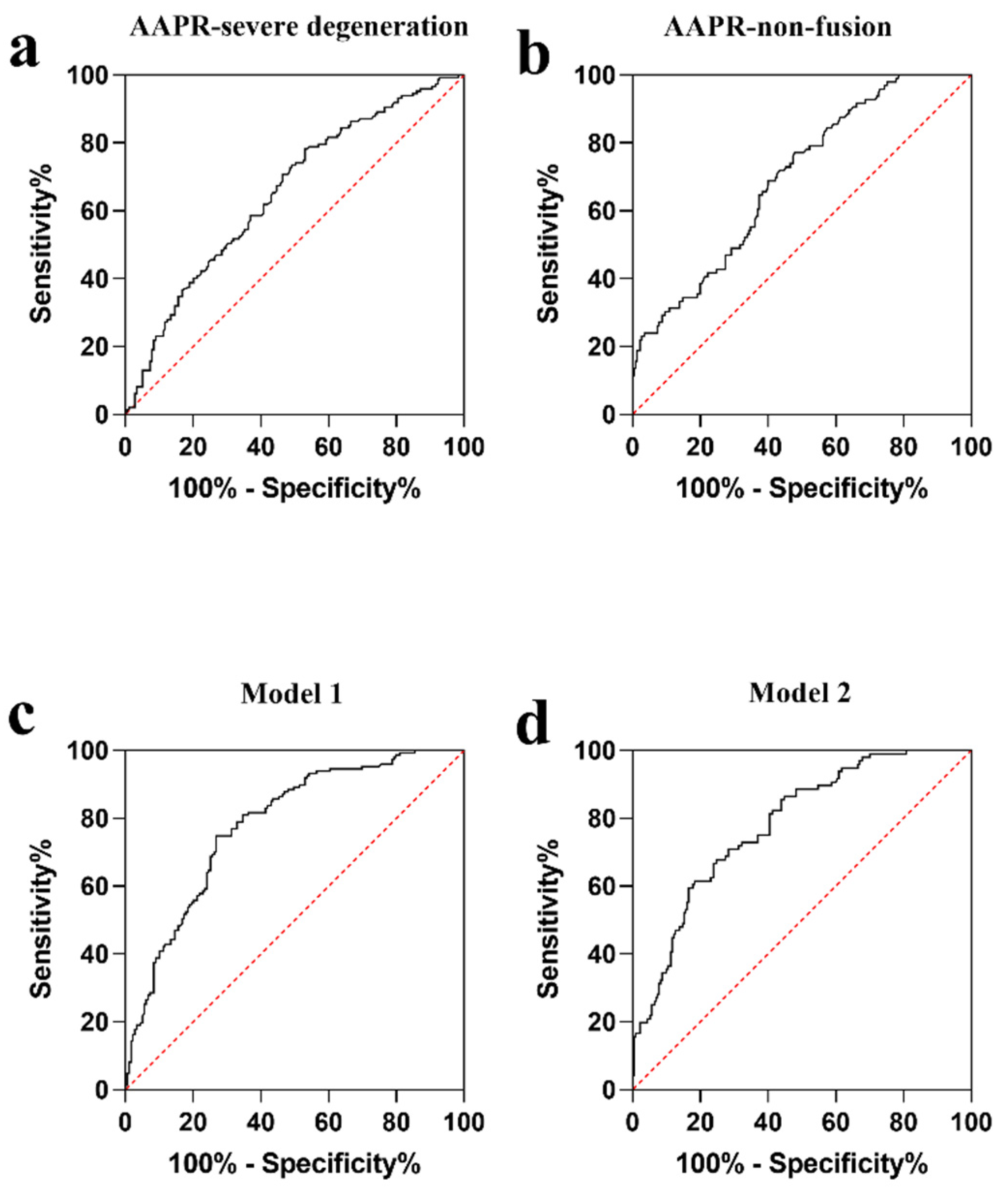
The continuous data were expressed as mean ± standard deviation (SD) and categorical data were presented as frequencies and percentages. The Chi-square and nonparametric tests were used to compare baseline characteristics between groups. Univariate and multivariate analyses were performed by binary logistic regression models to assess the prognostic effect of variables and estimate odds ratios (OR) with 95% confidence intervals (CI). Hosmer–Lemeshow goodness of fit test was used to assess the model fit (Hosmer-Lemeshow statistic ≥ 0.05). The receiver operating characteristic (ROC) curve and the area under the ROC curve (AUC) were also performed to assess the predictive ability of the built models. At the same time, ROC analysis determined AAPR’s predictive power and the optimal cut-off value. p-values for linear trends were calculated using the quartile median values. Collected data were encoded into SPSS 26.0 and analyzed.

3. Results

3.1. Patient Demographics and Outcomes

The baseline characteristics of study participants are shown in Table 1. There were 185 women (56.7%) among the patients with an average age of 63.48. The mean BMI of the patients was 24.80 kg/m2. There were significant differences in the following factors: age (p < 0.001), ALP (p < 0.001), RBP (p < 0.001), AST (p = 0.048), lumbar CT value (p < 0.001), AAPR (p < 0.001), fusion rate (p = 0.001), and the prevalence of hypertension (p = 0.042), disc calcification (p < 0.001) and osteoporosis (p = 0.018) between the high score group (accumulative grade > 18) and the low score group (accumulative grade ≤ 18). No significant differences were observed among the two groups regarding gender distribution, BMI, VAS, the length of hospital stay, hematological indicators other than ALP, AST and RBP, smoking history, etc.

3.2. IDD Severity Classification and Association with AAPR

Table 2 illustrates the distribution of disc grades among the target population. There were 41.7%, 35.9%, and 32.5% of grades for L1/2, L2/3, and L3/4, respectively, which were smaller than 4 (2, 3, and 3). For L4/5 and L5/S1, however, the majority (38.0% and 43.9%) were equal to or greater than 4. At the same time, the low score group showed the same trend as the whole population. On the other hand, all discs except L1/2 scored more than or equal to 4 in the high score group.
We defined mild to moderate degeneration as a score of less than 4 and severe degeneration as a score of more than or equal to 4. As shown in Table 3, the mean levels of LMR were substantially lower in the severe degeneration group (Pfirrmann grade ≥ 4) compared with the mild to moderate degeneration group (Pfirrmann grade < 4) in all lumbar discs except L5/S1. In addition, correlation analysis showed that LMR was significantly correlated with age, osteoporosis, calcification, Scr, VAS, CT value, non-fusion rate, and accumulative grade in all demographic and clinical parameters in Table 4. It is worth mentioning that there is a borderline positive correlation between LMR and UA. The LMR did not show any significant correlation with the length of hospital stay.

3.3. Univariable and Multivariable Analysis on Predictive Factors of Severe Disc Degeneration

Univariable binary logistic regression analysis based on the entire patient cohort showed that each additional unit of age (p < 0.001), hypertension (p = 0.043), osteoporosis (p = 0.019), calcification (p < 0.001), RBP (p = 0.001), and AAPR (p < 0.001) was significantly associated with severe disc degeneration (Table 5). ROC analysis was performed by defining severe disc degeneration as an endpoint, with the AUC(AAPR) being 0.652 (95% CI: 0.593–0.712) and the difference being statistically significant (p < 0.001) (Figure 1a). It is found that the optimal critical value for AAPR is 0.68, while the maximum approximate index is calculated at this point (0.251). Multivariable binary logistic regression model 1 built on clinical parameters further demonstrated that every one unit of increase in RBP (OR: 0.948; 95% CI: 0.919-0.977; p = 0.001), AAPR (OR: 0.114; 95% CI: 0.027-0.482; p = 0.003), and the occurrence of CHD (OR: 0.360; 95% CI: 0.155-0.834; p = 0.017) and disc calcification (OR: 3.215; 95% CI: 1.848-5.594; p < 0.001) were determined to be independent predictors of severe disc degeneration. Moreover, AAPR did not interact significantly with calcification, CAD, or RBP in the one-way ANOVA (p > 0.05). This model is also capable of calibration and discrimination (p > 0.05 and p < 0.05, respectively). The area under ROC curve is 0.782 (Figure 1c). At the same time, trend analysis showed that the higher the AAPR, the lower the risk of severe disc degeneration (p = 0.010; Table 6).

3.4. Univariable and Multivariable Analysis on Risk Factors of Non-Fusion

Univariable binary logistic regression analysis based on the entire patient cohort showed that each additional unit of AAPR (p < 0.001) and phosphorus (p = 0.002) were significantly associated with postoperative non-fusion rate, as shown in Table 7. ROC analysis was performed by defining non-fusion as an endpoint, with the AUC(AAPR) being 0.695 (95% CI: 0.636–0.755) and the difference being statistically significant (p < 0.001) (Figure 1b). The optimal cut-off value for AAPR is 0.63. After adjustment by all covariable estimates, multivariate binary logistic regression analysis model 2 showed that for every one unit increase in UA (OR: 1.005; 95% CI: 1.001-1.010; p = 0.014), phosphorus (OR: 16.677; 95% CI: 2.794–99.552; p = 0.002), AAPR (OR: 0.003; 95% CI: 0.0003–0.022; p < 0.001), and the prevalence of CHD (OR: 0.357; 95% CI: 0.128–0.998; p = 0.049) could be independent prognostic factors for non-fusion in patients with lumbar disease undergoing lumbar fusion surgery. At the time, the one-way ANOVA showed no significant interactions between AAPR and UA, CHD, or phosphorus. In addition, this model has effective calibration and discrimination (p > 0.05 and p < 0.05, respectively). The area under ROC curve of the model is 0.781(Figure 1d). Trend analysis showed that the higher the AAPR, the lower the risk of a non-fusion rate (p < 0.001; Table 6).

4. Discussion

In this study, we evaluated the prognostic value of AAPR after spinal fusion in patients with lumbar degenerative disease. According to analyses of patient characteristics, AAPR was closely related to non-fusion rate and severe disc degeneration. Binary logistic regression analysis showed that AAPR was an independent predictor of fusion rate and severe disc degeneration in the entire cohorts. Additionally, the levels of AAPR in the severe degeneration group were lower than that in the mild to moderate degeneration group, which verified the close relationship between AAPR and the severity of IDD to a certain extent. In addition, previous studies found that L4/5 or L5/S1 levels are the prone sites for lumbar diseases. In this study, we observed that the degree of disc degeneration was more severe at L4/5 and L5/S1 levels, and the cumulative grade was higher than L1/2, L2/3, and L3/4. At the same time, the ROC curve demonstrated that circulation AAPR levels could be used to predict severe IDD. Therefore, the low AAPR appeared to be an independent risk factor for severe disc degeneration. Additionally, AAPR is not the only factor contributing to disc degeneration and fusion rate. In the logistic regression analysis, serum phosphorus, UA, and CHD were also predictive factors for non-fusion, while the occurrence of CHD, disc calcification, and retinol-binding proteins appeared to have an impact on degeneration.
AAPR incorporates the two basic laboratory parameters, ALB and ALP, which are easily accessible and not too expensive. There is a high concentration of albumin in serum, which serves as a storage and transport system for many endogenous and exogenous substances [25]. It can reflect the human nutritional status and inflammatory state and be related to the severity of many diseases [26,27]. Several studies have demonstrated that ALB regulates inflammatory responses by binding to lipopolysaccharides and reactive oxygen species [28]. Moreover, data have been accumulating on the utility of albumin as a prognostic marker, including the prognostic value of different albumin parameters alone or when combined [29,30]. ALP catalyzes the hydrolysis of phosphate esters and is responsible for transferring phosphate groups, which are mainly produced in the liver and bone. ALP activity could reflect the metabolism and immunity of the body and be used as an immunometric [31,32]. ALP activity is increased in various hepatobiliary diseases, rickets, osteogenesis imperfection, osteomalacia, etc. [33,34]. A higher level of ALP alone has been associated with a poor prognosis. It has become increasingly important to determine the level of alkaline phosphatase in serum in clinical medicine for the detection and monitoring of many diseases. However, there is no study on the correlation between serum ALP and disc degeneration. It is worth mentioning that we separately analyzed the effects of ALB and ALP on severe disc degeneration and fusion rate (see the Supplementary Materials). The results showed that ALP was an independent predictor of non-fusion (OR: 1.047, 95% CI: 1.031–1.063, p < 0.001; Supplementary Materials Table S3), not severe disc degeneration (OR: 1.011, 95% CI: 0.999–1.022, p = 0.079; Supplementary Materials Table S1). However, Inose et al. found no significant correlation between serum ALP and non-fusion rate [22]. In sharp contrast, ALB is not significantly associated with severe degeneration (OR: 0.976, 95% CI: 0.889–1.072, p = 0.612; Supplementary Materials Table S2) and non-fusion rate (OR: 1.020, 95% CI: 0.927–1.122, p = 0.685; Supplementary Materials Table S4), which may be related to the sample size. Furthermore, this could be related to the nutritional status of the population included in this study, and there are no primary diseases such as liver and kidney disease, so the groups do not differ significantly. AAPR was applied in patients undergoing surgery for hepatocellular carcinoma for the first time by Chan et al.; this conclusion has been confirmed in the following studies [35,36]. Furthermore, previous research has shown that low levels of this indicator are associated with poor outcomes [37,38]. Despite varying cutoff values, these studies confirm that patients with high AAPR have a better prognosis than those with low AAPR. Therefore, we believe this ratio can provide insight into the microenvironment of local tissue inflammation and can be utilized to measure inflammation status in peripheral blood. It is undeniable that AAPR, a composite index, still has a particular clinical value, even though the effect of AAPR on fusion rate may be due to the mediating effect of ALP.
There were also other factors associated with fusion rate and disc degeneration identified. Multiple binary logistic regression showed that RBP acted as a protective factor against severe disc degeneration, while disc calcification as a risk factor. RBP, a vitamin transporter, is synthesized in the liver and is widely distributed in the blood, cerebrospinal fluid, urinary fluid, and other body fluids. RBP has a complex mechanism of action that exhibits both pro-oxidant and antioxidant effects [39,40]. However, no studies have been conducted on the relationship between RBP and IDD. In addition, intervertebral disc calcification occurs as a result of IDD, and it further aggravates the degeneration [41]. At the same time, the occurrence of calcification was positively correlated with advancing age and a reduced intervertebral height [42]. Calcium deposits in the cellular and extracellular space may cause cell death and decreased activity, resulting in disc degeneration, consistent with this study. A fascinating finding was that although CHD was negatively associated with severe disc degeneration, the effect of CHD on disc degeneration was not significant in models with only CHD and calcification. Furthermore, this study showed that preoperative high serum phosphorus levels were associated with fusion rate, while Shih et al. found no correlation between the fusion rate and the serum levels of calcium or phosphorus [43]. Additionally, we note that UA can enhance fusion rate, which may be related to its antioxidant abilities. Lastly, no significant influence of factors such as age or BMI on disc degeneration or fusion rate was detected, which may be the result of the small sample size.
However, this study has some limitations. Due to the fact that our cohort was a single-center retrospective one containing only Chinese patients, these results may not be generalizable to other populations. Therefore, it is suggested that further multicenter prospective studies be conducted. Additionally, the applicability of the current AAPR cut-off value to other conditions needs to be further examined. At the same time, we investigated the relationship between only one AAPR value and the severity of disc degeneration and fusion rate. Considering that serum AAPR may be affected by other factors, such as liver disease and diet, continuous monitoring may be necessary [44]. Third, larger sample sizes are necessary to test our results, primarily to determine whether or not the statistical significance of results is clinically significant and to measure the smallest clinically meaningful differences. A further research issue is how to exclude the effects of non-steroidal anti-inflammatory drugs, which have been taken by patients before surgery, on AAPR levels in vivo. In addition, since the included population mainly consisted of the elderly with the poor osteogenic ability and the follow-up period was two years, the fusion rate did not meet the expected results. Therefore, we will extend the follow-up period and examine more subtle characteristics of the elderly to verify the validity of this study.

5. Conclusions

The results of our study suggest that preoperative AAPR may be a prognostic predictor of postoperative fusion rate. At the same time, AAPR was related to severe disc degeneration, helping clinicians identify high-risk patients and guide individualized treatment.

Supplementary Materials

The following supporting information can be downloaded at: https://www.mdpi.com/article/10.3390/jcm11164719/s1, Table S1. Univariate and multivariate analysis model 3 (ALP) of risk factors for severe degeneration. Table S2. Univariate and multivariate analysis model 4 (ALB) of risk factors for severe degeneration. Table S3. Univariate and multivariate analysis model 5 (ALP) of risk factors for non-fusion. Table S4. Univariate and multivariate analysis model 6 (ALB) of risk factors for non-fusion.

Author Contributions

Y.G.: conceptualization, methodology, material preparation, data collection, analysis, and original draft preparation. H.Z., H.G., Y.C. and H.X.: investigation, data collection, and visualization. W.Z., T.L. and S.W.: development or design of methodology and creation of models. D.W., T.H. and K.L.: supervision and writing-reviewing and editing. All authors have read and agreed to the published version of the manuscript.

Funding

This study was supported by Shanghai East Hospital Xuri Young Excellent Talents Program 2019xrrcjh04 and Key Laboratory of Inorganic Coating Materials, Chinese Academy of Sciences (HX-2020-027).

Institutional Review Board Statement

The study was conducted in accordance with the Declaration of Helsinki and approved by the Shanghai East Hospital Ethics Committee (approval number EC. D (BG). 016. 02. 1).

Informed Consent Statement

Informed consent was obtained from all subjects involved in the study.

Data Availability Statement

The datasets generated and/or analyzed during the present study are available from the corresponding author upon reasonable request.

Conflicts of Interest

The authors have no relevant financial or non-financial interest to disclose.

References

  1. Luoma, K.; Riihimäki, H.; Luukkonen, R.; Raininko, R.; Viikari-Juntura, E.; Lamminen, A. Low Back Pain in Relation to Lumbar Disc Degeneration. Spine 2000, 25, 487–492. [Google Scholar] [CrossRef] [PubMed]
  2. Costi, J.J.; Stokes, I.; Gardner-Morse, M.; Iatridis, J. Frequency-Dependent Behavior of the Intervertebral Disc in Response to Each of Six Degree of Freedom Dynamic Loading. Spine 2008, 33, 1731–1738. [Google Scholar] [CrossRef] [PubMed]
  3. Suthar, P. MRI Evaluation of Lumbar Disc Degenerative Disease. J. Clin. Diagn. Res. 2015, 9, TC04–TC09. [Google Scholar] [CrossRef] [PubMed]
  4. Wang, S.; Rui, Y.; Lu, J.; Wang, C. Cell and molecular biology of intervertebral disc degeneration: Current understanding and implications for potential therapeutic strategies. Cell Prolif. 2014, 47, 381–390. [Google Scholar] [CrossRef] [PubMed]
  5. Vergroesen, P.-P.; Kingma, I.; Emanuel, K.; Hoogendoorn, R.; Welting, T.; van Royen, B.; van Dieën, J.; Smit, T. Mechanics and biology in intervertebral disc degeneration: A vicious circle. Osteoarthr. Cartil. 2015, 23, 1057–1070. [Google Scholar] [CrossRef]
  6. Molinos, M.; Almeida, C.R.; Caldeira, J.; Cunha, C.; Gonçalves, R.M.; Barbosa, M.A. Inflammation in intervertebral disc degeneration and regeneration. J. R. Soc. Interface 2015, 12, 20150429. [Google Scholar] [CrossRef] [PubMed]
  7. Taniguchi, K.; Karin, M. NF-κB, inflammation, immunity and cancer: Coming of age. Nat. Rev. Immunol. 2018, 18, 309–324. [Google Scholar] [CrossRef] [PubMed]
  8. Eck, J.C.; Sharan, A.; Ghogawala, Z.; Resnick, D.K.; Watters, W.C.; Mummaneni, P.V.; Dailey, A.T.; Choudhri, T.F.; Groff, M.W.; Wang, J.C.; et al. Guideline update for the performance of fusion procedures for degenerative disease of the lumbar spine. Part 7: Lumbar fusion for intractable low-back pain without stenosis or spondylolisthesis. J. Neurosurg. Spine 2014, 21, 42–47. [Google Scholar] [CrossRef] [PubMed]
  9. Loi, F.; Córdova, L.A.; Pajarinen, J.; Lin, T.; Yao, Z.; Goodman, S.B. Inflammation, fracture and bone repair. Bone 2016, 86, 119–130. [Google Scholar] [CrossRef]
  10. Małkowski, P.; Rozga, J.; Piątek, T. Human albumin: Old, new, and emerging applications. Ann. Transplant. 2013, 18, 205–217. [Google Scholar] [CrossRef] [PubMed]
  11. Eckart, A.; Struja, T.; Kutz, A.; Baumgartner, A.; Baumgartner, T.; Zurfluh, S.; Neeser, O.; Huber, A.; Stanga, Z.; Mueller, B.; et al. Relationship of Nutritional Status, Inflammation, and Serum Albumin Levels during Acute Illness: A Prospective Study. Am. J. Med. 2020, 133, 713–722.e7. [Google Scholar] [CrossRef] [PubMed]
  12. Vimalraj, S. Alkaline phosphatase: Structure, expression and its function in bone mineralization. Gene 2020, 754, 144855. [Google Scholar] [CrossRef]
  13. Demer, L.L.; Tintut, Y. Inflammatory, Metabolic, and Genetic Mechanisms of Vascular Calcification. Arter. Thromb. Vasc. Biol. 2014, 34, 715–723. [Google Scholar] [CrossRef]
  14. Estaki, M.; DeCoffe, D.; Gibson, D.L. Interplay between intestinal alkaline phosphatase, diet, gut microbes and immunity. World J. Gastroenterol. 2014, 20, 15650–15656. [Google Scholar] [CrossRef]
  15. Bottini, A.; Berruti, A.; Brizzi, M.P.; Bersiga, A.; Generali, D.; Allevi, G.; Aguggini, S.; Bolsi, G.; Bonardi, S.; Bertoli, G.; et al. Pretreatment haemoglobin levels significantly predict the tumour response to primary chemotherapy in human breast cancer. Br. J. Cancer 2003, 89, 977–982. [Google Scholar] [CrossRef]
  16. Li, D.; Yu, H.; Li, W. Albumin-to-alkaline phosphatase ratio at diagnosis predicts survival in patients with metastatic non-small-cell lung cancer. OncoTargets Ther. 2019, 12, 5241–5249. [Google Scholar] [CrossRef]
  17. Nie, M.; Sun, P.; Chen, C.; Bi, X.; Wang, Y.; Yang, H.; Liu, P.; Li, Z.; Xia, Y.; Jiang, W. Albumin-to-Alkaline Phosphatase Ratio: A Novel Prognostic Index of Overall Survival in Cisplatin-based Chemotherapy-treated Patients with Metastatic Nasopharyngeal Carcinoma. J. Cancer 2017, 8, 809–815. [Google Scholar] [CrossRef]
  18. Fauran-Clavel, M.; Oustrin, J. Alkaline phosphatase and bone calcium parameters. Bone 1986, 7, 95–99. [Google Scholar] [CrossRef]
  19. Skaliczki, G.; Schandl, K.; Weszl, M.; Major, T.; Kovács, M.; Skaliczki, J.; Szendrői, M.; Dobó-Nagy, C.; Lacza, Z. Serum albumin enhances bone healing in a nonunion femoral defect model in rats: A computer tomography micromorphometry study. Int. Orthop. 2013, 37, 741–745. [Google Scholar] [CrossRef]
  20. Horváthy, D.B.; Schandl, K.; Schwarz, C.M.; Renner, K.; Hornyák, I.; Szabó, B.T.; Niculescu-Morzsa, E.; Nehrer, S.; Dobó-Nagy, C.; Doros, A.; et al. Serum albumin-coated bone allograft (BoneAlbumin) results in faster bone formation and mechanically stronger bone in aging rats. J. Tissue Eng. Regen. Med. 2019, 13, 416–422. [Google Scholar] [CrossRef]
  21. Khalooeifard, R.; Oraee-Yazdani, S.; Keikhaee, M.; Shariatpanahi, Z.V. Protein Supplement and Enhanced Recovery after Posterior Spine Fusion Surgery: A Randomized, Double-blind, Placebo-controlled Trial. Clin. Spine Surg. 2022, 35, E356–E362. [Google Scholar] [CrossRef] [PubMed]
  22. Inose, H.; Yamada, T.; Mulati, M.; Hirai, T.; Ushio, S.; Yoshii, T.; Kato, T.; Kawabata, S.; Okawa, A. Bone Turnover Markers as a New Predicting Factor for Nonunion After Spinal Fusion Surgery. Spine 2018, 43, E29–E34. [Google Scholar] [CrossRef] [PubMed]
  23. Siepe, C.J.; Stosch-Wiechert, K.; Heider, F.; Amnajtrakul, P.; Krenauer, A.; Hitzl, W.; Szeimies, U.; Stäbler, A.; Mayer, H.M. Anterior stand-alone fusion revisited: A prospective clinical, X-ray and CT investigation. Eur. Spine J. 2015, 24, 838–851. [Google Scholar] [CrossRef]
  24. Pfirrmann, C.; Metzdorf, A.; Zanetti, M.; Hodler, J.; Boos, N. Magnetic Resonance Classification of Lumbar Intervertebral Disc Degeneration. Spine 2001, 26, 1873–1878. [Google Scholar] [CrossRef] [PubMed]
  25. Farrugia, A. Albumin Usage in Clinical Medicine: Tradition or Therapeutic? Transfus. Med. Rev. 2010, 24, 53–63. [Google Scholar] [CrossRef]
  26. Don, B.R.; Kaysen, G. Poor Nutritional Status and Inflammation: Serum Albumin: Relationship to Inflammation and Nutrition. Semin. Dial. 2004, 17, 432–437. [Google Scholar] [CrossRef] [PubMed]
  27. Deveci, B.; Gazi, E. Relation Between Globulin, Fibrinogen, and Albumin with the Presence and Severity of Coronary Artery Disease. Angiology 2021, 72, 174–180. [Google Scholar] [CrossRef] [PubMed]
  28. Belcher, D.A.; Williams, A.T.; Palmer, A.F.; Cabrales, P. Polymerized albumin restores impaired hemodynamics in endotoxemia and polymicrobial sepsis. Sci. Rep. 2021, 11, 10834. [Google Scholar] [CrossRef]
  29. Galata, C.; Busse, L.; Birgin, E.; Weiß, C.; Hardt, J.; Reissfelder, C.; Otto, M. Role of Albumin as a Nutritional and Prognostic Marker in Elective Intestinal Surgery. Can. J. Gastroenterol. Hepatol. 2020, 2020, 7028216. [Google Scholar] [CrossRef]
  30. Kawashima, A.; Yamamoto, Y.; Sato, M.; Nakata, W.; Kakuta, Y.; Ishizuya, Y.; Yamaguchi, Y.; Yamamoto, A.; Yoshida, T.; Takayama, H.; et al. FAN score comprising fibrosis-4 index, albumin–bilirubin score and neutrophil–lymphocyte ratio is a prognostic marker of urothelial carcinoma patients treated with pembrolizumab. Sci. Rep. 2021, 11, 21199. [Google Scholar] [CrossRef]
  31. Singh, S.B.; Lin, H.C. Role of Intestinal Alkaline Phosphatase in Innate Immunity. Biomolecules 2021, 11, 1784. [Google Scholar] [CrossRef] [PubMed]
  32. Vercalsteren, E.; Vranckx, C.; Lijnen, H.R.; Hemmeryckx, B.; Scroyen, I. Adiposity and metabolic health in mice deficient in intestinal alkaline phosphatase. Adipocyte 2018, 7, 149–155. [Google Scholar] [CrossRef]
  33. Gutman, A.B. Serum alkaline phosphatase activity in diseases of the skeletal and hepatobiliary systems: A consideration of the current status. Am. J. Med. 1959, 27, 875–901. [Google Scholar] [CrossRef]
  34. Peach, H.; Compston, J.E.; Vedi, S.; Horton, L.W. Value of plasma calcium, phosphate, and alkaline phosphatase measurements in the diagnosis of histological osteomalacia. J. Clin. Pathol. 1982, 35, 625–630. [Google Scholar] [CrossRef] [PubMed]
  35. Chen, Z.-H.; Zhang, X.-P.; Cai, X.-R.; Xie, S.-D.; Liu, M.-M.; Lin, J.-X.; Ma, X.-K.; Chen, J.; Lin, Q.; Dong, M.; et al. The Predictive Value of Albumin-to-Alkaline Phosphatase Ratio for Overall Survival of Hepatocellular Carcinoma Patients Treated with Trans-Catheter Arterial Chemoembolization Therapy. J. Cancer 2018, 9, 3467–3478. [Google Scholar] [CrossRef] [PubMed]
  36. Li, H.; Li, J.; Wang, J.; Liu, H.; Cai, B.; Wang, G.; Wu, H. Assessment of Liver Function for Evaluation of Long-Term Outcomes of Intrahepatic Cholangiocarcinoma: A Multi-Institutional Analysis of 620 Patients. Front. Oncol. 2020, 10, 525. [Google Scholar] [CrossRef] [PubMed]
  37. Acikgoz, Y.; Bal, O.; Dogan, M. Albumin-to-Alkaline Phosphatase Ratio: Does It Predict Survival in Grade 1 and Grade 2 Neuroendocrine Tumors? Pancreas 2021, 50, 111–117. [Google Scholar] [CrossRef]
  38. Li, J.; Zuo, M.; Zhou, X.; Xiang, Y.; Zhang, S.; Feng, W.; Liu, Y. Prognostic Significance of Preoperative Albumin to Alkaline Phosphatase Ratio in Patients with Glioblastoma. J. Cancer 2021, 12, 5950–5959. [Google Scholar] [CrossRef]
  39. Chen, J.; Shao, Y.; Sasore, T.; Moiseyev, G.; Zhou, K.; Ma, X.; Du, Y.; Ma, J.-X. Interphotoreceptor Retinol-Binding Protein Ameliorates Diabetes-Induced Retinal Dysfunction and Neurodegeneration through Rhodopsin. Diabetes 2021, 70, 788–799. [Google Scholar] [CrossRef]
  40. Wang, J.; Chen, H.; Liu, Y.; Zhou, W.; Sun, R.; Xia, M. Retinol binding protein 4 induces mitochondrial dysfunction and vascular oxidative damage. Atherosclerosis 2015, 240, 335–344. [Google Scholar] [CrossRef]
  41. Rutges, J.; Duit, R.; Kummer, J.; Oner, F.; van Rijen, M.; Verbout, A.; Castelein, R.; Dhert, W.; Creemers, L. Hypertrophic differentiation and calcification during intervertebral disc degeneration. Osteoarthr. Cartil. 2010, 18, 1487–1495. [Google Scholar] [CrossRef] [PubMed]
  42. Hawellek, T.; Hubert, J.; Hischke, S.; Rolvien, T.; Krause, M.; Püschel, K.; Rüther, W.; Niemeier, A. Microcalcification of lumbar spine intervertebral discs and facet joints is associated with cartilage degeneration, but differs in prevalence and its relation to age. J. Orthop. Res. 2017, 35, 2692–2699. [Google Scholar] [CrossRef]
  43. Shih, T.-Y.; Wu, Y.-C.; Tseng, S.-C.; Chen, K.-H.; Pan, C.-C.; Lee, C.-H. Correlation between Preoperative Serum Levels of Calcium, Phosphate, and Intact Parathyroid Hormone and Radiological Outcomes in Spinal Interbody Fusion among End-Stage Renal Disease Patients. J. Clin. Med. 2021, 10, 5447. [Google Scholar] [CrossRef] [PubMed]
  44. Sheng, G.; Peng, N.; Hu, C.; Zhong, L.; Zhong, M.; Zou, Y. The albumin-to-alkaline phosphatase ratio as an independent predictor of future non-alcoholic fatty liver disease in a 5-year longitudinal cohort study of a non-obese Chinese population. Lipids Health Dis. 2021, 20, 50. [Google Scholar] [CrossRef] [PubMed]
Figure 1. Receiver operating characteristic (ROC) curve to determine the predictive performance of AAPR for severe degeneration (a) and non-fusion (b). ROC curve analysis of severe degeneration (c) and non-fusion models (d) (see Table 5 and Table 7 for included variables).
Jcm 11 04719 g001
Table 1. Demographic characteristics of patients with disc degeneration disease.
AllLow Score GroupHigh Score Groupp-Value
(Accumulative
Grade ≤ 18)
(Accumulative
Grade > 18)
Subjects, n (%)326179147
Age (year)63.48 ± 13.3860.49 ± 14.9367.11 ± 10.13<0.001
Gender 0.562
Male, n (%)141 (43.3)80 (44.7)61 (41.5)
Female, n (%)185 (56.7)99 (55.3)86 (58.5)
BMI (kg/m2)24.80 ± 3.5424.96 ± 3.4924.60 ± 3.600.371
Smoking (y)42 (12.9)22 (12.3)20 (13.6)0.724
Alcohol abuse (y)27 (8.3)11 (6.1)16 (10.9)0.122
Hypertension (y)155 (47.5)76 (42.5)79 (53.7)0.042
DM (y)57 (17.5)30 (16.8)27 (18.4)0.704
CHD (y)40 (12.3)24 (13.4)16 (10.9)0.490
Osteoporosis (y)115 (35.3)53 (29.6)62 (42.2)0.018
Calcification (y)164 (50.3)60 (33.5)104 (70.7)<0.001
ALB (g/L)42.49 ± 3.2942.56 ± 3.1942.41 ± 3.410.923
ALP (U/L)74.18 ± 27.2768.65 ± 24.0280.90 ± 29.47<0.001
AAPR0.64 ± 0.220.69 ± 0.240.58 ± 0.17<0.001
Calcium (mmol/L)2.26 ± 0.102.25 ± 0.092.27 ± 0.100.092
Phosphorus (mmol/L)1.14 ± 0.171.15 ± 0.161.14 ± 0.170.505
FBG (mmol/L)5.54 ± 1.475.45 ± 1.365.65 ± 1.600.394
BUN (mmol/L)6.04 ± 1.776.05 ± 1.736.02 ± 1.820.547
Scr (μmol/L)72.33 ± 23.2272.09 ± 21.6572.62 ± 25.060.779
UA (μmol/L)320.44 ± 82.57323.42 ± 80.03316.82 ± 85.690.597
ALT (U/L)19.51 ± 14.3420.12 ± 16.0618.77 ± 11.920.549
AST (U/L)19.12 ± 8.6418.75 ± 9.8019.56 ± 6.990.048
RBP (mg/L)41.26 ± 9.5442.83 ± 9.4939.35 ± 9.27<0.001
Fusion (y)230 (70.6)140 (78.2)90 (61.2)0.001
VAS3.91 ± 1.843.73 ± 1.844.13 ± 1.840.067
Hospital stay (day)12.21 ± 4.3912.20 ± 4.3612.21 ± 4.450.854
CT value (HU)131.26 ± 49.20140.51 ± 52.51120.01 ± 42.35<0.001
Values are expressed as n (%) or mean ± SD. BMI, body mass index; VAS, visual analogue scale; AAPR, albumin-to-alkaline phosphatase ratio; FBG, fasting blood glucose; CHD, coronary heart disease; DM, diabetes mellitus; ALT, alanine transaminase; AST, aspartate transaminase; RBP, retinol-binding Protein; UA, uric acid; BUN, blood urea nitrogen; Scr, serum creatinine.
Table 2. The Pfirrmann grading system for lumbar disc degeneration.
12345
All (n = 326)
L1/20136 (41.7)106 (32.5)50 (15.3)34 (10.4)
L2/3079 (24.2)117 (35.9)78 (23.9)52 (16.0)
L3/41 (0.3)49 (15.0)106 (32.5)105 (32.2)65 (19.9)
L4/5024 (7.4)82 (25.2)124 (38.0)96 (29.4)
L5/S1027 (8.3)53 (16.3)103 (31.6)143 (43.9)
Low score group
(n = 179)
L1/20114 (63.7)47 (26.3)14 (7.8)4 (2.2)
L2/3077 (43.0)81 (45.3)19 (10.6)2 (1.1)
L3/41 (0.6)49 (27.4)90 (50.3)37 (20.7)2 (1.1)
L4/5024 (13.4)64 (35.8)74 (41.3)17 (9.5)
L5/S1027 (15.1)42 (23.5)65 (36.3)45 (25.1)
High score group
(n = 147)
L1/2022 (15.0)59 (40.1)36 (24.5)30 (20.4)
L2/302 (1.4)36 (24.5)59 (40.1)50 (34.0)
L3/40016 (10.9)68 (46.3)63 (42.9)
L4/50018 (12.2)50 (34.0)79 (53.7)
L5/S10011 (7.5)38 (25.9)98 (66.7)
Values are expressed as n (%).
Table 3. The relationship between the severity of individual disc degeneration and AAPR/.
AAPRp
L1/2Pfirrmann grade < 40.65 ± 0.210.003
Pfirrmann grade ≥ 40.59 ± 0.22
L2/3Pfirrmann grade < 40.68 ± 0.23<0.001
Pfirrmann grade ≥ 40.58 ± 0.18
L3/4Pfirrmann grade < 40.70 ± 0.25<0.001
Pfirrmann grade ≥ 40.58 ± 0.17
L4/5Pfirrmann grade < 40.68 ± 0.19<0.001
Pfirrmann grade ≥ 40.62 ± 0.23
L5/S1Pfirrmann grade < 40.67 ± 0.230.181
Pfirrmann grade ≥ 40.63 ± 0.21
Values are expressed as mean ± SD; AAPR, albumin-to-alkaline phosphatase ratio.
Table 4. Correlation of AAPR with demographic and clinical parameters.
rp
Age−0.1100.046
Gender0.0700.210
BMI0.0280.611
Smoking0.0260.640
Alcohol abuse−0.0700.209
Hypertension−0.0450.417
DM−0.0140.796
CHD−0.0580.295
Osteoporosis−0.1670.002
Calcification−0.422<0.001
Calcium0.0250.656
phosphorus0.0800.150
FBG−0.0340.546
BUN0.0460.408
Scr0.1790.001
UA0.1080.052
ALT−0.0190.728
AST−0.0640.249
RBP0.0360.521
Non-fusion−0.1320.017
VAS−0.1320.017
Hospital stay, day0.0870.118
CT value0.198<0.001
Accumulative grade−0.379<0.001
BMI, body mass index; VAS, visual analogue scale; AAPR, albumin-to-alkaline phosphatase ratio; FBG, fasting blood glucose; CHD, coronary heart disease; DM, diabetes mellitus; ALT, alanine transaminase; AST, aspartate transaminase; RBP, retinol-binding protein; UA, uric acid; BUN, blood urea nitrogen; Scr, serum creatinine.
Table 5. Univariate and multivariate analysis model 1 of risk factors for severe degeneration.
VariablesUnivariateMultivariate
OR (95% CI)pOR (95% CI)p
Age (year)1.042 (1.023–1.062)<0.0011.027 (0.999–1.055)0.062
Gender (male)0.878 (0.565–1.364)0.5620.953 (0.478–1.903)0.892
BMI0.971 (0.913–1.034)0.3580.979 (0.903–1.061)0.604
Smoking1.124 (0.587–2.151)0.7241.492 (0.623–3.574)0.369
Alcohol abuse1.865 (0.837–4.155)0.1272.037 (0.735–5.646)0.172
Hypertension1.574 (1.015–2.443)0.0431.179 (0.649–2.141)0.588
DM1.117 (0.630–1.982)0.7040.935 (0.397–2.201)0.877
CHD0.789 (0.402–1.548)0.4900.360 (0.155–0.834)0.017
Osteoporosis1.734 (1.096–2.742)0.0191.045 (0.581–1.880)0.883
Calcification4.797 (2.993–7.689)<0.0013.215 (1.848–5.594)<0.001
AAPR0.055 (0.015–0.194)<0.0010.114 (0.027–0.482)0.003
Calcium6.241 (0.649–59.996)0.11314.486 (0.796–263.562)0.071
phosphorus0.618 (0.166–2.308)0.4741.456 (0.296–7.161)0.644
FBG1.096 (0.944–1.274)0.2291.080 (0.868–1.344)0.489
BUN0.988 (0.873–1.118)0.8500.975 (0.822–1.157)0.773
Scr1.001 (0.992–1.010)0.8391.004 (0.988–1.021)0.590
UA0.999 (0.996–1.002)0.4721.001 (0.997–1.004)0.755
ALT0.993 (0.977–1.009)0.4050.996 (0.961–1.032)0.832
AST1.011 (0.985–1.038)0.4081.003 (0.947–1.061)0.928
RBP0.960 (0.937–0.984)0.0010.948 (0.919–0.977)0.001
BMI, body mass index; AAPR, albumin-to-alkaline phosphatase ratio; FBG, fasting blood glucose; CHD, coronary heart disease; DM, diabetes mellitus; ALT, alanine transaminase; AST, aspartate transaminase; RBP, retinol-binding protein; UA, uric acid; BUN, blood urea nitrogen; Scr, serum creatinine.
Table 6. Association of severe degeneration and non-fusion with AAPR.
VariableCasesModel 1 (Degeneration Model)Model 2 (Non-Fusion Model)
OR [95% CI]p for TrendOR [95% CI]p for Trend
AAPR (Median [Range])
Q1 (0.42 [≤0.49])82Reference Reference
Q2 (0.56 [0.49–0.61])810.632 [0.306–1.306] 0.653 [0.320–1.334]
Q3 (0.67 [0.61–0.75])820.731 [0.342–1.563] 0.400 [0.184–0.873]
Q4 (0.85 [>0.75])810.316 [0.139–0.719]0.0100.103 [0.038–0.277]<0.001
AAPR, albumin-to-alkaline phosphatase ratio.
Table 7. Univariate and multivariate analysis model 2 of risk factors for non-fusion.
VariablesUnivariateMultivariate
OR (95% CI)pOR (95% CI)p
Age (year)0.995 (0.977–1.013)0.5690.987 (0.960–1.014)0.345
Gender (male)0.630 (0.385–1.031)0.0660.570 (0.261–1.247)0.159
BMI0.973 (0.909–1.042)0.4320.959 (0.880–1.046)0.345
Smoking1.086 (0.538–2.191)0.8191.915 (0.743–4.936)0.179
Alcohol abuse0.826 (0.337–2.023)0.6750.808 (0.262–2.489)0.710
Hypertension0.855 (0.530–1.379)0.5201.101 (0.582–2.081)0.767
DM0.742 (0.385–1.432)0.3740.666 (0.237–1.873)0.441
CHD0.470 (0.200–1.102)0.0820.357 (0.128–0.998)0.049
Osteoporosis1.222 (0.746–2.002)0.4261.129 (0.591–2.156)0.713
Calcification1.581 (0.977–2.559)0.0620.834 (0.439–1.586)0.580
AAPR0.009 (0.002–0.047)<0.0010.003 (0.0003–0.022)<0.001
Calcium1.616 (0.144–18.189)0.6970.397 (0.017–9.500)0.569
phosphorus9.892 (2.270–43.106)0.00216.677 (2.794–99.552)0.002
FBG1.024 (0.873–1.201)0.7701.104 (0.868–1.405)0.418
BUN0.985 (0.859–1.128)0.8231.007 (0.837–1.212)0.938
Scr0.988 (0.976–1.001)0.0620.996 (0.976–1.016)0.673
UA1.001 (0.998–1.004)0.5311.005 (1.001–1.010)0.014
ALT1.002 (0.986–1.018)0.8080.988 (0.952–1.025)0.510
AST1.014 (0.987–1.041)0.3181.016 (0.959–1.076)0.595
RBP0.988 (0.963–1.014)0.3560.986 (0.956–1.016)0.344
BMI, body mass index; AAPR, albumin-to-alkaline phosphatase ratio; FBG, fasting blood glucose; CHD, coronary heart disease; DM, diabetes mellitus; ALT, alanine transaminase; AST, aspartate transaminase; RBP, retinol-binding protein; UA, uric acid; BUN, blood urea nitrogen; Scr, serum creatinine.
Publisher’s Note: MDPI stays neutral with regard to jurisdictional claims in published maps and institutional affiliations.

