Next Article in Journal
Prediction of the Responsiveness to Vagus-Nerve Stimulation in Patients with Drug-Resistant Epilepsy via Directed-Transfer-Function Analysis of Their Perioperative Scalp EEGs
Next Article in Special Issue
Treatment for the Thoracic Ossification of the Posterior Longitudinal Ligament and Ossification of the Ligamentum Flavum
Previous Article in Journal
Two Patterns of White Matter Connection in Multiple Gliomas: Evidence from Probabilistic Fiber Tracking
 
 
jcm-logo
Article Menu

Article Menu

Article

Clinical Characteristics of Patients with Ossification of the Posterior Longitudinal Ligament and a High OP Index: A Multicenter Cross-Sectional Study (JOSL Study)

by
Takashi Hirai
1,*,
Toshitaka Yoshii
1,
Jun Hashimoto
1,
Shuta Ushio
1,
Kanji Mori
2,
Satoshi Maki
3,
Keiichi Katsumi
4,
Narihito Nagoshi
5,
Kazuhiro Takeuchi
6,
Takeo Furuya
3,
Kei Watanabe
4,
Norihiro Nishida
7,
Soraya Nishimura
5,
Kota Watanabe
5,
Takashi Kaito
8,
Satoshi Kato
9,
Katsuya Nagashima
10,
Masao Koda
10,
Hiroaki Nakashima
11,
Shiro Imagama
11,
Kazuma Murata
12,
Yuji Matsuoka
12,
Kanichiro Wada
13,
Atsushi Kimura
14,
Tetsuro Ohba
15,
Hiroyuki Katoh
16,
Masahiko Watanabe
16,
Yukihiro Matsuyama
17,
Hiroshi Ozawa
18,
Hirotaka Haro
15,
Katsushi Takeshita
14,
Morio Matsumoto
5,
Masaya Nakamura
5,
Satoru Egawa
1,
Yu Matsukura
1,
Hiroyuki Inose
1,
Atsushi Okawa
1,
Masashi Yamazaki
10 and
Yoshiharu Kawaguchi
19
add Show full author list remove Hide full author list
1
Department of Orthopedic Surgery, Tokyo Medical and Dental University, Bunkyo, Tokyo 113-8519, Japan
2
Department of Orthopaedic Surgery, Shiga University of Medical Science, Ōtsu 520-2192, Japan
3
Department of Orthopedic Surgery, Chiba University Graduate School of Medicine, Chiba 260-8670, Japan
4
Department of Orthopedic Surgery, Niigata University Medical and Dental General Hospital, Niigata 951-8520, Japan
5
Department of Orthopedic Surgery, Keio University School of Medicine, Shinjuku, Tokyo 160-8582, Japan
6
Department of Orthopedic Surgery, National Hospital Organization Okayama Medical Center, Okayama 701-1192, Japan
7
Department of Orthopedic Surgery, Yamaguchi University Graduate School of Medicine, Ube 755-8505, Japan
8
Department of Orthopaedic Surgery, Osaka University Graduate School of Medicine, Suita 565-0871, Japan
9
Department of Orthopedic Surgery, Graduate School of Medical Sciences, Kanazawa University, Kanazawa 920-8641, Japan
10
Department of Orthopedic Surgery, Faculty of Medicine, University of Tsukuba, Tsukuba 305-8577, Japan
11
Department of Orthopedic Surgery, Graduate School of Medicine, Nagoya University, Nagoya 464-8601, Japan
12
Department of Orthopedic Surgery, Tokyo Medical University, Shinjuku, Tokyo 160-8402, Japan
13
Department of Orthopedic Surgery, Hirosaki University Graduate School of Medicine, Hirosaki 036-8562, Japan
14
Department of Orthopedics, Jichi Medical University, Shimotsuke 329-0498, Japan
15
Department of Orthopedic Surgery, University of Yamanashi, Chuo 409-3898, Japan
16
Department of Orthopedic Surgery, Surgical Science, Tokai University School of Medicine, Isehara 259-1193, Japan
17
Department of Orthopedic Surgery, Hamamatsu University School of Medicine, Hamamatsu 431-3125, Japan
18
Department of Orthopaedic Surgery, Tohoku Medical and Pharmaceutical University, Sendai 983-8536, Japan
19
Department of Orthopedic Surgery, Faculty of Medicine, University of Toyama, Toyama 930-8555, Japan
*
Author to whom correspondence should be addressed.
J. Clin. Med. 2022, 11(13), 3694; https://doi.org/10.3390/jcm11133694
Submission received: 29 April 2022 / Revised: 17 June 2022 / Accepted: 23 June 2022 / Published: 27 June 2022

Abstract

:
Background: The purpose of this study was to clarify the clinical features of ossification of the posterior longitudinal ligament (OPLL) and extreme ossification at multiple sites. Methods: This prospective study involved patients with a diagnosis of cervical OPLL at 16 institutions in Japan. Patient-reported outcome measures, including responses on the Japanese Orthopaedic Association (JOA) Cervical Myelopathy Evaluation Questionnaire (JOA-CMEQ), JOA Back Pain Evaluation Questionnaire (JOA-BPEQ), and visual analog scale pain score, were collected to investigate clinical status. In each patient, the sum of the levels at which OPLL was located (OP index) was evaluated on whole-spine computed tomography, along with ossification of other spinal ligaments including the anterior longitudinal ligament (OALL), ligament flavum (OLF), supra- and intraspinous ligaments (SSL), and diffuse idiopathic skeletal hyperostosis (DISH). The distribution of OP index values in the study population was investigated, and the clinical and radiologic characteristics of patients in the top 10% were assessed. Results: In total, 236 patients (163 male, 73 female; mean age 63.5 years) were enrolled. Twenty-five patients with OP index ≥ 17 were categorized into a high OP index group and the remainder into a moderate/low OP index group. There were significantly more women in the high OP index group. Patients in the high OP index group also had significantly poorer scores for lower extremity function and quality of life on the JOA-CMEQ and in each domain but not for body pain on the JOA-BPEQ compared with those in the moderate/low OP index group. Patients in the high OP index group had more OALL in the cervical spine and more OLF and SSL in the thoracic spine. The prevalence of DISH was also significantly higher in the high OP index group. In the high OP index group, interestingly, OPLL was likely to be present adjacent to DISH in the cervicothoracic and thoracolumbar spine, especially in men, and often coexisted with DISH in the thoracic spine in women. Conclusion: This prospective cohort registry study is the first to demonstrate the clinical and radiologic features of patients with OPLL and a high OP index. In this study, patients with a high OP index had poorer physical function in the lumbar spine and lower extremities and were also predisposed to extreme ossification of spinal ligaments other than the OPLL.

1. Introduction

Ossification of the spinal ligaments is the term used to describe heterotopic ossified lesions that can occur throughout the spine. Ossification of the posterior longitudinal ligament (OPLL) has been recognized as a common cause of neurologic disorders, including myelopathy, radiculopathy, and/or amyotrophy. Epidemiological studies have shown that cervical OPLL has a male predominance of 2:1 to 3:1 [1], whereas thoracic OPLL has a female predominance of 2:1 to 3:1 [2,3]. Moreover, a recent genome-wide association study identified six candidate genes for the pathogenesis of OPLL [4]. Therefore, the onset and expansion of ossified lesions may be closely associated with sex hormones and genetic background.
Various groups have reported on OPLL, including its epidemiology [1,2,3], surgical treatment [5,6,7], and radiological features [8,9,10,11,12,13,14,15,16]. Computed tomography (CT) has been widely used to evaluate bone structure and the distribution of ossification in the whole spine. It has been documented that the onset and progression of neurological symptoms are associated with the degree of canal narrowing caused by OPLL and segmental mobility at the level where OPLL is present [5]. Patients with OPLL and excessive spinal cord compression due to OPLL often have multiple lesions throughout the spine [9]. Given that thoracic OPLL sometimes leads to a concurrent spinal cord disorder with cervical ossification, it is important to evaluate ossified lesions and diagnose the level at which compression caused by ossification results in neurological deterioration.
Although the distribution of ossified lesions has been well investigated in other studies [10,11], there has been no research on the clinical characteristics of patients with OPLL in whom ossification occurs in a large number of segments. We have defined an OP index which reflects the number of segments where OPLL is present and evaluated the predisposition to ossification in individual patients with OPLL [12,13,14,15,16]. In this study, we investigated the clinical features of patients with a large number of ossifications and a high OP index and characteristics of bony bridging lesions of both DISH and OPLL in patients with a high OP index using data from a nationwide patient registry started by the Japanese Multicenter Research Organization for Ossification of the Spinal Ligament (JOSL), which was established by the Japanese Ministry of Health, Labour and Welfare.

2. Materials and Methods

This study had a multi-institutional prospective cross-sectional design and analyzed data from patients who visited 16 JOSL member institutions between September 2015 and December 2017. The patients in this study largely overlapped with those in our previous study, but here our analysis focused on the characteristics of only those patients with a high OP index. The inclusion criteria, clinical evaluation, and radiographic evaluation were described previously [9]. The inclusion criteria were age ≥ 20 years; cervical OPLL diagnosed based on radiographic findings; symptoms such as neck pain and upper and/or lower extremity numbness regardless of the need for surgery and presence of clumsiness and gait disturbance; and whole-spine CT scans available for localizing ossified lesions. Patients with a history of cervical spine surgery for OPLL were excluded. The study was approved by the institutional review board of each participating institution and conducted in accordance with the relevant guidelines and regulations.
The baseline demographic and clinical data listed in Table 1 and Table 2 were collected for all patients, and included evaluation of the cervical Japanese Orthopaedic Association (JOA) score for functional assessment in patients with cervical myelopathy [17]; the JOA Cervical Myelopathy Evaluation Questionnaire (JOA-CMEQ) for assessment of quality of life and functional assessment of the cervical spine, upper and lower extremities, and bladder and [18]; and the JOA Back Pain Evaluation Questionnaire (JOA-BPEQ) lumbar spine function, social dysfunction, mentality, locomotive function, and body pain [18]. A visual analog scale (VAS) was used to assess severity of pain or stiffness in the neck or shoulders, pain or numbness in the arms or hands, and low back pain.
Radiological evaluation was performed as in our previous study [9]. Briefly, whole-spine CT images were collected for each patient, including the cervical, thoracic, and lumbosacral spine. The incidence of OPLL was examined on mid-sagittal CT images in the cervical spine (the clivus to C7) and in the thoracic and lumbosacral regions from T1 to S1. As in previous studies [9,13,14,15,16], images were independently evaluated by senior spine surgeons (S.U., K.M., S.M., K.K., N.N., and K.T.) blinded to clinical outcomes. The OP index was defined as the number of levels with OPLL in the whole spine [9,16] and was also calculated for each patient.
The patients were then divided into a high OP index group (top 10% of OP index values) and a moderate/low OP index group. As in previous reports [13,14,19], the numbers of ossified spinal ligaments other than OPLL were also evaluated, namely, the anterior longitudinal ligament (OALL), ligament flavum (OLF), supra- and interspinous ligaments, and nuchal ligament. In addition, we evaluated the extent to which OPLL occupied the spinal canal in the cervical region and classified the most compressed segment according to the canal narrowing ratio (CNR) [7] as follows: Grade 1, 0–25%; Grade 2, 26–50%; Grade 3, 51–75%; Grade 4, >75%. OPLL was assessed as diffuse idiopathic skeletal hyperostosis (DISH) if it completely bridged at least 4 contiguous adjacent vertebral bodies according to the criteria reported by Nishimura et al. [20]. The distributions of OPLL and DISH in the whole spine were noted for each case in the high OP index group.
The two groups were compared using the unpaired t-test, Mann-Whitney U test, and chi-squared test as appropriate. The data are presented as the mean ± standard deviation. All statistical analyses were performed using SPSS for Windows (ver. 22.0; IBM Corp., Armonk, NY, USA). A p-value of less than 0.05 was considered statistically significant.

3. Results

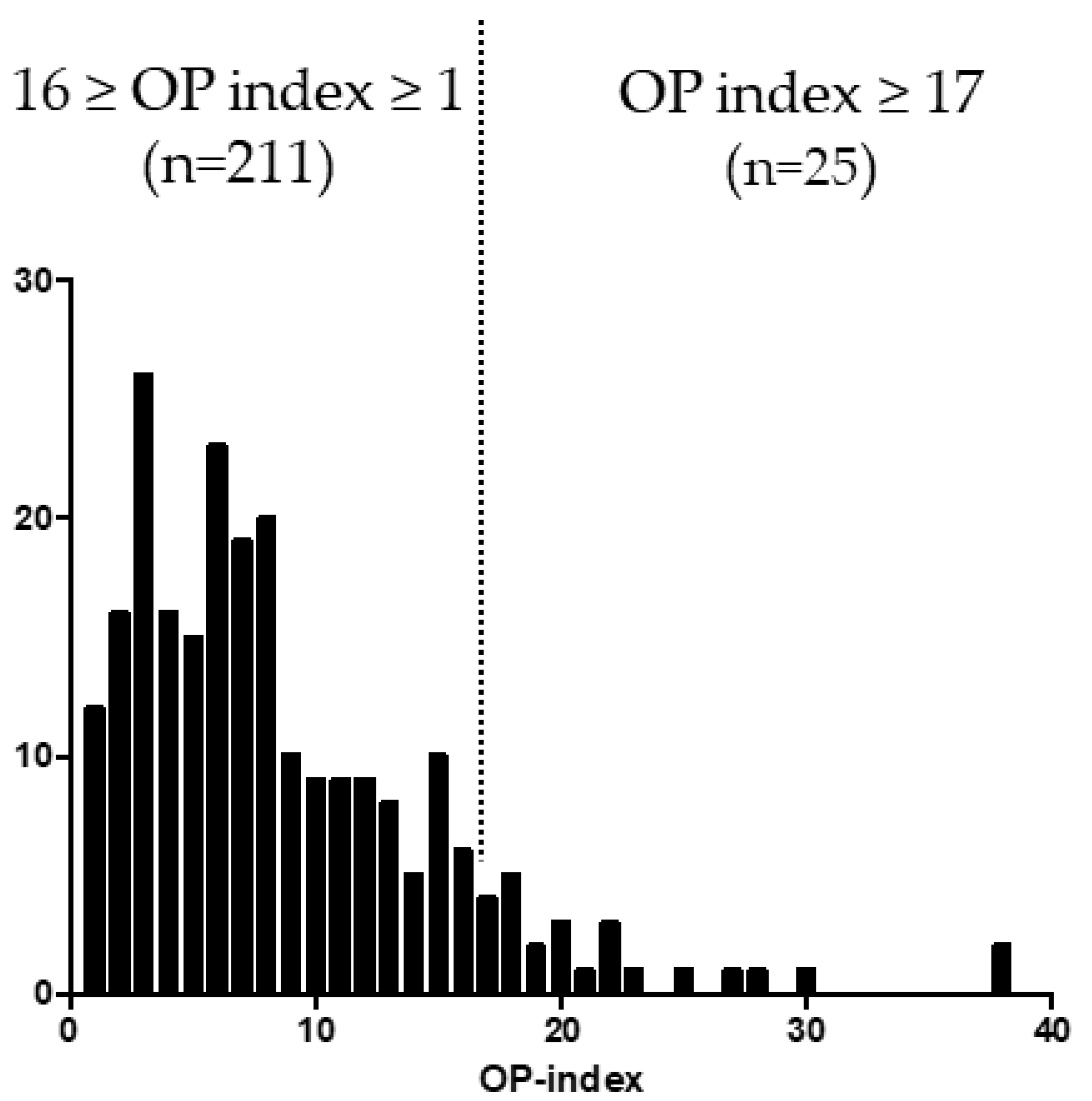
3.1. Distribution of OP-Index Values in Patients with OPLL

A single peak wave was found in the distribution of OP index values in the study population. The patients with the top 10% OP index value (n = 25) had at least 17 ossified lesions (Figure 1). These cases were defined as the high OP index group, and the remaining patients (n = 211) in whom the OP index was ≤16 were defined as the moderate/low OP index group.

3.2. Demographic Characteristics in the High and Moderate/Low OP Index Groups

The patient demographics are shown in Table 1. Mean age was 59.0 years in the high OP index group and 64.5 years in the moderate/low OP index group. Patients in the high OP index group were relatively younger, although the difference was not statistically significant. There were significantly fewer men in the high OP index group; however, there was no significant difference between the groups in body mass index, prevalence of diabetes mellitus, or cervical JOA score.

3.3. Deterioration of Lumbar Spine and Lower Extremity Function in the High OP-Index Group

Patient-reported evaluations, including JOA-CMEQ, JOA-BPEQ, and VAS scores, were assessed to determine whether neurological symptoms were associated with the severity of ossified lesions in the high OP index group (Table 2). There was no significant difference between the two groups in the prevalence of pain. On the JOA-CMEQ, patients in high OP index group had significantly poorer lower extremity function and quality of life scores compared with those in the moderate/low OP index group; however, there was no significant difference in cervical spine, upper extremity, or bladder function. There was also a significant difference in the score for each domain of the JOA-BPEQ, with the exception of body pain. VAS scores were significantly higher for numbness below the chest, low back pain, lower extremity numbness, and lower extremity pain in the high OP index group but no difference was noted in neck pain, numbness in the upper extremities, or chest constriction.

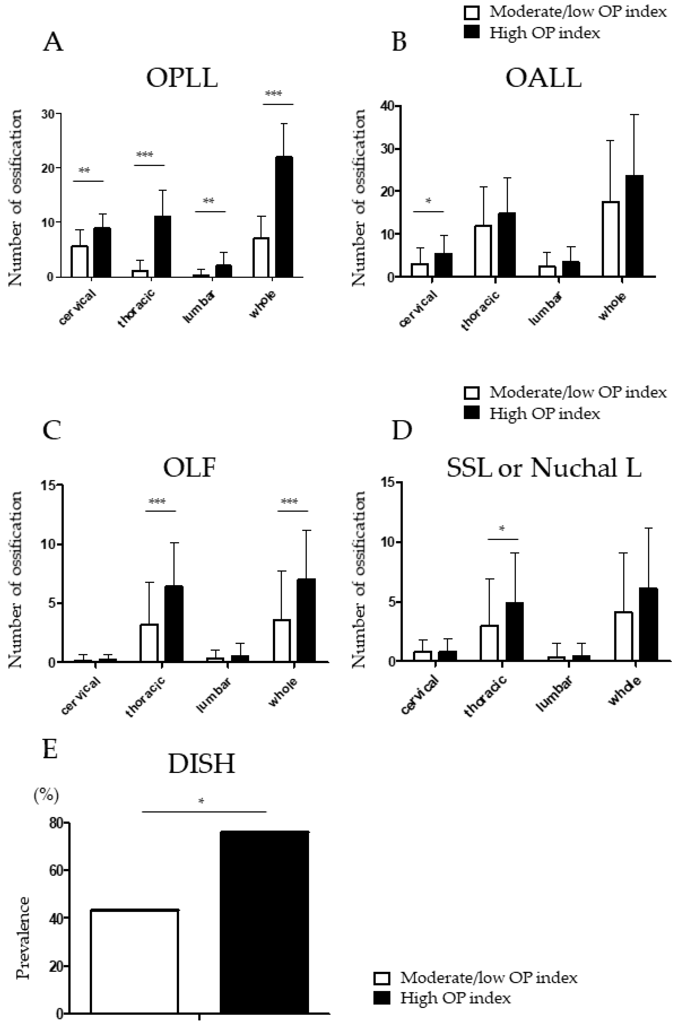
3.4. Higher Prevalence of Ossified Thoracic Lesions in Patients with a High OP Index

The distributions of OPLL and ossification of other spinal ligaments were compared between the two groups (Figure 2A–E). The number of ossifications, especially in the thoracic spine, was markedly higher in the high OP index group. (Figure 2A). Patients in the high OP index group also had more OALL in the cervical spine (Figure 2B) and more OLF (Figure 2C) and ossification of the supra- and interspinous ligaments (Figure 2D) in the thoracic spine. DISH was significantly more prevalent in the high OP index group than in the moderate/low OP index group (Figure 2E). The high OP index group had a significantly higher CNR compared with the moderate/low OP index group (Table 3).

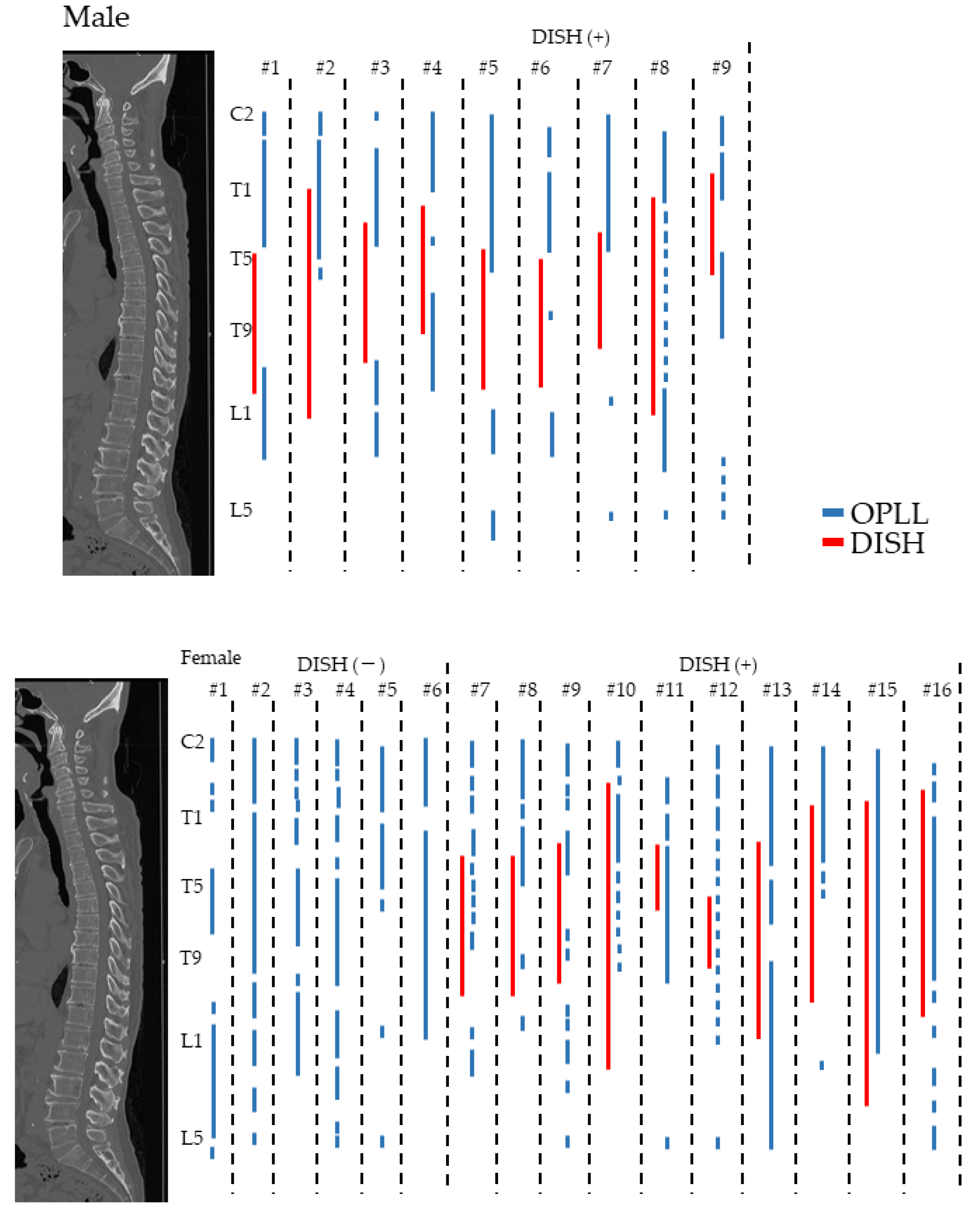
3.5. Distribution of OPLL and DISH in the High OP Index Group

The distribution of ossification in each patient in the high OP index group was investigated in detail. DISH was found in 9 men (100%) and 10 women (62.5%), suggesting that DISH is more prevalent in men (p = 0.03). OPLL was likely to be present adjacent to DISH in the cervicothoracic and thoracolumbar spine, especially in men, and often coexisted with DISH in the thoracic spine in women (Figure 3). Interestingly, patients with a relatively high OP index in the moderate/low OP group also showed similar differences according to sex.

4. Discussion

Ossification of the spinal ligaments is more prevalent in East Asian countries and has been recognized as heterotopic bone formation in the spine. The prevalence of OPLL in the Japanese population has been reported to range from 1.9% to 6.3% [1,8,21,22]. In particular, OPLL can be a common cause of myelopathy. Liao et al. reviewed three-dimensional CT scans for 7210 patients with degenerative cervical myelopathy and showed that the prevalence of OPLL was as high as 18% [23]. However, Fujimori et al. reviewed the registry data of OPLL patients and demonstrated that the prevalence of symptomatic OPLL was less than 1% in those with radiographically detected OPLL [21], demonstrating that most cases with radiographically detectable OPLL are asymptomatic. Thus, the mechanisms of onset and progression of myelopathy caused by OPLL is not fully understood.
A previous report [12] showed that the amount of OPLL (i.e., the OP index) was significantly correlated with the cervical OP index, female sex, and obesity. We also demonstrated that male sex was significantly less prevalent in the high OP index group. Furthermore, we have previously shown that the greater the OP index in patients with OPLL, the greater canal narrowing ratio [24]. Patients with a higher OP index may also be more likely to develop spinal cord compression and myelopathy. In the present study, the cervical JOA score was lower, albeit not significantly, in the high OP index group than in the moderate/low OP index group. In patients with a high OP index, ossified lesions can compress the spinal cord, leading to symptoms of myelopathy. Therefore, patients who are asymptomatic but have a high OP index should undergo long-term follow-up because neurological deterioration may occur.
In our study, evaluation of the patient-reported JOA-CMEQ measures showed that lower extremity function and quality of life were significantly worse in the high OP index group without any significant difference in cervical spine function, upper extremity function, bladder function, quality of life, or body pain. It has been reported that the OP index in the cervical spine is significantly associated with lower extremity motor function [24]. This indicates that the posterior column can be impaired by ossification in patients with a higher OP index. Haddas et al. compared the relationship between spatiotemporal parameters and the kinematics of the spine and lower extremities during the gait cycle between patients with myelopathy and healthy volunteers. Interestingly, they found that patients with myelopathy have significantly increased anterior pelvic tilt and lumbar lordosis as well as less cervical lordosis and head flexion, and consequently had less range of motion at the knee during the gait cycle [25]. Therefore, gait disturbance caused by myelopathy may not only influence lumbar spinal function but also cause locomotive dysfunction in the lower limbs.
Clinically, spinal disorders can result from spinal cord compression caused by OPLL or ossification of other spinal ligaments. OLF often coexists with OPLL and results in onset of neurological dysfunction at the cervicothoracic and thoracolumbar junctions [9,24]. Lee et al. reviewed 101 patients with symptomatic OLF and 102 patients with asymptomatic OLF that was discovered incidentally during investigation of thoracic or lumbar compression fractures to identify radiologic characteristics of symptomatic OLF and showed that the fused and tuberous types of OLF mostly lead to myelopathy [26]. They also identified an ossified cross-sectional area of the spinal canal of 33% as the cutoff value for a diagnosis of thoracic myelopathy caused by OLF. We found that the amount of OLF was greater in the high OP index group. Therefore, it is important for spine surgeons to evaluate not only OPLL but also OLF in the whole spine.
The present study also showed that patient-reported outcome measures related to the lumbar spine on the JOA-BPEQ were significantly worse in the high OP index group than in the moderate/low OP index group. In the high OP index group, ossification of multiple spinal ligaments, including OPLL, OLF, and SSL, was observed in the thoracic spine but not in the lumbar spine (Figure 2). Moreover, the prevalence of DISH was significantly higher in patients with a high OP index. Yamada et al. reviewed the CT myelograms for 61 patients who underwent surgery for symptomatic thoracic OLF and reported lumbar spinal canal stenosis as an incidental finding in up to 75% of cases [27]. Furthermore, a systematic review that included 524 patients with thoracic spinal stenosis found that the prevalence of lumbar canal stenosis was 35.9% [28]. These findings suggest that ankylosis in the thoracic spine might cause a long lever arm and then create excess loading on the lumbar spine. Despite the structural changes in the whole spine, patients with myelopathy due to ossification of spinal ligaments often present with pain, numbness, or impaired lower limb motor function. These symptoms likely increase the burden on the lumbar spine where mobile segments are preserved.
This study is the first to investigate the relationship between distribution of OPLL and DISH in patients with a predisposition to ossification in detail and found an extremely high prevalence of DISH in a high OP index group. In a retrospective case series by our group [20], DISH was found in 48.7% of patients with cervical OPLL and was basically distributed in the middle thoracic spine in younger cases but could extend to more proximal and/or distal levels in the whole spine in older cases. Although the mean age was relatively younger in the high OP index group, the prevalence of DISH was significantly higher than in the moderate/low group (76% vs. 43.6%). Our present findings indicate that OPLL and OALL can occur at the same time and extend even in younger patients who are predisposed to ossification. Interestingly, in the high OP index group, all of the men had DISH while 6 (37.5%) of the women did not. We also found that OPLL was likely to be present adjacent to DISH in the cervicothoracic and thoracolumbar spine in men, and that OPLL often coexisted with DISH in the thoracic spine in women. This evidence suggests that structural characteristics of ossification might be different between genders. Various factors have been shown to regulate the onset and extension of ossification and various sex-related mechanisms have been suggested [11,29,30,31]. Serum estrogen level has been shown to correlate with the onset and extension of ossified lesions, and estrogen is thought to stimulate osteoblast-like cells with the aid of trophic factors [11,30,31]. According to Ikeda et al., serum leptin and insulin levels were significantly higher in women with OPLL and there is a positive correlation between serum leptin level and both the affected levels with OPLL and the extent of ossification in the thoracic and/or lumbar spine [32]. These metabolic factors could affect the onset and development of OPLL in the whole spine over and above a sex-related difference in the prevalence of ossification.
Rheumatologic pathologies have also been shown to be associated with ossification predisposition. Notably, ankylosing spondylitis (AS), which is also known as “bamboo spine” [33] and consists of annulus fibrosus ossification and adjacent vertebral body bridging anteriorly and laterally, is characterized by chronic inflammatory disease and presence of the HLA-B27 antigen [33,34]. The Modified New York Criteria [35] defines AS based on radiographic features, including sacroiliitis grade ≥ 2 bilaterally or grade 3–4 unilaterally. Although vertebral ossification bridging can be found in DISH and/or OPLL [36,37] with morphology similar to that of AS, bone formation in patients with ossification differs from that in AS patients on a basic level. The bridging formation in ossification patients often has a candle-wax pattern. However, it is not presently clear how to recognize ossification of the spinal ligaments and AS. Nguyen et al. reviewed 104 DISH patients who also had OPLL and divided these patients into three groups according to bridging pattern [38]. They showed that patients with a flat bridging pattern, similar to the morphology of AS, had higher high-sensitivity CRP levels compared with patients with a jagged bridging pattern. This evidence suggests that flat vertebral bridging, which is often found in AS patients, might be caused by inflammatory pathogenesis rather than a degenerative process. However, further investigation is required to definitively determine the pathology.
This study has some limitations that should be noted. First, it had a cross-sectional cohort design, investigated a specific disease, and was not population-based. Second, the longitudinal design precludes any discussion of causality. Third, the JOA-CMEQ and JOA-BPEQ could not evaluate pain states in detail. These clinical questions should be addressed in future work by conducting population-based studies and adjusting for confounding factors for each spinal ligament. These limitations notwithstanding, this work contributes to understanding the clinical features of DISH in patients with cervical OPLL.

5. Conclusions

This prospective study used data from a nationwide cohort registry to investigate the clinical and radiologic features of patients with OPLL and a high OP index. We demonstrated that patients with a high OP index had poorer physical function in the lumbar spine and lower extremities and a predisposition to extreme ossification, including in spinal ligaments other than the OPLL.

Author Contributions

Conceptualization, T.H., S.N., T.Y., K.M. (Kanji Mori), S.U., S.M., K.K., N.N. (Narihito Nagoshi) and Y.K.; Methodology, T.H.; Software, T.H, S.U., H.N. and N.N. (Narihito Nagoshi); Validation, T.H., T.Y., K.M. (Kazuma Murata), S.U., S.M., K.K., N.N. (Narihito Nagoshi); Formal Analysis, T.H. and N.N. (Narihito Nagoshi); Investigation, K.M. (Kanji Mori), T.Y., T.H., N.N. (Narihito Nagoshi), S.N., K.T. (Kazuhiro Takeuchi), S.M., and K.K.; Resources, M.M., M.Y. (Masashi Yamazaki), and A.O.; Data Curation, T.H., T.Y., S.U., J.H., K.M., S.M., K.K., N.N. (Narihito Nagoshi), K.T. (Katsushi Takeshita), T.F., Kei W. (Kei Watanabe), N.N. (Norihiro Nishida), K.W. (Kota Watanabe), M.M., M.N., T.K., S.K., K.N., M.K., H.I., S.I., Y.M. (Yuji Matsuoka), K.W. (Kanichiro Wada), A.K., T.O., H.K., H.O., Y.M. (Yu Matsukura), H.I. and Y.K.; Writing—Original Draft Preparation, T.H., S.N., H.H., Y.M. (Yukihiro Matsuyama), K.T. (Katsushi Takeshita), H.O., A.O., and Y.K.; Writing—Review and Editing, T.H., S.N., T.Y., S.U., K.M. (Kanji Mori), S.M., K.K., N.N. (Norihiro Nishida), K.T. (Kazuhiro Takeuchi), T.F., K.W. (Kei Watanabe), N.N. (Norihiro Nishida), K.W. (Kota Watanabe), T.K., S.K., K.N., M.K., H.N., S.I., Y.M. (Yukihiro Matsuyama), K.W. (Kanichiro Wada), A.K., T.O., H.K., H.O. and Y.K.; Visualization, T.H., N.N. (Narihito Nagoshi), K.M. (Kazuma Murata), S.E. and S.U.; Supervision, M.M., M.W., M.N., M.Y., A.O. and Y.K.; Project Administration, T.H and Y.K.; Funding Acquisition, M.M., M.Y. and A.O. All authors have read and agreed to the published version of the manuscript.

Funding

This work was supported by a Health and Labour Science Research grant (201610008B) and by a grant from the Japan Agency for Medical Research and Development (16ek0109136h0002).

Institutional Review Board Statement

The study was conducted according to the guidelines of the Declaration of Helsinki and approved by the institutional review board of each participating institution (protocol code 28-34; 14 March 2017).

Informed Consent Statement

Informed consent was obtained from all subjects involved in the study.

Data Availability Statement

The data generated and analyzed in this study are available from the corresponding author upon reasonable request.

Acknowledgments

We thank Nobuko Nakajima and Yukiko Oya for data collection.

Conflicts of Interest

The authors declare no conflict of interest. The funders had no role in the design of the study; in the collection, analyses, or interpretation of data; in the writing of the manuscript, or in the decision to publish the results.

References

  1. Matsunaga, S.; Nakamura, K.; Seichi, A.; Yokoyama, T.; Toh, S.; Ichimura, S.; Satomi, K.; Endo, K.; Yamamoto, K.; Kato, Y.; et al. Radiographic predictors for the development of myelopathy in patients with ossification of the posterior longitudinal ligament: A multicenter cohort study. Spine 2008, 33, 2648–2650. [Google Scholar] [CrossRef] [PubMed]
  2. Mori, K.; Imai, S.; Kasahara, T.; Nishizawa, K.; Mimura, T.; Matsusue, Y. Prevalence, distribution, and morphology of thoracic ossification of the posterior longitudinal ligament in Japanese: Results of CT-based cross-sectional study. Spine 2014, 39, 394–399. [Google Scholar] [CrossRef] [PubMed]
  3. Ono, M.; Russell, W.J.; Kudo, S.; Kuroiwa, Y.; Takamori, M.; Motomura, S.; Murakami, J. Ossification of the thoracic posterior longitudinal ligament in a fixed population. Radiological and neurological manifestations. Radiology 1982, 143, 469–474. [Google Scholar] [CrossRef] [PubMed]
  4. Nakajima, M.; Takahashi, A.; Tsuji, T.; Karasugi, T.; Baba, H.; Uchida, K.; Kawabata, S.; Okawa, A.; Shindo, S.; Takeuchi, K.; et al. A genome-wide association study identifies susceptibility loci for ossification of the posterior longitudinal ligament of the spine. Nat. Genet. 2014, 46, 1012–1016. [Google Scholar] [CrossRef] [PubMed]
  5. Yoshii, T.; Sakai, K.; Hirai, T.; Yamada, T.; Inose, H.; Kato, T.; Enomoto, M.; Tomizawa, S.; Kawabata, S.; Arai, Y.; et al. Anterior decompression with fusion versus posterior decompression with fusion for massive cervical ossification of the posterior longitudinal ligament with a ≥50% canal occupying ratio: A multicenter retrospective study. Spine J. 2016, 16, 1351–1357. [Google Scholar] [CrossRef]
  6. Hirai, T.; Yoshii, T.; Egawa, S.; Sakai, K.; Inose, H.; Yuasa, M.; Yamada, T.; Ushio, S.; Kato, T.; Arai, Y.; et al. Increased height of fused segments contributes to early-phase strut dislodgement after anterior cervical corpectomy with fusion for multilevel ossification of the posterior longitudinal ligament. Spine Surg. Relat. Res. 2020, 4, 294–299. [Google Scholar] [CrossRef] [Green Version]
  7. Imagama, S.; Ando, K.; Takeuchi, K.; Kato, S.; Murakami, H.; Aizawa, T.; Ozawa, H.; Hasegawa, T.; Matsuyama, Y.; Koda, M.; et al. Perioperative Complications After Surgery for Thoracic Ossification of Posterior Longitudinal Ligament: A Nationwide Multicenter Prospective Study. Spine 2018, 43, E1389–E1397. [Google Scholar] [CrossRef]
  8. Boody, B.S.; Lendner, M.; Vaccaro, A.R. Ossification of the posterior longitudinal ligament in the cervical spine: A review. Int. Orthop. 2019, 43, 797–805. [Google Scholar] [CrossRef]
  9. Hirai, T.; Yoshii, T.; Ushio, S.; Hashimoto, J.; Mori, K.; Maki, S.; Katsumi, K.; Nagoshi, N.; Takeuchi, K.; Furuya, T.; et al. Associations between Clinical Symptoms and Degree of Ossification in Patients with Cervical Ossification of the Posterior Longitudinal Ligament: A Prospective Multi-Institutional Cross-Sectional Study. J. Clin. Med. 2020, 9, 4055. [Google Scholar] [CrossRef]
  10. Fujimori, T.; Watabe, T.; Iwamoto, Y.; Hamada, S.; Iwasaki, M.; Oda, T. Prevalence, Concomitance, and Distribution of Ossification of the Spinal Ligaments: Results of Whole Spine CT Scans in 1500 Japanese Patients. Spine 2016, 41, 1668–1676. [Google Scholar] [CrossRef]
  11. Kawaguchi, Y.; Nakano, M.; Yasuda, T.; Seki, S.; Hori, T.; Kimura, T. Ossification of the posterior longitudinal ligament in not only the cervical spine, but also other spinal regions: Analysis using multidetector computed tomography of the whole spine. Spine 2013, 38, E1477–E1482. [Google Scholar] [CrossRef] [PubMed]
  12. Hirai, T.; Yoshii, T.; Iwanami, A.; Takeuchi, K.; Mori, K.; Yamada, T.; Wada, K.; Koda, M.; Matsuyama, Y.; Takeshita, K.; et al. Prevalence and Distribution of Ossified Lesions in the Whole Spine of Patients with Cervical Ossification of the Posterior Longitudinal Ligament A Multicenter Study (JOSL CT study). PLoS ONE 2016, 11, e0160117. [Google Scholar] [CrossRef] [PubMed] [Green Version]
  13. Yoshii, T.; Hirai, T.; Iwanami, A.; Nagoshi, N.; Takeuchi, K.; Mori, K.; Yamada, T.; Seki, S.; Tsuji, T.; Fujiyoshi, K.; et al. Co-existence of ossification of the nuchal ligament is associated with severity of ossification in the whole spine in patients with cervical ossification of the posterior longitudinal ligament—A multi-center CT study. J. Orthop. Sci. 2019, 24, 35–41. [Google Scholar] [CrossRef]
  14. Mori, K.; Yoshii, T.; Hirai, T.; Maki, S.; Katsumi, K.; Nagoshi, N.; Nishimura, S.; Takeuchi, K.; Ushio, S.; Furuya, T.; et al. The characteristics of the young patients with cervical ossification of the posterior longitudinal ligament of the spine: A multicenter cross-sectional study. J. Orthop. Sci. 2021, in press. [CrossRef] [PubMed]
  15. Katsumi, K.; Hirai, T.; Yoshii, T.; Maki, S.; Mori, K.; Nagoshi, N.; Nishimura, S.; Takeuchi, K.; Ushio, S.; Furuya, T.; et al. The impact of ossification spread on cervical spine function in patients with ossification of the posterior longitudinal ligament. Sci. Rep. 2021, 11, 14337. [Google Scholar] [CrossRef] [PubMed]
  16. Hirai, T.; Yoshii, T.; Nagoshi, N.; Takeuchi, K.; Mori, K.; Ushio, S.; Iwanami, A.; Yamada, T.; Seki, S.; Tsuji, T.; et al. Distribution of ossified spinal lesions in patients with severe ossification of the posterior longitudinal ligament and prediction of ossification at each segment based on the cervical OP index classification: A multicenter study (JOSL CT study). BMC Musculoskelet. Disord. 2018, 19, 107. [Google Scholar] [CrossRef] [Green Version]
  17. Hirai, T.; Okawa, A.; Arai, Y.; Takahashi, M.; Kawabata, S.; Kato, T.; Enomoto, M.; Tomizawa, S.; Sakai, K.; Torigoe, I.; et al. Middle-term results of a prospective comparative study of anterior decompression with fusion and posterior decompression with laminoplasty for the treatment of cervical spondylotic myelopathy. Spine 2011, 36, 1940–1947. [Google Scholar] [CrossRef]
  18. Fukui, M.; Chiba, K.; Kawakami, M.; Kikuchi, S.; Konno, S.; Miyamoto, M.; Seichi, A.; Shimamura, T.; Shirado, O.; Taguchi, T.; et al. JOA Back Pain Evaluation Questionnaire (JOABPEQ)/JOA Cervical Myelopathy Evaluation Questionnaire (JOACMEQ). The report on the development of revised versions. April 16, 2007. The Subcommittee of the Clinical Outcome Committee of the Japanese Orthopaedic Association on Low Back Pain and Cervical Myelopathy Evaluation. J. Orthop. Sci. 2009, 14, 348–365. [Google Scholar] [CrossRef]
  19. Mori, K.; Yoshii, T.; Hirai, T.; Iwanami, A.; Takeuchi, K.; Yamada, T.; Seki, S.; Tsuji, T.; Fujiyoshi, K.; Furukawa, M.; et al. Prevalence and distribution of ossification of the supra/interspinous ligaments in symptomatic patients with cervical ossification of the posterior longitudinal ligament of the spine: A CT-based multicenter cross-sectional study. BMC Musculoskelet. Disord. 2016, 17, 492. [Google Scholar] [CrossRef]
  20. Nishimura, S.; Nagoshi, N.; Iwanami, A.; Takeuchi, A.; Hirai, T.; Yoshii, T.; Takeuchi, K.; Mori, K.; Yamada, T.; Seki, S.; et al. Prevalence and distribution of diffuse idiopathic skeletal hyperostosis on whole-spine computed tomography in patients with cervical ossification of the poosterior longitudinal ligament: A multicenter study. Clin. Spine Surg. 2018, 31, E460–E465. [Google Scholar] [CrossRef]
  21. Fujimori, T.; Nakajima, N.; Sugiura, T.; Ikegami, D.; Sakaura, H.; Kaito, T.; Iwasaki, M. Epidemiology of symptomatic ossification of the posterior longitudinal ligament: A nationwide registry survey. J. Spine Surg. 2021, 7, 485–494. [Google Scholar] [CrossRef] [PubMed]
  22. Yoshimura, N.; Nagata, K.; Muraki, S.; Oka, H.; Yoshida, M.; Enyo, Y.; Kagotani, R.; Hashizume, H.; Yamada, H.; Ishimoto, Y.; et al. Prevalence and progression of radiographic ossification of the posterior longitudinal ligament and associated factors in the Japanese population: A 3-year follow-up of the ROAD study. Osteoporos. Int. 2014, 25, 1089–1098. [Google Scholar] [CrossRef] [PubMed]
  23. Liao, X.; Jin, Z.; Shi, L.; Zhao, Y.; Zhou, S.; Chen, D.; Tang, D.; Yang, L.; Chen, X. Prevalence of Ossification of Posterior Longitudinal Ligament in Patients With Degenerative Cervical Myelopathy: Cervical Spine 3D CT Observations in 7210 Cases. Spine 2020, 45, 1320–1328. [Google Scholar] [CrossRef] [PubMed]
  24. Hirai, T.; Yoshii, T.; Ushio, S.; Mori, K.; Maki, S.; Katsumi, K.; Nagoshi, N.; Takeuchi, K.; Furuya, T.; Watanabe, K.; et al. Clinical characteristics in patients with ossification of the posterior longitudinal ligament: A prospective multi-institutional cross-sectional study. Sci. Rep. 2020, 10, 5532. [Google Scholar] [CrossRef]
  25. Haddas, R.; Patel, S.; Arakal, R.; Boah, A.; Belanger, T.; Ju, K.L. Spine and lower extremity kinematics during gait in patients with cervical spondylotic myelopathy. Spine J. 2018, 18, 1645–1652. [Google Scholar] [CrossRef]
  26. Lee, B.J.; Park, J.H.; Jeon, S.R.; Rhim, S.C.; Roh, S.W. Clinically significant radiographic parameter for thoracic myelopathy caused by ossification of the ligamentum flavum. Eur. Spine J. 2019, 28, 1846–1854. [Google Scholar] [CrossRef]
  27. Yamada, T.; Shindo, S.; Yoshii, T.; Ushio, S.; Kusano, K.; Miyake, N.; Arai, Y.; Otani, K.; Okawa, A.; Nakai, O. Surgical outcomes of the thoracic ossification of ligamentum flavum: A retrospective analysis of 61 cases. BMC Musculoskelet. Disord. 2021, 22, 7. [Google Scholar] [CrossRef]
  28. Chen, G.; Fan, T.; Yang, X.; Sun, C.; Fan, D.; Chen, Z. The prevalence and clinical characteristics of thoracic spinal stenosis: A systematic review. Eur. Spine J. 2020, 29, 2164–2172. [Google Scholar] [CrossRef]
  29. Kawaguchi, Y.; Furushima, K.; Sugimori, K.; Inoue, I.; Kimura, T. Association between polymorphism of the transforming growth factor-beta1 gene with the radiologic characteristic and ossification of the posterior longitudinal ligament. Spine 2003, 28, 1424–1426. [Google Scholar] [CrossRef]
  30. Komm, B.S.; Terpening, C.M.; Benz, D.J.; Graeme, K.A.; Gallegos, A.; Korc, M.; Greene, G.L.; O’Malley, B.W.; Haussler, M.R. Estrogen binding, receptor mRNA, and biologic response in osteoblast-like osteosarcoma cells. Science 1988, 241, 81–84. [Google Scholar] [CrossRef]
  31. Eriksen, E.F.; Colvard, D.S.; Berg, N.J.; Graham, M.L.; Mann, K.G.; Spelsberg, T.C.; Riggs, B.L. Evidence of estrogen receptors in normal human osteoblast-like cells. Science 1988, 241, 84–86. [Google Scholar] [CrossRef] [PubMed]
  32. Ikeda, Y.; Nakajima, A.; Aiba, A.; Koda, M.; Okawa, A.; Takahashi, K.; Yamazaki, M. Association between serum leptin and bone metabolic markers, and the development of heterotopic ossification of the spinal ligament in female patients with ossification of the posterior longitudinal ligament. Eur. Spine J. 2011, 20, 1450–1458. [Google Scholar] [CrossRef] [PubMed] [Green Version]
  33. Olivieri, I.; D’Angelo, S.; Palazzi, C.; Padula, A.; Mader, R.; Khan, M.A. Diffuse idiopathic skeletal hyperostosis: Differentiation from ankylosing spondylitis. Curr. Rheumatol. Rep. 2009, 11, 321–328. [Google Scholar] [CrossRef] [PubMed]
  34. Kuperus, J.S.; Waalwijk, J.F.; Regan, E.A.; van der Horst-Bruinsma, I.E.; Oner, F.C.; de Jong, P.A.; Verlaan, J.J. Simultaneous occurrence of ankylosing spondylitis and diffuse idiopathic skeletal hyperostosis: A systematic review. Rheumatology 2018, 57, 2120–2128. [Google Scholar] [CrossRef]
  35. van der Linden, S.; Valkenburg, H.A.; Cats, A. Evaluation of diagnostic criteria for ankylosing spondylitis. A proposal for modification of the New York criteria. Arthritis Rheum. 1984, 27, 361–368. [Google Scholar] [CrossRef]
  36. Hirai, T.; Nishimura, S.; Yoshii, T.; Nagoshi, N.; Hashimoto, J.; Mori, K.; Maki, S.; Katsumi, K.; Takeuchi, K.; Ushio, S.; et al. Associations between Clinical Findings and Severity of Diffuse Idiopathic Skeletal Hyperostosis in Patients with Ossification of the Posterior Longitudinal Ligament. J. Clin. Med. 2021, 10, 4137. [Google Scholar] [CrossRef]
  37. Nishimura, S.; Hirai, T.; Nagoshi, N.; Yoshii, T.; Hashimoto, J.; Mori, K.; Maki, S.; Katsumi, K.; Takeuchi, K.; Ushio, S.; et al. Association between Severity of Diffuse Idiopathic Skeletal Hyperostosis and Ossification of Other Spinal Ligaments in Patients with Ossification of the Posterior Longitudinal Ligament. J. Clin. Med. 2021, 10, 4690. [Google Scholar] [CrossRef]
  38. Nguyen, T.C.T.; Yahara, Y.; Yasuda, T.; Seki, S.; Suzuki, K.; Watanabe, K.; Makino, H.; Kamei, K.; Mori, K.; Kawaguchi, Y. Morphological characteristics of DISH in patients with OPLL and its association with high-sensitivity CRP: Inflammatory DISH. Rheumatology 2022, keac051. [Google Scholar] [CrossRef]
Figure 1. The distribution of OP index values.
Jcm 11 03694 g001
Figure 2. Radiologic findings, including OPLL (A), OALL (B), OLF (C), SSL or nuchal ligament (D), and DISH (E) in the high OP-index group and moderate/low OP-index group. DISH, diffuse idiopathic skeletal hyperostosis; OALL, ossification of the anterior longitudinal ligament; OLF, ossification of the ligamentum flavum; OPLL, ossification of the posterior longitudinal ligament; SSL, supra- and interspinous ligaments * p < 0.05, ** p < 0.01, *** p < 0.001.
Jcm 11 03694 g002
Figure 3. The distribution of OPLL and DISH in men and women in the high OP index group. DISH, diffuse idiopathic skeletal hyperostosis; OPLL, ossification of the posterior longitudinal ligament.
Jcm 11 03694 g003
Table 1. Demographic and clinical characteristics of patients with ossification of the posterior longitudinal ligament according to whether they had a high or moderate/low OP index.
High OP Index (n = 25)Moderate/Low OP Index (n = 211)p-Value
Age (years)59.0 ± 14.064.5 ± 12.00.07
Male sex (%)32.073.5<0.001 ***
Body mass index27.4 ± 5.825.7 ± 4.30.17
Diabetes mellitus (%)28.024.60.82
Cervical JOA score11.5 (6–17)12.4 (−2, 17)0.12
Data are expressed as the mean ± standard deviation. JOA, Japanese Orthopaedic Association. *** p < 0.001.
Table 2. Clinical characteristics in patients with ossification of the posterior longitudinal ligament according to whether the OP index was high or moderate/low.
High OP Index (n = 25)Moderate/Low OP Index (n = 211)p-Value
Prevalence of symptoms (%)
Neck pain60.060.20.98
Back pain32.028.00.67
Low back pain68.052.60.14
JOA-CMEQ score
Cervical spine function59.8 ± 33.866.6 ± 27.90.34
Upper extremity function74.5 ± 21.680.8 ± 21.50.18
Lower extremity function46.1 ± 35.768.3 ± 29.30.006 **
Bladder function73.0 ± 20.274.7 ± 22.30.69
Quality of life40.4 ± 20.251.0 ± 19.60.02 *
JOA-BPEQ score
Lumbar spine function54.0 ± 29.369.7 ± 31.60.02 *
Social dysfunction43.4 ± 32.857.9 ± 28.70.04 *
Mentality40.8 ± 18.650.1 ± 19.90.03 *
Locomotive function45.6 ± 37.166.4 ± 34.50.01 *
Body pain58.2 ± 39.172.3 ± 32.70.09
VAS score
Neck pain40.2 ± 31.338.6 ± 31.20.81
Upper extremity numbness47.5 ± 34.544.8 ± 33.20.71
Chest constriction13.0 ± 27.99.9 ± 21.00.60
Numbness below the chest60.1 ± 28.934.2 ± 33.6<0.001 ***
Low back pain44.1 ± 32.925.7 ± 27.90.01 *
Lower extremity numbness54.4 ± 34.928.1 ± 32.6<0.01 **
Lower extremity pain37.8 ± 33.621.4 ± 29.40.03 *
Data are expressed as the mean ± standard deviation. BPEQ, Back Pain Evaluation Questionnaire; CMEQ, Cervical Myelopathy Evaluation Questionnaire; JOA, Japanese Orthopaedic Association; VAS, visual analog scale. * p < 0.05, ** p < 0.01, *** p < 0.001.
Table 3. CNR grade according to high or moderate/low OP index values.
CNR GradeHigh OP Index (n = 25)Moderate/Low OP Index (n = 211)p-Value
Grade 12 (8%)70 (33.2%)
28 (32%)71 (33.6%)
312 (48%)56 (26.5%)
43 (12%)14 (6.6%)
0.03 *
CNR, canal narrowing ratio. * p < 0.05.
Publisher’s Note: MDPI stays neutral with regard to jurisdictional claims in published maps and institutional affiliations.

