Article

Impact of Oxygen Delivery on the Development of Acute Kidney Injury in Patients Undergoing Valve Heart Surgery

by
Elena Carrasco-Serrano
1,2,
Pablo Jorge-Monjas
1,2,3,
María Fé Muñoz-Moreno
4,
Esther Gómez-Sánchez
1,2,3,*,
Juan Manuel Priede-Vimbela
1,2,
Miguel Bardají-Carrillo
1,2,
Héctor Cubero-Gallego
5,
Eduardo Tamayo
1,2,3,6 and
Christian Ortega-Loubon
1
1
BioCritic, Group for Biomedical Research in Critical Care Medicine, 47003 Valladolid, Spain
2
Anesthesiology and Critical Care, Clinical University Hospital of Valladolid, 47003 Valladolid, Spain
3
Department of Surgery, University of Valladolid, 47003 Valladolid, Spain
4
Unit of Research, Clinical University Hospital of Valladolid, 47003 Valladolid, Spain
5
Interventional Cardiology Unit, Cardiology Department, Hospital del Mar, 08003 Barcelona, Spain
6
Centro de Investigación Biomédica en Red de Enfermedades Infecciosas (CIBERINFEC), Instituto de Salud Carlos III, 28029 Madrid, Spain
*
Author to whom correspondence should be addressed.
J. Clin. Med. 2022, 11(11), 3046; https://doi.org/10.3390/jcm11113046
Submission received: 11 April 2022 / Revised: 2 May 2022 / Accepted: 23 May 2022 / Published: 28 May 2022

Abstract

:
One of the strongest risk factors for death in individuals undergoing cardiac surgery is Cardiac Surgery Associated-Acute Kidney Injury (CSA-AKI). Although the minimum kidney oxygen delivery index (DO2i) during cardiopulmonary bypass (CPB) has been reported, the optimal threshold value has not yet been established. A prospective study was conducted from June 2012 to January 2016 to asses how DO2i influences the pathogenesis of CSA-AKI, as well as its most favorable cut-off value. DO2 levels were recorded at the beginning, middle, and end of the CPB. The association between DO2i and CSA-AKI was investigated using multivariable logistic regression analysis. The optimal cut-off of DO2i as a predictor of CSA-AKI was determined using Classification and Regression Tree (CART) analysis. A total of 782 consecutive patients were enrolled. Of these, 231 (29.5%) patients developed AKI. Optimal DO2i thresholds of 303 mL/min/m2 during the CPB and 295 mL/min/m2 at the end of the intervention were identified, which increased the odds of CSA-AKI almost two-fold (Odds Ratio (OR), 1.90; 95% CI, 1.12–3.24) during the surgery and maintained that risk (OR 1.94; 95% CI, 1.15–3.29) until the end. Low DO2i during cardiopulmonary bypass is a risk factor for CSA-AKI that cannot be ruled out. Continuous renal oxygen supply monitoring for adult patients could be a promising method for predicting AKI during CPB.

1. Introduction

Cardiac Surgery-Associated Acute Kidney Injury (CSA-AKI) is a severe complication and one of the strongest risk factors for mortality in patients after cardiac surgery, with an incidence varying from 8.9% to 42.5% according to the intricacy or complexity of the operation [1,2]. In its more severe form, it boosts the likelihood of death by 50–80% [3]. Even a mild rise in serum creatinine (SCr) after cardiac surgery can lead to a higher morbidity, as well as a longer term of stay in both hospital and intensive care unit (ICU) and a higher cost of care [4].
Non-modifiable predisposing variables for CSA-AKI include age, preoperative kidney insufficiency, female gender, low ejection fraction, diabetes, and emergency surgery. Modifiable factors include hemodilution, red blood cell transfusion, redo surgery, mean arterial pressure, postoperative hypotension, low cardiac output, cardiopulmonary bypass (CPB) period, and low oxygen delivery (DO2i) [5,6]. Low oxygen delivery is the amount of oxygen delivered to tissues throughout the body per minute, which is calculated from the cardiac output and arterial blood oxygen content. Hemoglobin content, oxygen saturation, pump flow, and arterial oxygen partial pressure all affect DO2 during CPB. When any of these components fall below critical levels, aerobic energy production can no longer keep up with oxygen requirements, triggering the anaerobic process to deliver energy to the cells and raise lactate levels. Innumerable efforts have been directed towards the control of modifiable risk factors [7]. Recently, the randomized goal-directed perfusion trial (GIFT), suggesting a novel goal directed perfusion (GDP) tactic, aimed to prevent a critical kidney oxygen delivery index lower than 280 mL/min/m2 diminished CSA-AKI in patients who had undergone moderately hypothermic CPB [8].
Despite all of this, not much is known about the effects of CPB on renal blood flow (RBF), the primary regulator of kidney oxygen supply (DO2). Renal vascular resistance increases from 15 to 23 percent during CPB as a result of the neuroendocrine response to CP. This includes increases in norepinephrine, vasopressin, and angiotensin II, causing blood flow to redistribute away from the kidneys [2]. Kidney hypoxia has been considered as a variable of paramount importance for the appearance of CSA-AKI [9,10]. The renal medulla is already on the verge of ischemia under normal circumstances [11]. This is related to a high medullary oxygen extraction rate and a poor perfusion rate, confirmed by the fact that the outer medulla has a tissue partial pressure of oxygen (pO2) of 10 to 20 mmHg, but the renal cortex has a pO2 around 50 mmHg [12,13]. Thus, the outer region of the kidney medulla is especially sensitive to low DO2 and is more vulnerable to harm, as commonly assessed through kidney tubular damage biomarkers. As a result, it is not unexpected that the kidneys could be one of the first organs to be damaged by a global decrease in DO2.
The minimum DO2i throughout CPB has been determined as an independent risk factor for dialysis [10] and the second state of Acute Kidney Injury Network (AKIN) [14]. The critical thresholds of DO2i have been identified to be 272 and 260 mL/min/m2, respectively. Nonetheless, none of these values are based on the novel CART analysis, which is a useful tool for classifying subjects into various risk categories and can partially overcome the risk of misclassification from the traditional Receiver Operating Characteristic (ROC) curve-based criteria to find the best cut-off value [15].
We hypothesize that normothermic CPB causes a diminished DO2i, leading to a kidney oxygen delivery/consumption imbalance. This study aims to determine the effects of DO2i on the appearance of AKI, in addition to determining the threshold value related to it.

2. Materials and Methods

2.1. Study Design

This investigation (identification number PI 19-1382) was authorized by both the Clinical Research Ethic Committee, which is a group of fifteen individuals, and the local Institutional Research Review Board.
We designed a prospective observational study to evaluate the effect of DO2i on CSA-AKI using the Kidney Disease: Improving Global Outcomes (KDIGO) criteria (i.e., an increment in SCr of ≥0.3 mg/dL within 48 h or an elevation in SCr to ≥1.5 times baseline, following 7 days after the intervention) [16]. Furthermore, we seek to determine the minimum DO2i threshold value related to CSA-AKI. We used the SCr levels recorded during the hospital entrance exam, as well as the peak SCr levels reached during the postoperative period of seven days.
This study took place in the Clinical University Hospital of Valladolid, a tertiary-level medical hospital with 800 beds, from June 2012 to January 2016. Approximately 450 open-heart interventions under CPB in adult patients are performed annually by the Cardiac Surgery Department, using two theaters on a daily basis. An intensive care unit (ICU) with 10 beds is dedicated exclusively for patients who have undergone cardiac surgery.

2.2. Study Patients

A total of 782 consecutive patients due to undergo elective heart surgery with cardiopulmonary bypass (CPB) were included in this study. Previous renal failure, off-pump surgery, urgent/emergency surgery, and patients with a kidney transplant were exclusion criteria [17].

2.3. Study Variables and Definitions

Population information included age, sex, body mass index (BMI), diabetes mellitus, hypertension, peripheral vascular disease, left ventricle ejection fraction (LVEF), logistic European System for Cardiac Operative Risk Evaluation (EuroSCORE), kidney depth, baseline bSo2, So2, preoperative hemoglobin, and preoperative SCr.
Perioperative variables collected included type of intervention; CPB duration; cross-clamp time; hematocrit (Hct); partial pressure of oxygen (pO2); oxygen delivery index (Do2i); and lactate at the beginning, during, and completion of CPB.
Oxygen delivery was calculated using the formula [18]:
DO2i = 10 × cardiac index (CI, L/min/m2) × oxygen content (mL/min/m2)
Oxygen content = (hemoglobin (g/dL) × 1.34 × oxygen saturation (%)) + (0.003 × pO2 (mmHg)), where hemoglobin, O2 saturation and pO2 were determined using blood gas analysis during CPB. Utilizing the same equation, the estimated DO2i for each patient was determined using the mean real time CI during CPB; the hemoglobin value; and the pO2 during the start, middle, and culmination of CPB.
Any degree of postoperative CSA-AKI as indicated by the KDIGO criteria was the primary endpoint.

2.4. Anesthesia and CPB Management

Intraoperative Care and Anesthesia

Intravenous midazolam and etomidate were used for the induction of anesthesia. Neuromuscular blockage was achieved using rocuronium. An oxygen/air mixture was used to ventilate the lungs to maintain normocapnia. A mixture of sevoflurane, midazolam and fentanyl was used to sustain anesthesia. To monitor arterial blood pressure and blood samples, a radial, brachial, or femoral artery catheter was placed. A trilumen internal jugular catheter was used to obtain central venous access for the assessment of central venous pressure, blood samples, and fluid/medication supply. An esophageal probe was used to detect the temperature. A Foley catheter was used to measure urine output.
Cardiac rhythm was continuously monitored. Systemic heparinization was achieved with 350 U/kg of heparin. To keep the active clotting time over 400 s, more heparin boluses were given. Pump output ranged between 45 and 55 mL/kg per minute. Intermittent cold blood cardioplegia and moderate systemic hypothermia (28–30 °C) were used. The mean arterial pressure was kept between 60 and 80 mmHg. Protamine was used to reverse the heparin effect at a 1.5:1 ratio after CPB completion and cannulae removal.

2.5. Statistical Analysis

According to a normal distribution, categorical data are represented as percentages, and continuous variables are reported as mean standard deviations (SDs) or medians (interquartile ranges (IQR)) as applicable. The Shapiro–Wilk and Kolmogorov–Smirnov tests were used to determine normality.
For categorical data, the relationship between CSA-AKI and other variables was identified using χ2, and for continuous data the Student’s t-test or the Mann–Whitney U test were utilized according to the normality criteria.
Data were used in a univariate logistic regression analysis to obtain 95% confidence intervals (CIs) for estimates. A stepwise approach was used to develop a multivariable logistic regression model. If the p value < 0.1, variables were included in the multivariable logistic regression model.
Regarding DO2, the optimal cut-off value for a higher risk for CSA-AKI was obtained using the Classification and Regression Tree (CART) Analysis, which is especially appropriate to the generation of clinical decision making. The capacity of this cut-off value to predict CSA-AKI development was further estimated using multivariate logistic regression analysis. The Hosmer–Lemeshow test was used to determine model calibration.
The odds ratio (OR) was calculated using a 95% confidence interval. Statistical significance was defined as p values <0.05. IBM SPSS Statistics for Windows version 24.0 was used to analyze the data (IBM Corp., Armonk, NY, USA). The R software was used to perform the CART analysis (Version 3.6.0. R Core Team, R foundation for Statistical computing, VIE, AU).

3. Results

A total of 231 (29.5%) patients developed CSA-AKI. In total, 48.7% of the individuals (n = 381) were female, and the median age was 70 (IQR, 63–77). Regarding the underlying conditions, the rates of hypertension, smoking, and EuroSCORE II were significantly higher in patients with CSA-AKI. They underwent longer CPB and cross-clamp periods, presented higher lactate levels, needed more red blood cell transfusions, and showed lower DO2i values throughout the procedure in comparison to those who do not develop CSA-AKI (p < 0.001). Table 1 shows the preoperative and intraoperative data.
CART analysis identified a DO2i < 303 mL/min/m2 during the CBP and <295 mL/min/m2 by the end of the intervention as the optimal cut-off values where there is already a risk for developing CSA-AKI (Figure 1).
The relationship between DO2i and CSA-AKI both during the surgery and at the end of the intervention is shown in Figure 2.

Multivariable Model for DO2i and CSA-AKI

Univariate regression analysis revealed age (OR, 1.06; 95% CI, 1.03 to 1.07), CPB (OR, 1.02; 95% CI, 1.00 to 1.02), and aortic cross-clamp (OR, 1.01; 95% CI, 1.01 to 1.02) as risk factors for CSA-AKI. Continuous DO2 index was significantly associated with CSA-AKI (Table 2).
Table 3 reports the final multivariable model for DO2i and CSA-AKI. DO2i < 303 mL/min/m2 during the surgery (OR 1.90; 95% CI, 1.12 to 3.24) and DO2i < 295 mL/min/m2 (OR 1.94; 95% CI, 1.15 to 3.29) at the end of the intervention were significantly related to CSA-AKI. Other independent risk factors for CSA-AKI were age (OR 1.07; 95% CI, 1.03 to 1.11) and CPB (OR 1.01; 95% CI, 1.01 to 1.02) (Table 3). Mixing these data in a multivariable logistic model, an AUROC of 0.711 (95% CI: 0.670–0.751, p < 0.021) (Figure 2) was obtained.

4. Discussion

There were two major findings in this study. First, age, CBP time, lactate levels, and DO2i were significantly associated with CSA-AKI. Second, a decline in DO2i greater than 303 mL/min/m2 was correlated to postoperative CSA-AKI, as depicted in Table 2.
The incidence of CSA-AKI varies from 7% to 40%, depending on its numerous classifications and definitions [19]. In this study, 29.5% of the participants had CSA-AKI, which is similar to what is currently reported in the literature [1,2].
Multivariable logistic regression analysis identified that age, CPB time, lactate level, and DO2i were significantly associated with CSA-AKI. These data are supported by reported data, which show CSA-AKI etiopathogenesis to be highly complex [20]. Firstly, age is a well-known risk factor for the occurrence of CSA-AKI, which is used as a persistent and steady variable in several risk scores, such as the Age, Creatinine, and Ejection Fraction score; the EuroSCORE; and the Society of Thoracic Surgeons score [21]. Secondly, CPB triggers a complex series of reactions, resulting in the initiation of a systemic inflammatory response, the complement cascade activation, coagulopathy state and hemolysis, and the release of microemboli. The greater the extent of CPB is, the greater its consequences will be [22].
Kidney function is particularly dependent on O2 supply [23]. In 2005, Ranucci et al. were the first to identify a critical threshold for DO2i of 272 mL/min/m2 as a risk for acute kidney failure needing dialysis and postoperative peak SCr levels in 1048 patients who underwent coronary revascularization under CPB [10]. Later, De Somer et al. [14] showed that even a DO2i level < 262 mL/min/m2 is independently related to AKI. Most recently, in 2017, Magruder et al. [24] found that keeping DO2i > 300 mL/min/m2 during CPB lowered the occurrence of CSA-AKI. Likewise, Ranucci et al. determined, in the GIFT trial with 326 patients, that a DO2i < 280 mL/min/m2 increases the appearance of CSA-AKI [8]. Our findings are in line with the findings of the GIFT trial and provide robust support for the notion that keeping a DO2i above 303 mL/min/m2 is related to a decreased likelihood of developing CSA-AKI. The risk of CSA-AKI rises by 2% when the minimum DO2i is lower than this threshold value.
Typically, to achieve full-body perfusion, CPB perfusion flows of 2.0 to 2.4 L/min/m2 [10] or 2.5 to 3 L/min/m2 [24] at a mean blood pressure of 50 to 75 mmHg are appropriate. However, current attention was diverted from a fixed approach to a flow, based on DO2i, as a strategy to lower the occurrence of AKI after heart surgery. The exterior kidney medulla is already on the verge of hypoxia under normal circumstances. This kidney oxygen delivery/consumption mismatch, which is already present before CPB, is even more exacerbated during intervention. Lannemyr et al. [2] showed that despite keeping a systemic oxygen delivery during CPB, the kidney oxygen delivery/consumption balance was disrupted, and renal oxygenation was additionally impaired even after the CPB ended. This plays an essential role in the generation of tubular damage markers and AKI after heart surgery. The reduced renal oxygenation was induced by a drop in DO2 at a constant Vo2, an impaired arterial oxygen content due to hemodilution, and a re-distribution of blood flow away from the kidneys, despite an increase in systemic perfusion flow rate during CPB. This persistent state causes later ischemia-reperfusion kidney injury, leading to a lower kidney oxygenation and proximal tubular damage. Hence, predictors for AKI after heart surgery may not be precisely evaluated without taking into account the duration of low DO2 as a continuous variable.
Previous studies have shown the association between nadir DO2 and renal failure after surgery [10]. However, it must be highlighted that the data computed in those studies were collected intermittently at 10–20-min intervals. DO2 dynamically changes during CPB. Oshita et al. [25] found that both an extended and profound decline of DO2 under the cutoff value of 300 mL/min/m2 is preferable compared with established or fixed nadir DO2 parameters in predicting CSA-AKI. In addition, preventing hemodilution and excessive transfusion and improving CPB pump flow are of paramount importance to maintaining a DO2 over 300 mL/min per m2 [26]. Recently, Mukaida et al. [27] assessed both duration and intensity under the DO2 cut-off value and showed that an AUC lower than the DO2 of 300 mL/min/m2 and the cumulative time beneath that threshold value were more useful parameters for predicting CSA-AKI compared with nadir DO2. They found that when the aggregated time under 300 mL/min/m2 DO2 exceeded 15 min, the incidence of CSA-AKI increased considerably. This measurement included both momentary and continuous drops in DO2. However, while research on renal hypoxia has found that repeated transitory renal ischemia (for example, four periods of four minutes of ischemia for a total of 16 min of ischemia) does not significantly harm the kidneys, continuous ischemia for 20 min caused moderate kidney tubular injury due to ischemia reperfusion lesion [28]. Rasmussen and colleagues [29] showed that only an exposure to at least 30 min of DO2 < 272 mL/min/m2 was independently related to CSA-AKI.
Recently, Hendrix et al. [30] suggested that VO2 is variable and every patient has unique oxygen requirements, recommending the use of a personalized O2 supply target. Relying solely on one critical DO2 value for every individual is not reasonable. Without a patient’s VO2, nadir DO2 cannot be determined, which is dependent on the patient’s unique features and can be influenced by factors such as temperature and anesthesia technique, resulting in varying VO2 levels (ranging from 38 to 76 mL/min/m2) and, hence, unique DO2 demands for each patient. Thus, a perfusion goal focused solely on a generic fixed level for DO2 right over a universal critical value cannot always guarantee adequate DO2 for every patient. Furthermore, adjusting DO2 to the patient BSA allows for differences in physique, but does not consider distinctions in variables such as gender and age. As a result, oxygen demands during CPB are influenced by a variety of parameters, including the anesthetic method used and temperature regulation. Hence, VO2 should indeed be considered when addressing DO2.
Because VO2 cannot be maintained via aerobic energy generation at a crucial DO2 level, anaerobic metabolism is initiated to provide energy to the cells [14].
In a conscious human under normothermic conditions, the crucial Do2 nadir is approximately 300 mL/min/m2. In this study, we identified that under anesthesia and normothermia, when the Do2 is lower than 303 mL/min/m2 kidney function begins to deteriorate.
Several investigations have evaluated the association between mean arterial pressure during CPB and the development of AKI after heart surgery. Nevertheless, there are still conflicting data. While Ranucci et al. showed that hypotension during CPB may lead to a low DO2, raising the odds of CSA-AKI [31], Azau et al. found that raising mean arterial pressure does not decrease AKI after cardiac surgery [32]. Similarly, raising the mean arterial pressure did not improve renal function after surgery, according to Urzua et al. [33] and Sirvinskas et al. [34,35].
Patients with AKI had considerably lower levels of hemoglobin during CPB, which is understandable considering hemoglobin’s role in oxygen supply. A reduced DO2 may also be induced by the dilution of hemoglobin due to the prime solution used for the CPB circuit, usually a cell-free crystalloid solution [30]. The extent of hemodilution and a decreased systemic oxygen supply have been shown as independent predictors for the development of AKI after heart surgery [36]. Furthermore, transfusion may aggravate the risk of AKI [37]. This is because stored RBCs undergo a series of irreversible morphological and physiological alterations, including reduced oxygen affinity, shorter lifespans, and lower deformability. They are also more prone to damage [37,38]. As a consequence, RBC transfusions can precipitate renal injury due to reduced tissue DO2, oxidative stress activation, and the intensification of inflammatory responses [39,40].
Physiological indications could be employed to provide continuous bedside monitoring of the hemodynamics and functional processes involved in the development of CSA-AKI, rather than the current practice of sampling blood gas. Such kidney function measurements may permit the continuous real-time assessment of renal function just as the ventilation and hemodynamic are monitored, providing a functional approach for the application of kidney-protective strategies. Such monitoring could comprise kidney venous oximetry (which translates into the VO2) and the regional assessment of kidney oxygenation using near-infrared spectroscopy [20]. This shows that in order to deal with AKI, a protective tactic for AKI is necessary. Further advancement may be achieved with the development of continuous DO2i monitoring and the patient-specific prediction of individual DO2i thresholds.

Study Limitations

First, we do not possess the Connect data management system (LivaNova), which can record the DO2i every 20 s to provide continuous real-time monitoring during CPB. We estimated the DO2i based on blood gas analysis. Second, DO2i was only calculated in three isolated periods of the CPB time. Third, during CPB we were not able to assess the time–dose effect of oxygen administration. Several researchers have defined the critical cut-off DO2i level by recording it every 10 to 20 min. Nevertheless, because the DO2i level is constantly and dynamically changing, it is possible that the reported DO2i levels do not accurately reflect a crucial condition. As a result, by analyzing nadir DO2i only, it is unclear whether appropriate DO2i was maintained during the CPB. Mukaida et al. identified that not only is maintaining levels of DO2i above specific threshold values important, but also the time below the nadir DO2i threshold level [27]. Thus, the understanding of the real consequences of DO2i throughout the entire CPB was not achieved.

5. Conclusions

The present data suggest that the appearance of renal failure after heart surgery is linked to DO2i during cardiopulmonary bypass. Low DO2i during cardiopulmonary bypass is a risk factor for CSA-AKI that cannot be ruled out. Continuous kidney oxygen supply monitoring may be a useful technique for preventing renal failure in adult patients undergoing cardiopulmonary bypass.

Author Contributions

Conceptualization, E.T., H.C.-G. and E.C.-S.; methodology, H.C.-G.; software, M.F.M.-M.; validation, E.T., C.O.-L. and E.G.-S.; formal analysis, M.F.M.-M.; investigation, E.C.-S. and C.O.-L.; resources, E.T.; data curation, H.C.-G. and E.C.-S.; writing—original draft preparation, C.O.-L. and J.M.P.-V.; writing—review and editing, E.T., E.G.-S. and C.O.-L.; visualization, P.J.-M.; supervision, E.C.-S.; project administration, M.B.-C. and P.J.-M. All authors have read and agreed to the published version of the manuscript.

Funding

This research received no external funding.

Institutional Review Board Statement

The study was conducted in accordance with the Declaration of Helsinki, and approved by the Ethics Committee of Clinical University Hospital of Valladolid, Spain (protocol code PI 19-1382, May 2012).

Informed Consent Statement

Written informed consent has been obtained from the patients to publish this paper.

Data Availability Statement

Data are available upon request to the corresponding author.

Acknowledgments

We appreciate the collaboration of the nursing staff and the patients from the Clinical University Hospital of Valladolid, Spain.

Conflicts of Interest

The authors declare no conflict of interest.

References

  1. Zakkar, M.; Bruno, V.D.; Guida, G.; Angelini, G.D.; Chivasso, P.; Suleiman, M.S.; Bryan, A.J.; Ascione, R. Postoperative acute kidney injury defined by RIFLE criteria predicts early health outcome and long-term survival in patients undergoing redo coronary artery bypass graft surgery. J. Thorac. Cardiovasc. Surg. 2016, 152, 235–242. [Google Scholar] [CrossRef] [PubMed] [Green Version]
  2. Lannemyr, L.; Bragadottir, G.; Krumbholz, V.; Redfors, B.; Sellgren, J.; Ricksten, S.E. Effects of Cardiopulmonary Bypass on Renal Perfusion, Filtration, and Oxygenation in Patients Undergoing Cardiac Surgery. Anesthesiology 2017, 126, 205–213. [Google Scholar] [CrossRef] [PubMed]
  3. Evans, R.G.; Ince, C.; Joles, J.A.; Smith, D.W.; May, C.N.; O’Connor, P.M.; Gardiner, B.S. Haemodynamic influences on kidney oxygenation: Clinical implications of integrative physiology. Clin. Exp. Pharmacol. Physiol. 2013, 40, 106–122. [Google Scholar] [CrossRef] [PubMed]
  4. Ortega-Loubon, C.; Fernandez-Molina, M.; Carrascal-Hinojal, Y.; Fulquet-Carreras, E. Cardiac surgery-associated acute kidney injury. Ann. Card. Anaesth. 2016, 19, 687–698. [Google Scholar] [CrossRef] [PubMed]
  5. Newland, R.F.; Baker, R.A.; Woodman, R.J.; Barnes, M.B.; Willcox, T.W.; Australian and New Zealand Collaborative Perfusion Registry. Predictive Capacity of Oxygen Delivery During Cardiopulmonary Bypass on Acute Kidney Injury. Ann. Thorac. Surg. 2019, 108, 1807–1814. [Google Scholar] [CrossRef]
  6. Magruder, J.T.; Dungan, S.P.; Grimm, J.C.; Harness, H.L.; Wierschke, C.; Castillejo, S.; Barodka, V.; Katz, N.; Shah, A.S.; Whitman, G.J. Nadir Oxygen Delivery on Bypass and Hypotension Increase Acute Kidney Injury Risk after Cardiac Operations. Ann. Thorac. Surg. 2015, 100, 1697–1703. [Google Scholar] [CrossRef]
  7. Patel, H.; Parikh, N.; Shah, R.; Patel, R.; Thosani, R.; Shah, P.; Prajapat, L. Effect of Goal-directed Hemodynamic Therapy in Postcardiac Surgery Patients. Indian J. Crit. Care Med. 2020, 24, 321–326. [Google Scholar] [CrossRef]
  8. Ranucci, M.; Johnson, I.; Willcox, T.; Baker, R.A.; Boer, C.; Baumann, A.; Justison, G.A.; de Somer, F.; Exton, P.; Agarwal, S.; et al. Goal-directed perfusion to reduce acute kidney injury: A randomized trial. J. Thorac. Cardiovasc. Surg. 2018, 156, 1918–1927.e2. [Google Scholar] [CrossRef] [Green Version]
  9. Ranucci, M. Perioperative renal failure: Hypoperfusion during cardiopulmonary bypass? Semin. Cardiothorac. Vasc. Anesth. 2007, 11, 265–268. [Google Scholar] [CrossRef]
  10. Ranucci, M.; Romitti, F.; Isgro, G.; Cotza, M.; Brozzi, S.; Boncilli, A.; Ditta, A. Oxygen delivery during cardiopulmonary bypass and acute renal failure after coronary operations. Ann. Thorac. Surg. 2005, 80, 2213–2220. [Google Scholar] [CrossRef]
  11. Lankadeva, Y.R.; Cochrane, A.D.; Marino, B.; Iguchi, N.; Hood, S.G.; Bellomo, R.; May, C.N.; Evans, R.G. Strategies that improve renal medullary oxygenation during experimental cardiopulmonary bypass may mitigate postoperative acute kidney injury. Kidney Int. 2019, 95, 1338–1346. [Google Scholar] [CrossRef] [PubMed] [Green Version]
  12. Brezis, M.; Rosen, S. Hypoxia of the renal medulla—Its implications for disease. N. Engl. J. Med. 1995, 332, 647–655. [Google Scholar] [CrossRef] [PubMed]
  13. Evans, R.G.; Lankadeva, Y.R.; Cochrane, A.D.; Marino, B.; Iguchi, N.; Zhu, M.Z.L.; Hood, S.G.; Smith, J.A.; Bellomo, R.; Gardiner, B.S.; et al. Renal haemodynamics and oxygenation during and after cardiac surgery and cardiopulmonary bypass. Acta Physiol. 2018, 222, e12995. [Google Scholar] [CrossRef] [PubMed]
  14. de Somer, F.; Mulholland, J.W.; Bryan, M.R.; Aloisio, T.; Van Nooten, G.J.; Ranucci, M. O2 delivery and CO2 production during cardiopulmonary bypass as determinants of acute kidney injury: Time for a goal-directed perfusion management? Crit. Care 2011, 15, R192. [Google Scholar] [CrossRef] [PubMed] [Green Version]
  15. Perkins, N.J.; Schisterman, E.F. The inconsistency of “optimal” cutpoints obtained using two criteria based on the receiver operating characteristic curve. Am. J. Epidemiol. 2006, 163, 670–675. [Google Scholar] [CrossRef] [Green Version]
  16. Abadeer, A.I.; Kurlansky, P.; Chiuzan, C.; Truby, L.; Radhakrishnan, J.; Garan, R.; Topkara, V.; Yuzefpolskaya, M.; Colombo, P.; Takeda, K.; et al. Importance of stratifying acute kidney injury in cardiogenic shock resuscitated with mechanical circulatory support therapy. J. Thorac. Cardiovasc. Surg. 2017, 154, 856–864.e4. [Google Scholar] [CrossRef] [Green Version]
  17. Choi, D.K.; Kim, W.J.; Chin, J.H.; Lee, E.H.; Don Hahm, K.; Yeon Sim, J.; Cheol Choi, I. Intraoperative renal regional oxygen desaturation can be a predictor for acute kidney injury after cardiac surgery. J. Cardiothorac. Vasc. Anesth. 2014, 28, 564–571. [Google Scholar] [CrossRef]
  18. Baker, R.A. Variation in Measurement and Reporting of Goal Directed Perfusion Parameters. J. Extra-Corpor. Technol. 2017, 49, P2–P7. [Google Scholar]
  19. Gaffney, A.M.; Sladen, R.N. Acute kidney injury in cardiac surgery. Curr. Opin. Anaesthesiol. 2015, 28, 50–59. [Google Scholar] [CrossRef] [Green Version]
  20. Ortega-Loubon, C.; Fernandez-Molina, M.; Fierro, I.; Jorge-Monjas, P.; Carrascal, Y.; Gomez-Herreras, J.I.; Tamayo, E. Postoperative kidney oxygen saturation as a novel marker for acute kidney injury after adult cardiac surgery. J. Thorac. Cardiovasc. Surg. 2019, 157, 2340–2351.e3. [Google Scholar] [CrossRef]
  21. Ortega-Loubon, C.; Fernandez-Molina, M.; Paneda-Delgado, L.; Jorge-Monjas, P.; Carrascal, Y. Predictors of Postoperative Acute Kidney Injury after Coronary Artery Bypass Graft Surgery. Braz. J. Cardiovasc. Surg. 2018, 33, 323–329. [Google Scholar] [CrossRef] [PubMed]
  22. Jorge-Monjas, P.; Bustamante-Munguira, J.; Lorenzo, M.; Heredia-Rodriguez, M.; Fierro, I.; Gomez-Sanchez, E.; Hernandez, A.; Alvarez, F.J.; Bermejo-Martin, J.F.; Gomez-Pesquera, E.; et al. Predicting cardiac surgery-associated acute kidney injury: The CRATE score. J. Crit. Care 2016, 31, 130–138. [Google Scholar] [CrossRef] [PubMed] [Green Version]
  23. Newland, R.F.; Baker, R.A. Low Oxygen Delivery as a Predictor of Acute Kidney Injury during Cardiopulmonary Bypass. J. Extra-Corpor. Technol. 2017, 49, 224–230. [Google Scholar] [PubMed]
  24. von Heymann, C.; Sander, M.; Foer, A.; Heinemann, A.; Spiess, B.; Braun, J.; Kramer, M.; Grosse, J.; Dohmen, P.; Dushe, S.; et al. The impact of an hematocrit of 20% during normothermic cardiopulmonary bypass for elective low risk coronary artery bypass graft surgery on oxygen delivery and clinical outcome—A randomized controlled study [ISRCTN35655335]. Crit. Care 2006, 10, R58. [Google Scholar] [CrossRef] [Green Version]
  25. Oshita, T.; Hiraoka, A.; Nakajima, K.; Muraki, R.; Arimichi, M.; Chikazawa, G.; Yoshitaka, H.; Sakaguchi, T. A Better Predictor of Acute Kidney Injury after Cardiac Surgery: The Largest Area under the Curve below the Oxygen Delivery Threshold during Cardiopulmonary Bypass. J. Am. Heart Assoc. 2020, 9, e015566. [Google Scholar] [CrossRef]
  26. Reagor, J.A.; Clingan, S.; Gao, Z.; Morales, D.L.S.; Tweddell, J.S.; Bryant, R.; Young, W.; Cavanaugh, J.; Cooper, D.S. Higher Flow on Cardiopulmonary Bypass in Pediatrics Is Associated with a Lower Incidence of Acute Kidney Injury. Semin. Thorac. Cardiovasc. Surg. 2020, 32, 1015–1020. [Google Scholar] [CrossRef]
  27. Mukaida, H.; Matsushita, S.; Kuwaki, K.; Inotani, T.; Minami, Y.; Saigusa, A.; Amano, A. Time-dose response of oxygen delivery during cardiopulmonary bypass predicts acute kidney injury. J. Thorac. Cardiovasc. Surg. 2019, 158, 492–499. [Google Scholar] [CrossRef]
  28. Jefayri, M.K.; Grace, P.A.; Mathie, R.T. Attenuation of reperfusion injury by renal ischaemic preconditioning: The role of nitric oxide. BJU Int. 2000, 85, 1007–1013. [Google Scholar] [CrossRef] [Green Version]
  29. Rasmussen, S.R.; Kandler, K.; Nielsen, R.V.; Cornelius Jakobsen, P.; Knudsen, N.N.; Ranucci, M.; Christian Nilsson, J.; Ravn, H.B. Duration of critically low oxygen delivery is associated with acute kidney injury after cardiac surgery. Acta Anaesthesiol. Scand. 2019, 63, 1290–1297. [Google Scholar] [CrossRef]
  30. Hendrix, R.H.J.; Ganushchak, Y.M.; Weerwind, P.W. Oxygen delivery, oxygen consumption and decreased kidney function after cardiopulmonary bypass. PLoS ONE 2019, 14, e0225541. [Google Scholar] [CrossRef]
  31. Ranucci, M.; Pavesi, M.; Mazza, E.; Bertucci, C.; Frigiola, A.; Menicanti, L.; Ditta, A.; Boncilli, A.; Conti, D. Risk factors for renal dysfunction after coronary surgery: The role of cardiopulmonary bypass technique. Perfusion 1994, 9, 319–326. [Google Scholar] [CrossRef] [PubMed]
  32. Azau, A.; Markowicz, P.; Corbeau, J.J.; Cottineau, C.; Moreau, X.; Baufreton, C.; Beydon, L. Increasing mean arterial pressure during cardiac surgery does not reduce the rate of postoperative acute kidney injury. Perfusion 2014, 29, 496–504. [Google Scholar] [CrossRef] [PubMed]
  33. Urzua, J.; Troncoso, S.; Bugedo, G.; Canessa, R.; Muñoz, H.; Lema, G.; Valdivieso, A.; Irarrazaval, M.; Moran, S.; Meneses, G. Renal function and cardiopulmonary bypass: Effect of perfusion pressure. J. Cardiothorac. Vasc. Anesth. 1992, 6, 299–303. [Google Scholar] [CrossRef]
  34. Sirvinskas, E.; Andrejaitiene, J.; Raliene, L.; Nasvytis, L.; Karbonskiene, A.; Pilvinis, V.; Sakalauskas, J. Cardiopulmonary bypass management and acute renal failure: Risk factors and prognosis. Perfusion 2008, 23, 323–327. [Google Scholar] [CrossRef]
  35. Sirvinskas, E.; Benetis, R.; Raliene, L.; Andrejaitiene, J. The influence of mean arterial blood pressure during cardiopulmonary bypass on postoperative renal dysfunction in elderly patients. Perfusion 2012, 27, 193–198. [Google Scholar] [CrossRef]
  36. Kanji, H.D.; Schulze, C.J.; Hervas-Malo, M.; Wang, P.; Ross, D.B.; Zibdawi, M.; Bagshaw, S.M. Difference between pre-operative and cardiopulmonary bypass mean arterial pressure is independently associated with early cardiac surgery-associated acute kidney injury. J. Cardiothorac. Surg. 2010, 5, 71. [Google Scholar] [CrossRef] [Green Version]
  37. Habib, R.H.; Zacharias, A.; Schwann, T.A.; Riordan, C.J.; Engoren, M.; Durham, S.J.; Shah, A. Role of hemodilutional anemia and transfusion during cardiopulmonary bypass in renal injury after coronary revascularization: Implications on operative outcome. Crit. Care Med. 2005, 33, 1749–1756. [Google Scholar] [CrossRef]
  38. Karkouti, K. Transfusion and risk of acute kidney injury in cardiac surgery. Br. J. Anaesth. 2012, 109 (Suppl. 1), i29–i38. [Google Scholar] [CrossRef] [Green Version]
  39. Almac, E.; Ince, C. The impact of storage on red cell function in blood transfusion. Best Pract. Res. Clin. Anaesthesiol. 2007, 21, 195–208. [Google Scholar] [CrossRef]
  40. Tinmouth, A.; Fergusson, D.; Yee, I.C.; Hebert, P.C.; Investigators, A.; the Canadian Critical Care Trials Group. Clinical consequences of red cell storage in the critically ill. Transfusion 2006, 46, 2014–2027. [Google Scholar] [CrossRef]
Figure 1. Decision-making analysis using the Classification and Regression Tree (CART) analysis, categorizing the kidney oxygen delivery index with an increased chance of developing acute renal injury after heart surgery.
Jcm 11 03046 g001
Figure 2. Relationship between DO2i and CSA-AKI both during the surgery and at the end of the intervention.
Jcm 11 03046 g002
Table 1. Preoperative and intraoperative features based on CSA-AKI presence.
CharacteristicsTotal (n 782)CSA-AKI (231)CSA-AKI Free (551)p Value
Preoperative data
Population characteristics
Age, y, median (IQR)70 (63–77)74 (67–78)69 (62–76)<0.001
Female 381 (48.7)112 (48.5)269 (48.8)0.932
BMI, kg/m2, median (IQR)27.0 (24.4–29.8)27.1 (24.6–29.3)27.0 (24.3–30.0)0.923
EuroSCORE II, median (IQR)1.7 (1.2–2.1)1.8 (1.4–2.2)1.6 (1.2–2.0)<0.001
Smoker163 (20.8)52 (22.5)111 (20.1)0.043
Current Smoker63 (8.1)10 (4.3)53 (9.6)0.043
Hypertension689 (88.1)216 (93.5)473 (85.8)0.003
Diabetes mellitus154 (19.7)54 (23.4)100 (18.1)0.141
Dyslipidemia574 (73.4)176 (76.2)398 (72.2)0.253
COPD43 (5.5)12 (5.2)31 (5.6)0.533
Peripheral Vascular Disease17 (2.2)8 (3.5)9 (1.6)0.109
Prior Cardiac Surgery49 (6.3)19 (8.2)30 (5.4)0.339
Prior Stroke36 (4.6)17 (7.4)19 (3.4)0.106
AF257 (33.0)75 (32.3)182 (33.0)0.948
NYHA 3230 (29.4)73 (31.6)157 (28.5)0.371
NYHA 48 (1.0)4 (1.7)4 (0.7)
Preoperative SCr, median (IQR)0.8 (0.7–0.9)1.21 (0.7–1.7)0.95 (0.7–1.2)<0.001
LVEF62 (58–65)62 (58–65)62 (59–65)0.740
Intraoperative data
CBP time, min, median (IQR)97 (80–123)102 (83–131)95 (80–117)<0.001
Aortic Cross-Clamp time, min, median (IQR)72 (57–93)75 (60–99)70 (56–91)<0.001
Surgical procedure
Aortic Surgery374 (47.7)105 (45.4)269 (48.8)0.007
Mitral Surgery170 (21.7)48 (20.8)122 (22.1)0.007
Mitral + Aortic Surgery115 (14.7)38 (16.5)77 (14.0)0.007
Mitral + Tricuspid Surgery87 (11.1)24 (10.4)63 (11.4)0.007
Mitral + Aortic + Tricuspid Surgery24 (3.1)11 (4.8)13 (2.4)0.007
Tricuspid Surgery12 (1.5)5 (2.2)7 (1.3)0.007
Red blood cell Transfusion94 (12.0)42 (18.2)52 (9.5)<0.001
Lactate, mg/dL25 (19–32)26 (21–33)24 (19–32)<0.001
DO2 indexed start CPB (mL min−1 m−2)315.6 (283.0–350.2)296.4 (330.6–271.3)322.2 (290.3–357.3)<0.001
DO2 indexed end CPB (mL min−1 m−2)316.8 (284.8–354.9)298.4 (272.1–330.8)323.3 (293.0–351.7)<0.001
AF, atrial fibrillation; CSA-AKI, Cardiac Surgery-Associated Acute Kidney Injury; BMI, body mass index; CABG, coronary artery bypass graft; COPD, chronic obstructive pulmonary disease; DO2, oxygen delivery; IQR, interquartile range; LVEF, left ventricle ejection fraction; MAP, mean arterial pressure; NYHA, New York Heart Association Classification; SCr, serum creatinine.
Table 2. Univariate logistic regression analysis for predisposing factors related to Cardiac Surgery-Associated Acute Renal Injury.
Univariate Analysis
VariablesOR (95% CI)p Value
Age, y1.06 (1.03–1.07)<0.001
CBP time, min1.01 (1.00–1.02)<0.001
Aortic Cross-Clamp time, min1.01 (1.01–1.02)0.001
DO2 indexed during CBP (mL/min/m2)0.99 (0.98–0.99)<0.001
DO2 indexed end CBP (mL/min/m2)0.99 (0.99–0.99)0.002
CPB, cardiopulmonary bypass; DO2, oxygen delivery; OR, odds ratio.
Table 3. Multivariable logistic regression analysis for Cardiac Surgery-Associated Acute Kidney Injury using DO2 as a binary categorical variable. N = 782.
Multivariable Analysis
VariablesOR (95% CI)p Value
Age, y1.07 (1.03–1.11)<0.001
CBP time, min1.01 (1.01–1.02)<0.001
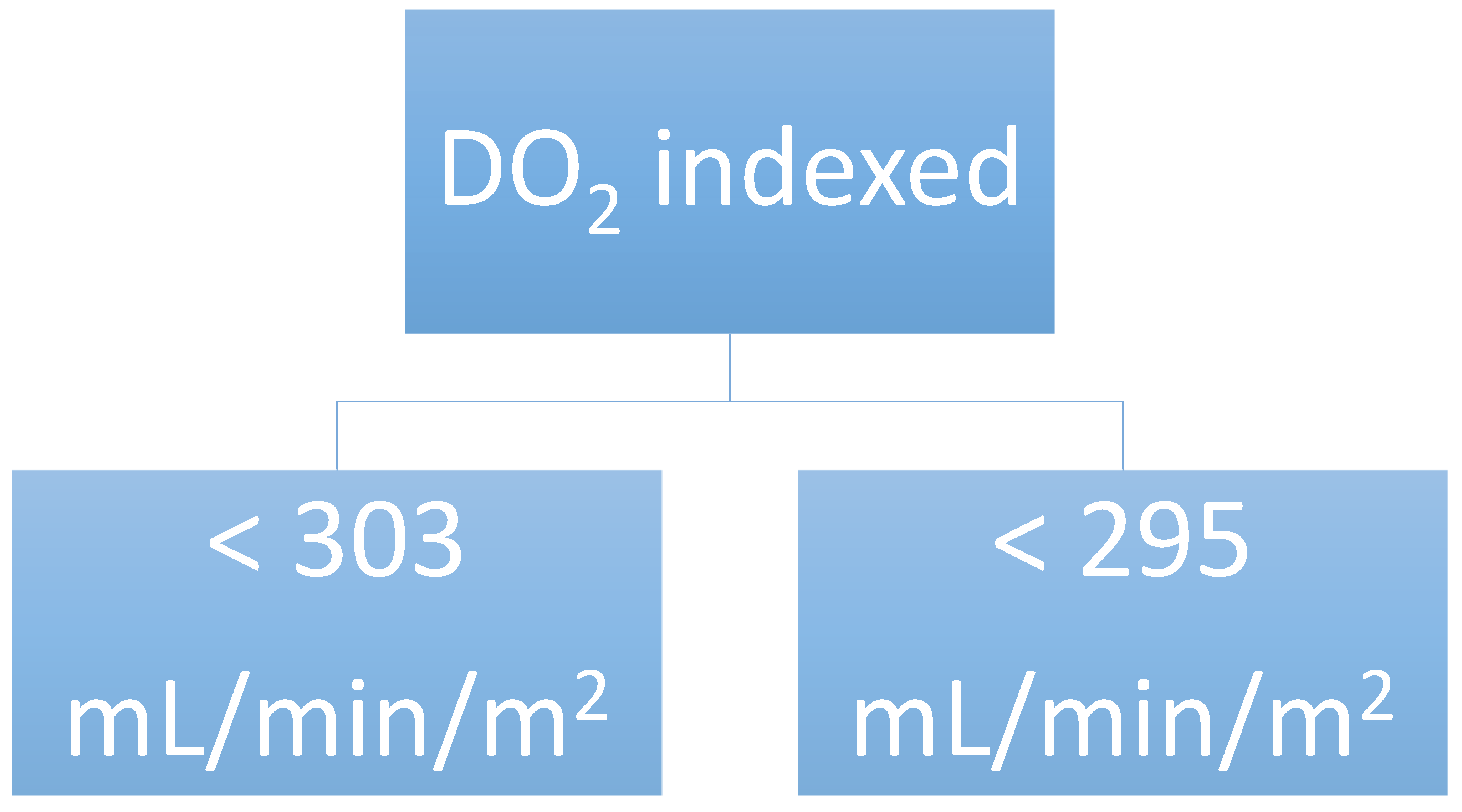
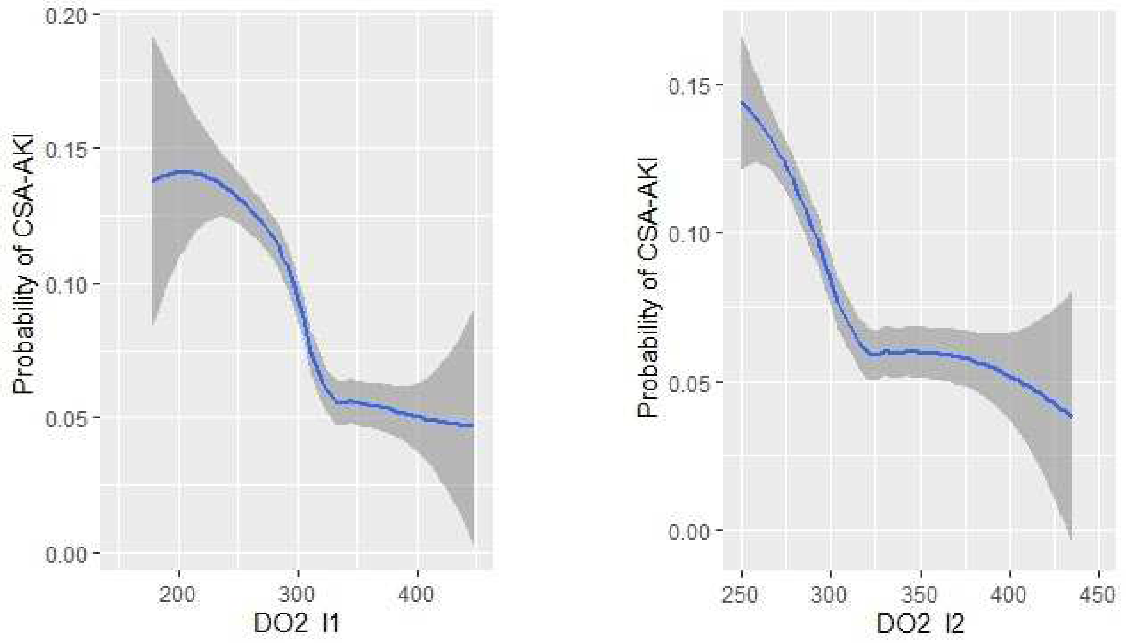
DO2 indexed during CPB < 303 mL min−1 m−21.90 (1.12–3.24)0.018
DO2 indexed end CPB < 295 mL min−1 m−21.94 (1.15–3.29)0.014
CPB, cardiopulmonary bypass; DO2, oxygen delivery; OR, odds ratio.
Publisher’s Note: MDPI stays neutral with regard to jurisdictional claims in published maps and institutional affiliations.

