Article

Potential of Stroke Imaging Using a New Prototype of Low-Field MRI: A Prospective Direct 0.55 T/1.5 T Scanner Comparison

by
Thilo Rusche
1,*,
Hanns-Christian Breit
1,
Michael Bach
1,
Jakob Wasserthal
1,
Julian Gehweiler
1,
Sebastian Manneck
1,
Johanna Maria Lieb
1,
Gian Marco De Marchis
2,
Marios Nikos Psychogios
1,† and
Peter B. Sporns
1,3,†
1
Department of Radiology, Clinic of Radiology & Nuclear Medicine, University Hospital Basel, University Basel, 4031 Basel, Switzerland
2
Department of Neurology, University Hospital Basel, University Basel, 4031 Basel, Switzerland
3
Department of Diagnostic and Interventional Neuroradiology, University Medical Center Hamburg-Eppendorf, 20246 Hamburg, Germany
*
Author to whom correspondence should be addressed.
These authors contributed equally to this work.
J. Clin. Med. 2022, 11(10), 2798; https://doi.org/10.3390/jcm11102798
Submission received: 23 March 2022 / Revised: 6 May 2022 / Accepted: 13 May 2022 / Published: 16 May 2022
(This article belongs to the Special Issue New Advances in Diagnostic Radiology of Ischemic Stroke)

Abstract

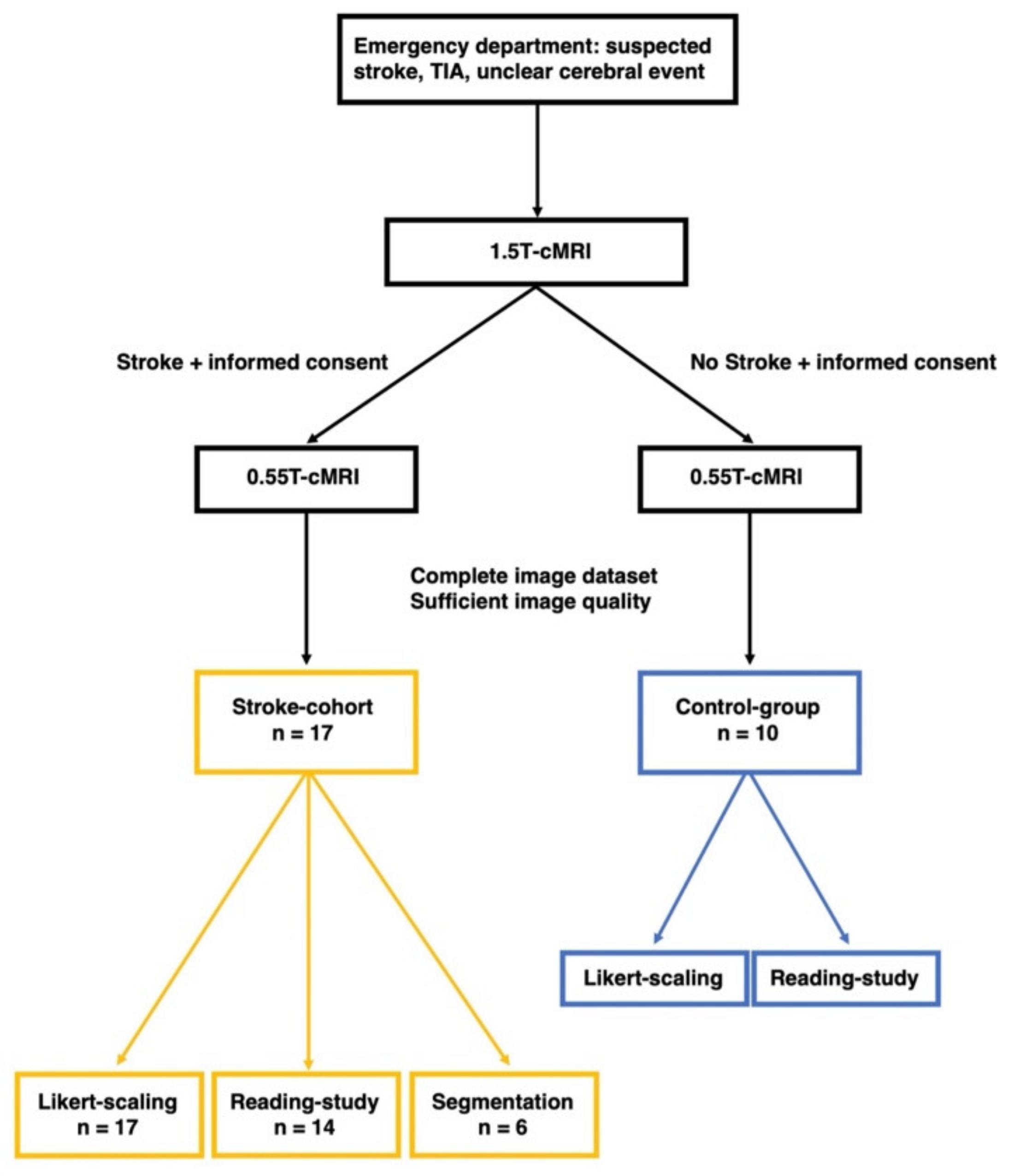
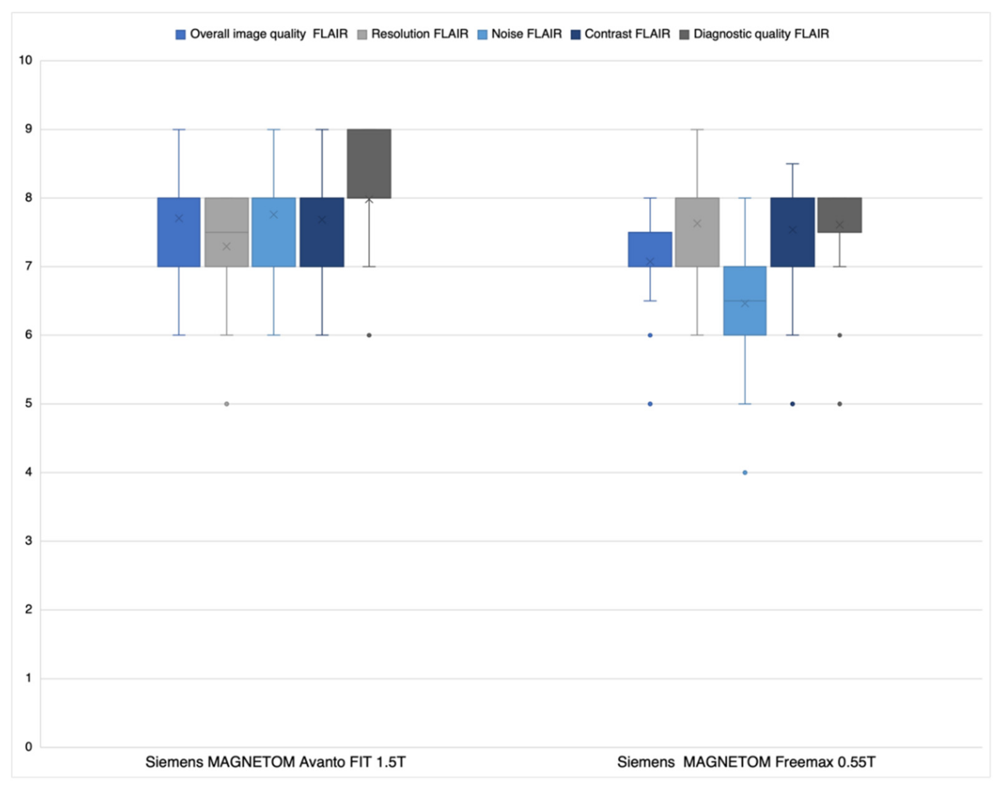
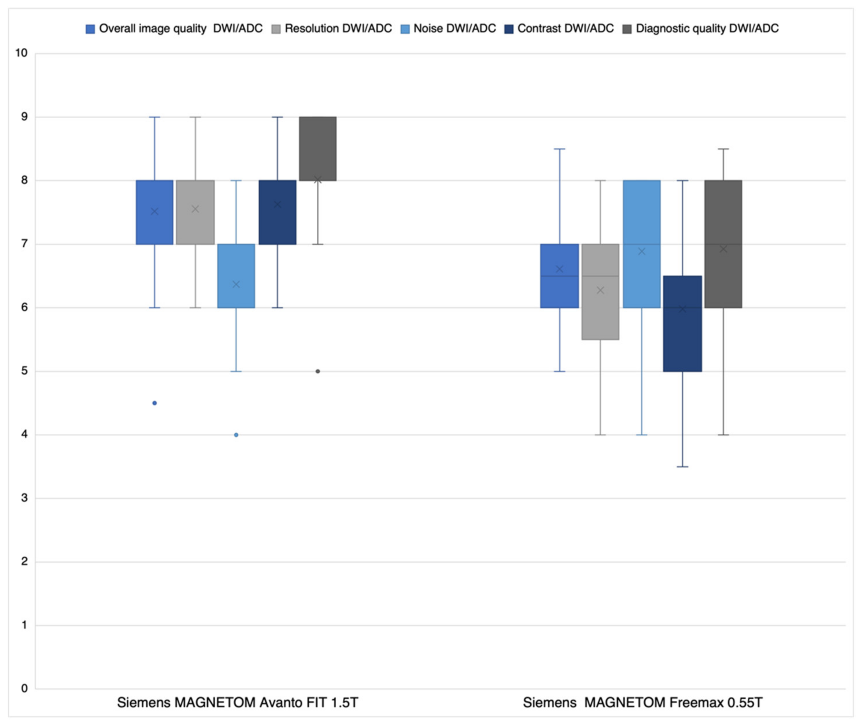
Objectives: Ischemic stroke is a leading cause of mortality and acquired disability worldwide and thus plays an enormous health-economic role. Imaging of choice is computed-tomographic (CT) or magnetic resonance imaging (MRI), especially diffusion-weighted (DW) sequences. However, MR imaging is associated with high costs and therefore has a limited availability leading to low-field-MRI techniques increasingly coming into focus. Thus, the aim of our study was to assess the potential of stroke imaging with low-field MRI. Material and Methods: A scanner comparison was performed including 27 patients (17 stroke cohort, 10 control group). For each patient, a brain scan was performed first with a 1.5T scanner and afterwards with a 0.55T scanner. Scan protocols were as identical as possible and optimized. Data analysis was performed in three steps: All DWI/ADC (apparent diffusion coefficient) and FLAIR (fluid attenuated inversion recovery) sequences underwent Likert rating with respect to image impression, resolution, noise, contrast, and diagnostic quality and were evaluated by two radiologists regarding number and localization of DWI and FLAIR lesions in a blinded fashion. Then segmentation of lesion volumes was performed by two other radiologists on DWI/ADC and FLAIR. Results: DWI/ADC lesions could be diagnosed with the same reliability by the most experienced reader in the 0.55T and 1.5T sequences (specificity 100% and sensitivity 92.9%, respectively). False positive findings did not occur. Detection of number/location of FLAIR lesions was mostly equivalent between 0.55T and 1.5T sequences. No significant difference (p = 0.789–0.104) for FLAIR resolution and contrast was observed regarding Likert scaling. For DWI/ADC noise, the 0.55T sequences were significantly superior (p < 0.026). Otherwise, the 1.5T sequences were significantly superior (p < 0.029). There was no significant difference in infarct volume and volume of infarct demarcation between the 0.55T and 1.5T sequences, when detectable. Conclusions: Low-field MRI stroke imaging at 0.55T may not be inferior to scanners with higher field strengths and thus has great potential as a low-cost alternative in future stroke diagnostics. However, there are limitations in the detection of very small infarcts. Further technical developments with follow-up studies must show whether this problem can be solved.
Keywords: stroke imaging; low-field MRI; reading study; scanner comparison stroke imaging; low-field MRI; reading study; scanner comparison