
TXI (Texture and Color Enhancement Imaging) for Serrated Colorectal Lesions


Abstract
1. Introduction
2. Methods
2.1. Patients
2.2. Ethics
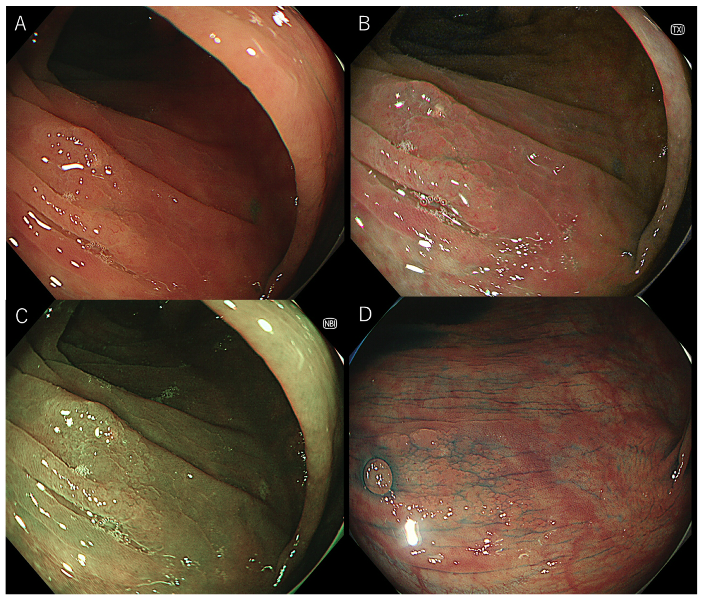
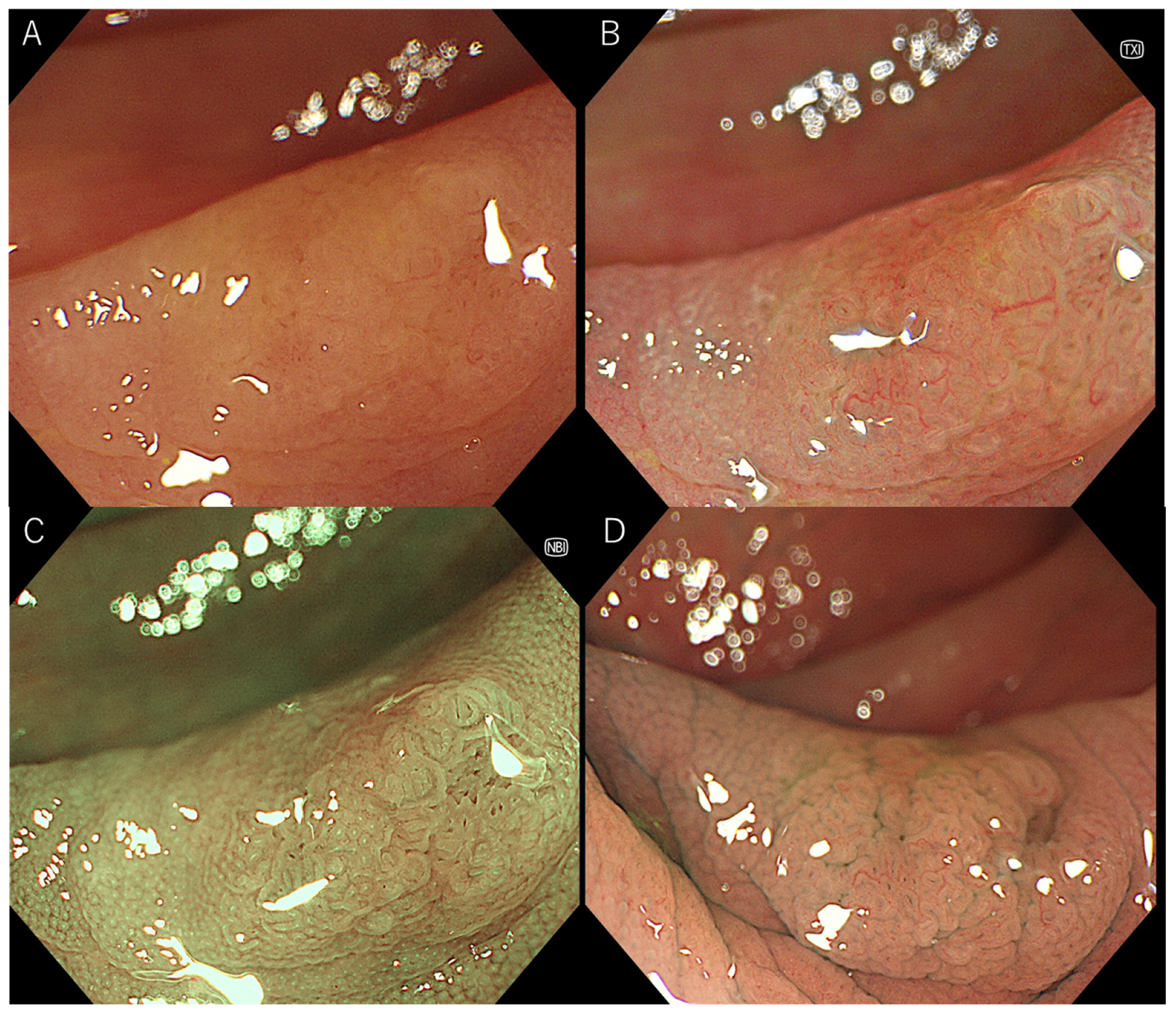
2.3. Endoscopy
2.4. Visibility Scoring
2.5. Statistical Analysis
3. Results
3.1. Patients
3.2. Visibility Score for the Lesion without Magnification
3.3. Visibility Score for Vessel Pattern with Magnification
3.4. Visibility Score for Surface Pattern with Magnification
3.5. Visibility Scores for WLI and TXI by Each Expert Endoscopist
4. Discussion
5. Conclusions
Author Contributions
Funding
Institutional Review Board Statement
Informed Consent Statement
Data Availability Statement
Conflicts of Interest
References
- Goyal, H.; Mann, R.; Gandhi, Z.; Perisetti, A.; Ali, A.; Aman Ali, K.; Sharma, N.; Saligram, S.; Tharian, B.; Inamdar, S. Scope of Artificial Intelligence in Screening and Diagnosis of Colorectal Cancer. J Clin. Med. 2020, 9, 3313. [Google Scholar] [CrossRef] [PubMed]
- Zauber, A.G.; Winawer, S.J.; O’Brien, M.J.; Lansdorp-Vogelaar, I.; van Ballegooijen, M.; Hankey, B.F.; Shi, W.; Bond, J.H.; Schapiro, M.; Panish, J.F.; et al. Colonoscopic polypectomy and long-term prevention of colorectal-cancer deaths. N. Engl. J. Med. 2012, 366, 687–696. [Google Scholar] [CrossRef] [PubMed]
- Albeniz, E.; Pellise, M.; Gimeno Garcia, A.Z.; Lucendo, A.J.; Alonso Aguirre, P.A.; Herreros de Tejada, A.; Alvarez, M.A.; Fraile, M.; Herraiz Bayod, M.; Lopez Roses, L.; et al. Clinical guidelines for endoscopic mucosal resection of non-pedunculated colorectal lesions. Gastroenterol. Hepatol. 2018, 41, 175–190. [Google Scholar] [CrossRef] [PubMed][Green Version]
- Nishizawa, T.; Yoshida, S.; Toyoshima, A.; Yamada, T.; Sakaguchi, Y.; Irako, T.; Ebinuma, H.; Kanai, T.; Koike, K.; Toyoshima, O. Endoscopic diagnosis for colorectal sessile serrated lesions. World J. Gastroenterol. 2021, 27, 1321–1329. [Google Scholar] [CrossRef]
- Kinoshita, S.; Nishizawa, T.; Uraoka, T. Progression to invasive cancer from sessile serrated adenoma/polyp. Dig. Endosc. 2018, 30, 266. [Google Scholar] [CrossRef]
- Dhillon, A.S.; Ibraheim, H.; Green, S.; Suzuki, N.; Thomas-Gibson, S.; Wilson, A. Curriculum review: Serrated lesions of the colorectum. Frontline Gastroenterol. 2020, 11, 243–248. [Google Scholar] [CrossRef]
- Riu Pons, F.; Andreu, M.; Naranjo, D.; Alvarez-Gonzalez, M.A.; Seoane, A.; Dedeu, J.M.; Barranco, L.; Bessa, X. Narrow-band imaging and high-definition white-light endoscopy in patients with serrated lesions not fulfilling criteria for serrated polyposis syndrome: A randomized controlled trial with tandem colonoscopy. BMC Gastroenterol. 2020, 20, 111. [Google Scholar] [CrossRef]
- JE, I.J.; van Doorn, S.C.; van der Brug, Y.M.; Bastiaansen, B.A.; Fockens, P.; Dekker, E. The proximal serrated polyp detection rate is an easy-to-measure proxy for the detection rate of clinically relevant serrated polyps. Gastrointest. Endosc. 2015, 82, 870–877. [Google Scholar] [CrossRef]
- Nishizawa, T.; Yoshida, S.; Toyoshima, O.; Matsuno, T.; Irokawa, M.; Arano, T.; Ebinuma, H.; Suzuki, H.; Kanai, T.; Koike, K. Risk Factors for Prolonged Hospital Stay after Endoscopy. Clin. Endosc. 2021, 54, 851–856. [Google Scholar] [CrossRef]
- Toyoshima, O.; Yoshida, S.; Nishizawa, T.; Yamakawa, T.; Sakitani, K.; Hata, K.; Takahashi, Y.; Fujishiro, M.; Watanabe, H.; Koike, K. CF290 for pancolonic chromoendoscopy improved sessile serrated polyp detection and procedure time: A propensity score-matching study. Endosc. Int. Open 2019, 7, E987–E993. [Google Scholar] [CrossRef]
- Toyoshima, O.; Nishizawa, T.; Yoshida, S.; Sekiba, K.; Kataoka, Y.; Hata, K.; Watanabe, H.; Tsuji, Y.; Koike, K. Expert endoscopists with high adenoma detection rates frequently detect diminutive adenomas in proximal colon. Endosc. Int. Open 2020, 8, E775–E782. [Google Scholar] [CrossRef]
- Yoshida, N.; Hisabe, T.; Hirose, R.; Ogiso, K.; Inada, Y.; Konishi, H.; Yagi, N.; Naito, Y.; Aomi, Y.; Ninomiya, K.; et al. Improvement in the visibility of colorectal polyps by using blue laser imaging (with video). Gastrointest. Endosc. 2015, 82, 542–549. [Google Scholar] [CrossRef] [PubMed]
- Suzuki, T.; Hara, T.; Kitagawa, Y.; Takashiro, H.; Nankinzan, R.; Sugita, O.; Yamaguchi, T. Linked-color imaging improves endoscopic visibility of colorectal nongranular flat lesions. Gastrointest. Endosc. 2017, 86, 692–697. [Google Scholar] [CrossRef] [PubMed]
- Kurumi, H.; Nonaka, K.; Ikebuchi, Y.; Yoshida, A.; Kawaguchi, K.; Yashima, K.; Isomoto, H. Fundamentals, Diagnostic Capabilities and Perspective of Narrow Band Imaging for Early Gastric Cancer. J. Clin. Med. 2021, 10, 2918. [Google Scholar] [CrossRef] [PubMed]
- Wagner, A.; Zandanell, S.; Kiesslich, T.; Neureiter, D.; Klieser, E.; Holzinger, J.; Berr, F. Systematic Review on Optical Diagnosis of Early Gastrointestinal Neoplasia. J. Clin. Med. 2021, 10, 2794. [Google Scholar] [CrossRef]
- Okumura, K.; Hojo, Y.; Tomita, T.; Kumamoto, T.; Nakamura, T.; Kurahashi, Y.; Ishida, Y.; Hirota, S.; Miwa, H.; Shinohara, H. Accuracy of Preoperative Endoscopy in Determining Tumor Location Required for Surgical Planning for Esophagogastric Junction Cancer. J. Clin. Med. 2021, 10, 3371. [Google Scholar] [CrossRef] [PubMed]
- Matsumura, S.; Dohi, O.; Yamada, N.; Harusato, A.; Yasuda, T.; Yoshida, T.; Ishida, T.; Azuma, Y.; Kitae, H.; Doi, T.; et al. Improved Visibility of Early Gastric Cancer after Successful Helicobacter pylori Eradication with Image-Enhanced Endoscopy: A Multi-Institutional Study Using Video Clips. J. Clin. Med. 2021, 10, 3649. [Google Scholar] [CrossRef]
- Min, M.; Deng, P.; Zhang, W.; Sun, X.; Liu, Y.; Nong, B. Comparison of linked color imaging and white-light colonoscopy for detection of colorectal polyps: A multicenter, randomized, crossover trial. Gastrointest. Endosc. 2017, 86, 724–730. [Google Scholar] [CrossRef]
- Fujimoto, D.; Muguruma, N.; Okamoto, K.; Fujino, Y.; Kagemoto, K.; Okada, Y.; Takaoka, Y.; Mitsui, Y.; Kitamura, S.; Kimura, T.; et al. Linked color imaging enhances endoscopic detection of sessile serrated adenoma/polyps. Endosc. Int. Open 2018, 6, E322–E334. [Google Scholar] [CrossRef]
- Oliveira Dos Santos, C.E.; Malaman, D.; Pereira-Lima, J.C.; de Quadros Onofrio, F.; Ribas Filho, J.M. Impact of linked-color imaging on colorectal adenoma detection. Gastrointest. Endosc. 2019, 90, 826–834. [Google Scholar] [CrossRef]
- Paggi, S.; Radaelli, F.; Senore, C.; Maselli, R.; Amato, A.; Andrisani, G.; Di Matteo, F.; Cecinato, P.; Grillo, S.; Sereni, G.; et al. Linked-color imaging versus white-light colonoscopy in an organized colorectal cancer screening program. Gastrointest. Endosc. 2020, 92, 723–730. [Google Scholar] [CrossRef] [PubMed]
- Kitagawa, Y.; Hara, T.; Ikebe, D.; Nankinzan, R.; Takashiro, H.; Kobayashi, R.; Nakamura, K.; Yamaguchi, T.; Suzuki, T. Magnified endoscopic observation of small depressed gastric lesions using linked color imaging with indigo carmine dye. Endoscopy 2018, 50, 142–147. [Google Scholar] [CrossRef] [PubMed]
- Sakamoto, T.; Inoki, K.; Takamaru, H.; Sekiguchi, M.; Yamada, M.; Nakajima, T.; Matsuda, T.; Saito, Y. Efficacy of linked colour imaging in magnifying chromoendoscopy with crystal violet staining: A pilot study. Int. J. Colorectal Dis. 2019, 34, 1341–1344. [Google Scholar] [CrossRef] [PubMed]
- Haralick, R.; Shanmugam, K.; Dinstein, I. Textural features for image classification. IEEE Trans. Syst. Man Cybern. 1973, 3, 610–621. [Google Scholar] [CrossRef]
- Yu, Q.; Vegh, V.; Liu, F.; Turner, I. A Variable Order Fractional Differential-Based Texture Enhancement Algorithm with Application in Medical Imaging. PLoS ONE 2015, 10, e0132952. [Google Scholar] [CrossRef]
- Sato, T. TXI: Texture and Color Enhancement Imaging for Endoscopic Image Enhancement. J. Healthc. Eng. 2021, 2021, 5518948. [Google Scholar] [CrossRef]
- Ishikawa, T.; Matsumura, T.; Okimoto, K.; Nagashima, A.; Shiratori, W.; Kaneko, T.; Oura, H.; Tokunaga, M.; Akizue, N.; Ohta, Y.; et al. Efficacy of Texture and Color Enhancement Imaging in visualizing gastric mucosal atrophy and gastric neoplasms. Sci. Rep. 2021, 11, 6910. [Google Scholar] [CrossRef]


| Serrated Polyps, n. | 29 |
|---|---|
| Histological subtype; n. | |
| Sessile serrated lesion | 18 |
| Microvesicular mucin-rich type hyperplastic polyp | 11 |
| Goblet cell-rich type hyperplastic polyp | 0 |
| Location; n., cecum, ascending, transverse, descending, sigmoid, rectum | 4, 12, 10, 0, 3, 0 |
| Size, mean (standard deviation, range); mm | 9.0 (4.29, 3–18) |
| Morphology; n., Ip, Is, IIa, IIb | 0, 0, 29, 0 |
| WLI | TXI | NBI | Chromoendoscopy | |
|---|---|---|---|---|
| All serrated polyps | ||||
| Mean visibility score (SD) | 2.27 (0.75) | 2.93 (0.76) *** | 2.74 (0.79) ** | 3.45 (0.68) ***, †††, ‡‡‡ |
| SSLs | ||||
| Mean visibility score (SD) | 2.25 (0.76) | 2.90 (0.77) *** | 2.65 (0.79) | 3.45 (0.64) ***, ††, ‡‡‡ |
| Hyperplastic polyps | ||||
| Mean visibility score (SD) | 2.30 (0.72) | 2.97 (0.67) ** | 2.88 (0.77) * | 3.45 (0.74) ***, ‡ |
| WLI | TXI | NBI | Chromoendoscopy | |
|---|---|---|---|---|
| All serrated polyps | ||||
| Vessel pattern (SD) | 2.30 (0.74) | 2.91 (0.80) ***, ††† | 3.23 (0.84) ***, ††† | 2.21 (0.83) |
| Surface pattern (SD) | 1.86 (0.64) | 2.75 (0.68) *** | 3.46 (0.70) ***, ‡‡‡, ††† | 2.79 (0.75) *** |
| SSLs | ||||
| Vessel pattern (SD) | 2.24 (0.74) | 2.89 (0.81) ***, †† | 3.19 (0.86) ***, ††† | 2.30 (0.87) |
| Surface pattern (SD) | 1.80 (0.62) | 2.70 (0.66) *** | 3.39 (0.78) ***, ‡‡‡, †† | 2.83 (0.76) *** |
| Hyperplastic polyps | ||||
| Vessel pattern (SD) | 2.41 (0.73) | 2.96 (0.79) ††† | 3.33 (0.77) ***, ††† | 2.04 (0.69) |
| Surface pattern (SD) | 2.00 (0.67) | 2.85 (0.70) ** | 3.59 (0.49) ***, ‡‡, ††† | 2.70 (0.71) * |
| WLI | TXI | |
|---|---|---|
| Mean visibility scores without magnification (SD) | ||
| Expert endoscopist 1 | 2.29 (0.71) | 2.75 (0.75) *** |
| Expert endoscopist 2 | 2.46 (0.83) | 3.00 (0.77) *** |
| Expert endoscopist 3 | 2.07 (0.66) | 3.04 (0.69) *** |
| Visibility scores of vessel pattern with magnification | ||
| Expert endoscopist 1 | 1.85 (0.60) | 2.37 (0.74) *** |
| Expert endoscopist 2 | 2.52 (0.85) | 2.89 (0.70) ** |
| Expert endoscopist 3 | 2.52 (0.58) | 3.48 (0.58) *** |
| Visibility scores of surface pattern with magnification | ||
| Expert endoscopist 1 | 1.62 (0.88) | 2.48 (0.80) *** |
| Expert endoscopist 2 | 1.89 (0.51) | 2.74 (0.59) *** |
| Expert endoscopist 3 | 2.07 (0.38) | 3.04 (0.52) *** |
Publisher’s Note: MDPI stays neutral with regard to jurisdictional claims in published maps and institutional affiliations. |
© 2021 by the authors. Licensee MDPI, Basel, Switzerland. This article is an open access article distributed under the terms and conditions of the Creative Commons Attribution (CC BY) license (https://creativecommons.org/licenses/by/4.0/).
Share and Cite
Nishizawa, T.; Toyoshima, O.; Yoshida, S.; Uekura, C.; Kurokawa, K.; Munkhjargal, M.; Obata, M.; Yamada, T.; Fujishiro, M.; Ebinuma, H.; et al. TXI (Texture and Color Enhancement Imaging) for Serrated Colorectal Lesions. J. Clin. Med. 2022, 11, 119. https://doi.org/10.3390/jcm11010119
Nishizawa T, Toyoshima O, Yoshida S, Uekura C, Kurokawa K, Munkhjargal M, Obata M, Yamada T, Fujishiro M, Ebinuma H, et al. TXI (Texture and Color Enhancement Imaging) for Serrated Colorectal Lesions. Journal of Clinical Medicine. 2022; 11(1):119. https://doi.org/10.3390/jcm11010119
Chicago/Turabian StyleNishizawa, Toshihiro, Osamu Toyoshima, Shuntaro Yoshida, Chie Uekura, Ken Kurokawa, Munkhbayar Munkhjargal, Miho Obata, Tomoharu Yamada, Mitsuhiro Fujishiro, Hirotoshi Ebinuma, and et al. 2022. "TXI (Texture and Color Enhancement Imaging) for Serrated Colorectal Lesions" Journal of Clinical Medicine 11, no. 1: 119. https://doi.org/10.3390/jcm11010119
APA StyleNishizawa, T., Toyoshima, O., Yoshida, S., Uekura, C., Kurokawa, K., Munkhjargal, M., Obata, M., Yamada, T., Fujishiro, M., Ebinuma, H., & Suzuki, H. (2022). TXI (Texture and Color Enhancement Imaging) for Serrated Colorectal Lesions. Journal of Clinical Medicine, 11(1), 119. https://doi.org/10.3390/jcm11010119