
Undetected Causes of Death in Hospitalized Elderly with COVID-19: Lessons from Autopsy

 , ,
, ,
Abstract
1. Introduction
2. Materials and Methods
2.1. Design, Setting and Population Study
2.2. Data Collection
2.3. Autopsy Techniques
2.4. Clinical and Pathological Confrontation
3. Results
3.1. Clinical Characteristics
3.2. Autopsy Findings
3.2.1. Lungs
3.2.2. Heart and Vessels
3.2.3. Kidney
3.2.4. Brain
3.2.5. Main Other Findings
3.3. Clinicians’ Cause of Death and Pathologists’ Findings
4. Discussion
5. Conclusions
Author Contributions
Funding
Institutional Review Board Statement
Informed Consent Statement
Data Availability Statement
Acknowledgments
Conflicts of Interest
Appendix A
- -
- Cumulative Illness Rating Scale-Geriatric (CIRS-G, range: 0–56) measures the cumulative comorbidity burden in 14 organ systems each rated from 0 (no impairment) to 5 (extremely severe life threatening impairment) [18]. This scale, designed for the elderly, evaluates the severity of illnesses as a function of the effect of disability.
- -
- Functional Independence Measure (FIM, range: 18–126): it is an 18 items tool that explores an individual’s physical, psychological and social function. It is used to assess the level of disability of a patient as well as the changes in his or her condition following rehabilitation or medical intervention [19]. The higher the score, the greater the patient’s functional abilities.
- -
- Clinical Frailty Scale (CFS, range: 1–9): it evaluates specific areas such as comorbidity, function and cognition to generate a fragility score ranging from 1 (very fit) to 9 (terminally ill). It thus summarizes the overall level of ability or frailty of an older adult after being assessed by an experienced clinician [20].
- -
- Body Mass Index (BMI), is a formula for measuring body-weight adjusted for height. For people 70 years of age and older, the cut-off value for screening for undernutrition is BMI < 21 kg/m2 [21].
- -
- Pneumonia Severity Index (PSI, range: 51–395) is a clinical prediction scale that physicians can use to calculate the likelihood of morbidity and mortality in patients with community-acquired pneumonia. This score is also frequently used to predict the need for hospitalization in people with pneumonia [22].
- -
- CURB-65 Severity Score (CURB-65, range: 0–5) determines the severity of a pneumopathy, and thus to decide whether the patient should be referred to an outpatient or an inpatient clinic [23].
References
- Chen, N.; Zhou, M.; Dong, X.; Qu, J.; Gong, F.; Han, Y.; Qiu, Y.; Wang, J.; Liu, Y.; Wei, Y.; et al. Epidemiological and clinical characteristics of 99 cases of 2019 novel coronavirus pneumonia in Wuhan, China: A descriptive study. Lancet 2020, 395, 507–513. [Google Scholar] [CrossRef]
- Weekly Epidemiological Update—20 October 2020. Available online: https://www.who.int/publications/m/item/weekly-epidemiological-update---20-october-2020 (accessed on 24 October 2020).
- Wang, L.; He, W.; Yu, X.; Hu, D.; Bao, M.; Liu, H.; Zhou, J.; Jiang, H. Coronavirus disease 2019 in elderly patients: Characteristics and prognostic factors based on 4-week follow-up. J. Infect. 2020, 80, 639–645. [Google Scholar] [CrossRef]
- Aguiar, D.; Lobrinus, J.A.; Schibler, M.; Fracasso, T.; Lardi, C. Inside the lungs of COVID-19 disease. Int. J. Leg. Med. 2020, 134, 1271–1274. [Google Scholar] [CrossRef]
- Youd, E.; Moore, L. COVID-19 autopsy in people who died in community settings: The first series. J. Clin. Pathol. 2020, 73, 840–844. [Google Scholar] [CrossRef]
- Wang, C.; Xie, J.; Zhao, L.; Fei, X.; Zhang, H.; Tan, Y.; Nie, X.; Zhou, L.; Liu, Z.; Ren, Y.; et al. Alveolar macrophage dysfunction and cytokine storm in the pathogenesis of two severe COVID-19 patients. EBioMedicine 2020, 57, 102833. [Google Scholar] [CrossRef]
- Falasca, L.; Nardacci, R.; Colombo, D.; Lalle, E.; Di Caro, A.; Nicastri, E.; Antinori, A.; Petrosillo, N.; Marchioni, L.; Biava, G.; et al. Postmortem Findings in Italian Patients with COVID-19: A Descriptive Full Autopsy Study of Cases with and Without Comorbidities. J. Infect. Dis. 2020, 222, 1807–1815. [Google Scholar] [CrossRef]
- Lax, S.F.; Skok, K.; Zechner, P.; Kessler, H.H.; Kaufmann, N.; Koelblinger, C.; Vander, K.; Bargfrieder, U.; Trauner, M. Pulmonary Arterial Thrombosis in COVID-19 With Fatal Outcome: Results from a Prospective, Single-Center, Clinicopathologic Case Series. Ann. Intern. Med. 2020, 173, 350–361. [Google Scholar] [CrossRef]
- Wichmann, D. Autopsy Findings and Venous Thromboembolism in Patients with COVID-19. Ann. Intern. Med. 2020, 173, 1030. [Google Scholar] [CrossRef]
- Maggi, E.; Canonica, G.W.; Moretta, L. COVID-19: Unanswered questions on immune response and pathogenesis. J. Allergy Clin. Immunol. 2020, 146, 18–22. [Google Scholar] [CrossRef]
- Merad, M.; Martin, J.C. Pathological inflammation in patients with COVID-19: A key role for monocytes and macrophages. Nat. Rev. Immunol. 2020, 20, 355–362. [Google Scholar] [CrossRef]
- Azkur, A.K.; Akdis, M.; Azkur, D.; Sokolowska, M.; van de Veen, W.; Brüggen, M.-C.; O’Mahony, L.; Gao, Y.; Nadeau, K.; Akdis, C.A. Immune response to SARS-CoV-2 and mechanisms of immunopathological changes in COVID-19. Allergy 2020, 75, 1564–1581. [Google Scholar] [CrossRef] [PubMed]
- Pomara, C.; Li Volti, G.; Cappello, F. COVID-19 Deaths: Are We Sure It Is Pneumonia? Please, Autopsy, Autopsy, Autopsy! J. Clin. Med. 2020, 9, 1259. [Google Scholar] [CrossRef]
- Sperhake, J.-P. Autopsies of COVID-19 deceased? Absolutely! Leg. Med. 2020, 47, 101769. [Google Scholar] [CrossRef] [PubMed]
- Cordasco, F.; Scalise, C.; Sacco, M.A.; Bonetta, C.F.; Zibetti, A.; Cacciatore, G.; Caputo, F.; Ricci, P.; Aquila, I. The silent deaths of the elderly in long-term care facilities during the Covid-19 pandemic: The role of forensic pathology. Med. Leg. J. 2020, 88, 66–68. [Google Scholar] [CrossRef]
- Sessa, F.; Bertozzi, G.; Cipolloni, L.; Baldari, B.; Cantatore, S.; D’Errico, S.; Di Mizio, G.; Asmundo, A.; Castorina, S.; Salerno, M.; et al. Clinical-Forensic Autopsy Findings to Defeat COVID-19 Disease: A Literature Review. J. Clin. Med. 2020, 9, 2026. [Google Scholar] [CrossRef]
- Mendes, A.; Serratrice, C.; Herrmann, F.R.; Genton, L.; Périvier, S.; Scheffler, M.; Fassier, T.; Huber, P.; Jacques, M.-C.; Prendki, V.; et al. Predictors of In-Hospital Mortality in Older Patients with COVID-19: The COVIDAge Study. J. Am. Med. Dir. Assoc. 2020, 21, 1546–1554.e3. [Google Scholar] [CrossRef]
- Salvi, F.; Miller, M.D.; Grilli, A.; Giorgi, R.; Towers, A.L.; Morichi, V.; Spazzafumo, L.; Mancinelli, L.; Espinosa, E.; Rappelli, A.; et al. A Manual of Guidelines to Score the Modified Cumulative Illness Rating Scale and Its Validation in Acute Hospitalized Elderly Patients. J. Am. Geriatr. Soc. 2008, 56, 1926–1931. [Google Scholar] [CrossRef]
- Linacre, J.M.; Heinemann, A.W.; Wright, B.D.; Granger, C.V.; Hamilton, B.B. The structure and stability of the Functional Independence Measure. Arch. Phys. Med. Rehabil. 1994, 75, 127–132. [Google Scholar] [CrossRef]
- Rockwood, K.; Song, X.; MacKnight, C.; Bergman, H.; Hogan, D.B.; McDowell, I.; Mitnitski, A. A global clinical measure of fitness and frailty in elderly people. CMAJ 2005, 173, 489–495. [Google Scholar] [CrossRef]
- Winter, J.E.; MacInnis, R.J.; Wattanapenpaiboon, N.; Nowson, C.A. BMI and all-cause mortality in older adults: A meta-analysis. Am. J. Clin. Nutr. 2014, 99, 875–890. [Google Scholar] [CrossRef]
- Fine, M.J.; Auble, T.E.; Yealy, D.M.; Hanusa, B.H.; Weissfeld, L.A.; Singer, D.E.; Coley, C.M.; Marrie, T.J.; Kappor, W.N. A prediction rule to identify low-risk patients with community-acquired pneumonia. N. Engl. J. Med. 1997, 336, 243–250. [Google Scholar] [CrossRef]
- Lim, W.; van der Eerden, M.M.; Laing, R.; Boersma, W.G.; Karalus, N.; Town, G.I.; Lewis, S.A.; Macfarlane, J.T. Defining community acquired pneumonia severity on presentation to hospital: An international derivation and validation study. Thorax 2003, 58, 377–382. [Google Scholar] [CrossRef]
- Edler, C.; Schröder, A.S.; Aepfelbacher, M.; Fitzek, A.; Heinemann, A.; Heinrich, F.; Klein, A.; Langenwalder, F.; Lütgehetmann, M.; Meissner, K.; et al. Dying with SARS-CoV-2 infection-an autopsy study of the first consecutive 80 cases in Hamburg, Germany. Int. J. Leg. Med. 2020, 134, 1275–1284. [Google Scholar] [CrossRef]
- Langford, B.J.; So, M.; Raybardhan, S.; Leung, V.; Westwood, D.; MacFadden, D.R.; Soucy, J.-P.R.; Daneman, N. Bacterial co-infection and secondary infection in patients with COVID-19: A living rapid review and meta-analysis. Clin. Microbiol. Infect. 2020, 26, 1622–1629. [Google Scholar] [CrossRef]
- Sieswerda, E.; de Boer, M.G.J.; Bonten, M.M.J.; Boersma, W.G.; Jonkers, R.E.; Aleva, R.M.; Kullberg, B.-J.; Schouten, J.A.; Van de Garde, E.M.W.; Verheij, T.J.; et al. Recommendations for antibacterial therapy in adults with COVID-19—an evidence based guideline. Clin. Microbiol. Infect. 2021, 27, 61–66. [Google Scholar] [CrossRef]
- Prendki, V.; Scheffler, M.; Huttner, B.; Garin, N.; Herrmann, F.; Janssens, J.-P.; Marti, C.; Carballo, S.; Roux, X.; Serratrice, C.; et al. Low-dose computed tomography for the diagnosis of pneumonia in elderly patients: A prospective, interventional cohort study. Eur. Respir. J. 2018, 51, 1702375. [Google Scholar] [CrossRef]
- Prendki, V.; Huttner, B.; Marti, C.; Mamin, A.; Fubini, P.E.; Meynet, M.P.; Scheffler, M.; Montet, X.; Janssens, J.-P.; Reny, J.L.; et al. Accuracy of comprehensive PCR analysis of nasopharyngeal and oropharyngeal swabs for CT-scan-confirmed pneumonia in elderly patients: A prospective cohort study. Clin. Microbiol. Infect. 2019, 25, 1114–1119. [Google Scholar] [CrossRef]
- Bartal, C.; Sagy, I.; Barski, L. Drug-induced eosinophilic pneumonia: A review of 196 case reports. Medicine 2018, 97, e9688. [Google Scholar] [CrossRef]
- Tseng, O.L.-I.; Kelsall, J.T.; Wilcox, P.G. Piperacillin-associated pulmonary infiltrates with eosinophilia: A case report. Can. Respir. J. 2010, 17, e24–e26. [Google Scholar] [CrossRef][Green Version]
- Murao, K.; Saito, A.; Kuronuma, K.; Fujiya, Y.; Takahashi, S.; Chiba, H. Acute eosinophilic pneumonia accompanied with COVID-19: A case report. Respirol. Case Rep. 2020, 8, e00683. [Google Scholar] [CrossRef]
- Lindsley, A.W.; Schwartz, J.T.; Rothenberg, M.E. Eosinophil responses during COVID-19 infections and coronavirus vaccination. J. Allergy Clin. Immunol. 2020, 146, 1–7. [Google Scholar] [CrossRef]
- Tseng, C.-T.; Sbrana, E.; Iwata-Yoshikawa, N.; Newman, P.C.; Garron, T.; Atmar, R.L.; Peters, C.J.; Couch, R.B. Immunization with SARS Coronavirus Vaccines Leads to Pulmonary Immunopathology on Challenge with the SARS Virus. PLoS ONE 2012, 7, e35421. [Google Scholar] [CrossRef]
- Tian, S.; Hu, W.; Niu, L.; Liu, H.; Xu, H.; Xiao, S.-Y. Pulmonary Pathology of Early-Phase 2019 Novel Coronavirus (COVID-19) Pneumonia in Two Patients with Lung Cancer. J. Thorac. Oncol. 2020, 15, 700–704. [Google Scholar] [CrossRef]
- Schaller, T.; Hirschbühl, K.; Burkhardt, K.; Braun, G.; Trepel, M.; Märkl, B.; Claus, R. Postmortem Examination of Patients With COVID-19. JAMA 2020, 323, 2518–2520. [Google Scholar] [CrossRef]
- Bösmüller, H.; Traxler, S.; Bitzer, M.; Häberle, H.; Raiser, W.; Nann, D.; Frauenfeld, L.; Vogelsberg, A.; Klingel, K.; Fend, F. The evolution of pulmonary pathology in fatal COVID-19 disease: An autopsy study with clinical correlation. Virchows Arch. 2020, 477, 349–357. [Google Scholar] [CrossRef]
- Carsana, L.; Sonzogni, A.; Nasr, A.; Rossi, R.S.; Pellegrinelli, A.; Zerbi, P.; Rech, R.; Colombo, R.; Antinori, S.; Corbellino, M.; et al. Pulmonary post-mortem findings in a series of COVID-19 cases from northern Italy: A two-centre descriptive study. Lancet Infect. Dis. 2020, 20, 1135–1140. [Google Scholar] [CrossRef]
- Rapkiewicz, A.V.; Mai, X.; Carsons, S.E.; Pittaluga, S.; Kleiner, D.E.; Berger, J.S.; Thomas, S.; Adler, N.M.; Charytan, D.M.; Gasmi, B.; et al. Megakaryocytes and platelet-fibrin thrombi characterize multi-organ thrombosis at autopsy in COVID-19: A case series. EClinicalMedicine 2020, 24, 100434. [Google Scholar] [CrossRef] [PubMed]
- Bradley, B.T.; Maioli, H.; Johnston, R.; Chaudhry, I.; Fink, S.L.; Xu, H.; Najafian, B.; Deutsch, G.; Lacy, J.M.; Williams, T.; et al. Histopathology and ultrastructural findings of fatal COVID-19 infections in Washington State: A case series. Lancet 2020, 396, 320–332. [Google Scholar] [CrossRef]
- Eketunde, A.O.; Mellacheruvu, S.P.; Oreoluwa, P. A Review of Postmortem Findings in Patients with COVID-19. Cureus 2020, 12, e9438. [Google Scholar]
- Migliaccio, M.G.; Di Mauro, M.; Ricciolino, R.; Spiniello, G.; Carfora, V.; Verde, N.; Mottola, F.F.; Coppola, N. Renal Involvement in COVID-19: A Review of the Literature. Infect. Drug Resist. 2021, 14, 895–903. [Google Scholar] [CrossRef]
- Pei, G.; Zhang, Z.; Peng, J.; Liu, L.; Zhang, C.; Yu, C.; Ma, Z.; Huang, Y.; Liu, W.; Yao, Y.; et al. Renal Involvement and Early Prognosis in Patients with COVID-19 Pneumonia. J. Am. Soc. Nephrol. 2020, 31, 1157–1165. [Google Scholar] [CrossRef] [PubMed]
- Jhaveri, K.D.; Meir, L.R.; Flores Chang, B.S.; Parikh, R.; Wanshoo, R.; Barilla-LaBarca, M.L.; Bijol, V.; Hajizadeh, N. Thrombotic microangiopathy in a patient with COVID-19. Kidney Int. 2020, 98, 509–512. [Google Scholar] [CrossRef]
- Ronco, C.; Reis, T. Kidney involvement in COVID-19 and rationale for extracorporeal therapies. Nat. Rev. Nephrol. 2020, 16, 308–310. [Google Scholar] [CrossRef]
- Matschke, J.; Lütgehetmann, M.; Hagel, C.; Sperhake, J.P.; Schröder, A.S.; Edler, C.; Mushumba, H.; Fitzek, A.; Allweiss, L.; Dandri, M.; et al. Neuropathology of patients with COVID-19 in Germany: A post-mortem case series. Lancet Neurol. 2020, 19, 919–929. [Google Scholar] [CrossRef]
- Pinal-Fernandez, I.; Casal-Dominguez, M.; Mammen, A.L. Immune-Mediated Necrotizing Myopathy. Curr. Rheumatol. Rep. 2018, 20, 21. [Google Scholar] [CrossRef] [PubMed]


| Patient No. | Sex | Age (years) | Clinical Medical History | Time from Symptoms to Death (Days) | CIRS-G (Range 0–56) | Clinical Frailty Scale (Range 1–9) | FIM (Range 18–126) | Radiologic Pulmonary Infiltrates |
|---|---|---|---|---|---|---|---|---|
| 1 | F | 87 | Arterial hypertension, primary biliary cirrhosis, chronic lymphocytic leukemia | 10 | 27 | 9 | 107 | multifocal |
| 2 | F | 86 | Asthma, Factor V Leiden, lower limb neuropathy | 7 | 19 | 8 | MD | multifocal |
| 3 | F | 83 | Arterial hypertension, ischemic and valvular heart disease, arteriosclerosis, stroke, dementia, diabetes, lower limb neuropathy | 3 | 23 | 6 | MD | multifocal |
| 4 | M | 86 | Dilated cardiomyopathy, pacemaker | 6 | 15 | 5 | 101 | multifocal |
| 5 | F | 95 | Arterial hypertension, dementia, diabetes | 8 | 24 | 9 | MD | multifocal |
| 6 | M | 91 | Arterial hypertension, ischemic heart disease, lower limb arterial insufficiency, dyslipidemia, asthma, deep vein thrombosis | 21 | 11 | 5 | 86 | local |
| 7 | F | 81 | Arterial hypertension, dementia, chronic hepatitis C, chronic kidney failure, breast cancer, depression | 15 | 22 | 8 | 25 | multifocal |
| 8 | F | 88 | Arterial hypertension, pulmonary embolism, epilepsy, chronic kidney failure | 8 | 24 | 8 | MD | local |
| 9 | M | 88 | Arterial hypertension, ischemic, valvular and rhythmic heart disease, COPD, pulmonary arterial hypertension, chronic kidney failure | 5 | 21 | 5 | MD | multifocal |
| 10 | M | 81 | Arterial hypertension, ischemic heart disease, diabetes, dyslipidemia, chronic kidney failure, metastatic bladder cancer | 22 | 21 | 8 | 103 | multifocal |
| 11 | M | 75 | Arterial hypertension, ischemic heart disease, arteriosclerosis, IgA nephropathy, chronic kidney failure, stroke, dementia, cirrhosis, arteriosclerosis | 25 | 32 | 6 | 73 | multifocal |
| 12 | M | 81 | Arterial hypertension, diabetes, dyslipidemia, stroke, dementia, chronic kidney failure, lower limb neuropathy | 11 | 13 | MD | 35 | multifocal |
| Patient No. | Time from Symptoms to Death (Days) | Lung Autopsy Findings | Other Non Pulmonary Death-Related Autopsy Findings | |||||||
|---|---|---|---|---|---|---|---|---|---|---|
| AD: Patchy Exudative | AD: Diffuse Exudative | AD: Patchy Proliferative | AD: Diffuse Prolferative | OP: Patchy | OP: Diffuse | Bacterial Pneumonia | Other Lung Findings | |||
| 1 | 10 | XXX | ||||||||
| 2 | 7 | XXX | Foci | Signs of heart failure | ||||||
| 3 | 3 | XX | Signs of heart failure | |||||||
| 4 | 6 | XXX | XX | Signs of heart failure | ||||||
| 5 | 8 | XXX | X | Foci | ||||||
| 6 | 21 | X | Large | Central left pulmonary embolism | ||||||
| 7 | 15 | XX | ||||||||
| 8 | 8 | X | XX | Interstitial pneumonia Rare peripheric thrombi | ||||||
| 9 | 5 | X | X | Signs of pulmonary hypertension | Ischemic heart disease | |||||
| 10 | 22 | X | XXX | Peripheric thrombi and microthrombi | Small acute septal myocardial infarct and signs of heart failure | |||||
| 11 | 25 | XX | XXX | Eosinophilic pneumonia | ||||||
| 12 | 11 | XXX | X | Foci | ||||||
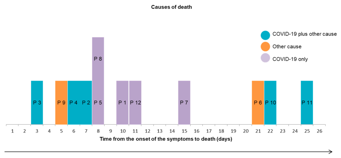
| Patient No. | Sex | Age (Years) | Time from Symptoms to Death (Days) | Complications during Hospitalization | Clinical Suspicion Cause of Death | Cause of Death at Autopsy * |
|---|---|---|---|---|---|---|
| 1 | F | 87 | 10 | Anemia due to bleeding; Acute renal failure | HRFdue to COVID-19 | HRFdue to COVID-19 |
| 2 | F | 86 | 7 | HRFdue to COVID-19 | HRFdue to COVID-19 and heart failure | |
| 3 | F | 83 | 3 | Urinary retention | HRFdue to COVID-19 | HRFdue to COVID-19 and heart failure |
| 4 | M | 86 | 6 | Global cardiac failure; Sacral ulcer; Suspicion of bacterial pneumonia; | HRFdue to COVID-19 and heart failure | HRFdue to COVID-19 and heart failure |
| 5 | F | 95 | 8 | Escherichia coli bacteremia; Pancytopenia; Suspicion of bacterial pneumonia; Acute renal failure | HRFdue to COVID-19 and sepsis with Escherichia coli bacteremia | HRFdue to COVID-19 |
| 6 | M | 91 | 21 | Left heart failure; Delirium; Hypernatremia; Macroscopic hematuria; Suspicion bacterial pneumonia | HRFdue to COVID-19 and delirium | Pulmonary embolism and bacterial pneumonia |
| 7 | F | 81 | 15 | HRFdue to COVID-19 | HRFdue to COVID-19 | |
| 8 | F | 88 | 8 | Arterial hypertention | HRFdue to COVID-19 | HRFdue to COVID-19 |
| 9 | M | 88 | 5 | Left heart failure; Suspicion of bacterial pneumonia | HRFdue to COVID-19 | Probable arythmia due to pulmonary hypertension and ischemic heart disease |
| 10 | M | 81 | 22 | Atrial fibrillation; Suspicion of bacterial pneumonia; Multiorganic failure | HRFdue to COVID-19 | HRFdue to COVID-19 and small acute myocardial infarct |
| 11 | M | 75 | 25 | Acute renal failure; Liver cholestase; Anemia; Suspicion of bacterial pneumonia; Unexplicated persisting inflammation | HRFdue to COVID-19 | HRFdue to COVID-19 and eosinophilic pneumonia |
| 12 | M | 81 | 11 | Hypoglycemia; Acute renal failure; Left heart failure; Suspicion of bacterial pneumonia | HRFdue to COVID-19 | HRFdue to COVID-19 |
Publisher’s Note: MDPI stays neutral with regard to jurisdictional claims in published maps and institutional affiliations. |
© 2021 by the authors. Licensee MDPI, Basel, Switzerland. This article is an open access article distributed under the terms and conditions of the Creative Commons Attribution (CC BY) license (http://creativecommons.org/licenses/by/4.0/).
Share and Cite
Malézieux-Picard, A.; Ferrer Soler, C.; De Macedo Ferreira, D.; Gaud-Luethi, E.; Serratrice, C.; Mendes, A.; Zekry, D.; Gold, G.; Lobrinus, J.A.; Arnoux, G.; et al. Undetected Causes of Death in Hospitalized Elderly with COVID-19: Lessons from Autopsy. J. Clin. Med. 2021, 10, 1337. https://doi.org/10.3390/jcm10071337
Malézieux-Picard A, Ferrer Soler C, De Macedo Ferreira D, Gaud-Luethi E, Serratrice C, Mendes A, Zekry D, Gold G, Lobrinus JA, Arnoux G, et al. Undetected Causes of Death in Hospitalized Elderly with COVID-19: Lessons from Autopsy. Journal of Clinical Medicine. 2021; 10(7):1337. https://doi.org/10.3390/jcm10071337
Chicago/Turabian StyleMalézieux-Picard, Astrid, Cecilia Ferrer Soler, David De Macedo Ferreira, Emilie Gaud-Luethi, Christine Serratrice, Aline Mendes, Dina Zekry, Gabriel Gold, Johannes Alexander Lobrinus, Grégoire Arnoux, and et al. 2021. "Undetected Causes of Death in Hospitalized Elderly with COVID-19: Lessons from Autopsy" Journal of Clinical Medicine 10, no. 7: 1337. https://doi.org/10.3390/jcm10071337
APA StyleMalézieux-Picard, A., Ferrer Soler, C., De Macedo Ferreira, D., Gaud-Luethi, E., Serratrice, C., Mendes, A., Zekry, D., Gold, G., Lobrinus, J. A., Arnoux, G., Serra, F., & Prendki, V. (2021). Undetected Causes of Death in Hospitalized Elderly with COVID-19: Lessons from Autopsy. Journal of Clinical Medicine, 10(7), 1337. https://doi.org/10.3390/jcm10071337