Article

Thermal Pain Thresholds Are Significantly Associated with Plasma Proteins of the Immune System in Chronic Widespread Pain—An Exploratory Pilot Study Using Multivariate and Network Analyses

1
Pain and Rehabilitation Centre, Department of Health, Medicine and Caring Sciences, Linköping University, SE-581 85 Linköping, Sweden
2
Department of Surgical Sciences, Uppsala University, SE-751 85 Uppsala, Sweden
*
Author to whom correspondence should be addressed.
J. Clin. Med. 2021, 10(16), 3652; https://doi.org/10.3390/jcm10163652
Submission received: 11 June 2021 / Revised: 13 August 2021 / Accepted: 16 August 2021 / Published: 18 August 2021
(This article belongs to the Section Clinical Laboratory Medicine)

Abstract

:
Chronic widespread pain (CWP), including fibromyalgia (FM), is characterized by generalized musculoskeletal pain. An important clinical feature is widespread increased pain sensitivity such as lowered pain thresholds for different stimuli such as heat (HPT) and cold (CPT). There is a growing interest in investigating the activated neurobiological mechanisms in CWP. This explorative proteomic study investigates the multivariate correlation pattern between plasma and muscle proteins and thermal pain thresholds in CWP and in healthy controls (CON). In addition, we analysed whether the important proteins and their networks for CPT and HPT differed between CWP and CON. We used a proteomic approach and analysed plasma and muscle proteins from women with CWP (n = 15) and CON (n = 23). The associations between the proteins and CPT/HPT were analysed using orthogonal partial least square (OPLS). The protein–protein association networks for the important proteins for the two thermal pain thresholds were analysed using STRING database. CWP had lowered pain thresholds for thermal stimulus. These levels were generally not related to the included clinical variables except in CWP for HPT. Highly interacting proteins mainly from plasma showed strong significant associations with CPT and HPT both in CWP and in CON. Marked differences in the important proteins for the two thermal pain thresholds were noted between CWP and CON; more complex patterns emerged in CWP. The important proteins were part of the immune system (acute phase proteins, complement factors, and immunoglobulin factors) or known to interact with the immune system. As expected, CWP had lowered pain thresholds for thermal stimulus. Although different proteins were important in the two groups, there were similarities. For example, proteins related to the host defence/immunity such as acute phase proteins, complement factors, immunoglobulin factors, and cytokines/chemokines (although not in CON for CPT) were important habitual/tonic factors for thermal pain thresholds. The fact that peripheral proteins contribute to thermal pain thresholds does not exclude that central factors also contribute and that complex interactions between peripheral and central factors determine the registered pain thresholds in CWP.

1. Introduction

Generalized musculoskeletal pain (i.e., chronic widespread pain; CWP) has a high population prevalence (5–10%) with a female predominance [1,2,3]. CWP is associated with increased prevalence of comorbidities such as depressive and anxiety symptoms, sleep problems, and cognitive difficulties [4,5,6]. Thus, CWP as well as other chronic pain conditions have negative consequences that lead to significant suffering for patients and their families and high socioeconomic costs [7].
Assessments, as well as design and choice of treatments, are hampered by lack of valid biomarkers [8,9]. Potential markers of neurobiological mechanisms in CWP including fibromyalgia (FM) such as cytokines/chemokines, lipids, and metabolites in blood, saliva, muscles, and cerebrospinal fluid are increasingly reported [10,11,12,13,14,15,16,17,18]. Omic methods are gaining increasing support as pain seemingly involves a myriad of molecular changes [19]. The proteome of a tissue regulates biological processes and integrates the effects of genes with environmental factors, comorbidities, behaviours, age, and drugs [19,20,21]. Compared to genome and transcriptome studies, investigating the proteome is more complex, including the choice of statistical methods [22]. We and others have reported marked significant differences both in blood and in muscle proteomes between CWP/FM and healthy controls [10,23,24,25,26]. Moreover, both muscle and plasma protein patterns show strong significant correlations with pain intensity [25,27,28].
Recently nociplastic pain was introduced as a pain mechanistic descriptor (IASP definition: ”Pain that arises from altered nociception despite no clear evidence of actual or threatened tissue damage causing the activation of peripheral nociceptors or evidence for disease or lesion of the somatosensory system causing the pain.” Source: https://www.iasp-pain.org/Education/Content.aspx?ItemNumber=1698#Nociplasticpain; access date: 1 June 2021); FM (a subgroup of CWP) is classified as a nociplastic pain condition [29]. Although validated and definite clinical criteria for nociplastic pain are still lacking, it can be reasonably assumed that these criteria would at the minimum include increased spatial distribution of pain and increased pain sensitivity [30,31,32]. CWP including FM is generally associated with increased pain sensitivity (i.e., lowered pain thresholds) [33]. Pain threshold is defined as when an acute stimulus becomes painful; the stimulus both activates peripheral nociceptors and the central nervous system (CNS). Pressure pain threshold (PPT) correlates significantly with certain plasma proteins obtained from proteomic analysis (i.e., proteins typically at nano and micro molar levels) and with proteins from targeted analysis of a panel of 92 inflammation-related substances (cytokines, chemokines, and growth factors—i.e., small molecules typically at picomolar levels) in CWP [17,34]. The strongest regression was obtained using proteomics. Recently, we reported that plasma proteins correlated with PPT in an FM cohort [25].
In addition to PPT, pain thresholds for cold (CPT) and heat (HPT) are used to develop a nuanced picture of the pain sensitivity. As both muscle and plasma proteins influence PPT in CWP, it may be appropriate to examine them together in relation to CPT and HPT. This study was further motivated by the fact that peripheral molecular mechanisms (in plasma and in muscle) underlying heat and cold pain thresholds are mainly lacking [35]. The need to investigate clinical variables such as pain thresholds in relation to peripheral biomarkers has been emphasized [36]. In chronic pain conditions, comorbidities such as psychological distress and obesity are common, and neurobiological alterations associated with these comorbidities may influence pain thresholds. For example, psychological distress and catastrophizing correlate with thermal pain thresholds [37,38].
We formulated three hypotheses: (1) thermal pain thresholds differ between CWP patients and controls; (2) thermal pain thresholds are significantly associated with peripheral protein patterns in blood (plasma) and in muscle; and (3) the important proteins differed between the two groups of subjects. Therefore, this exploratory study investigates the multivariate correlation pattern between thermal pain thresholds (CPT and HPT) and proteins from plasma and muscle in CWP (typically with lowered pain thresholds) and in healthy controls. We analysed whether the important proteins and their networks involved in the two thresholds differed between the two groups and to what extent comorbidities influenced the thresholds.

2. Materials and Methods

2.1. Subjects

The recruitment of subjects (patients with CWP and healthy controls (CON)) has been described elsewhere [39]. The American College of Rheumatology (ACR) criteria from 1990 was used to classify FM/CWP [6]. The criteria for CWP according to these criteria are chronic pain (>3 months), spinal pain, and pain in at least three out of four body quadrants (or in two contralateral body quadrants). As reported earlier, 13 out of 15 CWP patients also fulfilled the criteria for FM [28]. All subjects were women and none used any anticoagulatory, opioid, or steroidal medication. Exclusion criteria were medical history record of certain musculoskeletal conditions (bursitis, capsulitis, neck trauma, postoperative conditions in neck/shoulder area, spine disorders, tendonitis, rheumatoid arthritis), neurological disease, or systemic or metabolic diseases, malignancy, severe psychiatric illness, pregnancy, and difficulties understanding Swedish. The CON group were recruited through newspaper advertisements. CWP patients were recruited from former female patients at the Pain and Rehabilitation Centre at the University Hospital, Linköping, Sweden or from an organization for FM patients. A total of 19 CWP and 24 CON were initially recruited as reported earlier [10,39,40,41] and this study included 15 CWP patients and 23 CON subjects; see flow chart for details (Figure 1).
The study followed the guidelines in the Declaration of Helsinki and was approved by the Regional Ethical Review Board in Linköping, Sweden (Diarie-nr. M10–08, M233–09, Diarie-nr. 2010/164–32). In agreement with this, all subjects signed a written consent before the start of the study and after receiving verbal and written information about the aims and procedures.

2.2. Clinical Variables

All subjects answered a brief health questionnaire. As these data were previously published, we only provide summaries of the patient reported outcome measures (PROM) variables and instruments used.
Age and body mass index: Age (y), weight (kg), and height (m) were registered at the clinical examination. Body mass index (BMI) was calculated as weight/height2 (kg/m2).
Pain intensity: Each subject rated their pain intensity in the neck, the shoulders, and the whole body using an 11-grade (0–10) numeric rating scale (NRS) (endpoints: zero = no pain at all and 10 = worst possible pain) [42].
Psychological distress: The Hospital Anxiety and Depression Scale (HADS) was used for the self-assessments of anxiety and depression symptoms [43]. In agreement with a large psychometric analysis, a total score of HADS (denoted HADS; possible range 0–42)—including both the anxiety and depression scores—was used as a measure of psychological distress [44].
Catastrophizing: Catastrophizing aspects, i.e., rumination, magnification, and helplessness were measured using the Pain Catastrophizing Scale (PCS) [45]. The total PCS score was used (maximum score: 52).
Quality of life: This was measured using the Quality-of-Life Scale (QOLS) [46]. Sixteen items (each measured on a seven-point satisfaction scale) are added to a total score (possible range: 16–112). A lower score reflects lower satisfaction.

2.3. Pain Thresholds for Cold and Heat

Pain thresholds for cold (CPT) and heat (HPT) were determined using a modular sensory analyser (MSA) from Somedic, Hörby, Sweden; see earlier studies for details [47,48]. A skilled research nurse performed all tests. The testing was conducted over the upper part of the trapezius muscle (bilaterally approximately midway on a line between C7 and the acromion). The two main reasons for measuring CPT and HPT over the trapezius muscle were: (1) patients have pain in the neck-shoulder region as part of their CWP and (2) biopsies were taken from the trapezius muscle. A structured protocol according to the Marstock method was applied for all tests [49]. The thermode had a 25 × 50 mm stimulating surface consisting of Peltier elements and a temperature change range of 1 °C/s. The subjects sat comfortably in a quiet room with an ambient temperature (approximately 22 °C). Registrations of CPT and HPT were made a few days before the blood sampling.

2.4. Proteins and Other Biochemical Substances

We analysed proteins from plasma and muscle in relation to the thermal pain thresholds in CWP and CON.

2.4.1. Sample Collection

Blood Sampling

Venous blood samples were collected using EDTA tubes. A washout period of seven days for nonsteroidal anti-inflammatory drugs and 12 h for paracetamol medication was used. Plasma was extracted and prepared as previously described [10].

Muscle Biopsy Sampling

The sampling and preparation of muscle biopsies are fully described in our previous studies [23,27]. Briefly, biopsies were sampled from the upper trapezius muscle at the midpoint between the 7th cervical vertebra and the acromion on the most painful side (generally the dominant side) using Monopty BARD microbiopsy instrument (BARD Norden, Helsingborg, Sweden). Immediately after sampling, the muscle tissues were quickly frozen by immersing them in isopentane precooled with dry ice and then stored at −86 °C until analysis. Before analysis, the muscle tissues were heat stabilized using Denator Stabilizer T1 (Denator, Gothenburg, Sweden), placed in a tube containing urea sample buffer solution, homogenized, and prepared as previously described [23,27].

2.4.2. Biochemical Analyses

Both plasma and muscle biopsy samples were used for proteomic analyses. In addition, certain cytokines, chemokines, and growth factors in the plasma samples were identified using a proximal extension assay.

Proteomics of Muscle Biopsy and Plasma—Two-Dimensional Gel Electrophoresis (2-DE)

Here, we briefly summarise the 2-DE procedure as the procedure was described in detail earlier [10,23,50]. Proteomic analysis of depleted plasma samples and prepared muscle biopsies was carried out using Ettan IPGphor 3 IEF System (GE Healthcare, Buckinghamshire, UK) (first dimension) and Ettan DALTsix Electrophoresis Unit (Amersham, Pharmacia, Uppsala, Sweden) (second dimension). The fluorescent stain SYPRO Ruby (Bio-Rad Laboratories, Hercules, CA, USA) was applied to plasma protein gels, and silver stain was applied to muscle biopsy gels. The stained protein pattern was visualized using a charge coupled device camera (VersaDoc Imaging system 4000 MP, Bio-Rad). The PDQuest Advanced (v. 8.0.1, Bio-Rad) software was used to analyse and quantify the protein pattern. The amount of protein in a certain spot was assessed as background corrected optical density integrated over all pixels in the spot and expressed as integrated optical density (IOD). The parts per million (ppm) values for all proteins were generated and used for further statistical analysis.

Protein Identification

To identify proteins, spots of interest were excised from preparative fluorescently stained plasma and biopsy gels (400 µg of total protein), destained, tryptically digested, and prepared for mass spectrometry (MS) analysis [23]. Protein identification was carried out using two MS instruments: ultrafleXtreme matrix-assisted laser desorption/ionization time-of-flight (MALDI-TOF, Bruker Daltonik GmbH, Bremen, Germany) and nano liquid chromatography system (EASY-nLC, Thermo Scientific, Waltham, MA, USA) with a C18 column (100 mm × 0.75 µm, Agilent Technologies, Santa Clara, CA, USA) coupled to a LTQ Orbitrap Velos Pro mass spectrometer (Thermo Scientific).

Database Search

The search strategies for protein identification were described in detail earlier [10,14]. In brief, the acquired MS data from MALDI-TOF analysis were pre-processed using flexAnalysis v. 3.4 (Bruker Daltonik). The major peak list from each processed spectrum was imported into ProteinProspector MS-Fit search engineer (v. 5.14.4), including the Swiss-Prot database v. 2015.3.5 [10,14]. The acquired MS data from the Orbitrap was analysed with MaxQuant v. 1.5.8.3 (Max Planck Institute of Biochemistry, Martinsried, Germany) using the human UniProt/Swiss-Prot database (downloaded 4 April 2017) as reported previously [14].

Proximal Extension Assay for Identifying Cytokines, Chemokines, and Growth Factors

A multiplex proximity extension assay (PEA) was used to analyse 92 cytokines, chemokines, and growth factors simultaneously in the plasma samples as described previously [17]. The multiplex PEA was conducted using Proseek Multiplex Inflammation I (Olink Bioscience, Uppsala, Sweden) per the manufacturer’s instructions. The acquired data from the PEA analysis are expressed as normalized protein expression (NPX). The normalized protein expression (NPX) values were used for further statistical analysis.

2.5. Statistics

Student’s t-test was applied for comparison of group values of background variables and thermal pain thresholds (CPT and HPT) using IBM SPSS (version 24.0; IBM Corporation, Route 100 Somers, New York, USA); p < 0.05 was considered significant.
Multivariate data analysis (MVDA) is necessary when analysing omics data [36,51]. We used SIMCA-P+ (version 15.0; Sartorius Stedim Biotech, Umeå, Sweden) for MVDA and applied the recommendations concerning MVDA of omics data [51]. Orthogonal partial least squares (OPLS) regression analysis was used for the regression analyses of HPT and CPT of the trapezius as Y-variable and the proteins and clinical variables as regressors (X-variables) [52]. For detailed descriptions see previous studies: [10,14,23,34]. All variables were mean centred, scaled to unit variance (UV-scaling), and log-transformed if necessary. No multivariate outliers were identified according to the principal component analysis. Variables with variable influence on projection (predictive) value (VIPpred) >1.0 (combined with jack-knifed 95% confidence intervals in the regression coefficients plot not including zero) and with absolute p(corr) ≥ 0.40 were considered significant; p(corr) is the loading of each variable scaled as a correlation coefficient with a standardised range (−1 to +1) [51]. The OPLS analysis was made in two steps. In the first step, all proteins (several hundred) were included in the analysis. In the second step, the 20 proteins with the VIPpred ≥ 1.0 and p(corr) ≥ 0.40 were used in a new OPLS regression provided that the first analysis resulted in a significant component according to the internal rules used in SIMCA-P+ [52]. This article presents the results from the second step. R2 describes the goodness of fit and Q2 describes goodness of prediction [52]. Cross validated analysis of variance (CV-ANOVA) with a p ≤ 0.05 was used to validate the obtained model.

2.6. Network Analysis
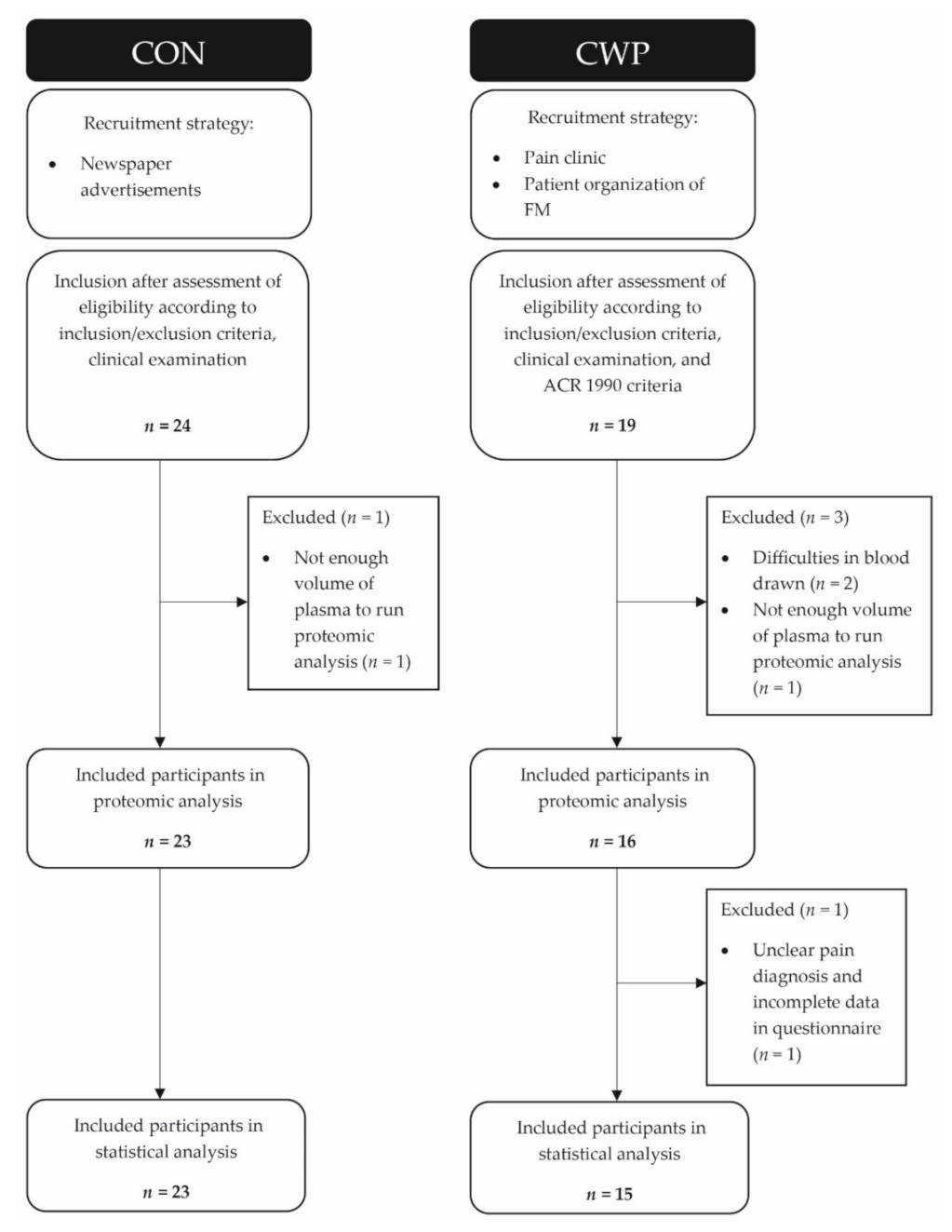
The protein–protein association network for the important proteins for the two thermal pain thresholds in CWP and CON were separately analysed using the online database Search Tool for Retrieval of Interacting Genes/Proteins (STRING; version 11) [53]. The search settings for the networks were set to: Homo sapiens (species); query proteins only (maximum number of protein interactions); minimum interaction score of medium confidence (0.400); and an FDR ≤ 0.001 for classifying the cellular component (CC), molecular function (MF), and biological processes (BP) according to Gene Ontology (GO; http://geneontology.org/docs/ontology-documentation/ access date: 15 March 2021). For each obtained network, PPI enrichment p-value and average local clustering coefficient were reported. In the network figure, each protein is represented by a coloured node, and protein–protein interaction and association are represented by an edge visualized as a line. Thicker lines/edges represent higher combined confidence scores.

3. Results

3.1. Clinical Variables

These data were presented elsewhere for the two groups and are summarized in Table 1 [28,34]. CWP patients reported higher levels of psychological distress and catastrophizing and lower quality of life. As expected, CWP had considerably higher pain intensities, and CON were pain free. CWP were significantly older.

3.2. Thermal Pain Thresholds

Significant group differences were found both for CPT and HPT (Table 1). CWP had significantly increased pain sensitivity according to both cold (reported at warmer temperature) and heat (reported at lower temperature).

3.3. Clinical Variables as Regressors of CPT and HPT

Neither CPT and HPT in CON nor CPT in CWP exhibited significant regressions, indicating that the clinical variables, including BMI and age, had little or no influence (for details, see Text S1). In contrast, the regression of HPT was significant (R2 = 0.67, Q2 = 0.41, CV-ANOVA p-value: 0.043; one predictive component) and HADS (VIPpred = 1.78; p(corr) = −0.85) and QOLS (VIPpred = 1.60; p(corr) = 0.77) were significant regressors (VIPpred > 1.0), whereas BMI, NRS-shoulders, NRS-neck, PCS, and age were not important (for details, see Text S1).

3.4. Proteins as Regressors of CPT and HPT

The proteins from muscle and plasma were used to regress CPT and HPT in CON and CWP.

3.4.1. CPT

Significant regressions of CPT were obtained both in CON (R2 = 0.94, Q2 = 0.76, CV-ANOVA p-value: 0.005; one predictive and three orthogonal components) and in CWP (R2 = 0.88, Q2 = 0.81, CV-ANOVA p-value: <0.001; one predictive component). The important significant proteins (including proteoforms), mainly from plasma, are shown in Table 2 and Table 3.
In CON, the protein with the strongest correlation with CPT was the muscle protein keratin type II cytoskeletal I, which is involved in mitochondrial function (Table 2). The most important plasma proteins (i.e., highest VIPpred) were proteoforms of alpha-2-macroglublin, apolipoprotein C-III, and ceruloplasmin from plasma (Table 2). Most proteins were acute phase proteins (Table 2). The significant proteins also included complement factors and 2 apolipoprotein-III proteoforms (associated with immunity). Several of the important proteins had proteoforms (i.e., alpha-1-antitrypsin, alpha-2-macroglobulin, ceruloplasmin, and complement factor C4-B and plasminogen) (Table 2). All proteins except one proteoform of alpha-1-antitrypsin correlated positively with CPT.
In CWP, the most important proteins for CPT were plasma proteins: apolipoprotein A-I, clusterin, Ig kappa chain C region, and alpha-2-macroglobulin (Table 3). The important proteins were acute phase proteins, three complement factors, one chemokine (CCL19), one cytokine (IL-7), two immunoglobulin factors, other factors associated with immunity (apolipoprotein A-I, CD244 (part of the Ig superfamily), and vitamin D-binding protein proteoforms) (Table 3). The only significant muscle protein was creatine kinase M-type, which is involved in the glycolytic pathway. Positive correlations with CPT were found in 12 of 20 proteins. Only two proteins and proteoforms were identical when comparing the important regressors for CPT between CON and CWP (i.e., a proteoform of alpha-1-antitrypsin and a proteoform of plasminogen) (Table 2andTable 3). Alpha-2-macroglobulin was important in both regressions, but the proteoforms differed.

3.4.2. HPT

For HPT, significant regressions were obtained in CON (R2 = 0.77, Q2 = 0.63, CV-ANOVA p-value: < 0.001; one predictive component) and in CWP (R2 = 0.77, Q2 = 0.61, CV-ANOVA p-value: 0.003; one predictive component) (Table 4 and Table 5).
In CON, the most important proteins—mainly plasma proteins—in the regression of HPT were one proteoform of serotransferrin, two proteoforms of alpha-2 macroglobulin, and complement C3 alpha chain (Table 4). In detail, acute phase proteins, two complement factors, two immunoglobulin factors, one chemokine (CCL20), one cytokine (CDCP1), as well as other molecules associated with immunity (i.e., beta-2-glycoprotein 1, EN-RAGE, and vitamin D-binding protein) were important (Table 4). The only muscle protein was phosphoglycerate mutase 2, which is involved in the glycolytic pathway. Several of the identified proteins were detected as different proteoforms: three proteoforms of alpha-2-macroglobulin, three of ceruloplasmin, and two of serotransferrin (Table 4).
In CWP the regression of HPT identified the plasma proteins clusterin, IL-10RB, and CSF-1 as most important (Table 5). In detail, three acute phase proteins; three complement factors; three cytokines (CSF-1, Flt3L, and IL-10RB); one neurotrophin (neurotrophin 3, NT-3); two immunoglobulin-related molecules; and other proteins related to inflammation and immunity (apolipoprotein A-I, TNFRSF9, and vitamin D-binding protein) were important plasma proteins (Table 5). Two muscle proteins related to the glycolytic pathway were also significant regressors (i.e., creatine kinase M-type and phosphoglycerate mutase 2). Two proteins—complement component C7 and immunoglobulin light chain—had two proteoforms among the significant proteins (Table 5). When comparing the important proteins for HPT between CON and CWP, some proteins were identical (phosphoglycerate mutase 2), but the proteoforms for alpha-2-macroglobulin and vitamin D-binding protein were different.

3.5. Both Proteins and Clinical Variables as Regressors of CPT and HPT

In the next step, the clinical variables were added to the regressions displayed in Table 2, Table 3, Table 4, Table 5. In the CON, these variables were not important compared to the 20 proteins nor in the regression of CPT (i.e., proteins: R2 = 0.94, Q2 = 0.76 and CV-ANOVA p-value: 0.005 vs. proteins and clinical variables: R2 = 0.97, Q2 = 0.75 and CV-ANOVA p-value: 0.004) nor in the regression of HPT (proteins: R2 = 0.77, Q2 = 0.63 and CV-ANOVA p-value: <0.001 vs. proteins and clinical variables: R2 = 0.78, Q2 = 0.62 and CV-ANOVA p-value: <0.001).
Some of the clinical variables were important for the regressions in CWP. In the regression of CPT, HADS was the 15th most important variable, whereas the other psychometric variables had little importance (proteins: R2 = 0.88, Q2 = 0.81 and CV-ANOVA p-value: <0.001 vs. proteins and clinical variables: R2 = 0.95, Q2 = 0.80 and CV-ANOVA p-value: 0.002). In the regression of HPT, HADS was the most important and QOL the fifth most important regressors. The explained variation increased when the clinical variables were added (proteins: R2 = 0.77, Q2 = 0.61 and CV-ANOVA p-value: 0.003 vs. proteins and clinical variables: R2 = 0.99, Q2 = 0.79 and CV-ANOVA p-value: 0.048). However, HADS was not markedly more important than the most important protein (HADS: VIPpred = 1.42 vs. IL-10rb: VIPpred = 1.40).

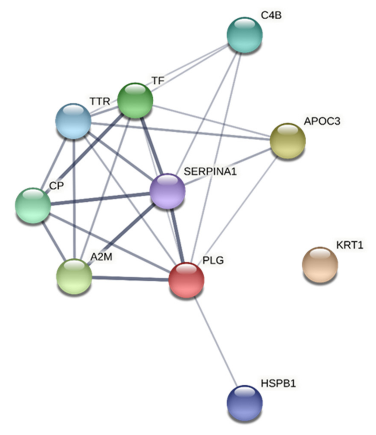
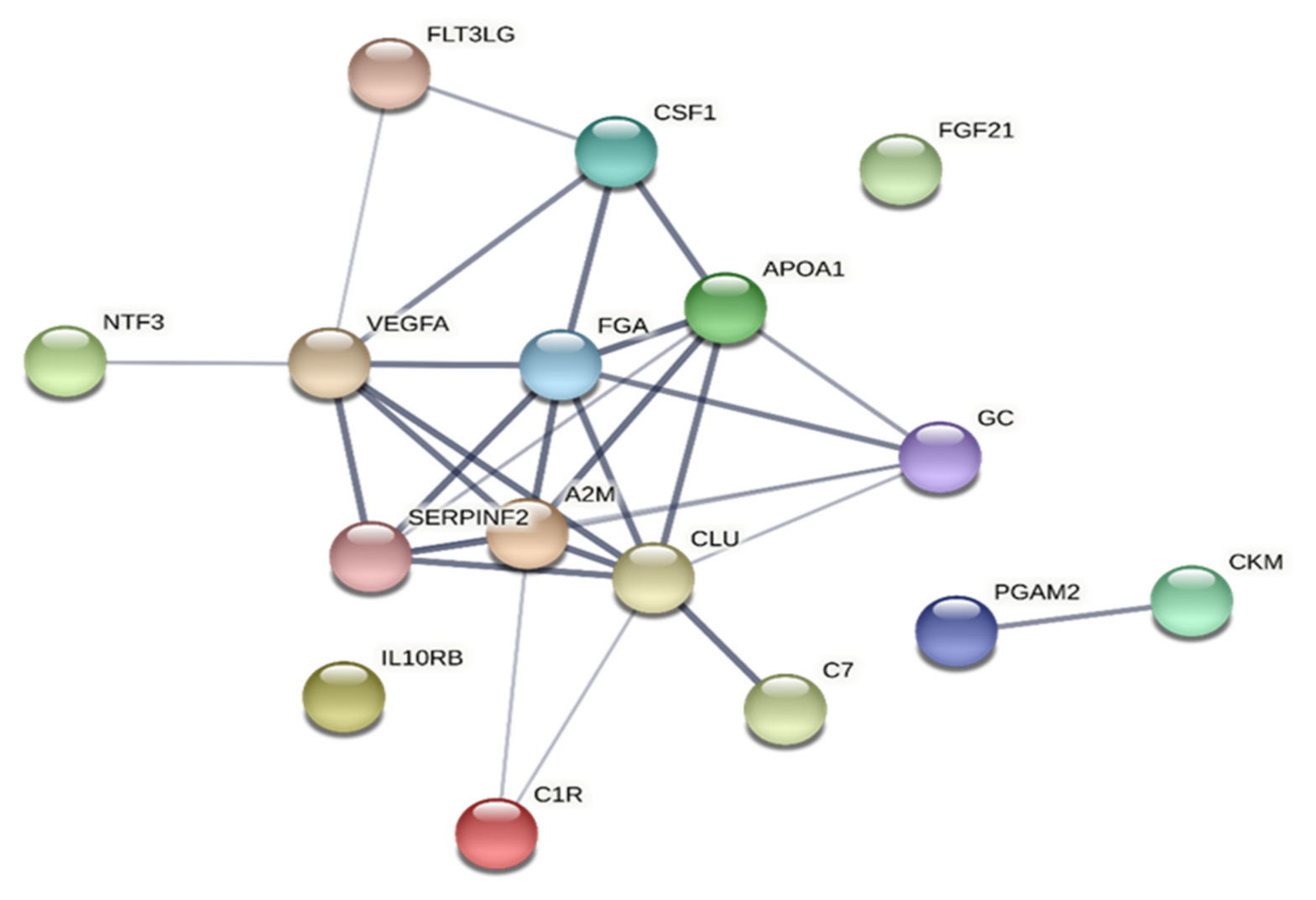
3.6. Network Analyses

3.6.1. CPT

The network and enrichment analysis of the important proteins in CON (Table 2) identified a protein–protein interaction network that was significantly enriched (Figure 2). Extracellular region and aspects of secretory and platelet granule had the lowest FDR according to CC (Table 6). MF terms with the lowest FDR were enzyme inhibitor activity and protein binding. The significant BP terms were platelet degranulation, transport aspects, regulated exocytosis, protein activation cascade, and protein metabolic process (Table 6).
In CWP, a significantly enriched protein–protein interaction network was identified (Table 7 and Figure 3). CC terms with lowest FDR were related to the extracellular regions and space as well as the lumens of vesicle secretory granule and platelets. MF terms with lowest FDR were signalling receptor and protein binding as well as enzyme and endopeptidase inhibitor activity. Most of the GO terms concerned BP; the most important were platelet degranulation and terms related to the immune system, response to stress and defence, protein activation/cascade, and cytokine regulations (Table 7). Fewer CC, MF, and BP terms were obtained in CON than in CWP (Table 6 and Table 7). Most terms obtained in CON within each of the three areas of GO were also found in CWP.

3.6.2. HPT

Significantly enriched protein–protein interaction networks were found for the proteins strongly associated with HPT both in CON (Table 8 and Figure 4) and in CWP (Table 9 and Figure 5). In CON, CC terms with lowest FDR were extracellular areas and secretory granule lumen (Table 8). An MF term with relatively low FDR was the signalling receptor binding. BP terms with low FDR were aspects of protein activation and complement activation, regulation and response of inflammatory and immune system (Table 8).
In CWP, the CC terms with lowest FDR were associated with extracellular areas, lumens of platelets, and secretory granules (Table 9). The MF terms were related to receptor-related activities and growth factor activity. Most of the GO terms were related to BP (i.e., platelet degranulation, protein activation cascade, protein regulation, complement activation, intracellular signal transduction, and inflammatory response). In addition, response to external stimuli had very low FDR (Table 9). More CC, MF, and BP terms were obtained in CWP than in CON. For example, the number of BP terms with FDR < 0.001 for HPT were 11 in CON and 45 in CWP. Most terms obtained in CON within each of the three areas of GO were also found in CWP.

4. Discussion

The three hypotheses stated were conformed and therefore the following major results were noted:
  • Patients with CWP had lowered pain thresholds for thermal stimulus; these levels were generally not related to the included clinical variables except for HPT in CWP.
  • Patterns of highly interacting proteins mainly from plasma showed strong associations with CPT and HPT both in CWP and in CON.
  • Differences in the important proteins for the two thermal pain thresholds were noted between CWP and CON; more complex patterns emerged in CWP.
CWP had lowered pain thresholds for cold and heat (Table 1), a finding that agrees with other studies [54,55,56,57,58]. For both the CWP and CON, CPT and HPT were generally not associated with clinical variables, which agrees with a study of FM [58]. The exception was in CWP: HADS and QOL showed significant associations with HPT, and HADS was a strong regressor of HPT although it was not markedly stronger than the most important proteins. Whiplash associated disorders and chronic pelvic pain were reported to have significant associations between thermal pain thresholds and clinical variables [37,38,59]. The resting-state brain connectome could predict pain thresholds for heat and pressure in healthy subjects [60]. Thus, larger studies need to be conducted to determine the importance of common clinical variables in relation to peripheral and central biomarkers of pain thresholds.
A broad sampling of plasma proteins was applied—i.e., proteins at nano and micro molar concentrations (proteomics) and at picomolar concentrations (cytokines, chemokines and growth factors)—to focus on protein interactions and possible biological processes. The latter approach was advocated before it will be possible to focus on key proteins [36,61] and is increasingly applied in proteomic blood and cerebrospinal fluid studies of CWP and FM [14,23,24,25,26,62]. Plasma/serum proteome studies of CWP and FM cohorts have focussed on differentiating patients from controls, and low-grade peripheral inflammation appears to be involved in pathogenesis and maintenance of CWP and FM [10,17,24,25,26,63]. It is also important to examine the peripheral protein patterns in relation to clinical variables since it cannot be assumed that the same proteins responsible for group differentiation are important for clinical variables [36]. Plasma proteomic studies of CWP and FM cohorts, including the present CWP/FM subjects, report that the important proteins largely differ across clinical variables [10,25,28,34]. As with PPT [34], the important proteins for both thermal pain thresholds differed between CON and CWP (Table 2, Table 3, Table 4, Table 5). When the same protein was identified, the proteoforms generally differed. The most important proteins explained a large proportion of the variations in CPT (88–94%) and in HPT (both 77%) in the two groups (Table 2, Table 3, Table 4, Table 5). These peripheral protein patterns reported in Table 2, Table 3, Table 4, Table 5 reflect tonic/habitual peripheral situations. With the present design, it is not possible to determine which proteins are specifically involved in the direct activation of the different receptors/channels of the nociceptors. If altered peripheral conditions such as inflammation or tissue injury are present peripherally in CWP, it is reasonable to expect sensitized and primed nociceptors [64]. In agreement with this, we report lowered pain thresholds as well as another pattern of peripheral proteins associated with the thermal pain thresholds in CWP.
Most of the proteins reported in Table 2, Table 3, Table 4, Table 5 were associated with either pain, nociception, or immune system, or in combination. Text S2 presents the results of brief literature reviews with special emphasis on earlier proteomic- and inflammation-related studies in relation to nociception and pain conditions. Although the exact proteins differed between the two groups, it was obvious that mostly plasma proteins related to the host defence/immunity—e.g., acute phase proteins, complement factors, immunoglobulin factors, and cytokines/chemokines—were important for the thermal pain thresholds. For example, in CWP, lower pain thresholds for cold (hyperalgesia) were associated with higher levels of most acute phase proteins, most complement factors, two cytokines/chemokines, and a muscle protein (creatine-kinase M-type), and low levels of proteins were associated with anti-inflammation/enhancement of immunity. Thus, in CWP, proteins representing different parts of the immune system were involved, including cytokine/chemokine, whereas in CON, acute phase proteins and their proteoforms dominated. Moreover, heat hyperalgesia (low pain thresholds for heat) in CWP was associated with low levels of two of three acute phase proteins, most complement factors, two immunoglobulin factors, high levels of four cytokines (CSF-1, IL-10Rb, FGF-21, and Flt3l), and neuroprotective factors (NT3 and VEGF-A). Thus, in CWP, more cytokines and fewer acute phase proteins were involved with respect to HPT compared to CON.
Is it reasonable to conclude that plasma proteins of the immune system are significant regressors of thermal pain thresholds? The circulatory system interacts with other tissues and is vital for the immune-system-based host defence mechanisms and tissue homeostasis [65,66]. Nociceptors in the skin are involved both in neuro-immune and neural-vascular interactions [67]. A network of immune cells is found in the skin [68] and immune cells are also recruited from the blood vascular systems [69]. Structural cells of the skin (epithelial cells, endothelial cells, fibroblasts, etc.) express immune regulators and cytokine signalling and contribute to immunity [70,71]. Dys-regulation in the bidirectional signalling between the nociceptive and immune systems may be associated with excitation [72]. The nociceptors are in direct association or near association with nociceptive Schwann cells in the epidermis and form a mesh-like network [73,74]. The intimate bidirectional relationships between the immune system and the nociceptors peripherally may indicate priming of the immune system [75]. The identified proteins correlating with thermal pain thresholds are consistent with the results from plasma/serum studies differentiating CWP vs. CON and FM vs. CON [10,24,25,26]. In addition, acute phase proteins (both positive and negative), complement factors, immunoglobulin factors, and coagulation factors were important. Inflammation is the biological defensive response of the immune system [76,77]. The crosstalk among immunity (innate and adaptive), coagulative/fibrinolytic pathways, and the nervous system is necessary for adequate inflammatory cascade [77]. We found that peripheral cytokines and chemokines are important, which is consistent with results from Bäckryd et al. [63]. The literature seems to focus on the cytokines/chemokines and their roles; indeed, these are important for pain thresholds in CWP. Our results indicate, however, that other parts of the immune system are also important for thermal pain thresholds. The classification of cytokines and chemokines as pro- or anti-inflammatory is not unproblematic since the properties may depend on the microenvironment [78]. The identified muscle proteins (creatine kinase M-type and phosphoglycerate mutase 2) in the regressions concerning CWP agree with our and other studies reporting muscle metabolic and mitochondrial disturbances in FM (a subgroup of CWP) [79,80,81]. Larger studies are needed to confirm these patterns of proteins and in detail elucidate the interactions between the different parts of the immune system and related interacting factors.
Both variables and groups of subjects revealed significantly enriched protein–protein interaction networks (Figure 2, Figure 3, Figure 4, Figure 5). Hence, the groups of plasma proteins generally interact, which is reasonable from a host defence perspective. Different proteoforms were important and sometimes had different signs within a regression (e.g., alpha-1 antitrypsin in the regression of CPT in CON and vitamin D-binding protein in the regression of CPT in CWP). Fewer GO terms were obtained in CON than in CWP. This finding suggests a more complex molecular relationships in CWP and therefore reflects peripheral molecular alterations associated with hyperalgesia in CWP. However, because STRING cannot handle different proteoforms, fewer proteins are included in analyses of CON than for CWP. In future larger studies, it will be important to scrutinize in more detail the pattern of proteoforms and how they may be altered in CWP.
The primary origin and maintenance factors for the pathophysiological alterations in CWP and in the FM subgroup are not known. Morphological and functional alterations both in the CNS (e.g., neuroinflammation, opioidergic dysregulation, and central sensitization) and in the periphery (e.g., systemic low-grade inflammation, small fibre impairment, reduced skin innervation, and muscle alterations such as mitochondrial disturbance) are found [77,82,83,84,85,86,87,88,89,90,91,92,93]. These mechanisms may be present simultaneously, indicating complicated interactions between peripheral and central processes in CWP including FM that contribute to pain and hypersensitivity. The present study (mainly consisting of FM patients), other proteomic and large cytokine/chemokine studies, and other peripheral studies may challenge the IASP’s definition that FM is a nociplastic condition [29].

Strengths and Limitations

An important strength of the study is that it examines a deficiently studied area in CWP including FM, i.e., whether peripheral molecular changes are associated with thermal pain thresholds. The cross-sectional design and the small sample size are limitations of this exploratory pilot study. Larger studies with repeated measures and population-based studies are important for validation [94]. As suggested in a previous systematic review [36], we applied MVDA for relating clinical parameters (i.e., pain thresholds) to many possibly intercorrelated proteins. The removal procedure of, e.g., albumin and IgG (i.e., large abundant proteins), could have removed important low abundant proteins. Proteoforms of a protein and post translational modifications can be detected by 2-DE, which is important as several of the significant proteins were expressed as different proteoforms, findings also reported earlier [10,28,34]. The ACR 1990 classification criteria for CWP and FM was used to simplify comparisons with earlier studies. In future studies, both ACR 1990 and the newer 2016 criteria should be used to optimize comparisons with other studies.

5. Conclusions and Clinical Implications

The present study contributes towards an emerging picture that peripheral proteins are associated with pain thresholds in CWP and FM; we have earlier reported such associations for pressure pain thresholds [25,34]. Hence, patterns of highly interacting proteins mainly from plasma showed strong associations with CPT and HPT both in CWP and in CON. Although different proteins were important in the two groups, there were also similarities; proteins related to the host defence/immunity such as acute phase proteins, complement factors, immunoglobulin factors, and cytokines/chemokines (not in CON for CPT) were important habitual/tonic factors for thermal pain thresholds. Although peripheral proteins contribute to thermal pain thresholds, central factors and complex interactions between peripheral and central factors may contribute to pain thresholds in CWP. The present study indicates that peripheral molecular factors are important for the clinical presentations in CWP and future larger studies may contribute to an increased understanding of the molecular mechanisms involved in CWP including FM, which in turn may contribute to the development of new therapies.

Supplementary Materials

The following are available online at https://www.mdpi.com/article/10.3390/jcm10163652/s1, Text S1: Regression of CPT an HPT using clinical variables as regressors, Text S2: Individual proteins.

Author Contributions

Conceptualization, B.G. (Björn Gerdle), B.G. (Bijar Ghafouri), K.W. and T.G.; methodology, B.G. (Björn Gerdle), B.G. (Bijar Ghafouri), K.W. and T.G.; formal analysis, B.G. (Björn Gerdle) and K.W.; investigation, B.G. (Björn Gerdle) and B.G. (Bijar Ghafouri); resources, B.G. (Bijar Ghafouri) and T.G.; data curation, B.G. (Björn Gerdle), B.G. (Bijar Ghafouri) and K.W.; writing—original draft preparation, B.G. (Björn Gerdle); writing—review and editing, B.G. (Björn Gerdle), B.G. (Bijar Ghafouri), K.W. and T.G.; visualization B.G. (Björn Gerdle), B.G. (Bijar Ghafouri) and K.W.; supervision, B.G. (Björn Gerdle) and B.G. (Bijar Ghafouri); project administration, B.G. (Björn Gerdle); funding acquisition, B.G. (Björn Gerdle) and B.G. (Bijar Ghafouri). All authors have read and agreed to the published version of the manuscript.

Funding

This research was funded by a grant from the Swedish Research Council, grant number 2018-02470.

Institutional Review Board Statement

The study was conducted according to the guidelines of the Declaration of Helsinki and approved by the Regional Ethical Review Board in Linköping, Sweden (Dnr. M10–08, M233–09, Dnr. 2010/164–32).

Informed Consent Statement

Informed consent was obtained from all subjects involved in the study.

Data Availability Statement

The datasets generated and analysed in this study are not publicly available as the Ethical Review Board has not approved the public availability of these data.

Conflicts of Interest

The authors declare no conflict of interest. The funder had no role in the design of the study; in the collection, analyses, or interpretation of data; in the writing of the manuscript, or in the decision to publish the results.

References

  1. Mansfield, K.E.; Sim, J.; Jordan, J.L.; Jordan, K.P. A systematic review and meta-analysis of the prevalence of chronic widespread pain in the general population. Pain 2016, 157, 55–64. [Google Scholar] [CrossRef] [PubMed]
  2. Cimmino, M.A.; Ferrone, C.; Cutolo, M. Epidemiology of chronic musculoskeletal pain. Best Pract. Res. Clin. Rheumatol. 2011, 25, 173–183. [Google Scholar] [CrossRef] [PubMed]
  3. Bergman, S.; Herrstrom, P.; Hogstrom, K.; Petersson, I.F.; Svensson, B.; Jacobsson, L.T. Chronic musculoskeletal pain, prevalence rates, and sociodemographic associations in a Swedish population study. J. Rheumatol. 2001, 28, 1369–1377. [Google Scholar] [PubMed]
  4. Perez de Heredia-Torres, M.; Huertas-Hoyas, E.; Maximo-Bocanegra, N.; Palacios-Cena, D.; Fernandez-De-Las-Penas, C. Cognitive performance in women with fibromyalgia: A case-control study. Aust. Occup. Ther. J. 2016, 63, 329–337. [Google Scholar] [CrossRef]
  5. Aparicio, V.A.; Ortega, F.B.; Carbonell-Baeza, A.; Gatto-Cardia, C.; Sjostrom, M.; Ruiz, J.R.; Delgado-Fernandez, M. Fibromyalgia’s key symptoms in normal-weight, overweight, and obese female patients. Pain Manag. Nurs. 2013, 14, 268–276. [Google Scholar] [CrossRef]
  6. Wolfe, F.; Smythe, H.A.; Yunus, M.B.; Bennett, R.M.; Bombardier, C.; Goldenberg, D.L.; Tugwell, P.; Campbell, S.M.; Abeles, M.; Clark, P.; et al. The American College of Rheumatology 1990 Criteria for the Classification of Fibromyalgia. Report of the Multicenter Criteria Committee. Arthritis Rheum. 1990, 33, 160–172. [Google Scholar] [CrossRef]
  7. Breivik, H.; Collett, B.; Ventafridda, V.; Cohen, R.; Gallacher, D. Survey of chronic pain in Europe: Prevalence, impact on daily life, and treatment. Eur. J. Pain 2006, 10, 287–333. [Google Scholar] [CrossRef]
  8. Tracey, I.; Woolf, C.J.; Andrews, N.A. Composite Pain Biomarker Signatures for Objective Assessment and Effective Treatment. Neuron 2019, 101, 783–800. [Google Scholar] [CrossRef] [Green Version]
  9. Galvez-Sanchez, C.M.; Reyes Del Paso, G.A. Diagnostic Criteria for Fibromyalgia: Critical Review and Future Perspectives. J. Clin. Med. 2020, 9, 1219. [Google Scholar] [CrossRef]
  10. Wåhlén, K.; Olausson, P.; Carlsson, A.; Ghafouri, N.; Gerdle, B.; Ghafouri, B. Systemic alterations in plasma proteins from women with chronic widespread pain compared to healthy controls: A proteomic study. J. Pain Res. 2017, 10, 797–809. [Google Scholar] [CrossRef] [Green Version]
  11. Olausson, P.; Gerdle, B.; Ghafouri, N.; Larsson, B.; Ghafouri, B. Identification of proteins from interstitium of trapezius muscle in women with chronic myalgia using microdialysis in combination with proteomics. PLoS ONE 2012, 7, e52560. [Google Scholar] [CrossRef] [Green Version]
  12. Hadrevi, J.; Bjorklund, M.; Kosek, E.; Hallgren, S.; Antti, H.; Fahlstrom, M.; Hellstrom, F. Systemic differences in serum metabolome: A cross sectional comparison of women with localised and widespread pain and controls. Sci. Rep. 2015, 5, 15925. [Google Scholar] [CrossRef] [PubMed] [Green Version]
  13. Culic, O.; Cordero, M.D.; Zanic-Grubisic, T.; Somborac-Bacura, A.; Pucar, L.B.; Detel, D.; Varljen, J.; Barisic, K. Serum activities of adenosine deaminase, dipeptidyl peptidase IV and prolyl endopeptidase in patients with fibromyalgia: Diagnostic implications. Clin. Rheumatol. 2016, 35, 2565–2571. [Google Scholar] [CrossRef] [PubMed]
  14. Olausson, P.; Ghafouri, B.; Backryd, E.; Gerdle, B. Clear differences in cerebrospinal fluid proteome between women with chronic widespread pain and healthy women—A multivariate explorative cross-sectional study. J. Pain Res. 2017, 10, 575–590. [Google Scholar] [CrossRef] [PubMed] [Green Version]
  15. Zanette, S.A.; Dussan-Sarria, J.A.; Souza, A.; Deitos, A.; Torres, I.L.S.; Caumo, W. Higher serum S100B and BDNF levels are correlated with a lower pressure-pain threshold in fibromyalgia. Mol. Pain 2014, 10, 46. [Google Scholar] [CrossRef] [Green Version]
  16. Bazzichi, L.; Ciregia, F.; Giusti, L.; Baldini, C.; Giannaccini, G.; Giacomelli, C.; Sernissi, F.; Bombardieri, S.; Lucacchini, A. Detection of potential markers of primary fibromyalgia syndrome in human saliva. Proteom. Clin. Appl. 2009, 3, 1296–1304. [Google Scholar] [CrossRef] [PubMed]
  17. Gerdle, B.; Ghafouri, B.; Ghafouri, N.; Backryd, E.; Gordh, T. Signs of ongoing inflammation in female patients with chronic widespread pain: A multivariate, explorative, cross-sectional study of blood samples. Medicine 2017, 96, e6130. [Google Scholar] [CrossRef]
  18. Jablochkova, A.; Backryd, E.; Kosek, E.; Mannerkorpi, K.; Ernberg, M.; Gerdle, B.; Ghafouri, B. Unaltered low nerve growth factor and high brain-derived neurotrophic factor levels in plasma from patients with fibromyalgia after a 15-week progressive resistance exercise. J. Rehabil. Med. 2019, 51, 779–787. [Google Scholar] [CrossRef] [Green Version]
  19. Gomez-Varela, D.; Barry, A.M.; Schmidt, M. Proteome-based systems biology in chronic pain. J. Proteom. 2019, 190, 1–11. [Google Scholar] [CrossRef] [PubMed]
  20. Niederberger, E.; Geisslinger, G. Proteomics in neuropathic pain research. J. Am. Soc. Anesthesiol. 2008, 108, 314–323. [Google Scholar] [CrossRef] [PubMed] [Green Version]
  21. Emilsson, V.; Ilkov, M.; Lamb, J.R.; Finkel, N.; Gudmundsson, E.F.; Pitts, R.; Hoover, H.; Gudmundsdottir, V.; Horman, S.R.; Aspelund, T.; et al. Co-regulatory networks of human serum proteins link genetics to disease. Science 2018, 361, 769–773. [Google Scholar] [CrossRef] [PubMed] [Green Version]
  22. Manzoni, C.; Kia, D.A.; Vandrovcova, J.; Hardy, J.; Wood, N.W.; Lewis, P.A.; Ferrari, R. Genome, transcriptome and proteome: The rise of omics data and their integration in biomedical sciences. Brief. Bioinform. 2018, 19, 286–302. [Google Scholar] [CrossRef] [PubMed]
  23. Olausson, P.; Gerdle, B.; Ghafouri, N.; Sjostrom, D.; Blixt, E.; Ghafouri, B. Protein alterations in women with chronic widespread pain--An explorative proteomic study of the trapezius muscle. Sci. Rep. 2015, 5, 11894. [Google Scholar] [CrossRef] [Green Version]
  24. Han, C.L.; Sheng, Y.C.; Wang, S.Y.; Chen, Y.H.; Kang, J.H. Serum proteome profiles revealed dysregulated proteins and mechanisms associated with fibromyalgia syndrome in women. Sci. Rep. 2020, 10, 12347. [Google Scholar] [CrossRef]
  25. Wåhlén, K.; Ernberg, M.; Kosek, E.; Mannerkorpi, K.; Gerdle, B.; Ghafouri, B. Significant correlation between plasma proteome profile and pain intensity, sensitivity, and psychological distress in women with fibromyalgia. Sci. Rep. 2020, 10, 12508. [Google Scholar] [CrossRef]
  26. Ramirez-Tejero, J.A.; Martinez-Lara, E.; Rus, A.; Camacho, M.V.; Del Moral, M.L.; Siles, E. Insight into the biological pathways underlying fibromyalgia by a proteomic approach. J. Proteom. 2018, 186, 47–55. [Google Scholar] [CrossRef]
  27. Olausson, P.; Ghafouri, B.; Ghafouri, N.; Gerdle, B. Specific proteins of the trapezius muscle correlate with pain intensity and sensitivity—An explorative multivariate proteomic study of the trapezius muscle in women with chronic widespread pain. J. Pain Res. 2016, 9, 345–356. [Google Scholar] [CrossRef] [Green Version]
  28. Wåhlén, K.; Ghafouri, B.; Ghafouri, N.; Gerdle, B. Plasma Protein Pattern Correlates With Pain Intensity and Psychological Distress in Women With Chronic Widespread Pain. Front. Psychol. 2018, 9, 2400. [Google Scholar] [CrossRef]
  29. Kosek, E.; Cohen, M.; Baron, R.; Gebhart, G.F.; Mico, J.A.; Rice, A.S.; Rief, W.; Sluka, A.K. Do we need a third mechanistic descriptor for chronic pain states? Pain 2016, 157, 1382–1386. [Google Scholar] [CrossRef]
  30. Gerdle, B.; Bäckryd, E.; Novo, M.; Roeck Hansen, E.; Rothman, M.; Stålnacke, B.-M.; Westergren, H.; Rivano Fischer, M. Smärtanalys—Diagnos, Smärtmekanismer, Psykologisk Och Social Bedömning; Studentlitteratur AB: Lund, Sweden, 2020. [Google Scholar]
  31. Nijs, J.; George, S.; Clauw, D.; Fernández-de-las-Peñas, C.; Kosek, E.; Ickmans, K.; Fernández-Carnero, J.; Polli, A.; Kapreli, E.; Huysmans, E.; et al. Central sensitisation in chronic pain conditions: Latest discoveries and their potential for precision medicine. Lancet Rheumatol. 2021, 3, e383–e392. [Google Scholar] [CrossRef]
  32. Kosek, E.; Clauw, D.; Nijs, J.; Baron, R.; Gilron, I.; Harris, R.E.; Mico, J.A.; Rice, A.S.; Sterling, M. Chronic nociplastic pain affecting the musculoskeletal system: Clinical criteria and grading system. Pain 2021, in press. [Google Scholar] [CrossRef]
  33. Potvin, S.; Marchand, S. Pain facilitation and pain inhibition during conditioned pain modulation in fibromyalgia and in healthy controls. Pain 2016, 157, 1704–1710. [Google Scholar] [CrossRef]
  34. Gerdle, B.; Wåhlén, K.; Ghafouri, B. Plasma protein patterns are strongly correlated with pressure pain thresholds in women with chronic widespread pain and in healthy controls-an exploratory case-control study. Medicine 2020, 99, e20497. [Google Scholar] [CrossRef]
  35. Muller, M.; Butikofer, L.; Andersen, O.K.; Heini, P.; Arendt-Nielsen, L.; Juni, P.; Curatolo, M. Cold pain hypersensitivity predicts trajectories of pain and disability after low back surgery: A prospective cohort study. Pain 2021, 162, 184–194. [Google Scholar] [CrossRef]
  36. Gerdle, B.; Ghafouri, B. Proteomic studies of common chronic pain conditions—A systematic review and associated network analyses. Expert Rev. Proteom. 2020, 17, 483–505. [Google Scholar] [CrossRef]
  37. Wallin, M.; Liedberg, G.; Börsbo, B.; Gerdle, B. Thermal detection and pain thresholds but not pressure pain thresholds are correlated with psychological factors in women with chronic whiplash-associated pain. Clin. J. Pain 2012, 28, 211–221. [Google Scholar] [CrossRef] [PubMed]
  38. Grundstrom, H.; Larsson, B.; Arendt-Nielsen, L.; Gerdle, B.; Kjolhede, P. Pain catastrophizing is associated with pain thresholds for heat, cold and pressure in women with chronic pelvic pain. Scand. J. Pain 2020, 20, 635–646. [Google Scholar] [CrossRef] [PubMed]
  39. Gerdle, B.; Larsson, B.; Forsberg, F.; Ghafouri, N.; Karlsson, L.; Stensson, N.; Ghafouri, B. Chronic widespread pain: Increased glutamate and lactate concentrations in the trapezius muscle and plasma. Clin. J. Pain 2014, 30, 409–420. [Google Scholar] [CrossRef] [PubMed]
  40. Gerdle, B.; Soderberg, K.; Salvador Puigvert, L.; Rosendal, L.; Larsson, B. Increased interstitial concentrations of pyruvate and lactate in the trapezius muscle of patients with fibromyalgia: A microdialysis study. J. Rehabil. Med. 2010, 42, 679–687. [Google Scholar] [CrossRef] [PubMed] [Green Version]
  41. Ghafouri, N.; Ghafouri, B.; Larsson, B.; Stensson, N.; Fowler, C.J.; Gerdle, B. Palmitoylethanolamide and stearoylethanolamide levels in the interstitium of the trapezius muscle of women with chronic widespread pain and chronic neck-shoulder pain correlate with pain intensity and sensitivity. Pain 2013, 154, 1649–1658. [Google Scholar] [CrossRef]
  42. Ferreira-Valente, M.A.; Pais-Ribeiro, J.L.; Jensen, M.P. Validity of four pain intensity rating scales. Pain 2011, 152, 2399–2404. [Google Scholar] [CrossRef] [PubMed]
  43. Zigmond, A.S.; Snaith, R.P. The hospital anxiety and depression scale. Acta Psychiatr. Scand. 1983, 67, 361–370. [Google Scholar] [CrossRef] [PubMed] [Green Version]
  44. LoMartire, R.; Äng, B.; Gerdle, B.; Vixner, L. Psychometric properties of SF-36, EQ-5D and HADS in patients with chronic pain. Pain 2020, 161, 83–95. [Google Scholar] [CrossRef] [Green Version]
  45. Sullivan, M.J.L.; Bishop, S.R.; Pivik, J. The Pain Catastrophizing Scale: Development and validation. Psychol. Assess. 1995, 7, 524–532. [Google Scholar] [CrossRef]
  46. Burckhardt, C.S.; Anderson, K.L. The Quality of Life Scale (QOLS): Reliability, validity, and utilization. Health Qual. Life Outcomes 2003, 1, 60. [Google Scholar] [CrossRef] [Green Version]
  47. Raak, R.; Wallin, M. Thermal Thresholds and Catastrophizing in Individuals with Chronic Pain after Whiplash Injury. Biol. Res. Nurs. 2006, 8, 138–146. [Google Scholar] [CrossRef]
  48. Wallin, M.K.; Raak, R.I. Quality of life in subgroups of individuals with whiplash associated disorders. Eur. J. Pain 2008, 12, 842–849. [Google Scholar] [CrossRef]
  49. Fruhstorfer, H.; Lindblom, U.; Schmidt, W. Method for quantitative estimation of thermal thresholds in patients. J. Neurol. Neurosurg. Psychiatry 1976, 39, 1071–1075. [Google Scholar] [CrossRef] [Green Version]
  50. Gorg, A.; Drews, O.; Luck, C.; Weiland, F.; Weiss, W. 2-DE with IPGs. Electrophoresis 2009, 30 (Suppl. S1), S122–S132. [Google Scholar] [CrossRef] [PubMed]
  51. Wheelock, A.M.; Wheelock, C.E. Trials and tribulations of omics data analysis: Assessing quality of SIMCA-based multivariate models using examples from pulmonary medicine. Mol. Biosyst. 2013, 9, 2589–2596. [Google Scholar] [CrossRef] [PubMed] [Green Version]
  52. Eriksson, L.; Byrne, T.; Johansson, E.; Trygg, J.; Vikström, C. Multi-and Megavariate Data Analysis: Basic Principles and Applications; Third Revised Edition; MKS Umetrics AB: Malmö, Sweden, 2013. [Google Scholar]
  53. Szklarczyk, D.; Gable, A.L.; Lyon, D.; Junge, A.; Wyder, S.; Huerta-Cepas, J.; Simonovic, M.; Doncheva, N.T.; Morris, J.H.; Bork, P.; et al. STRING v11: Protein-protein association networks with increased coverage, supporting functional discovery in genome-wide experimental datasets. Nucleic Acids Res. 2019, 47, D607–D613. [Google Scholar] [CrossRef] [Green Version]
  54. Blumenstiel, K.; Gerhardt, A.; Rolke, R.; Bieber, C.; Tesarz, J.; Friederich, H.C.; Eich, W.; Treede, R.D. Quantitative sensory testing profiles in chronic back pain are distinct from those in fibromyalgia. Clin. J. Pain 2011, 27, 682–690. [Google Scholar] [CrossRef]
  55. Hurtig, I.M.; Raak, R.I.; Kendall, S.A.; Gerdle, B.; Wahren, L.K. Quantitative sensory testing in fibromyalgia patients and in healthy subjects: Identification of subgroups. Clin. J. Pain 2001, 17, 316–322. [Google Scholar] [CrossRef] [PubMed]
  56. Smith, B.W.; Tooley, E.M.; Montague, E.Q.; Robinson, A.E.; Cosper, C.J.; Mullins, P.G. Habituation and sensitization to heat and cold pain in women with fibromyalgia and healthy controls. Pain 2008, 140, 420–428. [Google Scholar] [CrossRef]
  57. Brietzke, A.P.; Antunes, L.C.; Carvalho, F.; Elkifury, J.; Gasparin, A.; Sanches, P.R.S.; da Silva Junior, D.P.; Dussan-Sarria, J.A.; Souza, A.; da Silva Torres, I.L.; et al. Potency of descending pain modulatory system is linked with peripheral sensory dysfunction in fibromyalgia: An exploratory study. Medicine 2019, 98, e13477. [Google Scholar] [CrossRef] [PubMed]
  58. Rehm, S.; Sachau, J.; Hellriegel, J.; Forstenpointner, J.; Borsting Jacobsen, H.; Harten, P.; Gierthmuhlen, J.; Baron, R. Pain matters for central sensitization: Sensory and psychological parameters in patients with fibromyalgia syndrome. Pain Rep. 2021, 6, e901. [Google Scholar] [CrossRef] [PubMed]
  59. Grundstrom, H.; Gerdle, B.; Alehagen, S.; Bertero, C.; Arendt-Nielsen, L.; Kjolhede, P. Reduced pain thresholds and signs of sensitization in women with persistent pelvic pain and suspected endometriosis. Acta Obstet. Gynecol. Scand. 2019, 98, 327–336. [Google Scholar] [CrossRef] [PubMed] [Green Version]
  60. Tu, Y.; Zhang, B.; Cao, J.; Wilson, G.; Zhang, Z.; Kong, J. Identifying inter-individual differences in pain threshold using brain connectome: A test-retest reproducible study. Neuroimage 2019, 202, 116049. [Google Scholar] [CrossRef]
  61. Jin, P.; Lan, J.; Wang, K.; Baker, M.S.; Huang, C.; Nice, E.C. Pathology, proteomics and the pathway to personalised medicine. Expert Rev. Proteom. 2018, 15, 231–243. [Google Scholar] [CrossRef] [PubMed]
  62. Khoonsari, P.E.; Musunri, S.; Herman, S.; Svensson, C.I.; Tanum, L.; Gordh, T.; Kultima, K. Systematic analysis of the cerebrospinal fluid proteome of fibromyalgia patients. J. Proteom. 2019, 190, 35–43. [Google Scholar] [CrossRef]
  63. Bäckryd, E.; Tanum, L.; Lind, A.L.; Larsson, A.; Gordh, T. Evidence of both systemic inflammation and neuroinflammation in fibromyalgia patients, as assessed by a multiplex protein panel applied to the cerebrospinal fluid and to plasma. J. Pain Res. 2017, 10, 515–525. [Google Scholar] [CrossRef] [Green Version]
  64. Obreja, O.; Rukwied, R.; Nagler, L.; Schmidt, M.; Schmelz, M.; Namer, B. Nerve growth factor locally sensitizes nociceptors in human skin. Pain 2018, 159, 416–426. [Google Scholar] [CrossRef]
  65. Yurkovich, J.T.; Hood, L. Blood Is a Window into Health and Disease. Clin. Chem. 2019, 65, 1204–1206. [Google Scholar] [CrossRef] [PubMed]
  66. Bruderer, R.; Muntel, J.; Muller, S.; Bernhardt, O.M.; Gandhi, T.; Cominetti, O.; Macron, C.; Carayol, J.; Rinner, O.; Astrup, A.; et al. Analysis of 1508 Plasma Samples by Capillary-Flow Data-Independent Acquisition Profiles Proteomics of Weight Loss and Maintenance. Mol. Cell. Proteom. 2019, 18, 1242–1254. [Google Scholar] [CrossRef] [PubMed] [Green Version]
  67. Baral, P.; Udit, S.; Chiu, I.M. Pain and immunity: Implications for host defence. Nat. Rev. Immunol. 2019, 19, 433–447. [Google Scholar] [CrossRef] [PubMed]
  68. Nguyen, A.V.; Soulika, A.M. The Dynamics of the Skin’s Immune System. Int. J. Mol. Sci. 2019, 20, 1811. [Google Scholar] [CrossRef] [Green Version]
  69. Kabashima, K.; Honda, T.; Ginhoux, F.; Egawa, G. The immunological anatomy of the skin. Nat. Rev. Immunol. 2019, 19, 19–30. [Google Scholar] [CrossRef]
  70. Krausgruber, T.; Fortelny, N.; Fife-Gernedl, V.; Senekowitsch, M.; Schuster, L.C.; Lercher, A.; Nemc, A.; Schmidl, C.; Rendeiro, A.F.; Bergthaler, A.; et al. Structural cells are key regulators of organ-specific immune responses. Nature 2020, 583, 296–302. [Google Scholar] [CrossRef]
  71. Rice, F.L.; Kruger, L.; Albrecht, P.J. Histology of Nociceptors. In Encyclopedia of Pain; Gebhart, G.F., Schmidt, R.F., Eds.; Springer: Berlin/Heidelberg, Germany, 2013; pp. 1482–1490. [Google Scholar]
  72. Lacagnina, M.; Heijnen, C.; Watkins, L.; Grace, P. Autoimmune regulation of chronic pain. Pain Rep. 2021, 6, e905. [Google Scholar] [CrossRef]
  73. Abdo, H.; Calvo-Enrique, L.; Lopez, J.M.; Song, J.; Zhang, M.D.; Usoskin, D.; El Manira, A.; Adameyko, I.; Hjerling-Leffler, J.; Ernfors, P. Specialized cutaneous Schwann cells initiate pain sensation. Science 2019, 365, 695–699. [Google Scholar] [CrossRef]
  74. Bray, E.R.; Cheret, J.; Yosipovitch, G.; Paus, R. Schwann cells as underestimated, major players in human skin physiology and pathology. Exp. Dermatol. 2020, 29, 93–101. [Google Scholar] [CrossRef] [Green Version]
  75. Karshikoff, B.; Tadros, M.; Mackey, S.; Zouikr, I. Neuroimmune modulation of pain across the developmental spectrum. Curr. Opin. Behav. Sci. 2019, 28, 85–92. [Google Scholar] [CrossRef]
  76. Chen, L.; Deng, H.; Cui, H.; Fang, J.; Zuo, Z.; Deng, J.; Li, Y.; Wang, X.; Zhao, L. Inflammatory responses and inflammation-associated diseases in organs. Oncotarget 2018, 9, 7204–7218. [Google Scholar] [CrossRef] [PubMed] [Green Version]
  77. Coskun Benlidayi, I. Role of inflammation in the pathogenesis and treatment of fibromyalgia. Rheumatol. Int. 2019, 39, 781–791. [Google Scholar] [CrossRef] [PubMed]
  78. Goncalves Dos Santos, G.; Delay, L.; Yaksh, T.L.; Corr, M. Neuraxial Cytokines in Pain States. Front. Immunol. 2019, 10, 3061. [Google Scholar] [CrossRef] [PubMed] [Green Version]
  79. Gerdle, B.; Forsgren, M.; Bengtsson, A.; Dahlqvist Leinhard, O.; Sören, B.; Karlsson, A.; Brandejsky, V.; Lund, E.; Lundberg, P. Decreased muscle concentrations of ATP and PCR in the quadriceps muscle of fibromyalgia patients—A 31P MRS study. Eur. J. Pain 2013, 17, 1205–1215. [Google Scholar] [CrossRef] [PubMed]
  80. Park, J.H.; Phothimat, P.; Oates, C.T.; Hernanz-Schulman, M.; Olsen, N.J. Use of P-31 magnetic resonance spectroscopy to detect metabolic abnormalities in muscles of patients with fibromyalgia. Arthritis Rheum. 1998, 41, 406–413. [Google Scholar] [CrossRef]
  81. Gerdle, B.; Ghafouri, B.; Lund, E.; Bengtsson, A.; Lundberg, P.; Ettinger-Veenstra, H.V.; Leinhard, O.D.; Forsgren, M.F. Evidence of Mitochondrial Dysfunction in Fibromyalgia: Deviating Muscle Energy Metabolism Detected Using Microdialysis and Magnetic Resonance. J. Clin. Med. 2020, 9, 3527. [Google Scholar] [CrossRef]
  82. Sluka, K.A.; Clauw, D.J. Neurobiology of fibromyalgia and chronic widespread pain. Neuroscience 2016, 338, 114–129. [Google Scholar] [CrossRef] [Green Version]
  83. Schrepf, A.; Harper, D.E.; Harte, S.E.; Wang, H.; Ichesco, E.; Hampson, J.P.; Zubieta, J.K.; Clauw, D.J.; Harris, R.E. Endogenous opioidergic dysregulation of pain in fibromyalgia: A PET and fMRI study. Pain 2016, 157, 2217–2225. [Google Scholar] [CrossRef] [Green Version]
  84. Üçeyler, N.; Sommer, C. Small Nerve Fiber Pathology. In Fibromylagia Syndrome and Widespread Pain—From Construction to Relevant Recognition; Häuser, W., Perrot, S., Eds.; Wolters Kluwer: Philadelphia, PA, USA, 2018; pp. 204–214. [Google Scholar]
  85. Gerdle, B.; Larsson, B. Muscle. In Fibromyalgia Syndrome and Widespread Pain—From Construction to Relevant Recognition; Häuser, W., Perrot, S., Eds.; Wolters Kluwer: Philadelphia, PA, USA, 2018; pp. 215–231. [Google Scholar]
  86. Albrecht, D.S.; Forsberg, A.; Sandstrom, A.; Bergan, C.; Kadetoff, D.; Protsenko, E.; Lampa, J.; Lee, Y.C.; Hoglund, C.O.; Catana, C.; et al. Brain glial activation in fibromyalgia—A multi-site positron emission tomography investigation. Brain Behav. Immun. 2019, 75, 72–83. [Google Scholar] [CrossRef]
  87. Jensen, K.B.; Kosek, E.; Petzke, F.; Carville, S.; Fransson, P.; Marcus, H.; Williams, S.C.R.; Choy, E.; Giesecke, T.; Mainguy, Y.; et al. Evidence of dysfunctional pain inhibition in Fibromyalgia reflected in rACC during provoked pain. Pain 2009, 144, 95–100. [Google Scholar] [CrossRef] [PubMed]
  88. Littlejohn, G.; Guymer, E. Neurogenic inflammation in fibromyalgia. Semin. Immunopathol. 2018, 40, 291–300. [Google Scholar] [CrossRef] [PubMed]
  89. Van Ettinger-Veenstra, H.; Lundberg, P.; Alfoldi, P.; Sodermark, M.; Graven-Nielsen, T.; Sjors, A.; Engstrom, M.; Gerdle, B. Chronic widespread pain patients show disrupted cortical connectivity in default mode and salience networks, modulated by pain sensitivity. J. Pain Res. 2019, 12, 1743–1755. [Google Scholar] [CrossRef] [PubMed] [Green Version]
  90. Goubert, D.; Danneels, L.; Graven-Nielsen, T.; Descheemaeker, F.; Meeus, M. Differences in Pain Processing Between Patients with Chronic Low Back Pain, Recurrent Low Back Pain, and Fibromyalgia. Pain Physician 2017, 20, 307–318. [Google Scholar]
  91. Evdokimov, D.; Frank, J.; Klitsch, A.; Unterecker, S.; Warrings, B.; Serra, J.; Papagianni, A.; Saffer, N.; Meyer Zu Altenschildesche, C.; Kampik, D.; et al. Reduction of skin innervation is associated with a severe fibromyalgia phenotype. Ann. Neurol. 2019, 86, 504–516. [Google Scholar] [CrossRef] [Green Version]
  92. Fasolino, A.; Di Stefano, G.; Leone, C.; Galosi, E.; Gioia, C.; Lucchino, B.; Terracciano, A.; Di Franco, M.; Cruccu, G.; Truini, A. Small-fibre pathology has no impact on somatosensory system function in patients with fibromyalgia. Pain 2020, 161, 2385–2393. [Google Scholar] [CrossRef]
  93. Sawaddiruk, P.; Paiboonworachat, S.; Chattipakorn, N.; Chattipakorn, S.C. Alterations of brain activity in fibromyalgia patients. J. Clin. Neurosci. 2017, 38, 13–22. [Google Scholar] [CrossRef] [PubMed]
  94. Iordanova Schistad, E.; Kong, X.Y.; Furberg, A.S.; Backryd, E.; Grimnes, G.; Emaus, N.; Rosseland, L.A.; Gordh, T.; Stubhaug, A.; Engdahl, B.; et al. A population-based study of inflammatory mechanisms and pain sensitivity. Pain 2020, 161, 338–350. [Google Scholar] [CrossRef] [PubMed]
Figure 1. Flow chart of included chronic widespread pain (CWP) patients and healthy controls (CON).
Jcm 10 03652 g001
Figure 2. Network analyses of important proteins for CPT in CON. The network had the following characteristics: number of nodes: 10; number of edges: 24; average node degree: 4.8; avg. local clustering coefficient: 0.786; expected number of edges: 1; PPI enrichment p-value: <1.0 × 10⁻¹⁶. PLG = plasminogen; KRT1 = keratin, type II cytoskeletal 1; APOC3 = apolipoprotein C-III; A2M = alpha-2-macroglobulin; TF = serotransferrin; CP = ceruloplasmin; C4B = complement C4-B; TTR = transthyretin; HSPB1 = heat shock protein beta-1; SERPINA1 = alpha-1-antitrypsin; CON = healthy control group; CPT = cold pain threshold.
Jcm 10 03652 g002
Figure 3. Network analyses of important proteins for CPT in CWP. The network had the following characteristics: number of nodes: 16; number of edges: 45; average node degree: 5.62; avg. local clustering coefficient: 0.636; expected number of edges: 3; PPI enrichment p-value: <1.0 × 10⁻¹⁶. Note that Ig kappa chain C region is not included in STRING. HP = haptoglobin; CFB = complement factor B; GC = vitamin D-binding protein; C3 = complement C3; IL7 = interleukin-7; CKM = creatine kinase M-type; CCL19 = chemokine (C-C motif) ligand 19; CLU = clusterin; PLG = plasminogen; SERPINA1 = alpha-1-antitrypsin; HBA1 = haemoglobin subunit alpha; IGJ = immunoglobulin J chain; SERPINF2 = alpha-2-antiplasmin; APOA1 = apolipoprotein A-I; CD244 = cluster of differentiation 244, natural killer cell receptor 2B4; A2M = alpha-2-macroglobulin; CWP = chronic widespread pain group; CPT = cold pain threshold.
Jcm 10 03652 g003
Figure 4. Network analyses of important proteins for HPT in CON. The network had the following characteristics: number of nodes: 13; number of edges: 26; average node degree: 4; avg. local clustering coefficient: 0.586; expected number of edges: 2; PPI enrichment p-value: <1.0 × 10⁻¹⁶. Note that Ig alpha-2 chain C region and Ig kappa chain C region are not included in STRING. A2M = alpha-2-macroglobulin; APOH = beta-2-glycoprotein 1; CCL20 = chemokine (C-C motif) ligand 20; CDCP1 = CUB domain-containing protein 1; CP = ceruloplasmin; CLU = clusterin; C1R = complement C1r subcomponent; C3 = complement C3; S100A12 = protein S100-A12/EN-RAGE; FGF5 = fibroblast growth factor 5; TF = serotransferrin; PGAM2 = phosphoglycerate mutase 2; GC = vitamin D-binding protein; CON = healthy control group; HPT = heat pain threshold.
Jcm 10 03652 g004
Figure 5. Network analyses of important proteins for HPT in CWP. The network had the following characteristics: number of nodes: 16; number of edges: 29; average node degree: 3.62; avg. local clustering coefficient: 0.71; expected number of edges: 3; PPI enrichment p-value: < 1.0 × 10⁻¹⁶. Note that Ig light chain is not included in STRING. SERPINF2 = alpha-2-antiplasmin; A2M = alpha-2-macroglobulin; APOA1 = apolipoprotein A-I; CKM = creatine kinase M-type; PGAM2 = phosphoglycerate mutase 2; CLU = clusterin; C1R = complement C1r subcomponent; C7 = complement component C7; CSF1 = colony stimulating factor 1; FGF21 = fibroblast growth factor 21; FGA = fibrinogen alpha chain; FLT3LG = Fms-related tyrosine kinase 3 ligand; IL10RB = interleukin-10 receptor subunit beta; NTF3 = neurotrophin-3; TNFRSF9 = tumour necrosis factor receptor superfamily member 9; VEGFA = vascular endothelial growth factor A; GC = vitamin D-binding protein; CWP = chronic widespread pain group; HPT = heat pain threshold.
Jcm 10 03652 g005
Table 1. Background and PROM data (mean and SD) together with CPT and HPT over trapezius (mean of right and left sides and SD) for CON and CWP groups. Most of the background and PROM data were published elsewhere [28,34].
GroupCONn = 23CWPn = 15Statistics
VariablesMeanSDMeanSDp-Value
Age (y)41.010.249.28.90.014
BMI (kg/m2)24.02.826.05.00.185
NRS-neck0.00.05.72.4<0.001
NRS-shoulders0.00.05.71.9<0.001
NRS-whole body0.00.04.92.0<0.001
HADS3.32.814.05.3<0.001
PCS6.46.413.07.50.010
QOLS93.19.782.513.10.013
CPT (°C)11.64.015.85.70.020
HPT (°C)48.01.446.62.00.027
Notes: CON = healthy control group; CWP = chronic widespread pain group; BMI = body mass index; NRS = numeric rating scale for pain intensity; HADS = Hospital Anxiety and Depression Scale; PCS = Pain Catastrophizing Scale; QOLS = Quality of Life Scale; CPT = cold pain threshold; HPT = heat pain threshold. PROM = patient reported outcome measures.
Table 2. OPLS regression of CPT in CON; significant proteins (in alphabetical order) are presented according to VIPpred. Characterization is based upon brief reviews presented in Text S2. Proteins in italics are from muscle biopsy and the others are plasma proteins. A positive sign of p(corr) indicates that a high level of the protein is associated with a low pain threshold for cold.
Spot No.Accession No.ProteinVIPpredp(corr)Characterization
3420P01009Alpha-1-antitrypsin2.16−0.45Acute phase protein
3712P01009Alpha-1-antitrypsin2.140.44Acute phase protein
5811P01023Alpha-2-macroglobulin3.120.64Acute phase protein
4902P01023Alpha-2-macroglobulin2.190.45Acute phase protein
1055P02656Apolipoprotein C-III3.420.73Antiinflammation and enhance immunity
1054P02656Apolipoprotein C-III2.150.46Antiinflammation and enhance immunity
4814P00450Ceruloplasmin3.200.67Acute phase protein
3817P00450Ceruloplasmin2.780.56Acute phase protein
3904P00450Ceruloplasmin2.320.47Acute phase protein
4832P00450Ceruloplasmin2.200.43Acute phase protein
4816P00450Ceruloplasmin2.190.44Acute phase protein
7216P0C0L5Complement C4-B2.950.60Complement factors
8101P0C0L5Complement C4-B2.570.53Complement factors
B3540P04792Heat shock protein beta-12.590.63Protective
B1834P04264Keratin, type II cytoskeletal I2.990.82Mitochondrial function
8901P00747Plasminogen2.530.52Acute phase protein
7820P00747Plasminogen2.510.52Acute phase protein
7819P00747Plasminogen2.420.50Acute phase protein
7702P02787Serotransferrin2.290.50Acute phase protein
1003P02766Transthyretin2.380.49Acute phase protein
R2 = 0.94
Q2 = 0.76
CV-ANOVA p = 0.0047
VIPpred and p(corr) are reported for each regressor, i.e., the loading of each variable scaled as a correlation coefficient and therefore standardizing the range from −1 to +1. A variable/regressor was considered significant when VIPpred > 1.0 and absolute p(corr) ≥ 0.40. The sign of p(corr) indicates the direction of the correlation with the dependent variable (+ = positive correlation; − = negative correlation). CON = healthy control group; CPT = cold pain threshold. Spot no. refers to identified protein spots in previous publications. For visual location of proteins on 2-DE gel and comparisons of proteoforms see [10,23,27,28].
Table 3. OPLS regression of CPT in CWP; significant proteins (in alphabetical order) are presented according to VIPpred. Characterization is based on brief reviews presented in Text S2. Proteins in italics are muscle proteins and the others are plasma proteins. A positive sign of p(corr) indicates that a high level of the protein is associated with a low pain threshold for cold (hyperalgesia).
Spot No.Accession No.ProteinVIPpredp(corr)Characterization
3712P01009Alpha-1-antitrypsin2.170.58Acute phase protein +
5906P01023Alpha-2-macroglobulin2.620.70Acute phase protein +
4608P08697Alpha-2-antiplasmin2.100.56Acute phase protein +
3606P08697Alpha-2-antiplasmin1.980.53Acute phase protein +
1102P02647Apolipoprotein A-I2.81−0.75Antiinflammation
NAQ99731CCL192.320.66Chemokine
NAQ9BZW8CD2442.230.63Immunity, Ig superfamily
3214P10909Clusterin2.73−0.73Neuroprotective, etc.
7901P00751Complement factor B2.480.66Complement factors
6841P01024Complement C3c alpha chain1.980.53Complement factors
145P01024Complement C3c alpha chain fragment 21.92−0.52Complement factors
B5736P06732Creatine kinase M-type2.460.66ATP production
3105P00738Haptoglobin2.06−0.55Acute phase protein +
9009P69905Haemoglobin subunit alpha1.98−0.53Blood—haemoglobin +
9001P01834Ig kappa chain C region2.63−0.71Immunoglobulin
166P01591Ig J chain2.59−0.69Immunoglobulin
NAP13232IL-71.970.56Cytokine
8901P00747Plasminogen1.990.53Acute phase protein +
3408P02774Vitamin D-binding protein2.43−0.65Immunity, etc.
2502P02774Vitamin D-binding protein2.050.55Immunity, etc.
R2 = 0.88
Q2 = 0.81
CV-ANOVA p < 0.001
VIPpred and p(corr) are reported for each regressor, i.e., the loading of each variable scaled as a correlation coefficient and therefore standardizing the range from −1 to +1. A variable/regressor was considered significant when VIPpred > 1.0 and absolute p(corr) ≥ 0.40. The sign of p(corr) indicates the direction of the correlation with the dependent variable (+ = positive correlation; − = negative correlation). CCL19 = chemokine (C-C motif) ligand 19; CD244 = cluster of differentiation 244; IL-7 = interleukin-7; Ig = immunoglobulin; CWP = chronic widespread pain group; CPT = cold pain threshold. Spot no. refers to identified protein spots in previous publications. For visual location of proteins on 2-DE gel and comparisons of proteoforms see [10,23,27,28].
Table 4. OPLS regression of HPT in CON; significant proteins (in alphabetical order) are presented according to VIPpred. Characterization is based on brief reviews (Text S2). Proteins in italics are muscle proteins and the others are plasma proteins. A negative sign for p(corr) indicates that high level of the protein is associated with a low pain threshold for heat.
Spot No.Accession No.ProteinVIPpredp(corr)Characterization
5811P01023Alpha-2-macroglobulin2.77−0.59Acute phase protein
4902P01023Alpha-2-macroglobulin2.65−0.58Acute phase protein
5821P01023Alpha-2-macroglobulin2.01−0.43Acute phase protein
6528P02749Beta-2-glycoprotein 12.400.51Immunity, regulation of complement/coagulation
NAP78556CCL202.05−0.48Chemokine
NAQ9H5V8CDCP12.110.50Cytokine
4830P00450Ceruloplasmin2.59−0.53Acute phase protein
3817P00450Ceruloplasmin2.45−0.51Acute phase protein
4832P00450Ceruloplasmin2.13−0.44Acute phase protein
1105P10909Clusterin2.21−0.46Neuroprotective, etc.
4914P00736Complement C1r subcomponent2.15−0.44Complement factors
6842P01024Complement C3 alpha chain2.620.55Complement factors
NAP80511EN-RAGE2.040.48Immunity, etc.
NAP12034FGF-52.090.48Tissue repair and regulator Schwann cells
5511P01877Ig alpha-2 chain C region2.280.46Immunoglobulin
8001P01834Ig kappa chain C region2.05−0.43Immunoglobulin
B7525P15259Phosphoglycerate mutase 22.020.51Glycolytic pathway
7744P02787Serotransferrin2.510.54Acute phase protein
7731P02787Serotransferrin2.840.61Acute phase protein
3408P02774Vitamin D-binding protein2.19−0.47Immunity, etc.
R2 = 0.77
Q2 = 0.63
CV-ANOVA p < 0.001
VIPpred and p(corr) are reported for each regressor, i.e., the loading of each variable scaled as a correlation coefficient and therefore standardizing the range from −1 to +1. A variable/regressor was considered significant when VIPpred > 1.0 and absolute p(corr) ≥ 0.40. The sign of p(corr) indicates the direction of the correlation with the dependent variable (+ = positive correlation; − = negative correlation). CCL20 = chemokine (C-C motif) ligand 20; CDCP1 = CUB-domain containing protein 1; EN-RAGE = protein S100-A12; FGF-5 = fibroblast growth factor 5; Ig = immunoglobulin; CON = healthy control group; HPT = heat pain threshold. Spot no. refers to identified protein spots in previous publications. For visual location of proteins on 2-DE gel and comparisons of proteoforms see [10,23,27,28].
Table 5. OPLS regression of HPT in CWP; significant proteins (in alphabetical order) are presented according to VIPpred. Characterization is based on brief reviews (Text S2). Proteins in italics are muscle proteins and the others are plasma proteins. A negative sign for p(corr) indicates that high level of the protein is associated with a low pain threshold for heat (hyperalgesia).
Spot No.Accession No.ProteinVIPpredp(corr)Characterization
3619P08697Alpha-2-antiplasmin2.020.51Acute phase protein
6903P01023Alpha-2-macroglobulin2.020.51Acute phase protein
1102P02647Apolipoprotein A-I2.000.51Antiinflammation
3214P10909Clusterin2.710.69Neuroprotective, etc.
5819P00736Complement C1r subcomponent1.96−0.50Complement factors
6844P10643Complement component C72.330.59Complement factors
6845P10643Complement component C71.970.50Complement factors
B5736P06732Creatine kinase M-type2.09−0.53ATP production
NAP09603CSF-12.48−0.66Cytokine
NAQ9NSA1FGF-212.02−0.54Cytokine
8621P02671Fibrinogen alpha chain2.35−0.60Acute phase protein
NAP49771FIt3L2.35−0.62Cytokine
9006Q0KKI6Ig light chain2.150.55Immunoglobulin
9008Q0KKI6Ig light chain2.180.56Immunoglobulin
NAQ08334IL-10RB2.56−0.68Cytokine
NAP20783Neurotrophin 31.97−0.52Neurons- repair and growth
B7523P15259Phosphoglycerate mutase 21.970.50Glycolytic pathway
NAQ07011TNFRSF92.02−0.53Inflammation development
NAP15692VEGF-A1.99−0.53Neuroprotective and pro-nociceptive
2502P02774Vitamin D-binding protein1.97−0.50Immunity, etc.
R2 = 0.77
Q2 = 0.61
CV-ANOVA p = 0.0033
VIPpred and p(corr) are reported for each regressor, i.e., the loading of each variable scaled as a correlation coefficient and therefore standardizing the range from −1 to +1. A variable/regressor was considered significant when VIP > 1.0 and absolute p(corr) ≥ 0.40. The sign of p(corr) indicates the direction of the correlation with the dependent variable (+ = positive correlation; − = negative correlation). CSF-1 = colony stimulating factor 1; FGF-21 = fibroblast growth factor 21; FIt3L = FMS-like tyrosine kinase 3 ligand; IL-10RB = interleukin 10 receptor, beta subunit: TNFRSF9 = tumour necrosis factor receptor superfamily member 9: VEGF-A = vascular endothelial growth factor A; Ig = immunoglobulin; CWP = chronic widespread pain group; HPT = heat pain threshold. Spot no. refers to identified protein spots in previous publications. For visual location of proteins on 2-DE gel and comparisons of proteoforms see [10,23,27,28].
Table 6. The most significant GO terms within cellular component (CC), molecular function (MF), and biological process (BP) for CPT in CON.
Category.Term IDTerm DescriptionGene CountStrengthFDRMatching Proteins in Network
CCGO:0005576extracellular region90.851.08 × 10−5APOC3, TTR, KRT1, CP, PLG, A2M, TF, C4B, SERPINA1
CCGO:0034774secretory granule lumen51.481.96 × 10−5TTR, PLG, A2M, TF, SERPINA1
CCGO:0030141secretory granule61.152.77 × 10−5TTR, KRT1, PLG, A2M, TF, SERPINA1
CCGO:0031093platelet alpha granule lumen31.940.00010PLG, A2M, SERPINA1
CCGO:0031232extrinsic component of external side of plasma membrane22.750.00014PLG, TF
CCGO:0031410cytoplasmic vesicle70.790.00028APOC3, TTR, KRT1, PLG, A2M, TF, SERPINA1
MFGO:0004857enzyme inhibitor activity51.47.89 × 10−5APOC3, HSPB1, A2M, C4B, SERPINA1
MFGO:0005515protein binding100.470.0010APOC3, TTR, HSPB1, KRT1, CP, PLG, A2M, TF, C4B, SERPINA1
BPGO:0002576platelet degranulation41.780.00011PLG, A2M, TF, SERPINA1
BPGO:0006810transport100.680.00011APOC3, TTR, HSPB1, KRT1, CP, PLG, A2M, TF, C4B, SERPINA1
BPGO:0045055regulated exocytosis61.230.00011TTR, KRT1, PLG, A2M, TF, SERPINA1
BPGO:0065008regulation of biological quality90.690.00018APOC3, TTR, HSPB1, KRT1, CP, PLG, A2M, TF, SERPINA1
BPGO:0016192vesicle-mediated transport70.910.00024TTR, KRT1, PLG, A2M, TF, C4B, SERPINA1
BPGO:0072376protein activation cascade31.90.00037KRT1, A2M, C4B
BPGO:0019538protein metabolic process90.620.00038APOC3, TTR, KRT1, CP, PLG, A2M, TF, C4B, SERPINA1
BPGO:0043062extracellular structure organization41.360.00082APOC3, TTR, PLG, A2M
Gene count = observed gene count; FDR = false discovery rate; PLG = plasminogen; KRT1 = keratin, type II cytoskeletal 1; APOC3 = apolipoprotein C-III; A2M = alpha-2-macroglobulin; TF = serotransferrin; CP = ceruloplasmin; C4B = complement C4-B; TTR = transthyretin; HSPB1 = heat shock protein beta-1; SERPINA1 = alpha-1-antitrypsin; CON = healthy control group; CPT = cold pain threshold.
Table 7. The most significant GO terms within cellular component (CC), molecular function (MF), and biological process (BP) for CPT in CWP.
CategoryTerm IDTerm DescriptionGene CountStrengthFDRMatching Proteins in Network
CCGO:0060205cytoplasmic vesicle lumen91.511.88 × 10−10APOA1, C3, PLG, CLU, SERPINF2, HBA1, A2M, HP, SERPINA1
CCGO:0005576extracellular region140.831.14 × 10−9APOA1, C3, IGJ, IL7, CCL19, PLG, CLU, SERPINF2, HBA1, A2M, HP, SERPINA1, CFB, GC
CCGO:0005615extracellular space111.071.96 × 10−9APOA1, C3, IGJ, IL7, CCL19, PLG, CLU, SERPINF2, HBA1, HP, SERPINA1
CCGO:0034774secretory granule lumen81.481.96 × 10−9APOA1, C3, PLG, CLU, SERPINF2, A2M, HP, SERPINA1
CCGO:0031093platelet alpha granule lumen51.954.95 × 10−8PLG, CLU, SERPINF2, A2M, SERPINA1
CCGO:0034366spherical high-density lipoprotein particle32.611.37 × 10−6APOA1, CLU, HP
CCGO:0071682endocytic vesicle lumen32.296.95 × 10−6APOA1, HBA1, HP
CCGO:0031838haptoglobin-haemoglobin complex22.796.56 × 10−5HBA1, HP
CCGO:0009986cell surface50.950.00098APOA1, PLG, CLU, SERPINF2, CD244
MFGO:0005102signalling receptor binding90.869.94 × 10−5APOA1, C3, IGJ, IL7, CCL19, PLG, CLU, A2M, CD244
MFGO:0004857enzyme inhibitor activity51.20.00070APOA1, C3, SERPINF2, A2M, SERPINA1
MFGO:0004866endopeptidase inhibitor activity41.460.00070C3, SERPINF2, A2M, SERPINA1
MFGO:0005515protein binding140.410.00070APOA1, C3, IGJ, IL7, CCL19, PLG, CLU, SERPINF2, A2M, HP, CD244, SERPINA1, CFB, GC
BPGO:0002576platelet degranulation61.758.75 × 10−7APOA1, PLG, CLU, SERPINF2, A2M, SERPINA1
BPGO:0002697regulation of immune effector process71.374.74 × 10−6APOA1, C3, CCL19, CLU, A2M, CD244, CFB
BPGO:0032940secretion by cell91.065.70 × 10−6APOA1, C3, CCL19, PLG, CLU, SERPINF2, A2M, HP, SERPINA1
BPGO:0006950response to stress130.697.61 × 10−6APOA1, C3, IGJ, CCL19, PLG, CLU, SERPINF2, HBA1, A2M, HP, CD244, SERPINA1, CFB
BPGO:0006959humoral immune response61.467.61 × 10−6C3, IGJ, IL7, CCL19, CLU, CFB
BPGO:0045055regulated exocytosis81.157.61 × 10−6APOA1, C3, PLG, CLU, SERPINF2, A2M, HP, SERPINA1
BPGO:0070613regulation of protein processing51.727.61 × 10−6C3, CLU, SERPINF2, A2M, CFB
BPGO:0030449regulation of complement activation41.971.16 × 10−5C3, CLU, A2M, CFB
BPGO:0006952defence response90.951.23 × 10−5C3, IGJ, CCL19, CLU, SERPINF2, HP, CD244, SERPINA1, CFB
BPGO:0016192vesicle-mediated transport100.861.23 × 10−5APOA1, C3, IGJ, PLG, CLU, SERPINF2, HBA1, A2M, HP, SERPINA1
BPGO:2000257regulation of protein activation cascade41.961.23 × 10−5C3, CLU, A2M, CFB
BPGO:0001817regulation of cytokine production71.142.26 × 10−5APOA1, C3, IL7, CCL19, CLU, SERPINF2, CD244
BPGO:0032101regulation of response to external stimulus81.012.26 × 10−5APOA1, C3, CCL19, PLG, CLU, SERPINF2, A2M, CFB
BPGO:0072376protein activation cascade41.822.73 × 10−5C3, CLU, A2M, CFB
BPGO:0001819positive regulation of cytokine production61.272.82 × 10−5C3, IL7, CCL19, CLU, SERPINF2, CD244
BPGO:0002673regulation of acute inflammatory response41.735.46 × 10−5C3, CLU, A2M, CFB
BPGO:0006955immune response90.855.46 × 10−5C3, IGJ, IL7, CCL19, CLU, HP, CD244, SERPINA1, CFB
BPGO:0006954inflammatory response61.188.23 × 10−5C3, CCL19, CLU, SERPINF2, HP, SERPINA1
BPGO:0043086negative regulation of catalytic activity71.029.16 × 10−5APOA1, C3, IL7, SERPINF2, A2M, HP, SERPINA1
BPGO:0098542defence response to other organisms71.00.00013C3, IGJ, CCL19, CLU, HP, CD244, CFB
Gene count = observed gene count; FDR = false discovery rate. Note that for BP is shown the 20 terms with lowest FDR. HP = haptoglobin; CFB = complement factor B; GC = vitamin D-binding protein; C3 = complement C3; IL7 = interleukin-7; CKM = creatine kinase M-type; CCL19 = chemokine (C-C motif) ligand 19; CLU = clusterin; PLG = plasminogen; SERPINA1 = alpha-1-antitrypsin; HBA1 = haemoglobin subunit alpha; IGJ = immunoglobulin J chain; SERPINF2 = alpha-2-antiplasmin; APOA1 = apolipoprotein A-I; CD244 = cluster of differentiation 244, natural killer cell receptor 2B4; A2M = alpha-2-macroglobulin; CWP = chronic widespread pain group; CPT = cold pain threshold.
Table 8. The most significant GO terms within cellular component (CC), molecular function (MF), and biological Process (BP) for HPT in CON.
CategoryTerm IDTerm DescriptionGene CountStrengthFDRMatching Proteins in Network
CCGO:0005576extracellular region120.862.37 × 10−8APOH, C3, CP, CDCP1, FGF5, CLU, A2M, CCL20, S100A12, TF, GC, C1R
CCGO:0034774secretory granule lumen61.451.74 × 10−6APOH, C3, CLU, A2M, S100A12, TF
CCGO:0005615extracellular space70.975.86 × 10−5APOH, C3, CP, FGF5, CLU, CCL20, C1R
MFGO:0005102signalling receptor binding70.840.0025C3, FGF5, CLU, A2M, CCL20, S100A12, TF
MFGO:0005507copper ion binding21.730.0435CP, S100A12
BPGO:0072376protein activation cascade52.011.05 × 10−6APOH, C3, CLU, A2M, C1R
BPGO:0030449regulation of complement activation42.061.88 × 10−5C3, CLU, A2M, C1R
BPGO:2000257regulation of protein activation cascade42.051.88 × 10−5C3, CLU, A2M, C1R
BPGO:0002673regulation of acute inflammatory response41.826.69 × 10−5C3, CLU, A2M, C1R
BPGO:0006959humoral immune response51.486.69 × 10−5C3, CLU, CCL20, S100A12, C1R
BPGO:0070613regulation of protein processing41.720.00012C3, CLU, A2M, C1R
BPGO:0002576platelet degranulation41.670.00014APOH, CLU, A2M, TF
BPGO:0006958complement activation, classical pathway32.120.00016C3, CLU, C1R
BPGO:0050727regulation of inflammatory response51.350.00016C3, CLU, A2M, S100A12, C1R
BPGO:0045055regulated exocytosis61.120.00019APOH, C3, CLU, A2M, S100A12, TF
BPGO:0032101regulation of response to external stimulus60.980.00087APOH, C3, CLU, A2M, S100A12, C1R
Gene count = observed gene count; FDR = false discovery rate. Note that for MF none of the displayed terms had FDR < 0.001. A2M = alpha-2-macroglobulin; APOH = beta-2-glycoprotein 1; CCL20 = chemokine (C-C motif) ligand 20; CDCP1 = CUB domain-containing protein 1; CP = ceruloplasmin; CLU = clusterin; C1R = complement C1r subcomponent; C3 = complement C3; S100A12 = protein S100-A12/EN-RAGE; FGF5 = fibroblast growth factor 5; TF = serotransferrin; PGAM2 = phosphoglycerate mutase 2; GC = vitamin D-binding protein; CON = healthy control group; HPT = heat pain threshold.
Table 9. The most significant GO terms within cellular component (CC), molecular function (MF), and biological process (BP) for HPT in CWP.
CategoryTerm IDTerm DescriptionGene CountStrengthFDRMatching Proteins in Network
CCGO:0005576extracellular region140.811.36 × 10−8APOA1, FGA, CLU, SERPINF2, C7, A2M, CSF1, NTF3, GC, C1R, FLT3LG, FGF21, VEGFA, TNFRSF9
CCGO:0031093platelet alpha granule lumen51.931.67 × 10−7FGA, CLU, SERPINF2, A2M, VEGFA
CCGO:0005615extracellular space101.011.76 × 10−7APOA1, FGA, CLU, SERPINF2, CSF1, C1R, FLT3LG, FGF21, VEGFA, TNFRSF9
CCGO:0034774secretory granule lumen61.334.05 × 10−6APOA1, FGA, CLU, SERPINF2, A2M, VEGFA
CCGO:0009986cell surface71.071.11 × 10−5APOA1, FGA, CLU, SERPINF2, FLT3LG, VEGFA, TNFRSF9
CCGO:0005577fibrinogen complex22.460.00031FGA, SERPINF2
CCGO:0034366spherical high-density lipoprotein particle22.410.00035APOA1, CLU
MFGO:0005102signalling receptor binding90.840.00020APOA1, FGA, CLU, A2M, CSF1, NTF3, FLT3LG, FGF21, VEGFA
MFGO:0048018receptor ligand activity61.180.00020APOA1, CSF1, NTF3, FLT3LG, FGF21, VEGFA
MFGO:0008083growth factor activity41.460.00037CSF1, NTF3, FGF21, VEGFA
BPGO:0002576platelet degranulation61.757.32 × 10−7APOA1, FGA, CLU, SERPINF2, A2M, VEGFA
BPGO:0032101regulation of response to external stimulus101.117.32 × 10−7APOA1, FGA, CLU, SERPINF2, C7, A2M, CSF1, NTF3, C1R, VEGFA
BPGO:2000257regulation of protein activation cascade52.057.32 × 10−7FGA, CLU, C7, A2M, C1R
BPGO:0072376protein activation cascade51.921.47 × 10−6FGA, CLU, C7, A2M, C1R
BPGO:0051246regulation of protein metabolic process120.741.02 × 10−5APOA1, FGA, CLU, SERPINF2, C7, A2M, CSF1, NTF3, C1R, FLT3LG, FGF21, VEGFA
BPGO:0070613regulation of protein processing51.721.02 × 10−5CLU, SERPINF2, C7, A2M, C1R
BPGO:0019220regulation of phosphate metabolic process100.871.80 × 10−5APOA1, PGAM2, FGA, CLU, SERPINF2, CSF1, NTF3, FLT3LG, FGF21, VEGFA
BPGO:0030449regulation of complement activation41.971.80 × 10−5CLU, C7, A2M, C1R
BPGO:0001934positive regulation of protein phosphorylation81.023.63 × 10−5FGA, CLU, SERPINF2, CSF1, NTF3, FLT3LG, FGF21, VEGFA
BPGO:0001932regulation of protein phosphorylation90.93.74 × 10−5APOA1, FGA, CLU, SERPINF2, CSF1, NTF3, FLT3LG, FGF21, VEGFA
BPGO:0002682regulation of immune system process90.93.74 × 10−5APOA1, FGA, CLU, C7, A2M, CSF1, C1R, FLT3LG, VEGFA
BPGO:1902533positive regulation of intracellular signal transduction81.013.74 × 10−5APOA1, FGA, CLU, SERPINF2, CSF1, NTF3, FGF21, VEGFA
BPGO:0048584positive regulation of response to stimulus100.775.42 × 10−5APOA1, FGA, CLU, SERPINF2, C7, CSF1, NTF3, C1R, FGF21, VEGFA
BPGO:0002673regulation of acute inflammatory response41.736.68 × 10−5CLU, C7, A2M, C1R
BPGO:0009605response to external stimulus100.757.61 × 10−5APOA1, IL10RB, FGA, CLU, C7, CSF1, NTF3, C1R, FGF21, VEGFA
BPGO:0010811positive regulation of cell-substrate adhesion41.650.00011APOA1, FGA, CSF1, VEGFA
BPGO:1902531regulation of intracellular signal transduction90.80.00015APOA1, FGA, CLU, SERPINF2, A2M, CSF1, NTF3, FGF21, VEGFA
BPGO:0002684positive regulation of immune system process70.990.00017FGA, CLU, C7, CSF1, C1R, FLT3LG, VEGFA
BPGO:0006958complement activation, classical pathway32.030.00017CLU, C7, C1R
BPGO:0048583regulation of response to stimulus120.580.00017APOA1, FGA, CLU, SERPINF2, C7, A2M, CSF1, NTF3, C1R, FLT3LG, FGF21, VEGFA
Gene count = observed gene count; FDR = false discovery rate. Note that for BP is shown the 20 terms with lowest FDR. SERPINF2 = alpha-2-antiplasmin; A2M = alpha-2-macroglobulin; APOA1 = apolipoprotein A-I; CKM = creatine kinase M-type; PGAM2 = phosphoglycerate mutase 2; CLU = clusterin; C1R = complement C1r subcomponent; C7 = complement component C7; CSF1 = macrophage colony-stimulating factor 1; FGF21 = fibroblast growth factor 21; FGA = fibrinogen alpha chain; FLT3LG = Fms-related tyrosine kinase 3 ligand; IL10RB = interleukin-10 receptor subunit beta; NTF3 = neurotrophin-3; TNFRSF9 = tumour necrosis factor receptor superfamily member 9; VEGFA = vascular endothelial growth factor A; GC = vitamin D-binding protein; CWP = chronic widespread pain group; HPT = heat pain threshold.
Publisher’s Note: MDPI stays neutral with regard to jurisdictional claims in published maps and institutional affiliations.

