Article

Feasibility and Safety of the Left Distal Radial Approach in Percutaneous Coronary Intervention for Bifurcation Lesions

1
Division of Cardiology, Department of Internal Medicine, Yonsei University College of Medicine and Cardiovascular Center, Yongin Severance Hospital, Yongin 16995, Korea
2
Department of Cardiology, Chonnam National University Hospital, Gwangju 61469, Korea
*
Author to whom correspondence should be addressed.
J. Clin. Med. 2021, 10(10), 2204; https://doi.org/10.3390/jcm10102204
Submission received: 8 April 2021 / Revised: 3 May 2021 / Accepted: 18 May 2021 / Published: 19 May 2021
(This article belongs to the Special Issue Latest Advances in Complex Coronary Interventions)

Abstract

:
Recently, the left distal radial approach (DRA) for percutaneous coronary intervention (PCI) has been shown to be a feasible option, but there are limited data regarding the feasibility of performing bifurcation PCI via the left DRA. Therefore, this study aimed to describe our experience with the contemporary left DRA to perform PCI of bifurcation lesions. Between December 2017 and December 2019, we identified 106 patients treated with bifurcation PCI via the left DRA. We evaluated the success rate of PCI, access-site complications including major bleeding requiring surgery or transfusion, hematoma, distal and forearm radial artery occlusion, and 30-day mortality. Eleven patients (10.4%) treated with left main bifurcation and true bifurcations accounted for 39.6% of cases, with the left anterior descending artery/diagonal branch being the most frequent bifurcation site (57.5%, 61/106). PCI was performed using a 6-French guiding catheter in 101 (95.3%) cases. Successful PCI for bifurcation lesions via the left DRA was achieved in all 106 patients without access-site cross-over. There was no major bleeding, distal and forearm radial artery occlusion, forearm hematoma, or mortality at 30 days. The left DRA is a safe and feasible alternative access site for bifurcation PCI in selected patients.

Graphical Abstract

1. Introduction

The conventional radial approach is widely accepted, and current guidelines recommend the radial approach as the standard approach for coronary angiography (CAG) and percutaneous coronary intervention (PCI) irrespective of clinical presentation [1,2]. Cardiac catheterization with the radial approach has been reported to be more beneficial than femoral approaches with easier hemostasis, better patient comfort, earlier ambulation, shorter hospital stays, and fewer occurrences of short-term cardiac death, myocardial infarction, and access-site complications [3].
Although previous studies have shown significantly lower rates of subclavian tortuosity and slightly shorter fluoroscopy times with the left radial approach than with the right radial approach, operators prefer to perform CAG or PCI via the right radial approach as the left radial approach is not as ergonomic for the operator, especially when treating obese patients [4]. In this situation, CAG or PCI with the left distal radial approach (DRA) in the anatomical snuffbox is an alternative approach to radial artery cannulation that does not cause the operator physical discomfort [5]. The left DRA can show advantages over the conventional radial approach such as reduction of radial artery occlusion [6,7].
Coronary bifurcation lesions are involved in 15–20% of all PCIs and remain one of the challenging lesions in terms of the procedural success rate and long-term cardiac events [8]. Previous studies have shown that the conventional radial approach is a feasible approach to the transfemoral approaches for non-left main and left main bifurcation PCI [9,10]. However, there is a paucity of data regarding the feasibility of performing PCI with the left DRA for bifurcation lesions. Therefore, this study aimed to describe our experience with the contemporary left DRA to perform PCI of bifurcation lesions.

2. Materials and Methods

2.1. Subjects and Study Design

We retrospectively collected data from patients who underwent PCI for bifurcation lesions via the left DRA at Chonnam National University Hospital between December 2017 and December 2019 (Figure 1). Coronary bifurcation lesions with a side branch (SB) diameter ≥2.5 mm according to quantitative coronary angiography were included. The exclusion criteria were as follows: (1) an SB diameter <2.5 mm, (2) patients who underwent PCI via the right DRA, and (3) non-bifurcated lesions. This study protocol was approved by the Institutional Review Board of Chonnam National University Hospital (approval number: CNUH-2021-046) and registered with the Clinical Research Information Service (https://cris.nih.go.kr/cris/en (Approved date: 8 April 2021, unique identifier: KCT0006072).

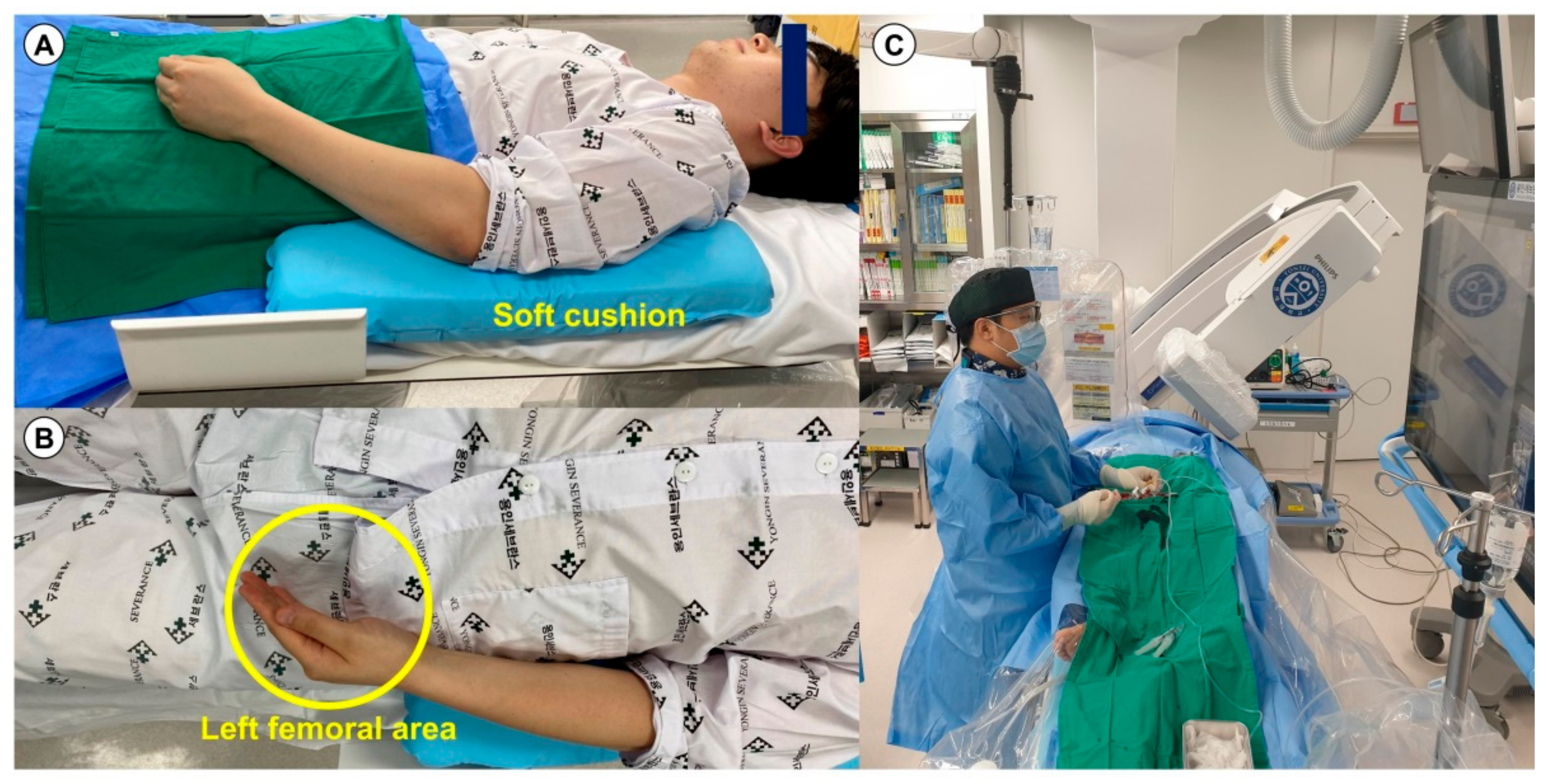
2.2. Preparation for the Left Distal Radial Approach

All patients had a palpable arterial pulse in the anatomical snuffbox. The patient’s left hand was turned clockwise at 90°, allowing the snuffbox to face the ceiling, and it was slightly bent toward the patient’s body and placed on his/her left groin (Figure 2A,B). The operator prepared for the left DRA and performed coronary procedures on the right side of the patients (Figure 2C). The puncture site of the left distal radial artery was sterilized in all patients. Local anesthesia of the anatomical snuffbox was achieved through a 1-cc lidocaine hydrochloride injection. Thereafter, puncture was performed by using a 20-gauge two-piece needle or a 21-gauge open needle. After a successful puncture, a 0.025-inch straight wire or a 0.018-inch hair wire was inserted, followed by insertion of the 5- or 6-French (Fr) radial sheath (Prelude® Radial, MERIT MEDICAL, South Jordan, UT, USA or Radiofocus® Introducer II, TERUMO Corp., Tokyo, Japan) (Figure 3A and Video S1). After successful sheath insertion, 3000 U of unfractionated heparin, 200 μg of nitroglycerin, and 2 mg of verapamil were administered intrasheath to reduce the rate of spasm and post-procedural radial arterial occlusion. After completion of PCI, the radial sheath was removed, and a compression cohesive elastic bandage with a 4 × 4-inch sterile gauze dressing for hemostasis was applied for 3 h in all patients (Figure 3B).

2.3. Procedural Details of Percutaneous Coronary Intervention for Bifurcation Lesions

Following placement of a sheath introducer, coronary angiography was performed according to the standard technique. Additional unfractionated heparin (50–70 U/kg) was administered during the procedure to maintain the activated clotting time at 250–300 s. The treatment strategy for SB including balloon angioplasty or stenting, use of a coronary stent, use of intravascular modalities, and use of an additional device (e.g., a microcatheter and guide extension catheter) were determined at the operator’s discretion.

2.4. Definition and Study Endpoints

Bifurcation lesions were assessed according to the Medina classification, which classifies the lesions according to the angiographic involvement of the three relevant segments of the bifurcation—the proximal main vessel, distal main branch, and SB [11]. Lesions that include both the main vessel and the SB are considered a true bifurcation.
The primary endpoint was the successful rate of PCI for bifurcation lesions via the left DRA. The secondary endpoints were major bleeding requiring any transfusion or surgery, access-site complications including hematoma and forearm radial and/or distal radial artery occlusion, and 30-day mortality.

2.5. Statistical Analysis

Normally distributed continuous variables are expressed as mean ± standard deviation. Non-normally distributed continuous variables are reported as median and interquartile range. All categorical variables are presented as number (percentage). All statistical analyses were performed using SPSS statistical software (SPSS version 25.0 for Windows; IBM Corp., Armonk, NY, USA).

3. Results

Between December 2017 and December 2019, we identified 1041 patients who underwent the DRA for CAG or PCI. Among them, we selected 106 patients who underwent PCI via the left DRA for bifurcation lesions (Figure 1). Baseline characteristics of the study population are presented in Table 1. The overall mean age of the patients was 63.3 ± 11.1 years (range, 35–89 years), and 79.2% (84/106) of patients were male. Of the 95 (89.6%) patients presenting with acute coronary syndrome, 14 (13.2%) patients had ST-elevation myocardial infarction. The mean left ventricular ejection fraction was 61.3 ± 11.0%. All patients, including 29 (27.4%) patients taking a potent P2Y12 inhibitor (ticagrelor, 12.3% (13/106); prasugrel, 15.1% (16/106)), were treated with dual antiplatelet therapy.
The success rate of PCI for bifurcation lesions was 100% in all 106 patients, and no patients required cross-over to the conventional radial or femoral approaches. Angiographic and procedural characteristics are provided in Table 2. The overall success rate of DRA was 95.1% (990/1041). Regarding the details of the left DRA, the mean and median times of distal radial artery puncture, defined as the time interval from local anesthesia induction to successful sheath cannulation, were 2.4 ± 1.6 and 1.8 min (quartiles 1–3: 1.3–3.4), respectively. The distal radial artery puncture was performed within 3 and 5 min in 71.7% (76/106) and 93.4% (99/106) of patients, respectively. Concerning lesion characteristics, 11 (10.4%) patients were treated for left main bifurcation lesions via the left DRA, and true bifurcations accounted for 39.6% of cases, with the left anterior descending artery/diagonal branch being the most frequent bifurcation site (57.5%, 61/106). Four patients were treated with a planned two-stent technique (mini-crush two-stent technique). Twenty-six (24.5%) and four (3.8%) patients were treated with plain balloon angioplasty and stent implantation of the SB, respectively. The kissing balloon technique for bifurcated lesions was performed in 20 (18.9%) patients. Implantation of ≥2 stents was performed in 23 (21.7%) patients, and intravascular imaging-guided PCI was conducted in 25 (23.6%) patients (10, intravascular ultrasound; 15, optical coherence tomography guidance). Overall, 11.3% of patients underwent multi-vessel PCI. An extra-backup type catheter was used in 71 of 96 cases (74.0%) with a left coronary artery lesion. Among 18 cases of right coronary artery lesion, Judkins right and Amplatz type catheters were used in four (22.2%) and 14 (77.8%) cases, respectively. PCI was performed using a 6-Fr guiding catheter in 101 patients (95.3%). In two (1.9%) patients, a 7-Fr guiding catheter was used.
Regarding puncture-site complications, there were no major bleeding complications requiring surgery or transfusion. Hand hematoma occurred in five patients (4.7%) including three (2.8%) patients with a >5-cm-diameter hematoma, but no case of forearm hematoma was observed. There was no incidence of distal and forearm radial artery occlusion and death at 30 days (Table 3).

4. Discussion

The principal findings of the current study were as follows: (1) the success rate of the bifurcation PCI via the left DRA was high without the need for access-site cross-over; (2) the occurrence rate of puncture site complications was low (4.7%), and there was no major bleeding, distal and forearm radial artery occlusion, forearm hematoma, or mortality at 30 days. To the best of our knowledge, this is the first clinical study to investigate the feasibility of the left DRA for bifurcation PCI, which remains a challenge.
Previous studies on the left DRA are summarized in Table 4, and these studies have reported a 97.6–98.9% success rate of PCI via the left DRA, except for the study on PCI for chronic total occlusion via the left DRA [5,12,13,14,15,16]. In the current study, bifurcation PCI via the left DRA was successfully performed in all 106 patients without access-site cross-over. Therefore, our study reaffirms the high success rate of PCI via the left DRA and suggests that the left DRA could be feasible as an alternative access site for bifurcation PCI.
The right conventional radial approach is commonly used by most operators probably because catheter manipulation can be performed from the patient’s right side without physical discomfort of the operator. However, with the right conventional radial approach, anatomic variations such as an increased incidence of tortuosity and loops in the arm and subclavian arteries may require extra catheter manipulation, and backup force for the right coronary artery is poor due to the S-shaped geometry of the subclavian–innominate–aorta axis, which can increase procedural complexity [17]. Furthermore, during the right conventional radial approach, the catheter needs to be passed from the innominate artery into the ascending aorta where the right carotid artery comes off, resulting in an increased risk of embolic stroke [4]. By contrast, the left conventional radial approach offers a very similar technique to the femoral approach, which makes it easy to manipulate the catheter and reduce the fluoroscopic time, procedure time, contrast volume, radiation exposure, and access-site change [17,18,19]. In the present study, the strength of the left side approach allowed us to achieve the high success rates of bifurcation PCI. Physical discomfort of the operator is generally recognized as a barrier to the left conventional radial approach, especially if the patient is obese or the operator is short [20]. This barrier can be overcome by the left DRA, in which the patient’s elbow is slightly bent, and the left hand is positioned above the left groin. As Figure 2 shows, there is no excessive bending of the elbow, which causes difficulties in wire or guiding catheter advancement, or kinking of the guiding catheter near the elbow and the operator position is the same as that in the right conventional radial approach, which may have led to the high success rate of PCI for bifurcation lesions via the left DRA in the current study.
Indeed, there are concerns regarding PCI via the DRA because the diameter of the distal radial artery in the anatomical snuffbox was significantly smaller than that of the conventional radial artery [14]. However, our study showed that 95.3% (101/106) of patients underwent successful PCI using a 6-Fr guiding catheter without major puncture-site complications. Moreover, with the recent introduction of the thin-wall 7-Fr sheath (Prelude IDealTM, MERIT MEDICAL; Glidesheath Slender®, TERUMO Corp.) or sheathless guiding catheter (Sheathless Eaucath, Asahi Intecc, Tokyo, Japan; RailwayTM, Cordis Corporation, Miami, FL, USA) [15,21,22,23], complex PCI, including bifurcation PCI, via the left DRA can be more easily and safely performed in the relatively smaller diameter vessels. Colletti et al. reported the feasibility and safety of performing bifurcation PCI (75%, 15/20) with a 7-Fr sheathless system via the DRA in a limited number (n = 20) of study patients [21]. Further studies are needed to evaluate the feasibility of PCI using a sheathless guiding catheter or thin-wall sheath via the DRA in a large study population.
Previous studies regarding the feasibility of the left DRA in patients who underwent CAG and PCI, as summarized in Table 4, showed that major complications did not occur. It is noteworthy that the current study showed the safety of the left DRA with a small number of minor complications and no major complications, which is similar to the findings of previous studies, despite the use of a 6-Fr sheath in most patients. However, the majority of patients in the current study are male and young and may have a relatively large diameter of distal radial artery. These demographic findings may have affected the result. Further large-scale, well-designed studies regarding the safety of complex PCI via the left DRA are required.
Some limitations of our study should be noted. First, the study was a retrospective, non-randomized, observational study, which has inherent selection and information biases. Second, the small number of patients in the present study rendered low statistical power. Nevertheless, this is the first clinical study to investigate the feasibility of the left DRA for bifurcation PCI, which remains a challenge. Third, details of the technical option for bifurcation PCI such as the jailed technique for SB protection (e.g., the jailed-wire, jailed-balloon, or jailed Corsair technique) or the selected balloon for the kissing balloon technique were not considered. Fourth, although the overall success rate of the DRA is high at over 95% in the current study, we should consider that puncture failure still remains a hurdle in the DRA. Fifth, the majority of patients in this study are male and young, and they may have relatively large diameter distal radial arteries compared with elderly and female patients. Furthermore, patients who had highly complex bifurcation lesions, requiring a 7-Fr guiding catheter, may have been excluded because the majority of patients were treated using a 6-Fr guiding catheter in our study. In interpreting the results of the current study, the groups of subjects should not be expanded, and the results should be interpreted with caution. Sixth, the lack of a comparator group of patients undergoing the conventional radial approach is another potential limitation. Seventh, the lack of routine post-procedural ultrasonography for forearm and radial artery may have contributed to underestimation of the puncture-site complications. Eighth, we did not investigate long-term clinical outcomes after 30 days because the present study focused on the technical success.

5. Conclusions

PCI via the left DRA was a feasible option in selected patients with bifurcation lesions, and this was supported by the high PCI success rate without access-site cross-over. Regarding the safety aspect of PCI via the left DRA for bifurcation lesions, there were no significant access-site complications or adverse clinical outcomes. Further large randomized trials should be conducted to evaluate the feasibility of the left DRA for bifurcation PCI.

Supplementary Materials

The following are available online at https://www.mdpi.com/article/10.3390/jcm10102204/s1, Video S1: How to perform left distal radial artery puncture.

Author Contributions

Conceptualization, Y.K.; data curation, Y.K. and M.H.J.; format analysis, O.-H.L.; investigation, O.-H.L. and Y.K.; project administration, Y.K.; resources, O.-H.L. and Y.K.; supervision, Y.K.; writing—original draft, O.-H.L. and Y.K.; writing—review and editing, O.-H.L., J.W.R., E.I., D.-K.C., M.H.J., D.C., and Y.K. All authors have read and agreed to the published version of the manuscript.

Funding

This is supported by a faculty research grant of Yonsei University College of Medicine (6-2020-0161) and a research seed money grant from Internal Medicine in Yongin Severance Hospital.

Institutional Review Board Statement

This study protocol was approved by the Institutional Review Board of Chonnam National University Hospital (approval number CNUH-2021-046; 22 February 2021).

Informed Consent Statement

Patient consent was waived by the Institutional Review Board of Chonnam National University Hospital because of the retrospective study design.

Data Availability Statement

The underlying data set is available from the corresponding author upon reasonable request.

Acknowledgments

The authors thank Medical Illustration & Design, part of the Medical Research Support Services of Yonsei University College of Medicine, for all artistic support related to this work.

Conflicts of Interest

The authors declare no conflict of interest.

References

  1. Ibanez, B.; James, S.; Agewall, S.; Antunes, M.J.; Bucciarelli-Ducci, C.; Bueno, H.; Caforio, A.L.; Crea, F.; Goudevenos, J.A.; Halvorsen, S.; et al. 2017 ESC Guidelines for the management of acute myocardial infarction in patients presenting with ST-segment elevation: The Task Force for the management of acute myocardial infarction in patients presenting with ST-segment elevation of the European Society of Cardiology (ESC). Eur. Heart J. 2018, 39, 119–177. [Google Scholar] [PubMed] [Green Version]
  2. Neumann, F.-J.; Sousa-Uva, M.; Ahlsson, A.; Alfonso, F.; Banning, A.P.; Benedetto, U.; Byrne, R.A.; Collet, J.-P.; Falk, V.; Head, S.J.; et al. 2018 ESC/EACTS Guidelines on myocardial revascularization. Eur. Heart J. 2019, 40, 87–165. [Google Scholar] [CrossRef] [PubMed]
  3. Kolkailah, A.A.; Alreshq, R.S.; Muhammed, A.M.; Zahran, M.E.; El-Wegoud, M.A.; Nabhan, A.F. Transradial versus transfemoral approach for diagnostic coronary angiography and percutaneous coronary intervention in people with coronary artery disease. Cochrane Database Syst. Rev. 2018, 4, CD012318. [Google Scholar] [CrossRef] [PubMed]
  4. Rashid, M.; Lawson, C.; Potts, J.; Kontopantelis, E.; Kwok, C.S.; Bertrand, O.F.; Shoaib, A.; Ludman, P.; Kinnaird, T.; de Belder, M.; et al. Incidence, determinants, and outcomes of left and right radial access use in patients undergoing percutaneous coronary intervention in the United Kingdom: A national perspective using the BCIS dataset. JACC Cardiovasc. Interv. 2018, 11, 1021–1033. [Google Scholar] [CrossRef]
  5. Kiemeneij, F. Left distal transradial access in the anatomical snuffbox for coronary angiography (ldTRA) and interventions (ldTRI). EuroIntervention 2017, 13, 851–857. [Google Scholar] [CrossRef]
  6. Sgueglia, G.A.; Di Giorgio, A.; Gaspardone, A.; Babunashvili, A. Anatomic basis and physiological rationale of distal radial artery access for percutaneous coronary and endovascular procedures. JACC Cardiovasc. Interv. 2018, 11, 2113–2119. [Google Scholar] [CrossRef]
  7. Sgueglia, G.A.; Lee, B.K.; Cho, B.R.; Babunashvili, A.; Lee, J.B.; Lee, J.W.; Schenke, K.; Lee, S.Y.; Harb, S. Distal radial access: Consensus report of the first Korea-Europe transradial intervention meeting. JACC Cardiovasc. Interv. 2021, 14, 892–906. [Google Scholar] [CrossRef] [PubMed]
  8. Sawaya, F.J.; Lefèvre, T.; Chevalier, B.; Garot, P.; Hovasse, T.; Morice, M.-C.; Rab, T.; Louvard, Y. Contemporary approach to coronary bifurcation lesion treatment. JACC Cardiovasc. Interv. 2016, 9, 1861–1878. [Google Scholar] [CrossRef]
  9. Chung, S.; Her, S.-H.; Song, P.S.; Song, Y.B.; Hahn, J.-Y.; Choi, J.-H.; Lee, S.H.; Jang, Y.; Yoon, J.H.; Tahk, S.-J.; et al. Trans-radial versus trans-femoral intervention for the treatment of coronary bifurcations: Results from coronary bifurcation stenting registry. J. Korean Med. Sci. 2013, 28, 388. [Google Scholar] [CrossRef] [Green Version]
  10. Chung, S.; Yang, J.H.; Choi, S.-H.; Song, Y.B.; Hahn, J.-Y.; Choi, J.-H.; Lee, S.H.; Jang, Y.; Yoon, J.H.; Tahk, S.-J.; et al. Transradial versus transfemoral intervention for the treatment of left main coronary bifurcations: Results from the COBIS (coronary bifurcation stenting) II registry. J. Invasive Cardiol. 2015, 27, 35–40. [Google Scholar]
  11. Medina, A.; Suárez de Lezo, J.; Pan, M. A new classification of coronary bifurcation lesions. Rev. Esp. Cardiol. 2006, 59, 183. [Google Scholar] [CrossRef] [PubMed] [Green Version]
  12. Soydan, E.; Akın, M. Coronary angiography using the left distal radial approach-an alternative site to conventional radial coronary angiography. Anatol. J. Cardiol. 2018, 19, 243. [Google Scholar] [CrossRef] [PubMed]
  13. Al-Azizi, K.M.; Grewal, V.; Gobeil, K.; Maqsood, K.; Haider, A.; Mohani, A.; Giugliano, G.; Lotfi, A.S. The left distal transradial artery access for coronary angiography and interventions: A US experience. Cardiovasc. Revasc. Med. 2019, 20, 786–789. [Google Scholar] [CrossRef] [PubMed]
  14. Kim, Y.; Ahn, Y.; Kim, I.; Lee, D.H.; Kim, M.C.; Sim, D.S.; Hong, Y.J.; Kim, J.H.; Jeong, M.H. Feasibility of coronary angiography and percutaneous coronary intervention via left snuffbox approach. Korean Circ. J. 2018, 48, 1120. [Google Scholar] [CrossRef]
  15. Gasparini, G.L.; Garbo, R.; Gagnor, A.; Oreglia, J.; Mazzarotto, P. First prospective multicentre experience with left distal transradial approach for coronary chronic total occlusion interventions using a 7 Fr Glidesheath Slender. EuroIntervention 2019, 15, 126–128. [Google Scholar] [CrossRef]
  16. Lee, J.-W.; Park, S.W.; Son, J.-W.; Ahn, S.-G.; Lee, S.-H. Real-world experience of the left distal transradial approach for coronary angiography and percutaneous coronary intervention: A prospective observational study (LeDRA). EuroIntervention 2018, 14, e995–e1003. [Google Scholar] [CrossRef] [Green Version]
  17. Sciahbasi, A.; Romagnoli, E.; Burzotta, F.; Trani, C.; Sarandrea, A.; Summaria, F.; Pendenza, G.; Tommasino, A.; Patrizi, R.; Mazzari, M.; et al. Transradial approach (left vs right) and procedural times during percutaneous coronary procedures: TALENT study. Am. Heart J. 2011, 161, 172–179. [Google Scholar] [CrossRef]
  18. Park, J.Y.; Rha, S.-W.; Choi, B.G.; Oh, D.J.; Choi, C.U.; Youn, Y.-J.; Yoon, J. Comparison of clinical outcomes between the right and left radial artery approaches from the Korean transradial coronary intervention registry. Yonsei Med. J. 2017, 58, 521. [Google Scholar] [CrossRef] [Green Version]
  19. Biondi-Zoccai, G.; Sciahbasi, A.; Bodí, V.; Fernández-Portales, J.; Kanei, Y.; Romagnoli, E.; Agostoni, P.; Sangiorgi, G.; Lotrionte, M.; Modena, M.G. Right versus left radial artery access for coronary procedures: An international collaborative systematic review and meta-analysis including 5 randomized trials and 3210 patients. Int. J. Cardiol. 2013, 166, 621–626. [Google Scholar] [CrossRef] [Green Version]
  20. Kado, H.; Patel, A.M.; Suryadevara, S.; Zenni, M.M.; Box, L.C.; Angiolillo, D.J.; Bass, T.A.; Guzman, L.A. Operator radiation exposure and physical discomfort during a right versus left radial approach for coronary interventions: A randomized evaluation. JACC Cardiovasc. Interv. 2014, 7, 810–816. [Google Scholar] [CrossRef] [Green Version]
  21. Colletti, G.; Auslender, J.; Antoine De Meester, M.; Aminian, A.; Kayaert, P.; Ungureanu, C. Feasibility and safety of performing complex coronary interventions by distal radial artery using the railway sheathless vascular system. J. Invasive Cardiol. 2020, 32, 459–462. [Google Scholar]
  22. Youn, Y.J.; Yoon, J.; Han, S.W.; Lee, J.-W.; Sung, J.K.; Ahn, S.-G.; Kim, J.-Y.; Yoo, B.-S.; Lee, S.-H.; Choe, K.-H. Feasibility of transradial coronary intervention using a sheathless guiding catheter in patients with small radial artery. Korean Circ. J. 2011, 41, 143–148. [Google Scholar] [CrossRef] [Green Version]
  23. Aminian, A.; Iglesias, J.F.; Van Mieghem, C.; Zuffi, A.; Ferrara, A.; Manih, R.; Dolatabadi, D.; Lalmand, J.; Saito, S. First prospective multicenter experience with the 7 French Glidesheath slender for complex transradial coronary interventions. Catheter. Cardiovasc. Interv. 2017, 89, 1014–1020. [Google Scholar] [CrossRef]
Figure 1. Study flowchart. Abbreviations: CAG, coronary artery angiography; DRA, distal radial approach; PCI, percutaneous coronary intervention.
Jcm 10 02204 g001
Figure 2. Positions of the patient and operator during the left DRA. The lateral (A) and front views (B) of the patient’s preparation position during the left DRA. (C) The natural working position of the operation while performing coronary angiography via the left DRA. Abbreviation: DRA, distal radial approach.
Jcm 10 02204 g002
Figure 3. Inserted 6-Fr sheath via the left distal radial artery in the anatomical snuffbox area (blue arrowhead: puncture site) (A) and dressing for hemostasis with compressive bandage with gauze (B) and summary of the study outcomes. Abbreviations: DRA, distal radial approach; PCI, percutaneous coronary intervention, RAO, radial artery occlusion; STEMI, ST-elevation myocardial infarction; Fr, French.
Jcm 10 02204 g003
Table 1. Baseline characteristics (n = 106).
CharacteristicValue
Age (years)63.3 ± 11.1
Male sex84 (79.2%)
Height (cm)167.5 (160, 172)
Weight (kg)69.0 ± 11.1
Body mass index (kg/m2)25.0 ± 2.9
Hypertension64 (60.4%)
Diabetes mellitus33 (31.1%)
Dyslipidemia54 (50.9%)
Current smoker29 (27.4%)
Chronic kidney disease, ≥stage 315 (14.2%)
Clinical presentation
Stable angina11 (10.4%)
Unstable angina54 (50.9%)
NSTEMI27 (25.5%)
STEMI14 (13.2%)
LVEF (%)61.3 ± 11.0
Periprocedural medication
DAPT106 (100%)
Aspirin106 (100%)
P2Y12 inhibitor
Clopidogrel77 (72.6%)
Ticagrelor13 (12.3%)
Prasugrel16 (15.1%)
Oral anticoagulation3 (2.8%)
Glycoprotein IIb/IIIa inhibitors5 (4.7%)
Data are presented as mean ± standard deviation, median (interquartile range), or number (%). Abbreviations: DAPT, dual antiplatelet therapy; LVEF, left ventricular ejection fraction; NSTEMI, non-ST-elevation myocardial infarction; STEMI, ST-elevation myocardial infarction.
Table 2. Angiographic and procedural characteristics (n = 106).
CharacteristicValue
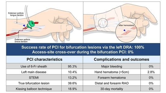
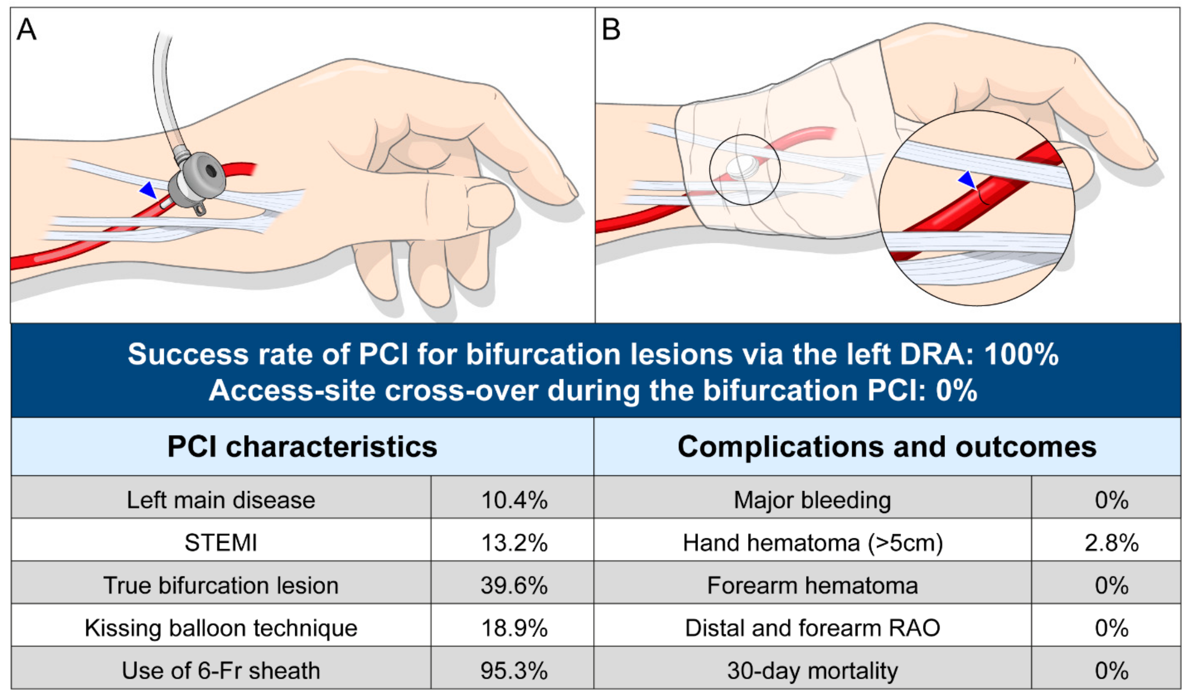
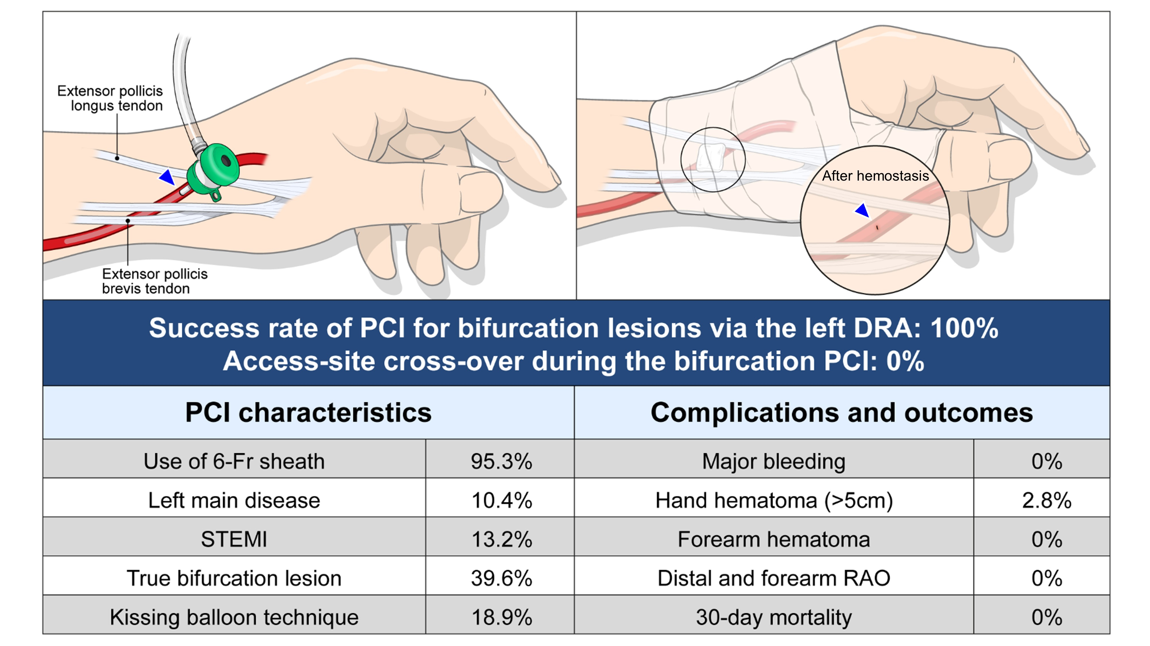
Successful bifurcation PCI via the left DRA106 (100%)
Cross-over to another vascular approach0 (0)
Characteristics of the left DRA
Puncture time (min)
Mean ± SD2.4 ± 1.6
Median (IQR)1.8 (1.3, 3.4)
Puncture time <3 min76 (71.7%)
Puncture time <5 min99 (93.4%)
Lesion characteristics
Target lesion
Left main coronary artery11 (10.4%)
Left anterior descending artery61 (57.5%)
Left circumflex artery18 (17.0%)
Right coronary artery16 (15.1%)
Medina classification
0, 0, 12 (1.9%)
0, 1, 021 (19.8%)
0, 1, 19 (8.5%)
1, 0, 016 (15.1%)
1, 0, 19 (8.5%)
1, 1, 025 (23.6%)
1, 1, 124 (22.6%)
True bifurcation42 (39.6%)
ACC/AHA type B2/C lesion57 (53.8%)
Procedural characteristics
Treatment of the side branch
Plain balloon angioplasty26 (24.5%)
Stenting (two-stent technique)4 (3.8%)
Kissing balloon technique20 (18.9%)
Intravascular imaging-guided PCI25 (23.6%)
IVUS guidance10 (9.4%)
OCT guidance15 (14.2%)
Multi-vessel PCI12 (11.3%)
Total no. of implanted stents1.26 ± 0.54
Cases with implantation of ≥2 stents23 (21.7%)
Stent diameter in the main vessel (mm)2.98 ± 0.38
Total stent length in the main vessel (mm)31.8 ± 14.3
Left guiding catheter (n = 96)
EBU type catheter71 (74.0%)
Judkins left type catheter25 (26.0%)
Right guiding catheter (n = 18)
Judkins right type catheter4 (22.2%)
Amplatz type catheter14 (77.8%)
Guiding catheter size
5-Fr3 (2.8%)
6-Fr101 (95.3%)
7-Fr2 (1.9%)
Data are presented as mean ± SD, median (interquartile range), or number (%). Abbreviations: ACC, American College of Cardiology; AHA, American Heart Association; DRA, distal radial approach; EBU, extra-backup; IVUS, intravascular ultrasound; IQR, interquartile range; OCT, optical coherence tomography; PCI, percutaneous coronary intervention; SD, standard deviation; Fr, French; no., number.
Table 3. Safety outcomes.
VariableTotal Patients (n = 106)
30-day mortality0
Any bleeding complication requiring surgery or transfusion0
Access-site complication
Distal radial artery occlusion0
Forearm radial artery occlusion0
Hand hematoma5 (4.7%)
≤5 cm in diameter2 (1.9%)
>5 cm in diameter3 (2.8%)
Forearm hematoma0
Data are presented as number (%).
Table 4. Summary of previous articles regarding the left DRA.
Study, Author Name (y of Publication)No. of PatientsMean Age (y)Male SexLeft DRASuccess Rate of the Puncture SitePCIBifurcationSuccess Rate of PCICross-over in PCIDistal RAOMajor Bleeding Requiring Transfusion or Surgery
Kiemeneji et al. (2017) [5]7068 ± 1155 (79%)100%89%25 (36%)N/AN/A11 (1.5%)0
Kim et al. (2018) [14]15066 ± 1394 (71%)100%88%42 (28%)N/A97.6%1N/A0
Lee et al. (2018) [16]20066 ± 12132 (66%)100%95.5%87 (44%)28/87 (32.2%)98.9%000
Soydan et al. (2018) [12]5459 ± 1243 (80%)100%100%20 (37%)N/AN/A200
Al-Azizi et al. (2018) [13]617046 (75%)100%98.434 (56%)N/AN/A000
Gasparini et al. (2019) [15]4168 ± 531 (76%)100%82.9%100%N/A78.1%N/A1 (4.3%)0
Data are presented as number (%). Abbreviations: DRA, distal radial approach; PCI, percutaneous coronary intervention; RAO, radial artery occlusion; no., number; N/A, not applicable.
Publisher’s Note: MDPI stays neutral with regard to jurisdictional claims in published maps and institutional affiliations.

