Emerging Approaches to Mitigate Neural Cell Degeneration with Nanoparticles-Enhanced Polyelectrolyte Systems
Abstract
1. Introduction
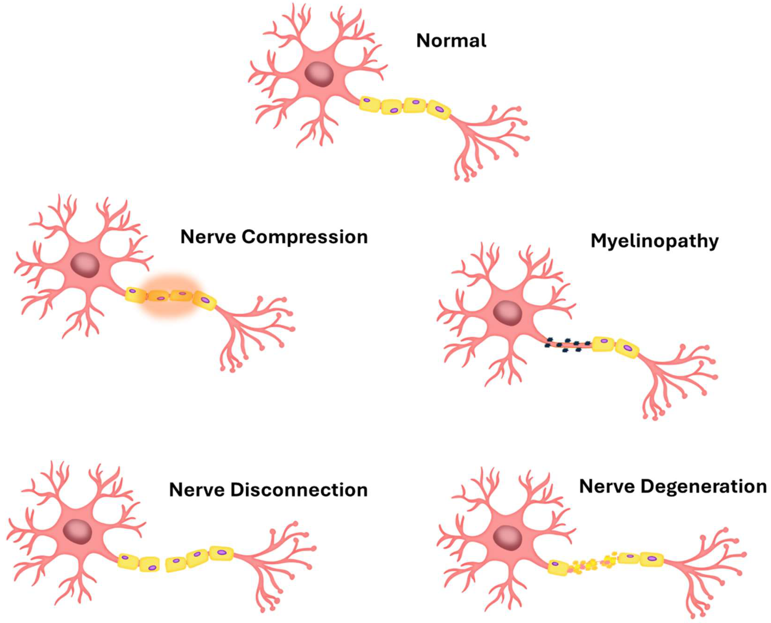
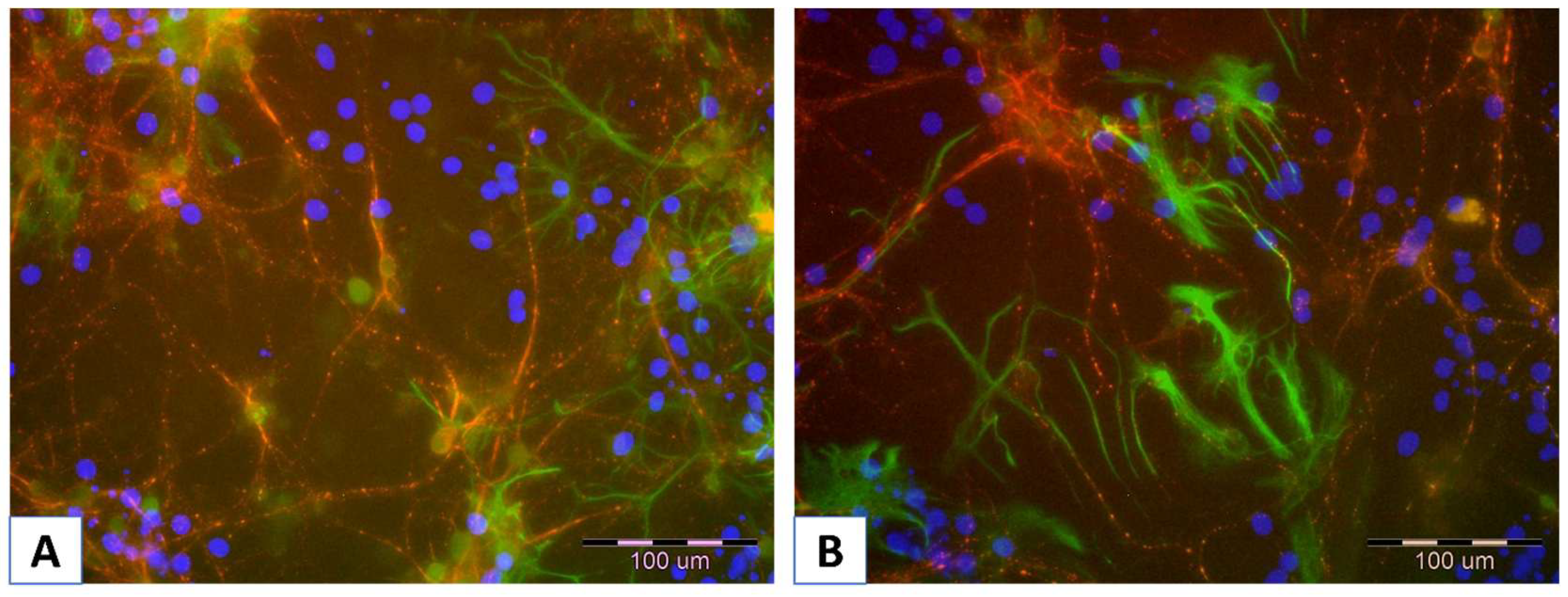
2. Trouble in Paradise—Short Overview of the Neuronal Cell Cultures
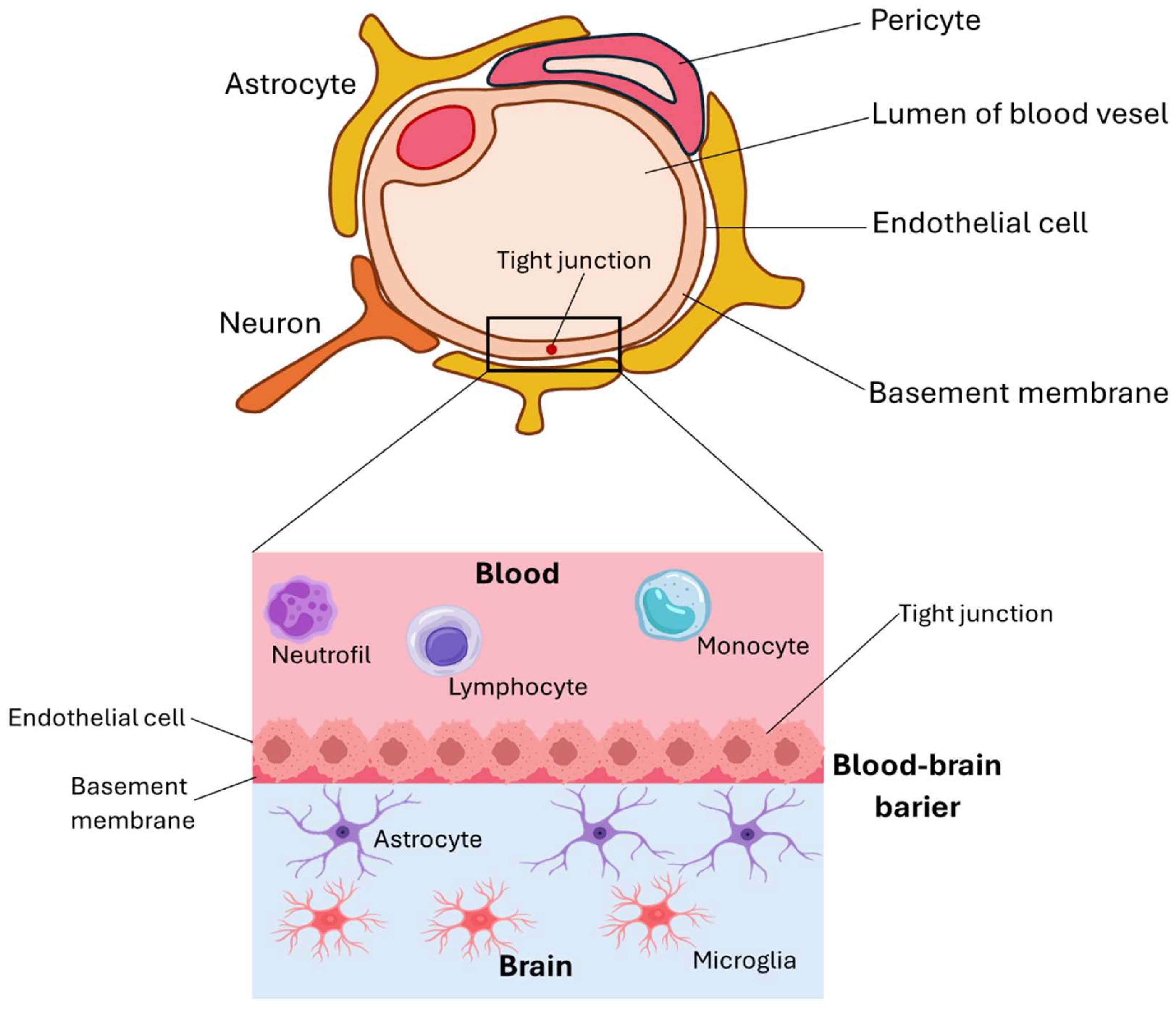
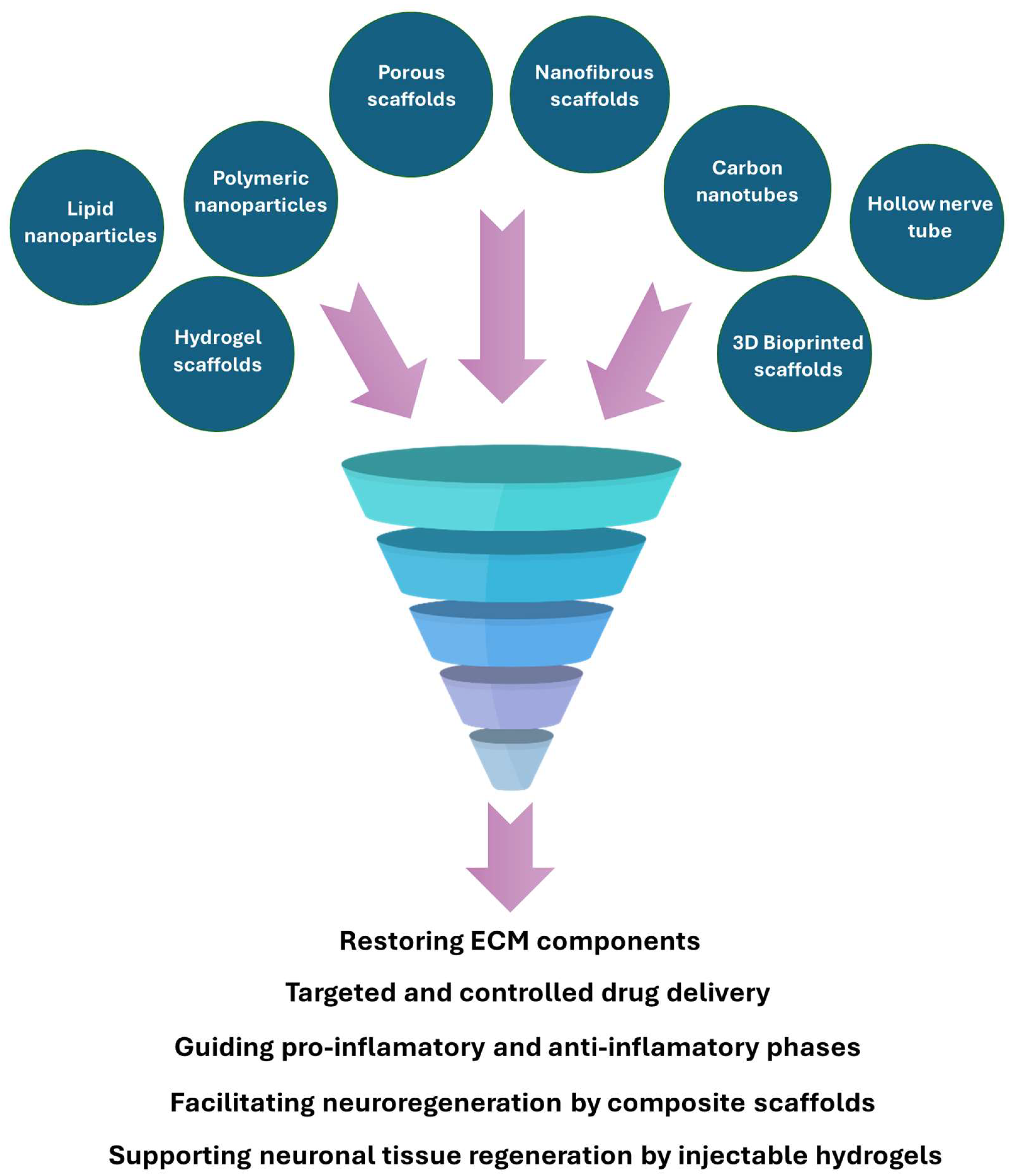
3. Nanomaterials in Nerve Regeneration
3.1. The Influence of Nanomaterial Structure and Its Role in Neuroregeneration
3.2. Multifaceted Approach to Nerve Regeneration Using Nanomaterials
3.2.1. NPs Mitigating Oxidative Stress and Modulating Inflammation
3.2.2. Remyelination, Axonal Growth, and NPs for Its Promotion
3.2.3. NPs in Neutrophic/Therapeutic Factors Delivery
4. Polyelectrolyte Materials
4.1. Mechanism of Self-Assembly
4.2. Polyelectrolytes for Neural Cell Regeneration in Terms of Mechanical Properties and Potential
4.3. Polyelectrolytes for Interface with Neural Cells
5. Application of PE and PE-Based Nanocomposites for Neuronal Cell Immobilization
6. Discussion
7. Conclusions and Outlook
Author Contributions
Funding
Data Availability Statement
Conflicts of Interest
References
- The Molecular and Cellular Basis of Neurodegenerative Diseases|ScienceDirect. Available online: https://www.sciencedirect.com/book/9780128113042/the-molecular-and-cellular-basis-of-neurodegenerative-diseases?via=ihub= (accessed on 31 May 2025).
- Ciurea, A.V.; Mohan, A.G.; Covache-Busuioc, R.A.; Costin, H.P.; Glavan, L.A.; Corlatescu, A.D.; Saceleanu, V.M. Unraveling Molecular and Genetic Insights into Neurodegenerative Diseases: Advances in Understanding Alzheimer’s, Parkinson’s, and Huntington’s Diseases and Amyotrophic Lateral Sclerosis. Int. J. Mol. Sci. 2023, 24, 10809. [Google Scholar] [CrossRef]
- Ayers, J.I.; Borchelt, D.R. Phenotypic diversity in ALS and the role of poly-conformational protein misfolding. Acta Neuropathol. 2021, 142, 41–55. [Google Scholar] [CrossRef]
- Gadhave, D.G.; Sugandhi, V.V.; Jha, S.K.; Nangare, S.N.; Gupta, G.; Singh, S.K.; Dua, K.; Cho, H.; Hansbro, P.M.; Paudel, K.R. Neurodegenerative disorders: Mechanisms of degeneration and therapeutic approaches with their clinical relevance. Ageing Res. Rev. 2024, 99, 102357. [Google Scholar] [CrossRef] [PubMed]
- Feigin, V.L.; Vos, T.; Nichols, E.; Owolabi, M.O.; Carroll, W.M.; Dichgans, M.; Deuschl, G.; Parmar, P.; Brainin, M.; Murray, C. The global burden of neurological disorders: Translating evidence into policy. Lancet Neurol. 2020, 19, 255–265. [Google Scholar] [CrossRef] [PubMed]
- Van Schependom, J.; D’haeseleer, M. Advances in Neurodegenerative Diseases. J. Clin. Med. 2023, 12, 1709. [Google Scholar] [CrossRef] [PubMed]
- Huang, Y.; Li, Y.; Pan, H.; Han, L. Global, regional, and national burden of neurological disorders in 204 countries and territories worldwide. J. Glob. Health 2023, 13, 04160. [Google Scholar] [CrossRef]
- Steinmetz, J.D.; Seeher, K.M.; Schiess, N.; Nichols, E.; Cao, B.; Servili, C.; Cavallera, V.; Cousin, E.; Hagins, H.; Moberg, M.E.; et al. Global, regional, and national burden of disorders affecting the nervous system, 1990–2021: A systematic analysis for the Global Burden of Disease Study 2021. Lancet Neurol. 2024, 23, 344–381. [Google Scholar] [CrossRef]
- Thakur, R.; Saini, A.K.; Taliyan, R.; Chaturvedi, N. Neurodegenerative diseases early detection and monitoring system for point-of-care applications. Microchem. J. 2025, 208, 112280. [Google Scholar] [CrossRef]
- Tay, L.X.; Ong, S.C.; Tay, L.J.; Ng, T.; Parumasivam, T. Economic Burden of Alzheimer’s Disease: A Systematic Review. Value Health Reg. Issues 2024, 40, 1–12. [Google Scholar] [CrossRef]
- Chen, S.; Cao, Z.; Nandi, A.; Counts, N.; Jiao, L.; Prettner, K.; Kuhn, M.; Seligman, B.; Tortorice, D.; Vigo, D.; et al. The global macroeconomic burden of Alzheimer’s disease and other dementias: Estimates and projections for 152 countries or territories. Lancet Glob. Health 2024, 12, e1534–e1543. [Google Scholar] [CrossRef]
- Hammam, I.A.; Winters, R.; Hong, Z. Advancements in the application of biomaterials in neural tissue engineering: A review. Biomed. Eng. Adv. 2024, 8, 100132. [Google Scholar] [CrossRef]
- Kovacs, G.G. Molecular pathology of neurodegenerative diseases: Principles and practice. J. Clin. Pathol. 2019, 72, 725–735. [Google Scholar] [CrossRef]
- Collins, M.N.; Zamboni, F.; Serafin, A.; Escobar, A.; Stepanian, R.; Culebras, M.; Reis, R.L.; Oliveira, J.M. Emerging scaffold- and cellular-based strategies for brain tissue regeneration and imaging. Vitr. Model. 2022, 1, 129–150. [Google Scholar] [CrossRef] [PubMed]
- Gordon, J.; Amini, S. General Overview of Neuronal Cell Culture. Methods Mol. Biol. 2021, 2311, 1–8. [Google Scholar] [CrossRef] [PubMed]
- Landry, M.J.; Gu, K.; Harris, S.N.; Al-Alwan, L.; Gutsin, L.; De Biasio, D.; Jiang, B.; Nakamura, D.S.; Corkery, T.C.; Kennedy, T.E.; et al. Tunable Engineered Extracellular Matrix Materials: Polyelectrolyte Multilayers Promote Improved Neural Cell Growth and Survival. Macromol. Biosci. 2019, 19, e1900036. [Google Scholar] [CrossRef] [PubMed]
- Stil, A.; Liberelle, B.; Guadarrama Bello, D.; Lacomme, L.; Arpin, L.; Parent, P.; Nanci, A.; Dumont, É.C.; Ould-Bachir, T.; Vanni, M.P.; et al. A simple method for poly-D-lysine coating to enhance adhesion and maturation of primary cortical neuron cultures in vitro. Front. Cell. Neurosci. 2023, 17, 1212097. [Google Scholar] [CrossRef]
- Hendler, R.M.; Weiss, O.E.; Morad, T.; Sion, G.; Kirby, M.; Dubinsky, Z.; Barbora, A.; Minnes, R.; Baranes, D. A Poly-D-lysine-Coated Coralline Matrix Promotes Hippocampal Neural Precursor Cells’ Differentiation into GFAP-Positive Astrocytes. Polymers 2023, 15, 4054. [Google Scholar] [CrossRef]
- Wu, C.; Liu, S.; Zhou, L.; Chen, Z.; Yang, Q.; Cui, Y.; Chen, M.; Li, L.; Ke, B.; Li, C.; et al. Cellular and Molecular Insights into the Divergence of Neural Stem Cells on Matrigel and Poly-l-lysine Interfaces. ACS Appl. Mater. Interfaces 2024, 16, 31922–31935. [Google Scholar] [CrossRef]
- Rahimi Darehbagh, R.; Mahmoodi, M.; Amini, N.; Babahajiani, M.; Allavaisie, A.; Moradi, Y. The effect of nanomaterials on embryonic stem cell neural differentiation: A systematic review. Eur. J. Med. Res. 2023, 28, 576. [Google Scholar] [CrossRef]
- Liaw, K.; Zhang, Z.; Kannan, S. Neuronanotechnology for brain regeneration. Adv. Drug Deliv. Rev. 2019, 148, 3–18. [Google Scholar] [CrossRef]
- Krsek, A.; Jagodic, A.; Baticic, L. Nanomedicine in Neuroprotection, Neuroregeneration, and Blood–Brain Barrier Modulation: A Narrative Review. Medicina 2024, 60, 1384. [Google Scholar] [CrossRef]
- Wei, Z.; Hong, F.F.; Cao, Z.; Zhao, S.Y.; Chen, L. In Situ Fabrication of Nerve Growth Factor Encapsulated Chitosan Nanoparticles in Oxidized Bacterial Nanocellulose for Rat Sciatic Nerve Regeneration. Biomacromolecules 2021, 22, 4988–4999. [Google Scholar] [CrossRef] [PubMed]
- Zhang, Q.; Wu, P.; Chen, F.; Zhao, Y.; Li, Y.; He, X.; Huselstein, C.; Ye, Q.; Tong, Z.; Chen, Y. Brain Derived Neurotrophic Factor and Glial Cell Line-Derived Neurotrophic Factor-Transfected Bone Mesenchymal Stem Cells for the Repair of Periphery Nerve Injury. Front. Bioeng. Biotechnol. 2020, 8, 563158. [Google Scholar] [CrossRef] [PubMed]
- dos Santos Rodrigues, B.; Kanekiyo, T.; Singh, J. ApoE-2 Brain-Targeted Gene Therapy Through Transferrin and Penetratin Tagged Liposomal Nanoparticles. Pharm. Res. 2019, 36, 161. [Google Scholar] [CrossRef] [PubMed]
- Arora, S.; Layek, B.; Singh, J. Design and Validation of Liposomal ApoE2 Gene Delivery System to Evade Blood-Brain Barrier for Effective Treatment of Alzheimer’s Disease. Mol. Pharm. 2021, 18, 714–725. [Google Scholar] [CrossRef]
- Moreira, R.; Nóbrega, C.; de Almeida, L.P.; Mendonça, L. Brain-targeted drug delivery—Nanovesicles directed to specific brain cells by brain-targeting ligands. J. Nanobiotechnol. 2024, 22, 260. [Google Scholar] [CrossRef]
- Yang, Y.; Chu, Y.; Li, C.; Fan, L.; Lu, H.; Zhan, C.; Zhang, Z. Brain-targeted drug delivery by in vivo functionalized liposome with stable D-peptide ligand. J. Control. Release 2024, 373, 240–251. [Google Scholar] [CrossRef]
- Dhariwal, R.; Jain, M.; Mir, Y.R.; Singh, A.; Jain, B.; Kumar, P.; Tariq, M.; Verma, D.; Deshmukh, K.; Yadav, V.K.; et al. Targeted drug delivery in neurodegenerative diseases: The role of nanotechnology. Front. Med. 2025, 12, 1522223. [Google Scholar] [CrossRef]
- Musielak, M.; Potoczny, J.; Boś-liedke, A.; Kozak, M. The Combination of Liposomes and Metallic Nanoparticles as Multifunctional Nanostructures in the Therapy and Medical Imaging—A Review. Int. J. Mol. Sci. 2021, 22, 6229. [Google Scholar] [CrossRef]
- Cai, X.; Drummond, C.J.; Zhai, J.; Tran, N. Lipid Nanoparticles: Versatile Drug Delivery Vehicles for Traversing the Blood Brain Barrier to Treat Brain Cancer. Adv. Funct. Mater. 2024, 34, 2404234. [Google Scholar] [CrossRef]
- Pérez-Carrión, M.D.; Posadas, I. Dendrimers in Neurodegenerative Diseases. Processes 2023, 11, 319. [Google Scholar] [CrossRef]
- Ma, J.; Zhan, M.; Sun, H.; He, L.; Zou, Y.; Huang, T.; Karpus, A.; Majoral, J.P.; Mignani, S.; Shen, M.; et al. Phosphorus Dendrimers Co-deliver Fibronectin and Edaravone for Combined Ischemic Stroke Treatment via Cooperative Modulation of Microglia/Neurons and Vascular Regeneration. Adv. Healthc. Mater. 2024, 13, 2401462. [Google Scholar] [CrossRef]
- Escobar, A.; Carvalho, M.R.; Maia, F.R.; Reis, R.L.; Silva, T.H.; Oliveira, J.M. Glial Cell Line-Derived Neurotrophic Factor-Loaded CMCht/PAMAM Dendrimer Nanoparticles for Peripheral Nerve Repair. Pharmaceutics 2022, 14, 2408. [Google Scholar] [CrossRef] [PubMed]
- Xue, J.; Wu, T.; Qiu, J.; Xia, Y. Spatiotemporally Controlling the Release of Biological Effectors Enhances Their Effects on Cell Migration and Neurite Outgrowth. Small Methods 2020, 4, 2000125. [Google Scholar] [CrossRef] [PubMed]
- Donsante, A.; Xue, J.; Poth, K.M.; Hardcastle, N.S.; Diniz, B.; O’Connor, D.M.; Xia, Y.; Boulis, N.M. Controlling the Release of Neurotrophin-3 and Chondroitinase ABC Enhances the Efficacy of Nerve Guidance Conduits. Adv. Healthc. Mater. 2020, 9, e2000200. [Google Scholar] [CrossRef] [PubMed]
- Du, W.; Wang, T.; Hu, S.; Luan, J.; Tian, F.; Ma, G.; Xue, J. Engineering of electrospun nanofiber scaffolds for repairing brain injury. Eng. Regen. 2023, 4, 289–303. [Google Scholar] [CrossRef]
- Shi, S.; Ou, X.; Cheng, D. How Advancing is Peripheral Nerve Regeneration Using Nanofiber Scaffolds? A Comprehensive Review of the Literature. Int. J. Nanomed. 2023, 18, 6763. [Google Scholar] [CrossRef]
- Alla, K.; Yuri, C.; Anatoliy, L.; Volodymyr, L.; Yuriy, S.; Alla, K.; Yuri, C.; Anatoliy, L.; Volodymyr, L.; Yuriy, S. Interface Nerve Tissue-Silicon Nanowire for Regeneration of Injured Nerve and Creation of Bio-Electronic Device. In Neurons—Dendrites Axons; InTech: Houston, TX, USA, 2019. [Google Scholar] [CrossRef]
- Cortés-Llanos, B.; Rauti, R.; Ayuso-Sacido, Á.; Pérez, L.; Ballerini, L. Impact of Magnetite Nanowires on In Vitro Hippocampal Neural Networks. Biomolecules 2023, 13, 783. [Google Scholar] [CrossRef]
- Raos, B.J.; Maddah, M.; Graham, E.S.; Plank, N.O.V.; Unsworth, C.P. ZnO nanowire florets promote the growth of human neurons. Materialia 2020, 9, 100577. [Google Scholar] [CrossRef]
- Rodilla, B.L.; Arché-Núñez, A.; Ruiz-Gómez, S.; Domínguez-Bajo, A.; Fernández-González, C.; Guillén-Colomer, C.; González-Mayorga, A.; Rodríguez-Díez, N.; Camarero, J.; Miranda, R.; et al. Flexible metallic core–shell nanostructured electrodes for neural interfacing. Sci. Rep. 2024, 14, 3729. [Google Scholar] [CrossRef]
- Harberts, J.; Siegmund, M.; Hedrich, C.; Kim, W.; Fontcuberta i Morral, A.; Zierold, R.; Blick, R.H. Generation of Human iPSC-Derived Neurons on Nanowire Arrays Featuring Varying Lengths, Pitches, and Diameters. Adv. Mater. Interfaces 2022, 9, 2200806. [Google Scholar] [CrossRef]
- Convertino, D.; Trincavelli, M.L.; Giacomelli, C.; Marchetti, L.; Coletti, C. Graphene-based nanomaterials for peripheral nerve regeneration. Front. Bioeng. Biotechnol. 2023, 11, 1306184. [Google Scholar] [CrossRef] [PubMed]
- Ławkowska, K.; Pokrywczyńska, M.; Koper, K.; Kluth, L.A.; Drewa, T.; Adamowicz, J. Application of Graphene in Tissue Engineering of the Nervous System. Int. J. Mol. Sci. 2021, 23, 33. [Google Scholar] [CrossRef]
- Huang, H.; Fan, C.; Li, M.; Nie, H.L.; Wang, F.B.; Wang, H.; Wang, R.; Xia, J.; Zheng, X.; Zuo, X.; et al. COVID-19: A Call for Physical Scientists and Engineers. ACS Nano 2020, 14, 3747–3754. [Google Scholar] [CrossRef]
- Qian, Y.; Wang, X.; Song, J.; Chen, W.; Chen, S.; Jin, Y.; Ouyang, Y.; Yuan, W.E.; Fan, C. Preclinical assessment on neuronal regeneration in the injury-related microenvironment of graphene-based scaffolds. npj Regen. Med. 2021, 6, 31. [Google Scholar] [CrossRef]
- Aleemardani, M.; Zare, P.; Seifalian, A.; Bagher, Z.; Seifalian, A.M. Graphene-Based Materials Prove to Be a Promising Candidate for Nerve Regeneration Following Peripheral Nerve Injury. Biomedicines 2021, 10, 73. [Google Scholar] [CrossRef]
- Wang, J.; Lin, Y.; Li, C.; Lei, F.; Luo, H.; Jing, P.; Fu, Y.; Zhang, Z.; Wang, C.; Liu, Z.; et al. Zein-Based Triple-Drug Nanoparticles to Promote Anti-Inflammatory Responses for Nerve Regeneration after Spinal Cord Injury. Adv. Healthc. Mater. 2024, 13, 2304261. [Google Scholar] [CrossRef]
- Lang, Y.; Liu, H.; Zhang, C. Study on antibacterial activity and repair of peripheral nervous system injury by Zno nanocomposites. Mater. Lett. 2023, 340, 134180. [Google Scholar] [CrossRef]
- Qu, Y.; Ma, Y.; Zhang, M.; Haoran, J.; Xing, B.; Wang, B.; Heng, A.; Wen, Y.; Zhang, P. Design of AgNPs Loaded γ-PGA Chitosan Conduits with Superior Antibacterial Activity and Nerve Repair Properties. Front. Bioeng. Biotechnol. 2025, 16, 1561330. [Google Scholar] [CrossRef]
- Aili, T.; Zong, J.B.; Zhou, Y.F.; Liu, Y.X.; Yang, X.L.; Hu, B.; Wu, J.H. Recent advances of self-assembled nanoparticles in the diagnosis and treatment of atherosclerosis. Theranostics 2024, 14, 7505–7533. [Google Scholar] [CrossRef]
- Nasr Azadani, M.; Abed, A.; Mirzaei, S.A.; Mahjoubin-Tehran, M.; Hamblin, M.; Rahimian, N.; Mirzaei, H. Nanoparticles in Cancer Theranostics: Focus on Gliomas. Bionanoscience 2025, 15, 129. [Google Scholar] [CrossRef]
- Shanahan, K.; Coen, D.; Nafo, W. Polymer-based nanoparticles for cancer theranostics: Advances, challenges, and future perspectives. Open Explor. 2025, 2, 101342. [Google Scholar] [CrossRef]
- Gao, C.; Xiong, R.; Zhang, Z.Y.; Peng, H.; Gu, Y.K.; Xu, W.; Yang, W.T.; Liu, Y.; Gao, J.; Yin, Y. Hybrid nanostructures for neurodegenerative disease theranostics: The art in the combination of biomembrane and non-biomembrane nanostructures. Transl. Neurodegener. 2024, 13, 43. [Google Scholar] [CrossRef] [PubMed]
- Demirdöğen, B.C. Theranostic potential of graphene quantum dots for multiple sclerosis. Mult. Scler. Relat. Disord. 2022, 68, 104232. [Google Scholar] [CrossRef]
- Chakraborty, P.; Das, S.S.; Dey, A.; Chakraborty, A.; Bhattacharyya, C.; Kandimalla, R.; Mukherjee, B.; Gopalakrishnan, A.V.; Singh, S.K.; Kant, S.; et al. Quantum dots: The cutting-edge nanotheranostics in brain cancer management. J. Control. Release 2022, 350, 698–715. [Google Scholar] [CrossRef]
- Hu, Y.; Wang, X.; Niu, Y.; He, K.; Tang, M. Application of quantum dots in brain diseases and their neurotoxic mechanism. Nanoscale Adv. 2024, 6, 3733–3746. [Google Scholar] [CrossRef]
- Villalva, M.D.; Agarwal, V.; Ulanova, M.; Sachdev, P.S.; Braidy, N. Quantum Dots as A Theranostic Approach in Alzheimer’s Disease: A Systematic Review. Nanomedicine 2021, 16, 1595–1611. [Google Scholar] [CrossRef]
- Zia, S.; Islam Aqib, A.; Muneer, A.; Fatima, M.; Atta, K.; Kausar, T.; Zaheer, C.N.F.; Ahmad, I.; Saeed, M.; Shafique, A. Insights into nanoparticles-induced neurotoxicity and cope up strategies. Front. Neurosci. 2023, 17, 1127460. [Google Scholar] [CrossRef]
- Lojk, J.; Repas, J.; Veranič, P.; Bregar, V.B.; Pavlin, M. Toxicity mechanisms of selected engineered nanoparticles on human neural cells in vitro. Toxicology 2020, 432, 152364. [Google Scholar] [CrossRef]
- Xuan, L.; Ju, Z.; Skonieczna, M.; Zhou, P.K.; Huang, R. Nanoparticles-induced potential toxicity on human health: Applications, toxicity mechanisms, and evaluation models. MedComm 2023, 4, e327. [Google Scholar] [CrossRef]
- Gao, M.; Yang, Z.; Zhang, Z.; Chen, L.; Xu, B. Nervous system exposure of different classes of nanoparticles: A review on potential toxicity and mechanistic studies. Environ. Res. 2024, 259, 119473. [Google Scholar] [CrossRef]
- Teixeira, M.I.; Lopes, C.M.; Amaral, M.H.; Costa, P.C. Navigating Neurotoxicity and Safety Assessment of Nanocarriers for Brain Delivery: Strategies and Insights. Acta Biomater. 2024, 189, 25–56. [Google Scholar] [CrossRef]
- Ma, Y.; Yang, H.; Niu, S.; Guo, M.; Xue, Y. Mechanisms of micro- and nanoplastics on blood-brain barrier crossing and neurotoxicity: Current evidence and future perspectives. Neurotoxicology 2025, 109, 92–107. [Google Scholar] [CrossRef] [PubMed]
- Fathian-Nasab, M.H.; Manavi, M.A.; Gelivarisarshari, M.; Daghighi, S.M.; Beyer, C.; Baeeri, M.; Sanadgol, N. Unveiling the hidden risks of human exposure to nanomedicine and nanopollutants: Nanoparticle-induced blood barrier disruption and tissue toxicity. Colloids Surf. B Biointerfaces 2025, 255, 114909. [Google Scholar] [CrossRef] [PubMed]
- Soltysova, A.; Ludwig, N.; Diener, C.; Sramkova, M.; Kozics, K.; Jakic, K.; Balintova, L.; Bastus, N.G.; Moriones, O.H.; Liskova, A.; et al. Gold and titania nanoparticles accumulated in the body induce late toxic effects and alterations in transcriptional and miRNA landscape. Environ. Sci. Nano 2024, 11, 1296–1313. [Google Scholar] [CrossRef]
- Jakic, K.; Selc, M.; Razga, F.; Nemethova, V.; Mazancova, P.; Havel, F.; Sramek, M.; Zarska, M.; Proska, J.; Masanova, V.; et al. Long-Term Accumulation, Biological Effects and Toxicity of BSA-Coated Gold Nanoparticles in the Mouse Liver, Spleen, and Kidneys. Int. J. Nanomed. 2024, 19, 4103. [Google Scholar] [CrossRef]
- Kumar, R.; Kumar, A.; Bhardwaj, S.; Sikarwar, M.; Sriwastaw, S.; Sharma, G.; Gupta, M. Nanotoxicity unveiled: Evaluating exposure risks and assessing the impact of nanoparticles on human health. J. Trace Elem. Miner. 2025, 13, 100252. [Google Scholar] [CrossRef]
- Antsiferova, A.A.; Kopaeva, M.Y.; Kochkin, V.N.; Kashkarov, P.K. Kinetics of silver accumulation in tissues of laboratory mice after long-term oral administration of silver nanoparticles. Nanomaterials 2021, 11, 3204. [Google Scholar] [CrossRef]
- Nolte, T.M.; Lu, B.; Hendriks, A.J. Nanoparticles in bodily tissues: Predicting their equilibrium distributions. Environ. Sci. Nano 2023, 10, 424–439. [Google Scholar] [CrossRef]
- Havelikar, U.; Ghorpade, K.B.; Kumar, A.; Patel, A.; Singh, M.; Banjare, N.; Gupta, P.N. Comprehensive insights into mechanism of nanotoxicity, assessment methods and regulatory challenges of nanomedicines. Discov. Nano 2024, 19, 165. [Google Scholar] [CrossRef]
- Rahman, M.; Mahady Dip, T.; Padhye, R.; Houshyar, S. Review on electrically conductive smart nerve guide conduit for peripheral nerve regeneration. J. Biomed. Mater. Res. Part A 2023, 111, 1916–1950. [Google Scholar] [CrossRef]
- Qian, Y.; Lin, H.; Yan, Z.; Shi, J.; Fan, C. Functional nanomaterials in peripheral nerve regeneration: Scaffold design, chemical principles and microenvironmental remodeling. Mater. Today 2021, 51, 165–187. [Google Scholar] [CrossRef]
- Wang, J.; Fang, J.; Weng, Z.; Nan, L.; Chen, Y.; Shan, J.; Chen, F.; Liu, J. Advanced development of conductive biomaterials for enhanced peripheral nerve regeneration: A review. RSC Adv. 2025, 15, 12997–13009. [Google Scholar] [CrossRef] [PubMed]
- Sharifi, M.; Farahani, M.K.; Salehi, M.; Atashi, A.; Alizadeh, M.; Kheradmandi, R.; Molzemi, S. Exploring the Physicochemical, Electroactive, and Biodelivery Properties of Metal Nanoparticles on Peripheral Nerve Regeneration. ACS Biomater. Sci. Eng. 2023, 9, 106–138. [Google Scholar] [CrossRef] [PubMed]
- Jin, L.; Nie, L.; Deng, Y.; Khana, G.J.; He, N. The Application of Polymeric Nanoparticles as Drug Delivery Carriers to Cells in Neurodegenerative Diseases. Cell Prolif. 2025, 58, e13804. [Google Scholar] [CrossRef]
- Romero-Ben, E.; Goswami, U.; Soto-Cruz, J.; Mansoori-Kermani, A.; Mishra, D.; Martin-Saldaña, S.; Muñoz-Ugartemendia, J.; Sosnik, A.; Calderón, M.; Beloqui, A.; et al. Polymer-based nanocarriers to transport therapeutic biomacromolecules across the blood-brain barrier. Acta Biomater. 2025, 196, 17–49. [Google Scholar] [CrossRef] [PubMed]
- Zhang, J.; Yang, X.; Chang, Z.; Zhu, W.; Ma, Y.; He, H. Polymeric nanocarriers for therapeutic gene delivery. Asian J. Pharm. Sci. 2025, 20, 101015. [Google Scholar] [CrossRef]
- Kim, H.J.; Lee, J.S.; Park, J.M.; Lee, S.; Hong, S.J.; Park, J.S.; Park, K.H. Fabrication of Nanocomposites Complexed with Gold Nanoparticles on Polyaniline and Application to Their Nerve Regeneration. ACS Appl. Mater. Interfaces 2020, 12, 30750–30760. [Google Scholar] [CrossRef]
- Lacko, C.S.; Singh, I.; Wall, M.A.; Garcia, A.R.; Porvasnik, S.L.; Rinaldi, C.; Schmidt, C.E. Magnetic particle templating of hydrogels: Engineering naturally derived hydrogel scaffolds with 3D aligned microarchitecture for nerve repair. J. Neural Eng. 2020, 17, 016057. [Google Scholar] [CrossRef]
- Garcés, M.; Cáceres, L.; Chiappetta, D.; Magnani, N.; Evelson, P. Current understanding of nanoparticle toxicity mechanisms and interactions with biological systems. New J. Chem. 2021, 45, 14328–14344. [Google Scholar] [CrossRef]
- Egbuna, C.; Parmar, V.K.; Jeevanandam, J.; Ezzat, S.M.; Patrick-Iwuanyanwu, K.C.; Adetunji, C.O.; Khan, J.; Onyeike, E.N.; Uche, C.Z.; Akram, M.; et al. Toxicity of Nanoparticles in Biomedical Application: Nanotoxicology. J. Toxicol. 2021, 2021, 9954443. [Google Scholar] [CrossRef] [PubMed]
- Abbasi, R.; Shineh, G.; Mobaraki, M.; Doughty, S.; Tayebi, L. Structural parameters of nanoparticles affecting their toxicity for biomedical applications: A review. J. Nanopart. Res. 2023, 25, 43. [Google Scholar] [CrossRef] [PubMed]
- Mota, W.S.; Severino, P.; Kadian, V.; Rao, R.; Zielińska, A.; Silva, A.M.; Mahant, S.; Souto, E.B. Nanometrology: Particle sizing and influence on the toxicological profile. Front. Nanotechnol. 2025, 7, 1479464. [Google Scholar] [CrossRef]
- Eker, F.; Duman, H.; Akdaşçi, E.; Bolat, E.; Sarıtaş, S.; Karav, S.; Witkowska, A.M. A Comprehensive Review of Nanoparticles: From Classification to Application and Toxicity. Molecules 2024, 29, 3482. [Google Scholar] [CrossRef]
- Cook, A.B.; Clemons, T.D. Bottom-Up versus Top-Down Strategies for Morphology Control in Polymer-Based Biomedical Materials. Adv. NanoBiomed Res. 2022, 2, 2100087. [Google Scholar] [CrossRef]
- Abid, N.; Khan, A.M.; Shujait, S.; Chaudhary, K.; Ikram, M.; Imran, M.; Haider, J.; Khan, M.; Khan, Q.; Maqbool, M. Synthesis of nanomaterials using various top-down and bottom-up approaches, influencing factors, advantages, and disadvantages: A review. Adv. Colloid Interface Sci. 2022, 300, 102597. [Google Scholar] [CrossRef]
- Vinnacombe-Willson, G.A.; Conti, Y.; Stefancu, A.; Weiss, P.S.; Cortés, E.; Scarabelli, L. Direct Bottom-Up In Situ Growth: A Paradigm Shift for Studies in Wet-Chemical Synthesis of Gold Nanoparticles. Chem. Rev. 2023, 123, 8488–8529. [Google Scholar] [CrossRef]
- Municoy, S.; Álvarez Echazú, M.I.; Antezana, P.E.; Galdopórpora, J.M.; Olivetti, C.; Mebert, A.M.; Foglia, M.L.; Tuttolomondo, M.V.; Alvarez, G.S.; Hardy, J.G.; et al. Stimuli-Responsive Materials for Tissue Engineering and Drug Delivery. Int. J. Mol. Sci. 2020, 21, 4724. [Google Scholar] [CrossRef]
- Redolfi Riva, E.; Özkan, M.; Stellacci, F.; Micera, S. Combining external physical stimuli and nanostructured materials for upregulating pro-regenerative cellular pathways in peripheral nerve repair. Front. Cell Dev. Biol. 2024, 12, 1491260. [Google Scholar] [CrossRef]
- Yang, J.; des Rieux, A.; Malfanti, A. Stimuli-Responsive Nanomedicines for the Treatment of Non-cancer Related Inflammatory Diseases. ACS Nano 2025, 19, 15189–15219. [Google Scholar] [CrossRef]
- Mehta, M.; Bui, T.A.; Yang, X.; Aksoy, Y.; Goldys, E.M.; Deng, W. Lipid-Based Nanoparticles for Drug/Gene Delivery: An Overview of the Production Techniques and Difficulties Encountered in Their Industrial Development. ACS Mater. Au 2023, 3, 600–619. [Google Scholar] [CrossRef] [PubMed]
- Hammami, I.; Alabdallah, N.M.; Al Jomaa, A.; Kamoun, M. Gold nanoparticles: Synthesis properties and applications. J. King Saud Univ.-Sci. 2021, 33, 101560. [Google Scholar] [CrossRef]
- Asem, H.; Zheng, W.; Nilsson, F.; Zhang, Y.; Hedenqvist, M.S.; Hassan, M.; Malmström, E. Functional Nanocarriers for Drug Delivery by Surface Engineering of Polymeric Nanoparticle Post-Polymerization-Induced Self-Assembly. ACS Appl. Bio Mater. 2021, 4, 1045–1056. [Google Scholar] [CrossRef]
- Hochreiner, E.G.; van Ravensteijn, B.G.P. Polymerization-induced self-assembly for drug delivery: A critical appraisal. J. Polym. Sci. 2023, 61, 3186–3210. [Google Scholar] [CrossRef]
- Varma, L.T.; Singh, N.; Gorain, B.; Choudhury, H.; Tambuwala, M.M.; Kesharwani, P.; Shukla, R. Recent Advances in Self-Assembled Nanoparticles for Drug Delivery. Curr. Drug Deliv. 2020, 17, 279–291. [Google Scholar] [CrossRef]
- Floyd, T.G.; Gurnani, P.; Rho, J.Y. Characterisation of polymeric nanoparticles for drug delivery. Nanoscale 2025, 17, 7738–7752. [Google Scholar] [CrossRef]
- Baig, N.; Kammakakam, I.; Falath, W.; Kammakakam, I. Nanomaterials: A review of synthesis methods, properties, recent progress, and challenges. Mater. Adv. 2021, 2, 1821–1871. [Google Scholar] [CrossRef]
- Shi, S.; Ou, X.; Cheng, D. Nanoparticle-Facilitated Therapy: Advancing Tools in Peripheral Nerve Regeneration. Int. J. Nanomed. 2024, 19, 19. [Google Scholar] [CrossRef]
- Wu, C.; Chen, S.; Zhou, T.; Wu, K.; Qiao, Z.; Zhang, Y.; Xin, N.; Liu, X.; Wei, D.; Sun, J.; et al. Antioxidative and Conductive Nanoparticles-Embedded Cell Niche for Neural Differentiation and Spinal Cord Injury Repair. ACS Appl. Mater. Interfaces 2021, 13, 52346–52361. [Google Scholar] [CrossRef]
- Liu, X.; Mao, Y.; Huang, S.; Li, W.; Zhang, W.; An, J.; Jin, Y.; Guan, J.; Wu, L.; Zhou, P. Selenium nanoparticles derived from Proteus mirabilis YC801 alleviate oxidative stress and inflammatory response to promote nerve repair in rats with spinal cord injury. Regen. Biomater. 2022, 9, rbac042. [Google Scholar] [CrossRef]
- Chen, T.; Wan, L.; Xiao, Y.; Wang, K.; Wu, P.; Li, C.; Huang, C.; Liu, X.; Xue, W.; Sun, G.; et al. Curcumin/pEGCG-encapsulated nanoparticles enhance spinal cord injury recovery by regulating CD74 to alleviate oxidative stress and inflammation. J. Nanobiotechnol. 2024, 22, 653. [Google Scholar] [CrossRef] [PubMed]
- Yang, Q.; Lu, D.; Wu, J.; Liang, F.; Wang, H.; Yang, J.; Zhang, G.; Wang, C.; Yang, Y.; Zhu, L.; et al. Nanoparticles for the treatment of spinal cord injury. Neural Regen. Res. 2024, 20, 1665. [Google Scholar] [CrossRef] [PubMed]
- Voiţă-Mekereş, F.; Mekeres, G.M.; Voiță, I.B.; Galea-Holhoș, L.B.; Manole, F. A Review of the Protective Effects of Nanoparticles in the Treatment of Nervous System Injuries. Int. J. Pharm. Res. Allied Sci. 2023, 12, 149–155. [Google Scholar] [CrossRef]
- Liu, H.; Qing, X.; Peng, L.; Zhang, D.; Dai, W.; Yang, Z.; Zhang, J.; Liu, X. Mannose-coated nanozyme for relief from chemotherapy-induced peripheral neuropathic pain. iScience 2023, 26, 106414. [Google Scholar] [CrossRef]
- Li, X.; Han, Z.; Wang, T.; Ma, C.; Li, H.; Lei, H.; Yang, Y.; Wang, Y.; Pei, Z.; Liu, Z.; et al. Cerium oxide nanoparticles with antioxidative neurorestoration for ischemic stroke. Biomaterials 2022, 291, 121904. [Google Scholar] [CrossRef]
- Chen, X.; Wang, B.; Mao, Y.; Al Mamun, A.; Wu, M.; Qu, S.; Zhang, X.; Zhang, J.; Pan, J.; Zhu, Y.; et al. Zein nanoparticles loaded with chloroquine improve functional recovery and attenuate neuroinflammation after spinal cord injury. Chem. Eng. J. 2022, 450, 137882. [Google Scholar] [CrossRef]
- Jian, C.; Hong, Y.; Liu, H.; Yang, Q.; Zhao, S. ROS-responsive quercetin-based polydopamine nanoparticles for targeting ischemic stroke by attenuating oxidative stress and neuroinflammation. Int. J. Pharm. 2025, 669, 125087. [Google Scholar] [CrossRef]
- Tamjid, M.; Abdolmaleki, A.; Mahmoudi, F.; Mirzaee, S.; Tamjid, M.; Abdolmaleki, A.; Mahmoudi, F.; Mirzaee, S.; Tamjid, M.; Abdolmaleki, A.; et al. Neuroprotective Effects of Fe3O4 Nanoparticles Coated with Omega-3 as a Novel Drug for Recovery of Sciatic Nerve Injury in Rats. Gene Cell Tissue 2023, 10, e124110. [Google Scholar] [CrossRef]
- Hu, Y.; Chen, Z.; Wang, H.; Guo, J.; Cai, J.; Chen, X.; Wei, H.; Qi, J.; Wang, Q.; Liu, H.; et al. Conductive Nerve Guidance Conduits Based on Morpho Butterfly Wings for Peripheral Nerve Repair. ACS Nano 2022, 16, 1868–1879. [Google Scholar] [CrossRef]
- Zhou, X.; Tang, A.; Xiong, C.; Zhang, G.; Huang, L.; Xu, F. Oriented Graphene Oxide Scaffold Promotes Nerve Regeneration in vitro and in vivo. Int. J. Nanomed. 2024, 19, 2573–2589. [Google Scholar] [CrossRef]
- Hanwright, P.J.; Qiu, C.; Rath, J.; Zhou, Y.; von Guionneau, N.; Sarhane, K.A.; Harris, T.G.W.; Howard, G.P.; Malapati, H.; Lan, M.J.; et al. Sustained IGF-1 delivery ameliorates effects of chronic denervation and improves functional recovery after peripheral nerve injury and repair. Biomaterials 2022, 280, 121244. [Google Scholar] [CrossRef] [PubMed]
- Lopes, C.D.F.; Gonçalves, N.P.; Gomes, C.P.; Saraiva, M.J.; Pêgo, A.P. BDNF gene delivery mediated by neuron-targeted nanoparticles is neuroprotective in peripheral nerve injury. Biomaterials 2017, 121, 83–96. [Google Scholar] [CrossRef] [PubMed]
- Robinson, A.P.; Zhang, J.Z.; Titus, H.E.; Karl, M.; Merzliakov, M.; Dorfman, A.R.; Karlik, S.; Stewart, M.G.; Watt, R.K.; Facer, B.D.; et al. Nanocatalytic activity of clean-surfaced, faceted nanocrystalline gold enhances remyelination in animal models of multiple sclerosis. Sci. Rep. 2020, 10, 1936. [Google Scholar] [CrossRef]
- Luo, W.; Wang, Y.; Lin, F.; Liu, Y.; Gu, R.; Liu, W.; Xiao, C. Selenium-doped carbon quantum dots efficiently ameliorate secondary spinal cord injury via scavenging reactive oxygen species. Int. J. Nanomed. 2020, 15, 10113–10125. [Google Scholar] [CrossRef]
- Antman-Passig, M.; Giron, J.; Karni, M.; Motiei, M.; Schori, H.; Shefi, O. Magnetic Assembly of a Multifunctional Guidance Conduit for Peripheral Nerve Repair. Adv. Funct. Mater. 2021, 31, 2010837. [Google Scholar] [CrossRef]
- Manoukian, O.S.; Rudraiah, S.; Arul, M.R.; Bartley, J.M.; Baker, J.T.; Yu, X.; Kumbar, S.G. Biopolymer-nanotube nerve guidance conduit drug delivery for peripheral nerve regeneration: In vivo structural and functional assessment. Bioact. Mater. 2021, 6, 2881–2893. [Google Scholar] [CrossRef]
- Sandoval-Castellanos, A.M.; Claeyssens, F.; Haycock, J.W. Bioactive 3D Scaffolds for the Delivery of NGF and BDNF to Improve Nerve Regeneration. Front. Mater. 2021, 8, 734683. [Google Scholar] [CrossRef]
- Xia, B.; Lv, Y. Dual-delivery of VEGF and NGF by emulsion electrospun nanofibrous scaffold for peripheral nerve regeneration. Mater. Sci. Eng. C 2018, 82, 253–264. [Google Scholar] [CrossRef]
- Chen, X.; Ge, X.; Qian, Y.; Tang, H.; Song, J.; Qu, X.; Yue, B.; Yuan, W.E. Electrospinning Multilayered Scaffolds Loaded with Melatonin and Fe3O4 Magnetic Nanoparticles for Peripheral Nerve Regeneration. Adv. Funct. Mater. 2020, 30, 2004537. [Google Scholar] [CrossRef]
- Zhou, Y.; Notterpek, L. Promoting peripheral myelin repair. Exp. Neurol. 2016, 283, 573. [Google Scholar] [CrossRef]
- Yu, Q.; Guan, T.; Guo, Y.; Kong, J. The Initial Myelination in the Central Nervous System. ASN Neuro 2023, 15, 17590914231163040. [Google Scholar] [CrossRef]
- Liao, S.; Chen, Y.; Luo, Y.; Zhang, M.; Min, J. The phenotypic changes of Schwann cells promote the functional repair of nerve injury. Neuropeptides 2024, 106, 102438. [Google Scholar] [CrossRef]
- Ghezzi, L.; Bollman, B.; De Feo, L.; Piccio, L.; Trapp, B.D.; Schmidt, R.E.; Cross, A.H. Schwann Cell Remyelination in the Multiple Sclerosis Central Nervous System. Lab. Investig. 2023, 103, 100128. [Google Scholar] [CrossRef]
- Wei, C.; Guo, Y.; Ci, Z.; Li, M.; Zhang, Y.; Zhou, Y. Advances of Schwann cells in peripheral nerve regeneration: From mechanism to cell therapy. Biomed. Pharmacother. 2024, 175, 116645. [Google Scholar] [CrossRef] [PubMed]
- Frenkel, D.; Hanani, M.; Shurin, M.R.; Wheeler, S.E.; Zhong, H.; Zhou, Y. Pathologic and Therapeutic Schwann Cells. Cells 2025, 14, 1336. [Google Scholar] [CrossRef] [PubMed]
- Eldridge, C.F.; Bunge, M.B.; Bunge, R.P.; Wood, P.M. Differentiation of axon-related Schwann cells in vitro. I. Ascorbic acid regulates basal lamina assembly and myelin formation. J. Cell Biol. 1987, 105, 1023–1034. [Google Scholar] [CrossRef] [PubMed]
- Yoshioka, Y.; Taniguchi, J.B.; Homma, H.; Tamura, T.; Fujita, K.; Inotsume, M.; Tagawa, K.; Misawa, K.; Matsumoto, N.; Nakagawa, M.; et al. AAV-mediated editing of PMP22 rescues Charcot-Marie-Tooth disease type 1A features in patient-derived iPS Schwann cells. Commun. Med. 2023, 3, 170. [Google Scholar] [CrossRef]
- Hajimirzaei, P.; Tabatabaei, F.S.A.; Nasibi-Sis, H.; Razavian, R.S.; Nasirinezhad, F. Schwann cell transplantation for remyelination, regeneration, tissue sparing, and functional recovery in spinal cord injury: A systematic review and meta-analysis of animal studies. Exp. Neurol. 2025, 384, 115062. [Google Scholar] [CrossRef]
- Rao, Z.; Lin, Z.; Song, P.; Quan, D.; Bai, Y. Biomaterial-Based Schwann Cell Transplantation and Schwann Cell-Derived Biomaterials for Nerve Regeneration. Front. Cell. Neurosci. 2022, 16, 926222. [Google Scholar] [CrossRef]
- Johnson, C.D.L.; Ganguly, D.; Zuidema, J.M.; Cardinal, T.J.; Ziemba, A.M.; Kearns, K.R.; McCarthy, S.M.; Thompson, D.M.; Ramanath, G.; Borca-Tasciuc, D.A.; et al. Injectable, Magnetically Orienting Electrospun Fiber Conduits for Neuron Guidance. ACS Appl. Mater. Interfaces 2018, 11, 356. [Google Scholar] [CrossRef]
- Zong, H.; Zhao, H.; Zhao, Y.; Jia, J.; Yang, L.; Ma, C.; Zhang, Y.; Dong, Y. Nanoparticles carrying neurotrophin-3-modified Schwann cells promote repair of sciatic nerve defects. Neural Regen. Res. 2013, 8, 1262. [Google Scholar] [CrossRef] [PubMed]
- Toader, C.; Dumitru, A.V.; Eva, L.; Serban, M.; Covache-Busuioc, R.A.; Ciurea, A.V. Nanoparticle Strategies for Treating CNS Disorders: A Comprehensive Review of Drug Delivery and Theranostic Applications. Int. J. Mol. Sci. 2024, 25, 13302. [Google Scholar] [CrossRef] [PubMed]
- Shi, J.; Tan, C.; Ge, X.; Qin, Z.; Xiong, H. Recent advances in stimuli-responsive controlled release systems for neuromodulation. J. Mater. Chem. B 2024, 12, 5769–5786. [Google Scholar] [CrossRef] [PubMed]
- Pinto, M.; Silva, V.; Barreiro, S.; Silva, R.; Remião, F.; Borges, F.; Fernandes, C. Brain drug delivery and neurodegenerative diseases: Polymeric PLGA-based nanoparticles as a forefront platform. Ageing Res. Rev. 2022, 79, 101658. [Google Scholar] [CrossRef]
- Ozceylan, O.; Sezgin-Bayindir, Z. Current Overview on the Use of Nanosized Drug Delivery Systems in the Treatment of Neurodegenerative Diseases. ACS Omega 2024, 9, 35223–35242. [Google Scholar] [CrossRef]
- Duan, L.; Li, X.; Ji, R.; Hao, Z.; Kong, M.; Wen, X.; Guan, F.; Ma, S. Nanoparticle-Based Drug Delivery Systems: An Inspiring Therapeutic Strategy for Neurodegenerative Diseases. Polymers 2023, 15, 2196. [Google Scholar] [CrossRef]
- Wu, D.; Chen, Q.; Chen, X.; Han, F.; Chen, Z.; Wang, Y. The blood–brain barrier: Structure, regulation, and drug delivery. Signal Transduct. Target. Ther. 2023, 8, 217. [Google Scholar] [CrossRef]
- Teleanu, D.M.; Chircov, C.; Grumezescu, A.M.; Volceanov, A.; Teleanu, R.I. Blood-Brain Delivery Methods Using Nanotechnology. Pharmaceutics 2018, 10, 269. [Google Scholar] [CrossRef]
- Guillama Barroso, G.; Narayan, M.; Alvarado, M.; Armendariz, I.; Bernal, J.; Carabaza, X.; Chavez, S.; Cruz, P.; Escalante, V.; Estorga, S.; et al. Nanocarriers as Potential Drug Delivery Candidates for Overcoming the Blood-Brain Barrier: Challenges and Possibilities. ACS Omega 2020, 5, 12583–12595. [Google Scholar] [CrossRef]
- Hersh, A.M.; Alomari, S.; Tyler, B.M. Crossing the Blood-Brain Barrier: Advances in Nanoparticle Technology for Drug Delivery in Neuro-Oncology. Int. J. Mol. Sci. 2022, 23, 4153. [Google Scholar] [CrossRef]
- Wu, J.R.; Hernandez, Y.; Miyasaki, K.F.; Kwon, E.J. Engineered nanomaterials that exploit blood-brain barrier dysfunction for delivery to the brain. Adv. Drug Deliv. Rev. 2023, 197, 114820. [Google Scholar] [CrossRef]
- Zha, S.; Liu, H.; Li, H.; Li, H.; Wong, K.L.; All, A.H. Functionalized Nanomaterials Capable of Crossing the Blood-Brain Barrier. ACS Nano 2024, 18, 1820–1845. [Google Scholar] [CrossRef]
- Sadat Razavi, Z.; Sina Alizadeh, S.; Sadat Razavi, F.; Souri, M.; Soltani, M. Advancing neurological disorders therapies: Organic nanoparticles as a key to blood-brain barrier penetration. Int. J. Pharm. 2025, 670, 125186. [Google Scholar] [CrossRef] [PubMed]
- Luo, Q.; Yang, J.; Yang, M.; Wang, Y.; Liu, Y.; Liu, J.; Kalvakolanu, D.V.; Cong, X.; Zhang, J.; Zhang, L.; et al. Utilization of nanotechnology to surmount the blood-brain barrier in disorders of the central nervous system. Mater. Today Bio 2025, 31, 101457. [Google Scholar] [CrossRef] [PubMed]
- Wang, C.; Xue, Y.; Markovic, T.; Li, H.; Wang, S.; Zhong, Y.; Du, S.; Zhang, Y.; Hou, X.; Yu, Y.; et al. Blood–brain-barrier-crossing lipid nanoparticles for mRNA delivery to the central nervous system. Nat. Mater. 2025, 24, 1653–1663. [Google Scholar] [CrossRef]
- Sun, R.; Liu, M.; Lu, J.; Chu, B.; Yang, Y.; Song, B.; Wang, H.; He, Y. Bacteria loaded with glucose polymer and photosensitive ICG silicon-nanoparticles for glioblastoma photothermal immunotherapy. Nat. Commun. 2022, 13, 5127. [Google Scholar] [CrossRef]
- Nagri, S.; Rice, O.; Chen, Y. Nanomedicine strategies for central nervous system (CNS) diseases. Front. Biomater. Sci. 2023, 2, 1215384. [Google Scholar] [CrossRef]
- Lu, Y.J.; Lan, Y.H.; Chuang, C.C.; Lu, W.T.; Chan, L.Y.; Hsu, P.W.; Chen, J.P. Injectable Thermo-Sensitive Chitosan Hydrogel Containing CPT-11-Loaded EGFR-Targeted Graphene Oxide and SLP2 shRNA for Localized Drug/Gene Delivery in Glioblastoma Therapy. Int. J. Mol. Sci. 2020, 21, 7111. [Google Scholar] [CrossRef]
- Li, J.; Wei, Y.; Zhang, C.; Bi, R.; Qiu, Y.; Li, Y.; Hu, B. Cell-Membrane-Coated Nanoparticles for Targeted Drug Delivery to the Brain for the Treatment of Neurological Diseases. Pharmaceutics 2023, 15, 621. [Google Scholar] [CrossRef]
- Pinheiro, R.G.R.; Coutinho, A.J.; Pinheiro, M.; Neves, A.R. Nanoparticles for Targeted Brain Drug Delivery: What Do We Know? Int. J. Mol. Sci. 2021, 22, 11654. [Google Scholar] [CrossRef]
- Wang, K.; Yang, R.; Li, J.; Wang, H.; Wan, L.; He, J. Nanocarrier-based targeted drug delivery for Alzheimer’s disease: Addressing neuroinflammation and enhancing clinical translation. Front. Pharmacol. 2025, 16, 1591438. [Google Scholar] [CrossRef] [PubMed]
- Bai, M.; Cui, N.; Liao, Y.; Guo, C.; Li, L.; Yin, Y.; Wen, A.; Wang, J.; Ye, W.; Ding, Y. Astrocytes and microglia-targeted Danshensu liposomes enhance the therapeutic effects on cerebral ischemia-reperfusion injury. J. Control. Release 2023, 364, 473–489. [Google Scholar] [CrossRef] [PubMed]
- Zhao, L.; Peng, T.; Wu, S.Y.; Zhao, L.; Peng, T.; Wu, S.Y. Iron Oxide Nanoparticles Application in Toxicity Therapeutics of CNS Disorders Indicated by Molecular MRI. In Recent Progress and Development in Nanostructures; IntechOpen: London, UK, 2025. [Google Scholar] [CrossRef]
- Ibarra-Ramírez, E.; Montes, M.; Urrutia, R.A.; Reginensi, D.; Segura González, E.A.; Estrada-Petrocelli, L.; Gutierrez-Vega, A.; Appaji, A.; Molino, J. Metallic Nanoparticles Applications in Neurological Disorders: A Review. Int. J. Biomater. 2025, 2025, 4557622. [Google Scholar] [CrossRef] [PubMed]
- Agarwal, R.; Adhikary, S.; Bhattacharya, S.; Goswami, S.; Roy, D.; Dutta, S.; Ganguly, A.; Nanda, S.; Rajak, P. Iron oxide nanoparticles: A narrative review of in-depth analysis from neuroprotection to neurodegeneration. Environ. Sci. Adv. 2024, 3, 635–660. [Google Scholar] [CrossRef]
- Qiao, R.; Fu, C.; Forgham, H.; Javed, I.; Huang, X.; Zhu, J.; Whittaker, A.K.; Davis, T.P. Magnetic iron oxide nanoparticles for brain imaging and drug delivery. Adv. Drug Deliv. Rev. 2023, 197, 114822. [Google Scholar] [CrossRef]
- Choi, M.; Ryu, J.; Vu, H.D.; Kim, D.; Youn, Y.J.; Park, M.H.; Huynh, P.T.; Hwang, G.B.; Youn, S.W.; Jeong, Y.H. Transferrin-Conjugated Melittin-Loaded L-Arginine-Coated Iron Oxide Nanoparticles for Mitigating Beta-Amyloid Pathology of the 5XFAD Mouse Brain. Int. J. Mol. Sci. 2023, 24, 14954. [Google Scholar] [CrossRef]
- Li, X.; Vemireddy, V.; Cai, Q.; Xiong, H.; Kang, P.; Li, X.; Giannotta, M.; Hayenga, H.N.; Pan, E.; Sirsi, S.R.; et al. Reversibly Modulating the Blood–Brain Barrier by Laser Stimulation of Molecular-Targeted Nanoparticles. Nano Lett. 2021, 21, 9805. [Google Scholar] [CrossRef]
- Zheng, M.; Pan, M.; Zhang, W.; Lin, H.; Wu, S.; Lu, C.; Tang, S.; Liu, D.; Cai, J. Poly(α-l-lysine)-based nanomaterials for versatile biomedical applications: Current advances and perspectives. Bioact. Mater. 2020, 6, 1878–1909. [Google Scholar] [CrossRef]
- Delgado, M.Z.; Aranda, F.L.; Hernandez-Tenorio, F.; Garrido-Miranda, K.A.; Meléndrez, M.F.; Palacio, D.A. Polyelectrolytes for Environmental, Agricultural, and Medical Applications. Polymers 2024, 16, 1434. [Google Scholar] [CrossRef]
- Muronetz, V.I.; Pozdyshev, D.V.; Semenyuk, P.I. Polyelectrolytes for Enzyme Immobilization and the Regulation of Their Properties. Polymers 2022, 14, 4204. [Google Scholar] [CrossRef]
- Lim, C.; Shin, Y.; Oh, K.T. Polyelectrolyte Complex for Drug Delivery. Drug Targets Ther. 2024, 3, 62–73. [Google Scholar] [CrossRef]
- Cazorla-Luna, R.; Notario-Pérez, F.; Martín-Illana, A.; Ruiz-Caro, R.; Rubio, J.; Tamayo, A.; Veiga, M.D. Bigels based on polyelectrolyte complexes as vaginal drug delivery systems. Int. J. Pharm. 2025, 669, 125065. [Google Scholar] [CrossRef] [PubMed]
- Cazorla-Luna, R.; Martín-Illana, A.; Notario-Pérez, F.; Ruiz-Caro, R.; Veiga, M.D. Naturally Occurring Polyelectrolytes and Their Use for the Development of Complex-Based Mucoadhesive Drug Delivery Systems: An Overview. Polymers 2021, 13, 2241. [Google Scholar] [CrossRef] [PubMed]
- Antropenko, A.; Caruso, F.; Fernandez-Trillo, P. Stimuli-Responsive Delivery of Antimicrobial Peptides Using Polyelectrolyte Complexes. Macromol. Biosci. 2023, 23, 2300123. [Google Scholar] [CrossRef] [PubMed]
- Ramirez, C.A.B.; Carriero, M.M.; Leomil, F.S.C.; Moro de Sousa, R.L.; de Miranda, A.; Mertins, O.; Mathews, P.D. Complexation of a Polypeptide-Polyelectrolytes Bioparticle as a Biomaterial of Antibacterial Activity. Pharmaceutics 2022, 14, 2746. [Google Scholar] [CrossRef]
- Szczęsna-Górniak, W.; Lamch, Ł.; Surowiak, A.; Zboińska, E.; Szyk-Warszyńska, L.; Bartman, M.; Warszyński, P.; Wilk, K.A. Novel Antimicrobial-Decorated Polyelectrolytes as Versatile Building Blocks for Multifunctional Hydrogel Nano- and Microparticles. ACS Omega 2025, 10, 22165–22183. [Google Scholar] [CrossRef]
- Alsayyed, R.; Ribeiro, A.; Cabral-Marques, H. Polyelectrolytes and Polyelectrolyte Complexes as Future Antibacterial Agents. Bacteria 2024, 3, 452–475. [Google Scholar] [CrossRef]
- Verma, A.; Chauhan, A.; Sharma, T.; Thakur, S.; Bhatia, R.; Singh, T.G.; Awasthi, A. Advances in gene therapy: Polysaccharide nanoparticles as pioneers in safe and effective gene delivery. J. Drug Deliv. Sci. Technol. 2024, 102, 106287. [Google Scholar] [CrossRef]
- Ting, J.M.; Fisher, J.D.; Conyers, T.; Patil, S.; Robohn, C.G.; Tamayo-Mendoza, T.; Oviedo, F.; Murthy, S.K. Predictive design of multimonomeric polyelectrolytes enables lung-specific gene delivery. Polym. Chem. 2024, 15, 2627–2633. [Google Scholar] [CrossRef]
- Zhang, P.; Khan, M. Polymers as Efficient Non-Viral Gene Delivery Vectors: The Role of the Chemical and Physical Architecture of Macromolecules. Polymers 2024, 16, 2629. [Google Scholar] [CrossRef]
- Jackson, L.A. Polyelectrolyte polymers—Types, forms, and function. In Water-Formed Deposits: Fundamentals and Mitigation Strategies; Elsevier: Amsterdam, The Netherlands, 2022; pp. 747–764. [Google Scholar] [CrossRef]
- Zhang, Z.; Zeng, J.; Groll, J.; Matsusaki, M. Layer-by-layer assembly methods and their biomedical applications. Biomater. Sci. 2022, 10, 4077–4094. [Google Scholar] [CrossRef]
- Jia, G.; Schmidl, G.; Pezoldt, M.; Zedler, L.; Dellith, A.; Dellith, J.; Simon, A.; Ritter, U.; Voigt, I.; Dietzek-Ivanšić, B.; et al. Self-assembled layer-by-layer deposition of ultrathin graphene membranes for high performance gas separation. J. Memb. Sci. 2025, 717, 123617. [Google Scholar] [CrossRef]
- Hu, W.X.; Jin, C.S.; Li, S.L.; Fu, J.Y.; Liao, L.S.; Yu, X.; Du, S. kui Layer-by-layer assembly mechanism of particulate-continuous phase for sandwich-like nanoparticles in oral colon-targeted protein delivery. Food Chem. 2025, 492, 145398. [Google Scholar] [CrossRef]
- Rowland, S.; Aghakhani, A.; Whalley, R.D.; Ferreira, A.M.; Kotov, N.; Gentile, P. Layer-by-Layer Nanoparticle Assembly for Biomedicine: Mechanisms, Technologies, and Advancement via Acoustofluidics. ACS Appl. Nano Mater. 2024, 7, 15874. [Google Scholar] [CrossRef]
- Liu, Y.; Bai, Z.; Lin, G.; Xia, Y.; Tong, L.; Li, T.; Liu, C.; Liu, S.; Jia, K.; Liu, X. Tannic acid-mediated interfacial layer-by-layer self-assembly of nanofiltration membranes for high-efficient dye separation. Appl. Surf. Sci. 2022, 602, 154264. [Google Scholar] [CrossRef]
- Meng, W.; Sun, H.; Mu, T.; Garcia-Vaquero, M. Pickering emulsions with chitosan and macroalgal polyphenols stabilized by layer-by-layer electrostatic deposition. Carbohydr. Polym. 2023, 300, 120256. [Google Scholar] [CrossRef]
- Grunberger, J.W.; Ghandehari, H. Layer-by-Layer Hollow Mesoporous Silica Nanoparticles with Tunable Degradation Profile. Pharmaceutics 2023, 15, 832. [Google Scholar] [CrossRef] [PubMed]
- Borges, J.; Zeng, J.; Liu, X.Q.; Chang, H.; Monge, C.; Garot, C.; Ren, K.F.; Machillot, P.; Vrana, N.E.; Lavalle, P.; et al. Recent Developments in Layer-by-Layer Assembly for Drug Delivery and Tissue Engineering Applications. Adv. Healthc. Mater. 2024, 13, 2302713. [Google Scholar] [CrossRef] [PubMed]
- Liu, H.; Tian, F.; Lei, L.; Zhang, C.; Bai, Y.; Zhao, Y.; Dong, L. Metal-incorporated covalent organic framework membranes via layer-by-layer self-assembly for efficient antibiotic desalination. Desalination 2025, 600, 118537. [Google Scholar] [CrossRef]
- Huang, G.Q.; Cheng, L.Y.; Xiao, J.X.; Han, X.N. Preparation and characterization of O-carboxymethyl chitosan–sodium alginate polyelectrolyte complexes. Colloid Polym. Sci. 2015, 293, 401–407. [Google Scholar] [CrossRef]
- Zhang, C.; Li, C.; Aliakbarlu, J.; Cui, H.; Lin, L. Typical application of electrostatic layer-by-layer self-assembly technology in food safety assurance. Trends Food Sci. Technol. 2022, 129, 88–97. [Google Scholar] [CrossRef]
- Díez-Pascual, A.M.; Rahdar, A. LbL Nano-Assemblies: A Versatile Tool for Biomedical and Healthcare Applications. Nanomaterials 2022, 12, 949. [Google Scholar] [CrossRef]
- Iqbal, M.H.; Kerdjoudj, H.; Boulmedais, F. Protein-based layer-by-layer films for biomedical applications. Chem. Sci. 2024, 15, 9408–9437. [Google Scholar] [CrossRef]
- Wen, Z.; Zhou, R.; Zheng, Z.; Zhao, Y. PM6/L8-BO Thin Films through Layer-by-Layer Engineering: Formation Mechanism, Energetic Disorder, and Carrier Mobility. Aggregate 2025, 6, e729. [Google Scholar] [CrossRef]
- Li, W.; Lei, X.; Feng, H.; Li, B.; Kong, J.; Xing, M. Layer-by-Layer Cell Encapsulation for Drug Delivery: The History, Technique Basis, and Applications. Pharmaceutics 2022, 14, 297. [Google Scholar] [CrossRef] [PubMed]
- Granicka, L.H.; Grzeczkowicz, A. The Polyelectrolyte Shells for Immobilization of Cells. J. Nanomed. Nanotech. Nanomater. 2021, 2, 106. [Google Scholar]
- Popkov, A.; Su, Z.; Sigurdardóttir, S.B.; Luo, J.; Malankowska, M.; Pinelo, M. Engineering polyelectrolyte multilayer coatings as a strategy to optimize enzyme immobilization on a membrane support. Biochem. Eng. J. 2023, 193, 108838. [Google Scholar] [CrossRef]
- Enriquez-Ochoa, D.; Robles-Ovalle, P.; Mayolo-Deloisa, K.; Brunck, M.E.G. Immobilization of Growth Factors for Cell Therapy Manufacturing. Front. Bioeng. Biotechnol. 2020, 8, 620. [Google Scholar] [CrossRef]
- Wang, P.; Zhang, C.; Zou, Y.; Li, Y.; Zhang, H. Immobilization of lysozyme on layer-by-layer self-assembled electrospun films: Characterization and antibacterial activity in milk. Food Hydrocoll. 2021, 113, 106468. [Google Scholar] [CrossRef]
- Zhang, J.; Huang, X.; Zhang, L.; Si, Y.; Guo, S.; Su, H.; Liu, J. Layer-by-layer assembly for immobilizing enzymes in enzymatic biofuel cells. Sustain. Energy Fuels 2019, 4, 68–79. [Google Scholar] [CrossRef]
- Jung, Y.S.; Park, W.; Na, K. Temperature-modulated noncovalent interaction controllable complex for the long-term delivery of etanercept to treat rheumatoid arthritis. J. Control. Release 2013, 171, 143–151. [Google Scholar] [CrossRef]
- Lorena Cortez, M.; De Matteis, N.; Ceolín, M.; Knoll, W.; Battaglini, F.; Azzaroni, O. Hydrophobic interactions leading to a complex interplay between bioelectrocatalytic properties and multilayer meso-organization in layer-by-layer assemblies. Phys. Chem. Chem. Phys. 2014, 16, 20844–20855. [Google Scholar] [CrossRef]
- Khadria, A. Tools to measure membrane potential of neurons. Biomed. J. 2022, 45, 749. [Google Scholar] [CrossRef]
- Kopach, O.; Sindeeva, O.A.; Zheng, K.; McGowan, E.; Sukhorukov, G.B.; Rusakov, D.A. Brain neurons internalise polymeric micron-sized capsules: Insights from in vitro and in vivo studies. Mater. Today Bio 2025, 31, 101493. [Google Scholar] [CrossRef] [PubMed]
- Grzeczkowicz, A.; Gruszczynska-Biegala, J.; Czeredys, M.; Kwiatkowska, A.; Strawski, M.; Szklarczyk, M.; Koźbiał, M.; Kuźnicki, J.; Granicka, L.H. Polyelectrolyte membrane scaffold sustains growth of neuronal cells. J. Biomed. Mater. Res. Part A 2019, 107, 839–850. [Google Scholar] [CrossRef]
- Tseng, T.C.; Tao, L.; Hsieh, F.Y.; Wei, Y.; Chiu, I.M.; Hsu, S.H. An injectable, self-healing hydrogel to repair the central nervous system. Adv. Mater. 2015, 27, 3518–3524. [Google Scholar] [CrossRef] [PubMed]
- Liu, Y.; Hsu, S.H. Biomaterials and neural regeneration. Neural Regen. Res. 2020, 15, 1243. [Google Scholar] [CrossRef] [PubMed]
- Discher, D.E.; Mooney, D.J.; Zandstra, P.W. Growth factors, matrices, and forces combine and control stem cells. Science 2009, 324, 1673–1677. [Google Scholar] [CrossRef]
- Fan, B.; Zhang, K.; Liu, Q.; Eelkema, R. Self-Healing Injectable Polymer Hydrogel via Dynamic Thiol-Alkynone Double Addition Cross-Links. ACS Macro Lett. 2020, 9, 776–780. [Google Scholar] [CrossRef]
- Clément, J.P.; Al-Alwan, L.; Glasgow, S.D.; Stolow, A.; Ding, Y.; Quevedo Melo, T.; Khayachi, A.; Liu, Y.; Hellmund, M.; Haag, R.; et al. Dendritic Polyglycerol Amine: An Enhanced Substrate to Support Long-Term Neural Cell Culture. ASN Neuro 2022, 14, 17590914211073276. [Google Scholar] [CrossRef]
- Martin-Saldaña, S.; Al-Waeel, M.; Bagnoli, E.; Chevalier, M.T.; Bu, Y.; Lally, C.; Fitzgerald, U.; Pandit, A. Hyaluronic acid-based hydrogels modulate neuroinflammation and extracellular matrix remodelling in multiple sclerosis: Insights from a primary cortical cell model. Mater. Horiz. 2025, 12, 3841–3854. [Google Scholar] [CrossRef] [PubMed]
- Ranjan, S.; Choudhary, P.; Shivalkar, S.; Dwivedi, S.; Singh, S. Potential of hyaluronic acid and collagen-based scaffolds in promoting stem cell neuronal differentiation for neuroregenerative therapies: A review. Int. J. Biol. Macromol. 2025, 309, 142981. [Google Scholar] [CrossRef] [PubMed]
- Pereira, I.; Lopez-Martinez, M.J.; Villasante, A.; Introna, C.; Tornero, D.; Canals, J.M.; Samitier, J. Hyaluronic acid-based bioink improves the differentiation and network formation of neural progenitor cells. Front. Bioeng. Biotechnol. 2023, 11, 1110547. [Google Scholar] [CrossRef] [PubMed]
- Janzen, D.; Bakirci, E.; Faber, J.; Andrade Mier, M.; Hauptstein, J.; Pal, A.; Forster, L.; Hazur, J.; Boccaccini, A.R.; Detsch, R.; et al. Reinforced Hyaluronic Acid-Based Matrices Promote 3D Neuronal Network Formation. Adv. Healthc. Mater. 2022, 11, 2201826. [Google Scholar] [CrossRef]
- Bajic, A.; Andersson, B.; Ossinger, A.; Tavakoli, S.; Varghese, O.P.; Schizas, N. Physically and Chemically Crosslinked Hyaluronic Acid-Based Hydrogels Differentially Promote Axonal Outgrowth from Neural Tissue Cultures. Biomimetics 2024, 9, 140. [Google Scholar] [CrossRef]
- Liu, Y.; Xu, S.; Deng, Y.; Luo, J.; Zhang, K.; Yang, Y.; Sha, L.; Hu, R.; Xu, Z.; Yin, E.; et al. SWCNTs/PEDOT:PSS nanocomposites-modified microelectrode arrays for revealing locking relations between burst and local field potential in cultured cortical networks. Biosens. Bioelectron. 2024, 253, 116168. [Google Scholar] [CrossRef]
- Dąbkowska, M.; Stukan, I.; Kowalski, B.; Donerowicz, W.; Wasilewska, M.; Szatanik, A.; Stańczyk-Dunaj, M.; Michna, A. BDNF-loaded PDADMAC-heparin multilayers: A novel approach for neuroblastoma cell study. Sci. Rep. 2023, 13, 17939. [Google Scholar] [CrossRef]
- Lee, I.C.; Wu, Y.C. Facilitating neural stem/progenitor cell niche calibration for neural lineage differentiation by polyelectrolyte multilayer films. Colloids Surf. B Biointerfaces 2014, 121, 54–65. [Google Scholar] [CrossRef]
- Tsai, H.C.; Wang, J.H.; Chen, Y.A.; Tsai, L.K.; Young, T.H.; Ethan Li, Y.C. A self-assembled layer-by-layer surface modification to fabricate the neuron-rich model from neural stem/precursor cells. J. Formos. Med. Assoc. 2020, 119, 430–438. [Google Scholar] [CrossRef]
- Wong, F.S.Y.; Chan, B.P.; Lo, A.C.Y. Carriers in Cell-Based Therapies for Neurological Disorders. Int. J. Mol. Sci. 2014, 15, 10669. [Google Scholar] [CrossRef]
- Yu, X.; Zhang, T.; Li, Y. 3D Printing and Bioprinting Nerve Conduits for Neural Tissue Engineering. Polymers 2020, 12, 1637. [Google Scholar] [CrossRef] [PubMed]
- Mamidi, N.; De Silva, F.F.; Vacas, A.B.; Gutiérrez Gómez, J.A.; Montes Goo, N.Y.; Mendoza, D.R.; Reis, R.L.; Kundu, S.C. Multifaceted Hydrogel Scaffolds: Bridging the Gap between Biomedical Needs and Environmental Sustainability. Adv. Healthc. Mater. 2024, 13, 2401195. [Google Scholar] [CrossRef] [PubMed]
- Li, C.S.; Xu, Y.; Li, J.; Qin, S.H.; Huang, S.W.; Chen, X.M.; Luo, Y.; Gao, C.T.; Xiao, J.H. Ultramodern natural and synthetic polymer hydrogel scaffolds for articular cartilage repair and regeneration. Biomed. Eng. Online 2025, 24, 13. [Google Scholar] [CrossRef] [PubMed]
- Kwiatkowska, A.; Granicka, L.H.; Grzeczkowicz, A.; Stachowiak, R.; Kamiński, M.; Grubek, Z.; Bielecki, J.; Strawski, M.; Szklarczyk, M. Stabilized nanosystem of nanocarriers with an immobilized biological factor for anti-tumor therapy. PLoS ONE 2017, 12, e0170925. [Google Scholar] [CrossRef]
- He, P.; Bayachou, M. Layer-by-layer fabrication and characterization of DNA-wrapped single-walled carbon nanotube particles. Langmuir 2005, 21, 6086–6092. [Google Scholar] [CrossRef]
- Byon, H.R.; Lee, S.W.; Chen, S.; Hammond, P.T.; Shao-Horn, Y. Thin films of carbon nanotubes and chemically reduced graphenes for electrochemical micro-capacitors. Carbon N. Y. 2011, 49, 457–467. [Google Scholar] [CrossRef]
- Lee, D.W.; Hong, T.K.; Kang, D.; Lee, J.; Heo, M.; Kim, J.Y.; Kim, B.S.; Shin, H.S. Highly controllable transparent and conducting thin films using layer-by-layer assembly of oppositely charged reduced graphene oxides. J. Mater. Chem. 2011, 21, 3438–3442. [Google Scholar] [CrossRef]
- Srivastava, S.; Kotov, N.A. Composite Layer-by-Layer (LBL) assembly with inorganic nanoparticles and nanowires. Acc. Chem. Res. 2008, 41, 1831–1841. [Google Scholar] [CrossRef]
- Kellaway, S.C.; Ullrich, M.M.; Dziemidowicz, K. Electrospun drug-loaded scaffolds for nervous system repair. Wiley Interdiscip. Rev. Nanomed. Nanobiotechnol. 2024, 16, e1965. [Google Scholar] [CrossRef]
- Qu, X.; Lu, G.; Tsuchida, E.; Komatsu, T. Protein nanotubes comprised of an alternate layer-by-layer assembly using a polycation as an electrostatic glue. Chemistry 2008, 14, 10303–10308. [Google Scholar] [CrossRef]
- Nikolova, M.P.; Chavali, M.S. Metal Oxide Nanoparticles as Biomedical Materials. Biomimetics 2020, 5, 27. [Google Scholar] [CrossRef]
- Kanaan, A.F.; Piedade, A.P. Electro-responsive polymer-based platforms for electrostimulation of cells. Mater. Adv. 2022, 3, 2337–2353. [Google Scholar] [CrossRef]
- Dybowska-Sarapuk, Ł.; Sosnowicz, W.; Grzeczkowicz, A.; Krzemiński, J.; Jakubowska, M. Ultrasonication effects on graphene composites in neural cell cultures. Front. Mol. Neurosci. 2022, 15, 992494. [Google Scholar] [CrossRef] [PubMed]
- Zhuang, A.; Huang, X.; Fan, S.; Yao, X.; Zhu, B.; Zhang, Y. One-Step Approach to Prepare Transparent Conductive Regenerated Silk Fibroin/PEDOT:PSS Films for Electroactive Cell Culture. ACS Appl. Mater. Interfaces 2022, 14, 123–137. [Google Scholar] [CrossRef] [PubMed]
- Bhaskar, N.; Kachappilly, M.C.; Bhushan, V.; Pandya, H.J.; Basu, B. Electrical field stimulated modulation of cell fate of pre-osteoblasts on PVDF/BT/MWCNT based electroactive biomaterials. J. Biomed. Mater. Res. A 2023, 111, 340–353. [Google Scholar] [CrossRef] [PubMed]
- Rocha, I.; Cerqueira, G.; Varella Penteado, F.; Córdoba de Torresi, S.I. Electrical Stimulation and Conductive Polymers as a Powerful Toolbox for Tailoring Cell Behaviour in vitro. Front. Med. Technol. 2021, 3, 670274. [Google Scholar] [CrossRef]
- Qi, T.; Wang, X.; Zhang, J.; Gou, M.; He, C. Percutaneous electrical stimulation combined with conductive nerve guidance conduits for peripheral nerve regeneration. Chem. Eng. J. 2025, 511, 162124. [Google Scholar] [CrossRef]
- Stocco, E.; Barbon, S.; Faccio, D.; Petrelli, L.; Incendi, D.; Zamuner, A.; De Rose, E.; Confalonieri, M.; Tolomei, F.; Todros, S.; et al. Development and preclinical evaluation of bioactive nerve conduits for peripheral nerve regeneration: A comparative study. Mater. Today Bio 2023, 22, 100761. [Google Scholar] [CrossRef]
- Liu, N.; Tong, L.; Li, K.; Dong, Q.; Jing, J. Copper-Nanoparticle-Induced Neurotoxic Effect and Oxidative Stress in the Early Developmental Stage of Zebrafish (Danio rerio). Molecules 2024, 29, 2414. [Google Scholar] [CrossRef]
- Cendrowska-Pinkosz, M.; Krauze, M.; Juśkiewicz, J.; Fotschki, B.; Ognik, K. The Influence of Copper Nanoparticles on Neurometabolism Marker Levels in the Brain and Intestine in a Rat Model. Int. J. Mol. Sci. 2023, 24, 11321. [Google Scholar] [CrossRef]
- Teixeira, M.I.; Lopes, C.M.; Amaral, M.H.; Costa, P.C. Silver nanoparticles for the management of neurological diseases. In Silver Nanoparticles for Drug Delivery; Academic Press: Cambridge, MA, USA, 2024; pp. 209–239. [Google Scholar] [CrossRef]
- Sharma, H.S.; Sharma, A. Nanoparticles aggravate heat stress induced cognitive deficits, blood-brain barrier disruption, edema formation and brain pathology. Prog. Brain Res. 2007, 162, 245–273. [Google Scholar] [CrossRef]
- Lawal, S.K.; Olojede, S.O.; Alabi, B.A.; Dithole, K.S.; Matula, S.T.; Naidu, E.C.; Rennie, C.O.; Azu, O.O. Evaluation of Hippocampal Microanatomy and Neuro-Biomarkers Following Administration of Silver Nanoparticles Conjugated with Tenofovir Disoproxil Fumarate in Experimental Diabetic Rats. Pharmaceuticals 2024, 17, 1635. [Google Scholar] [CrossRef] [PubMed]
- Wang, G.; Shen, X.; Song, X.; Wang, N.; Wo, X.; Gao, Y. Protective mechanism of gold nanoparticles on human neural stem cells injured by β-amyloid protein through miR-21–5p/SOCS6 pathway. Neurotoxicology 2023, 95, 12–22. [Google Scholar] [CrossRef] [PubMed]
- Chiang, M.C.; Nicol, C.J.B.; Lin, C.H.; Chen, S.J.; Yen, C.; Huang, R.N. Nanogold induces anti-inflammation against oxidative stress induced in human neural stem cells exposed to amyloid-beta peptide. Neurochem. Int. 2021, 145, 104992. [Google Scholar] [CrossRef] [PubMed]
- Ohta, S.; Kikuchi, E.; Ishijima, A.; Azuma, T.; Sakuma, I.; Ito, T. Investigating the optimum size of nanoparticles for their delivery into the brain assisted by focused ultrasound-induced blood–brain barrier opening. Sci. Rep. 2020, 10, 18220. [Google Scholar] [CrossRef]


| Action | Description | Examples |
|---|---|---|
| Antioxidant and Anti-Inflammatory Actions | ||
| Oxidative Stress Mitigation | NPs can scavenge reactive oxygen species (ROS), protecting neurons from oxidative damage. |
|
| Inflammation Modulation | NPs can decrease proinflammatory cytokines, creating a favorable environment for nerve regeneration. | |
| Enhancing neuronal viability | NPs can reduce cellular apoptosis. | |
| Promotion of Axonal Growth and Myelination | ||
| Axonal Regeneration | Surface-modified NPs can guide axonal growth by mimicking extracellular matrix structures. |
|
| Myelination Support | NPs can promote axon remyelination. | |
| Enhanced Drug Delivery | ||
| Targeted Delivery | Functionalized NPs can cross biological barriers and deliver therapeutic agents directly to injury sites, minimizing off-target effects. |
|
| Controlled Release | NPs can provide sustained release of therapeutics, reducing the need for frequent dosing and maintaining effective drug concentrations at the injury site. | |
Disclaimer/Publisher’s Note: The statements, opinions and data contained in all publications are solely those of the individual author(s) and contributor(s) and not of MDPI and/or the editor(s). MDPI and/or the editor(s) disclaim responsibility for any injury to people or property resulting from any ideas, methods, instructions or products referred to in the content. |
© 2025 by the authors. Licensee MDPI, Basel, Switzerland. This article is an open access article distributed under the terms and conditions of the Creative Commons Attribution (CC BY) license (https://creativecommons.org/licenses/by/4.0/).
Share and Cite
Kwiatkowska, A.; Grzeczkowicz, A.; Lipko, A.; Kazimierczak, B.; Granicka, L.H. Emerging Approaches to Mitigate Neural Cell Degeneration with Nanoparticles-Enhanced Polyelectrolyte Systems. Membranes 2025, 15, 313. https://doi.org/10.3390/membranes15100313
Kwiatkowska A, Grzeczkowicz A, Lipko A, Kazimierczak B, Granicka LH. Emerging Approaches to Mitigate Neural Cell Degeneration with Nanoparticles-Enhanced Polyelectrolyte Systems. Membranes. 2025; 15(10):313. https://doi.org/10.3390/membranes15100313
Chicago/Turabian StyleKwiatkowska, Angelika, Anna Grzeczkowicz, Agata Lipko, Beata Kazimierczak, and Ludomira H. Granicka. 2025. "Emerging Approaches to Mitigate Neural Cell Degeneration with Nanoparticles-Enhanced Polyelectrolyte Systems" Membranes 15, no. 10: 313. https://doi.org/10.3390/membranes15100313
APA StyleKwiatkowska, A., Grzeczkowicz, A., Lipko, A., Kazimierczak, B., & Granicka, L. H. (2025). Emerging Approaches to Mitigate Neural Cell Degeneration with Nanoparticles-Enhanced Polyelectrolyte Systems. Membranes, 15(10), 313. https://doi.org/10.3390/membranes15100313


