Article

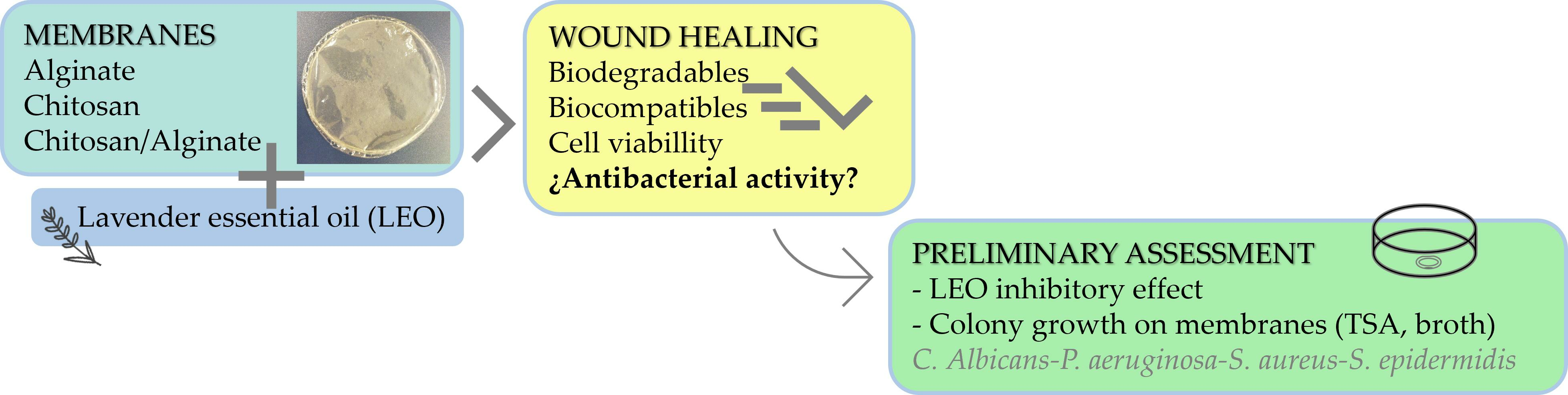
Antibacterial Activity Assessment of Chitosan/Alginate Lavender Essential Oil Membranes for Biomedical Applications

by
Encarnación Cruz Sánchez
1,
María Teresa García
1,
Ignacio Gracia
1,
Soledad Illescas Fernández-Bermejo
2,3,
Juan Francisco Rodríguez
1,
Jesús Manuel García-Vargas
1 and
Dolors Vidal Roig
2,*
1
Department of Chemical Engineering, Facultad de Ciencias y Tecnologías Químicas, University of Castilla-La Mancha, Avda. Camilo José Cela 12, 13071 Ciudad Real, Spain
2
Department of Medical Sciences, Microbiology Area, Facultad de Medicina, University of Castilla-La Mancha, Paseo de Moledores s/n, 13071 Ciudad Real, Spain
3
Department of Microbiology, Hospital General Universitario de Ciudad Real, Obispo Rafael Torija s/n, 13005 Ciudad Real, Spain
*
Author to whom correspondence should be addressed.
Membranes 2024, 14(1), 12; https://doi.org/10.3390/membranes14010012
Submission received: 14 November 2023 / Revised: 29 December 2023 / Accepted: 30 December 2023 / Published: 1 January 2024

Abstract

:
The demand for natural products in the treatment of dermatological pathologies has boosted the use of bioactive substances such as lavender essential oil (LEO), which stands out for its anti-inflammatory and antioxidant properties and its antimicrobial potential. Biopolymers such as chitosan (CHT) and alginate (ALG) are biodegradable and biocompatible and have proven their viability in biomedical applications such as skin regeneration. The inhibitory effect of LEO on the growth of skin-related bacterial species Staphylococcus aureus, Staphylococcus epidermidis, Pseudomonas aeruginosa and the fungus Candida albicans was studied by incorporating 1% v/v LEO encapsulated in CHT, ALG, and CHT/ALG membranes. Despite the verification of the antimicrobial effect of all type of membranes, no synergistic effect was observed following the addition of LEO. S. aureus and P. aeruginosa showed the most growth on the different substrates and C. albicans demonstrated the highest inhibition. This is a first approach using microorganisms isolated from clinical samples or skin microbiota. Further investigation would be advisable using more clinical strains for each microorganism to validate their biomedical applicability.

Graphical Abstract

1. Introduction

Skin anomalies are a leading epidemiological burden worldwide, ranking fourth with respect to the number of diseases they cause [1]. Skin is a complex organism inhabited by bacteria, fungi, and viruses. These microbes are essential for the proper functioning of the human body. Diseases or pathologies can, however, result from their disruption or interaction with other microorganisms or hosts [2]. Staphylococcus epidermidis (S. epidermidis) and Staphylococcus aureus (S. aureus), both gram-positive cocci, are naturally present on the skin and in the mucous membranes of the nose [3,4]. S. epidermidis is present in 100% of humans, while S. aureus is present in a lower percentage [5]. The gram-negative bacillus Pseudomonas aeruginosa (P. aeruginosa) is also present, but is less abundant and frequently behaves as an opportunistic pathogen causing burn wound infections [6] and skin and soft tissue injuries [7]. One of the most common fungI in the human microbiota is Candida albicans (C. albicans), which asymptomatically colonizes numerous parts of the body, including the gastrointestinal and genitourinary tracts of healthy people [8,9].
The demand for products of natural origin in all areas driven by new life patterns, as well as the side effects of traditional medicines for the treatment of dermatological disorders, have boosted the presence of natural extracts, such as essential oils, in the pharmaceutical industry [10]. Lavender essential oil (LEO) has been one of the most popular essential oils since ancient times. It has been traditionally used as a natural remedy in aromatherapy and herbal medicine. Currently, it is present in cosmetics, perfumes, and, particularly, in nutraceuticals and pharmaceuticals [11]. This is due to LEO therapeutic attributes, such as its anti-inflammatory and antioxidant properties and its beneficial effects on diseases related to the nervous system [12,13]. It also plays an important role in products related to the treatment of stress or anxiety or in aromatherapy to alleviate various pathologies [14,15].
Regarding the biological activity of LEO, its antifungal and antibacterial capacities are also noteworthy. This essential oil, isolated or encapsulated in various materials, has shown antibacterial activity against bacteria such as S. aureus and Escherichia coli as well as a yeast, C. albicans [16,17]. Consequently, LEO is a very interesting component at the pharmacological level for biomedical applications such as products for the treatment of skin diseases.
In addition to the use of high added-value compounds from natural resources, environmental issues are of great relevance to biomedicine. For this reason, biopolymers are the most commonly used materials for the encapsulation of bioactive substances [18]. Biopolymers are a type of polymers synthesised by living organisms, including natural polymers present in the environment or those produced artificially from natural resources. These materials are readily available, inexpensive, and stand out for their biodegradability and biocompatibility [19].
The most widely used in health-related applications are polysaccharides because of their biological activity, especially alginate (ALG) and chitosan (CHT). These biopolymers, apart from their ability to form membranes, have antimicrobial capacity, antioxidant potential, and even possess the ability to accelerate healing [20,21,22,23]. The combination of both polymers has been shown to work as an effective pathway for skin regeneration [24,25,26,27], with the incorporation of essential oils able to increase their biological activity and therapeutic potential [28,29,30,31,32]. In previous research, the cell viability and biocompatibility of CHT/ALG + LEO membranes with HaCaT, a human epidermal keratinocyte cell line, has been demonstrated [33].
In this study, ALG, CHT, and CHT/ALG membranes were prepared with the addition of LEO. Because these membranes are created with the aim of improving wound healing and curing skin pathologies, bacteria present in the human microbiota and residing on the skin and/or mucous membranes were used to test their antimicrobial capacity, namely S. epidermidis, S. aureus, P. aeruginosa, and C. albicans. While S. aureus and P. aeruginosa are mainly responsible for chronic skin infections [34], S. epidermidis and C. albicans have not been frequently associated with skin infections, but have been sporadically implicated in post-surgical infections [8,35]. Considering that biopolymers may have a bactericidal effect, the addition of LEO as an extra bactericidal additive should reflect a synergy. The aim of this work is to provide a preliminary result of the possible synergistic inhibition effect of LEO-additivated polymers using clinical strains implicated in wound or skin microbiota infections. The results obtained in this study provide a reference for further study of the antibacterial mechanism of LEO in conjunction with the proposed biopolymers and possible interactions between them.

2. Materials and Methods

2.1. Preparation of Membranes

2.1.1. Materials

CHT and Tween 80® were supplied by Sigma-Aldrich (St. Louis, MO, USA). Sodium alginate, ALG, from brown algae was obtained from Fluka-BioChemika (Buchs, Swizerland) and Calcium chloride from PanReac AppliChem (Barcelona, Spain). The LEO was from Peñarrubia del Alto Guadiana S. L. (Albacete, Spain). Acetone, supplied by LabChem (Santo Antão do Tojal, Portugal), acetic acid from Carlo Erba Reagents (Milan, Italy), and deionized water were also used. A total of 20 mM PBS pH 7.4 (including milli Q water, Na2HPO4·7H2O, and NaH2PO4·H2O) was used.

2.1.2. Membranes Synthesis Procedure

Six types of membranes were prepared, two with a single polymer (Alginate, ALG; Chitosan, CHT) and a third mixed (CHT–ALG), combined with and without lavender essential oil (LEO).
Membranes were prepared as described in other work [33] using the method of Rodrigues et al. [25]. For the preparation of the CHT/ALG membranes, 90 mL of an aqueous solution of ALG at 0.5% (w/w) was added to 90 mL of CHT solution at 0.5% (w/w) in 2% aqueous acetic acid (v/v) and acetone 1:1 (v/v) using a syringe pump (KDS Legato 200 Series) with a flow rate of 40 mL/h and stirring at 500 rpm.
The tests were conducted in a glass vessel at a temperature of 25 °C with a mechanical stirrer. After obtaining the suspension, it was homogenized for 10 min while being stirred at 1000 rpm. The pH was then raised to 5.28 by adding NaOH (1 M), which was then agitated for 10 min at 1000 rpm. For cross-linking, 1.8 mL of a 2% (w/v) aqueous CaCl2 solution was added last. The mixture was then put into Petri dishes with a 15 cm internal diameter and dried for 20 h at 37 °C in an oven with moving air. Membranes were dried before being submerged for 1 h in 150 mL of a 2% (w/v) CaCl2 aqueous solution to cross-link alginate L-glucuronic acid residues on neighboring chains that were not attached to chitosan. They were then submerged twice for 1 h in 200 mL of deionized water, and then allowed to air dry at room temperature.
In the case of membranes composed of a single biopolymer, the same procedure was followed, although the first step of mixing the solutions was excluded. The only difference between CHT and ALG membranes is that ALG membranes do not require pH neutralization. For the preparation of ALG, CHT, and CHT/ALG membranes with essential oil, LEO was added to the initial solution with a concentration of 1% (v/v), as well as Tween 80® 1% (v/v) which acts as an emulsifying agent for the dispersion and solubilization of the essential oil [36].

2.1.3. Sterilization of Membranes

Membranes were cut into square shapes with a mean area of 0.25 cm2. They were then sterilized by ultraviolet irradiation on both sides in a laminar flow cabinet for 1 h on each side in sterile Petri dishes. Once sterilized, they were stored in the sterile plates until use, within 72 h.

2.2. Antimicrobial Capacity Analysis

This study was carried out in two stages: prior to the study of the antimicrobial capacity of the membranes synthesized with the different polymers and LEO, the inhibition capacity of the LEO itself was determined (Experiment 1). Once the behavior of the LEO against the strains used had been checked, the antimicrobial activity of the membranes was analyzed using two methods, culturing by direct contact with a solid medium (Experiment 2) and by immersion in culture broth (Experiment 3).

2.2.1. Strains and Culture Media

Table 1 shows the strains used. All of them came from the area of Microbiology of the Faculty of Medicine of Ciudad Real of the UCLM as collection strains of the area for laboratory practice in the subject of Microbiology in the Degree of Medicine.
Two types of culture media were used, a solid media Tryptone Soy Agar (TSA) and a broth Tryptone Soy Broth (TSB) (Scharlab, Spain). Tryptone Soy Agar (TSA) is a versatile, non-selective medium that offers enough nutrients to support the development of a broad range of pathogenic microorganisms. It was decided to use TSA because it is a medium where the micro-organisms to be tested grow optimally.

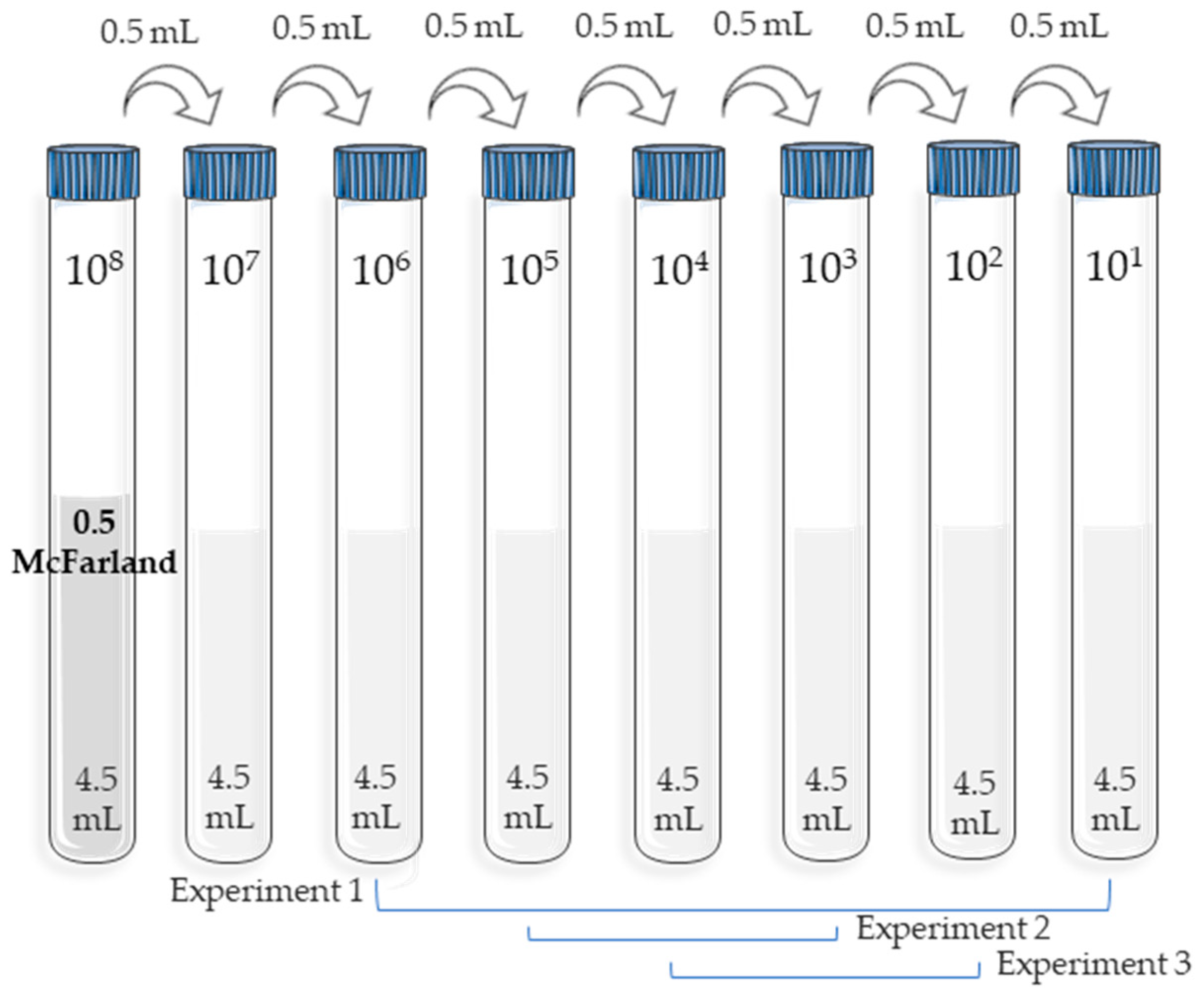
2.2.2. Inoculum Solutions

For both the measurement of the antimicrobial capacity of the oil and the membranes, the inocula for each of the strains used were prepared in the same way.
A schematic of the procedure followed is shown in Figure 1. A set of 10-fold serial tube dilutions was performed under aseptic conditions with 4.5 mL of sterile NaCl solution (0.9%). The initial dilution was the one with the highest microbial concentration, measured with a McFarland 0.5 standard (bioMérieux, Craponne, France) equivalent to 108 CFU/mL. In parallel, for each experiment, plate counts with medium were performed to determine the starting CFU.

2.2.3. Determination of Bactericidal Effect of LEO

First, TSA medium was prepared for distribution in sterile 90 mm Petri plates (Scharlab, Barcelona, Spain). Once the medium was prepared and prior to sterilisation, LEO was added. After sterilising the prepared medium with the essential oil in the autoclave, it was distributed in sterile Petri dishes used in the microbiological experiment. It should be noted that, at this stage, in order to obtain a homogeneous distribution of the LEO in the medium, continuous magnetic stirring of the container was performed.
In order to test the influence of LEO concentration on its antibacterial activity, different concentrations of 1, 0.75, 0.5, 0.25, 0.1, and 0% v/v were prepared, the latter serving as control. In addition, different concentrations of inoculum of each strain were also used (from 108 to 101). A total of 10 µL of each of the dilutions was inoculated in quadruplicates on a TSA plate, from dilution 106 to 101, and left to incubate at 35–37 °C for 18–24 h. After this time, colony counts were conducted for the S. epidermidis, S. aureus, and P. aeruginosa tests. In the case of C. albicans, the count was done at 48 h of incubation.

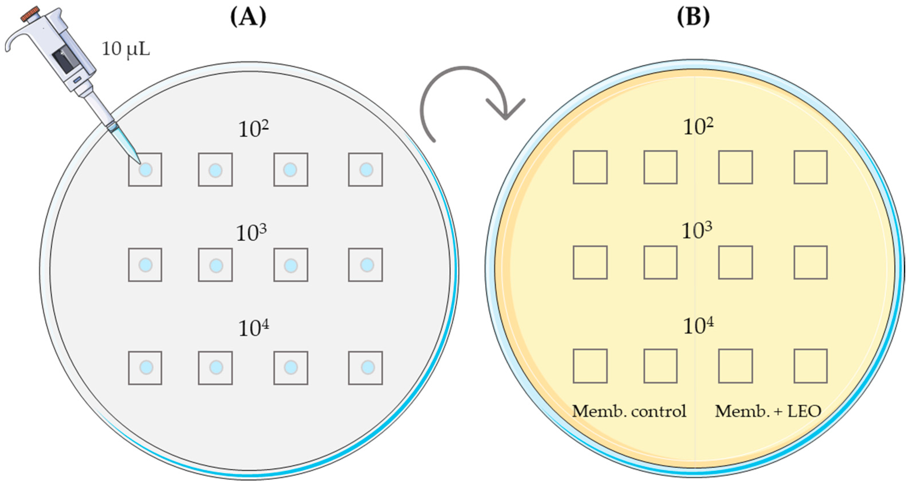
2.2.4. Microbiological Study of Direct-Contact Membranes in Solid Media

In this case, the 10 µL inoculum was cultured directly onto the membrane pieces and placed in contact with the TSA medium on the plates, previously prepared as in the previous experiment (Figure 2A). Three different inoculum concentrations (103, 104, and 105) were used, combined with three different times (from inoculum application on the membranes to incubation on the culture plate) at 0 h, 2 h, and 6 h. Each test was duplicated on the same plate, comparing the control with the membrane, including LEO (Figure 2B), for each type of membrane synthesized, CHT, ALG, and CHT/ALG. Plates were incubated in aerobic conditions at 35–37 °C for 18–24 h for all three bacteria and at 48 h for Candida. After incubation, plates were analyzed for the presence or absence of microbial growth.

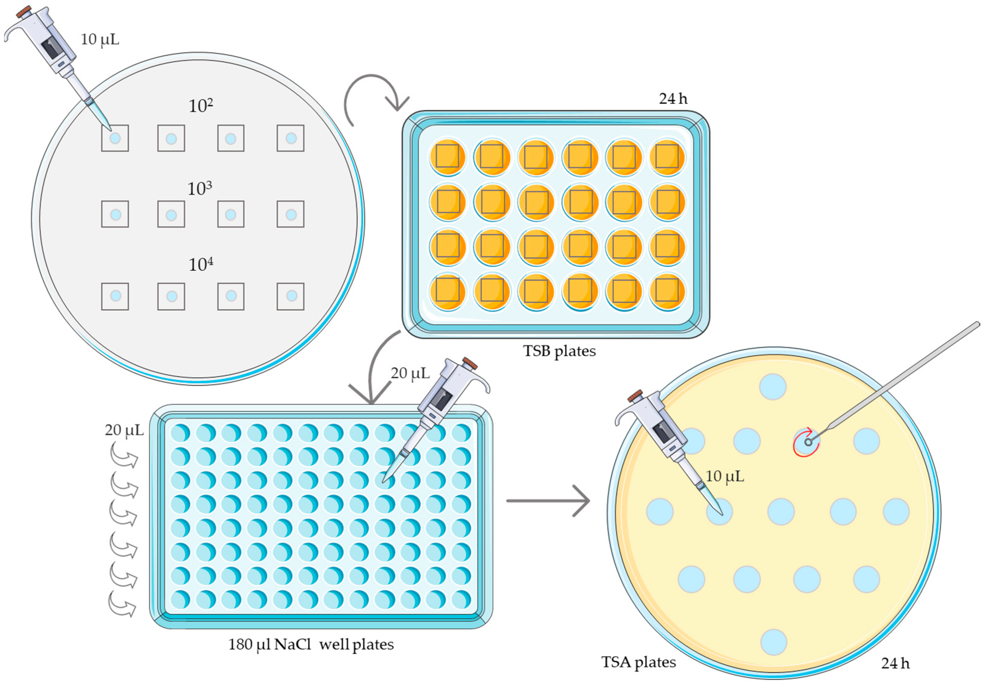
2.2.5. Microbiological Study of Membranes in Broth

The key difference between this method and the contact experiment is that, after the membrane inoculation phase, instead of placing the membranes on TSA solid medium plates, they were immersed in TSB liquid medium for 24 h in sterile 24-well plates (Deltalab, Barcelona, Spain). Inoculation of the membranes was attained following the same method used in the contact experiment, and, in this case, the used concentrations were 102, 103, 104 CFU/mL. As in the experiment in the solid medium, membranes were grown on the plates at three different times after the inoculum was placed on them: at 0 h, 2 h, and 6 h.
After 24 h, 20 µL of liquid medium was taken from each well. In this experiment, not only the presence or absence of microbial growth was analyzed, but also a CFU/mL count was performed using 10-fold serial dilutions and 0.9% NaCl as dilutant. These dilutions were carried out in sterile 96-well plates (Deltalab, Spain) in aseptic conditions. A total of 10 µL of each dilution were taken and cultured on TSA plates and left to incubate at 35–37 °C for 18–24 h (Figure 3). After this time, colony counts were performed for the S. epidermidis, S. aureus, and P. aeruginosa tests. In the case of C. albicans, the count was done at 48 h.

2.2.6. Database and Statistical Analysis

A database was created using Microsoft Excel 2021, with the following variables included: membrane type (ALG, CHT, CHT/ALG, ALG + LEO, CHT + LEO, CHT/ALG + LEO), experiment (1,2,3), strain (C. albicans, P. aeruginosa, S. aureus, S. epidermidis), inoculum (106, 105, 104, 103, 102, 101), time (0 h, 2 h, 6 h), growing (NEG, POS), count (only for Experiment 3: 0, <100, 100–1000, >1000). To evaluate significant differences between qualitative discrete variables, the Pearson’s Chi-square (χ2) test was used, with a statistical significance level of p < 0.05, using the Winepi program (www.winepi.net, (accessed on 2 October 2023); basic statistics, Ignacio de Blas, University of Zaragoza). As there was a low sample size for each experiment, different variables such as total strains, pre-inoculum times, or membrane type (with or without LEO) were grouped together, since, in most of the analyses, the expected results in the test were lower than 5, rendering invalid the significance of χ2 test.

3. Results

3.1. Bactericidal Effect of LEO

The objective of this experiment was to evaluate what concentration of LEO is sufficient to inhibit microbial growth or produce changes in the phenotype of the colonies, which could be indicative of a microbial stress effect, by cultivating the different strains in a solid medium supplemented with the essential oil.
Table 2 shows the average count results adjusted for inoculum dilution 103 for the strains studied. The main finding was that the 1% v/v LEO concentration inhibited the growth of all strains. The count was similar between control and LEO dilutions lower than or equal to 0.5%. The 0.75 v/v LEO concentration also inhibited S. epidermidis and C. albicans, while for S. aureus and P. aeruginosa, although there was growth, this concentration subtly reduced the number of colonies.
Although there were no differences in counts between 0.5% v/v LEO and the controls, a change in the phenotype was observed in the case of S. epidermidis and C. albicans (Figure 4), showing smaller colonies. This change in phenotype was not observed in either S. aureus or P. aeruginosa.

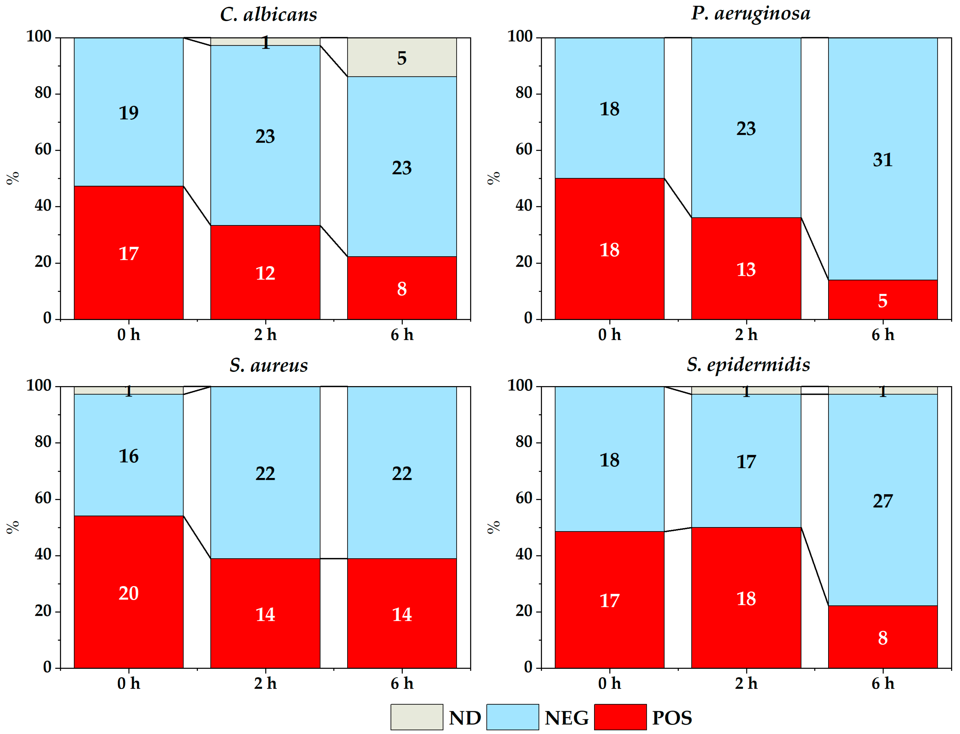
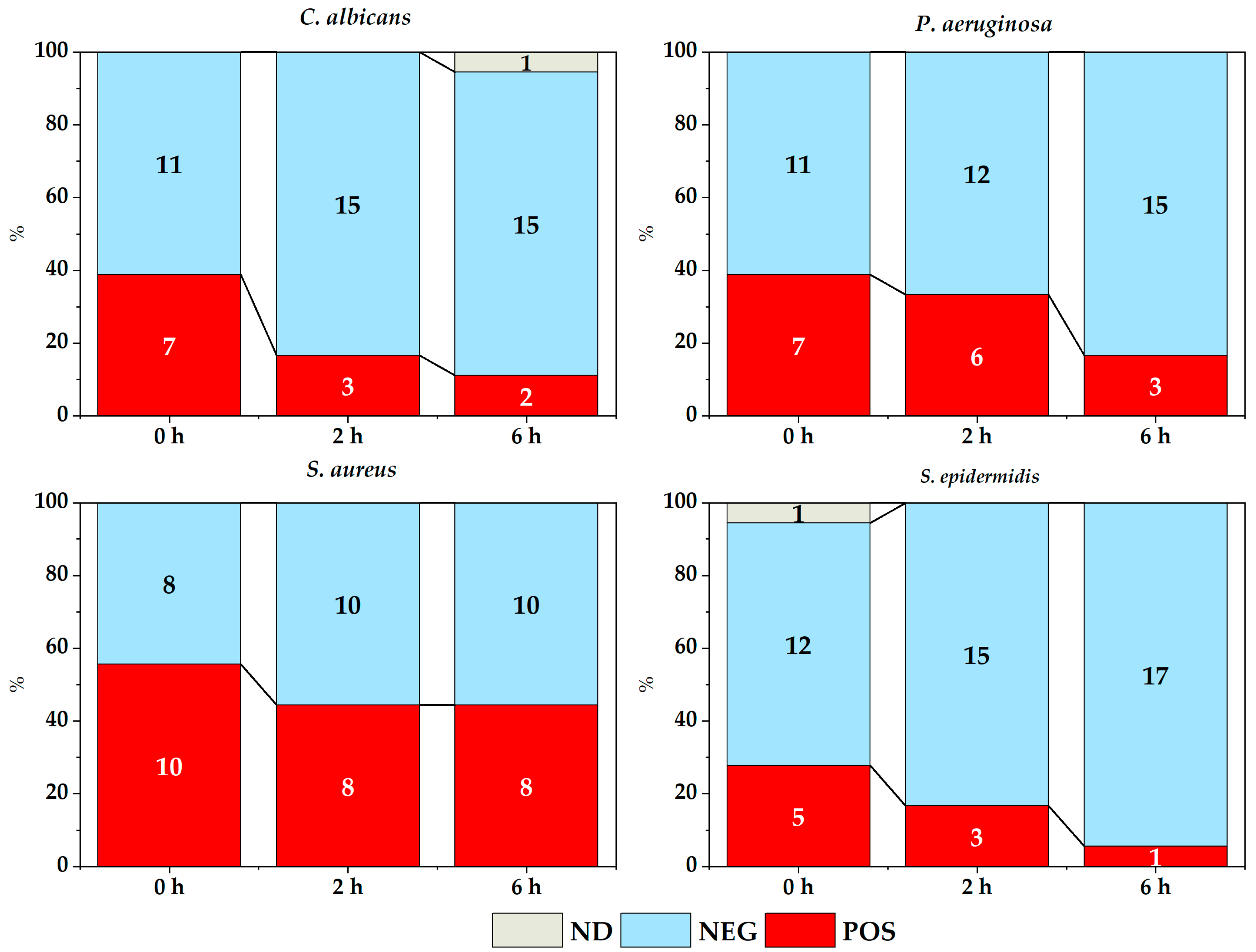
3.2. Growth Inhibition Depending on the Time between Inoculum Addition and Culture

Figure 5 and Figure 6 show the count of membranes with and without microbial growth depending on the time elapsed between placing the inoculum on the different membranes and its corresponding culture, in both the experiment in solid medium (Figure 5) and the experiment with broth (Figure 6).
Overall, there was an effect of the time elapsed between inoculum addition and subsequent cultivation for almost all strains studied, both in the contact experiment (Figure 5) and in the broth experiment (Figure 6), except for S. aureus in both cases. For this strain, it should be noted that the number of positive cases remained constant for the experiment in the solid medium (χ2 = 2.700, p = 0.2592) and also in the experiment with membranes in broth (χ2 = 1.637, p = 0.4412), except for a slight decrease of positive cases in the broth over time.

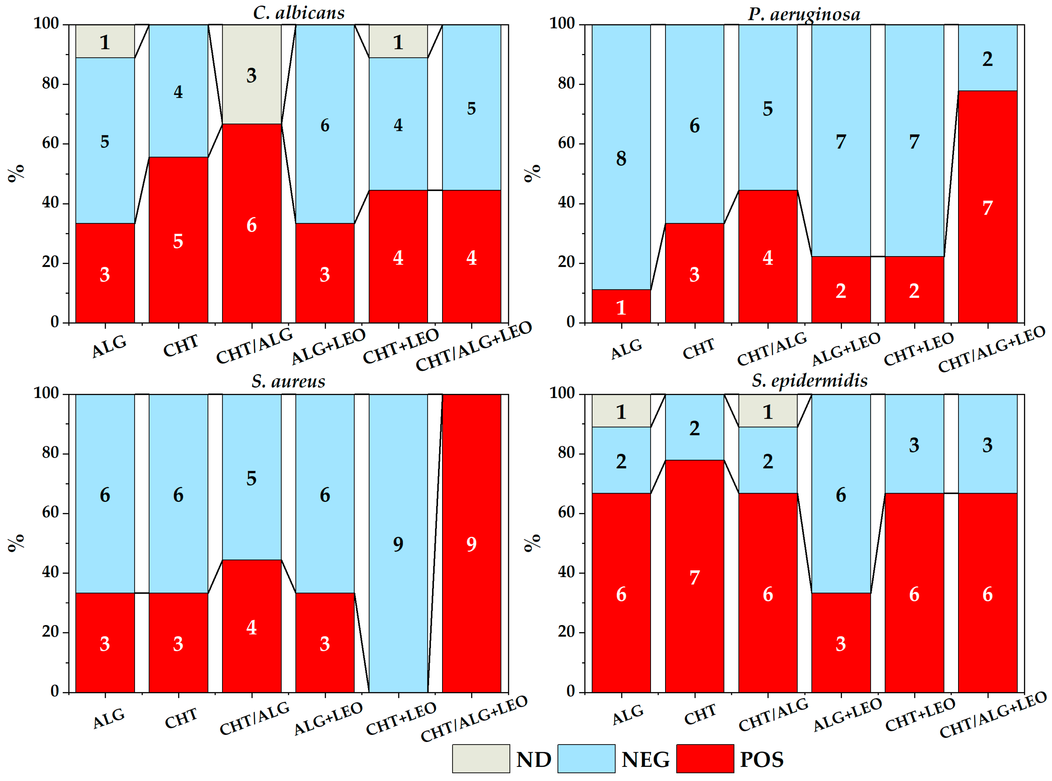
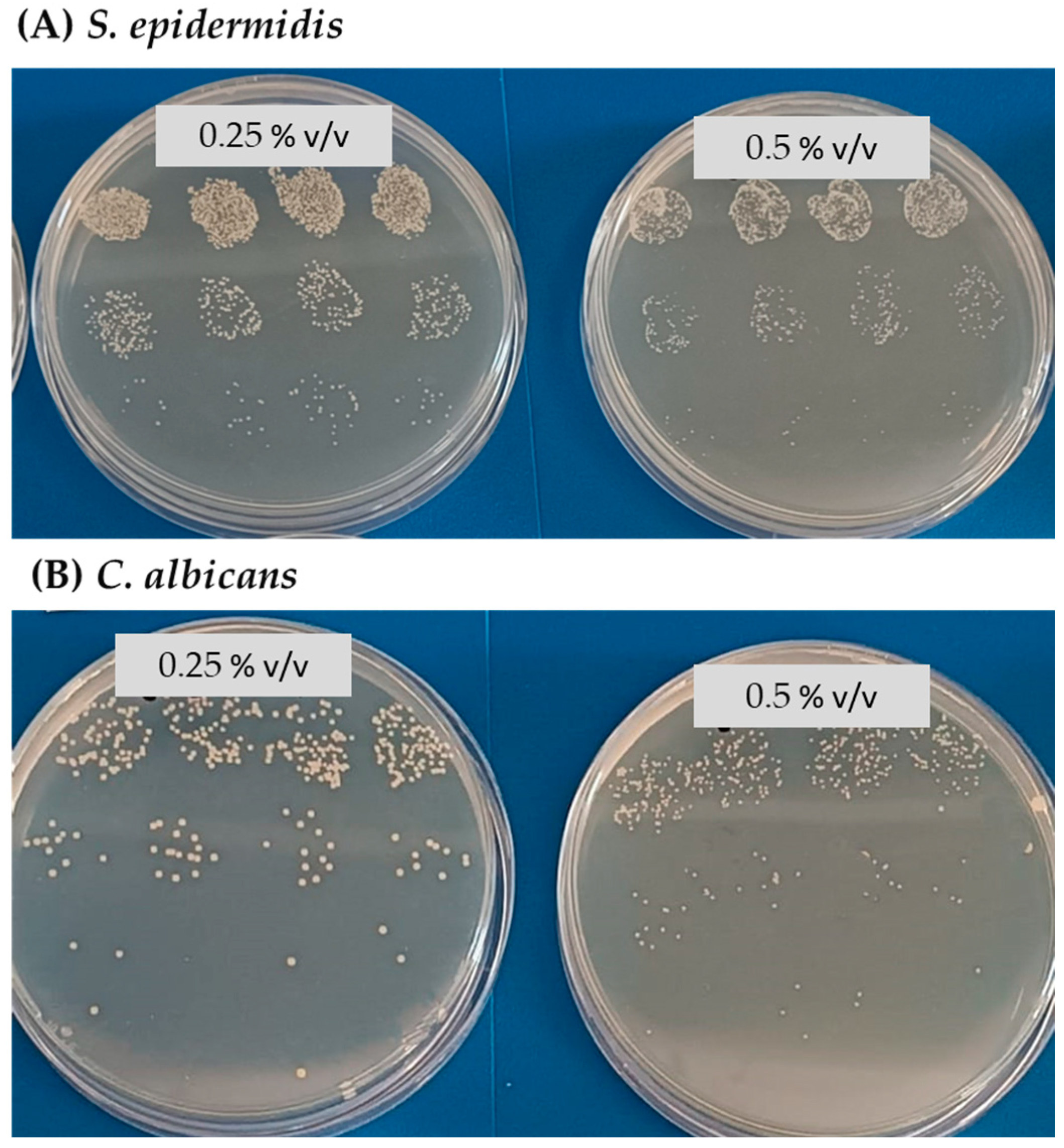
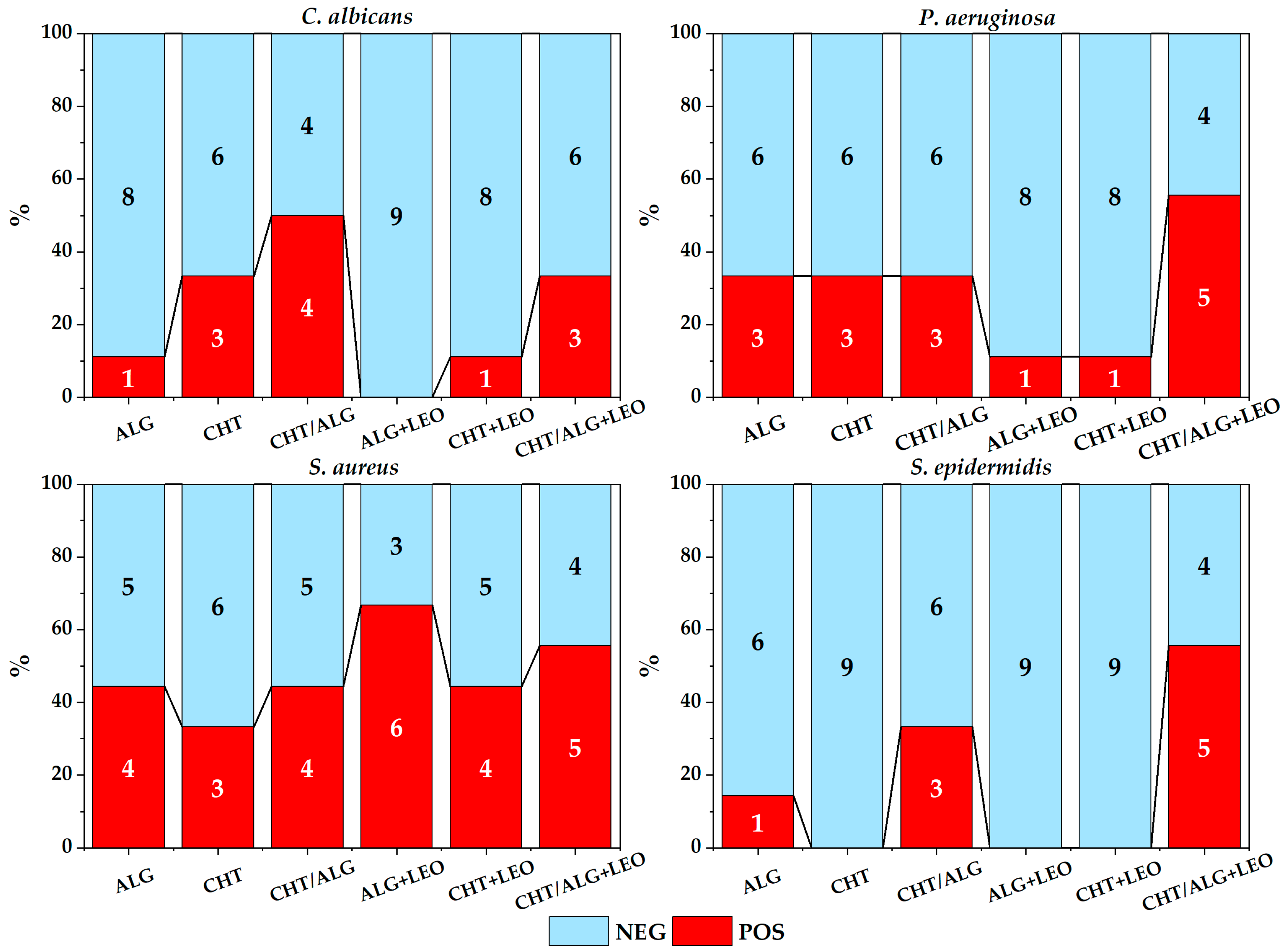
3.3. Bacterial Growth Resulting from Membranes Cultured Directly on Plates

Figure 7 summarizes all the results of experiment of direct contact, for each type of membrane and strain. In some cases, it was not possible to obtain a result because the membranes were detached from the culture medium or there was some type of contamination, mainly in the oil-free membranes.
To evaluate the effect of membrane oil addition (LEO), all strains and membranes with and without LEO were grouped, but no significant differences were observed (χ2 = 0.370, p = 0.5429). On the other hand, upon separation by type of membrane, no differences were found either, although there was a slight tendency of greater inhibition in CHT membranes with LEO (χ2 = 1.796, p = 0.1802).
In addition, the difference in total membranes was evaluated for each of the strains (χ2 = 11.6, p = 0.0089), concluding that S. epidermidis was the best-growing strain.

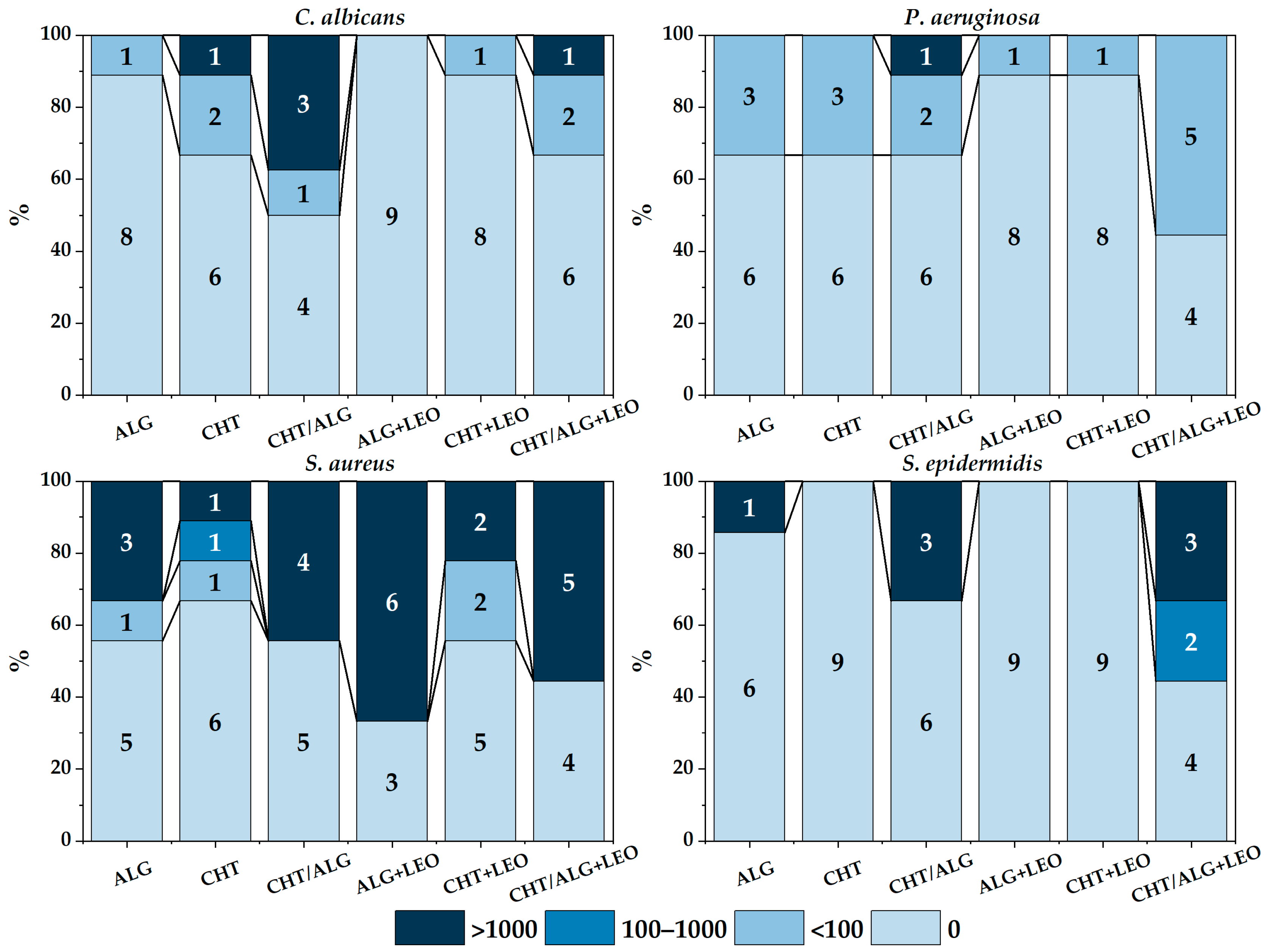
3.4. Bacterial Growth Resulting from Membranes Cultured in Broth

Figure 8 summarizes all the results of experiment in broth, for each type of membrane and strain.
In this experiment, in addition to assessing the presence or absence of growth, counts were made from the culture broth (CFU/mL). Although it was not possible to analyze significant differences in counts for each strain individually due to the small sample size (less than 5 samples), for C. albicans further growth was observed on CHT/ALG membranes; also, by comparing membranes with or without LEO, the number of positive cases was found to be slightly higher when LEO was included.
No significant differences were obtained by grouping the results of counts with respect to the presence of LEO in the total strains (χ2 = 0.080, p = 0.7769), but there were differences in growth among strains (χ2 = 13.951, p = 0.0029), with S. aureus and P. aeruginosa growing better in broth than C. albicans and S. epidermidis2 = 9.8945, p = 0.0016).
Furthermore, by grouping all strains together, no differences in positivity were found in the total number of membranes (χ2 = 0.080, p = 0.7769) or with respect to each membrane type. By comparing strains, S. aureus was found to be the strain with the highest number of positive cases, with no differences when LEO was used.
By grouping by the type of polymer used between oil and non-oil membranes, it was observed that there was a slightly higher growth in mixed membranes with respect to all strains (χ2 = 11.0652, p = 0.0039), with the exception of S aureus.
Figure 9 breaks down the number of positive colonies into intervals in order to really see the influence of LEO on the total percentage of inhibition of each of the strains. It can be seen that S. aureus shows less difference in the number of positive colonies between the membranes with or without LEO and between the biopolymers used.

4. Discussion

The study of the concentration of LEO that does or does not inhibit the growth of the microbial strains chosen for this study, C. albicans, P. aeruginosa, S. aureus, and S. epidermidis, provides preliminary information on how the addition of this essential oil can influence different biopolymers for biomedical applications. In this work, the antimicrobial effect has been analyzed using strains isolated from clinical cases or from the microbiota itself, trying to demonstrate real cases, as other researchers also do using LEO, with environmental strains from hospital settings [37] or with different strains of a single species from clinical isolates [38].
Initially, it was found that LEO inhibited the growth of all micro-organisms tested at 1% concentration in the medium. However, in the case of S. epidermidis and C. albicans total inhibition already occurred at a concentration of 0.75%. Although no changes in colony counts were observed at 0.5% v/v LEO for S. epidermidis and C. albicans, changes were observed in the phenotype of the colonies, which grew smaller and with a rougher appearance. This phenotypic change may indicate possible cellular alterations. In general, the antimicrobial power of essential oils is related to changes in membrane permeability, as oils penetrate membrane lipids, destroying them and making them more permeable [39]. With respect to S. aureus, in a study using aguaribay oil, in addition to increasing membrane permeability, an ability to inhibit biofilm formation was demonstrated [40]. Again with respect to S. aureus, other research using clove essential oil demonstrated, by electron microscopy, morphological changes at the cellular level [41]. In particular for S. epidermidis, the use of Zanthoxylum schinifolium oil not only resulted in changes in membrane permeability and integrity, but also in alterations in the physiological functions of the bacterium [42].
According to the literature, it is evident that essential oils have antibacterial effects, and, among all of them, LEO has been extensively studied [43]. In this case, it has been observed that the higher the concentration of LEO, the higher the degree of inhibition of the four strains studied, as it has also been observed in other works [44]. However, in experiments testing membranes, we did not observe differences between the addition or non-addition of LEO, unlike other researchers who have observed more antimicrobial efficacy when LEO was added at concentrations of 0.5% v/v and 1% v/v using a carboxymethylchitosan-based substrate [41]. On the one hand, the biopolymers themselves already have an antimicrobial effect [41,45,46]. In fact, this work shows that the mixed CHT/ALG membrane has proven to be the least inhibitory, perhaps due to the dilution effect of both polymers in the membrane which has already been observed to have a lower toxicity effect in cell cultures compared to single-component polymers [33]. On the other hand, because LEO in the biopolymer is less bioaccessible, it is necessary to analyze the surface of the membranes to revise the composition or to add LEO in the form of nanoemulsions as in other works [47,48]. Another possible explanation could be a low concentration of the oil used, as other studies have found more inhibition at higher concentrations of the essential oil, as in the case of LEO [44] or other essential oils [49].
In general, differences in inhibition were found depending on the experiment performed. It is difficult to make comparative studies with what is available in the scientific literature. Most published works use disk diffusion techniques [27,38,50] or MIC techniques [44,46,50,51]. In this case, two experiments have been performed that aim to get closer to reality by using a dressing on a wound. On the one hand, transferring the wound to the culture medium itself (solid or broth) with the dressing on it, on the other, analyzing different inoculum concentrations, as these microorganisms reside in the normal skin in low concentrations prior to a skin infection.
Another consideration is that it is difficult to determine differences between microbial species, as there may be differences caused by usage of different strains of the same microbial species [38] and even between two collection strains of the same species with different antibioresistance profiles [37]. Overall, in the present work if there is growth inhibition in all the strains studied, with or without LEO.
S. aureus and P. aeruginosa have grown the most on the different substrates. Although these two bacteria are not very frequent in the skin microbiota, they have great clinical repercussions and are the ones that colonize most skin wounds, thus causing chronic infections [34]. However, they did show growth inhibition in all the membranes tested. In the experiment involving contact between the biopolymer and the solid culture medium, S. epidermidis was found to be the strain that obtained the most positive cases, but this was not the case when it was cultured in broth. The broth culture experiment was more inhibitory than the contact experiment. This may be due to a release of both the oil and the biopolymer itself into the liquid culture medium, as the membranes were partially degraded after incubation. In the case of C. albicans, in the present work the fungus suffered overall more inhibition than the bacteria. In contrast to other work using LEO, there was almost no inhibitory effect using also a strain isolated from a clinical case [50], nor using a reference strain, with a null [44].

5. Conclusions

This work provides information on the preliminary study of the antimicrobial potential of LEO and its incorporation into CHT and ALG membranes. Firstly, it can be deduced that LEO has an inhibitory capacity in a concentration of 1% v/v in TSA medium with respect to P. aeruginosa and S. aureus, as well as with respect to S. epidermidis and the fungus C. albicans from a concentration of 0.75% v/v, with C. albicans suffering an alteration in the phenotype of the colonies. Despite the verification of the antimicrobial effect of LEO, in the membrane tests, in general, no significant differences were observed between the results obtained with and without its addition. Furthermore, the CHT/ALG membrane proved to be the least inhibitory, probably due to the dilution effect of both polymers in that configuration. As for the difference between microbial species, differences were reflected with S. aureus and P. aeruginosa showing the most growth on the different substrates despite demonstrating some inhibition on all membranes. It was the fungus C. albicans that demonstrated the highest inhibition. The main limitation of this study is the few strains analyzed and the low number of replicates. Expansion of the experiments through an increase in the LEO concentration in membranes, along with the introduction of different variants of each specie could yield broader outcomes.

Author Contributions

Conceptualization, D.V.R.; methodology, D.V.R. and E.C.S.; software, D.V.R.; validation, D.V.R., E.C.S., M.T.G. and J.M.G.-V.; formal analysis, D.V.R. and S.I.F.-B.; investigation, E.C.S. and D.V.R.; resources, D.V.R. and S.I.F.-B.; data curation, D.V.R. and E.C.S.; writing—original draft preparation, E.C.S. and D.V.R.; writing—review and editing, E.C.S., D.V.R., M.T.G., J.M.G.-V., I.G., J.F.R. and S.I.F.-B.; visualization, M.T.G. and J.M.G.-V.; supervision, D.V.R., M.T.G., J.M.G.-V., I.G. and J.F.R.; project administration, M.T.G., J.M.G.-V., I.G. and J.F.R.; funding acquisition, M.T.G., I.G. and J.F.R. All authors have read and agreed to the published version of the manuscript.

Funding

This research was funded by the project Ref. SBPLY/17/180501/000311 from the Consejería de Educación, Cultura y Deportes of Junta de Comunidades de Castilla-La Mancha.

Institutional Review Board Statement

Not applicable.

Data Availability Statement

The data presented in this study are available upon request from the corresponding author. The data are not publicly available due to privacy.

Acknowledgments

We gratefully acknowledge funding from the Consejería de Educación, Cultura y Deportes of Junta de Comunidades de Castilla La Mancha provided for this project.

Conflicts of Interest

The authors declare no conflicts of interest; they have no known competing financial interests or personal relationship that could have appeared to influence the work reported in this paper.

References

  1. Maddheshiya, S.; Ahmad, A.; Ahmad, W.; Zakir, F.; Aggarwal, G. Essential oils for the treatment of skin anomalies: Scope and potential. S. Afr. J. Bot. 2022, 151, 187–197. [Google Scholar] [CrossRef]
  2. Chen, Y.E.; Fischbach, M.A.; Belkaid, Y. Skin microbiota–host interactions. Nature 2018, 553, 427–436. [Google Scholar] [CrossRef] [PubMed]
  3. Vuong, C.; Otto, M. Staphylococcus epidermidis infections. Microbes Infect. 2002, 4, 481–489. [Google Scholar] [CrossRef] [PubMed]
  4. Tong, S.Y.C.; Davis, J.S.; Eichenberger, E.; Holland, T.L.; Fowler, V.G.J. Staphylococcus aureus infections: Epidemiology, pathophysiology, clinical manifestations, and management. Clin. Microbiol. Rev. 2015, 28, 603–661. [Google Scholar] [CrossRef] [PubMed]
  5. Gimeno Cardona, C. “Microbiología Clínica” de Guillem Prats. Enferm. Infecc. Microbiol. Clin. 2006, 24, 215. [Google Scholar] [CrossRef]
  6. Salerian, A.J. Burn wound infections and Pseudomonas aeruginosa. Burns 2020, 46, 257–258. [Google Scholar] [CrossRef] [PubMed]
  7. Nagoba, B.; Davane, M.; Gandhi, R.; Wadher, B.; Suryawanshi, N.; Selkar, S. Treatment of skin and soft tissue infections caused by Pseudomonas aeruginosa—A review of our experiences with citric acid over the past 20 years. Wound Med. 2017, 19, 5–9. [Google Scholar] [CrossRef]
  8. Nobile, C.J.; Johnson, A.D. Candida albicans Biofilms and Human Disease. Annu. Rev. Microbiol. 2015, 69, 71–92. [Google Scholar] [CrossRef]
  9. Lu, H.; Hong, T.; Jiang, Y.; Whiteway, M.; Zhang, S. Candidiasis: From cutaneous to systemic, new perspectives of potential targets and therapeutic strategies. Adv. Drug Deliv. Rev. 2023, 199, 114960. [Google Scholar] [CrossRef]
  10. Mohd Zaid, N.A.; Sekar, M.; Bonam, S.R.; Gan, S.H.; Lum, P.T.; Begum, M.Y.; Mat Rani, N.N.I.; Vaijanathappa, J.; Wu, Y.S.; Subramaniyan, V.; et al. Promising Natural Products in New Drug Design, Development, and Therapy for Skin Disorders: An Overview of Scientific Evidence and Understanding Their Mechanism of Action. Drug Des. Devel. Ther. 2022, 16, 23–66. [Google Scholar] [CrossRef]
  11. Djilani, A.; Dicko, A. The therapeutic benefits of essential oils. Nutr. Well-Being Health 2012, 7, 155–179. [Google Scholar]
  12. But, V.M.; Bulboacă, A.E.; Rus, V.; Ilyés, T.; Gherman, M.L.; Bolboacă, S.D. Anti-inflammatory and antioxidant efficacy of lavender oil in experimentally induced thrombosis. Thromb. J. 2023, 21, 85. [Google Scholar] [CrossRef] [PubMed]
  13. Chen, W.N.; Chin, K.W.; Tang, K.S.; Agatonovic-Kustrin, S.; Yeong, K.Y. Neuroprotective, neurite enhancing, and cholinesterase inhibitory effects of Lamiaceae family essential oils in Alzheimer’s disease model. J. Herb. Med. 2023, 41, 100696. [Google Scholar] [CrossRef]
  14. de Melo Alves Silva, L.C.; de Oliveira Mendes, F.d.C.; de Castro Teixeira, F.; de Lima Fernandes, T.E.; Barros Ribeiro, K.R.; da Silva Leal, K.C.; Dantas, D.V.; Neves Dantas, R.A. Use of Lavandula angustifolia essential oil as a complementary therapy in adult health care: A scoping review. Heliyon 2023, 9, e15446. [Google Scholar] [CrossRef]
  15. Rivaz, M.; Rahpeima, M.; Khademian, Z.; Dabbaghmanesh, M.H. The effects of aromatherapy massage with lavender essential oil on neuropathic pain and quality of life in diabetic patients: A randomized clinical trial. Complement. Ther. Clin. Pract. 2021, 44, 101430. [Google Scholar] [CrossRef]
  16. Sofi, H.S.; Akram, T.; Tamboli, A.H.; Majeed, A.; Shabir, N.; Sheikh, F.A. Novel lavender oil and silver nanoparticles simultaneously loaded onto polyurethane nanofibers for wound-healing applications. Int. J. Pharm. 2019, 569, 118590. [Google Scholar] [CrossRef]
  17. Liakos, I.; Rizzello, L.; Scurr, D.J.; Pompa, P.P.; Bayer, I.S.; Athanassiou, A. All-natural composite wound dressing films of essential oils encapsulated in sodium alginate with antimicrobial properties. Int. J. Pharm. 2014, 463, 137–145. [Google Scholar] [CrossRef]
  18. Singh, N.K.; Baranwal, J.; Pati, S.; Barse, B.; Khan, R.H.; Kumar, A. Application of plant products in the synthesis and functionalisation of biopolymers. Int. J. Biol. Macromol. 2023, 237, 124174. [Google Scholar] [CrossRef]
  19. Syed, M.H.; Zahari, M.A.K.M.; Khan, M.M.R.; Beg, M.D.H.; Abdullah, N. An overview on recent biomedical applications of biopolymers: Their role in drug delivery systems and comparison of major systems. J. Drug Deliv. Sci. Technol. 2023, 80, 104121. [Google Scholar] [CrossRef]
  20. Ren, W.; Cheng, W.; Wang, G.; Liu, Y. Developments in antimicrobial polymers. J. Polym. Sci. Part A Polym. Chem. 2017, 55, 632–639. [Google Scholar] [CrossRef]
  21. López-Mata, M.A.; Ruiz-Cruz, S.; de Jesús Ornelas-Paz, J.; Del Toro-Sánchez, C.L.; Márquez-Ríos, E.; Silva-Beltrán, N.P.; Cira-Chávez, L.A.; Burruel-Ibarra, S.E. Mechanical, Barrier and Antioxidant Properties of Chitosan Films Incorporating Cinnamaldehyde. J. Polym. Environ. 2018, 26, 452–461. [Google Scholar] [CrossRef]
  22. Bano, I.; Arshad, M.; Yasin, T.; Ghauri, M.A.; Younus, M. Chitosan: A potential biopolymer for wound management. Int. J. Biol. Macromol. 2017, 102, 380–383. [Google Scholar] [CrossRef]
  23. Zhou, H.; Yang, N.; Hou, J.; Yu, C.; Jin, Z.; Zeng, P.; Yang, L.; Fu, Y.; Shen, Y.; Guo, S. Effects of CaCl2, HCl, acetic acid or citric acid on dynamic mechanical performances and physicochemical properties of sodium alginate edible films. Food Packag. Shelf Life 2022, 34, 100935. [Google Scholar] [CrossRef]
  24. Baysal, K.; Aroguz, A.Z.; Adiguzel, Z.; Baysal, B.M. Chitosan/alginate crosslinked hydrogels: Preparation, characterization and application for cell growth purposes. Int. J. Biol. Macromol. 2013, 59, 342–348. [Google Scholar] [CrossRef] [PubMed]
  25. Rodrigues, A.P.; Sanchez, E.M.S.; Da Costa, A.C.; Moraes, A.M. The influence of preparation conditions on the characteristics of chitosan-alginate dressings for skin lesions. J. Appl. Polym. Sci. 2008, 109, 2703–2710. [Google Scholar] [CrossRef]
  26. Caetano, G.F.; Frade, M.A.C.; Andrade, T.A.M.; Leite, M.N.; Bueno, C.Z.; Moraes, Â.M.; Ribeiro-Paes, J.T. Chitosan-alginate membranes accelerate wound healing. J. Biomed. Mater. Res. Part B Appl. Biomater. 2015, 103, 1013–1022. [Google Scholar] [CrossRef] [PubMed]
  27. Feketshane, Z.; Adeyemi, S.A.; Ubanako, P.; Ndinteh, D.T.; Ray, S.S.; Choonara, Y.E.; Aderibigbe, B.A. Dissolvable sodium alginate-based antibacterial wound dressing patches: Design, characterization, and in vitro biological studies. Int. J. Biol. Macromol. 2023, 232, 123460. [Google Scholar] [CrossRef]
  28. Istiqomah, A.; Utami, M.R.; Firdaus, M.; Suryanti, V.; Kusumaningsih, T. Antibacterial chitosan-Dioscorea alata starch film enriched with essential oils optimally prepared by following response surface methodology. Food Biosci. 2022, 46, 101603. [Google Scholar] [CrossRef]
  29. Benavides, S.; Villalobos-Carvajal, R.; Reyes, J.E. Physical, mechanical and antibacterial properties of alginate film: Effect of the crosslinking degree and oregano essential oil concentration. J. Food Eng. 2012, 110, 232–239. [Google Scholar] [CrossRef]
  30. Frank, K.; Garcia, C.V.; Shin, G.H.; Kim, J.T. Alginate Biocomposite Films Incorporated with Cinnamon Essential Oil Nanoemulsions: Physical, Mechanical, and Antibacterial Properties. Int. J. Polym. Sci. 2018, 2018, 1519407. [Google Scholar] [CrossRef]
  31. Pereira Dos Santos, E.; Nicácio, P.H.M.; Coêlho Barbosa, F.; Nunes da Silva, H.; Andrade, A.L.S.; Lia Fook, M.V.; de Lima Silva, S.M.; Farias Leite, I. Chitosan/Essential Oils Formulations for Potential Use as Wound Dressing: Physical and Antimicrobial Properties. Materials 2019, 12, 2223. [Google Scholar] [CrossRef] [PubMed]
  32. Wei, Z.; Huang, L.; Feng, X.; Cui, F.; Wu, R.; Kong, Q.; Sun, K.; Gao, J.; Guo, J. Development of functional, sustainable pullulan-sodium alginate-based films by incorporating essential oil microemulsion for chilled pork preservation. Int. J. Biol. Macromol. 2023, 253, 127257. [Google Scholar] [CrossRef] [PubMed]
  33. Cruz Sánchez, E.; García, M.T.; Pereira, J.; Oliveira, F.; Craveiro, R.; Paiva, A.; Gracia, I.; García-Vargas, J.M.; Duarte, A.R.C. Alginate-Chitosan Membranes for the Encapsulation of Lavender Essential Oil and Development of Biomedical Applications Related to Wound Healing. Molecules 2023, 28, 3689. [Google Scholar] [CrossRef] [PubMed]
  34. Serra, R.; Grande, R.; Butrico, L.; Rossi, A.; Settimio, U.F.; Caroleo, B.; Amato, B.; Gallelli, L.; de Franciscis, S. Chronic wound infections: The role of Pseudomonas aeruginosa and Staphylococcus aureus. Expert Rev. Anti-Infect. Ther. 2015, 13, 605–613. [Google Scholar] [CrossRef] [PubMed]
  35. Wilson, A.P.R.; Grüneberg, R.N.; Treasure, T.; Sturridge, M.F. Staphylococcus epidermidis as a cause of postoperative wound infection after cardiac surgery: Assessment of pathogenicity by a wound-scoring method. Br. J. Surg. 2005, 75, 168–170. [Google Scholar] [CrossRef] [PubMed]
  36. Gutiérrez-Jara, C.; Bilbao-Sainz, C.; McHugh, T.; Chiou, B.; Williams, T.; Villalobos-Carvajal, R. Physical, mechanical and transport properties of emulsified films based on alginate with soybean oil: Effects of soybean oil concentration, number of passes and degree of surface crosslinking. Food Hydrocoll. 2020, 109, 106133. [Google Scholar] [CrossRef]
  37. Gismondi, A.; Di Marco, G.; Redi, E.L.; Ferrucci, L.; Cantonetti, M.; Canini, A. The antimicrobial activity of Lavandula angustifolia Mill. essential oil against Staphylococcus species in a hospital environment. J. Herb. Med. 2021, 26, 100426. [Google Scholar] [CrossRef]
  38. Dănilă, E.; Moldovan, Z.; Popa, M.; Chifiriuc, M.C.; Kaya, A.D.; Kaya, M.A. Chemical composition, antimicrobial and antibiofilm efficacy of C. limon and L. angustifolia EOs and of their mixtures against Staphylococcus epidermidis clinical strains. Ind. Crops Prod. 2018, 122, 483–492. [Google Scholar] [CrossRef]
  39. Dhifi, W.; Bellili, S.; Jazi, S.; Bahloul, N.; Mnif, W. Essential Oils’ Chemical Characterization and Investigation of Some Biological Activities: A Critical Review. Medicines 2016, 3, 25. [Google Scholar] [CrossRef]
  40. Cutro, A.C.; Coria, M.S.; Bordon, A.; Rodriguez, S.A.; Hollmann, A. Antimicrobial properties of the essential oil of Schinus areira (Aguaribay) against planktonic cells and biofilms of S. aureus. Arch. Biochem. Biophys. 2023, 744, 109670. [Google Scholar] [CrossRef]
  41. Liu, J.; Liu, Y.; Shao, S.; Zheng, X.; Tang, K. Soluble soybean polysaccharide/carboxymethyl chitosan coatings incorporated with lavender essential oil: Structure, properties and application in fruit preservation. Prog. Org. Coat. 2022, 173, 107178. [Google Scholar] [CrossRef]
  42. Liao, S.; Yang, G.; Ou, Y.; Huang, S.; Li, B.; Li, A.; Kan, J. Inhibitory impacts of essential oil (Zanthoxylum schinifolium Sieb. et Zucc) on the growth of Staphylococcus epidermidis. Food Biosci. 2022, 49, 101906. [Google Scholar] [CrossRef]
  43. Cavanagh, H.M.A.; Wilkinson, J.M. Lavender essential oil: A review. Aust. Infect. Control 2005, 10, 35–37. [Google Scholar] [CrossRef]
  44. Danila, A.; Muresan, E.I.; Ibanescu, S.-A.; Popescu, A.; Danu, M.; Zaharia, C.; Türkoğlu, G.C.; Erkan, G.; Staras, A.-I. Preparation, characterization, and application of polysaccharide-based emulsions incorporated with lavender essential oil for skin-friendly cellulosic support. Int. J. Biol. Macromol. 2021, 191, 405–413. [Google Scholar] [CrossRef] [PubMed]
  45. Alavi, M.; Nokhodchi, A. An overview on antimicrobial and wound healing properties of ZnO nanobiofilms, hydrogels, and bionanocomposites based on cellulose, chitosan, and alginate polymers. Carbohydr. Polym. 2020, 227, 115349. [Google Scholar] [CrossRef]
  46. Sukhodub, L.; Kumeda, M.; Sukhodub, L.; Bielai, V.; Lyndin, M. Metal ions doping effect on the physicochemical, antimicrobial, and wound healing profiles of alginate-based composite. Carbohydr. Polym. 2023, 304, 120486. [Google Scholar] [CrossRef]
  47. Aditya Wardana, A.; Tanaka, F.; Tanaka, F. Inhibition of Botrytis cinerea by alginate/cajuput essential oil and the composite’s surface properties as potential antifungal coating. Mater. Today Proc. 2021, 45, 5263–5268. [Google Scholar] [CrossRef]
  48. Choi, Y.; Jang, J.; Koo, H.-J.; Tanaka, M.; Lee, K.-H.; Choi, J. Alginate-chitosan Hydrogel Patch with Beta-glucan Nanoemulsion for Antibacterial Applications. Biotechnol. Bioprocess Eng. 2021, 26, 71–77. [Google Scholar] [CrossRef]
  49. Costa-Fernandez, S.; Matos, J.K.R.; Scheunemann, G.S.; Salata, G.C.; Chorilli, M.; Watanabe, I.-S.; de Araujo, G.L.B.; Santos, M.F.; Ishida, K.; Lopes, L.B. Nanostructured lipid carriers containing chitosan or sodium alginate for co-encapsulation of antioxidants and an antimicrobial agent for potential application in wound healing. Int. J. Biol. Macromol. 2021, 183, 668–680. [Google Scholar] [CrossRef]
  50. Agarwal, V.; Lal, P.; Pruthi, V. Effect of plant oils on Candida albicans. J. Microbiol. Immunol. Infect. 2010, 43, 447–451. [Google Scholar] [CrossRef]
  51. Wichai, S.; Chuysinuan, P.; Chaiarwut, S.; Ekabutr, P.; Supaphol, P. Development of bacterial cellulose/alginate/chitosan composites incorporating copper (II) sulfate as an antibacterial wound dressing. J. Drug Deliv. Sci. Technol. 2019, 51, 662–671. [Google Scholar] [CrossRef]
Figure 1. Serial dilutions of the bacterial inocula.
Membranes 14 00012 g001
Figure 2. (A): placement of the inoculum on the membranes in a sterile Petri dish; (B): culture of the inoculated membranes in the TSA medium.
Membranes 14 00012 g002
Figure 3. Procedure for the culturing of membranes in broth and subsequent colony counting.
Membranes 14 00012 g003
Figure 4. Colony phenotype comparing 0.25% v/v LEO and 0.5% v/v LEO for S. epidermidis and C. albicans.
Membranes 14 00012 g004
Figure 5. Microbial growth depending on the time elapsed between placing the inoculum in experiment of membranes in solid medium (POS: positive, NEG: negative, ND: not determined).
Membranes 14 00012 g005
Figure 6. Microbial growth depending on the time elapsed between placing the inoculum in the experiment of membranes in broth (POS: positive, NEG: negative, ND: not determined).
Membranes 14 00012 g006
Figure 7. Summary of bacterial growth for each strain and membrane type for experiment of direct contact in solid medium (POS: positive, NEG: negative, ND: not determined).
Membranes 14 00012 g007
Figure 8. Summary of bacterial growth for each strain and membrane type for the experiment involving membranes in broth (POS: positive, NEG: negative).
Membranes 14 00012 g008
Figure 9. Summary of the number of colonies determined for each membrane and strain in experiment in broth.
Membranes 14 00012 g009
Table 1. Microbiological strains used.
Bacterial SpeciesSource
Staphylococcus aureusStrain isolated in 2017 from the nasal cavity of a human carrier, as part of its saprophytic microbiota.
Pseudomonas aeruginosaStrain isolated in 2022 from a clinical case from the microbiology department of the Hospital General Universitario de Ciudad Real.
Candida albicansStrain isolated in 2017 from a clinical case from the microbiology department of the Hospital General Universitario de Ciudad Real.
Staphylococcus epidermididisStrain isolated in 2017 from skin microbiota of a human carrier, as part of its saprophytic microbiota.
Table 2. LEO inhibition in different strains expressed as mean number of colonies at a concentration of 103 CFU/mL.
StrainControl0.1% v/v LEO0.25% v/v LEO0.5% v/v LEO0.75% v/v LEO1% v/v LEO
C. albicans118420901613148800
P. aeruginosa18502325230032309500
S. aureus81507100140001165043500
S. epidermidis18001775103395000
Disclaimer/Publisher’s Note: The statements, opinions and data contained in all publications are solely those of the individual author(s) and contributor(s) and not of MDPI and/or the editor(s). MDPI and/or the editor(s) disclaim responsibility for any injury to people or property resulting from any ideas, methods, instructions or products referred to in the content.

