Article

Molecular Modeling of Cardiac Sodium Channel with Mexiletine

by
Boris S. Zhorov
1,2,3
1
Department of Biochemistry and Biomedical Sciences, McMaster University, Hamilton, ON L8S 4K1, Canada
2
Sechenov Institute of Evolutionary Physiology and Biochemistry, Russian Academy of Sciences, 194223 St. Petersburg, Russia
3
Almazov National Medical Research Centre, 197341 St. Petersburg, Russia
Membranes 2022, 12(12), 1252; https://doi.org/10.3390/membranes12121252
Submission received: 16 November 2022 / Revised: 4 December 2022 / Accepted: 6 December 2022 / Published: 10 December 2022
(This article belongs to the Special Issue Biological Membrane and Bioactive Compounds Interactions)

Abstract

:
A sodium channel blocker mexiletine (MEX) is used to treat chronic pain, myotonia and some arrhythmias. Mutations in the pore domain (PD) of voltage-gated sodium channels differently affect tonic block (TB) and use-dependent block (UDB) by MEX. Previous studies identified several MEX-sensing residues in the hNav1.5 channel and demonstrated that the channel block by MEX increases with activation of the voltage-sensing domain III (VSDIII), whereas MEX stabilizes the activated state of VSDIII. Structural rationales for these observations are unclear. Here, Monte Carlo (MC) energy minimizations were used to dock MEX and its more potent analog, Thio-Me2, into the hNav1.5 cryo-EM structure with activated VSDs and presumably inactivated PD. Computations yielded two ensembles of ligand binding poses in close contacts with known MEX-sensing residues in helices S6III, S6IV and P1IV. In both ensembles, the ligand NH3 group approached the cation-attractive site between backbone carbonyls at the outer-pore bottom, while the aromatic ring protruded ether into the inner pore (putative UDB pose) or into the III/IV fenestration (putative TB pose). In silico deactivation of VSDIII shifted helices S4–S5III, S5III, S6III and S6IV and tightened the TB site. In a model with activated VSDIII and three resting VSDs, MC-minimized energy profile of MEX pulled from the TB site towards lipids shows a deep local minimum due to interactions with 11 residues in S5III, P1III, S6III and S6IV. The minimum may correspond to an interim binding site for MEX in the hydrophobic path to the TB site along the lipid-exposed sides of repeats III and IV where 15 polar and aromatic residues would attract cationic blockers. The study explains numerous experimental data and suggests the mechanism of allosteric modification of the MEX binding site by VSDIII.

1. Introduction

The inward current through sodium channels Nav1.5 initiates the action potential in cardiomyocytes [1]. Recent cryo-EM structures [2,3,4,5] and the AlphaFold model (Q14524) [6] of the Nav1.5 channel show common structural features of Nav1.x channels many of which have long been proposed in functional studies [7]. The pore-forming α-subunit of the Nav1.x channels folds from a polypeptide chain of four homologous repeat domains (I–IV). Each repeat contains six transmembrane (TM) helices (S1–S6) linked by intra- and extra-cellular loops. A large extracellular membrane-reentering P-loop has membrane-descending helix P1 and membrane-ascending helix P2. In each repeat, helices S1–S4 form a voltage-sensing domain (VSD), whereas helices S5, S6 and the P-loop contribute a quarter to the pore domain (PD). Residues Asp, Glu, Lys, Ala form the selectivity-filter ring DEKA, which divides the ion permeation pathway into the extracellularly exposed outer pore and the inner pore that in open channel joins the cytoplasm. Different conformations of sodium channel are categorized in electrophysiological studies as the resting (closed), open, fast inactivated and slow inactivated states [7].
Interactions of various small-molecule blockers with sodium channels are studied with methods of molecular biology, electrophysiology and molecular modeling [8,9,10,11,12,13,14]. When a channel is activated by infrequent depolarizations of the membrane, a blocker causes so called resting or tonic block (TB). Increasing the stimulation frequency enhances the blocking effect, causing the use-dependent block (UDB) due to the drug access from the cytoplasm through the open activation gate. The maximal channel blockade occurs after a long pre-conditioning depolarization of the membrane that causes a steady-state inactivation of the channel. These phenomena are explained by the modulated receptor hypothesis by Bertil Hille [15] according to which a blocker has the lowest affinity to the closed state, a larger affinity to the open state, and the maximal affinity to the inactivated state. Bertil Hille further proposed that some blockers may reach the pore of the closed channel through the hydrophobic access pathway [15].
A local anesthetics (LA) lidocaine and its elongated derivative, tetracaine, are examples of drugs that inhibit sodium currents by the UDB of the open channels or TB of the resting channels. Tetracaine and lidocaine are predicted to bind in the inner pore of Nav1.x channels and block the ion permeation pathway with the ammonium group approaching the outer pore and the aromatic moiety extending either along the inner pore during UDB or in the III/IV fenestration during TB [16,17]. Crystal and cryo-EM structures of drug-bound sodium channels are consistent with major aspects of these predictions [4,18].
A sodium channel blocker mexiletine (MEX) is used to treat chronic pain, myotonia and some arrhythmias. Mutations in sodium channels differently affect UDB and TB by MEX [19]. Inhibition of the hNav1.5 channel by MEX increases with activation of the voltage-sensing domain III (VSDIII) [20], whereas the bound MEX stabilizes the activated conformation of VSDIII. Furthermore, for 15 variants of hNav1.5, which are associated with a cardiac arrhythmia (long-QT syndrome), a correlation is found between the voltage dependence of VSDIII activation and tonic block of the channel by MEX [20]. Structural rationales for these intriguing observations are unclear.
Here, a cryo-EM structure of the hNav1.5 channel with activated VSDs and partially inactivated PD [4] was used to dock MEX and its more potent analog, Thio-Me2. Hundreds of random starting poses of MEX were generated in the region involving MEX-sensing residues, which are previously identified in mutational studies [19,21]. Monte Carlo energy minimizations yielded ensembles of ligand binding poses that likely represent the TB and UDB block of the channel. In silico deactivation of VSDIII tightened the III/IV fenestration, which harbors a part of the TB-bound ligand. The tightening is consistent with analysis of crystal [22] and cryo-EM [2,23] structures of sodium channels with activated and deactivated VSDs.
Among four lipid-exposed sides of the hNav1.5, the side with the III/IV fenestration has the maximal number of polar and aromatic residues that may provide interim binding sites for ligands as they move to the closed channel to achieve the TB block. The MC-minimized energy profile for MEX pulled from the TB site towards the membrane, which was computed in the model with three resting VSDs and activated VSDIII, shows an energy minimum midway the III/IV fenestration where several channel residues form close contacts with the ligand. Analogous computations for the model with four resting VSDs show the same minimum, but a higher energy barrier between the interim and TB sites. The study explains mutational and structure-activity data and identifies several residues in the III/IV fenestration that may be targets for experimental studies of ligand action on the hNav1.5 channel.

2. Methods

The cryo-EM structure of the hNav1.5 channel (PDB ID: 6LQA) obtained at resolution of 3.3 Å [4] was used in this study. Employed methodology of ion channel modeling and ligand docking is described, e.g., in [24,25]. Briefly, the ZMM program (www.zmmsoft.ca) was used to minimize energy in the space of internal (generalized) coordinates with the Monte Carlo (MC) energy minimization method [26] and the AMBER force field [27,28]. Electrostatic interactions were calculated with the distance- and environment-dependent dielectric function [24]. Atomic charges in ligands were calculated with the MOPAC program [29]. Atom-atom interactions were calculated with the distance cutoff of 9 Å and a shifting function [30], but no cutoff was used for electrostatic interactions involving ionized groups.
The MCM sampling protocol randomized the channel side chain torsions as well as positions, orientation and torsion angles of the ligand. During energy minimizations these variables, as well as backbone torsions and bond angles of the ligand were flexible. To prevent large deformations of the channel backbones seen in the cryo-EM structures, “pin” constraints were used. A pin constraint is a flat-bottom parabolic energy function, which imposes an energy penalty when a model Cα atom deviates from the template position by >1 Ǻ. The energy penalty was calculated using the force constant of 10 kcal∙mol−1∙Å−2.
Several protocols were employed for ligand docking. Initially, hundreds random positions and orientations of the ligand were generated with its mass center in the sphere of radius 8 Å located in the pore region accommodating experimentally known MEX-sensing residues. Each stating point was optimized in 100 MC-minimizations without imposing any ligand-channel constraints. A hundred energetically best complexes were further optimized in MCM trajectories that were terminated when 1000 consecutive minimizations did not improve the energy.
For each ligand-channel complex, structures with energy < 7 kcal/mol from the apparent global minimum were clustered in a stack of up to 100 records [24]. Each record in the stack represented a set of complexes with similar positions, orientations, and conformations of the ligand and similar conformations of the channel side chains. (R)-MEX was docked, which in the skeletal muscle fibers produced a stronger UDB and TB of sodium currents than (S)-MEX [31].
In silico deactivating VSDIII. Methodology of in silico deactivation of VSDs was developed and applied to deactivate VSDII in an insect sodium channel with a scorpion toxin [32], VSDIII in a homology model of hNav1.5 [33], and VSDIII in a cockroach sodium [34]. Here, the cryo-EM structure of hNav1.5 (PDB ID: 6lqa) with all VSDs activated [4] was used as the starting point for in silico deactivation of VSDIII. Cryo-EM structures of the rat Nav1.5 channel in the apo-form and in complex with the deathstalker scorpion toxin [2] show that the toxin causes a large downshift of helix S4IV, significant changes in loop S3–S4IV and in the extracellular half of helix S3IV, but rather small shifts of other helices in VSDIV. Based on these data, Cα atoms in helices S1III, S2III and the cytoplasmic half of helix S3III, were pinned, while other channel segments were free to move. Cα atoms of five basic residues in helix IIIS4 were pulled down through 21 sets of planes (5 planes for five Cα atoms in each set), which were normal to the pore axis. Two adjacent planes were 0.5 Å from each other. At each step of the in silico deactivation, the Cα atoms of basic residues were free to move within corresponding plane, but not to leave it. The MCM trajectory at each step was terminated when the last 200 consecutive energy minimizations did not improve the apparent global minimum obtained at the step. The starting structure for the next step of S4III downshifting corresponded to the MC-minimized structure found in the previous step.
Residues in the pore domain are designed by sequential numbers of the hNav1.5 channel (Q14524) and/or by labels, which are universal for P-loop channels [35]. A label contains repeat number, letter “k”, “o”, “p” or “i”, for the linker helix, outer helix, P-loop or inner helix, respectively, and the relative residue number in the segment (Table 1). For example, the outer carboxylate in the first repeat is designated E1p54 where the label contains repeat number, character “p” for P-loop, and the residue number relative to position p50 where the selectivity filter residue is located. VSD residues are labeled with their UniProt numbers.
Besides the cryo-EM structures of Nav1.5 channels, crystal structures of the NavAb prokaryotic sodium channel with activated (6p6y) and resting (6p6w) VSDs [22], as well as with the open and closed PD [36] were compared. All the experimental structures were 3D-aligned by minimizing root mean square deviations of Cα atoms in P1 helices, the most 3D-conserved parts of P-loop channels, from matching positions in the Kv1.2-Kv2.1 channel (PDB code: 2R9R), the first eukaryotic P-loop potassium channels whose crystal structure was obtained with the resolution below 2.5 Å [38].
Limitations of the employed computational approach should be mentioned. Entropy contributions to the free energy were not computed. Explicit water molecules and lipids were not included. Rather simple treatment of electrostatic interactions was used. Other computational studies with the ZMM program, e.g., [16,39,40] had the same limitations, but their major predictions are confirmed by later published experimental data.

3. Results and Discussions

3.1. Docking MEX and Its More Potent Analog

Earlier computational studies with homology models of Nav1.x channels, which are based on crystal structures of prokaryotic P-loop channels, predicted that the ammonium group of LAs approaches the outer pore, while the aromatic ring extends either along the inner pore in the UDB binding pose or along the III/IV fenestration in the TB binding pose [16,17,41]. Cryo-EM structure of the hNav1.5 channel with anti-arrhythmic drug quinidine [4] was used for drug docking. Quinidine and other small-molecule ligands were removed from the cryo-EM structure and coordinates of the apo-channel with presumably inactivated PD and activated VSDs were used for intensive docking of MEX as described in Methods. Hundreds of random starting positions, orientations, and conformations of MEX were generated in the inner-pore region, in the sphere of radius 8 Å with the center at the focus of P1 helices. The region involves MEX-sensing residues L3i19, F4i15 and S4p49 that are identified in mutational studies [19,21]. The docking in this region rather than unbiased docking in the entire channel protein was performed to minimize false-positive binding poses. The later are possible due to the limited precision of force field, ignoring entropy contribution to the free energy, and the lack of explicit water and lipid molecules in the model.
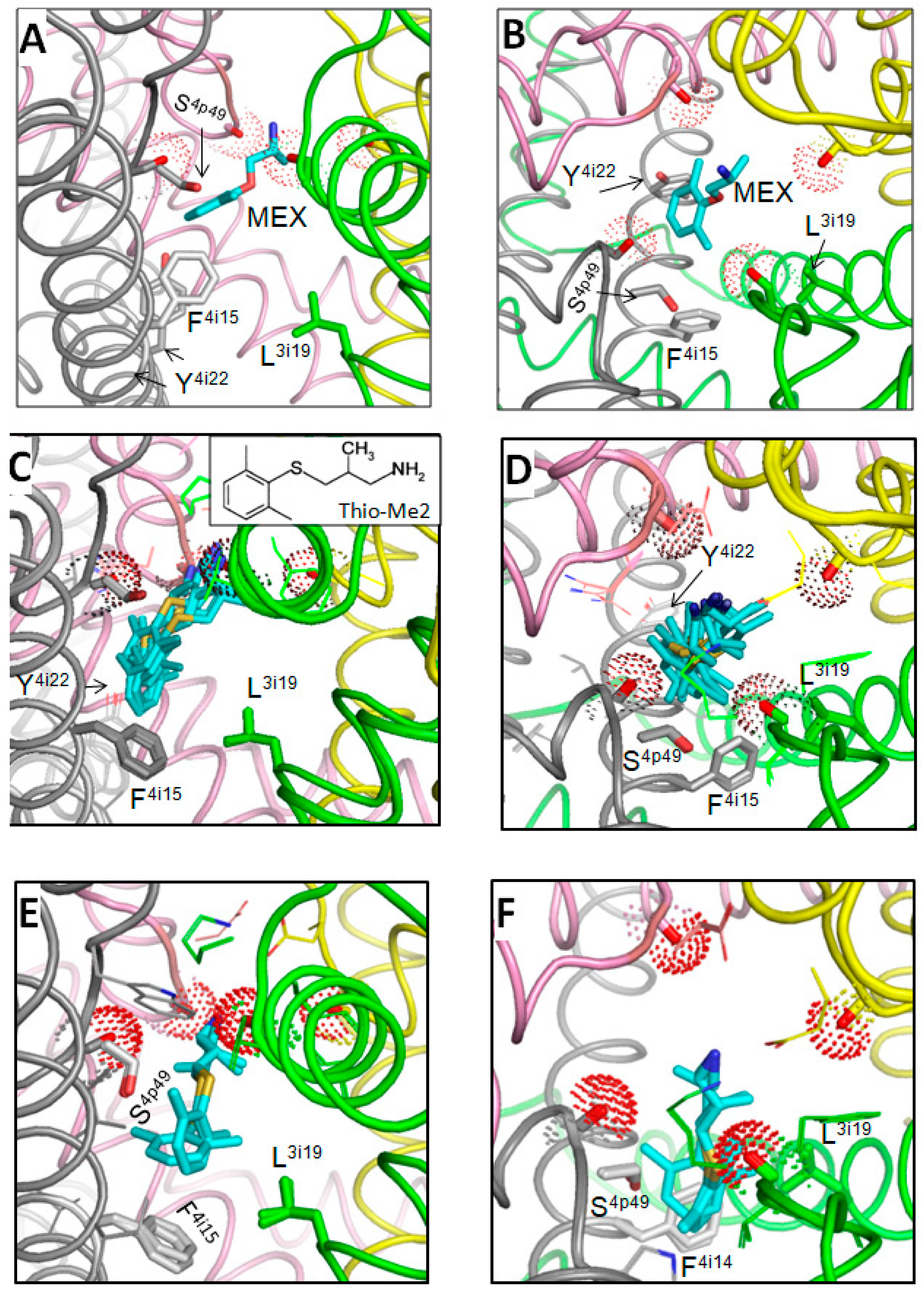
Computations yielded a stack with 100 MEX binding poses. Figure 1A,B show the side and extracellular views at the superposition of 50 lowest-energy structures with the ligand-channel interaction energy (enthalpy) within 10 kcal/mol from the apparent global minimum. Structures with the lowest energy include MEX binding poses, which resemble those suggested to represent TB and USB of LAs [16]. Figure 1C–F shows a low-energy structure, which may represent the TB binding pose of MEX. The ligand ammonium group was attracted by the backbone carbonyls of T3p48 and T4p48, which face the outer-pore bottom. The ligand contacts with backbones are hardly possible to test by mutational analysis. However, the same carbonyls attract the ammonium group of the DEKA lysine K3p50 in several cryo-EM strictures of Nav1.x channels, see [42] and references therein. In the cryo-EM structure of hNav1.5, K3p50 was in the “dunking” orientation, but during MC-minimizations its ammonium group was repelled by the MEX ammonium group and moved “up” to form a salt bridge with the DEKA glutamate E2p50. The aromatic group of MEX partially extended into the III/IV fenestration and formed favorable contacts with L3i19 and F4i15, whereas the middle part of the ligand interacted with S4p49. Mutations of these residues do affect the TB by MEX [19,21]. The TB binding pose of MEX shown in Figure 1E,F resembles that predicted by molecular dynamics simulations of MEX steered through the III/IV fenestration of the NavAb-based model of the hNav1.5 closed channel [43].
One of the “vertical” binding poses of MEX, which likely represent the UDB, is shown in Figure 2A,B. The primary ammonium group of MEX deeply penetrated into the outer pore and interacted with backbone carbonyls in positions p48. So deep penetration of the cationic group into the outer pore is hardly possible for the tertiary ammonium group of lidocaine or the quaternary ammonium group of QX-314. This may explain the peculiarities of pharmacological properties of MEX vs. those of lidocaine [44], which are further discussed at the end of this section. The aromatic ring of MEX bound to F4i15 and approached Y4i22, the residue which is important for UDB by some LAs [8,45]. Mutational data on MEX interaction with Y4i22 are unavailable. However, the fact that a MEX derivative with meta-hydroxyl in the aromatic ring is more potent than MEX [46] suggests that the hydroxyl may form an H-bond with Y4i22.
Neither TB nor USB binding poses correspond to those proposed in the primarily electrophysiological studies of MEX action on Nav1.5 [19,47]. However, the predicted TB binding pose is consistent with the experimental data [19] according to which mutation F4i15A decreased the TB by MEX, mutations N4i20A strongly enhanced it, and L3i19E somehow enhanced the TB. In the TB binding pose (Figure 1E,F) the aromatic group of MEX is engaged in stacking interactions with F4i15 that would be eliminated by mutation F4i15A. MEX interactions with L3i19 are rather weak, but in the L3i19E mutant they would be enhanced by attraction of the aromatic ring edge to the glutamate. Favorable aromatic-carboxylate interactions are revealed by the analysis of crystal structures of many proteins [48]. Mutation N4i15A enhanced the closed-channel block (TB), but decreased the open-channel block (UDB) by MEX [19]. This is consistent with the prediction that N4i15A would destabilize the open conformation of PD, but stabilize its closed conformation [49,50].
The data that mutation S4p49L enhanced the TB by MEX [21] is also consistent with the proposed TB binding pose where engineered S4p49L would provide additional hydrophobic contact to MEX (Figure 1E). In the proposed UDB binding pose, MEX interacts with F4i15 but it is rather far from L3i19 and S4p49 (Figure 2A,B). This is consistent with data that mutations F4i15A and N1i20A decreased UDB, while mutation L3i19F enhanced it [19]. The fact that mutation S4p49L slightly attenuated the UDB by MEX and accelerated recovery from the block is also consistent with the UDB model where MEX approaches S4p49. Mutation S4p49L would make the III/IV fenestration more hydrophobic and thus facilitate the drug egress through the fenestration.
A strong UDB and TB are demonstrated for Tio-Me2 [51] that has the elongated sulfur-containing chain between the aromatic ring and the amino group. Docking of Thio-Me2 yielded structures consistent with the UDB (Figure 2C,D) and TB (Figure 2E,F) binding poses of MEX. The longer and more hydrophobic Tio-Me2 would experience stronger interactions with the channel. As compared with the earlier models of Nav1.5-bound lidocaine and tetracaine, which have bulky tertiary amino group [16,17], the protonated primary amino group of MEX and Thio-Me2 penetrated deeper into the outer pore and occupied the site where the primary amino group of the dunked lysine is seen in cryo-EM structures of Nav1.x channel.
The peculiarities of MEX and lidocaine binding pose are consistent with biophysical characteristics of their action on hNav1.5 channels [44]. Thus, (i) both MEX and lidocaine enhanced the slow component of closed-state inactivation, (ii) MEX was a more potent inhibitor than lidocaine, (iii) the recovery from inactivation of Nav1.5 was significantly prolonged by MEX compared to lidocaine, and (iv) MEX displayed a stronger use-dependent inhibition of Nav1.5 than lidocaine. In view of the proposed model, the stronger block by MEX is due to the deeper penetration of the primary amino group into the outer pore as compared to the tertiary amino group in lidocaine. MEX would shift “dunked” DEKA lysine closer to the outer ring of carboxylates thus facilitating closure of the slow-inactivation gate. Due to the same reason, dissociation of the MEX ammonium group from the outer pore would be delayed resulting in delayer recovery from inactivation.

3.2. In Silico Deactivation of VSDIII

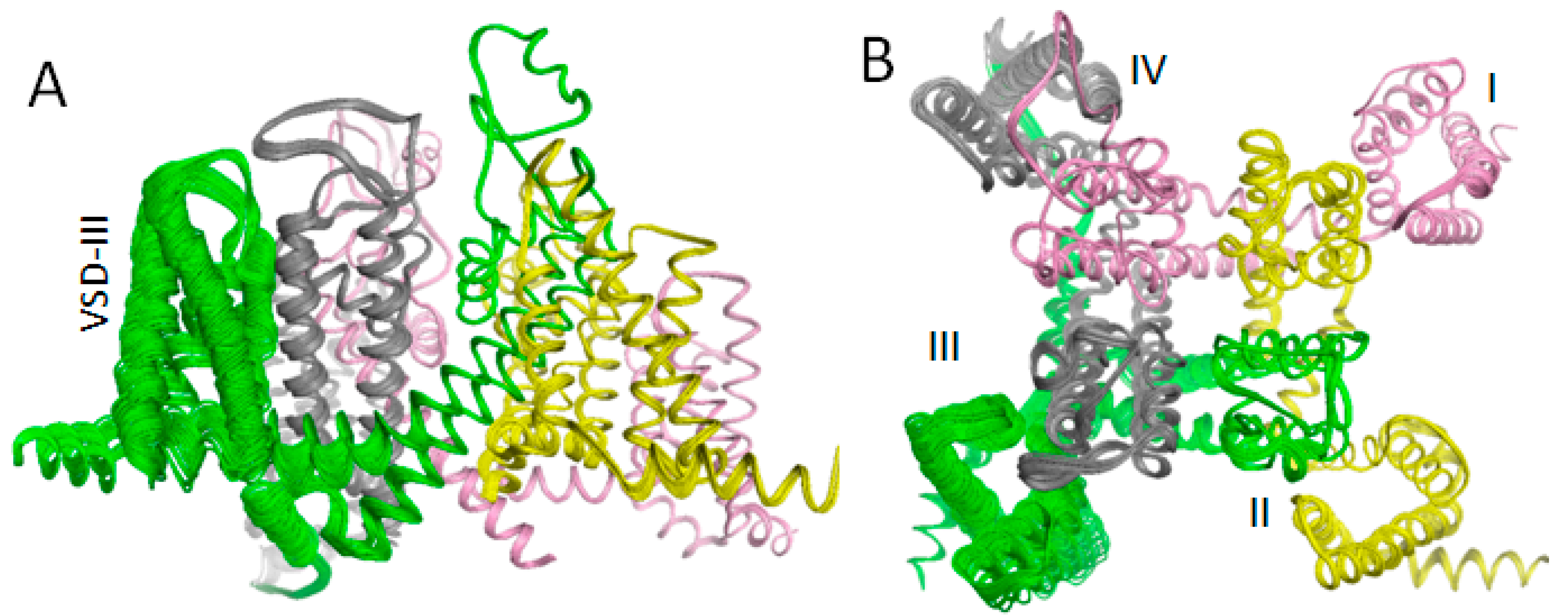
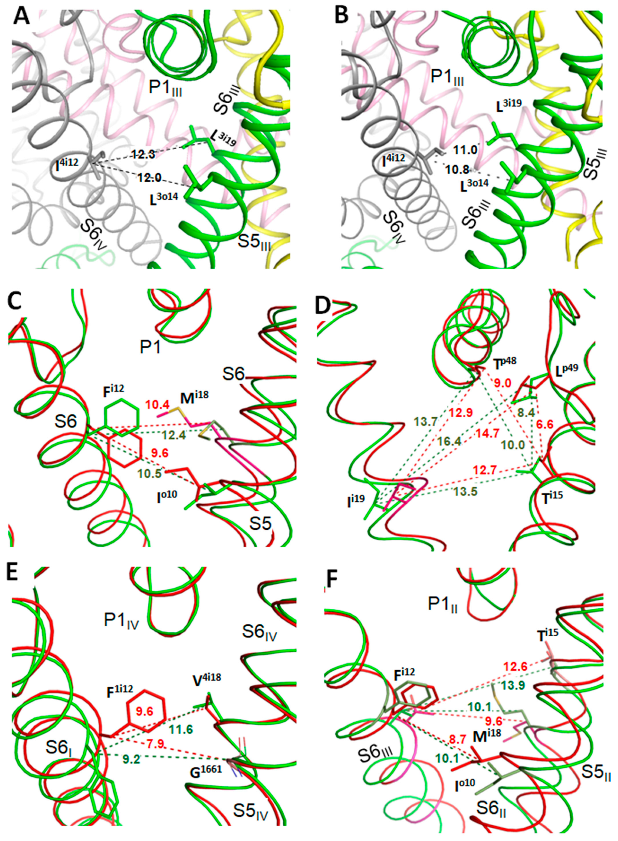
The above results support the earlier predicted binding poses of lidocaine and tetracaine that may correspond, respectively, to the UDB and TB in Nav1.5 [16]. The results also explain mutational data on MEX-sensing residues and biophysical characteristics of the channel block by MEX. However, the proposed models do not explain the paradoxical relations between VSDIII states and MEX action [20,52]. To address the problem, VSDIII was in silico deactivated (Figure 3). The forced downshift of S4III caused significant perturbations in other channel domains, particularly in PDIV, due to contacts of helices S4III and S4–S5III with helices S5IV and S6IV. Similar perturbations are observed upon in silico deactivation of VSDIII in a homology model of hNav1.5 [33] and AlphaFold2 model of the cockroach sodium channel [34]. In silico deactivation of VSDIII by downshifting S4III decreased the III/IV fenestration as compared with the cryo-EM structure. The fenestration is lined by several residues, including L3o14, L3i19 and I4i12 (Figure 4A,B). The distances between atom Cβ_I4i12 on one hand and atoms Cβ_L3o14 and Cβ_L3i19 on the other hand in the cryo-EM structure are over 1 Å bigger than in the model with in silico deactivated VSDIII.

3.3. State-Dependent Geometry of Fenestration in Experimental Structures

Similar tendency of widening fenestrations upon activation of VSDs are seen upon comparison of experimental structures of other P-loop channels. Thus, the NavAb channel with VSDs in the resting state is stabilized by engineered disulfide bridges between G94C and Q150C [22]. Figure 4C,D show that in the crystal structure with VSDs in the activated state (PDB ID: 6p6y), the distances between Cβ atoms of residues, which line the fenestrations, are up to 2 Å larger than in the crystal structure with VSDs in the resting state (PDB ID: 6p6w). In the rat Nav1.5 channel, VSDIV was trapped in the resting state by the deathstalker scorpion toxin [2]. Figure 4E shows that in the cryo-EM structure of the channel in the apo-state (PDB ID: 6uz3), the distance between Cβ atoms of residues F1i12/399 and V4i18/1765 at the opposite sides of fenestration I/IV, is also 2 Å larger than in the structure with the resting VSDIV (PDB ID: 7k18).
VSDs in the homotetrameric chimera channel NavAb/Nav1.7-VS2 with the pore domain from NavAb and VSDs from Nav1.7_VSDII are trapped in the resting state by tarantula toxin m3-Huwentoxin-IV [53]. Distances between Cβ atoms in diametrically opposed residues of the fenestration decrease upon deactivation of VSDs (Figure 4F). Cryo-EM structures of the NavAb-Nav1.7_VSDII chimera in complexes with the gating modifier toxin ProTx2 bound to the activated and resting VSDII [23] also show significant narrowing of the fenestrations upon VSD deactivation.
Different-width fenestrations in experimental structures of the same channel are likely due to the same mechanisms as seen upon in silico deactivating hNav1.5: contacts of S4–S5 linkers with S5 and S6 helices in the subunit/repeat interface and some downshift of helices S5, which are linked to the S4–S5 helices. The fact that the activated VSDIII of Nav1.x channels is stabilized by lidocaine [54,55] or MEX [20], whereas bound ligands stabilize VSDIII in the activated state suggests that the states with the activated VSDIII and wider III/IV fenestration are more complementary to the ligands than those with deactivated VSDIII.

3.4. Hydrophobic Access Pathway to the Nav1.5 Inner Pore through III/IV Fenestrations Is Paved by Aromatic and Polar Residues

To achieve the TB block of the resting Nav1.5 channel, LAs are proposed to move through the III/IV fenestration, approach the selectivity filter by the ammonium group and remain in the horizontal position [16]. Such binding pose of ligands in the resting channel is implied [56] and modeled [57] for LAs in the closed NavMs channel and for lidocaine in the NavMs-based model of the Nav1.4 channel [17]. More recently, such a binding pose is shown in the crystal structure of flecainide-bound closed NavAb channel [18] where the ammonium nitrogen of the drug closely approaches the backbone carbonyl in position p48, whereas the trifluoromethyl group approaches Ti15 on one side of the fenestration and Mi18 on the opposite side.
Access of cationic drugs such as MEX and lidocaine to the inner pore of the Nav1.x channels through the membrane likely involves deprotonation of the ammonium group and subsequent re-protonation in the cytoplasm or within the pore. However, the permanently charged QX-314 moving through entirely hydrophobic path would encounter a large energy barrier. The fact that mutation S4p49L strongly increases TB, attenuates UDB and accelerates recovery from the block by MEX and QX-314 suggests the drugs motion through the III/IV fenestration and lipid-channel interface is accelerated [21]. The TB acceleration by S4p49L indicates that the ligand motion through the membrane is not the rate-limiting step for the ligand access, implying that polar or aromatic residues in the lipid-channel interface would provide interim binding sites for the ligand cationic group. The types of lipids that solvate the Nav1.5 channel are unknown, but the channel cryo-EM structure allows visualizing the channel residues, which face the lipids.
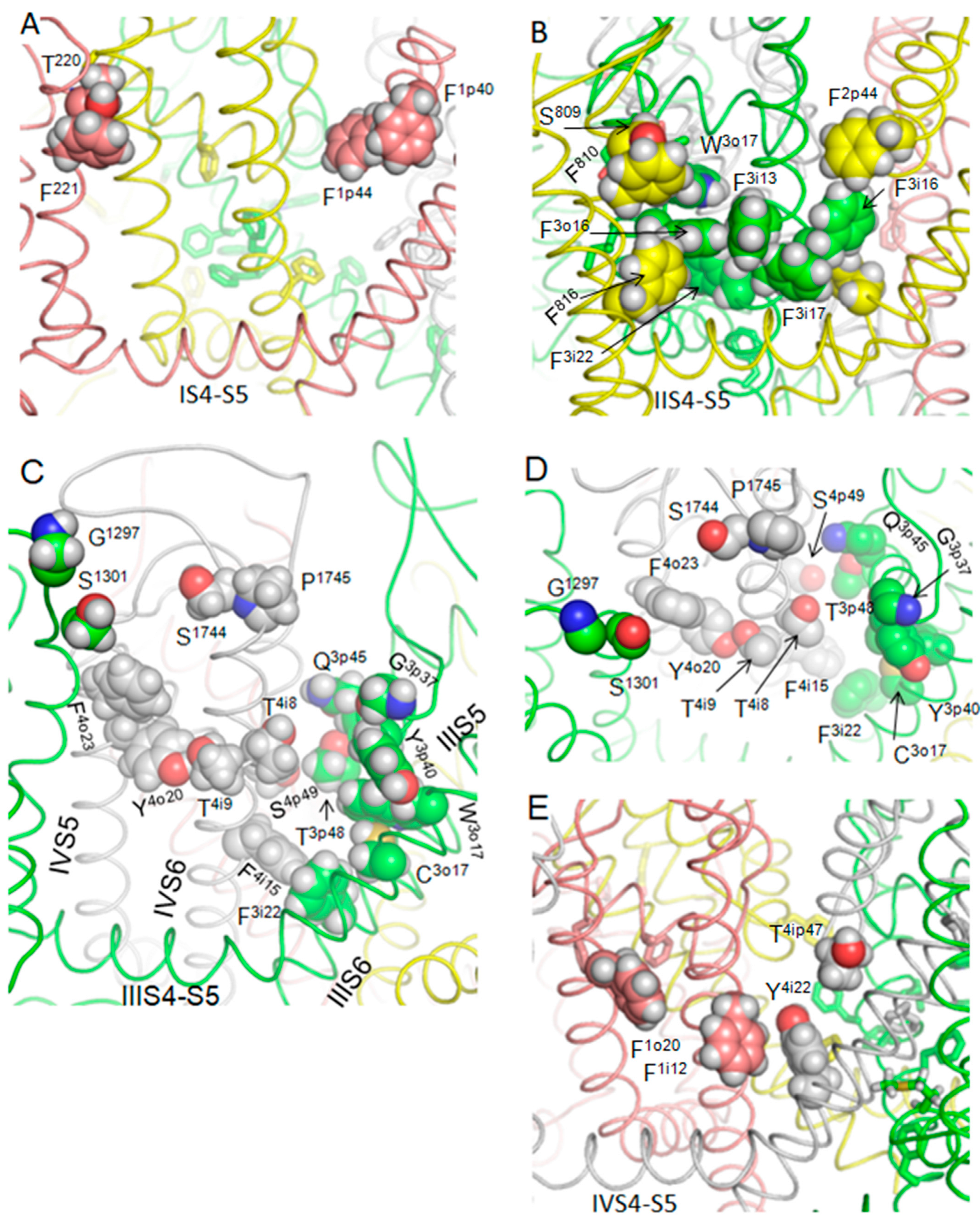
Figure 5 shows lipid-exposed sides of the four fenestrations where polar and aromatic residues could provide interim binding sites for the ligand ammonium group. The lipid-exposed side of fenestration II/III has nine aromatic residues (Figure 5B), whereas the lipid-exposed side of fenestration III/IV has six aromatic residues and seven polar residues (serines, threonines and glutamine). Besides, the backbone oxygens of glycines G1247 and G3p37, the sidechain nitrogen in P1745, and the sulfur atom in C3o17 may also attract the permanently charged ammonium group of QX-314 and protonatable amino groups of other ligands. This analysis suggests that the cationic ligands may move from the extracellular space towards the inner pore and III/IV fenestration by hopping through multiple interim binding sites where the ammonium group would be attracted by electronegative atoms and π-electrons in aromatic rings. It should be noted that due to limitations of the MCM methodology, lipid and water molecules were not included in the model. Interactions with lipids may affect side chain conformations of some lipid-exposed residues. However, most of these residues would enjoy interaction with both lipids and hydrophobic moieties of the ligands, whereas aromatic groups and polar atoms in the III/IV fenestration would attract the ligand ammonium group.

3.5. State-Dependent Energy Barrier for TB-Bound MEX Egress through Fenestration III/IV

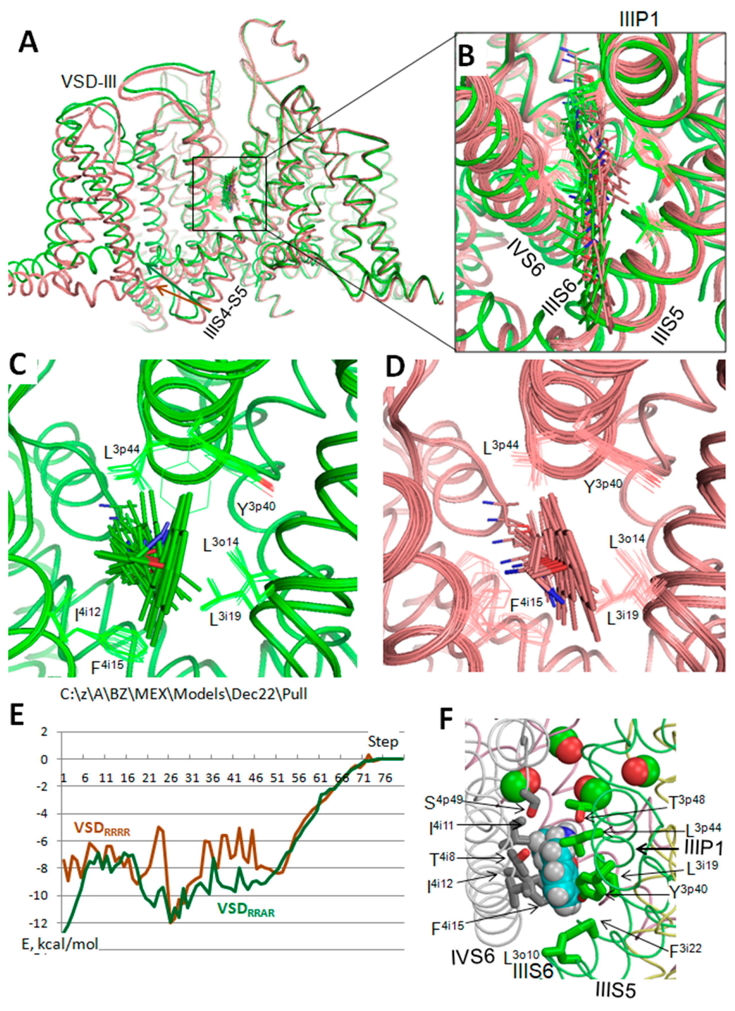
The above analysis illustrates that the III/IV fenestration would provide the access pathway for the ingress and egress of cationic ligands to/from the closed-channel pore. However, it does not explain how activation of VSDIII may affect the ligand motion through the fenestration. To address this problem, the cryo-EM structure of hNav1.5 was in silico transformed to yield model VSDRRRR with four VSDs in the resting state and model VSDRRAR with three VSDs in the resting state and VSDIII in the activated state. The MEX TB pose (Figure 1) was reproduced in the two models and the ligand was pulled away the pore through the III/IV fenestration by increasing the distance between the pore axis and the para carbon of the ligand aromatic ring. The distance was increased with the step of 0.4 Å and at each step the energy was MC-minimized.
The channel folding was preserved by pin constraints, which allow Cα atoms to deviate penalty-free up to 1 Å from the starting position (see Methods). At each step, the distance between the MEX para-carbon and the pore axis was constrained. The constraint did not preclude the para-carbon motion over the surface of the sphere whose center was free to translate along the pore axis. In other words, this constraint did not restrict the ligand mobility at the given separation from the pore axis. All side chain torsions and MEX torsion and bond angles were allowed to vary during MC-minimizations. Such setting allowed the flexible MEX molecule to find the energetically optimal egress path, which could be curved. It should be noted that the MEX egress from rather than ingress into the pore was modeled because the starting point for the ingress is undefined.
Figure 6A,B show superposed models VSDRRRR (brown) and VSDRRAR (green) with 80 steps of MEX during which the para-carbon moved 32 Å away the pore axis from the TB pose. In both models, the egress paths were rather straight but not identical (Figure 6B). The MEX egress in model VSDRRAR did not cause significant deviations of the channel sidechains, including MEX-sensing F4i15 and L3i19 (Figure 6C). In contrast, the egress of MEX through model VSDRRRR caused significant motion of the F4i15 side chain (Figure 6D). The plots of MEX-channel energy (Figure 6E) show that the energy of the starting point (TB pose) in model VSDRRAR is ~5 kcal/mol more preferable than in model VSDRRRR, explaining why activation of VSDIII stabilizes binding of MEX, whereas TB-bound MEX stabilizes VSDIII in the activated state [20].
Both models show a minimum of MEX-channel energy at step 27. In model VSDRRAR this is a local energy minimum, which is separated from the global minimum at the TB site by the energy barrier of ~5 kcal/mol. In model VSDRRRR the step 27 corresponds to the global energy minimum along the MEX egress path, which is separated from the TB site by the energy barrier of ~7 kcal/mol. The results suggest that while access of MEX to the closed/inactivated channel with activated VSDIII is possible, MEX targeting the closed channel with all VSDs in the resting state is more restricted, but MEX may remain in the interim site at the pathway midpoint until VSDIII activates.
Figure 6F shows MEX in model VSDRRAR midway from the TB binding site to the lipids, at step 27 corresponding to the energy minimum. Residues within 4 Å from the MEX are shown by sticks and backbone carbonyls at the outer-pore bottom are shown by spheres. Besides residues F4i15, L3i19 and S4p49, which are known to affect the TB of MEX, the midway site involves other residues, in particular L3o10, L3i19, F3i22, Y3p40, L3p44, T4i8, I4i11 and I4i12. Mutational analysis of these residues is beyond the goals of this theoretical study.

3.6. Relations between the Half-Voltage of VSDIII Activation (aV0.5) and TB by MEX

In a set of Nav1.5 disease variants, aV0.5 of VSDIII correlates with TB by MEX [20]: the easier is VSDIII activation, the stronger is the TB block. Many of the disease mutations are located in S4IV. The fact that these mutations affect the voltage-dependent activation of VSDIII seems surprising. The general explanation is suggested by in silico deactivation of VSDIII, which disturbs VSDIV (Figure 3) due to strong hydrophobic contacts of S4III with S5IV, and contacts of S4–S5III with S5IV and S6IV. Similar contacts are described for hNav1. 5 [33] and for an insect sodium channel [34]. On the other hand, contacts with S5IV and S6IV may affect motion of S4–S5III and S4III. Structural rationale for the observed decrease in TB by MEX in several disease variants of Nav1.5 [20] are proposed below.
Variant R1626P at the extracellular half of S4IV demonstrates the maximal normalized TB of 0.54 and aV0.5 of −165 mV. In the cryo-EM structure of hNav1.5, R1626 forms a salt bridge with E1548 in loop S1–S2IV. The salt bridge stabilizes the ”up” state of helix S4IV and thus restricts its motion. Mutation R1626P would eliminate the salt bridge and facilitate the motion of S4IV and S4–S5IV, thus facilitating activation of S4III. The activated VSDIII would widen the III/IV fenestration and strengthen TB by MEX.
Variant R1623Q, one helical turn above R1626, also demonstrates rater strong normalized TB of 0.307. In the cryo-EM structure, R1623 forms inter-repeat contact with M273/1o29 at the C-end of helix S5I. The electrostatic attraction between the guanidinium group of arginine and the sulfur atom of methionine would also stabilize the “up” conformation of S4III, but this attraction is weaker than the salt bridge R1626----E1548.
On the opposite end of the correlation curve TB vs. aV0.5 [20], variant M1652/4o1R at the N-end of S5IV has the minimal normalized TB by MEX of 0.175 and aV0.5 of −102.1 mV. Substitution of M4o1R may attract the arginine and S5IV towards the negatively charged inner leaflet of the membrane. Through strong hydrophobic contacts of S5IV with S4III and S4–S5III, this motion would shift down the later helices. This would restrain the outward shift of S4III, activation of VSDIII and keep fenestration III/IV in the narrower state, which disfavors TB.
VSDIV regulates the onset of fast inactivation, while VSDIII determines its recovery [58]. Mutation R1626P in S4IV most strongly decelerates inactivation [59]. In the cryo-EM structures of hNav1.5, R1626 forms close contacts with M273 and N275 in loop S5-P1I, and it is engaged in electrostatic interactions with D1550 in loop S1–S2IV. Mutation R1626P eliminates these interactions, which stabilize the activated state of S4IV in agreement with the data that activation of S4IV is necessary for fast inactivation [60,61].

3.7. Limitations of the Study

In silico transformation of cryo-EM structures yields models that are less precise than experimental structures. Among multiple low-energy binding poses found for USB and TB by MEX, shown only those where ligands form contacts with experimentally known MEX-sensing residues. Energy profiles of MEX pulled through the III/IV fenestration are just approximate estimates of the enthalpy contribution to the free energy. Nevertheless, the results, which are obtained without biasing contacts of the ligand with specific residues, are consistent with multiple experimental data.

4. Conclusions

Computations suggest that MEX blocks the ion permeation by its ammonium group that competes with sodium ions for the binding site at the outer-pore bottom where four pore-facing backbone carbonyls create a cation-attractive site. The predicted MEX binding modes are consistent with available experimental studies that identified several MEX-sensing residues in hNav1.5. In silico deactivation of VSDIII tightens the III/IV fenestration, likely decreasing the TB site affinity to MEX. This may explain the paradoxical data that the channel block by MEX increases with activation of VSDIII, whereas bound MEX stabilizes the activated state of VSDIII. Extracellularly applied MEX would reach its binding site in the closed channel by hopping over lipid-exposed polar and aromatic residues in the III/IV domain interface. Along this way, a low-energy interim binding site for MEX is predicted with eleven residues from S5III, P1III, S6III, P1IV and S6IV.

Funding

This research was funded by grants to B.S.Z. from the Natural Sciences and Engineering Research Council of Canada (RGPIN-2020-07100) and the Russian Science Foundation (22-15-00186).

Acknowledgments

Computations were performed using facilities provided by Compute Ontario (https://www.computeontario.ca/) and the Digital Research Alliance of Canada (https://www.alliancecan.ca).

Conflicts of Interest

The author declares no conflict of interest.

Abbreviations

LA, local anesthetic; MCM, Monte Carlo minimizations; MEX, Mexiletine; Nav1.5, cardiac voltage-gated sodium channel; PD, Pore domain; TB, Tonic block; UDB, Use-dependent bock; VSD, Voltage-sensing domain.

References

  1. Rudy, Y.; Silva, J.R. Computational biology in the study of cardiac ion channels and cell electrophysiology. Q. Rev. Biophys. 2006, 39, 57–116. [Google Scholar] [CrossRef] [PubMed] [Green Version]
  2. Jiang, D.; Tonggu, L.; Gamal El-Din, T.M.; Banh, R.; Pomes, R.; Zheng, N.; Catterall, W.A. Structural basis for voltage-sensor trapping of the cardiac sodium channel by a deathstalker scorpion toxin. Nat. Commun. 2021, 12, 128. [Google Scholar] [CrossRef] [PubMed]
  3. Jiang, D.; Shi, H.; Tonggu, L.; Gamal El-Din, T.M.; Lenaeus, M.J.; Zhao, Y.; Yoshioka, C.; Zheng, N.; Catterall, W.A. Structure of the Cardiac Sodium Channel. Cell 2020, 180, 122–134.e110. [Google Scholar] [CrossRef] [PubMed]
  4. Li, Z.; Jin, X.; Wu, T.; Huang, G.; Wu, K.; Lei, J.; Pan, X.; Yan, N. Structural Basis for Pore Blockade of the Human Cardiac Sodium Channel Nav 1.5 by the Antiarrhythmic Drug Quinidine*. Angew. Chem. Int. Ed. 2021, 60, 11474–11480. [Google Scholar] [CrossRef]
  5. Pan, X.; Li, Z.; Jin, X.; Zhao, Y.; Huang, G.; Huang, X.; Shen, Z.; Cao, Y.; Dong, M.; Lei, J.; et al. Comparative structural analysis of human Na(v)1.1 and Na(v)1.5 reveals mutational hotspots for sodium channelopathies. Proc. Natl. Acad. Sci. USA 2021, 118, e2100066118. [Google Scholar] [CrossRef]
  6. Jumper, J.; Evans, R.; Pritzel, A.; Green, T.; Figurnov, M.; Ronneberger, O.; Tunyasuvunakool, K.; Bates, R.; Zidek, A.; Potapenko, A.; et al. Highly accurate protein structure prediction with AlphaFold. Nature 2021, 596, 583–589. [Google Scholar] [CrossRef]
  7. Hille, B. Ion Channels of Excitable Membranes, 3rd ed.; Sinauer Associates, Inc.: Sunderlamd, MA, USA, 2001; p. 814. [Google Scholar]
  8. Ragsdale, D.S.; McPhee, J.C.; Scheuer, T.; Catterall, W.A. Molecular determinants of state-dependent block of Na+ channels by local anesthetics. Science 1994, 265, 1724–1728. [Google Scholar] [CrossRef]
  9. Ragsdale, D.S.; McPhee, J.C.; Scheuer, T.; Catterall, W.A. Common molecular determinants of local anesthetic, antiarrhythmic, and anticonvulsant block of voltage-gated Na+ channels. Proc. Natl. Acad. Sci. USA 1996, 93, 9270–9275. [Google Scholar] [CrossRef] [Green Version]
  10. Wang, G.K.; Quan, C.; Wang, S.Y. Local anesthetic block of batrachotoxin-resistant muscle Na+ channels. Mol. Pharmacol. 1998, 54, 389–396. [Google Scholar] [CrossRef] [Green Version]
  11. Wang, G.K.; Russell, C.; Wang, S.Y. Mexiletine block of wild-type and inactivation-deficient human skeletal muscle hNav1.4 Na+ channels. J. Physiol. 2004, 554, 621–633. [Google Scholar] [CrossRef]
  12. Yarov-Yarovoy, V.; Brown, J.; Sharp, E.M.; Clare, J.J.; Scheuer, T.; Catterall, W.A. Molecular determinants of voltage-dependent gating and binding of pore-blocking drugs in transmembrane segment IIIS6 of the Na(+) channel alpha subunit. J. Biol. Chem. 2001, 276, 20–27. [Google Scholar] [CrossRef] [PubMed] [Green Version]
  13. Yarov-Yarovoy, V.; McPhee, J.C.; Idsvoog, D.; Pate, C.; Scheuer, T.; Catterall, W.A. Role of amino acid residues in transmembrane segments IS6 and IIS6 of the Na+ channel alpha subunit in voltage-dependent gating and drug block. J. Biol. Chem. 2002, 277, 35393–35401. [Google Scholar] [CrossRef] [PubMed] [Green Version]
  14. Ahern, C.A.; Eastwood, A.L.; Dougherty, D.A.; Horn, R. Electrostatic contributions of aromatic residues in the local anesthetic receptor of voltage-gated sodium channels. Circ. Res. 2008, 102, 86–94. [Google Scholar] [CrossRef] [PubMed] [Green Version]
  15. Hille, B. Local anesthetics: Hydrophilic and hydrophobic pathways for the drug-receptor reaction. J. Gen. Physiol. 1977, 69, 497–515. [Google Scholar] [CrossRef] [PubMed] [Green Version]
  16. Bruhova, I.; Tikhonov, D.B.; Zhorov, B.S. Access and binding of local anesthetics in the closed sodium channel. Mol. Pharmacol. 2008, 74, 1033–1045. [Google Scholar] [CrossRef] [PubMed]
  17. Tikhonov, D.B.; Zhorov, B.S. Mechanism of sodium channel block by local anesthetics, antiarrhythmics, and anticonvulsants. J. Gen. Physiol. 2017, 149, 465–481. [Google Scholar] [CrossRef] [Green Version]
  18. Gamal El-Din, T.M.; Lenaeus, M.J.; Zheng, N.; Catterall, W.A. Fenestrations control resting-state block of a voltage-gated sodium channel. Proc. Natl. Acad. Sci. USA 2018, 115, 13111–13116. [Google Scholar] [CrossRef] [Green Version]
  19. Nakagawa, H.; Munakata, T.; Sunami, A. Mexiletine Block of Voltage-Gated Sodium Channels: Isoform- and State-Dependent Drug-Pore Interactions. Mol. Pharmacol. 2019, 95, 236–244. [Google Scholar] [CrossRef] [Green Version]
  20. Zhu, W.; Mazzanti, A.; Voelker, T.L.; Hou, P.; Moreno, J.D.; Angsutararux, P.; Naegle, K.M.; Priori, S.G.; Silva, J.R. Predicting Patient Response to the Antiarrhythmic Mexiletine Based on Genetic Variation. Circ. Res. 2019, 124, 539–552. [Google Scholar] [CrossRef]
  21. Sasaki, K.; Makita, N.; Sunami, A.; Sakurada, H.; Shirai, N.; Yokoi, H.; Kimura, A.; Tohse, N.; Hiraoka, M.; Kitabatake, A. Unexpected mexiletine responses of a mutant cardiac Na+ channel implicate the selectivity filter as a structural determinant of antiarrhythmic drug access. Mol. Pharmacol. 2004, 66, 330–336. [Google Scholar] [CrossRef]
  22. Wisedchaisri, G.; Tonggu, L.; McCord, E.; Gamal El-Din, T.M.; Wang, L.; Zheng, N.; Catterall, W.A. Resting-State Structure and Gating Mechanism of a Voltage-Gated Sodium Channel. Cell 2019, 178, 993–1003.e12. [Google Scholar] [CrossRef] [PubMed]
  23. Xu, H.; Li, T.; Rohou, A.; Arthur, C.P.; Tzakoniati, F.; Wong, E.; Estevez, A.; Kugel, C.; Franke, Y.; Chen, J.; et al. Structural Basis of Nav1.7 Inhibition by a Gating-Modifier Spider Toxin. Cell 2019, 176, 1238–1239. [Google Scholar] [CrossRef] [PubMed] [Green Version]
  24. Garden, D.P.; Zhorov, B.S. Docking flexible ligands in proteins with a solvent exposure- and distance-dependent dielectric function. J. Comput. Mol. Des. 2010, 24, 91–105. [Google Scholar] [CrossRef] [PubMed]
  25. Tikhonov, D.B.; Zhorov, B.S. Architecture and pore block of eukaryotic voltage-gated sodium channels in view of NavAb bacterial sodium channel structure. Mol. Pharmacol. 2012, 82, 97–104. [Google Scholar] [CrossRef] [PubMed] [Green Version]
  26. Li, Z.; Scheraga, H.A. Monte Carlo-minimization approach to the multiple-minima problem in protein folding. Proc. Natl. Acad. Sci. USA 1987, 84, 6611–6615. [Google Scholar] [CrossRef] [PubMed] [Green Version]
  27. Weiner, S.J.; Kollman, P.A.; Case, D.A.; Singh, U.C.; Chio, C.; Alagona, G.; Profeta, S.; Weiner, P.K. A new force field for molecular mechanical simulation of nucleic acids and proteins. J. Am. Chem. Soc. 1984, 106, 765–784. [Google Scholar] [CrossRef]
  28. Weiner, S.J.; Kollman, P.A.; Nguyen, D.T.; Case, D.A. An All Atom Force-Field for Simulations of Proteins and Nucleic-Acids. J. Comput. Chem. 1986, 7, 230–252. [Google Scholar] [CrossRef]
  29. Dewar, M.J.S.; Zoebisch, E.G.; Healy, E.F.; Stewart, J.J.P. AM1: A New General Purpose Quantum Mechanical Model. J. Am. Chem. Soc. 1985, 107, 3902–3909. [Google Scholar] [CrossRef]
  30. Brooks, C.L.; Pettitt, M.; Karplus, M. Structural and energetic effects of truncating long ranged interactions in ionic and polar fluids. J. Chem. Phys. 1985, 83, 5897–5908. [Google Scholar] [CrossRef]
  31. De Luca, A.; Natuzzi, F.; Lentini, G.; Franchini, C.; Tortorella, V.; Conte Camerino, D. Stereoselective effects of mexiletine enantiomers on sodium currents and excitability characteristics of adult skeletal muscle fibers. Naunyn Schmiedebergs Arch. Pharmacol. 1995, 352, 653–661. [Google Scholar] [CrossRef]
  32. Zhorov, B.S.; Du, Y.; Song, W.; Luo, N.; Gordon, D.; Gurevitz, M.; Dong, K. Mapping the interaction surface of scorpion beta-toxins with an insect sodium channel. Biochem. J. 2021, 478, 2843–2869. [Google Scholar] [CrossRef] [PubMed]
  33. Zaytseva, A.K.; Boitsov, A.S.; Kostareva, A.A.; Zhorov, B.S. Possible Interactions of Extracellular Loop IVP2-S6 With Voltage-Sensing Domain III in Cardiac Sodium Channel. Front. Pharmacol. 2021, 12, 742508. [Google Scholar] [CrossRef] [PubMed]
  34. Gurevitz, M.; Zhorov, B.S.; Dong, K. Allosteric interactions among voltage-sensor modules of sodium channels probed by scorpion toxin modifiers. J. Neurobiol. Physiol. 2022, 4, 9–12. [Google Scholar]
  35. Zhorov, B.S.; Tikhonov, D.B. Potassium, sodium, calcium and glutamate-gated channels: Pore architecture and ligand action. J. Neurochem. 2004, 88, 782–799. [Google Scholar] [CrossRef]
  36. Lenaeus, M.J.; Gamal El-Din, T.M.; Ing, C.; Ramanadane, K.; Pomes, R.; Zheng, N.; Catterall, W.A. Structures of closed and open states of a voltage-gated sodium channel. Proc. Natl. Acad. Sci. USA 2017, 114, E3051–E3060. [Google Scholar] [CrossRef] [Green Version]
  37. Tikhonov, D.B.; Zhorov, B.S. Predicting Structural Details of the Sodium Channel Pore Basing on Animal Toxin Studies. Front. Pharmacol. 2018, 9, 880. [Google Scholar] [CrossRef] [Green Version]
  38. Long, S.B.; Tao, X.; Campbell, E.B.; MacKinnon, R. Atomic structure of a voltage-dependent K+ channel in a lipid membrane-like environment. Nature 2007, 450, 376–382. [Google Scholar] [CrossRef]
  39. Bruhova, I.; Zhorov, B.S. A homology model of the pore domain of a voltage-gated calcium channel is consistent with available SCAM data. J. Gen. Physiol. 2010, 135, 261–274. [Google Scholar] [CrossRef]
  40. Du, Y.; Nomura, Y.; Satar, G.; Hu, Z.; Nauen, R.; He, S.Y.; Zhorov, B.S.; Dong, K. Molecular evidence for dual pyrethroid-receptor sites on a mosquito sodium channel. Proc. Natl. Acad. Sci. USA 2013, 110, 11785–11790. [Google Scholar] [CrossRef] [Green Version]
  41. Tikhonov, D.B.; Bruhova, I.; Zhorov, B.S. Atomic determinants of state-dependent block of sodium channels by charged local anesthetics and benzocaine. FEBS Lett. 2006, 580, 6027–6032. [Google Scholar] [CrossRef] [Green Version]
  42. Zhorov, B.S. Possible Mechanism of Ion Selectivity in Eukaryotic Voltage-Gated Sodium Channels. J. Phys. Chem. B 2021, 125, 2074–2088. [Google Scholar] [CrossRef]
  43. O’Reilly, A.O.; Eberhardt, E.; Weidner, C.; Alzheimer, C.; Wallace, B.A.; Lampert, A. Bisphenol A binds to the local anesthetic receptor site to block the human cardiac sodium channel. PLoS ONE 2012, 7, e41667. [Google Scholar] [CrossRef] [PubMed]
  44. Wang, Y.; Mi, J.; Lu, K.; Lu, Y.; Wang, K. Comparison of Gating Properties and Use-Dependent Block of Nav1.5 and Nav1.7 Channels by Anti-Arrhythmics Mexiletine and Lidocaine. PLoS ONE 2015, 10, e0128653. [Google Scholar] [CrossRef] [PubMed] [Green Version]
  45. Li, H.L.; Galue, A.; Meadows, L.; Ragsdale, D.S. A molecular basis for the different local anesthetic affinities of resting versus open and inactivated states of the sodium channel. Mol. Pharmacol. 1999, 55, 134–141. [Google Scholar] [CrossRef] [PubMed]
  46. Catalano, A.; Budriesi, R.; Bruno, C.; Di Mola, A.; Defrenza, I.; Cavalluzzi, M.M.; Micucci, M.; Carocci, A.; Franchini, C.; Lentini, G. Searching for new antiarrhythmic agents: Evaluation of meta-hydroxymexiletine enantiomers. Eur. J. Med. Chem. 2013, 65, 511–516. [Google Scholar] [CrossRef] [PubMed]
  47. Li, G.; Woltz, R.L.; Wang, C.Y.; Ren, L.; He, P.X.; Yu, S.D.; Liu, X.Q.; Yarov-Yarovoy, V.; Hu, D.; Chiamvimonvat, N.; et al. Gating Properties of Mutant Sodium Channels and Responses to Sodium Current Inhibitors Predict Mexiletine-Sensitive Mutations of Long QT Syndrome 3. Front. Pharmacol. 2020, 11, 1182. [Google Scholar] [CrossRef]
  48. Philip, V.; Harris, J.; Adams, R.; Nguyen, D.; Spiers, J.; Baudry, J.; Howell, E.E.; Hinde, R.J. A survey of aspartate-phenylalanine and glutamate-phenylalanine interactions in the protein data bank: Searching for anion-pi pairs. Biochemistry 2011, 50, 2939–2950. [Google Scholar] [CrossRef]
  49. Tikhonov, D.B.; Bruhova, I.; Garden, D.P.; Zhorov, B.S. State-dependent inter-repeat contacts of exceptionally conserved asparagines in the inner helices of sodium and calcium channels. Pflug. Arch. Eur. J. Physiol. 2015, 467, 253–266. [Google Scholar] [CrossRef]
  50. Du, Y.; Tikhonov, D.B.; Nomura, Y.; Dong, K.; Zhorov, B.S. Mutational analysis of state-dependent contacts in the pore module of eukaryotic sodium channels. Arch. Biochem. Biophys. 2018, 652, 59–70. [Google Scholar] [CrossRef]
  51. De Luca, A.; Talon, S.; De Bellis, M.; Desaphy, J.F.; Franchini, C.; Lentini, G.; Catalano, A.; Corbo, F.; Tortorella, V.; Conte-Camerino, D. Inhibition of skeletal muscle sodium currents by mexiletine analogues: Specific hydrophobic interactions rather than lipophilia per se account for drug therapeutic profile. Naunyn Schmiedebergs Arch. Pharmacol. 2003, 367, 318–327. [Google Scholar] [CrossRef]
  52. Muroi, Y.; Chanda, B. Local anesthetics disrupt energetic coupling between the voltage-sensing segments of a sodium channel. J. Gen. Physiol. 2009, 133, 1–15. [Google Scholar] [CrossRef] [PubMed]
  53. Wisedchaisri, G.; Tonggu, L.; Gamal El-Din, T.M.; McCord, E.; Zheng, N.; Catterall, W.A. Structural Basis for High-Affinity Trapping of the NaV1.7 Channel in Its Resting State by Tarantula Toxin. Mol. Cell 2021, 81, 38–48.e4. [Google Scholar] [CrossRef] [PubMed]
  54. Sheets, M.F.; Hanck, D.A. Molecular action of lidocaine on the voltage sensors of sodium channels. J. Gen. Physiol. 2003, 121, 163–175. [Google Scholar] [CrossRef] [PubMed] [Green Version]
  55. Arcisio-Miranda, M.; Muroi, Y.; Chowdhury, S.; Chanda, B. Molecular mechanism of allosteric modification of voltage-dependent sodium channels by local anesthetics. J. Gen. Physiol. 2010, 136, 541–554. [Google Scholar] [CrossRef] [PubMed] [Green Version]
  56. Bagneris, C.; DeCaen, P.G.; Naylor, C.E.; Pryde, D.C.; Nobeli, I.; Clapham, D.E.; Wallace, B.A. Prokaryotic NavMs channel as a structural and functional model for eukaryotic sodium channel antagonism. Proc. Natl. Acad. Sci. USA 2014, 111, 8428–8433. [Google Scholar] [CrossRef] [PubMed] [Green Version]
  57. Korkosh, V.S.; Zhorov, B.S.; Tikhonov, D.B. Modeling interactions between blocking and permeant cations in the NavMs channel. Eur. J. Pharmacol. 2016, 780, 188–193. [Google Scholar] [CrossRef] [PubMed]
  58. Hsu, E.J.; Zhu, W.; Schubert, A.R.; Voelker, T.; Varga, Z.; Silva, J.R. Regulation of Na(+) channel inactivation by the DIII and DIV voltage-sensing domains. J. Gen. Physiol. 2017, 149, 389–403. [Google Scholar] [CrossRef] [Green Version]
  59. Ruan, Y.; Liu, N.; Bloise, R.; Napolitano, C.; Priori, S.G. Gating properties of SCN5A mutations and the response to mexiletine in long-QT syndrome type 3 patients. Circulation 2007, 116, 1137–1144. [Google Scholar] [CrossRef] [Green Version]
  60. Goldschen-Ohm, M.P.; Capes, D.L.; Oelstrom, K.M.; Chanda, B. Multiple pore conformations driven by asynchronous movements of voltage sensors in a eukaryotic sodium channel. Nat. Commun. 2013, 4, 1350. [Google Scholar] [CrossRef] [Green Version]
  61. Capes, D.L.; Goldschen-Ohm, M.P.; Arcisio-Miranda, M.; Bezanilla, F.; Chanda, B. Domain IV voltage-sensor movement is both sufficient and rate limiting for fast inactivation in sodium channels. J. Gen. Physiol. 2013, 142, 101–112. [Google Scholar] [CrossRef]
Figure 1. MEX in hNa1.5. Repeats I, II, III and IV are pink, yellow, green and gray, respectively. Carbon atoms in MEX are cyan. (A,B). Membrane (A) and extracellular (B) views of top 50 docking poses after refinement of hundreds randomly generates initial structures. (C,D) Intra-membrane (C) and extracellular (D) views of the Nav1.5 channel with a representative MEX docking pose in the III/IV fenestration, which presumably corresponds to TB. Space-filled are residues, which are in direct contact with MEX and whose mutations are known to affect the MEX TB. Backbone carbonyls in the outer pore (two positions N-terminal to the DEKA selectivity-filter ring), which create an electronegative site attractive for cationic ligands, are shown by sticks surrounded by dots. (E,F) Expanded views of panels (C,D).
Membranes 12 01252 g001
Figure 2. UDB binding pose of MEX and its potent analog, Thio-Me2, in the cryo-EM structure of Nav1.5. Pore-facing backbone carbonyls in position p48 are shown by sticks and dots. (A,B) Intra-membrane (A) and extracellular (B), views of the MEX binding pose that corresponds to UDB. The aromatic ring of MEX binds between two aromatic residues in IVS6 in the edge-to-plane mode. The ammonium group deeply penetrates into the outer pore and interacts with the pore-facing backbone carbonyls (red dots) in positions p48 to which the ammonium group of the selectivity-filter lysine is attracted in the ligand-free channel [42]. (C,D) UDB of Nav1.5 by Thio-Me2. Intra-membrane (C) and extracellular (D) views of low-energy structures. (E,F) TB of Nav1.5 by Thio-Me2. Membrane (E) and extracellular (F) views of low-energy structures.
Membranes 12 01252 g002
Figure 3. In silico deactivation of VSDIII. Z-coordinates of Cα atoms (z-axis coincides with the pore axis) in S4III arginines were displaced from their positions in hNav1.5 in the cytoplasmic direction with the step of 0.5 Å. At each of the 21 steps the Cα atoms were free to move within plane x0y but not to leave it, and the energy was MC-minimized until the last 200 energy minimizations did not improve the apparent global minimum found at the step. No other constraints were imposed. The entire displacements of the z-coordinates of arginines by 10.5 Å were approximately equal to those in the NavAb and NavAb/Nav1.7 experimental structures with up/down positions of VSDs. (A,B) Intra-membrane (A) and extracellular (B) views at the superposition of 21 MC-minimized structures. Repeat domains I, II, III and IV are pink, yellow, green, gray, respectively. Note that besides large perturbations in VSDIII and IIIS4–S5, significant changes are seen in repeat domain IV, where S5IV directly contacts S4III and S4–S5III wile S6IV contacts S4–S5III.
Membranes 12 01252 g003
Figure 4. Deactivation of VSDs narrows fenestrations in the pore domain. Channels are viewed from lipids along helix P1 (panels AC,E,F) or from the pore axis (panel D). Distances (Å) are shown between Cβ atoms (Cα atom in glycine). In panels (CF), structures with the activated and deactivated VSDs are green and red, respectively. (A) Cryo-EM structure of hNav1.5. (B) hNav1.5 model with in silico deactivated VSDIII. (C) NavAb crystal structure with activated VSDs (PDB ID: 6p6x) and deactivated VSDs (PDB ID: 6p6w). (D) Structures from panel C viewed from the pore axis. Sticks show side chains of residues, which are in sequentially matching positions with experimentally known MEX-sensing residues in Nav1.5, as well as the backbone carbonyl Tp48. Activation of VSDs increases the room where MEX bind in eukaryotic Navs. (E) Cryo-EM structures of the rNav1.5 channel in the apo form (PDB ID: 6uz3) and with tarantula toxin bound to VSDIV (PDB ID: 7k18). Residues are numbered as in hNav1.5. (F) Fenestration II/III in the cry-EM structures of the chimeric NavAb/Nav1.7 channel in the apo state (PDB ID: 6N4Q) and with gating-modifier spider toxin (PDB ID: 6N4R) [23].
Membranes 12 01252 g004
Figure 5. Intra-membrane views at four sides of Nav1.5. (A) Interface I/II. (B) Interface II/III. (C) Interface III/IV. (D) Extracellular view of interface III/IV. (E) Interface IV/I. Space-filled are lipid-facing polar and aromatic residues, which could attract the ammonium group of MEX in the hydrophobic path from the extracellular space to the pore. Side III/IV has the largest number of polar residues, which may attract polar groups of lipids. MEX could move through the interface between the III/IV side and lipids into the closed-channel pore.
Membranes 12 01252 g005
Figure 6. MEX in the III/IV fenestration of hNav1.5. (A) Superposition of 80 positions of MEX in model VSDRRRR with four deactivated VSDs (brown) and model VSDRRAR with activated VSDIII and three deactivated VSDs (green). The starting position of MEX corresponds to the TB binding pose. Subsequent positions were generated by pulling p-carbon of MEX away the pore axis with the step of 0.4 Å. At each step, two MC-minimizations were computed staring from the structure found in the previous step. During the first MCM, backbones were fixed, the p–carbon was restrained at the given distance from the pore axis, and all other degrees of freedom were allowed to vary. In the second MCM, no constraints were imposed. Each MCM trajectory was terminated when the last 100 energy minimizations did not decrease the energy of the apparent global minim found at the given step. (B) Expanded view of fenestration III/IV along helix P1III. Note that in model VSDRRRR fenestration III/IV is narrower than in model VSDRRAR. (C,D) Expanded intra-membrane views of MEX in fenestration III/IV. Side chains of residues whose conformations change with moving MEX are shown by lines. (E) Energy of MEX interaction with the channel plotted against the step number. Note that energy of the TB binding pose of MEX in model VSDRRAR is lower than in model VSDRRRR R. (F) MEX in model VSDRRAR at step 27, which corresponds to the local energy minimum. Side chains of residues within 4 Å from MEX are shown as sticks. MEX and the pore-facing backbone carbonyls in positions p48 are shown by spheres.
Membranes 12 01252 g006
Table 1. Sequences of transmembrane helices and P-loops in the pore domain of NavAb and hNav1.5 channels a.
ChannelSegmentResidue #Membranes 12 01252 i001
NavAb bS4-S5, SS113Membranes 12 01252 i002
hNav1.5 cIS4-S5, IS5232
IIS4-S5, IIS5822
IIIS4-S5, IIIS51317
IVS4-S5, IVS51640
Membranes 12 01252 i003
NavAbP163Membranes 12 01252 i004
hNav1.5IP358
IIP884
IIIP1405
IVP1697
Membranes 12 01252 i005
NavAbS6192Membranes 12 01252 i006
hNav1.5IS6387
IIS6913
IIIS61444
IVS61746
a Rows above sequences show residue relative positions in labels, which are universal for P-loop channels [35]. Letters “k”, “o”, “p” and “i” stand, respectively, for the lin¬ker helix (S4–S5), outer helix (S5), P-loop, and ¬inner helix (S6). For example, F1760 in helix S6IV of hNav1.5 is designated F4i15. b Voltage-gated sodium channels from Aliarcobacter butzleri (A8EVM5_ALIB4). Residues that in the crystal structure of the open channel (PDB ID: 5vb8) [36] face the pore, a fenestration, or both the pore and the fenestration are underlined, respectively, with black, blue or red lines. c Cardiac voltage-gated sodium channel (SCN5A_HUMAN). Insertions/deletions in the P-loop sequences are according to [37]. Residues whose mutations affect the MEX action [19,21] are red. Residues that in the cryo-EM structure with presumably inactivated pore domain (PDB ID: 6lqa) [4] face the pore, III/IV fenestration or both the pore and III/IV fenestration are underlined, respectively, with black, blue or red lines.
Publisher’s Note: MDPI stays neutral with regard to jurisdictional claims in published maps and institutional affiliations.