Review

Role of NKT Cells during Viral Infection and the Development of NKT Cell-Based Nanovaccines

Department of Basic Health Sciences, College of Applied Medical Sciences, Qassim University, Buraydah 51452, Saudi Arabia
*
Author to whom correspondence should be addressed.
Vaccines 2021, 9(9), 949; https://doi.org/10.3390/vaccines9090949
Submission received: 9 August 2021 / Revised: 16 August 2021 / Accepted: 23 August 2021 / Published: 26 August 2021

Abstract

:
Natural killer T (NKT) cells, a small population of T cells, are capable of influencing a wide range of the immune cells, including T cells, B cells, dendritic cells and macrophages. In the present review, the antiviral role of the NKT cells and the strategies of viruses to evade the functioning of NKT cell have been illustrated. The nanoparticle-based formulations have superior immunoadjuvant potential by facilitating the efficient antigen processing and presentation that favorably elicits the antigen-specific immune response. Finally, the immunoadjuvant potential of the NKT cell ligand was explored in the development of antiviral vaccines. The use of an NKT cell-activating nanoparticle-based vaccine delivery system was supported in order to avoid the NKT cell anergy. The results from the animal and preclinical studies demonstrated that nanoparticle-incorporated NKT cell ligands may have potential implications as an immunoadjuvant in the formulation of an effective antiviral vaccine that is capable of eliciting the antigen-specific activation of the cell-mediated and humoral immune responses.

1. Introduction

Natural Killer T (NKT) cells belong to a subset of T cells that can influence the status of the innate and adaptive immune systems because they secrete huge amounts of Th1 and Th2 cytokines (Figure 1) [1]. Earlier, the NKT cells were characterized by the NK and T cell properties as they express the natural killer (NK) cell lineage markers (NK1.1 or DX5 in mice and CD161 in human) and αβ T-cell receptor (TCR) [1,2]. NKT cells are more appropriately defined as “CD1d-restricted and TCR-αβ positive T cells”. In mice, the NKT cells constitute about 0.2–2.0% of lymphocytes in the blood, spleen, bone marrow and thymus, and about 15–35% of total lymphocytes in the liver. On the other hand, the levels of NKT cells are lower in humans, comprising about 0.04–1.3% of circulating lymphocytes in the blood, spleen and bone marrow. They make up about 0.001–0.01% of lymphocytes in the thymus and about 1% in the liver [3]. The greater part of the NKT cells, called canonical or invariant NKT cells (iNKT cells) or type I NKT cells have a specific TCR α-chain rearrangement (Vα14-Jα18 in mice; Vα24-Jα18 in humans), associated with limited diverse Vβ chains (Figure 2). Type II NKT cells, also called non-classical NKT cells, are more diverse in TCR α-chain (but some Vα3.2-Jα9, Vα8 in mice) and TCR-β chains (but some Vβ8.2 in mice) (Figure 2).
NKT cells have the ability to recognize lipid or glycolipid antigens that are presented by the non-classical Major histocompatibility complex (MHC) I-like CD1 molecule [1]. The professional antigen-presenting cells (APCs), including macrophages, B cells and dendritic cells express the CD1 molecules. The CD1 family consists of two groups: group 1 CD1 includes CD1a, CD1b, and CD1c, whereas CD1d is the only member of CD1 group 2. There are two CD1d molecules (CD1d1 and CD1d2) in mice. CD1d molecules bind to their specific ligands that activate the NKT cells [1]. Contrary to type I and II NKT cells, the NKT-like cells are CD3+ CD56+ and are independent of CD1d (Figure 2) [4]. The NKT-like cells express the diverse TCR-α and TCR-β chains. They are nonreactive to α-Galcer and secrete Th1 cytokines. They are not detectable in newborns, whereas their numbers are increased in elderly individuals [4]. In a recent report, Terrazzano G. et al. defined CD3+ CD56+ T cells as TR3-56 cells that play a regulatory role by controlling the effector function of CD8+ T cells. The frequency of TR3-56 cells was found to be reduced in type 1 diabetes [5].
Besides type I and type II NKT cells, other CD1d-restricted cells also express a semi-invariant Vα10-Jα50 TCR and are CD1d/α-GalCer tetramer-positive cells that have been demonstrated in Jα18−/− mice [6]. Mucosal-associated invariant T cells (MAIT Cells), a subset of T cells restricted to the MHC class I-related molecule (MR1-restricted T cells), are characterized by the expression of a semi-invariant TCR composed of a canonical TCRα chain (Vα19-Jα33 in mice and Vα7.2-Jα33 in humans) associated with a restricted set of Vβ segments [7]. Several subsets of the human and mouse MAIT cells have recently been identified using diverse αβ TCR. The MAIT cells can respond to TCR signals or to various activating cytokines, including IL-12, IL-1β, IL-18, and IL-23 [8,9,10]. Upon activation, MAIT cells produce huge amounts of Th1- and Th17-related cytokines, such as IFN-γ, TNF-α, IL-17A, and IL-22 [11]. Additionally, MAIT cells have the ability to kill bacteria-infected cells [12,13].

2. NKT Cell Ligands

NKT cells are activated by the specific exogenous or endogenous ligands presented by CD1d molecules. The most common exogenous CD1d ligand, α-Galactosylceramide (α-GalCer), was originally extracted from a marine sponge Agelas mauritians [14]. Later on, α-GalCer was also found to be secreted by the two human gut commensals, including Bacteroides vulgatus and Prevotella Capri [15,16]. Glycosphingolipids, from a lipopolysaccharide (LPS)-free bacteria Sphingomonas paucimobilis, activate the NKT cells in a CD1d-dependent manner [17]. Some microbial components such as lipophosphoglycan from Leishmania donovani andasperamide B from Aspergillus fumigatus can stimulate type I NKT cells, whereas sulfatide, lysophosphatidylcholine, lyso-GL-1, phosphatidylglycerol (PG), di-phosphatidylglycerol (DPG), phosphatidylinositol (PI) from Corynebacterium glutamicum, PG and DPG from Listeria monocytogenes and DPG from Mycobacterium tuberculosis can activate type II NKT cells [18,19,20]. Upon activation with specific ligands, NKT cells secrete a copious amount of Th1 (IFN-γ, IL-2 and GM-CSF) and Th2 (IL-4, IL-10 and IL-13) cytokines that act on the cells of the innate and adaptive immunity [21,22].
NKT cells play a very important role against a wide range of pathogens, including viruses, protozoans, bacteria and fungi [23]. The NKT cell ligands have been suggested to be effective immunoadjuvants in the formulation of synthetic vaccines [24]. Taking their immune-stimulatory role into consideration, NKT cell ligands may be very useful in the preparation of vaccine formulation and immunotherapeutics in order to prevent infectious diseases. Since NKT cells have a role in the regulation of the innate and adaptive immune responses, the pathogens try to evade the functioning of NKT cells in order to establish the infection. In the present review, we describe the antiviral roles of NKT cells and also discuss the implications of NKT cell-based nanoparticle vaccines to protect against viral infections.

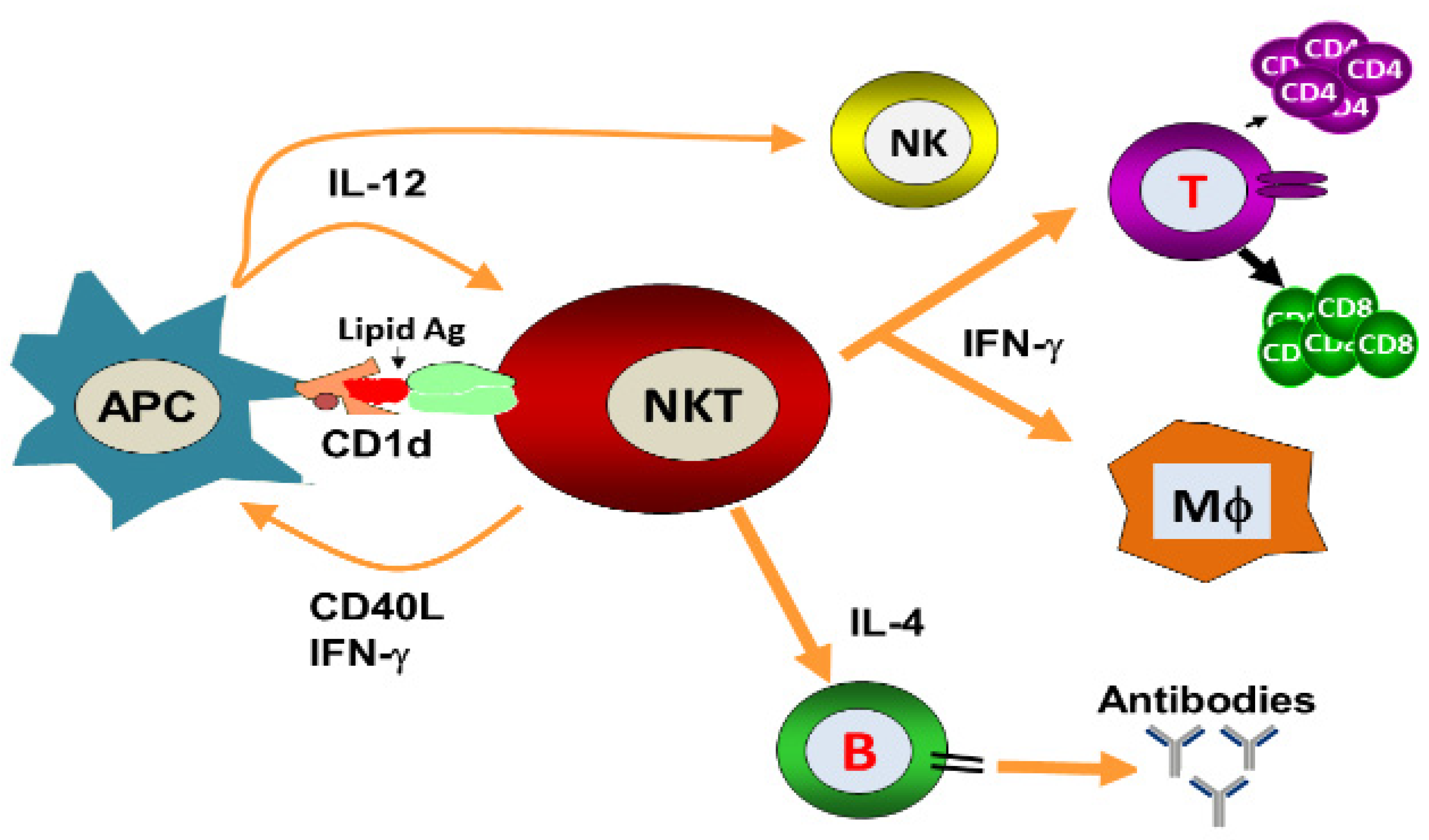
3. Role of iNKT Cells against Viral Infections

NKT cells have antiviral potential against hepatitis B virus (HBV), respiratory syncytial virus (RSV), encephalomyocarditis virus (EMCV), Herpes simplex virus-1 (HSV-1), coxsackievirus B (CVB), lymphocytic choriomeningitis virus (LCMV), influenza A virus (IAV) and murine cytomegalovirus (MCMV) [25,26,27,28,29,30]. They constitute an important arm of the innate immune response against viruses and can also regulate the adaptive immune responses by modulating the antigen-presenting cells (APCs). NKT cells exert the direct cytolytic effects and restrict the replication of viruses. Moreover, they can indirectly induce an antiviral state through the secretion of important cytokines. α-GalCer-activated NKT cells reduced the replication of murine cytomegalovirus [30]. Likewise, α-GalCer administration protected the mice against viral encephalomyocarditis [27]. Wu et al. showed that α-GalCer protected the mice against coxsackievirus B3 (CVB3)-induced myocarditis [31]. Johnson et al. demonstrated that NKT cell activation has been shown to induce the expansion of Cytotoxic T lymphocytes (CTLs) response and enhance the immune response against RSV [32]. The activation of the NKT cell resulted in the reduction of the viral load in the pancreas of LCMV-infected mice and this effect was mediated through the secretion of type I interferon [28]. Whereas, α-GalCer-induced activation of NKT cells against HBV was shown to be mediated by IFN-α/β and IFN-γ secretion [25]. However, the antiviral effect of NKT cells against the IAV is mediated through the activation of the innate immune responses [29]. Type I NKT cells exhibited a critical role against IAV infection by increasing IAV-specific CD8+ T cell response and viral clearance [33]. Singh et al. showed that cytokines produced by iNKT cells were associated with non-progressive HIV-I infection and patients had a lower viral load in their plasma [34]. There have been many studies that support the role of NKT cells against viral infections in humans. For example, some patients with mutated adaptor protein SAP (signaling lymphocyte activation molecule-associated protein) were found to be deficient in invariant iNKT cells [35]. These patients have been found to be susceptible to EBV infection. In an another study, patients mutated in the X-linked inhibitor of the apoptosis protein (XIAP) showed the selective reduction in iNKT cell numbers without affecting B cells, T cells and NK cells [36]. The Wiskott−Aldrich syndrome (WAS), a primary immune deficiency disease in humans, has been shown to be associated with an iNKT cell deficiency that impaired the functioning of the innate and adaptive immune systems and therefore predisposed the patients to viral infections [37]. The NKT cells play a very important role in linking the innate and adaptive immunity, their deficiency causes severe immunodeficiency in those persons for whom vaccination with attenuated pathogens is a serious challenge. This was evident from a report that a girl with impaired iNKT cell number and function developed severe respiratory distress after vaccination with the attenuated varicella-zoster virus (VZV) [38].
NKT cells have been actively involved in immune responses against HSV-1 and HSV-2 as NKT cell-deficient mice showed severe HSV-1 infection and impaired viral clearance [39]. The immune evasion strategy of HSV includes the inhibition of NKT cell recognition by curbing the CD1d recycling from the late endosomal compartments to the cell surface [40]. The protective role of NKT cells against RSV was evident from the fact that Cd1d−/− mice infected with RSV showed a poor ability to clear the viral load [39]. MCMV is considered a study model for human cytomegalovirus (HCMV). The iNKT cell activation by MCMV cells requires the involvement of IL-12 and type I interferon, but was independent of CD1d [41]. In an another study, a role of iNKT cells and CD1d has been suggested to counter the MCMV-induced suppression of hematopoiesis in mice [42]. The CD1d-dependent activation of NKT cells plays an important role in resisting EMCV infection because Cd1d−/− mice demonstrated more brutal paralysis due to an acute cytopathic effect of EMCV on neuronal cells [27]. The iNKT cells also played an important role in protecting against IAV because iNKT cell-deficient mice showed more severe bronchopneumonia. The adoptive transfer of iNKT cells before the infection reversed the effect of IAV-induced bronchopneumonia [43].
The NKT cells are found in the highest numbers in the livers of mice and therefore are supposed to play a very important role in understanding the pathology of liver diseases [44]. The activation of NKT cells had a protective role against HBV infection [25,45]. The absence of NKT cells or CD1d in mice resulted in diminished HBV-specific T and B cell responses with delayed viral clearance [46]. Toll-like receptor (TLR) ligands have been shown to activate iNKT cells [47]. Viruses altered the antigen presentation by CD1d through the activation of pattern recognition receptors, such as TLRs. The endosomal TLRs (TLR 3, 7, 8, and 9) are involved in detecting viruses by recognizing the nucleic acid structures [48]. TLR-mediated recognition of viruses may lead to altered CD1d-mediated antigen presentation, thereby affecting the activation of iNKT cells. The role of NKT cells has been extensively reported against viral infections in pigs. The percentage of iNKT cells were increased in the blood, lymph node and broncho-alveolar lavage of pigs upon IAV infection [49]. Renukaradhya et al. demonstrated the role of iNKT cells in the regulation of airway hyper-reactivity [50]. The intranasal administration of NKT cell ligand ameliorated H1N1 IAV infection in piglets [51], while the adjuvant effect of α-GalCer potentiated the immune response of the inactivated HIN1 influenza virus in pigs [52].

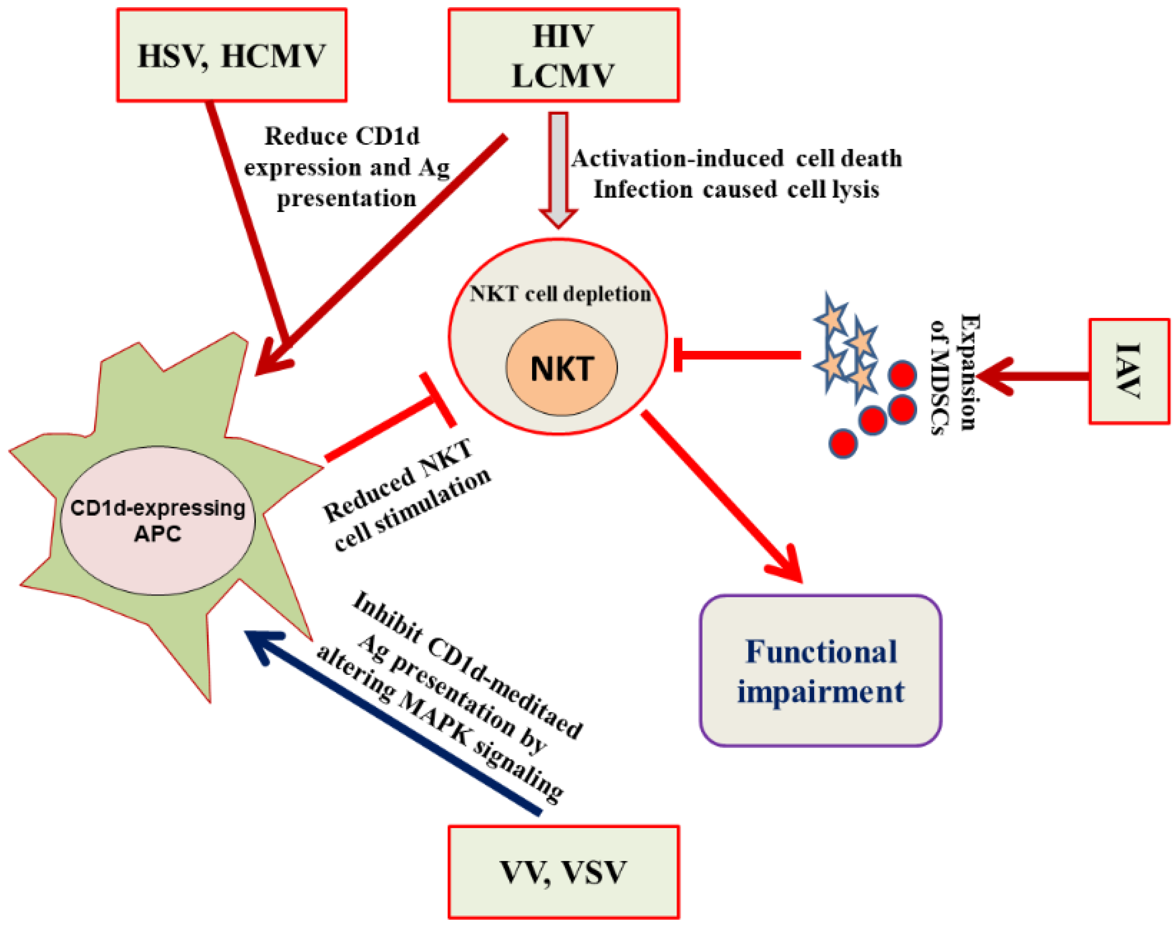
4. Evasion of the NKT Cell Functioning by Viruses

Microbial pathogens can cause recurrent or persistent infections by avoiding the onslaught of the normal host immunity. Viruses adopt different strategies to evade both the innate and adaptive arms of the immune system (Figure 3). For example, HIV weakens the immune system by depleting the numbers of CD4+ T cells [53]. Some viruses, such as the herpes virus and Epstein−Barr virus (EBV), have the ability to enter into a latent state. Although the viruses do not replicate in the latent state, they cannot, however, be eliminated and can reactivate themselves to a fully virulent form. Invariant NKT (iNKT) cells have shown an antiviral immunity upon stimulation with α-GalCer against HIV, MCMV, RSV, HBV and influenza virus infections [25,26,27,28,29,30,54]. There has been a reported reduction in the iNKT cell numbers in HIV-1+ patients, particularly a depletion in the CD4+ iNKT cell subset [55]. Moreover, HIV also causes the functional impairment of iNKT cells as the CD4+ and CD4 iNKT cells secrete a lower amount of IFN-γ, TNF-α, and IL-4 in response to α-GalCer/IL-2/PMA stimulation [56]. Viruses have adopted many tactics to avoid the assault by the immune system. For example, HIV-1 reduces the expression of CD1d molecules by increasing its internalization and retains them in the trans-Golgi network. The downregulation in the cell surface CD1d is caused by the interaction of CD1d intra-cytoplasmic tyrosine with HIV-1 Nef protein [57,58]. The CD4+ NKT cells showed greater susceptibility, as compared to conventional CD4+ T cells, to HIV-1 infection owing to the elevated level of CCR5 coreceptor expression on iNKT cells [59]. Fernandez et al. demonstrated an early NKT cell depletion in HIV-infected individuals [60]. Besides, iNKT cells showed their functional impairment as they produced lower levels of Th1 and Th2 cytokines in response to α-GalCer [61]. The functional status of NKT cells is significantly preserved in the long term nonprogressors (LTNPs) as compared to the progressors [34].
Cell signaling pathways play an important role in modulating the CD1d-mediated antigen presentation to NKT cells by viruses [62]. We have earlier shown that vaccinia virus (VV) evaded the NKT cell activity by inhibiting the CD1d-mediated antigen presentation through the alteration of the mitogen-activated protein kinases (MAPKs) (Figure 3) [63]. A VV infection inhibited the CD1d-mediated antigen presentation by activating the p38 MAPK. The inhibition of p38 MAPK signaling using a specific inhibitor SB203580 rescued the VV-induced inhibition of CD1d-mediated antigen presentation. The alteration of MAPKs activation changed the intracellular trafficking of CD1d through the ligand-loading endocytic compartments [63]. In another study, we demonstrated that JNK2 negatively regulated the antigen presentation by CD1d and also contributed to the IL-12 induced activation of iNKT cells [64].
The signal transducer and activator of transcription-3 (STAT-3) has been shown to activate NKT cells by promoting CD1d-mediated antigen presentation [65]. The STAT-3 signaling induces an antiviral immunity as the treatment with a specific STAT-3 inhibitor increased the viral load in VV-infected mice [66]. In an another study, we and colleagues showed that a vesicular stomatitis virus (VSV) inhibited the CD1d-mediated antigen presentation much faster as compared to that inhibited by VV. A matrix protein, M protein, of VSV played a key role in inhibiting CD1d-mediated antigen presentation [67]. Moreover, a chronic LCMV infection caused a long-term loss of NKT cells in mice [68,69]. Interestingly, the acute infection of mice with LCMV caused a reduction in CD1d cell surface expression on dendritic cells and macrophages [68]. Moreover, West Nile virus (WNV) interferes with the interaction of dendritic cells (DCs) with NKT cells and thus inhibits the secretion of proinflammatory cytokines [70]. Kovats et al. showed that WNV-infected human dendritic cells failed to fully activate NKT cells [70]. HSV-1 has been shown to inhibit CD1d-mediated antigen presentation by suppressing the recycling of CD1d on the cell surface [71]. Viral glycoprotein protein (gB) and viral serine-threonine kinase US3 are required to inhibit the CD1d antigen presentation and NKT cell activation.
Programmed death (PD)-1 is a member of the CD28 family of the costimulatory molecules and its interaction with its ligands PD-L1 and PD-L2 on APC sends the inhibitory signals to T cells [72]. The PD-1:PD-L interaction contributes to the induction of NKT cell anergy [73]. It has been shown that an increased expression of PD-1 on the T cells in HIV-1 infection induces T cell exhaustion and contributes to the progression of the disease [74]. Moll et al. demonstrated the elevated expression of PD-1 and functional impairment in CD1d-restricted NKT cells in the chronic infection of HIV-1 [56]. The coadministration of anti-PDL1 monoclonal antibody and α-GalCer modulated the NKT cell activity and inhibited HBV infection [44]. Recently, Zingaropoli et al. showed a reduction in NKT cells in the peripheral blood of COVID-19 patients [75].

5. Nanotechnology-Based Vaccine Delivery Platforms and the Development of an NKT Cell-Based Nanovaccine

The development of nanotechnology-based formulations has shown great promise for the development of new generation vaccines. The nanoparticle-based delivery systems not only improve the stability of the vaccine, but also increase the immunogenicity of the encapsulated antigens by delivering them to the intracellular locations of APCs. The shape, size and the surface properties of the delivery systems determine their efficacy as an immunoadjuvant. Various types of vaccine delivery systems, including gold and silver nanoparticles, liposomes and chitosan nanoparticles have the ability to induce the antigen-specific T cell responses and antibody responses [76]. The important vaccine nanocarriers include liposomes, inorganic nanoparticles, chitosan nanoparticles, PLGA nanoparticles and virus-like particles (VLPs) [77,78].

5.1. Inorganic Nanoparticles
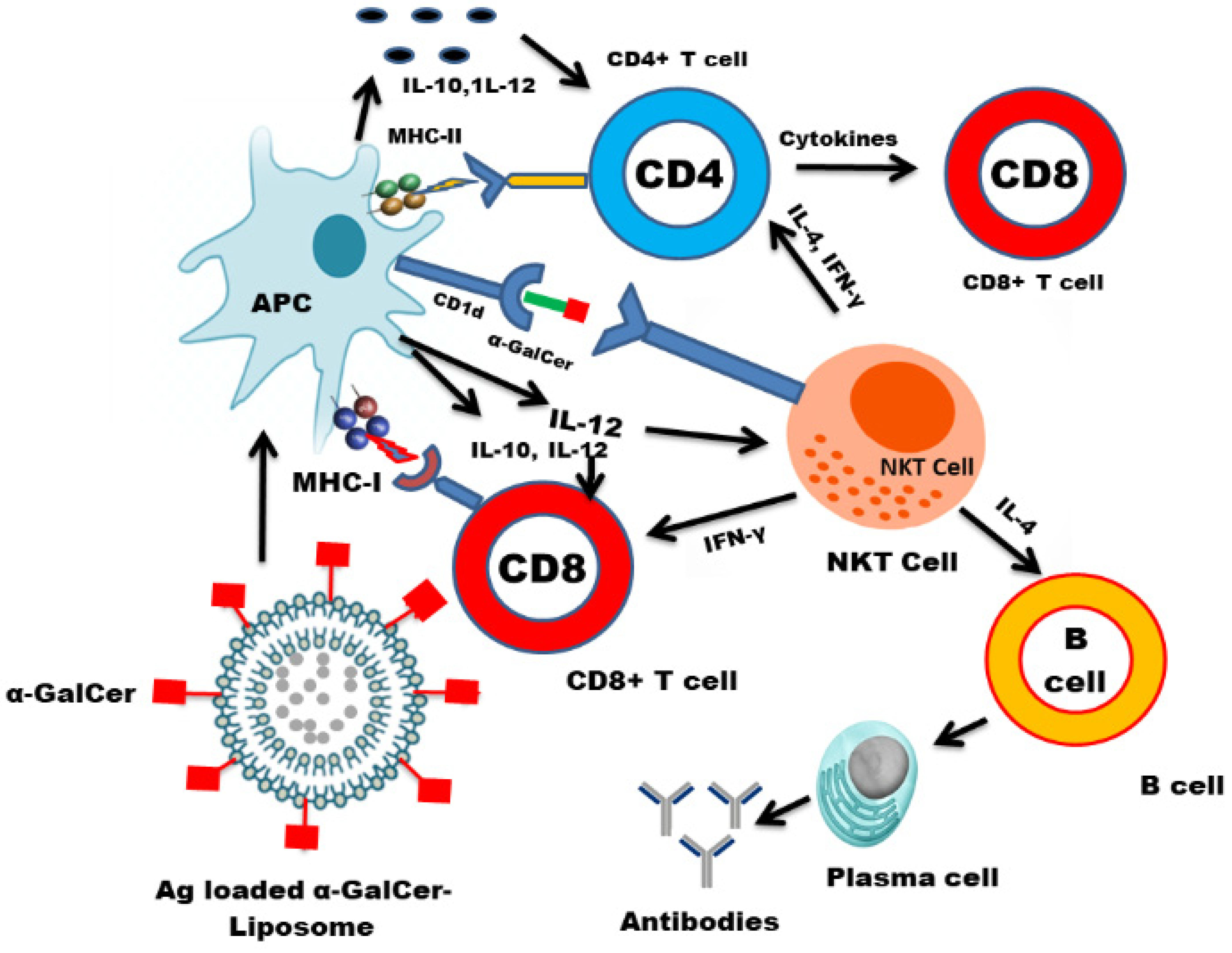
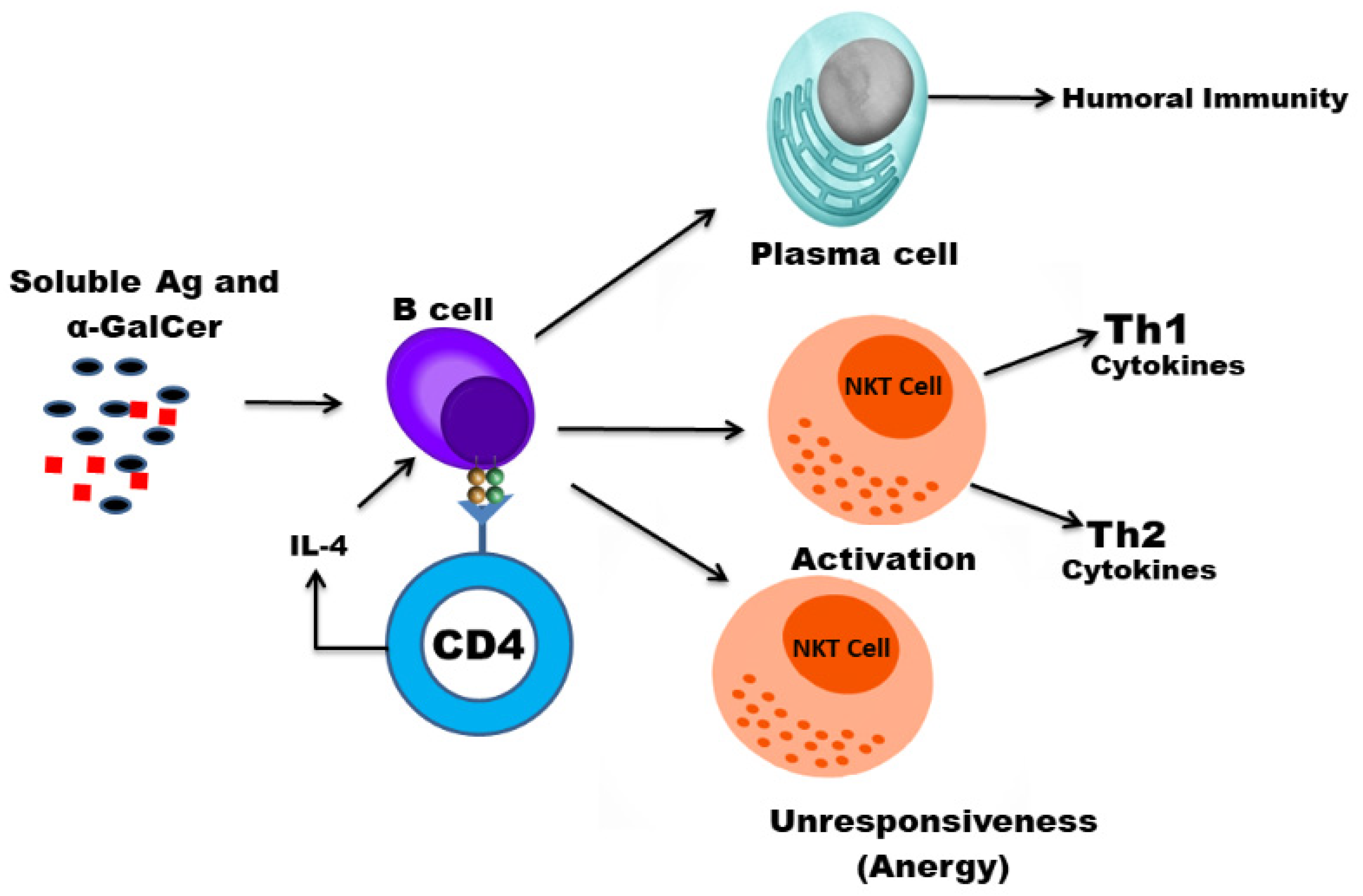
Several inorganic nanoparticles, including gold, silver, silica and iron have been formulated as vaccine delivery systems [79]. Influenza virus M2 membrane protein immobilized on gold-nanoparticles induced protective immunity against influenza A subtypes [80]. Silica nanoparticles loaded with CV2-ORF2 proteins elicited antigen-specific cell- and antibody-mediated immune responses [81]. Carbon nanotube-conjugated peptides from the foot-and-mouth disease virus elicited a strong antigen-specific immune response [82].

5.2. Liposomes

Liposomes are the unilamellar or multilamellar lipid vesicles that are composed of biodegradable phospholipids, including phosphatidylserine, phosphatidylcholine and cholesterol. Liposomes deliver the antigens to the cytoplasmic compartments by fusing with the membrane of the APCs [78]. The modification of the liposomal surface with certain molecules increases the immunoadjuvant potential of the liposomes [83,84]. Liposomes composed of cationic lipids elicited a strong immune response against the hepatitis B surface antigen (HBsAg) [85]. In addition, liposomes have been shown to be very effective in inducing antigen-specific immune responses against IAV and RSV [86,87].

5.3. Polymeric Nanoparticles

The polymer-based vaccine carriers are composed of biodegradable polymers, including chitosan, polylactic acid, polyglutamic acid, and poly(lactic-glycolic acid) [88]. Among them, the chitosan-based nanoparticle vaccine carriers have been extensively studied due to their biodegradability and reduced toxicity [89]. Chitosan-bearing nanoparticles have been shown to improve the immunogenicity of the influenza virus vaccine [90]. The design of poly(lactic-co-glycolic acid) (PLGA) nanoparticles improved the antigen-specific immune responses [91] Moreover, PLGA nanoparticles increased the antigen-specific lymphocyte proliferation against H1N2 antigens [92].
A successful vaccine should be able to induce a strong CD8+ T cell immune response in order to protect against viral infections (Figure 4). Generally, the vaccines composed of the live attenuated pathogens can generate CD8+ T-cell-mediated immune responses [93,94]. The antiviral activity of the NKT cells is evident from the fact that NKT-cell-deficient mice showed a greater susceptibility to viruses [95,96]. Since NKT cells have a unique ability to stimulate the innate and adaptive immune responses, the NKT-cell ligand may prove to be an effective immunoadjuvant in the development of a successful vaccine.
The NKT cells show immune-stimulating activity through the maturation of the dendritic cells and boosting of antibody production [97,98]. Since they induce a rapid maturation of the dendritic cells (DCs) and B cells, their activation may elicit a strong antigen-specific immune responses. Various studies have shown the immunoadjuvant effect of α-GalCer in protection against influenza virus infections [99,100]. The use of a synthetic glycolipid ABX196, an NKT ligand, demonstrated an immunoadjuvant effect and stimulated an anti-HB antibody response in human volunteers [101]. Huang et al. reported that α-GalCer enhanced the immunogenicity of the DNA vaccine by increasing antigen-specific T-cell and antibody immune responses [102]. A major drawback with the use of α-GalCer is that its sequential systemic administration results in the NKT cell anergy that hampers the development of the NKT-based immunotherapeutics (Figure 5). Moreover, the presentation of α-GalCer by the nonprofessional APCs may induce NKT cell anergy. In order to exploit the NKT cell-mediated immune stimulation, it is important to codeliver the NKT cell ligand and antigens to the professional APCs such as dendritic cells. The co-administration of α-GalCer and soluble antigens enhanced the antigen-specific cell-mediated and humoral immunity [103]. However, the coadministration of α-GalCer and soluble antigens does not necessarily ensure that they are taken up by the same APC. In order to fully exploit the iNKT role to induce an effective CD8+ T-cell response, it is essential that α-GalCer and antigens are delivered to the same professional APC. This goal can be achieved by using the nanoparticles as delivery systems for antigens and NKT cell ligand. The liposome-mediated delivery of α-GalCer and tumor-associated antigens (TAA) has resulted in an increased antigen-specific CD8+ T-cell response [104]. Another study showed that the delivery of α-GalCer with ovalbumin (OVA) in poly(lactic-co-glycolic acid) (PLGA) nanoparticles induced a very strong antigen-specific CD8+ T-cell response [105].
We have earlier shown that liposomes can fuse with the membrane of APC and deliver the antigens to the cytoplasmic compartment for processing [74]. These antigens are presented by MHC class I molecules to induce the activation of CD8+ T cells. The activation of antigen-specific CTLs is critical in the development of an antiviral vaccine. Moreover, the liposome-encapsulated antigens are also taken by the APCs through phagocytosis where the antigens are presented by MHC class II molecules to CD4+ T cells resulting in the secretion of various cytokines. On the other hand, MHC class I-like CD1d molecules present glycolipid antigens to NKT cells. The iNKT cell-specific ligands recruit NKT cells, like CD4+ T cells, that play an important role in the modulation of the humoral and cytotoxic T-cell responses. The stimulation of NKT cells leads to the activation of downstream immune cells, including NK cells, DCs, macrophages, B cells, and conventional T cells. Many of these immune cells secrete immune-modulating cytokines, creating an entire activation cascade.
In contrast to DCs, B cells express lower levels of costimulatory molecules and contribute to NKT cell anergy [106]. Interestingly, liposome-incorporated-α-GalCer is preferentially taken up by the dendritic cells and activates iNKT cells without inducing anergy [107]. Thapa et al. demonstrated that the soluble α-GalCer is presented by B cells, whereas the nanoparticle-incorporated α-GalCer is preferentially presented by DCs. Moreover, glycosphingolipids (GSLs), isolated from S. paucimobilis, specifically activated the iNKT cells in a CD1d-dependent manner [17]. Liposomes composed of GSLs elicited a strong antigen-specific immune response [108]. Moreover, they induced the activation of DCs and increased the production of IFN-γ by the splenocytes [108,109]. It suggested that the incorporation of iNKT-specific ligands in nanoparticles or liposomes may further potentiate the immunogenicity of encapsulated antigens by enhancing the generation of antigen-specific CD8 + T cells and CD4 + T cells that contribute to the development of an effective vaccine against viruses.

6. Conclusions

In spite of constituting a very small population of T cells, NKT cells play a very critical role in protecting against viruses. They have both the direct and indirect antiviral effect through the secretion of important cytokines. Contrarily, viruses evade the functioning of the NKT cells by utilizing multiple immune evasion mechanisms. While the NKT-cell ligands have been extensively studied as a potential therapeutic agent in the treatment of cancer, their role, however, as immunoadjuvants has not been widely explored. Some studies showed that NKT-cell ligands have immunoadjuvant potential in eliciting the antigen-specific cell-mediated and humoral immune responses. Nevertheless, their frequent systemic administration results in NKT cell anergy. Nanoparticle-based vaccine formulations have shown their efficacy in the generation of CD4+ and CD8+ T cell responses, the latter is critical to control intracellular infections, particularly viral infections. The codelivery of NKT cell ligand and antigens by nanoparticles is suggested to be a very important strategy to prepare an antiviral vaccine because it will not only activate CD4+ and CD8+ T cells, but also stimulate iNKT cells that further contribute in the activation of the cell-mediated and humoral immune responses through the secretion of various cytokines. Moreover, iNKT cell ligand incorporated into nanoparticles can persistently activate iNKT cells without inducing anergy. Thus the codelivery of nanoparticle-incorporated iNKT cell ligand and antigens may be considered an important prophylactic approach to mount a strong antigen-specific antiviral immune response.

Author Contributions

Conceptualization, writing, review and editing, M.A.K.; Review and editing A.K. All authors have read and agreed to the published version of the manuscript.

Funding

This research received no external funding.

Institutional Review Board Statement

Not applicable.

Informed Consent Statement

Not applicable.

Data Availability Statement

Not applicable.

Acknowledgments

The authors would like to thank the Deanship of Scientific Research, Qassim University for the Grant # 5575-cams1-2019-2-2-I to Masood A. Khan.

Conflicts of Interest

There is no conflict of interest to declare.

References

  1. Bendelac, A.; Savage, P.B.; Teyton, L. The Biology of NKT Cells. Annu. Rev. Immunol. 2007, 25, 297–336. [Google Scholar] [CrossRef] [PubMed] [Green Version]
  2. Godfrey, D.I.; Stankovic, S.; Baxter, A. Raising the NKT cell family. Nat. Immunol. 2010, 11, 197–206. [Google Scholar] [CrossRef] [PubMed]
  3. Berzins, S.P.; Smyth, M.; Baxter, A. Presumed guilty: Natural killer T cell defects and human disease. Nat. Rev. Immunol. 2011, 11, 131–142. [Google Scholar] [CrossRef] [PubMed]
  4. Peralbo, E.; Alonso, C.; Solana, R. Invariant NKT and NKT-like lymphocytes: Two different T cell subsets that are differentially affected by ageing. Exp. Gerontol. 2007, 42, 703–708. [Google Scholar] [CrossRef] [PubMed]
  5. Terrazzano, G.; Bruzzaniti, S.; Rubino, V.; Santopaolo, M.; Palatucci, A.T.; Giovazzino, A.; La Rocca, C.; de Candia, P.; Puca, A.; Perna, F.; et al. T1D progression is associated with loss of CD3+CD56+ regulatory T cells that control CD8+ T cell effector functions. Nat. Metab. 2020, 2, 142–152. [Google Scholar] [CrossRef] [PubMed]
  6. Uldrich, A.; Patel, O.; Cameron, G.; Pellicci, D.; Day, E.B.; Sullivan, L.; Kyparissoudis, K.; Kjer-Nielsen, L.; Vivian, J.; Cao, B.; et al. A semi-invariant Vα10+ T cell antigen receptor defines a population of natural killer T cells with distinct glycolipid antigen—Recognition properties. Nat. Immunol. 2011, 12, 616–623. [Google Scholar] [CrossRef]
  7. Reantragoon, R.; Corbett, A.; Sakala, I.G.; Gherardin, N.; Furness, J.B.; Chen, Z.; Eckle, S.; Uldrich, A.; Birkinshaw, R.; Patel, O.; et al. Antigen-loaded MR1 tetramers define T cell receptor heterogeneity in mucosal-associated invariant T cells. J. Exp. Med. 2013, 210, 2305–2320. [Google Scholar] [CrossRef]
  8. Tang, X.-Z.; Jo, J.; Tan, A.T.; Sandalova, E.; Chia, A.; Tan, K.C.; Lee, K.H.; Gehring, A.; De Libero, G.; Bertoletti, A. IL-7 Licenses Activation of Human Liver Intrasinusoidal Mucosal-Associated Invariant T Cells. J. Immunol. 2013, 190, 3142–3152. [Google Scholar] [CrossRef]
  9. Loh, L.; Wang, Z.; Sant, S.; Koutsakos, M.; Jegaskanda, S.; Corbett, A.J.; Liu, L.; Fairlie, D.; Crowe, J.; Rossjohn, J.; et al. Human mucosal-associated invariant T cells contribute to antiviral influenza immunity via IL-18-dependent activation. Proc. Natl. Acad. Sci. USA 2016, 113, 10133–10138. [Google Scholar] [CrossRef] [PubMed] [Green Version]
  10. Le Bourhis, L.; Martin, E.; Péguillet, I.; Guihot, A.; Froux, N.; Coré, M.; Lévy, E.; Dusseaux, M.; Meyssonnier, V.; Premel, V.; et al. Antimicrobial activity of mucosal-associated invariant T cells. Nat. Immunol. 2010, 11, 701–708. [Google Scholar] [CrossRef] [Green Version]
  11. Salio, M.; Silk, J.D.; Jones, Y.; Cerundolo, V. Biology of CD1- and MR1-Restricted T Cells. Annu. Rev. Immunol. 2014, 32, 323–366. [Google Scholar] [CrossRef] [PubMed]
  12. Gold, M.C.; Cerri, S.; Smyk-Pearson, S.; Cansler, M.E.; Vogt, T.M.; Delepine, J.; Winata, E.; Swarbrick, G.M.; Chua, W.-J.; Yu, Y.Y.L.; et al. Human Mucosal Associated Invariant T Cells Detect Bacterially Infected Cells. PLoS Biol. 2010, 8, e1000407. [Google Scholar] [CrossRef] [PubMed] [Green Version]
  13. Lepore, M.; Kalinichenko, A.; Colone, A.; Paleja, B.; Singhal, A.; Tschumi, A.; Lee, B.; Poidinger, M.; Zolezzi, F.; Quagliata, L.; et al. Parallel T-cell cloning and deep sequencing of human MAIT cells reveal stable oligoclonal TCRβ repertoire. Nat. Commun. 2014, 5, 3866. [Google Scholar] [CrossRef] [Green Version]
  14. Morita, M.; Motoki, K.; Akimoto, K.; Natori, T.; Sakai, T.; Sawa, E.; Yamaji, K.; Koezuka, Y.; Kobayashi, E.; Fukushima, H. Structure-Activity relationship of α-Galactosylceramide against B-16 bearing mice. J. Med. Chem. 1995, 38, 2176–2187. [Google Scholar] [CrossRef] [PubMed]
  15. Brown, L.C.W.; Penaranda, C.; Kashyap, P.C.; Williams, B.B.; Clardy, J.; Kronenberg, M.; Sonnenburg, J.L.; Comstock, L.E.; Bluestone, J.A.; Fischbach, M.A. Production of α-Galactosylceramide by a Prominent Member of the Human Gut Microbiota. PLoS Biol. 2013, 11, e1001610. [Google Scholar] [CrossRef] [Green Version]
  16. von Gerichten, J.; Schlosser, K.; Lamprecht, D.; Morace, I.; Eckhardt, M.; Wachten, D.; Jennemann, R.; Gröne, H.-J.; Mack, M.; Sandhoff, R. Diastereomer-specific quantification of bioactive hexosylceramides from bacteria and mammals. J. Lipid Res. 2017, 58, 1247–1258. [Google Scholar] [CrossRef] [PubMed] [Green Version]
  17. Sriram, V.; Du, W.; Gervay-Hague, J.; Brutkiewicz, R.R. Cell wall glycosphingolipids of Sphingomonas paucimobilis are CD1d-specific ligands for NKT cells. Eur. J. Immunol. 2005, 35, 1692–1701. [Google Scholar] [CrossRef]
  18. Brutkiewicz, R.R. CD1d ligands: The good, the bad, and the ugly. J. Immunol. 2006, 177, 769–775. [Google Scholar] [CrossRef] [Green Version]
  19. Albacker, L.A.; Chaudhary, V.; Chang, Y.J.; Kim, H.Y.; Chuang, Y.T.; Pichavant, M.; DeKruyff, R.H.; Savage, P.B.; Umetsu, D.T. Invariant natural killer T cells recognize a fungal glycosphingolipid that can induce airway hyper reactivity. Nat. Med. 2013, 19, 1297–1304. [Google Scholar] [CrossRef] [Green Version]
  20. Dhodapkar, M.V.; Kumar, V. Type II NKT Cells and Their Emerging Role in Health and Disease. J. Immunol. 2017, 198, 1015–1021. [Google Scholar] [CrossRef]
  21. Brutkiewicz, R.R.; Lin, Y.; Cho, S.; Hwang, Y.K.; Sriram, V.; Roberts, T.J. CD1d-mediated antigen presentation to natural killer T (NKT) cells. Crit. Rev. Immunol. 2003, 23, 403–419. [Google Scholar] [CrossRef] [PubMed]
  22. Coquet, J.; Chakravarti, S.; Kyparissoudis, K.; McNab, F.W.; Pitt, L.A.; McKenzie, B.S.; Berzins, S.P.; Smyth, M.; Godfrey, D.I. Diverse cytokine production by NKT cell subsets and identification of an IL-17-producing CD4-NK1.1- NKT cell population. Proc. Natl. Acad. Sci. USA 2008, 105, 11287–11292. [Google Scholar] [CrossRef] [PubMed] [Green Version]
  23. James, E.E.; Andrew, J.K.; Webb, T.J. Raising the roof: The preferential pharmacological stimulation of Th1 and Th2 responses mediated by NKT cells. Med. Res. Rev. 2014, 34, 45–76. [Google Scholar]
  24. Liu, Z.; Guo, J. NKT-Cell glycolipid agonist as adjuvant in synthetic vaccine. Carbohydr. Res. 2017, 452, 78–90. [Google Scholar] [CrossRef]
  25. Kakimi, K.; Guidotti, L.G.; Koezuka, Y.; Chisari, F. Natural Killer T Cell Activation Inhibits Hepatitis B Virus Replication in Vivo. J. Exp. Med. 2000, 192, 921–930. [Google Scholar] [CrossRef] [PubMed] [Green Version]
  26. Liu, J.; Glosson, N.L.; Du, W.; Gervay-Hague, J.; Brutkiewicz, R.R. A Thr/Ser dual residue motif in the cytoplasmic tail of human CD1d is important for the down-regulation of antigen presentation following a herpes simplex virus 1 infection. Immunology 2013, 140, 191–201. [Google Scholar] [CrossRef]
  27. Exley, M.A.; Bigley, N.J.; Cheng, O.; Tahir, S.M.; Smiley, S.T.; Carter, Q.L.; Stills, H.F.; Grusby, M.J.; Koezuka, Y.; Taniguchi, M.; et al. CD1d-Reactive T-cell activation leads to amelioration of disease caused by diabetogenic encephalomyocarditis viruses. J. Leukoc. Biol. 2001, 69, 713–718. [Google Scholar] [PubMed]
  28. Diana, J.; Griseri, T.; Lagaye, S.; Beaudoin, L.; Autrusseau, E.; Gautron, A.-S.; Tomkiewicz, C.; Herbelin, A.; Barouki, R.; von Herrath, M.; et al. NKT Cell-Plasmacytoid Dendritic Cell Cooperation via OX40 Controls Viral Infection in a Tissue-Specific Manner. Immunity 2009, 30, 289–299. [Google Scholar] [CrossRef]
  29. Ho, L.; Denney, L.; Luhn, K.; Teoh, D.; Clelland, C.; McMichael, A.J. Activation of invariant NKT cells enhances the innate immune response and improves the disease course in influenza A virus infection. Eur. J. Immunol. 2008, 38, 1913–1922. [Google Scholar] [CrossRef] [PubMed]
  30. van Dommelen, S.L.H.; Tabarias, H.A.; Smyth, M.J.; Degli-Esposti, M.A.; Law, L.M.J.; Everitt, J.C.; Beatch, M.D.; Holmes, C.F.B.; Hobman, T.C. Activation of Natural Killer (NK) T Cells during Murine Cytomegalovirus Infection Enhances the Antiviral Response Mediated by NK Cells. J. Virol. 2003, 77, 1764–1771. [Google Scholar] [CrossRef] [Green Version]
  31. Wu, C.Y.; Feng, Y.; Qian, G.C.; Wu, J.H.; Luo, J.; Wang, Y.; Chen, G.J.; Guo, X.-K.; Wang, Z.J. α-Galactosylceramide protects mice from lethal Coxsackievirus B3 infection and subsequent myocarditis. Clin. Exp. Immunol. 2010, 162, 178–187. [Google Scholar] [CrossRef]
  32. Johnson, T.R.; Hong, S.; Van Kaer, L.; Koezuka, Y.; Graham, B.S. NK T Cells Contribute to Expansion of CD8 + T Cells and Amplification of Antiviral Immune Responses to Respiratory Syncytial Virus. J. Virol. 2002, 76, 4294–4303. [Google Scholar] [CrossRef] [Green Version]
  33. De Santo, C.; Salio, M.; Masri, S.H.; Lee, L.Y.-H.; Dong, T.; Speak, A.; Porubsky, S.; Booth, S.; Veerapen, N.; Besra, G.; et al. Invariant NKT cells reduce the immunosuppressive activity of influenza A virus–induced myeloid-derived suppressor cells in mice and humans. J. Clin. Investig. 2008, 118, 4036–4048. [Google Scholar] [CrossRef] [Green Version]
  34. Singh, D.; Ghate, M.; Godbole, S.; Kulkarni, S.; Thakar, M. Functional Invariant Natural Killer T Cells Secreting Cytokines Are Associated with Non-Progressive Human Immunodeficiency Virus-1 Infection but Not With Suppressive Anti-Retroviral Treatment. Front. Immunol. 2018, 9, 1152. [Google Scholar] [CrossRef] [PubMed] [Green Version]
  35. Nichols, K.E.; Hom, J.; Gong, S.; Ganguly, A.; Ma, C.; Cannons, J.L.; Tangye, P.S.; Schwartzberg, P.L.; Koretzky, G.A.; Stein, P.L. Regulation of NKT cell development by SAP, the protein defective in XLP. Nat. Med. 2005, 11, 340–345. [Google Scholar] [CrossRef]
  36. Rigaud, S.; Fondanèche, M.-C.; Lambert, N.C.; Pasquier, B.; Mateo, V.; Soulas-Sprauel, P.; Galicier, L.; Le Deist, F.; Rieux-Laucat, F.; Revy, P.; et al. XIAP deficiency in humans causes an X-linked lymphoproliferative syndrome. Nature 2006, 444, 110–114. [Google Scholar] [CrossRef] [PubMed]
  37. Locci, M.; Draghici, E.; Marangoni, F.; Bosticardo, M.; Catucci, M.; Aiuti, A.; Cancrini, C.; Marodi, L.; Espanol, T.; Bredius, R.G.; et al. The Wiskott-Aldrich syndrome protein is required for iNKT cell maturation and function. J. Exp. Med. 2009, 206, 735–742. [Google Scholar] [CrossRef] [PubMed]
  38. Levy, O.; Orange, J.S.; Hibberd, P.; Steinberg, S.; LaRussa, P.; Weinberg, A.; Wilson, S.B.; Shaulov, A.; Fleisher, G.; Geha, R.S.; et al. Disseminated varicella infection due to the vaccine strain of varicella-zoster virus, in a patient with a novel deficiency in natural killer T cells. J. Infect. Dis. 2003, 188, 948–953. [Google Scholar] [CrossRef] [PubMed] [Green Version]
  39. Grubor-Bauk, B.; Simmons, A.; Mayrhofer, G.; Speck, P.G. Impaired clearance of herpes simplex virus type 1 from mice lacking CD1d or NKT cells expressing the semivariant V alpha 14-J alpha 281 TCR. J. Immunol. 2003, 170, 1430–1434. [Google Scholar] [CrossRef]
  40. Yuan, W.; Dasgupta, A.; Cresswell, P. Herpes simplex virus evades naturalkiller T cell recognition by suppressing CD1d recycling. Nat. Immunol. 2006, 7, 835–842. [Google Scholar] [CrossRef]
  41. Tyznik, A.J.; Tupin, E.; Nagarajan, N.A.; Her, M.J.; Benedict, C.A.; Kronenberg, M. Cutting edge: The mechanism of invariant NKT cell responses to viral danger signals. J. Immunol. 2008, 181, 4452–4456. [Google Scholar] [CrossRef] [Green Version]
  42. Broxmeyer, H.E.; Dent, A.; Cooper, S.; Hangoc, G.; Wang, Z.-Y.; Du, W.; Gervay-Haque, J.; Sriram, V.; Renukaradhya, G.J.; Brutkiewicz, R.R. A role for natural killer T cells and CD1d molecules in counteracting suppression of hematopoiesis in mice induced by infection with murine cytomegalovirus. Exp. Hematol. 2007, 35, 87–93. [Google Scholar] [CrossRef]
  43. Paget, C.; Ivanov, S.; Fontaine, J.; Blanc, F.; Pichavant, M.; Renneson, J.; Bialecki, E.; Pothlichet, J.; Vendeville, C.; Barba-Speath, G.; et al. Potential Role of Invariant NKT Cells in the Control of Pulmonary Inflammation and CD8+ T Cell Response during Acute Influenza A Virus H3N2 Pneumonia. J. Immunol. 2011, 186, 5590–5602. [Google Scholar] [CrossRef] [PubMed] [Green Version]
  44. Ajuebor, M.N. Role of NKT cells in the digestive system. I. Invariant NKT cells and liver diseases: Is there strength in numbers? Am. J. Physiol. Gastrointest. Liver Physiol. 2007, 293, G651–G656. [Google Scholar] [CrossRef] [PubMed] [Green Version]
  45. Wang, X.F.; Lei, Y.; Chen, M.; Chen, C.B.; Ren, H.; Shi, T.D. PD-1/PDL1 and CD28/CD80 pathways modulate naturalkiller T cell function to inhibit hepatitis B virus replication. J. Viral. Hepat. 2013, 20, 27–39. [Google Scholar] [CrossRef] [PubMed]
  46. Zeissig, S.; Murata, K.; Sweet, L.; Publicover, J.; Hu, Z.; Kaser, A.; Bosse, E.; Iqbal, J.; Hussain, M.M.; Balschun, K.; et al. Hepatitis B virus-induced lipid alterations contribute to naturalkiller T cell-dependent protective immunity. Nat. Med. 2012, 18, 1060–1068. [Google Scholar] [CrossRef] [Green Version]
  47. Villanueva, A.I.; Haeryfar, S.M.; Mallard, B.A.; Kulkarni, R.R.; Sharif, S. Functions of NKT cells are modulated by TLR ligands and IFNα. Innate Immun. 2015, 21, 275–288. [Google Scholar] [CrossRef] [PubMed]
  48. Bowie, A.G.; Haga, I.R. The role of Toll-like receptors in the host response to viruses. Mol. Immunol. 2005, 42, 859–867. [Google Scholar] [CrossRef] [PubMed]
  49. Schäfer, A.; Hühr, J.; Schwaiger, T.; Dorhoi, A.; Mettenleiter, T.C.; Blome, S.; Schröder, C.; Blohm, U. Porcine Invariant Natural Killer T Cells: Functional Profiling and Dynamics in Steady State and Viral Infections. Front. Immunol. 2019, 10, 1380. [Google Scholar] [CrossRef] [PubMed]
  50. Renukaradhya, G.J.; Manickam, C.; Khatri, M.; Rauf, A.; Li, X.; Tsuji, M.; Rajashekara, G.; Dwivedi, V. Functional invariant NKT cells in pig lungs regulate the airway hyperreactivity: A potential animal model. J. Clin. Immunol. 2010, 31, 228–239. [Google Scholar] [CrossRef] [PubMed] [Green Version]
  51. Artiaga, B.L.; Yang, G.; Hutchinson, T.E.; Loeb, J.C.; Richt, J.A.; Lednicky, J.A.; Salek-Ardakani, S.; Driver, J.P. Rapid control of pandemic H1N1 influenza by targeting NKT-cells. Sci. Rep. 2016, 6, 37999. [Google Scholar] [CrossRef] [PubMed]
  52. Dwivedi, V.; Manickam, C.; Dhakal, S.; Binjawadagi, B.; Ouyang, K.; Hiremath, J.; Khatri, M.; Hague, J.G.; Lee, C.W.; Renukaradhya, G.J. Adjuvant effects of invariant NKT cell ligand potentiates the innate and adaptive immunity to an inactivated H1N1 swine influenza virus vaccine in pigs. Veter Microbiol. 2016, 186, 157–163. [Google Scholar] [CrossRef] [PubMed]
  53. Pegu, A.; Asokan, M.; Wu, L.; Wang, K.; Hataye, J.; Casazza, J.P.; Guo, X.; Shi, W.; Georgiev, I.; Zhou, T.; et al. Activation and lysis of human CD4 cells latently infected with HIV-1. Nat. Commun. 2015, 6, 8447. [Google Scholar] [CrossRef]
  54. Courtney, A.N.; Nehete, P.; Nehete, B.P.; Thapa, P.; Zhou, D.; Sastry, K.J. Alpha-galactosylceramide is an effective mucosal adjuvant for repeated intranasal or oral delivery of HIV peptide antigens. Vaccine 2009, 27, 3335–3341. [Google Scholar] [CrossRef] [Green Version]
  55. Sandberg, J.K.; Fast, N.M.; Palacios, E.H.; Fennelly, G.; Dobroszycki, J.; Palumbo, P.; Wiznia, A.; Grant, R.M.; Bhardwaj, N.; Rosenberg, M.G.; et al. Selective loss of innate CD4+ V alpha 24 natural killer T cells in human immunodeficiency virus infection. J. Virol. 2002, 76, 7528–7534. [Google Scholar] [CrossRef] [Green Version]
  56. Moll, M.; Kuylenstierna, C.; Gonzalez, V.D.; Andersson, S.K.; Bosnjak, L.; Sönnerborg, A.; Quigley, M.F.; Sandberg, J.K. Severe functional impairment and elevated PD-1 expression in CD1d-restricted NKT cells retained during chronic HIV-1 infection. Eur. J. Immunol. 2009, 39, 902–911. [Google Scholar] [CrossRef] [PubMed] [Green Version]
  57. Cho, S.; Knox, K.S.; Kohli, L.M.; He, J.J.; Exley, M.A.; Wilson, S.B.; Brutkiewicz, R.R. Impaired cell surface expression of human CD1d by the formation of an HIV-1 Nef/CD1d complex. Virology 2005, 337, 242–252. [Google Scholar] [CrossRef] [Green Version]
  58. Chen, N.; McCarthy, C.; Drakesmith, H.; Li, D.; Cerundolo, V.; McMichael, A.J.; Screaton, G.R.; Xu, X.-N. HIV-1 down-regulates the expression of CD1d via Nef. Eur. J. Immunol. 2006, 36, 278–286. [Google Scholar] [CrossRef] [PubMed]
  59. Motsinger-Reif, A.; Haas, D.W.; Stanic-Kostic, A.; Van Kaer, L.; Joyce, S.; Unutmaz, D. CD1d-restricted Human Natural Killer T Cells Are Highly Susceptible to Human Immunodeficiency Virus 1 Infection. J. Exp. Med. 2002, 195, 869–879. [Google Scholar] [CrossRef]
  60. Fernandez, C.S.; Kelleher, A.D.; Finlayson, R.; Godfrey, D.I.; Kent, S.J. NKT cell depletion in humans during early HIV infection. Immunol. Cell Biol. 2014, 92, 578–590. [Google Scholar] [CrossRef]
  61. Mureithi, M.W.; Cohen, K.; Moodley, R.; Poole, D.; Mncube, Z.; Kasmar, A.; Moody, D.B.; Goulder, P.J.; Walker, B.D.; Altfeld, M.; et al. Impairment of CD1d-Restricted Natural Killer T Cells in Chronic HIV Type 1 Clade C Infection. AIDS Res. Hum. Retroviruses 2011, 27, 501–509. [Google Scholar] [CrossRef] [Green Version]
  62. Brutkiewicz, R.R. Cell Signaling Pathways That Regulate Antigen Presentation. J. Immunol. 2016, 197, 2971–2979. [Google Scholar] [CrossRef] [Green Version]
  63. Renukaradhya, G.J.; Webb, T.; Khan, M.A.; Lin, Y.L.; Du, W.; Gervay-Hague, J.; Brutkiewicz, R. Virus-Induced Inhibition of CD1d1-Mediated Antigen Presentation: Reciprocal Regulation by p38 and ERK. J. Immunol. 2005, 175, 4301–4308. [Google Scholar] [CrossRef] [Green Version]
  64. Liu, J.; Gallo, R.M.; Khan, M.A.; Iyer, A.K.; Kratzke, I.M.; Brutkiewicz, R.R. JNK2 modulates the CD1d-Dependent and -Independent activation of iNKT cells. Eur. J. Immunol. 2018, 49, 255–265. [Google Scholar] [CrossRef] [Green Version]
  65. Iyer, A.K.; Liu, J.; Gallo, R.M.; Kaplan, M.H.; Brutkiewicz, R.R. STAT3 promotes CD1d-mediated lipid antigen presentation by regulating a critical gene in glycosphingolipid biosynthesis. Immunology 2015, 146, 444–455. [Google Scholar] [CrossRef] [Green Version]
  66. He, Y.; Fisher, R.; Chowdhury, S.; Sultana, I.; Pereira, C.P.; Bray, M.; Reed, J.L. Vaccinia Virus Induces Rapid Necrosis in Keratinocytes by a STAT3-Dependent Mechanism. PLoS ONE 2014, 9, e113690. [Google Scholar] [CrossRef] [Green Version]
  67. Renukaradhya, G.J.; Khan, M.A.; Shaji, D.; Brutkiewicz, R. Vesicular Stomatitis Virus Matrix Protein Impairs CD1d-Mediated Antigen Presentation through Activation of the p38 MAPK Pathway. J. Virol. 2008, 82, 12535–12542. [Google Scholar] [CrossRef] [PubMed] [Green Version]
  68. Lin, Y.; Roberts, T.J.; Wang, C.; Cho, S.; Brutkiewicz, R. Long-term loss of canonical NKT cells following an acute virus infection. Eur. J. Immunol. 2005, 35, 879–889. [Google Scholar] [CrossRef] [PubMed]
  69. Hobbs, J.A.; Cho, S.; Roberts, T.J.; Sriram, V.; Zhang, J.; Xu, M.; Brutkiewicz, R.R. Selective Loss of Natural Killer T Cells by Apoptosis following Infection with Lymphocytic Choriomeningitis Virus. J. Virol. 2001, 75, 10746–10754. [Google Scholar] [CrossRef] [PubMed] [Green Version]
  70. Kovats, S.; Turner, S.; Simmons, A.; Powe, T.; Chakravarty, E.; Alberola-Ila, J. West Nile virus-Infected human dendritic cells fail to fully activate invariant natural killer T cells. Clin. Exp. Immunol. 2016, 186, 214–226. [Google Scholar] [CrossRef] [PubMed] [Green Version]
  71. Rao, V.; Pham, H.T.; Kulkarni, A.; Yang, Y.; Liu, X.; Knipe, D.M.; Cresswell, P.; Yuan, W. Herpes Simplex Virus 1 Glycoprotein B and US3 Collaborate To Inhibit CD1d Antigen Presentation and NKT Cell Function. J. Virol. 2011, 85, 8093–8104. [Google Scholar] [CrossRef] [Green Version]
  72. Keir, M.E.; Butte, M.; Freeman, G.J.; Sharpe, A.H. PD-1 and Its Ligands in Tolerance and Immunity. Annu. Rev. Immunol. 2008, 26, 677–704. [Google Scholar] [CrossRef] [PubMed] [Green Version]
  73. Parekh, V.V.; Lalani, S.; Kim, S.; Halder, R.; Azuma, M.; Yagita, H.; Kumar, V.; Wu, L.; Van Kaer, L. PD-1/PD-L Blockade Prevents Anergy Induction and Enhances the Anti-Tumor Activities of Glycolipid-Activated Invariant NKT Cells. J. Immunol. 2009, 182, 2816–2826. [Google Scholar] [CrossRef] [PubMed]
  74. Day, C.L.; Kaufmann, D.E.; Kiepiela, P.; Brown, J.A.; Moodley, E.S.; Reddy, S.; Mackey, E.W.; Miller, J.D.; Leslie, A.; DePierres, C.; et al. PD-1 expression on HIV-specific T cells is associated with T-cell exhaustion and disease progression. Nature 2006, 443, 350–354. [Google Scholar] [CrossRef] [PubMed]
  75. Zingaropoli, M.A.; Perri, V.; Pasculli, P.; Dezza, F.C.; Nijhawan, P.; Savelloni, G.; La Torre, G.; D’Agostino, C.; Mengoni, F.; Lichtner, M.; et al. Major reduction of NKT cells in patients with severe COVID-19 pneumonia. Clin. Immunol. 2020, 222, 108630. [Google Scholar] [CrossRef] [PubMed]
  76. Fifis, T.; Gamvrellis, A.; Crimeen-Irwin, B.; Pietersz, G.A.; Li, J.; Mottram, P.L.; McKenzie, I.F.C.; Plebanski, M. Size-Dependent Immunogenicity: Therapeutic and Protective Properties of Nano-Vaccines against Tumors. J. Immunol. 2004, 173, 3148–3154. [Google Scholar] [CrossRef] [Green Version]
  77. Syed, F.M.; Khan, M.A.; Nasti, T.H.; Ahmad, N.; Mohammad, O. Antigen entrapped in escheriosomes leads to the generation of CD4+ helper and CD8+ cytotoxic T cell response. Vaccine 2003, 21, 2383–2393. [Google Scholar] [CrossRef]
  78. Ahmad, N.; Khan, M.A.; Owais, M. Fusogenic potential of prokaryotic membrane lipids: Implication in vaccine development. Eur. J. Biochem. 2001, 268, 1–10. [Google Scholar] [CrossRef]
  79. Kumari, S.; Chatterjee, K. Biomaterials-based formulations and surfaces to combat viral infectious diseases. APL Bioeng. 2021, 5, 011503. [Google Scholar] [CrossRef]
  80. Tao, W.; Gill, H.S. M2e-immobilized gold nanoparticles as influenza A vaccine: Role of soluble M2e and longevity of protection. Vaccine 2015, 33, 2307–2315. [Google Scholar] [CrossRef] [Green Version]
  81. Guo, H.-C.; Feng, X.-M.; Sun, S.-Q.; Wei, Y.-Q.; Sun, D.-H.; Liu, X.-T.; Liu, Z.-X.; Luo, J.-X.; Yin, H. Immunization of mice by Hollow Mesoporous Silica Nanoparticles as carriers of Porcine Circovirus Type 2 ORF2 Protein. Virol. J. 2012, 9, 108. [Google Scholar] [CrossRef] [Green Version]
  82. Pantarotto, D.; Partidos, C.D.; Hoebeke, J.; Brown, F.; Kramer, E.; Briand, J.P.; Muller, S.; Prato, M.; Bianco, A. Immunization with peptide-functionalized carbon nanotubes enhances virus-specific neutralizing antibody responses. Chem. Biol. 2003, 10, 61–66. [Google Scholar] [CrossRef] [Green Version]
  83. Khan, M.A.; Ahmad, N.; Moin, S.; Mannan, A.; Wajahul, H.; Pasha, S.T.; Khan, A.; Owais, M. Tuftsin-mediated immunoprophylaxis against an isolate of Aspergillus fumigatus shows less in vivo susceptibility to amphotericin B. FEMS Immunol. Med. Microbiol. 2005, 44, 269–276. [Google Scholar] [CrossRef] [PubMed]
  84. Khan, A.A.; Allemailem, K.S.; Almatroodi, S.A.; Almatroudi, A.; Rahmani, A.H. Recent strategies towards the surface modification of liposomes: An innovative approach for different clinical applications. 3 Biotech 2020, 10, 1–15. [Google Scholar] [CrossRef] [PubMed]
  85. Brunel, F.; Darbouret, A.; Ronco, J. Cationic lipid DC-Chol induces an improved and balanced immunity able to overcome the unresponsiveness to the hepatitis B vaccine. Vaccine 1999, 17, 2192–2203. [Google Scholar] [CrossRef]
  86. Joseph, A.; Itskovitz-Cooper, N.; Samira, S.; Flasterstein, O.; Eliyahu, H.; Simberg, D.; Goldwaser, I.; Barenholz, Y.; Kedar, E. A new intranasal influenza vaccine based on a novel polycationic lipid—Ceramide carbamoyl-spermine (CCS): I. Immunogenicity and efficacy studies in mice. Vaccine 2006, 24, 3990–4006. [Google Scholar] [CrossRef] [PubMed]
  87. Blom, R.A.M.; Erni, S.T.; Krempaská, K.; Schaerer, O.; Van Dijk, R.M.; Amacker, M.; Moser, C.; Hall, S.R.R.; Von Garnier, C.; Blank, F. A Triple Co-Culture Model of the Human Respiratory Tract to Study Immune-Modulatory Effects of Liposomes and Virosomes. PLoS ONE 2016, 11, e0163539. [Google Scholar] [CrossRef] [Green Version]
  88. Jin, Z.; Gao, S.; Cui, X.; Sun, D.; Zhao, K. Adjuvants and delivery systems based on polymeric nanoparticles for mucosal vaccines. Int. J. Pharm. 2019, 572, 118731. [Google Scholar] [CrossRef]
  89. Mehrabi, M.; Montazeri, H.; Dounighi, N.M.; Rashti, A.; Vakili-Ghartavol, R. Chitosan-Based Nanoparticles in Mucosal Vaccine Delivery. Arch. Razi Inst. 2018, 73, 165–176. [Google Scholar]
  90. Liu, Q.; Zheng, X.; Zhang, C.; Shao, X.; Zhang, X.; Zhang, Q.; Jiang, X. Conjugating influenza a (H1N1) antigen to n-trimethylaminoethylmethacrylate chitosan nanoparticles improve the immunogenicity of the antigen after nasal administration. J. Med. Virol. 2015, 87, 1807–1815. [Google Scholar] [CrossRef]
  91. Gu, P.; Wusiman, A.; Zhang, Y.; Liu, Z.; Bo, R.; Hu, Y.; Liu, J.; Wang, D. Rational Design of PLGA Nanoparticle Vaccine Delivery Systems to Improve Immune Responses. Mol. Pharm. 2019, 16, 5000–5012. [Google Scholar] [CrossRef]
  92. Dhakal, S.; Hiremath, J.; Bondra, K.; Lakshmanappa, Y.S.; Shyu, D.-L.; Ouyang, K.; Kang, K.-I.; Binjawadagi, B.; Goodman, J.; Tabynov, K.; et al. Biodegradable nanoparticle delivery of inactivated swine influenza virus vaccine provides heterologous cell-mediated immune response in pigs. J. Control. Release 2017, 247, 194–205. [Google Scholar] [CrossRef] [Green Version]
  93. Akondy, R.S.; Monson, N.D.; Miller, J.D.; Edupuganti, S.; Teuwen, D.; Wu, H.; Quyyumi, F.; Garg, S.; Altman, J.D.; Del Rio, C.; et al. The Yellow Fever Virus Vaccine Induces a Broad and Polyfunctional Human Memory CD8+ T Cell Response. J. Immunol. 2009, 183, 7919–7930. [Google Scholar] [CrossRef] [Green Version]
  94. Sei, J.J.; Cox, K.S.; Dubey, S.A.; Antonello, J.M.; Krah, D.L.; Casimiro, D.R.; Vora, K.A. Effector and Central Memory Poly-Functional CD4+ and CD8+ T Cells are Boosted upon ZOSTAVAX® Vaccination. Front. Immunol. 2015, 6, 553. [Google Scholar] [CrossRef]
  95. Kulkarni, R.R.; Haeryfar, S.M.; Sharif, S. The invariant NKT cell subset in anti-viral defenses: A dark horse in anti-influenza immunity? J. Leukoc. Biol. 2010, 88, 635–643. [Google Scholar] [CrossRef]
  96. Kok, W.L.; Denney, L.; Benam, K.; Cole, S.; Clelland, C.; McMichael, A.J.; Ho, L.-P. Pivotal Advance: Invariant NKT cells reduce accumulation of inflammatory monocytes in the lungs and decrease immune-pathology during severe influenza A virus infection. J. Leukoc. Biol. USA 2011, 91, 357–368. [Google Scholar] [CrossRef]
  97. Fujii, S.-I.; Shimizu, K.; Hemmi, H.; Fukui, M.; Bonito, A.J.; Chen, G.; Franck, R.W.; Tsuji, M.; Steinman, R.M. Glycolipid alpha-C-galactosylceramide is a distinct inducer of dendritic cell function during innate and adaptive immune responses of mice. Proc. Natl. Acad. Sci. USA 2006, 103, 11252–11257. [Google Scholar] [CrossRef] [PubMed] [Green Version]
  98. Galli, G.; Nuti, S.; Tavarini, S.; Galli-Stampino, L.; De Lalla, C.; Casorati, G.; Dellabona, P.; Abrignani, S. CD1d-restricted Help to B Cells By Human Invariant Natural Killer T Lymphocytes. J. Exp. Med. 2003, 197, 1051–1057. [Google Scholar] [CrossRef] [PubMed] [Green Version]
  99. Artiaga, B.L.; Yang, G.; Hackmann, T.J.; Liu, Q.; Richt, J.A.; Salek-Ardakani, S.; Castleman, W.L.; Lednicky, J.A.; Driver, J.P. α-Galactosylceramide protects swine against influenza infection when administered as a vaccine adjuvant. Sci. Rep. 2016, 6, 23593. [Google Scholar] [CrossRef] [PubMed] [Green Version]
  100. Driver, J.P.; Madrid, D.M.D.C.; Gu, W.; Artiaga, B.L.; Richt, J.A. Modulation of Immune Responses to Influenza A Virus Vaccines by Natural Killer T Cells. Front. Immunol. 2020, 11, 2172. [Google Scholar] [CrossRef] [PubMed]
  101. Tefit, J.N.; Crabé, S.; Orlandini, B.; Nell, H.; Bendelac, A.; Deng, S.; Savage, P.B.; Teyton, L.; Serra, V. Efficacy of ABX196, a new NKT agonist, in prophylactic human vaccination. Vaccine 2014, 32, 6138–6145. [Google Scholar] [CrossRef] [PubMed] [Green Version]
  102. Huang, Y.; Chen, A.; Li, X.; Chen, Z.; Zhang, W.; Song, Y.; Gurner, D.; Gardiner, D.; Basu, S.; Ho, D.D.; et al. Enhancement of HIV DNA vaccine immunogenicity by the NKT cell ligand, α-galactosylceramide. Vaccine 2008, 26, 1807–1816. [Google Scholar] [CrossRef]
  103. Fujii, S.-I.; Shimizu, K.; Smith, C.L.; Bonifaz, L.; Steinman, R.M. Activation of Natural Killer T Cells by α-Galactosylceramide Rapidly Induces the Full Maturation of Dendritic Cells In Vivo and Thereby Acts as an Adjuvant for Combined CD4 and CD8 T Cell Immunity to a Coadministered Protein. J. Exp. Med. 2003, 198, 267–279. [Google Scholar] [CrossRef] [PubMed]
  104. Neumann, S.; Young, K.; Compton, B.; Anderson, R.; Painter, G.; Hook, S. Synthetic TRP2 long-peptide and α-galactosylceramide formulated into cationic liposomes elicit CD8 + T-cell responses and prevent tumour progression. Vaccine 2015, 33, 5838–5844. [Google Scholar] [CrossRef] [PubMed]
  105. Dolen, Y.; Kreutz, M.; Gileadi, U.; Tel, J.; Vasaturo, A.; Van Dinther, E.A.W.; Van Hout-Kuijer, M.A.; Cerundolo, V.; Figdor, C.G. Co-delivery of PLGA encapsulated invariant NKT cell agonist with antigenic protein induce strong T cell-mediated antitumor immune responses. OncoImmunology 2015, 5, e1068493. [Google Scholar] [CrossRef] [Green Version]
  106. Singh, A.K.; Gaur, P.; Das, S.N. Natural killer T cell anergy, co-stimulatory molecules and immunotherapeutic interventions. Hum. Immunol. 2014, 75, 250–260. [Google Scholar] [CrossRef]
  107. Thapa, P.; Zhang, G.; Xia, C.; Gelbard, A.; Overwijk, W.W.; Liu, C.; Hwu, P.; Chang, D.Z.; Courtney, A.; Sastry, J.K.; et al. Nanoparticle formulated alpha-galactosylceramide activates NKT cells without inducing anergy. Vaccine 2009, 27, 3484–3488. [Google Scholar] [CrossRef] [PubMed] [Green Version]
  108. Inoue, J.; Ideue, R.; Takahashi, D.; Kubota, M.; Kumazawa, Y. Liposomal glycosphingolipids activate natural killer T cell-mediated immune responses through the endosomal pathway. J. Control. Release 2009, 133, 18–23. [Google Scholar] [CrossRef]
  109. Khan, M.A.; Aljarbou, A.N.; Aldebasi, Y.H.; Alorainy, M.S.; Rahmani, A.H.; Younus, H.; Khan, A. Liposomal formulation of glycosphingolipids from Sphingomonas paucimobilis induces antitumor immunity in mice. J. Drug Target. 2018, 26, 709–719. [Google Scholar] [CrossRef]
Figure 1. Activation of NKT cells results in the secretion of Th1 and Th2 cytokines. Lipid antigens presented by CD1d activate NKT cells, resulting in the secretion of IFN-γ and IL-4. IFN-γ activates the T cells and macrophages that are important players in cell-mediated immunity, whereas IL-4 acts on B cells and contributes to humoral immunity.
Vaccines 09 00949 g001
Figure 2. Similarities and differences between different types of NKT cells.
Vaccines 09 00949 g002
Figure 3. HIV, LCMV, HSV, HCMV, VV, VSV and IAV adopt different strategies to evade the NKT cell-mediated immune responses. HSV, HCMV, HIV and LCMV reduce the expression of CD1d on APCs. HIV and LCMV induce the activation-induced cell death in NKT cells. IAV induces the expansion of MDSCs that cause the impairment of NKT cell functioning. VV and VSV impede the NKT cell stimulation by inhibiting the CD1d-mediated antigen presentation.
Vaccines 09 00949 g003
Figure 4. Liposome-incorporated α-GalCer and antigens are preferentially taken up and presented by dendritic cells. Liposomes deliver the encapsulated molecules to the cytoplasm and intracellular compartments of the APC for the processing of antigens. It results in the activation of CD8+ T cells, CD4+ T cells and NKT cells. NKT cells secrete both Th1 and Th2 cytokines that induce the proliferation of the cell-mediated and humoral immune responses.
Vaccines 09 00949 g004
Figure 5. Soluble α-GalCer presented by B cells anergize NKT cells. In contrast to dendritic cells, α-GalCer-pulsed B cells express low levels of costimulatory molecules and anergize NKT cells.
Vaccines 09 00949 g005
Publisher’s Note: MDPI stays neutral with regard to jurisdictional claims in published maps and institutional affiliations.

