Article

Peptide Vaccination against Cytomegalovirus Induces Specific T Cell Response in Responses in CMV Seronegative End-Stage Renal Disease Patients

1
Department of Nephrology, University Hospital Heidelberg, University of Heidelberg, 69117 Heidelberg, Germany
2
German Center for Infection Research DZIF, 69117 Heidelberg, Germany
3
Department of Internal Medicine V, University of Heidelberg, 69117 Heidelberg, Germany
4
Institute of Immunology, University of Heidelberg, 69117 Heidelberg, Germany
5
Institute of Medical Biometry and Informatics, University of Heidelberg, 69117 Heidelberg, Germany
6
Department of Virology, University Hospital Heidelberg, University of Heidelberg, 69117 Heidelberg, Germany
*
Author to whom correspondence should be addressed.
Vaccines 2021, 9(2), 133; https://doi.org/10.3390/vaccines9020133
Submission received: 29 December 2020 / Revised: 23 January 2021 / Accepted: 4 February 2021 / Published: 6 February 2021
(This article belongs to the Special Issue Advances in Vaccine Development and Immunotherapies)

Abstract

:
Introduction: Cytomegalovirus (CMV) reactivation occurs in seronegative patients after solid organ transplantation (SOT) particularly from seropositive donors and can be lethal. Generation of CMV-specific T cells helps to prevent CMV reactivation. Therefore, we initiated a clinical phase I CMVpp65 peptide vaccination trial for seronegative end-stage renal disease patients waiting for kidney transplantation. Methods: The highly immunogenic nonamer peptide NLVPMVATV derived from CMV phosphoprotein 65(CMVpp65) in a water-in-oil emulsion (Montanide™) plus imiquimod (Aldara™) as an adjuvant was administered subcutaneously four times biweekly. Clinical course as well as immunological responses were monitored using IFN-γ ELISpot assays and flow cytometry for CMV-specific CD8+ T cells. Results: Peptide vaccination was well tolerated, and no drug-related serious adverse events were detected except for Grade I–II local skin reactions. Five of the 10 patients (50%) mounted any immune response (responders) and 40% of the patients presented CMV-specific CD8+ T cell responses elicited by these prophylactic vaccinations. No responders experienced CMV reactivation in the 18 months post-transplantation, while all non-responders reactivated. Conclusion: CMVpp65 peptide vaccination was safe, well tolerated, and clinically encouraging in seronegative end-stage renal disease patients waiting for kidney transplantation. Further studies with larger patient cohorts are planned.

1. Introduction

Renal allograft recipients are at high risk for cytomegalovirus (CMV) infection, particularly during the first three months after transplantation due to high initial immunosuppression [1]. Ten to fifty percent of renal allograft recipients develop symptomatic CMV disease. Transmission of CMV infection occurs by endogenous reactivation, by donor-derived infection transmitted via the allograft, or by de novo infection. The range of clinical manifestations of CMV infection is very broad: CMV infection may occur as asymptomatic viremia or lead to more general symptoms like fever and bone marrow suppression (CMV syndrome). CMV viremia may develop into symptomatic CMV disease with tissue invasion, which increases morbidity and mortality in transplant recipients. CMV infection is recognized as a risk factor for other poor short-term outcomes including acute allograft rejection. Not only CMV disease but also subclinical CMV infection correlates with increased long-term morbidity, graft loss [2,3], diabetes [3,4], atherosclerosis [5,6,7], neoplasia [8], and mortality [2,3].
Antiviral prophylaxis is standard of care at least in patients with CMV high-risk constellation, i.e., donor CMV-seropositive/recipient seronegative (D+R−). Its efficacy has been demonstrated in large randomized multicenter trials [9,10]. Antiviral CMV prophylaxis reduces the incidence of CMV disease and other infectious complications (e.g., other herpes, polyoma, and noroviruses). Prophylactic treatment of CMV is often associated with pronounced side effects as hematological toxicity requiring reduction of immunosuppression. Late CMV infection might occur after cessation of CMV prophylaxis three or six months after transplantation [11].
No commercial CMV vaccine is currently available. Several products are under investigation in phase I–III clinical trials: attenuated viruses, truncated proteins, and DNA vaccines [12,13,14,15,16,17]. Cellular immune response is essential for controlling CMV infection [18]. Patients might be protected, once a detectable T cell response against CMV has been reached. Recently, a first phase I trial in patients after hematopoietic stem cell transplantation has shown that this CMV peptide vaccination was safe, well tolerated, and efficacious [19].
The aim of the present trial was to prove safety and feasibility of a CMVpp65-derived vaccine in CMV-seronegative end-stage renal disease (ESRD) patients on the kidney transplant waiting list.

2. Material and Methods

2.1. Study Design

The CMVPepVac study (RCHD-CMV-1001, EudraCT No. 2012-002486-35; ISRCTN11842403) was a prospective phase I trial in CMV negative end-stage renal disease patients prior to renal transplantation performed at the Renal Clinic Heidelberg and the University Hospital Heidelberg, Germany. The study protocol was approved by the ethical committee (IRB No. AFmo-256/2013) as well as by Federal Regulatory Authority, the Paul-Ehrlich-Institute, Langen, Germany (PEI registration No. 1855/02).
The primary objective of this phase I clinical trial was to test the safety and feasibility of CMV peptide vaccination. Secondary objectives were the evaluation of cellular and humoral immune responses to the virus and the assessment of the CMV antigenemia status before and after peptide vaccination.
Main inclusion criteria were age ≥ 18 years, end-stage renal disease patients, CMV IgG seronegative, HLA-A*02 expression positivity, liver function tests (alanine aminotransferase, aspartate aminotransferase, alkaline phosphatase, and gamma-glutamyl-transpeptidase) below the threefold of the normal upper values (ultraviolet test according to IFCC (International Federation of Clinical Chemistry and Laboratory Medicine)), no active infection, and written informed consent. Main exclusion criteria were prednisolone therapy > 25 mg/day and planned vaccination of other indication within the study period.
Written informed consent was obtained from all patients prior to study inclusion. The peptide in a water-in-oil emulsion plus an adjuvant was administered subcutaneously four times biweekly. Clinical course, CMVpp65 and IE-1 antigen specific IFNγ release, and CMV-specific CD8+ T cells were monitored. Duration of the core study was 56 days, with a follow-up of six months after transplantation followed by an extended observation until months 18 after transplantation. All patients had preemptive CMV therapy after transplantation with careful observations after transplantation including CMV PCR every second week. CMV reactivation was classified as CMV replication, CMV disease (viral detection in body fluid or tissue specimen), and CMV syndrome (two of the following symptoms: fever, malaise, cytopenia, or elevation of hepatic aminotransferases) [20].
The trial was conducted in compliance with Good Clinical Practice (GCP) Guidelines and the Helsinki Declaration of 1975, as revised in 2008.

2.2. Patient Samples

Samples were collected from all patients before the first vaccination (T0), prior to each vaccination (T1–T4), and two weeks (T5) after the last vaccination. Peripheral blood mononuclear cells (PBMC) from patients were prepared by Ficoll (Biochrom, Berlin, Germany) separation and tested freshly or after cryopreservation in FCS serum (PAN Biotech, Aidenbach, Germany) containing 10% DMSO (Sigma Aldrich, Steinheim, Germany) and stored in liquid nitrogen.

2.3. Vaccine Preparation and Peptide Vaccination

CMVpp65 peptide vaccines were manufactured according to Good Manufacturing Practice (GMP) Guidelines at the GMP Core Facility in Heidelberg as described previously [19]. Three hundred micrograms of CMVpp65-derived peptide (495-NLVPMVATV-503, Bachem Distribution Services, Weil am Rhein, Germany) were emulsified with incomplete Freund’s adjuvant ISA-51, Montanide® (Seppic, Paris, France), and 1400 µL of the emulsion were administered subcutaneously four times in the proximal upper leg. Imiquimod 5% (Aldara®, MEDA Pharma, Solna, Sweden) was administered on the skin at the site of the peptide vaccine on the day of vaccination, as well as one day before and two days after peptide vaccination, i.e., four times per peptide administration. All vaccinations had to be performed four times biweekly prior to transplantation. The membrane-bound method was used for sterility testing after validation for bacteria and fungi as required per Ph. Eur. 2.6.1

2.4. CMV-Specific Antibodies

CMV-specific antibodies, i.e., the CMV immunoglobulin G index, was assessed by standard assays (Enzygnost anti-CMV IgG/IgM, Siemens, Eschborn, Germany).

2.5. CMV Quantitative PCR

DNA was extracted from 200 μL EDTA blood samples and purified using the QIAamp blood kit (QIAGEN, Hilden, Germany) according to the manufacturer’s instructions. A TaqMan real-time PCR assay was performed targeting the UL 86 region in the CMV genome. For quantitative analysis of CMV DNA, 5 μL of extracted nucleic acids were amplified with forward primer CMV1 (5′-CAG CCT ACC CGT ACC TTT CCA-3′) and reverse primer CMV2 (5′-GCG TTT AAT GTC GTC GCT CAA-3′) and detected with the probe 5′-FAM-TTC TAC TCA AAC CCC ACC ATC TGC GC-TAMRA-3′. Additionally, a CMV DNA quantification standard was used threefold in all assays in order to allow quantification of the amplified CMV DNA from patient samples. Quantified CMV DNA was expressed as copies/mL. PCR was performed in a reaction volume of 20 μL with a ready-to-use master mix (Roche Diagnostics, Mannheim, Germany) containing Taq DNA polymerase and dNTPs. Amplification and detection were performed on a LightCycler 480 instrument (Roche Diagnostics, Mannheim, Germany) with a thermocycling profile at 95 °C for 5 min followed by 50 cycles of 95 °C for 5 s and 60 °C for 20 s.

2.6. Tetramer Staining for CMV-Specific CD8+ T Cells

The frequency of CMV-specific CD8+ T lymphocytes was determined by staining with a combination of antibodies (Table S1) including anti-CD8 FITC antibody (BD, Heidelberg, Germany) and HLA-A2/CMV-tetramer PE as described previously [21]. The acquisition was performed on a LSRII device (BD Biosciences, San Diego, USA) and the analysis was done by BD FACSDiva software (BD bioscience).
A positive immunological response of CMV-specific CD8+ T cells was defined as more than 0.1% of CMV-specific CD8+ T cells out of the population of the CD8+ T cells.

2.7. T-Track Assays

Commercially available IFN-γ ELISpot T-Track® CMV (Lophius Biosciences GmbH, Regensburg, Germany) assays were used for the assessment of CMVpp65 and IE-1 antigen specific IFNγ release. T-Track® CMV assays were performed and interpreted according to the manufacturer’s instructions. Briefly, PBMC were stimulated with T-activated® CMV-specific immediate-early 1 (IE-1) and phosphoprotein pp65 (pp65) proteins for 19 h at 37 °C. In the IFN-γ ELISpot assay 2.0 × 10⁵ cells were seeded per well and spot-forming cells (SFC) were enumerated on an automated reader (Bioreader® 5000 Pro-Eα, BIO-SYS GmbH, Karben, Germany). Test results were considered positive if the geometric mean of four replicate spot counts resulting from pp65 or/and IE-1 stimulation was ≥10 SFC/200,000 cells and when the ratio of the geometric means of stimulated to non-stimulated conditions was ≥2.5. SFC counts (geometric mean of four replicates) from unstimulated conditions were subtracted from those of the respective IE-1- and pp65-stimulated conditions, and a minimum SFC value of 0.1 was chosen.

2.8. Sample Size Calculation and Statistical Analyses

Patients were enrolled in a two-step 5 + 5 study design to ensure patient safety as appropriate for a clinical phase I study. The first five patients had to complete all four vaccinations as well as the “end of study visit” 14 days after the last vaccination. Solely one patient per day was allowed to receive the first vaccination within the first five patients. If more than one patient had developed toxicity signs above Grade II, the study would have been stopped. If the true rate of toxicity (>Grade II) is 0.50, then the probability that at least two patients out of five suffer from this event and therefore the early termination of the study is about 97%. The probability to find an event of at least Grade II out of ten patients is 99%, when the true rate is 0.5. On the other hand, when the true rate of toxicity is 0.1, the probability to recommend the vaccine is about 93% in the second stage (5–10 patients).
This 5 + 5 design provided the necessary statistical quality for a phase I study.
The results are presented in a descriptive manner with number and percentages for adverse events, and median and ranges for non-parametric data. Parametric data are shown as mean, standard deviation, minimum, and maximum.

3. Results

3.1. Manufacturing of the Vaccine

All 40 vaccines were individually produced under sterile conditions in full compliance with Good Manufacturing Practice (GMP) requirements. All release criteria including weight and volume, visual control, drop test for consistency, and microscopy for homogeneity of micellular structure were achieved. The content of peptide in emulsion was in the range of 300 µg ± 20% per injection as measured by gas chromatography followed by mass spectrometry using the enantiomer labeling method. In validated post vaccination tests, all vaccines tested sterile according to Ph. Eur. 2.6.1. (Table S2). The time between vaccine release and acceptance at the hospital for patient application was 9 ± 0.2 min (range 4–15 min). The vaccine was transferred from the GMP unit to the hospital under temperature control. In all 40 vaccine preparations, the temperature was within the prescribed range with a mean increase of 1.93 ± 1.89 °C during transportation.
All patients completed all four vaccinations as well as the 56-day study period before transplantation.

3.2. Patients’ Demographics and Clinical Characteristics

Ten patients (six male and four female) were included consecutively at our institution, between February 2015 and May 2016. Table 1 summarizes clinical data of the patients along with dialysis procedures and basic clinical data. All patients were active on the waiting list for renal transplantation. Detailed characteristics of vaccinated patients are shown in Table 2.

3.3. Clinical Adverse Events

All ten patients received all four vaccinations. No serious adverse events were detected (Table 3). Altogether, 34 adverse events were documented within the study period, including 13 events without association to vaccination. All 21 vaccination associated side effects (mostly pruritus and pressure pain) were classified as CTC (common toxicity criteria) Grade I reactions of the skin at the site of injection. These side effects resolved without sequels. No other toxicities were observed.

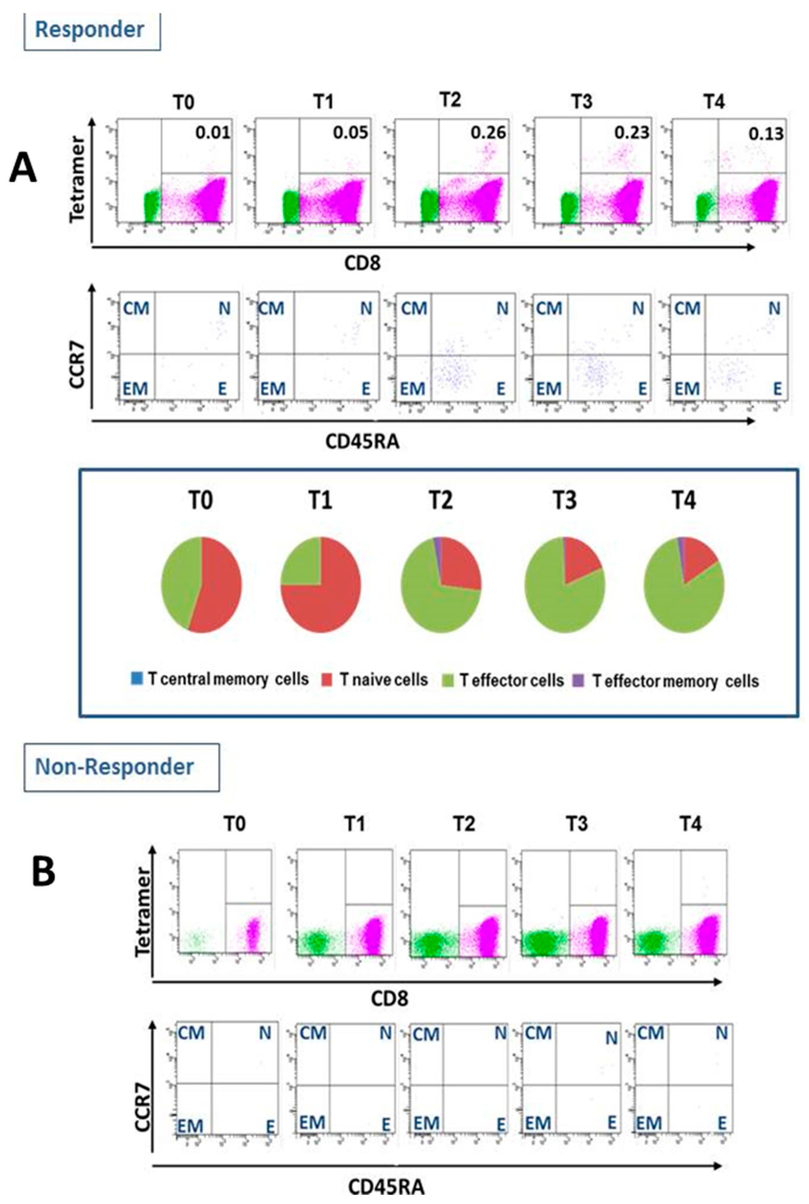
3.4. CMV-Specific T Cells and Release

All enrolled patients were CMV IgM/IgG negative prior to vaccination. At baseline, participants showed neither pre-existing CMV-specific CD8+ T cells in tetramer-based flow cytometry nor significant (>10/200.000) IFNγ spot-forming cells (SFC).
In five of 10 patients, any immune response was detected by an increase in IFNγ production in the T-Track™ assay and/or an increase of CMV-specific CD8+ T cells were observed (Table 4, exemplary Patient 03 in Figure 1). All patients with CMV-specific CD8+ T cells presented an increase of CMV-specific effector T cells (Table 4).
None of the patients presented IgG seroconversion after the vaccination.

3.5. Clinical Follow-Up

None of the patients developed any CMV disease within the core study period (end of study = Day 56). At the time of analysis, nine patients have had a renal transplantation and one patient has died on the waiting list due to cardiac failure. Renal allograft function was stable in the follow-up period of six months and the following 12-month observation period. All four transplanted patients with immune response did not develop CMV replication in the follow-up period of six months, but one renal allograft recipient presenting with CMVpp65 and IE-1 antigen specific IFNγ release, but missing CMV-specific CD8+ T cells, developed CMV disease with a CMV colitis at Month 7. This patient also had a high immunosuppressive load with tacrolimus (Tac) trough levels of about 10 ug/L and mycophenolic acid (MPA) 1000 mg BID. One patient classified as responder received a seronegative organ and did not develop any CMV infection after transplantation including the extended observational period. Four patients classified as non-responders developed CMV replication after transplantation, while the fifth non-responding patient received a CMV negative organ. Without any inoculation with CMV, of course no reactivation could occur. Two of these four patients presented with CMV syndrome, one with CMV disease (pneumonia) and one with CMV replication only. The last patient had presented with very small CMVpp65 and IE-1 antigen specific IFNγ release after vaccination, but missing CMV-specific CD8+ T cells. Due to very small immune response, the patient was primarily classified as non-responder. Further information is given in Table 5.

4. Discussion

In this investigator-initiated phase I study, safety and feasibility of a specific CMV peptide vaccination in end-stage renal disease patients on preparation to renal transplantation was assessed for the first time.
In the most recent phase I study, the present CMVpp65-derived peptide was used to vaccinate ten patients receiving an allogeneic stem cell graft from CMV-seronegative donors with encouraging results [19]. As in the current study, vaccination was well tolerated. Seven of nine patients cleared CMVpp65 antigenemia after four vaccinations. In that study, only one patient received prophylactic vaccination, but this patient did not develop antigenemia. This observation underlines our prophylactic approach in end-stage renal patients on the transplant waiting list.
In the present study, the novel method of peptide vaccination was used. Conventional vaccine strategies have been highly efficacious for several decades in reducing mortality and morbidity due to infectious diseases. However, conventional vaccines, such as those that include whole organisms or large proteins, appear to have some adverse side effects due to inclusion of unnecessary antigenic load [22]. A high antigenic load might complicate the vaccination due to induction of allergenic responses. Peptide vaccination is an attractive alternative strategy that relies on usage of short peptide fragments to engineer the induction of highly targeted immune responses. On the other side, peptide vaccines are often weakly immunogenic and require adjuvants. In the present study, a specific CMV peptide vaccine was used in combination with incomplete Freund’s Adjuvant (Montanide®) and local application of imiquimod (Aldara® 5% cream). Both adjuvants had been used successfully and safely in earlier studies.
In our present study on further kidney transplant recipients, patients received four vaccinations as per protocol. No serious adverse drug reactions or serious adverse events were detected. All 19 side effects (mostly pruritus and pressure pain) were classified as CTC (common toxicity criteria) Grade I reactions of the skin at the site of injection. These side effects resolved without sequels. No other toxicities were observed (Table 3).
All enrolled patients were CMV IgM/IgG negative prior to vaccination. Five of the 10 patients (50%) mounted any immune response. Four patients developed CMV-specific effector T cells and one patient developed significant IE-1- and pp65-specific spot formatting cells in the IFN-γ ELISpot assay only, all five patients were classified as CMV peptide vaccination responders (Table 4 and Figure 1).
These results are corresponding to other studies on vaccination response in end-stage renal disease patients. In dialysis patients, response rates between 35% and 67% are reported after hepatitis B and influenza vaccination depending on the type of vaccine and number of applied dosages as well as additional boost vaccinations [23,24,25,26].
Protective immunity can be induced by the formation of protective antibodies which requires an effective cross-linking of B cell receptors on B cells stimulating B cell affinity maturation. Monomeric peptide vaccines are rather poorly immunogenic with regard to B cell stimulation and antibody formation [27]. This is consistent with our observation in the study.
Epitope-specific T cell stimulation is another mechanism by which vaccines can induce protective immunity. Peptides can be presented by antigen-presenting cells (APCs) on the peptide binding groove of Class I or II major histocompatibility complexes (MHCs) to the T cell receptor (TCR) of T cells and can lead to a peptide-specific T cell clone expansion. For T-cell epitopes, immunodominance is an important consideration for peptide vaccine design. Moreover, the vaccine is injected with an adjuvant (e.g., Freund’s adjuvant) to boost the immune response by increasing the half-life of the epitope by decreasing the susceptibility to proteolytic degradation.
Next to the optimal vaccine, the immune system of the patient is of major relevance for the immune response. Patients with end-stage renal disease have an altered immune system with an impaired innate and adaptive immune response. Monocytes and monocyte-derived dendritic cells as the key players for antigen presentation in the vaccination strategy have been shown to display decreased endocytosis and impaired maturation in end-stage renal disease [28]. This might be the major reason for an impaired immune response to an active vaccination strategy in end-stage renal disease patients. It is known that cellular immunity through effector CD4+ and CD8+ T cells play a critical role for controlling CMV replication after transplantation [29]. Therefore, the novel T-Track® CMV IFN-γ ELISpot assay was used to measure sensitively the response of a large spectrum of clinically relevant CMV-reactive effector cells including T helper cells, cytotoxic T lymphocytes, as well as natural killer and natural killer T-like cells via bystander activation to immediate early-1 (IE-1) and phosphoprotein 65 (pp65) antigens [30]. IE-1 specific reaction is supposed to show immediate immune response, whereas the development of pp65 specific activation is expected to represent long-term immune response. In three patients, a significant increase of IE-1 and pp65-specific SFC could be detected after vaccination.
Nickel et al. described an association between CMV disease with low IE-1-specific T-cell frequencies in a pilot study on renal allograft recipients [31]. In a previous observational study, it has been shown that IE-1 CMV specific T cell frequencies before transplantation could help to discriminate those patients with no need of CMV prophylactic treatment form those in whom prophylaxis is indicated [32]. Intrinsic impairment of IE-1 specific T cell response but not pp65-specific T cells was associated with post-transplant CMV infection as well as CMV disease. In addition, in this previous study only patients with adequate pre-transplant anti-IE-1-specific T cell frequencies were at significant low-risk for CMV infection demonstrating that although CMV triggers both humoral and cellular immune responses, especially the cellular response directed to IE-1 CMV antigen seemed to be important for post-transplant viral replication control. In parallel to our study, Bestard et al. [32] also showed that patients who had not experienced CMV infection showed a significantly lower T cell response when compared to patients with an earlier infection period. Concerning IE-1-specific T cell response the threshold of 7 spots in 3 × 10⁵ showed a high negative predictive value of 95.7%.
None of the patients presented IgG seroconversion after the vaccination. Humoral immunity after primary viral infection is long-lasting. However, the contribution of antibodies (as assessed by standard serology) towards protection against CMV replication in transplant recipients is questionable [33].
After transplantation, immunosuppression may interfere with CMV immune response [29]. T-cell depleting agents increased the risk for CMV infection due to direct depletion of functional CMV-specific T cells or induction of proinflammatory cytokine release which is involved in the activation of latent CMV. Mycophenolic acid blocking activated lymphocytes may facilitate CMV infection especially in high doses. While T-cell depleting agents were prohibited in this study, the use of mycophenolic acid might have influenced even the after vaccination existing cellular immune response. However, none of the vaccinated patients with immune response suffered from clinically overt CMV disease within the first six months, whereas one patient who received a renal allograft more than four years after vaccination developed CMV disease seven months after transplantation. This patient presented only with CMVpp65 and IE-1 antigen specific IFNγ release, but missing CMV-specific CD8+ T cells (Table 5). In addition, this patient had a high immunosuppressive load in the weeks prior to CMV disease. Forty percent of the patients showed CMV-specific CD8+ T cell responses elicited by these prophylactic vaccinations. All responders did never experience CMV reactivation in the 18 months after transplantation, while all non-responders reactivated. In summary, prophylactic CMV specific peptide vaccination before kidney transplantation induces a T cell mediated response and therefore may prevent CMV infection after transplantation. Since immune response among the patients’ is heterogenous and sometimes unpredictable, a pre-vaccination immune test as the CD4 T cell count might be helpful to detect patients with potential response to vaccination. Because of the small numbers of patients, the correlation between CMV-specific T-cell reactivity and vaccine response requires further investigation. Further studies with an increased patient number and multi-center assessment are necessary to confirm our results. Future vaccines might also include more viral antigen peptides including HLA class II antigens.

Supplementary Materials

The following are available online at https://www.mdpi.com/2076-393X/9/2/133/s1. Table S1. Antibody list; Table S2. Release Criteria for peptide vaccines.

Author Contributions

C.S. set up the study concept and design; participated in the performance of the study, study conduct, data collection, data analysis; and wrote the manuscript. A.S. was head of production of vaccines. A.H.-K. was responsible for quality control (QC) of the vaccines and logistics. T.B. performed statistical analysis and wrote the manuscript. T.G. participated in the investigation. L.W. participated in vaccine preparation. P.S. was responsible for CMV serology and PCR. S.M. and M.Z. supervised the study conception and performance of the study. M.S. set up the study concept and design, was qualified person (QP) for vaccine preparation, and wrote the manuscript. All authors were involved in the study conduct and review of the study data. All authors have read and agreed to the published version of the manuscript.

Funding

The study was partly funded by the Else Kröner-Fresenius Foundation, Germany, the Division of Nephrology and Renal Clinic Heidelberg, and the Department of Internal Medicine V, University Hospital Heidelberg.

Institutional Review Board Statement

The study was conducted according to the guidelines of the Declaration of Helsinki, and approved by the Institutional Ethics Committee (IRB No. AFmo-256/2013, date of approval 10 October 2013) as well as by Federal Regulatory Authority, the Paul-Ehrlich-Institute, Langen, Germany (PEI registration No. 1855/02, date of approval 10 October 2013).

Informed Consent Statement

Informed consent was obtained from all subjects involved in the study.

Data Availability Statement

The data presented in this study are available on request from the corresponding author. Data are not publicly available due to the national law.

Conflicts of Interest

The following authors of this manuscript have conflicts of interest to disclose: Conflict of interest (COI) declaration for MS: Apogenix: Funding for collaborative research. Hexal: Financial support for research on biosimilars, travel grants. Kite: Financial support of educational activities and conference, travel grants. Co-PI of clinical trials on CAR-T cells. MSD: Ad board member, PI of clinical trials on letermovir. Novartis: Collaborative research grant CAR-T cells. Co-PI of clinical trials on CAR-T cells. TolerogenixX: Co-Founder and shareholder. Conflict of interest (COI) declaration for AS: TolerogenixX: Co-Founder and shareholder. Conflict of interest (COI) declaration for CS: Chiesi: Grant for research on biomarkers independent of the present study provided to the institution. Novartis: PI of clinical trials on immunosuppression. Calliditas Therapeutics: PI of a clinical trial on renal disease. All other authors declared no COIs.

References

  1. De Keyzer, K.; Van Laecke, S.; Peeters, P.; Vanholder, R. Human cytomegalovirus and kidney transplantation: A clinician’s update. Am. J. Kidney Dis. 2011, 58, 118–126. [Google Scholar] [CrossRef]
  2. Sagedal, S.; Hartmann, A.; Nordal, K.P.; Osnes, K.; Leivestad, T.; Foss, A.; Degre, M.; Fauchald, P.; Rollag, H. Impact of early cytomegalovirus infection and disease on long-term recipient and kidney graft survival. Kidney Int. 2004, 66, 329–337. [Google Scholar] [CrossRef] [Green Version]
  3. Hodson, E.M.; Jones, C.A.; Webster, A.C.; Strippoli, G.F.; Barclay, P.G.; Kable, K.; Vimalachandra, D.; Craig, J.C. Antiviral medications to prevent cytomegalovirus disease and early death in recipients of solid-organ transplants: A systematic review of randomised controlled trials. Lancet 2005, 365, 2105–2115. [Google Scholar] [CrossRef]
  4. Hjelmesaeth, J.; Sagedal, S.; Hartmann, A.; Rollag, H.; Egeland, T.; Hagen, M.; Nordal, K.P.; Jenssen, T. Asymptomatic cytomegalovirus infection is associated with increased risk of new-onset diabetes mellitus and impaired insulin release after renal transplantation. Diabetologia 2004, 47, 1550–1556. [Google Scholar] [CrossRef] [PubMed]
  5. Sharples, L.D.; Jackson, C.H.; Parameshwar, J.; Wallwork, J.; Large, S.R. Diagnostic accuracy of coronary angiography and risk factors for post-heart-transplant cardiac allograft vasculopathy. Transplantation 2003, 76, 679–682. [Google Scholar] [CrossRef]
  6. Fateh-Moghadam, S.; Bocksch, W.; Wessely, R.; Jager, G.; Hetzer, R.; Gawaz, M. Cytomegalovirus infection status predicts progression of heart-transplant vasculopathy. Transplantation 2003, 76, 1470–1474. [Google Scholar] [CrossRef]
  7. Ducloux, D.; Bamoulid, J.; Crepin, T.; Rebibou, J.-M.; Courivaud, C.; Saas, P. Posttransplant Immune Activation: Innocent Bystander or Insidious Culprit of Posttransplant Accelerated Atherosclerosis. Cell Transplant. 2017, 26, 1601–1609. [Google Scholar] [CrossRef] [PubMed] [Green Version]
  8. Rozen-Zvi, B.; Lichtenberg, S.; Green, H.; Cohen, O.; Chagnac, A.; Mor, E.; Rahamimov, R. Cytomegalovirus-negative kidney transplant recipients are at an increased risk for malignancy after kidney transplantation. Clin. Transplant. 2016, 30, 980–985. [Google Scholar] [CrossRef]
  9. Kliem, V.; Fricke, L.; Wollbrink, T.; Burg, M.; Radermacher, J.; Rohde, F. Improvement in long-term renal graft survival due to CMV prophylaxis with oral ganciclovir: Results of a randomized clinical trial. Am. J. Transplant. 2008, 8, 975–983. [Google Scholar] [CrossRef]
  10. Witzke, O.; Hauser, I.A.; Bartels, M.; Wolf, G.; Wolters, H.; Nitschke, M.; Group, V.S. Valganciclovir prophylaxis versus preemptive therapy in cytomegalovirus-positive renal allograft recipients: 1-year results of a randomized clinical trial. Transplantation 2012, 93, 61–68. [Google Scholar] [CrossRef] [PubMed]
  11. Helantera, I.; Kyllonen, L.; Lautenschlager, I.; Salmela, K.; Koskinen, P. Primary CMV infections are common in kidney transplant recipients after 6 months valganciclovir prophylaxis. Am. J. Transplant. 2010, 10, 2026–2032. [Google Scholar] [CrossRef]
  12. Hill, A.B. The immune response to CMV infection and vaccination in mice, monkeys and humans: Recent developments. Curr. Opin. Virol. 2018, 28, 161–166. [Google Scholar] [CrossRef] [PubMed]
  13. Nakamura, R.; La Rosa, C.; Longmate, J.; Drake, J.; Slape, C.; Zhou, Q.; Lampa, M.G.; O′Donnell, M.; Cai, J.L.; Farol, L.; et al. Viraemia, immunogenicity, and survival outcomes of cytomegalovirus chimeric epitope vaccine supplemented with PF03512676 (CMVPepVax) in allogeneic haemopoietic stem-cell transplantation: Randomised phase 1b trial. Lancet Haematol. 2016, 3, e87–e98. [Google Scholar] [CrossRef] [Green Version]
  14. Bernstein, D.I.; Munoz, F.M.; Callahan, S.T.; Rupp, R.; Wootton, S.H.; Edwards, K.M.; Turley, C.B.; Stanberry, L.R.; Patel, S.M.; McNeal, M.M.; et al. Safety and efficacy of a cytomegalovirus glycoprotein B (gB) vaccine in adolescent girls: A randomized clinical trial. Vaccine 2016, 34, 313–319. [Google Scholar] [CrossRef] [Green Version]
  15. Pass, R.F.; Zhang, C.; Evans, A.; Simpson, T.; Andrews, W.; Huang, M.L.; Corey, L.; Hill, J.; Davis, E.; Flanigan, C.; et al. Vaccine prevention of maternal cytomegalovirus infection. N. Engl. J. Med. 2009, 360, 1191–1199. [Google Scholar] [CrossRef] [PubMed]
  16. Wloch, M.K.; Smith, L.R.; Boutsaboualoy, S.; Reyes, L.; Han, C.; Kehler, J.; Smith, H.D.; Selk, L.; Nakamura, R.; Brown, J.M.; et al. Safety and immunogenicity of a bivalent cytomegalovirus DNA vaccine in healthy adult subjects. J. Infect. Dis 2008, 197, 1634–1642. [Google Scholar] [CrossRef] [PubMed]
  17. Kharfan-Dabaja, M.A.; Boeckh, M.; Wilck, M.B.; Langston, A.A.; Chu, A.H.; Wloch, M.K.; Guterwill, D.F.; Smith, L.R.; Rolland, A.P.; Kenney, R.T. A novel therapeutic cytomegalovirus DNA vaccine in allogeneic haemopoietic stem-cell transplantation: A randomised, double-blind, placebo-controlled, phase 2 trial. Lancet Infect. Dis. 2012, 12, 290–299. [Google Scholar] [CrossRef] [Green Version]
  18. Reusser, P.; Riddell, S.R.; Meyers, J.D.; Greenberg, P.D. Cytotoxic T-lymphocyte response to cytomegalovirus after human allogeneic bone marrow transplantation: Pattern of recovery and correlation with cytomegalovirus infection and disease. Blood 1991, 78, 1373–1380. [Google Scholar] [CrossRef] [PubMed] [Green Version]
  19. Schmitt, M.; Schmitt, A.; Wiesneth, M.; Huckelhoven, A.; Wu, Z.; Kuball, J.; Wang, L.; Schauwecker, P.; Hofmann, S.; Gotz, M.; et al. Peptide vaccination in the presence of adjuvants in patients after hematopoietic stem cell transplantation with CD4+ T cell reconstitution elicits consistent CD8+ T cell responses. Theranostics 2017, 7, 1705–1718. [Google Scholar] [CrossRef]
  20. Ljungman, P.; Boeckh, M.; Hirsch, H.H.; Josephson, F.; Lundgren, J.; Nichols, G.; Pikis, A.; Razonable, R.R.; Miller, V.; Griffiths, P.D. Definitions of Cytomegalovirus Infection and Disease in Transplant Patients for Use in Clinical Trials. Clin. Infect. Dis. 2017, 64, 87–91. [Google Scholar] [CrossRef] [Green Version]
  21. Schmitt, A.; Bechter, C.; Yao, J.; Goetz, M.; Maccari, B.; Schauwecker, P.; Wiesneth, M.; Schmitt, M. Cytomegalovirus vaccination of leukemia and lymphoma patients after allogeneic stem cell transplantation--validation of a peptide vaccine. J. Immunol. Methods 2009, 343, 140–147. [Google Scholar] [CrossRef]
  22. Li, W.; Joshi, M.D.; Singhania, S.; Ramsey, K.H.; Murthy, A.K. Peptide Vaccine: Progress and Challenges. Vaccines (Basel) 2014, 2, 515–536. [Google Scholar] [CrossRef] [PubMed] [Green Version]
  23. Ayub, M.A.; Bacci, M.R.; Fonseca, F.L.; Chehter, E.Z. Hemodialysis and hepatitis B vaccination: A challenge to physicians. Int. J. Gen. Med. 2014, 7, 109–114. [Google Scholar] [CrossRef] [Green Version]
  24. Chaves, S.S.; Daniels, D.; Cooper, B.W.; Malo-Schlegel, S.; Macarthur, S.; Robbins, K.C.; Kobetitsch, J.F.; McDaniel, A.; D′Avella, J.F.; Alter, M.J. Immunogenicity of hepatitis B vaccine among hemodialysis patients: Effect of revaccination of non-responders and duration of protection. Vaccine 2011, 29, 9618–9623. [Google Scholar] [CrossRef]
  25. Dikow, R.; Eckerle, I.; Ksoll-Rudek, D.; Hampel, H.; Schwenger, V.; Zeier, M.; Schnitzler, P.; Sommerer, C. Immunogenicity and efficacy in hemodialysis patients of an AS03(A)-adjuvanted vaccine for 2009 pandemic influenza A(H1N1): A nonrandomized trial. Am. J. Kidney Dis. 2011, 57, 716–723. [Google Scholar] [CrossRef] [PubMed]
  26. Mastalerz-Migas, A.; Gwiazda, E.; Brydak, L.B. Effectiveness of influenza vaccine in patients on hemodialysis—A review. Med. Sci Monit 2013, 19, 1013–1018. [Google Scholar] [CrossRef] [PubMed] [Green Version]
  27. Malonis, R.J.; Lai, J.R.; Vergnolle, O. Peptide-Based Vaccines: Current Progress and Future Challenges. Chem. Rev. 2020, 120, 3210–3229. [Google Scholar] [CrossRef] [Green Version]
  28. Kato, S.; Chmielewski, M.; Honda, H.; Pecoits-Filho, R.; Matsuo, S.; Yuzawa, Y.; Tranaeus, A.; Stenvinkel, P.; Lindholm, B. Aspects of immune dysfunction in end-stage renal disease. Clin. J. Am. Soc. Nephrol. 2008, 3, 1526–1533. [Google Scholar] [CrossRef] [PubMed] [Green Version]
  29. Lucia, M.; Crespo, E.; Cruzado, J.M.; Grinyo, J.M.; Bestard, O. Human CMV-specific T-cell responses in kidney transplantation; toward changing current risk-stratification paradigm. Transpl. Int. 2014, 27, 643–656. [Google Scholar] [CrossRef]
  30. Barabas, S.; Spindler, T.; Kiener, R.; Tonar, C.; Lugner, T.; Batzilla, J.; Bendfeldt, H.; Rascle, A.; Asbach, B.; Wagner, R.; et al. An optimized IFN-gamma ELISpot assay for the sensitive and standardized monitoring of CMV protein-reactive effector cells of cell-mediated immunity. BMC Immunol. 2017, 18, 14. [Google Scholar] [CrossRef] [Green Version]
  31. Nickel, P.; Bold, G.; Presber, F.; Biti, D.; Babel, N.; Kreutzer, S.; Pratschke, J.; Schonemann, C.; Kern, F.; Volk, H.D.; et al. High levels of CMV-IE-1-specific memory T cells are associated with less alloimmunity and improved renal allograft function. Transpl. Immunol. 2009, 20, 238–242. [Google Scholar] [CrossRef] [PubMed]
  32. Bestard, O.; Lucia, M.; Crespo, E.; Van Liempt, B.; Palacio, D.; Melilli, E.; Torras, J.; Llaudo, I.; Cerezo, G.; Taco, O.; et al. Pretransplant immediately early-1-specific T cell responses provide protection for CMV infection after kidney transplantation. Am. J. Transplant. 2013, 13, 1793–1805. [Google Scholar] [CrossRef] [PubMed]
  33. Humar, A.; Mazzulli, T.; Moussa, G.; Razonable, R.R.; Paya, C.V.; Pescovitz, M.D.; Covington, E.; Alecock, E. Valganciclovir Solid Organ Transplant Study, G. Clinical utility of cytomegalovirus (CMV) serology testing in high-risk CMV D+/R- transplant recipients. Am. J. Transplant. 2005, 5, 1065–1070. [Google Scholar] [CrossRef] [PubMed]
Figure 1. Cytomegalovirus(CMV)-specific CD8+ T cells frequency and subsets in patients receiving CMV peptide vaccination. (A): Example of a patient(#003) with positive immune response to cytomegalovirus (CMV)-specific peptide vaccination (responder). a. Classical AMV-specific ab CD8+ T cells were assessed with tetramer-based flow cytometry. b. Change in T-cell subsets. (B): Example of patient (#001) with negative immune response to cytomegalovirus(CMV)-specific peptide vaccination (non- responder). a. Classical AMV-specific ab CD8+ T cells were assessed with tetramer-based flow cytometry.
Vaccines 09 00133 g001
Table 1. Demographic data of study participants.
ParameterResult
Gender (female/male)n (%)4/6 (40/60)
Age(years), mean ± SD49.7 ± 12.7
Body Mass Index(kg/m2), mean ± SD24.1 ± 2.1
Renal disease
  Glomerulonephritisn (%)5 (50)
  ADPKDn (%)1 (10)
  Alport Syndromen (%)1 (10)
  Nephrocalcinosisn (%)1 (10)
  Analgesic Nephropathyn (%)1 (10)
  Unknown (shrunken kidney)n (%)1 (10)
Pre-emptive transplantationn (%)1 (10)
Dialysis
  Hemodialysisn (%)7 (70)
  Peritoneal Dialysisn (%)2 (20)
Time to Renal Replacement Therapy(months), mean ± SD84.6 ± 29.4
Table 2. Clinical characteristics of vaccinated patients.
Pat #GenderAge (Years)BMI (kg/m2)Underlying Renal DiseaseType of RRTTime on RRT (Months)Transplant
Program
Karnofsky IndexBlood Pressure (mmHg)Creatinine (mg/dL)Albumin (g/L)
01F4626.9ADPKDPre-emptive0Living0.9120/705.9044.0
02 *M6726.7GNHD39ESP0.8162/936.9547.5
03M2521.1Alport
syndrome
HD115ETKAS0.9150/959.1840.9
04F4526.7unknownHD90ETKAS0.9124/869.4042.9
05M5922.8GNHD125ETKAS0.9130/807.3144.3
06M3922.5GNPD75ETKAS0.9140/9012.538.2
07M5722.2GNHD42ETKAS1115/846.7644.3
08F4525.1Analgesic nephropathyPD80ETKAS0.7130/8511.437.1
09F6523.0Nephro-calcinosisHD98ETKAS1180/808.1738.9
10M4924.1GNHD97ETKAS1110/7512.142.7
ADPKD, autosomal-dominant polycystic kidney disease; BMI, body mass index; ESP, Eurotransplant Senior Program; ETKAS, Euro Transplant Kidney Allocation System; F, female; GN, glomerulonephritis; HD, hemodialysis; M, male; PD, peritoneal dialysis; RRT, renal replacement therapy; * Patient 02 had already undergone a preceding kidney transplantation.
Table 3. Adverse events in vaccinated patients: (a) associated to vaccination; and (b) not associated to vaccination.
(a)
(a)
Pat #Number of EventsInflammationSwellingPruritusPainHematomaFatigue
014111 1
021 1
0321 1
041 1
052 2
062 1 1
073 111
0841 111
091 1
101 1
(b)
(b)
Pat #Number of EventsRespiratory
Tract Infection
Gastro-Intestinal
Infection
Muscle CrampsHyperkalemiaHypotensionPollinosisRenal Cyst Bleeding Abrasions after Bicycle Accident
012 11
02
0321 1
0443 1
0511
061 1
07
081 1
09
102 11
Table 4. Virus status and immunological responses up to the end of the study. Prior to vaccination, none of the 10 patients had experienced a CMV infection. All patients had a negative serostatus for CMV. No CMV-neutralizing antibodies were found in the serum. IFT for CMVpp65 and qPCR for CMV tested native in all patients.
Pat #T-Track® CMV
(Antigen-Specific SFC/200.000 Lymphocytes) *
CMV-Specific
CD8+ T Cells % #
before → after
Vaccination
CMV-Specific Effector T Cells (EM) %
before → after
Vaccination
Any CMV Peptide Immune Reaction
(FACS and/or Elispot)
IE-1+
before → after
Vaccination
pp65+
before → after
vaccination
011 → 10 → 10 → 0NANo
022 → 141 → 130 → 0NANo
031 → 420 → 120 → 0.336 → 87Yes
043 → 12 → 30 → 0NANo
054 → 240 → 40 → 0.133 → 86Yes
061 → 201 → 100 → 0.131 → 72Yes
070 → 1040 → 500 → 0NAYes
084 → 22 → 40 → 0NANo
091 → 11 → 00 → 0.10 → 91Yes
101 → 11 → 20 → 0NANo
CMV, cytomegalovirus; PCR, polymerase chain reaction; IFNy, interferon y, NA, not applicable. * T-Track® CMV: positive >10 antigen-specific spot-forming colonies (SFC) per 200,000 lymphocytes. # Tetramer staining: (0) Negative → 0.0%; (1) low → 0.1–1.0% tetramer+ CD8+ T cell; (2) medium → 1–5%; (3) high → >5%.
Table 5. Virus status, immunological responses and clinical outcome within the follow-up after transplantation.
Pat #Patient OutcomeTime from Vaccination to Transplantation (Months)CMV StatusProphylactic CMV TreatmentImmunosuppressive RegimenS-Creatinine (mg/dL), Month 6CMV
Replication/Disease
until Month 18 after tx
CMV Specific T Cell Response
01Living kidney tx *
7D+/R-pre-emptiveplasmapheresis, rituximab, ATG, steroids, MPA, Tac +1.20Yes (CMV syndrome)
(month 18 after tx)
No
02Deceased kidney tx2D+/R-pre-emptivebasiliximab, steroids, MPA, Tac1.54Yes (CMV replication)
(month 5 after tx)
No
03Deceased kidney tx 43D+/R-pre-emptivebasiliximab, steroids, MPA, CsA2.05NoYes
04Deceased kidney tx *46D+/R-pre-emptiveATG, steroids, MPA, Tac1.59Yes (CMV disease)
(month 5 after Tx)
No
05Deceased kidney tx17D+/R-pre-emptiveATG, steroids, MPA, Tac1.94NoYes
06Deceased kidney tx48D-/R-pre-emptivebasiliximab, steroids, MPA, Tac1.59NoYes
07Deceased kidney tx53D+/R-pre-emptivebasiliximab, steroids, MPA, Tac1.28Yes (CMV disease)
(month 7 after tx)
Yes
08Deceased kidney tx4D+/R-pre-emptivebasiliximab, steroids, MPA, CsA1.67Yes (CMV syndrome)
(month 12 after tx)
No
09Died from cardiac failure on waiting listNANANANANANAYes
10Deceased kidney tx18 D-/R-pre-emptivebasiliximab, steroids, MPA, CsA1.75NoNo
CMV, cytomegalovirus; CsA, Ciclosporin A; IFNy, interferon y; MPA, mycophenolate acid; NA, not applicable; PCR, polymerase chain reaction; Tac, tacrolimus; Tx, transplantation.
Publisher’s Note: MDPI stays neutral with regard to jurisdictional claims in published maps and institutional affiliations.

