Review

Immunomodulatory Strategies for Parapoxvirus: Current Status and Future Approaches for the Development of Vaccines against Orf Virus Infection

by
Alhaji Modu Bukar
1,2,*,
Faez Firdaus Abdullah Jesse
3,
Che Azurahanim Che Abdullah
4,
Mustapha M. Noordin
1,
Zaharaddeen Lawan
1,
Hassana Kyari Mangga
1,
Krishnan Nair Balakrishnan
1 and
Mohd-Lila Mohd Azmi
1,*
1
Department of Pathology and Microbiology, Faculty of Veterinary Medicine, Universiti Putra Malaysia, Serdang 43400, Selangor, Malaysia
2
Department of Science Laboratory Technology, School Agriculture and Applied Sciences, Ramat Polytechnic Maiduguri, Maiduguri 1070, Borno, Nigeria
3
Department of Veterinary Clinical Studies, Faculty of Veterinary Medicine, Universiti Putra Malaysia, Serdang 43400, Selangor, Malaysia
4
Department of Physics, Faculty of Science, Universiti Putra Malaysia, Serdang 43400, Selangor, Malaysia
*
Authors to whom correspondence should be addressed.
Vaccines 2021, 9(11), 1341; https://doi.org/10.3390/vaccines9111341
Submission received: 9 August 2021 / Revised: 25 August 2021 / Accepted: 25 August 2021 / Published: 17 November 2021
(This article belongs to the Special Issue Vaccines in Small Ruminants)

Abstract

:
Orf virus (ORFV), the prototype species of the parapoxvirus genus, is the causative agent of contagious ecthyma, an extremely devastating skin disease of sheep, goats, and humans that causes enormous economic losses in livestock production. ORFV is known for its ability to repeatedly infect both previously infected and vaccinated sheep due to several immunomodulatory genes encoded by the virus that temporarily suppress host immunity. Therefore, the development of novel, safe and effective vaccines against ORFV infection is an important priority. Although, the commercially licensed live-attenuated vaccines have provided partial protection against ORFV infections, the attenuated viruses have been associated with major safety concerns. In addition to safety issues, the persistent reinfection of vaccinated animals warrants the need to investigate several factors that may affect vaccine efficacy. Perhaps, the reason for the failure of the vaccine is due to the long-term adaptation of the virus in tissue culture. In recent years, the development of vaccines against ORFV infection has achieved great success due to technological advances in recombinant DNA technologies, which have opened a pathway for the development of vaccine candidates that elicit robust immunity. In this review, we present current knowledge on immune responses elicited by ORFV, with particular attention to the effects of the viral immunomodulators on the host immune system. We also discuss the implications of strain variation for the development of rational vaccines. Finally, the review will also aim to demonstrate future strategies for the development of safe and efficient vaccines against ORFV infections.

1. Introduction

Small ruminants are of great economic importance to humans as they provide a tremendous source of protein, calcium, vitamins, fiber, hides, and especially wool for hundreds of millions of people worldwide. However, it is believed that the economic losses caused by viruses are not adequately recorded because it is an acute infection. Moreover, a larger percentage of infectious diseases that hinder the livestock industry today are caused by pathogenic animal viruses [1,2,3,4,5]. To meet the high demand in livestock production, animals must be protected from infectious viruses such as ORFV [6,7,8,9] and several other pathogenic viruses of veterinary importance [10,11,12,13,14,15,16]. Therefore, the prevention of infectious diseases in animals can lead to widespread eradication of viruses [17,18,19,20], sustainable livestock production [21] and consequently improved life expectancy of farmers and veterinarians [6,22,23,24].
Orf virus (ORFV), the prototype species of the genus Parapoxvirus in the family Poxviridae causes highly contagious skin lesions popularly known as orf in several species of small ruminant particularly sheep and goats [17,25,26,27,28,29,30], although it can also infect humans [31,32,33,34]. ORFV infection may result in up to 100 percent morbidity in lambs, mortality can be up to 50 percent in animals with secondary infections [35] and immunocompromised conditions [36]. The virus is well known for its resistance to harsh environmental conditions [37] and its ability to cause persistent reinfections in sheep [38] and goats which has contributed to the further spread of infections in other animals such as deer, guinea pigs, and dogs [28,30,31,32,33,34,35,36,37,38,39]. The virus can be inadvertently transmitted to a susceptible animal through injured skin, readily replicates, and causes proliferative lesions that give rise to scab [19,20,21,22,23,24,25,26,28,29,40]. The classic pathological signs are scabs on the mucosal borders of the skin, lips, nose, eyelids, feet, teats of lactating ewes, and mouth [26,31,32]. Likewise, the virus incurred huge financial losses in the small ruminants industry by reducing both the quality [39,41] and quantity of milk [3,42,43], and even the death of infected animals [6,44,45]. In addition, ORFV infection has serious socio-cultural and economic challenges for livestock farmers, most of whom rely on small ruminants as their main source of livelihood [17,27,33,34,35,36,37]. Thus, ORFV infections increase the demand for the huge cost of management; resulting in reduced average income [27,40,46,47,48,49]. However, the acute infection caused by ORFV, which usually affects the lips, nose, teats, and sometimes on mucosal parts, has been described as a self-limiting disease, [32,50,51,52,53,54,55,56,57], and as such it has no positive impact on management although in several instances, particularly in lambs, it can turn to be persistent and even death [32,48]. The severe infection, which usually affects the eyelids, feet, vulva, and in some cases the udder has reportedly led to outbreaks in young animals [37] and kids resulted in tremendous losses in dairy farming across the world [20,47,49,50].
Furthermore, several strategies have been explored to develop effective antiviral drugs and prophylactic vaccines to control ORFV infection in sheep and goats, most of which are derived from immunogenic envelope proteins. Treatment of infected animals with antiviral drugs can only reduce the severity of the disease but cannot eliminate the virus [37,51]. Although, treating an infected animal with antibiotics can only minimize secondary bacterial infections such as staphylococcal dermatitis associated with ORFV infection [20,58,59,60,61,62,63,64,65]. It is well documented that misuse of antibiotics and chemotherapeutic agents has resulted in acaricide antibiotic resistance and accumulation of residues in the affected animals [41,52,53,54]. So far, vaccination has been the most effective alternative to antibiotics and antiviral agents [66]. Thus, the use of prophylactic vaccines and quarantine are the most cost-effective approaches to minimize the need for antimicrobials by controlling ORFV infections in sheep and goats of all ages [67,68,69,70]. As such, strict compliance to vaccination, extensive precautions combined with prompt treatment of infected animals, [71,72,73] and other containment measures such as improved sanitation appear to prevent further spread of the disease [74,75,76,77,78,79].
To date, vaccination of an infected animal is the best, if not the only, alternative, to effectively eradicate ORFV [78,79]. Vaccines are biological or synthetic preparations of immunogens-proteins, carbohydrates, and lipids derived from microorganisms or other synthetic sources- that when administered to the host, induce sustained immunogenic memory for the antigen [79]. Vaccines are part of an anti-infection medicine that promotes the stimulation of the desired immune response against infectious agents in both humans and animals of veterinary importance [79,80,81,82,83,84]. Vaccines intended for animals and humans should be safe, stable, highly immunogenic, and most importantly excellent at eliciting protective immunity [78,85]. Therefore, vaccines against ORFV should be used for recurrent infections and vaccinated sheep or goats should be kept isolated from unimmunized animals [48,85]. Nevertheless, lambs and young animals lacking ORFV-specific antibodies can acquire significant immunity if immunized within the first week after birth [86]. However, there are few scientific publications on the development of vaccine and vaccination strategies against ORFV infections [86,87,88]. Additionally, the existing live-attenuated vaccines against ORFV infections do not elicit protective immunity against the virus [56,88]. Therefore, current attenuated vaccines present a serious risk of reversion to virulence [56,89]. These varying degrees of vaccine failure warrant the re-examination of several determining factors particularly genes and proteins involved in virulence and pathogenesis of the virus, which may influence the safety and efficacy of existing vaccines against ORFV infection [53,60,90,91,92,93].
Detailed knowledge of viral structure and its immunomodulatory properties has the potential to reveal underlying factors that promote more rational vaccine design [56,88,89,94]. ORFV is an ovoid epitheliotropic linear double-stranded DNA [59,95] with dimensions of approximately 260 nm × 160 nm [9,96]. The genome is approximately 138 kbp long and encodes 132 genes [93,97]. The most important physical feature of this virus is the tubular, filamentous arrangement of the outer layer of the viral particle [97,98] as shown in Figure 1. The viral capsid proteins are arranged on the surface in tabular filamentous structures [99]. The inner membrane of the virion particle surrounded by a thick wall consists of the nuclear membrane, the palisade layer, and the nucleoprotein [63,100]. The 138 kbp ORFV genome consists of 17% highly variable genes involved in pathogenesis, virulence, host range, [101,102,103,104,105,106,107,108,109,110,111,112,113] and immunomodulatory activities of the virus [70,104]. The variable regions surrounded by 3 kbp of inverted terminal repeats (ITRs), which are similar but oppositely arranged nucleotide sequences, are located at the extreme terminal ends of the genomic DNA [104] and are strongly bonded with hairpin loops [32,93,105]. On the other hand, the central conserved genomic region of ORFV accounts for approximately 80% of the entire genome which is responsible for viral replication, transcription, and morphogenesis [32,54,63,105,106,107,108].
It is well-documented that major immunogenic proteins located within the conserved region particularly B2L (ORFV011) and F1L (ORFV059), are exploited prophylactically as vaccines against ORFV infections [78,109]. Stimulation of humoral and T-cell responses against such conserved (ORFV 011 and ORFV 059) antigens has been reported to elicit robust protection against ORFV infections [110]. Interestingly, the discovery of highly neutralizing antibodies and T-cells elicited by immunogenic proteins (ORFV 011 and ORFV 059) has led to the development of chimeric recombinant DNA and subunit vaccines [111]. A more recent study has demonstrated that the chimeric recombinant ORFV DNA vaccine has elicited vigorous neutralizing ORFV-specific IgG and T-cell (CD4+ and CD8+) responses in mice [109,110,111].
This review is aimed at a broader approach to vaccine development against ORFV infections. The review also discusses the impact of the host immune response against ORFV infections, paying special attention to the efficiency and efficiency of existing vaccines to provide in-depth knowledge into the major challenges faced by existing vaccines. In addition, the article also focuses on the impact of immunomodulatory proteins encoded by ORFV and the role of some highly conserved genes, encoding immuno-dominant proteins, to provide a new strategy for future vaccine development. The well-documented problems of genetic variation among ORFV strains will also be discussed. Likewise, the review will also seek to demonstrate new strategies and future approaches for the development of efficacious vaccines to control ORFV infections. Such challenges can be addressed through careful observation and the use of recent advances in molecular biology to fill the existing knowledge gaps.

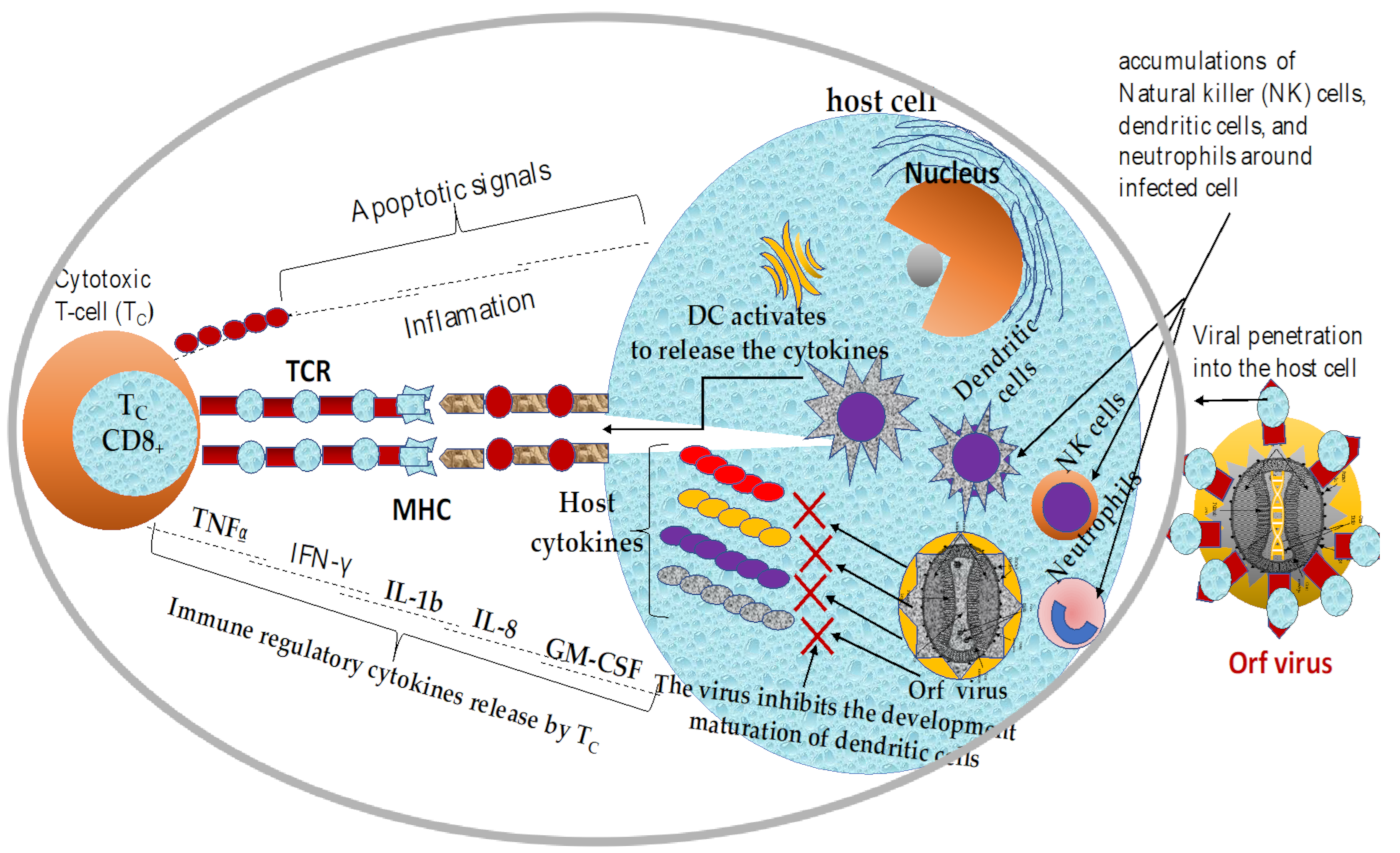
2. Mechanisms of Immune Response to ORFV Infection

2.1. Overview of Current Knowledge in Immune Responses Elicited by ORFV Infections

The immune system is versatile, and the collective network of specialized cells, tissues, and organs has evolved to protect the host from invasion by microorganisms and their toxic products [68,112]. The immune response to ORFV infection consists of both innate and adaptive responses [9,113]. Despite several studies conducted on the immune response to ORFV infection, many mechanisms of host interaction with the virus are still not clearly understood [114,115,116].
However, ORFV penetrates a susceptible host and temporarily replicates transiently by undermining host immunity [117]. Once the viral pathogen is internalized, the cells of the innate immune system such as neutrophils, MHC class II-dendritic cells (DCs), and natural killer (NK) cells are constantly recruited to the sites of infection to capture the viral antigens, which facilitates the migration of the captured antigens to the peripheral lymph node where the antigen presentation to newly recruited naïve T-cells and memory cells take place [82,117,118,119]. The dendritic cells (DCs) facilitate the transport of the internalized virus to the lymph nodes for further proliferation [119] and the presentation of specialized cytokines such as granulocyte-macrophage colony-stimulating factor (GM-CSF), interleukin-1β (IL-1b), interferon-α (IFN-α), and IL-8, and IFN-γ [54,56,73,80,120]. In addition to innate immune cell recruitment, B-cell, CD4+, and CD8+ T-cell mediated immune responses are the most common adaptive immunity associated with infected host cells [121]. However, the activated T-cell immunity could not destroy this antigens because the virus releases several virulence factors that prevent possible neutralization of the virion particles, as shown in Figure 2 [122].

2.2. Innate and Adaptive Immune Responses against ORFV Infection

Innate immunity is the first form of defense mechanism against an invading virus and is considered a nonspecific response due to the lack of immunological memory [55,69,79,123]. In addition, ORFV has strong immunomodulatory activities [118] and has evolved a strategy to evade the immune system by developing a number of virulence factors [124]. The virus targets numerous non-specific mediator defenses including complement, chemokines interleukins, inflammation, interferons (IFN), and tumor necrosis factors [79,125]. Another important line defense next to the innate response is the adaptive defense mechanism, which includes antibody-mediated and cell-mediated responses [83,111,125].
Adaptive immunity is often referred to as cell-mediated and antibody-mediated responses that promote host cell recognition by invading viral antigens [65,70,126] and tailoring immune system adaptation to a specific antigen [127]. Several studies suggested that T-cells play an important role in eliciting protective immunity against ORFV [83,128]. However, the ability of the virus to repeatedly infect the susceptible host does not appear to be associated with the production of the desired memory T-cells as a promising hypersensitivity reaction to ORFV antigen, as reported by several recent publications [54,79,83,129]. Thus, such an immune response cannot be considered abnormal for ORFV infection, as significant levels of CD4+ T-cells have been detected among the infected animals when compared to CD8+ cytotoxic T-cells and/or B-cell response [115,130,131,132,133,134,135]. On the other hand, the humoral immune response does not play an important role in protecting animals from ORFV infection [136]. It is well-documented that the abundant antibody titers detected in the sera of infected sheep with ORFV, the passage of antibodies from sheep to their offspring via colostrum [115,137] or, inoculation of sera from infected animals to healthy animals do not elicit protective immunity [138]. Therefore, immunity against ORFV is not long-lived and the virus can readily reinfect both vaccinated and unvaccinated sheep [38,136,139]. Interestingly, smaller lesions that resolve more rapidly have been observed in the case of vaccinated and reinfected sheep [67,136,140]. Presumably, the absence of ORFV-specific neutralizing antibodies in the vaccinated or previously infected animals is the main reason why ORFV to causes repeated reinfections in its host [136,141]. In addition to the lack of neutralizing antibodies, the ability of ORFV to repeatedly reinfect previously infected and/or vaccinated sheep is attributed to the immunomodulatory genes and proteins encoded by the virus that temporarily suppress host immunity [142].

2.3. Immunomodulatory Properties of Orf Virus

Parapoxvirus (ORFV) is well recognized for its ability to withstand numerous challenges to the host-triggered immune response. ORFV is able to partially circumvent the host immune response because upon acute infection of the mucocutaneous boundaries of the epidermis, the virus immediately releases multiple virulence proteins that subvert host immunity [122,143]. Therefore, the virus may ultimately utilize the various immunomodulatory strategies to modulate, subvert, evade, [108] and/or suppress host immunity [144,145,146]. The major immunomodulatory genes (IMGs) encoded by ORFV include chemokine binding protein (CBP), interleukin-10 (IL-10), vascular endothelial growth factor (VEGF), the GM-CSF inhibitory factor (GIF) [98,105] and interferon-resistance gene (OVIFNR), which inhibits protein synthesis by preventing an enzyme dsRNA-dependent kinase [48,68] (Table 1). It is well-documented that IMGs and immunomodulatory proteins (IMPs) of ORFV are acquired either from vaccinia viruses (VACV) [119,147] as a result of continuous interactions with their host [44,91]. For instance, CBP, GIP, VIR, and dUTP were acquired from VACV homologue proteins [44,105,148] and their origin from the ancestral poxviral genes [92,93,94]. Thus, the discovery of immunomodulatory genes [98] and proteins secreted by ORFV may explain how the virus escape elimination by host immunity [105,149].
In addition to therapeutic benefits, IMPs are highly involved in viral pathogenesis, and virulence activities [88,105,149]. Accordingly, such advantages would draw attention to the immunology [45,91,150] and the pathogenesis of the virus [53,85], as well as potential treatment opportunities [88,96,151]. On the other hand, intracellular virus also produces immune-modulatory proteins (IMPs) that subvert the host immune response, further blocking cell interactions [45,152].
One of the well-characterized immunomodulatory genes is the ORFV orthologue of interleukin-10. ORFV-IL-10 (ORFV127) is one of the early genes [99,153] synthesized by the virus and is located at the terminal right end of the viral genomic DNA and encodes a ~550bp, 21.7kDa protein [97,106,111,113,154]. Interestingly, parapoxvirus (ORFV) is not the only virus in the family of poxvirus that can secrete the vIL-10 homologue, which is actively involved in the suppressing and subverting of the host cellular [106] and humoral immune responses [85,103,155]. In addition, ORFV-IL-10 protein can inhibit the synthesis and trafficking of host cytokines and chemokines [103,106,156] such as IL-1b, IL-8, TNF-a, IFN-γ which is likely inhibit inflammatory reactions that in turn can prevent recruitment [157] and activation of immune cells at the site of the infection [99,108,109]. In addition, ORFV-IL-10 induces the proliferation of thymocytes and mast cells [72]. Nevertheless, ORFV-IL-10 also modulates and/or inhibits T-cell proliferation by suppressing the functions of MHC II (major histocompatibility complex class II) molecules, which decrease the recruitment of innate immune cells such as mast cells, macrophages, monocyte, and dendritic cells (DCs) to the sites of damaged skin [100,110,158].
Another virulence factor of parapoxvirus, ORFV that suppresses host immunity is chemokine-binding protein (CBP), which is encoded by the gene ORFV112 (~861 bp) [99,103,104,105,158]. ORFV-CBP is one of the early virulence genes synthesized by the virus after successful host cell invasion [104,159]. CBP is located in the highly variable terminal regions of the Orf viral genome and encodes a 31.18kDa protein [104,157]. It is well documented that the ORFV-CBP (ORFV112) allows the temporary replication of viral antigen in infected cells by inhibiting the reactivation of cellular immune responses such as IL-1β, IFN-α, IL-8, and IFN-γ [80,157]. The ORFV-CBP protein inhibits the recruitment and migration of dendritic cells and other immune cells to peripheral lymph nodes to activate an adaptive immune response [87,157]. Unlike other immunomodulators, CBP has no mammalian homolog [105]. In addition, the lack of trafficking of the immune cells can lead to inhibition of the MHC class-II pathway [102,106,157], affecting the recruitment and/or activation of cytotoxic T cells (CD8+T-cell) at the site of infected skin [87,106,107,160].
ORFV also encodes a vascular endothelial growth factor (VEGF) gene, one of the immunomodulatory proteins responsible for inhibiting the proliferation of host immunity [94,160]. However, host VEGF is a regulatory protein actively involved in the elimination of tumors [112], virus-infected cells [113], and in the wound healing process [114]. The genetic analysis of ORFV has shown that the ORFV-IL-10 [106,160] and the ORFV-VEGF (ORFV132) genes were purloined from host organisms during the evolutionary coexistence [85,115]. Therefore, the viral protein ORFV-VEGF is characteristic only of viruses of the genus Parapoxvirus. ORFV132 (~462 bp) is also one of the early genes of the virus and is located in the highly variable terminal regions at the right end of the conserved region [84,88,94,97,116]. Previous studies indicate that the ORFV-VEGF may promote the continued proliferation of epithelial cells which would facilitate the creation of more binding sites for ORFV replication [94,116,160]. In addition to the viral replication, ORFV-VEGF protects the virus from the violent effects of collective immune responses and subverts the effects of host antiviral apoptosis [55,69]. This suggests that ORFV-VEGF enhances intracellular replication of the virus by subverting the apoptotic activity of the host cell [92,98]. However, recent studies have shown that knockout of the ORFV-VEGF protein from genomic DNA attenuates has been reported to attenuate the virus and presumably reduces the severity of the disease in the host cells [84,94,160].
On the other hand, GIF (GM-CSF inhibitory factor) is a hemopoietin produced by macrophages and T-cells (among other cell types) that stimulates the development of neutrophils, macrophages/monocytes, and eosinophils from hematopoietic progenitor cells [88,103,110,160]. Unlike the other immunomodulatory proteins, ORFV-GIP has no counterpart in the host genome. Interestingly, a more recent study revealed that ORFV-GIP is one of the new immunomodulatory proteins that suppress the functions of the host cytokines such as IL-2 and GM-CSF [65]. In addition, the interferon-resistance genes of ORFV act within an infected host cell to inhibit the possible anti-inflammatory proteins blockage induced by interferons, on the ORFV orthologue of interleukin-10 and ORFV GM-CSF inhibitory factor (GIF) proteins are released from infected tissues and induce their immunomodulatory activities away from infected cells [103,157,158,159,160]. Therefore, blockage of the activities of both the innate and acquired immune response is one of the main strategies in successful ORFV infections. However, the functional mechanisms of GIP in ORFV pathogenesis and virulence are yet to be determined [84,117,160].

3. Overview of Vaccines against Animal Viral Infections

3.1. Common Viral Vaccines of Veterinary Importance

Prophylactic animal vaccines have played a significant role in the eradication of contagious diseases [5,14,25,160]. So far, vaccination has been one of the efficient and cost-effective means of preventing viral infections of both humans and veterinary importance [157,159,160,161]. Besides the intended improvement of animal health and productivity, veterinary vaccines play a tremendous role in safeguarding consumers’ health by offsetting the use of antimicrobials and antiviral drugs for the treatment of animal diseases [56,161]. Interestingly, the livestock sector has grown consistently as the result of proper and timely animal vaccinations with the existing vaccine platforms shown in Table 2 [84,162]. There are six main types of viral vaccines that are commonly used in animals; live attenuated, DNA vaccines, inactivated vaccines, recombinant vaccines, subunit vaccines, and peptide vaccines [8,120,162].

3.2. Live Attenuated Vaccines

Despite their drawbacks, live-attenuated vaccines have been the most effective and cost-effective strategy for eradicating ORFV [74,162]. The virus possesses a peculiar capacity to modulate and hence suppresses host immunity, which warrants further improvement [74,162]. The live-attenuated vaccines, commonly known as live attenuated vaccines are highly effective in eliciting long-lived immunity against infectious organisms. The main characteristics of live-attenuated vaccines are the elimination or complete loss of virulence factors while retaining the immunogenicity of the pathogen [144,162]. The live-attenuated vaccines can be produced by continuous passages of the virus in vitro cell culture-based [56] or in vivo chicken embryonated eggs [78,162], with the prime target of losing the virulence factors, but still retaining immunogenicity pathogens [163]. The production of cell-culture-based, and egg-based attenuated vaccines have been in use for more than two decades, many are being licensed for veterinary use [120,162]. So far, attenuated vaccines have been developed for numerous animals including cats, horses, dogs, cattle, and other domesticated animals worldwide [103,163]. This vaccination strategy offers some considerable advantages, not the least of which is a desirable immune response against viral diseases that can induce long-lived immunity [124]. Attenuated vaccines can usually elicit durable immunity after a single injection and additional boosters aren’t required [130]. The ORFV D1701 strain is an excellent example of an efficient attenuated vaccine against ORFV infection in sheep that requires a single or two boosters to induce durable immunity [128,163]. Despite these advantages, several drawbacks have been reported ranging from reversion to virulence [25], gene deletions, mutations, and vaccination failure have been observed so far as the results of the replication nature of the vaccine strains that would enable the virus to multiply in the vaccinated animals [8,120,162,163] (Table 2). Furthermore, vaccines produced by live virus modification are reported to have difficulty in understanding the true nature of the immune response as well as difficulties in optimization during administration and subsequent boosters [116,129]. In addition, the conventional viral attenuation approach is highly time-consuming and not promising against highly virulent ORFV variants.

3.3. Inactivated (Killed) Vaccines

Inactivated vaccines are prepared by killing or inactivating the viral antigens by heat or chemicals such as formaldehyde and/or formal saline [25]. Thus, inactivated vaccine is relatively safe, cannot reverse virulence, and most importantly, it can be used to vaccinate even immunocompromised animals [121,162,163,164,165]. It is well-documented that inactivation facilitates the induction of Th1and other T-cell mediated immune responses. Despite these benefits, inactivated vaccines can induce a short-lived immune response [144,165]. As such, there is a need for adjuvants in the vaccine formulations to improve immunogenicity [84,166]. In addition, inactivated vaccines are relatively expensive to produce due to the additional cost involvement of an adjuvant, purifications, chemicals for inactivation, and most importantly cost implications of multiple uses of booster dosage [121,167]. The inactivated or killed vaccines are further classified into several types including; (1) Inactivated complete (whole) viral pathogens by heat or chemicals e.g., scab-based inactivated vaccine against Orf virus and rabies vaccine; (2) purified protein-based vaccine obtained from killed viral particles or purified antigens produced [120,168].

3.4. Subunit Vaccines

Subunit vaccines are a sub-type of killed vaccines that contain purified or recombinant fragments obtained from the desired genes that are cloned and expressed in a bacterial vector [120,163,164,165,166,167,168]. Therefore, the production of subunit vaccines often involves the ultimate use of genetic engineering techniques that target specific immunogenic proteins of a particular virus that can trigger the production of durable humoral and cellular immunity [168,169]. The production of subunit vaccines requires: (1) careful selection of immunogenic microbial protein, (2) efficient adjuvant and (3) suitable delivery systems [164]. In comparison to their live-attenuated equivalents, subunit vaccines have shown numerous advantages: minimal or no adverse effects, less reactogenic profiles, chemically defined, inexpensive, stable shelf life, and most importantly can be used for vaccination of immunocompromised animals [169]. However, the downside of this type of vaccine may confer lower immunity, and therefore, there is a need for multiple booster doses vaccination [170]. It has been reported that rabbits vaccinated with highly immunogenic F1L subunit recombinant proteins derived from ORFV induce the production of humoral immunity [78,109,170].

3.5. Recombinant Live Viral Vaccines

Recombinant live vaccines are viral vector-based immunogenic products that are made of a live virus strain as a vector carrying an encoding target gene interest [157,159,160,161,162]. Thus, large DNA viruses such as poxviruses, herpesviruses, and adenoviruses are commonly exploited as helper-independent vectors for the expression of an immunogenic gene of the desired virus [124,169,170]. Recombinant DNA technology has made it possible to use viruses as vectors for the expression of a wide variety of genres. Therefore, it is scientifically applicable to use existing live-attenuated vaccines such as the one derived from ORFV as a destination vector to express certain immunogenic proteins of the viral pathogen as vaccines [74,103]. Interestingly, this class of vaccine is gaining greater attention due to its ability to induce neutralizing antibodies and cytotoxic T-cell immune responses [124,170]. A recent study demonstrated that the product of recombinant ORFVD1701-V-RabG carrying the rabies viral protein has elicited a durable immune response after a single dosage of dog vaccination [157,159,160,161,162,163,164,165,166,167,168,169,170].

3.6. DNA Vaccines

DNA vaccines, less commonly-known as nucleic acid vaccines, show significant efficacy owing to their huge capacity for conveying additional copies of genes that can lead to the production of chimeric (multivalent) vaccines [111,170]. The replication of parapoxvirus DNA vaccines in the host cytoplasm have shown to induce long-lived immune response at single time point injection [120,150,171]. Thus, the advantages of this type of vaccine over the killed vaccines include prompt antigen presentation [25] and activation of the immune cells, relatively lower cost of production, and highly efficacious in small ruminants [86,121,122]. DNA vaccines are emerging as novel and essential strategies for animal vaccine development [4,12,52,64,170]. Such a type of vaccine is developed from a plasmid encoding a particular gene of interest that is derived from a target viral pathogen. Therefore, the gene of interest can be cloned into designated expression vectors, together with suitable genetic elements [122,171]. The direct injection of such plasmid encoding the gene of interest confers sustained immune responses [172,173]. Despite the advantages, there are serious health concerns to both animals and meat consumers owing to the presence of an antibiotic-resistant gene contained in the vector backbones [8]. Another drawback of this class of vaccine is its limitation to small animals such as sheep, goats, cats, and fish [122,173]. However, DNA vaccines against ORFV infection have been reported to trigger T-cells and ORFV-specific IgG in young and adult mice [124,173].
Table 2. Common viral vaccines of veterinary importance.
Vaccine TypeAdvantagesDisadvantagesBeample(s)Immune ResponseRef.
Live
attenuated
vaccines
They are highly immunogenic and are excellent for inducing sustained cell-mediated and humoral immunity with a single dose.Risk of mutation and reversion to virulent strain in an immunised host. High cost of production and storage facilities.Parvovirus, adenovirus-2
vaccines Rubella (MMR), Influenza,
Evoke vigorous T- and B-cell responses.[134,157,169,170]
Inactivated vaccinesThey are highly safe and are made from non-replicating whole-cell viruses that pose no risk of reversion to cause disease.Poorly immunogenic and inefficient to stimulate prolonged duration of immunity, so vaccination of 2 to 4 weeks is neededRabies Vaccines
Feline Leukemia vaccines,
canine influenza vaccines
Immune
response
T helper 1-type
cytokines
[151,168]
Subunit
vaccines
Poorly immunogenic and inefficient to stimulate prolonged duration of immunity, so vaccination of 2 to 4 weeks is neededWeaker immune
response and it
requires adjuvants
F1L recombinant protein was able to stimulate the production of antibodyORFV-specific IgG,[25,84,124,170]
DNA vaccinesinduces both cellular and
humoral immunity, ensuring a sustained immune response
once the animal encounters
the wild-type virus at a later time
Lower efficacy in large animals and serious safety issues due to activation of oncogenes and antibiotic resistance.Virus-specific IgG, T-cell immune responses.Influenza and Herpes vaccines, parapoxvirus vaccinesViral
specific IgG, T-cell immune responses.
[8,134,150]
Peptide
Vaccines
effective in preventing animal infections no possibility of return to pathogenic phenotype.controlled antigen displays and relatively low immunogenicity.Influenza
vaccines
Evoke CD4+, CD8+, B-cells and IFN-γ[165,166]
Recombinant VaccinesAttenuated virus used to introduce microbial DNA into host cells. It induces a strong immune response.Risk of mutation and reversion to a virulent strain in an immunized host.ORFVD1701-V-RabG Recombinant vaccineElici
tviral-specific
IgG, and
T-cell
[3,25,86,160,168]

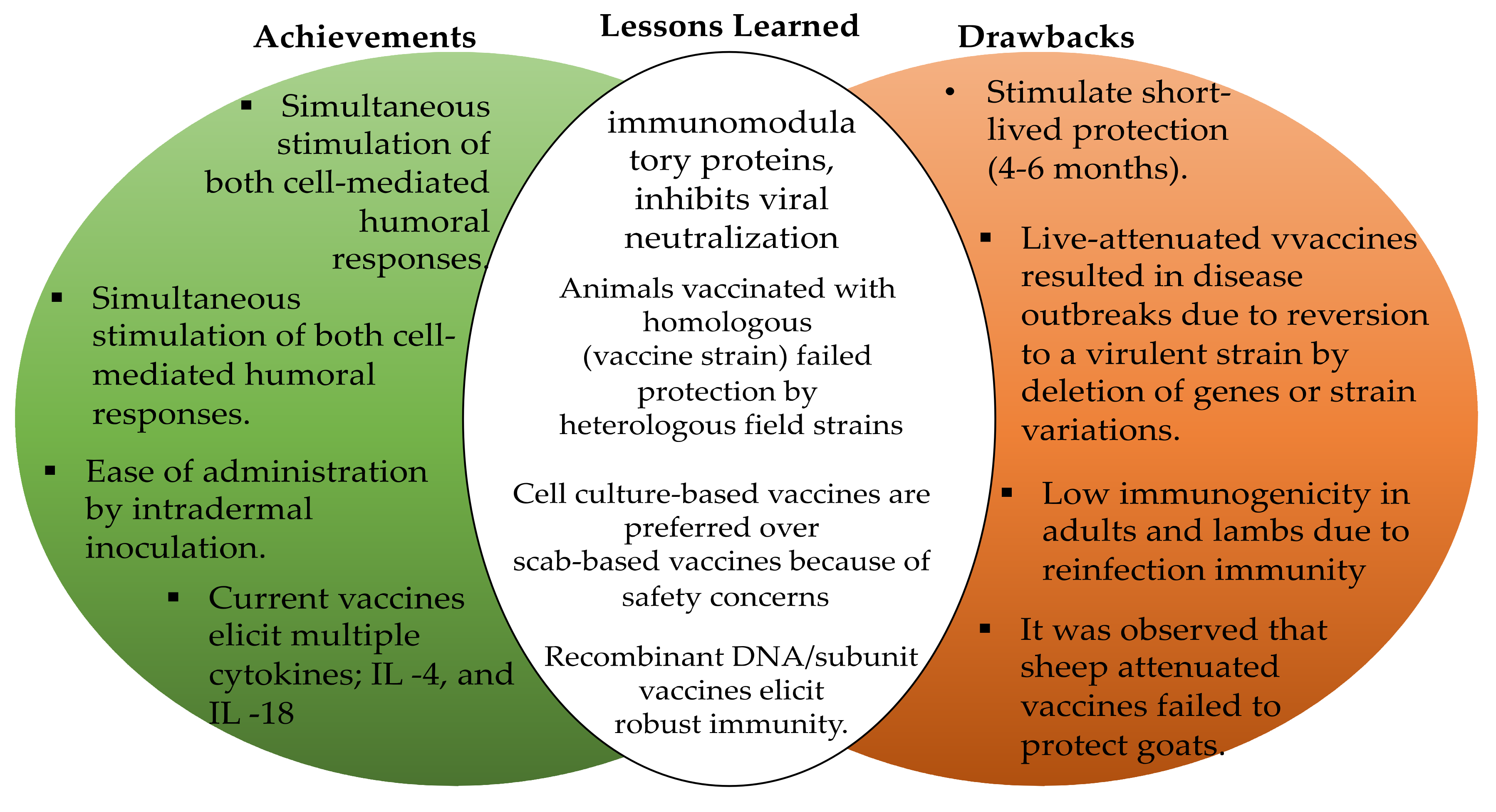
4. Current Status of Vaccines Development against ORFV Infections

4.1. The Current State of ORFV Vaccines

The historical events in vaccine developments didn’t just begin with the initial inoculation of human skin with the material collected from cowpox pustules by Edward Jenner to protect humans against smallpox [120,121,122,123,124,125,126,127,128,129,172,173]. Rather, it can be traced back to the history of infectious diseases of the human and veterinary importance [129]. Historically, ORFV vaccines containing tissue culture attenuated live virus or live scab-based virus preparations have been available since the late 1930s [25,86,129,159]. Parapoxvirus (ORFV) is currently used as attenuated live virus and/or as a vector for antigen delivery systems for other viral vaccines such as vectored rabies vaccines [25,78,128,174]. In spite of the abundant studies published, on both vaccine development ‘and immune response to ORFV infections, few vaccines against ORFV infection are licensed [56,174]. As of today, there are no universally approved sheep or goat vaccines against ORFV infections. Therefore, there is a need to develop a universal ORFV vaccine that could provide protection against worldwide strains of the virus [78,122,174]. However, the humoral, and T-cell analysis revealed that live-attenuated vaccines have shown several benefits. In addition, all the current licensed vaccines used to protect sheep and goats against ORFV infections are based upon live (scab-based) and live-attenuated (Figure 3). Currently, no subunit, peptides, or DNA vaccines are licensed, but some recent publications have demonstrated that chimeric DNA vaccines and recombinant vaccines against ORFV infection have shown promising results [126,129,175].

4.2. Safety and Efficiency Profile of Current Vaccines against ORFV Infections

This section discusses currently available vaccines against ORFV infections to analyze the safety, efficiency, effectiveness, and limitations of the existing vaccine platforms [144,174]. Vaccines are the most powerful approach to prevent infectious disease outbreaks [141,176]. So far, vaccination has been one of the most effective strategies designed to prevent ORFV infections [128,174]. The commercially available vaccines for vaccinating goats and sheep against ORFV infections are generally classified into two types: purified scab-based vaccine [176,177] and cell culture-based live-attenuated vaccine. The first cell culture-based vaccine against ORFV infection was developed in the 1930s at Texas Agricultural Experiment Station in Sonora, Texas (USA) [56,120,124,176]. After the successful discovery of the first vaccine, several other researchers made it their mission to improve the live-attenuated vaccines [78,120,176]. However, the live attenuated vaccine has been reported to provide short-lived protection against ORFV infection of up to 4–6 months [78,178].
The scab-based vaccine was prepared from a virulent ORFV strain derived from goat or sheep scab mouth virus [5,14,150]. However, live scab-based vaccines can induce a robust immune response [179]. Unfortunately, the development of such a vaccine is labor intensive [56,86,179] and it can serve as a potential source of environmental contamination by scab derived from vaccinated lesions [180]. Therefore, cell culture-based vaccines are relatively more preferred owing to increased safety as compared to the counterpart of scab-based vaccines [56,120,178]. Several studies suggest that the vaccine developed based on tissue culture has significantly higher efficacy [120,144,181] and safety compared to the scab-based vaccine [182].
At present, live-attenuated vaccines derived from the ORFV field strain are used prophylactically to prevent ORFV infection in sheep and goats, which play a significant role in reducing disease outbreak [182]. Unfortunately, the current vaccine platform is not safe or able to induce protective immunity and the virus can repeatedly reinfect the vaccinated animals [129]. For example, despite the immune response elicited by the current attenuated vaccines, ORFV continues to infect the susceptible animals as the results of the inefficiency of the vaccines [5,14,150,182].
In addition to the short-lived immune response, live-attenuated vaccines also present a serious risk of reversion to virulence [128,129,179]. The reason for reversion to its original virulent strain is still not clear but could occur due to possible deletion of one or more terminal genes encoding immunomodulatory proteins of the virus leading to attenuation of ORFV in sheep and goats as the results of long-term adaptation of the virus in tissue culture could render current vaccines inefficient [124,125,126,127,128,129,130,131,132,133,134,135,136,137,138,139,140,141,142,143,144,145,146,147,148,149,150,151,152,153,154,155,156,157,158,159,160,161,162,163,164,165,166,167,168,169,170,171,173,174,175,176,177,183]. Other reasons for vaccine failure include possible contamination of the environment by scab derived from vaccinated animals [178], improper storage of the vaccine, genetic [78,84,182] and physiological variation between animals, [121,144,182] and antigenic variations between the vaccine and field strain with increased virulence [55,56,57,61,62], nutritional factors, physical factors [59], and coinfection by bacteria [58,59,75,139,140,141,142,182]. Such factors may influence one or more features for the development of an effective vaccine, which may enhance the desired immune response against the target antigens [176,182]. Perhaps, the notable vaccine failure can be associated with one of these factors [129,144]. The effectiveness of current vaccine platform strategies against ORFV infections are shown in Table 3
However, the antibodies detected in the vaccinated animals are not considered significant for sustained protection against ORFV infection, but the presence of an antibody in the vaccinated sheep indicated prior exposure to ORFV [124,129,179,180,182]. On the other hand, live-attenuated ORFV vaccines based on cell culture elicited the desired immunity against originally virulent ORFV strain obtained from a British isolate of Orf virus [56,92,129,181]. On a contrary note, a tissue culture-prepared live attenuated vaccine derived from the OKA ORFV strain failed to protect sheep from ORFV infection because the vaccine did not induce a neutralizing ORFV-specific antibody [125] and/or T-cell immune response in sheep [14,56,78,124,159,182]. A similar study conducted at Western Texas demonstrated that ORFV the ORFV vaccine has been reported to cause an outbreak in vaccinated sheep and goats [146,179,180].
More recent studies demonstrated that plasmid DNA-based ORFV vaccines derived from virulent strains induced protective T cells and ORFV-specific antibody responses in neonates [3,27,64,130,135]. The result indicated that the plasmid DNA-based vaccines induced similar responses in mice compared to live-attenuated vaccines [179]. Zhao et al. also reported that DNA vaccines elicited ORFV-specific IgG and T-cell (CD4+ and CD8+) responses in mice after two repeated booster doses [56,132,133,134,135,136]. In another study, subunit ORFV vaccines derived from sheep scabs have been reported to induce an anamnestic response, with a significant increase in T-cell response and ORFV-specific IgG response against virulent strains [63,66,80,124,125,126,127,128,129,130,131,132,133,134,135,136,137,138,139,140,141,142,143,144,145,182].

4.3. Implications of Strain Genetic Variation for the Rational Design of ORFV Vaccines

A proper understanding of the implications associated with strain genetic variation among viral pathogens could pave the way for the design of universal vaccines with improved efficacy [60,76,144,168,169,170,171,173,174,175,176,177,178]. However, careful design of an efficacious vaccine candidate against viral infections is a huge challenge guided by years of studies on viral biology and host immune response [176,182,184]. Thus, strain genetic variation is an important characteristic of ORFV [171,184] and remains one of the major constraints of current vaccines against ORFV infections [178,179,180,181,182,184]. It has been observed that genetic variation can occur even within genes at the conserved regions leading to the alteration of structural proteins targeted in a possible universal vaccine formulation [92,145,171,173,174,175,176,177,178,179,180,181,182,184]. In addition to alteration in immunogenic proteins, ORFV can undergo highly antigenic variation in other essential proteins, which can often lead to continuous reinfection of the virus in previously vaccinated animals [125,145,157,177]. It has been reported that the possible outbreaks in vaccinated sheep often due to genetic reassortment between ORFV from district species [75,182]. The most obvious of genetic reassortment is that involving the viral defensive immunomodulatory proteins [180,182] or inverted terminal repeats (ITR) of the virus [171,174,182]. Therefore, genes in the terminal and ITR regions are highly variable [174] and responsible for antigenic variations, pathogenesis, virulence [184], and/or tissue tropisms [101,102,103,104,105,106,142,184]. It is well documented that knockout of an immunomodulatory protein (IMPs) conserved among parapoxviruses of the family poxviridae can reflect variability in animals [174] and tissue tropism facilitating the phenotypic variation observed at the point of ORFV disease manifestations [92,133,184].
Work by Cottone et al. reported that a highly attenuated tissue culture-based vaccine derived from ORFV strain D1701 stimulates an immune response against the wild-type ORFV infection [129,133,159]. Unfortunately, the immunity elicited has only lasted for about 4–6 months [3,60]. Thus, the immunity induced by the attenuated D1701 vaccine is inefficient in protecting the animal from reinfection [60,174,175,176,177,178,179,180,181]. Cottone et al. also reported that the enlargement of inverted terminal repeats (ITR) up to 18 kbp due to recombination between nonhomologous sequences during cell culture adaptation [133]. Immuno-modulation of the host defense mechanism is a common strategy used by the ORFV to overcome host-specific antibodies [133]. Thus, such variations at the genetic level may significantly contribute to poor immune response to vaccines, [177,178] and consequently, such critical insights could be useful strategies in future vaccine design and development [167,168,169,170,171,173].
Variation may occur in several ways depending on the biology of the causative agents [171]. For example, some animal viruses such as aphthoviruses, rhinoviruses, and parapoxviruses have been shown to have multiple serotypes circulating within a particular geographical region [25,78,144,160]. However, the rate of genetic recombination, as well as the emergence of new antigenic variants have not been clearly elucidated [63,66,80]. Genetic variations in the ORFV strain may be caused by several factors arising from a different mechanism, including: (1) genomic rearrangement and subsequent deletion of one or more genes as the result of the attenuation of the virus in tissue culture [58], (2) gene enlargement as a result of duplications in the ITR region, which can result in a huge increase in the overall genomic sequence of the virus, and (3) nucleotide substitution within the coding regions of GC-rich sequence [171,174,185]. Host variation can often lead to vaccine failure which is not insignificantly related to the fact that ORFV strains from sheep and goats are clusters but belong to different branches of the phylogenetic tree [59,63].
ORFV can also exhibit huge variability in circulating strains worldwide. It has been demonstrated that vaccination of goats with goat derived attenuated ORFV vaccine did not clearly minimize the severity of the lesions when it was exposed to field strain ORFV with increased virulence [78,120,124,173]. Additionally, vaccination of goats with the attenuated vaccine produced from relatively increased virulent field ORFV strains significantly reduced the number of previously infected animals but failed to reduce the severity of the infection [74,173,182]. This suggests that the vaccine does not provide cross-protection between the vaccine strains and field strains with increased virulence [179]. Recent studies demonstrated that the goats vaccinated with live-attenuated vaccines did not provide complete protection against ORFV infection when exposed to another ORFV strain derived from goats [124]. Such vaccination failure may have been related to the genetic factors that result in a serious outbreak of the disease in vaccinated animals [179]. In another study, sheep immunized with the live-attenuated vaccine developed from a virulent strain of ORFV did not elicit desired antibody and cell-mediated immune responses that can be seen with natural infections with field strain ORFV [79,144,157,159,160,161,162,163,164,165,166]. In several instances, lambs vaccinated with the scab-based vaccine developed from goats-based Orf virus failed to protect the animals from the heterologous virulent field virus strains [139]. Repeated re-infections [120]. and vaccine failures have been observed in both vaccinated goats and sheep [175]. Thus, such variations at the genetic level may significantly contribute to a poor immune response to vaccines, [177] and consequently, such critical insights could be useful strategies in future vaccine design and development [173,174].

4.4. Enhancement of the Current ORFV Vaccines

The tissue-culture-based live attenuated virus vaccines have played a significant role in preventing the spread of viruses [124,173]. Although live attenuated and scab-based vaccines protect animals ORFV infection worldwide, the continued reinfection vaccinated animals are a major concern that needs to be addressed [174]. Additionally, ORFV reinfection in vaccinated animals might be due to the global movement of vaccinated sheep and/or goats and genetic variation [171,173,174,175,177]. The enhancement of existing vaccines in terms of their safety, efficiency, efficacy, and cost-effectiveness is a key priority. Improvement of current vaccines can be achieved through careful selection of the desired ORFV strain for vaccine seed virus, proper use of tissue culture for vaccine seed virus propagation, and proper use of potent adjuvants and/or sustained delivery systems [152,153,154,155,168,169,170,171,173].
Over the last few years, several strategies have been explored to accomplish attenuation of the virus through genetic engineering rather than the traditional approaches; based on long-term adaptation of the virus in tissue culture [56,173]. In addition, advances in recombinant DNA technology and genetic engineering have opened a new avenue to enhance the current vaccine by deleting immunomodulatory genes of ORFV, to promote the development of vaccines with robust immune response [65,92,93,94,171]. A recent study indicates that the deletion of IMGs such as ORFV020, ORFV117 and ORFV132 from the Orf virus genomic DNA led to production of modified live-attenuated virus that can elicit protective immunity [92,98]. In addition, production of modified live-vaccine makes the virus a suitable vectored vaccine candidate [65,171,173,174,175,176,177,178,184]. Several studies demonstrated that the attenuation of vaccine strains by deletion of immunomodulatory genes has resulted in a significant increase in the immunogenicity of the vaccine [99,108,109,171]. Thus, the future vaccine can be engineered by targeting ORFV immunomodulatory proteins that inhibit the host immune response [85,92,93,94].

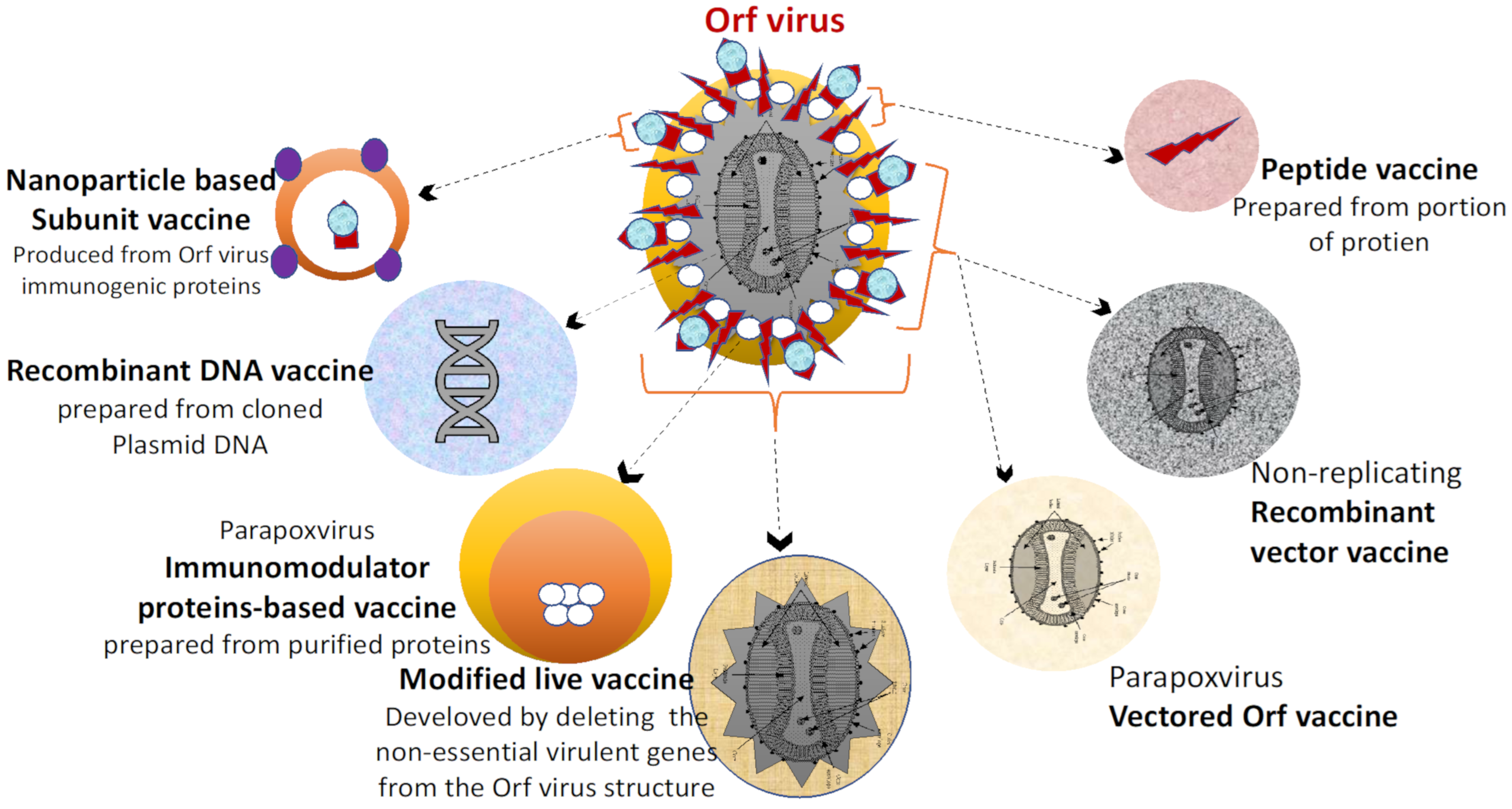
5. Future Strategies for the Development of Vaccines against ORFV Infection

In recent years, great success has been recorded in the development of vaccines. Recent advances in recombinant DNA technologies will continue to play a key role in shaping future ORFV vaccine development. Future vaccine design technologies would focus on various strategies that are particularly promising in their potential to mimic desired immune responses to challenging antigens for which current vaccination strategies have proven ineffective [167,174,175,176,177,178,179,180,181,182,184]. As with current ORFV vaccine platforms, there is a need for the development of a novel vaccine with increased efficiency/safety profiles [144,159,173].and improved adjuvants and sustained delivery strategies to overcome the current limitation observed in vaccines against ORFV infection [162]. The novel technologies are expected to improve the safety [144] and efficacy of future vaccines [144,174,182].
Furthermore, vaccines against ORFV and other small ruminants in the live-stock production industries are under huge demand in order to increase their application in the field. The future vaccine platforms should be designed to fulfill three main constraints; (1) compatibility with mass vaccination, (2) cost-effectiveness for large-scale production and delivery, (3) most importantly, they should be safe, effective, and efficacious. However, such constraints can serve as a benchmark for determining the efficacy of future vaccine platforms [4,25,84,173,185]. A schematic outline of some future vaccines against ORFV (Figure 4). The future vaccines against ORFV infections include nanoparticle-based subunit and/or peptide vaccines, DNA vaccine, and parapoxvirus vectored vaccines. Thus, the application of such vaccines can minimize the use of the attenuated virus that might easily reverse to virulent [147,174,184].
Subunit and peptide vaccines are composed of either glycoproteins [163] or proteins derived from specific ORFV genes that are actively involved in eliciting robust immunity [25,84,124,170]. Additionally, subunit and peptide vaccines are relatively safe [109,110,111] and easy to produce compared to efficient conventional live-attenuated and/or inactivated vaccines [170,182]. Unfortunately, subunit vaccines induce short-lived immune responses, so adjuvants are needed to boost immunity [152,153,154,155]. Recent significant advances in the field of nanotechnology has revealed that nanoparticles (NPs) are capable of enhancing the cellular immune response as well as the delivery of specific viral antigens to a target cell [154]. Therefore, nano-adjuvanted vaccines can be used to enhance the immunogenicity of existing and future vaccine candidates [168,169,170,171]. For example, peptide, subunit, and inactivated vaccine platforms are relatively safer but tend to be less immunogenic compared to live-attenuated viral vaccines [170]. Therefore, the low immunogenicity of such vaccine candidates can be enhanced by the addition of potent such as alum or nano-adjuvants that stimulate durable immune response [148,149,150,151]. Therefore, nano-adjuvants are used for vaccine development due following benefits: (1) enhancement of vaccine antigens efficacy; (2) entrapment and release of the vaccine antigens to the target cells in a sustained manner; (3) protection of delivered vaccine antigens from possible degradation; (4) it minimizes the dose, amount of vaccine antigen required vaccination in order to minimize production cost; (5) enchantment of humoral and cell-mediated immune response; (6) enhancing antibody specificity, affinity, avidity, and (7) prompt dissemination of the antigens to target cells [152,153,154,155,156,157,158,159,160,161,162,163,164,165,166,167,168].
Furthermore, due to the nanosized structure and shape, nanoparticles can be easily engulfed by the antigen-presenting cells, particularly the dendritic cells [144,150], and consequently trapped into the draining lymph nodes to stimulate the production of humoral and cell-mediated immunity [155,156]. Hence, the use of nano-adjuvants will further improve both the efficacy and effectiveness of vaccines [159]. If the concept of nanoparticle-based vaccines truly works, it would be a great approach in future strategies to control ORFV infections [158,159]. The immunity to nanoparticle-based (NPs) vaccines that result in enhancement of vaccine-stimulated immunity is summarized in Figure 5.
Peptide vaccines are a sub-type of subunit vaccine that are synthesized in vitro using peptides that containing immunogenic fragments critical immunodominant proteins of the target virus [157,159,160,161,162,163]. However, peptide-based vaccines are relatively easier to produce and show high stability than subunit vaccines (whole proteins). Interestingly, peptide vaccines have shown numerous advantages compared to their subunit counterpart, lower antigen complexity, lower toxicity, and most importantly low production costs [166]. Peptide-based viral vaccines can enhance immunogenicity by the addition of potent immunoadjuvants that stimulate the T-cell response (CD4+ and CD8+), sometimes the humoral response, and Interferon-gamma (IFN-γ) to destroy the virus from an infected cell. Currently, there are no peptide vaccines against ORFV and other viral infections. However, peptide-based vaccines against influenza virus and human papillomavirus vaccines have been reported to trigger the induction of desirable immune responses [167]. Thus, this class of vaccines can be developed using bioinformatics and molecular biological approaches to be designed in order to overcome the undesirable effects of conventional vaccines [164,182]. Interestingly, peptide vaccines are theoretically considered as a promising class of future vaccines due to the following advantages; easy synthesis, stability, and highly safe to be administered to a wider host range [165,166].
Recombinant viral vector vaccines are composed of a highly competent engineered vector backbone for the production of recombinant vaccine antigens [160,161]. The selected DNA fragment is subsequently recombined with a suitable expression vector that can lead to the production of the recombinant protein of interest [167,173]. Interestingly, advances in the understanding of host immunity to viral infections and pathogenicity coupled with efficient genetic engineering approaches have led to the development of novel vaccines [147,148,162]. Commonly used viral vectors include adenoviruses, alphaviruses, flaviviruses, herpesviruses, Newcastle disease virus, parvoviruses, and poxviruses have been manipulated to develop recombinant viral-vectored vaccines [160]. The most striking features of ORFV vectored vaccines include the induction of durable T-cells and humoral immune response, lack of systemic dissemination of the virus, and restricted target host. Such vaccines are expected to address the huge challenges posed by existing conventional vaccines [25]. A recent study has demonstrated that recombinant vaccines have been observed to stimulate long-lived both humoral and cellular immune responses in animals [162]. Vectored vaccines combined the benefits of both replicating and non-replicating subunit vaccines [52,164]. However, the process of developing such a vaccine has been observed to be highly complex and costly [140,163,186].

6. Conclusions

The development of prophylactic vaccines and subsequent comprehensive immunization have resulted in partial control of ORFV infection worldwide. However, existing live attenuated vaccines based on traditional attenuation by serial passage of the virus have been found to be inefficient [38,119]. Nevertheless, numerous drawbacks of the existing attenuated ORFV vaccines have been observed, such as low vaccine efficacy, short-lived immunity, and return to virulence [38]. In addition to the poor immunogenicity of current vaccines, there are other critical challenges and knowledge gaps that need to be addressed regarding vaccine safety and efficacy [186,187]. In order to develop safe and effective vaccines against ORFV infections, we envision a new strategy that utilizes recent advances in molecular biology techniques as the basis for developing future vaccines [186]. In recent years, numerous approaches have been used to successfully attenuate the virus through modern genetic manipulation, rather than traditional strategies based on a series of passages of the virus in tissue culture techniques [129,144,186]. As mentioned earlier, the virulence genes of ORFV are known to suppress the host immune response [187]. Therefore, engineering silencing of one or more immunosuppressive genes by genetic modification of the viral genome represents a new strategy for the development of subunits, chimeric DNA [3,27,64] and recombinant vectorized vaccines, the main goal of which is to address issues of safety and efficacy [144,159,188]. Therefore, vaccines will continue to be the basis for new advances in veterinary medicine [65,92,93,94]. We are optimistic that the improvement of current and the development of future vaccines will not only focus on the improvement of viral attenuation and the deletion of immunomodulatory genes, but also on the exploration of immunogenic ORFV proteins that will further enhance the immunogenicity of the new vaccine candidates. If the concept of recombinant vaccines really works, it would be a great strategy to fight ORFV infections [188].

Author Contributions

Conceptualization, M.-L.M.A., A.M.B., F.F.A.J., M.M.N., C.A.C.A., K.N.B., H.K.M., Z.L.; writing—original draft preparation, A.M.B.; writing—review and editing, A.M.B., M.-L.M.A., F.F.A.J., M.M.N., C.A.C.A. All authors have read and agreed to the published version of the manuscript.

Funding

This research received no external funding. This work was supported by grants from Malaysian Skim Geran Penyelidikan Fundamental (FRGS) FASA 1/2019 University Putra Malaysia (FRGS/1/2019/STG03/UPM/01/2).

Conflicts of Interest

The authors declare no commercial or financial relationships which can be interpreted as a potential conflict of interest.

References

  1. Chakraborty, S.; Kumar, A.; Tiwari, R.; Rahal, A.; Malik, Y.; Dhamma, K.; Prasad, M. Advances in diagnosis of respiratory diseases of small ruminants. Vet. Med. Int. 2014, 138, 1–16. [Google Scholar] [CrossRef] [Green Version]
  2. Babji, A.S.; Seri, S.C. The nutritional value of some processed meat products in Malaysia, Malays. J. Nutr. 1995, 1, 83–94. [Google Scholar]
  3. Minguijón, E.; Reina, R.; Pérez, M.; Polledo, L.; Villoria, M.; Ramírez, H. Juste, R.A. Small ruminant lentivirus infections and diseases. Vet. Microbiol. 2015, 181, 75–89. [Google Scholar] [CrossRef]
  4. Barnett, P.V.; Cox, S.J. The role of small ruminants in the epidemiology and transmission of foot-and-mouth disease. Vet. J. 1999, 158, 6–13. [Google Scholar] [CrossRef] [PubMed]
  5. Yadav, D.K.; Yadav, N.; Khurana, S.M.P. Vaccines: Present Status and Applications. Vaccines 2014, 31, 491–508. [Google Scholar]
  6. Ouedraogo, A.; Luciani, L.; Zannou, O.; Biguezoton, A.; Pezzi, L.; Thirion, L.; Lempereur, L. Detection of two species of the genus parapoxvirus (Bovine papular stomatitis virus and pseudocowpox virus) in ticks infesting cattle in Burkina Faso. Microorganisms 2020, 8, 644. [Google Scholar] [CrossRef] [PubMed]
  7. Shams, H. Recent developments in veterinary vaccinology. Vet. J. 2005, 170, 289–299. [Google Scholar] [CrossRef] [PubMed]
  8. Ismail, R.; Allaudin, Z.N.; Lila, M.-A.M. Scaling-up recombinant plasmid DNA for clinical trial: Current concern, solution and status. Vaccine 2012, 30, 5914–5920. [Google Scholar] [CrossRef] [PubMed] [Green Version]
  9. Vikøren, T.; Lillehaug, A.; Åkerstedt, J.; Bretten, T.; Haugum, M.; Tryland, M. A severe outbreak of contagious ecthyma (Orf) in a free-ranging musk ox (Ovibos moschatus) population in Norway. Vet. Microbiol. 2008, 127, 10–20. [Google Scholar] [CrossRef]
  10. Vandenberg, G.W. Oral vaccines for finfish: Academic theory or commercial reality? Anim. Health Res. Rev. 2004, 5, 301–304. [Google Scholar] [CrossRef]
  11. Poland, G.A.; Jacobson, R.M.; Poland, G.A.; Jacobson, R.M. Understanding those who do not understand: A brief review of the anti-vaccine movement. Vaccine 2001, 19, 2440–2445. [Google Scholar] [CrossRef]
  12. Bala, J.A.; Balakrishnan, K.N.; Abdullah, A.A.; Kimmy, T.; Abba, Y.; Bin Mohamed, R.; Jesse, F.F.A.; Haron, A.W.; Noordin, M.M.; Bitrus, A.A.; et al. Dermatopathology of Orf Virus (Malaysian Isolates) in Mice Experimentally Inoculated at Different Sites with and without Dexamethasone Administration. J. Pathog. 2018, 2018, 1–12. [Google Scholar] [CrossRef]
  13. Lafar, S.; Zro, K.; Ennaji, M.M. Capripoxvirus Diseases: Current Updates and Developed Strategies for Control. Emerg. Reemerging Viral Pathog. 2019, 8, 635–655. [Google Scholar] [CrossRef]
  14. Lee, N.-H.; Lee, J.-A.; Park, S.-Y.; Song, C.-S.; Choi, I.-S.; Lee, J.-B. A review of vaccine development and research for industry animals in Korea. Clin. Exp. Vaccine Res. 2012, 1, 18–34. [Google Scholar] [CrossRef] [PubMed] [Green Version]
  15. Mohamed, Z.; Chiew, E.; Shamsudin, M. Government Incentives and Comparative Advantage of the Sheep Industry in Ma-laysia, Pertanika. J. Soc. Sci. Humanit. 1995, 3, 173–179. [Google Scholar]
  16. Reguzova, A.; Ghosh, M.; Müller, M.; Rziha, H.-J.; Amann, R. Orf Virus-Based Vaccine Vector D1701-V Induces Strong CD8+ T Cell Response against the Transgene but Not against ORFV-Derived Epitopes. Vaccines 2020, 8, 295. [Google Scholar] [CrossRef] [PubMed]
  17. Khoo, C.K.; Norlina, D.; Roshaslinda, D.; Zuraidah, O.; Zunaida, B.; Mohd, H.A.H.; Roslina, H. Case Report Molecular Diagnosis of Caprine ORF Virus (ORFV) from Penang, Malaysia. J. Malays. Vet. 2019, 10, 98–102. [Google Scholar]
  18. Bergqvist, C.; Kurban, M.; Abbas, O. Orf virus infection. Rev. Med. Virol. 2017, 27, e1932. [Google Scholar] [CrossRef]
  19. Jesse, F.F.A.; Hambali, I.U.; Abba, Y.; Lin, C.C.; Chung, E.L.T.; Bitrus, A.A.; Abdullah, A.A.; Balakrishnan, K.N.; Bala, J.A.; Lila, M.A.M. Effect of dexamethasone administration on the pathogenicity and lesion severity in rats experimentally inoculated with Orf virus (Malaysian isolates). Comp. Haematol. Int. 2018, 27, 1227–1236. [Google Scholar] [CrossRef]
  20. Dreesen, D.W. Animal Vaccines; Elsevier: Amsterdam, The Netherlands, 2007; pp. 517–530. [Google Scholar]
  21. Bahaman, A.R.; Joseph, P.G.; Siti-Khairani, B. A Review of the Epidemiology and Control of Brucellosis in Malaysia. J. Vet. Malays. 2007, 19, 1–6. [Google Scholar]
  22. Hassan, L.; Zainalabidin, M. The economic impact attributable to bru-cellosis among goat farms in Peninsula Malaysia and cost benefit analysis. Res. Opin. Anim. Vet. Sci. 2015, 5, 57–64. [Google Scholar]
  23. Zainalabidin, F.A.; Raimy, N.; Yaacob, M.H.; Musbah, A.; Bathmanaban, P.; Ismail, E.A.; Mamat, Z.C.; Zahari, Z.; Ismail, M.I.; Panchadcharam, C. The Prevalence of Parasitic Infestation of Small Ruminant Farms in Perak, Malaysia. Trop. Life Sci. Res. 2015, 26, 1–8. [Google Scholar]
  24. Souza, A.; Haut, L.; Reyes-Sandoval, A.; Pinto, A. Recombinant viruses as vaccines against viral diseases. Braz. J. Med. Biol. Res. 2005, 38, 509–522. [Google Scholar] [CrossRef] [Green Version]
  25. Jorge, S.; Dellagostin, O.A. The development of veterinary vaccines: A review of traditional methods and modern biotechnology approaches. Biotechnol. Res. Innov. 2017, 1, 6–13. [Google Scholar] [CrossRef]
  26. Abdullah, A.A.; Bin Ismail, M.F.; Balakrishnan, K.N.; Bala, J.A.; Hani, H.; Abba, Y.; Isa, M.K.A.; Abdullah, F.F.J.; Arshad, S.S.; Nazariah, Z.A.; et al. Isolation and phylogenetic analysis of caprine Orf virus in Malaysia. Virus Dis. 2015, 26, 255–259. [Google Scholar] [CrossRef] [PubMed] [Green Version]
  27. McKeever, D.; Jenkinson, D.M.; Hutchison, G.; Reid, H. Studies of the pathogenesis of orf virus infection in sheep. J. Comp. Pathol. 1988, 99, 317–328. [Google Scholar] [CrossRef]
  28. Tryland, M.; Beckmen, K.B.; Burek-Huntington, K.A.; Breines, E.M.; Klein, J. Orf virus infection in Alaskan mountain goats, Dall’s sheep, muskoxen, caribou and Sitka black-tailed deer. Acta Vet. Scand. 2018, 60, 1–11. [Google Scholar] [CrossRef] [Green Version]
  29. Andreani, J.; Fongue, J.; Khalil, J.Y.B.; David, L.; Mougari, S.; Le Bideau, M.; Abrahão, J.; Berbis, P.; La Scola, B. Human Infection with Orf Virus and Description of Its Whole Genome, France, 2017. Emerg. Infect. Dis. 2019, 25, 2197–2204. [Google Scholar] [CrossRef] [PubMed]
  30. Teshale, A. Contagious Ecthyma and its Public Health Significance. J. Dairy Vet. Sci. 2018, 7, 1–6. [Google Scholar] [CrossRef] [Green Version]
  31. Vakhshiteh, F.; Allaudin, Z.N.; Lila, M.A.B.M.; Hani, H. Size-related assessment on viability and insulin secretion of caprine islets in vitro. Xenotransplantation 2013, 20, 82–88. [Google Scholar] [CrossRef]
  32. Spyrou, V.; Valiakos, G. Orf virus infection in sheep or goats. Vet. Microbiol. 2015, 181, 178–182. [Google Scholar] [CrossRef] [PubMed]
  33. Buttner, M.; Rziha, H.-J. Parapoxviruses: From the Lesion to the Viral Genome. J. Vet. Med. Ser. B 2002, 49, 7–16. [Google Scholar] [CrossRef]
  34. Zarnke, R.L.; Dieterich, R.A.; Neiland, K.A.; Ranglack, G. Serologic and experimental investigations of contagious ecthyma in alaska. J. Wildl. Dis. 1983, 19, 170–174. [Google Scholar] [CrossRef] [PubMed]
  35. Srinivasa, B.T.; Rathnamma, D.; Isloor, S.; Chandranaik, B.M.; Veeregowda, B.M.; Manjunath, R.G.B. Diagnosis of Orf virus infection in sheep and goats by virus isolation, polymerase chain reaction and sequencing. J. Exp. Biol. Agric. Sci. 2018, 6, 176–187. [Google Scholar] [CrossRef]
  36. Paiba, G.A.; Thomas, D.R.; Morgan, K.L.; Bennett, M.; Salmon, R.L.; Chalmers, R.; Kench, S.M.; Coleman, T.J.; Meadows, D.; Morgan-Capner, P.; et al. Orf (contagious pustular dermatitis) in farmworkers: Prevalence and risk factors in three areas of England. Vet. Rec. 1999, 145, 7–11. [Google Scholar] [CrossRef]
  37. Al Saad, K.M.; Thweni, H.T.; Abdali, D.A.; Tarik, A.S. Clinical and Diagnostic Studies of Contagious Ecthyma (ORF) in Sheep. IOSR J. Agric. Vet. Sci. 2017, 10, 64–69. [Google Scholar] [CrossRef]
  38. Hosamani, M.; Scagliarini, A.; Bhanuprakash, V.; McInnes, C.J.; Singh, R.K. Orf: An update on current research and future perspectives. Expert Rev. Anti-Infect. Ther. 2009, 7, 879–893. [Google Scholar] [CrossRef]
  39. Adedeji, A.; Adole, J.; Chima, N.; Maguda, A.; Dyek, D.; Jambol, A.; Anefu, E.; Shallmizhili, J.; Luka, P. Contagious ecthyma in three flocks of goats in Jos-south LGA, Plateau State, Nigeria. Sokoto J. Vet. Sci. 2018, 16, 107. [Google Scholar] [CrossRef] [Green Version]
  40. Nadeem, M.; Curran, P.; Cooke, R.; Ryan, C.A.; Connolly, K. Orf: Contagious pustular dermatitis. Ir. Med. J. 2010, 103, 971–972. [Google Scholar]
  41. Robinson, A.J. Prevalence of contagious pustular dermatitis (orf) in six million lambs at slaughter: A three-year study. N. Zealand Vet. J. 1983, 31, 161–163. [Google Scholar] [CrossRef]
  42. Lederman, E.R.; Green, G.M.; DeGroot, H.E.; Dahl, P.; Goldman, E.; Greer, P.W.; Li, Y.; Zhao, H.; Paddock, C.D.; Damon, I.K. Progressive Orf Virus Infection in a Patient with Lymphoma: Successful Treatment Using Imiquimod. Clin Infect. Dis. 2007, 44, e100–e103. [Google Scholar] [CrossRef] [Green Version]
  43. Yan, M.H.; Wang, L.L.; Hao, J.H.; Zhang, X.G.; Shen, C.C.; Zhang, D.J.; Zhang, K.S. Orf Virus VIR Antagonizes p53-Mediated Antiviral Effects to Facilitate Viral Replication. Viral Immunol. 2020, 33, 468–476. [Google Scholar] [CrossRef]
  44. Ghilardi, N.; Pappu, R.; Arron, J.R.; Chan, A.C. 30 Years of Biotherapeutics Development—What Have We Learned? Annu. Rev. Immunol. 2020, 38, 249–287. [Google Scholar] [CrossRef]
  45. Lucas, A.; McFadden, G. Secreted Immunomodulatory Viral Proteins as Novel Biotherapeutics. J. Immunol. 2004, 173, 4765–4774. [Google Scholar] [CrossRef] [PubMed] [Green Version]
  46. Vellucci, A.; Manolas, M.; Jin, S.; Dwyer, J.; Vick, G.; Wang, A.; Swiatlo, E.; Zheng, C. Orf virus infection after Eid al-Adha. IDCases 2020, 21, e00854. [Google Scholar] [CrossRef]
  47. De La Concha-Bermejillo, A.; Guo, J.; Zhang, Z.; Waldron, D. Severe persistent orf in young goats. J. Vet. Diagn. Investig. 2003, 15, 423–431. [Google Scholar] [CrossRef] [PubMed] [Green Version]
  48. Bala, J.A.; Balakrishnan, K.N.; Abdullah, A.; Mohamed, R.; Haron, A.W.; Jesse, F.F.A.; Noordin, M.M.; Mohd-Azmi, M.L. The re-emerging of orf virus infection: A call for surveillance, vaccination and effective control measures. Microb. Pathog. 2018, 120, 55–63. [Google Scholar] [CrossRef]
  49. Batra, S.K.; Chand, P.; Rajpurohit, B.S. A severe outbreak of contagious pustular dermatitis in a sheep flock. Indian J. Anim. Sci. 1999, 69, 34–35. [Google Scholar]
  50. Yirrell, D.; Reid, H.; Norval, M.; Howie, S. Immune response of lambs to experimental infection with Orf virus. Vet. Immunol. Immunopathol. 1989, 22, 321–332. [Google Scholar] [CrossRef]
  51. Anziliero, D.; Weiblen, R.; Kreutz, L.C.; Spilki, F.; Flores, E. Inactivated Parapoxvirus ovis induces a transient increase in the expression of proinflammatory, Th1-related, and autoregulatory cytokines in mice. Braz. J. Med. Biol. Res. 2014, 47, 110–118. [Google Scholar] [CrossRef] [PubMed] [Green Version]
  52. Jia, H.; Zhan, L.; Wang, X.; He, X.; Chen, G.; Zhang, Y.; Feng, Y.; Wei, Y.; Zhang, Y.; Jing, Z. Transcriptome analysis of sheep oral mucosa response to Orf virus infection. PLoS ONE 2017, 12, e0186681. [Google Scholar] [CrossRef]
  53. Gregory, A.E.; Titball, R.; Williamson, D. Vaccine delivery using nanoparticles. Front. Cell. Infect. Microbiol. 2013, 3, 13. [Google Scholar] [CrossRef] [PubMed] [Green Version]
  54. Boughton, I.; Hardy, W. Immunization of Sheep and Goats against Sore Mouth (Contagious Ecthyma). Tex. FARMER Collect. 1935, 504, 16. [Google Scholar]
  55. Haig, D.M.; McInnes, C. Immunity and counter-immunity during infection with the parapoxvirus orf virus. Virus Res. 2002, 88, 3–16. [Google Scholar] [CrossRef]
  56. Pye, D. Vaccination of sheep with cell culture grown orf virus. Aust. Vet. J. 1990, 67, 182–186. [Google Scholar] [CrossRef]
  57. Nandi, S.; De, U.K.; Chowdhury, S. Current status of contagious ecthyma or orf disease in goat and sheep—A global perspective. Small Rumin. Res. 2011, 96, 73–82. [Google Scholar] [CrossRef]
  58. Bala, J.A.; Balakrishnan, K.N.; Abdullah, A.A.; Yi, L.C.; Bitrus, A.A.; Abba, Y.; Aliyu, I.A.; Peter, I.D.; Hambali, I.U.; Bin Mohamed, R.; et al. Sero-epidemiology of Contagious Ecthyma Based on Detection of IgG Antibody in Selected Sheep and Goats Farms in Malaysia. Adv. Anim. Vet. Sci. 2018, 6, 219–226. [Google Scholar] [CrossRef] [Green Version]
  59. Wang, R.; Wang, Y.; Liu, F.; Luo, S. Orf virus: A promising new therapeutic agent. Rev. Med. Virol. 2019, 29, 1–10. [Google Scholar] [CrossRef] [Green Version]
  60. Rziha, H.-J.; Büttner, M.; Müller, M.; Salomon, F.; Reguzova, A.; Laible, D.; Amann, R. Genomic Characterization of Orf Virus Strain D1701-V (Parapoxvirus) and Development of Novel Sites for Multiple Transgene Expression. Viruses 2019, 11, 127. [Google Scholar] [CrossRef] [Green Version]
  61. Friebe, A.; Friederichs, S.; Scholz, K.; Janssen, U.; Scholz, C.; Schlapp, T.; Mercer, A.; Siegling, A.; Volk, H.-D.; Weber, O. Characterization of immunostimulatory components of orf virus (parapoxvirus ovis). J. Gen. Virol. 2011, 92, 1571–1584. [Google Scholar] [CrossRef]
  62. Da Costa, R.A.; Cargnelutti, J.F.; Schild, C.O.; Flores, E.F.; Riet-Correa, F.; Giannitti, F. Outbreak of contagious ecthyma caused by Orf virus (Parapoxvirus ovis) in a vaccinated sheep flock in Uruguay. Braz. J. Microbiol. 2019, 50, 565–569. [Google Scholar] [CrossRef] [PubMed]
  63. Yahaya, M.A.F.; Lila, M.A.M.; Ismail, S.; Zainol, M.; Afizan, N. Tumour-Associated Macrophages (TAMs) in Colon Cancer and How to Reeducate Them. J. Immunol. Res. 2019, 2019, 1–9. [Google Scholar] [CrossRef] [PubMed]
  64. Fleming, S.B.; Wise, L.M.; Mercer, A.A. Molecular Genetic Analysis of Orf Virus: A Poxvirus That Has Adapted to Skin. Viruses 2015, 7, 1505–1539. [Google Scholar] [CrossRef] [PubMed]
  65. McLane, L.M.; Hakeem, M.A.; Wherry, E.J. CD8 T Cell Exhaustion during Chronic Viral Infection and Cancer. Annu. Rev. Immunol. 2019, 37, 457–495. [Google Scholar] [CrossRef] [PubMed] [Green Version]
  66. Zamri-Saad, M.; Effendy, A.; Israf, D.; Azmi, M.L.M. Cellular and humoral responses in the respiratory tract of goats following intranasal stimulation using formalin-killed Pasteurella haemolytica A2. Vet. Microbiol. 1999, 65, 233–240. [Google Scholar] [CrossRef]
  67. Hu, M.-M.; Shu, H.-B. Innate Immune Response to Cytoplasmic DNA: Mechanisms and Diseases. Annu. Rev. Immunol. 2020, 38, 79–98. [Google Scholar] [CrossRef]
  68. Janeway, C.A., Jr.; Medzhitov, R. Innate immune recognition. Annu. Rev. Immunol. 2002, 20, 197–216. [Google Scholar] [CrossRef] [PubMed] [Green Version]
  69. Haig, D.M.; McInnes, C.J.; Hutchison, G.; Seow, H.-F.; Reid, H.W. Cyclosporin A abrogates the acquired immunity to cutaneous reinfection with the parapoxvirus orf virus. Immunology 1996, 89, 524–531. [Google Scholar] [CrossRef]
  70. Johnston, J.B.; Mcfadden, G. Minireview Poxvirus Immunomodulatory Strategies: Current Perspectives. J. Virol. 2003, 77, 6093–6100. [Google Scholar] [CrossRef] [PubMed] [Green Version]
  71. Joshi, L.R.; Okda, F.A.; Singrey, A.; Maggioli, M.F.; Faccin, T.C.; Fernandes, M.H.V.; Diel, D.G. Passive immunity to porcine epidemic diarrhea virus following immunization of pregnant gilts with a recombinant orf virus vector expressing the spike protein. Arch. Virol. 2018, 163, 2327–2335. [Google Scholar] [CrossRef]
  72. McKeever, D.; Reid, H.; Inglis, N.; Herring, A. A qualitative and quantitative assessment of the humoral antibody response of the sheep to orf virus infection. Vet. Microbiol. 1987, 15, 229–241. [Google Scholar] [CrossRef]
  73. Jenkinson, D.M.; McEwan, P.E.; Onwuka, S.K.; Moss, V.A.; Elder, H.Y.; Hutchison, G.; Reid, H.W. The Pathological Changes and Polymorphonuclear and Mast Cell Responses in the Skin of Specific Pathogen-free Lambs Following Primary and Secondary Challenge with Orf Virus. Vet. Dermatol. 1990, 1, 139–150. [Google Scholar] [CrossRef]
  74. Nettleton, P.F.; Brebner, J.; Pow, I.; Gilray, J.A.; Bell, G.D.; Reid, H.W. Tissue culture-propagated Orf virus vaccine protects lambs from orf virus challenge. Vet. Rec. 1996, 138, 184–186. [Google Scholar] [CrossRef] [PubMed]
  75. Bath, G.; Van Wyk, J.; Pettey, K. Control measures for some important and unusual goat diseases in southern Africa. Small Rumin. Res. 2005, 60, 127–140. [Google Scholar] [CrossRef]
  76. McKeever, D.; Reid, H. The response of the supramammary lymph node of the sheep to secondary infection with orf virus. Vet. Microbiol. 1987, 14, 3–13. [Google Scholar] [CrossRef]
  77. Haig, D.M.; Deane, D.; Myatt, N.; Thomson, J.; Entrican, G.; Rothel, J.; Reid, H. The activation status of ovine CD45R+ and CD45R− efferent lymph T cells after orf virus reinfection. J. Comp. Pathol. 1996, 115, 163–174. [Google Scholar] [CrossRef]
  78. Wassie, T.; Fanmei, Z.; Jiang, X.; Liu, G.; Girmay, S.; Min, Z.; Chenhui, L.; Bo, D.D.; Ahmed, S. Recombinant B2L and Kisspeptin-54 DNA Vaccine Induces Immunity against Orf Virus and Inhibits Spermatogenesis in Rats. Sci. Rep. 2019, 9, 1–11. [Google Scholar] [CrossRef] [Green Version]
  79. Förster, R.; Wolf, G.; Mayr, A. Highly attenuated poxviruses induce functional priming of neutrophils in vitro. Arch. Virol. 1994, 136, 219–226. [Google Scholar] [CrossRef]
  80. McGuire, M.J.; Johnston, S.A.; Sykes, K.F. Novel immune-modulator identified by a rapid, functional screen of the parapoxvirus ovis (Orf virus) genome. Proteome Sci. 2012, 10, 4. [Google Scholar] [CrossRef] [Green Version]
  81. Jenkinson, D.M.; Hutchison, G.; Onwuka, S.K.; Reid, H.W. Changes in the MHC Class II+ Dendritic Cell Population of Ovine Skin in Response to Orf Virus Infection. Vet. Dermatol. 1991, 2, 1–9. [Google Scholar] [CrossRef]
  82. Chung, E.L.T.; Abdullah, F.F.J.; Ibrahim, H.H.; Marza, A.D.; Zamri-Saad, M.; Haron, A.W.; Lila, M.A.M.; Norsidin, M.J. Clinico-pathology, hematology and biochemistry responses in buffaloes towards Pasteurella multocida type B: 2 immunogen lypopolysaccharide via oral and intravenous routes of infection. Microb. Pathog. 2016, 91, 141–154. [Google Scholar] [CrossRef] [PubMed]
  83. Smith, G.L.; Symons, J.A.; Alcamı́, A. Poxviruses: Interfering with Interferon. Semin. Virol. 1998, 8, 409–418. [Google Scholar] [CrossRef]
  84. Brun, A.; Albina, E.; Barret, T.; Chapman, D.A.; Czub, M.; Dixon, L.K.; Keil, G.M.; Klonjkowski, B.; Le Potier, M.-F.; Libeau, G.; et al. Antigen delivery systems for veterinary vaccine development: Viral-vector based delivery systems. Vaccine 2008, 26, 6508–6528. [Google Scholar] [CrossRef] [PubMed]
  85. Alcami, A.; Koszinowski, U.H. Viral mechanisms of immune evasion. Trends Microbiol. 2000, 8, 410–418. [Google Scholar] [CrossRef]
  86. Weber, O.; Siegling, A.; Friebe, A.; Limmer, A.; Schlapp, T.; Knolle, P.A.; Mercer, A.; Schaller, H.; Volk, H.-D. Inactivated parapoxvirus ovis (Orf virus) has antiviral activity against hepatitis B virus and herpes simplex virus. J. Gen. Virol. 2003, 84, 1843–1852. [Google Scholar] [CrossRef] [PubMed]
  87. Chen, Y.-L.; Hardman, C.S.; Yadava, K.; Ogg, G. Innate Lymphocyte Mechanisms in Skin Diseases. Annu. Rev. Immunol. 2020, 38, 171–202. [Google Scholar] [CrossRef]
  88. Tan, J.L.; Ueda, N.; Mercer, A.; Fleming, S.B. Investigation of Orf virus structure and morphogenesis using recombinants expressing FLAG-tagged envelope structural proteins: Evidence for wrapped virus particles and egress from infected cells. J. Gen. Virol. 2009, 90, 614–625. [Google Scholar] [CrossRef]
  89. Mancini, M.; Vidal, S.M. Mechanisms of Natural Killer Cell Evasion through Viral Adaptation. Annu. Rev. Immunol. 2020, 38, 511–539. [Google Scholar] [CrossRef]
  90. Hani, H.; Allaudin, Z.N.; Lila, M.A.M.; Ibrahim, T.A.T.; Othman, A.M. Caprine pancreatic islet xenotransplantation into diabetic immunosuppressed BALB /c mice. Xenotransplantation 2014, 21, 174–182. [Google Scholar] [CrossRef] [Green Version]
  91. Shen Ni, L.; Allaudin, Z.N.; Mohd Lila, M.A.; Othman, A.M.; Othman, F. Selective apoptosis induction in MCF-7 cell line by truncated minimal functional region of Apoptin. BMC Cancer 2013, 13. [Google Scholar] [CrossRef] [PubMed] [Green Version]
  92. Mercer, A.A.; Wise, L.M.; Scagliarini, A.; McInnes, C.; Büttner, M.; Rziha, H.J.; McCaughan, C.A.; Fleming, S.B.; Ueda, N.; Nettleton, P.F. Vascular endothelial growth factors encoded by Orf virus show surprising sequence variation but have a conserved, functionally relevant structure. J. Gen. Virol. 2002, 83, 2845–2855. [Google Scholar] [CrossRef]
  93. Mercer, A.; Fraser, K.; Barns, G.; Robinson, A.J. The structure and cloning of orf virus DNA. Virology 1987, 157, 1–12. [Google Scholar] [CrossRef]
  94. Wise, L.M.; Veikkola, T.; Mercer, A.; Savory, L.J.; Fleming, S.B.; Caesar, C.; Vitali, A.; Makinen, T.; Alitalo, K.; Stacker, S.A. Vascular endothelial growth factor (VEGF)-like protein from orf virus NZ2 binds to VEGFR2 and neuropilin-1. Proc. Natl. Acad. Sci. USA 1999, 96, 3071–3076. [Google Scholar] [CrossRef] [PubMed] [Green Version]
  95. Slobedman, B.; Barry, P.A.; Spencer, J.V.; Avdic, S.; Abendroth, A. Virus-Encoded Homologs of Cellular Interleukin-10 and Their Control of Host Immune Function. J. Virol. 2009, 83, 9618–9629. [Google Scholar] [CrossRef] [PubMed] [Green Version]
  96. Fleming, S.B.; Anderson, I.E.; Thomson, J.; Deane, D.L.; McInnes, C.; McCaughan, C.A.; Mercer, A.; Haig, D.M. Infection with recombinant orf viruses demonstrates that the viral interleukin-10 is a virulence factor. J. Gen. Virol. 2007, 88, 1922–1927. [Google Scholar] [CrossRef]
  97. Lateef, Z.; Fleming, S.; Halliday, G.; Faulkner, L.; Mercer, A.; Baird, M.; Rziha, H.-J.; Bauer, B.; Adam, K.-H.; Röttgen, M.; et al. Orf virus-encoded interleukin-10 inhibits maturation, antigen presentation and migration of murine dendritic cells. J. Gen. Virol. 2003, 84, 1101–1109. [Google Scholar] [CrossRef] [PubMed]
  98. Martins, M.; Rodrigues, F.S.; Joshi, L.R.; Jardim, J.C.; Flores, M.M.; Weiblen, R.; Diel, D.G. Orf virus ORFV112, ORFV117 and ORFV127 contribute to ORFV IA82 virulence in sheep. Vet. Microbiol 2021, 257. [Google Scholar] [CrossRef] [PubMed]
  99. Alcamí, A.; Symons, J.A.; Collins, P.D.; Williams, T.J.; Smith, G.L. Blockade of chemokine activity by a soluble chemokine binding protein from vaccinia virus. J. Immunol. 1998, 160, 624–633. [Google Scholar]
  100. Lacasta, D.; Reina, R.; Ruiz de Arcaute, M.; Ferrer, L.M.; Benito, A.A.; Tejedor, M.T.; Windsor, P.A. Effect of a Topical Formulation on Infective Viral Load in Lambs Naturally Infected with Orf Virus. Vet. Med. Res. Rep. 2021, 12, 149–158. [Google Scholar] [CrossRef]
  101. Hani, H.; Ibrahim, T.A.T.; Othman, A.M.; Lila, M.A.M.; Allaudin, Z.N.B. Isolation, density purification, and in vitro culture maintenance of functional caprine islets of Langerhans as an alternative islet source for diabetes study. Xenotransplantation 2010, 17, 469–480. [Google Scholar] [CrossRef]
  102. Basic Neurochemistry; Elsevier: Amsterdam, The Netherlands, 2012.
  103. González-Motos, V.; Kropp, K.A.; Viejo-Borbolla, A. Chemokine binding proteins: An immunomodulatory strategy going viral. Cytokine Growth Factor Rev. 2016, 30, 71–80. [Google Scholar] [CrossRef] [Green Version]
  104. Heidarieh, H.; Hernáez, B.; Alcamí, A. Immune modulation by virus-encoded secreted chemokine binding proteins. Virus Res. 2015, 209, 67–75. [Google Scholar] [CrossRef] [PubMed] [Green Version]
  105. Lateef, Z.; Baird, M.A.; Wise, L.M.; Young, S.; Mercer, A.; Fleming, S.B. The chemokine-binding protein encoded by the poxvirus orf virus inhibits recruitment of dendritic cells to sites of skin inflammation and migration to peripheral lymph nodes. Cell Microbiol. 2010, 12, 665–676. [Google Scholar] [CrossRef]
  106. Mcfadden, G.; Moyer, R. Parapoxvirus (Orf Virus) IL-10 Homolog; Arizona State University: Tempe, AZ, USA, 2014. [Google Scholar] [CrossRef]
  107. Rojas, J.M.; Avia, M.; Martín, V.; Sevilla, N. IL-10: A Multifunctional Cytokine in Viral Infections. J. Immunol. Res. 2017, 2017, 1–14. [Google Scholar] [CrossRef] [Green Version]
  108. Anderson, P.M. Delivery Systems for Immunomodulatory Proteins and Peptides. BioDrugs 1997, 7, 51–65. [Google Scholar] [CrossRef] [PubMed]
  109. Yogisharadhya, R.; Kumar, A.; Bhanuprakash, V.; Shivachandra, S.B. Evaluation of a recombinant major envelope protein (F1L) based indirect—ELISA for sero-diagnosis of orf in sheep and goats. J. Virol. Methods 2018, 261, 112–120. [Google Scholar] [CrossRef] [PubMed]
  110. Gallina, L.; Scagliarini, A.; Ciulli, S.; Prosperi, S. Cloning and Expression of the Orf Virus F1L Gene: Possible Use as a Subunit Vaccine. Vet. Res. Commun. 2004, 28, 291–293. [Google Scholar] [CrossRef] [PubMed]
  111. Zhao, K.; He, W.; Gao, W.; Lu, H.; Han, T.; Gaili, W.; Zhang, X.; Zhang, B.; Wang, G.; Tiesuo, H.; et al. Orf virus DNA vaccines expressing ORFV 011 and ORFV 059 chimeric protein enhances immunogenicity. Virol. J. 2011, 8, 562. [Google Scholar] [CrossRef] [Green Version]
  112. Nash, A.D.; Baca, M.; Wright, C.; Scotney, P.D. The biology of vascular endothelial growth factor-B (VEGF-B). Pulm. Pharmacol. Ther. 2006, 19, 61–69. [Google Scholar] [CrossRef] [PubMed]
  113. Achen, M.G.; Jeltsch, M.; Kukk, E.; Makinen, T.; Vitali, A.; Wilks, A.F.; Alitalo, K.; Stacker, S. Vascular endothelial growth factor D (VEGF-D) is a ligand for the tyrosine kinases VEGF receptor 2 (Flk1) and VEGF receptor 3 (Flt4). Proc. Natl. Acad. Sci. USA 1998, 95, 548–553. [Google Scholar] [CrossRef] [PubMed] [Green Version]
  114. Ni, L.S.; Allaudin, Z.N.B.; Lila, M.A.B.M.; Othman, A.M.B.; Othman, F.B. Selective apoptosis induction in MCF-7 cell line by truncated minimal functional region of Apoptin. BMC Cancer 2013, 13, 488. [Google Scholar] [CrossRef] [Green Version]
  115. Deane, D.; McInnes, C.; Percival, A.; Wood, A.; Thomson, J.; Lear, A.; Gilray, J.; Fleming, S.; Mercer, A.; Haig, D. Orf Virus Encodes a Novel Secreted Protein Inhibitor of Granulocyte-Macrophage Colony-Stimulating Factor and Interleukin-2. J. Virol. 2000, 74, 1313–1320. [Google Scholar] [CrossRef] [PubMed] [Green Version]
  116. Balamurugan, V.; Kumar, R.M.; Suryanarayana, V.V.S. Past and present vaccine development strategies for the control of foot-and-mouth disease. Acta. Virol. 2004, 48, 201–214. [Google Scholar]
  117. Zahoor, M.A.; Khurshid, M.; Qureshi, R.; Naz, A.; Shahid, M. Cell culture-based viral vaccines: Current status and future prospects. Futur. Virol. 2016, 11, 549–562. [Google Scholar] [CrossRef]
  118. Plotkin, S. History of vaccination. Proc. Natl. Acad. Sci. USA 2014, 111, 12283–12287. [Google Scholar] [CrossRef] [PubMed] [Green Version]
  119. Weber, O.; Mercer, A.; Friebe, A.; Knolle, P.A.; Volk, H.-D. Therapeutic immunomodulation using a virus—The potential of inactivated orf virus. Eur. J. Clin. Microbiol. Infect. Dis. 2012, 32, 451–460. [Google Scholar] [CrossRef] [PubMed]
  120. Dory, D.; Fischer, T.; Béven, V.; Cariolet, R.; Rziha, H.-J.; Jestin, A. Prime-boost immunization using DNA vaccine and recombinant Orf virus protects pigs against Pseudorabies virus (Herpes suid 1). Vaccine 2006, 24, 6256–6263. [Google Scholar] [CrossRef]
  121. Chen, S.; Miao, B.; Chen, N.; Zhang, X.; Zhang, X.; Du, Q.; Huang, Y.; Tong, D. A novel porcine parvovirus DNA-launched infectious clone carrying stable double labels as an effective genetic platform. Vet. Microbiol. 2020, 240, 108502. [Google Scholar] [CrossRef]
  122. Gurunathan, S.; Klinman, D.M.; Seder, R.A. DNA Vaccines: Immunology, Application, and Optimization. Annu. Rev. Immunol. 2000, 18, 927–974. [Google Scholar] [CrossRef] [Green Version]
  123. Delany, I.; Rappuoli, R.; De Gregorio, E. Vaccines for the 21st century. EMBO Mol. Med. 2014, 6, 708–720. [Google Scholar] [CrossRef]
  124. Buddle, B.; Pulford, H. Effect of passively-acquired antibodies and vaccination on the immune response to contagious ecthyma virus. Vet. Microbiol. 1984, 9, 515–552. [Google Scholar] [CrossRef]
  125. Fenner, F. Adventures with poxviruses of vertebrates. FEMS Microbiol. Rev. 2000, 24, 123–133. [Google Scholar] [CrossRef]
  126. Martins, M.; Joshi, L.; Rodrigues, F.D.S.; Anziliero, D.; Frandoloso, R.; Kutish, G.F.; Rock, D.L.; Weiblen, R.; Flores, E.F.; Diel, D.G. Immunogenicity of ORFV-based vectors expressing the rabies virus glycoprotein in livestock species. Virology 2017, 511, 229–239. [Google Scholar] [CrossRef]
  127. Mercer, A.; Yirrell, D.L.; Reid, H.W.; Robinson, A.J. Lack of cross-protection between vaccinia virus and orf virus in hysterectomy-procured, barrier-maintained lambs. Vet. Microbiol. 1994, 41, 373–382. [Google Scholar] [CrossRef]
  128. Friebe, A.; Siegling, A.; Friederichs, S.; Volk, H.-D.; Weber, O. Immunomodulatory Effects of Inactivated Parapoxvirus Ovis (Orf Virus) on Human Peripheral Immune Cells: Induction of Cytokine Secretion in Monocytes and Th1-Like Cells. J. Virol. 2004, 78, 9400–9411. [Google Scholar] [CrossRef] [Green Version]
  129. Barry, P.A. Exploiting viral natural history for vaccine development. Med. Microbiol. Immunol. 2015, 204, 255–262. [Google Scholar] [CrossRef] [PubMed] [Green Version]
  130. Small, S.; Cresswell, L.; Lovatt, F.; Gummery, E.; Onyango, J.; McQuilkin, C.; Wapenaar, W. Do UK sheep farmers use orf vaccine correctly and could their vaccination strategy affect vaccine efficacy? Vet. Rec. 2019, 185, 305. [Google Scholar] [CrossRef] [PubMed] [Green Version]
  131. Hain, K.S.; Joshi, L.; Okda, F.; Nelson, J.; Singrey, A.; Lawson, S.; Martins, M.; Pillatzki, A.; Kutish, G.F.; Nelson, E.A.; et al. Immunogenicity of a recombinant parapoxvirus expressing the spike protein of Porcine epidemic diarrhea virus. J. Gen. Virol. 2016, 97, 2719–2731. [Google Scholar] [CrossRef]
  132. Holmes, D.I.R.; Zachary, I. The vascular endothelial growth factor (VEGF) family: Angiogenic factors in health and disease. Genome Biol. 2005, 6, 209. [Google Scholar] [CrossRef] [Green Version]
  133. Cottone, R.; Büttner, M.; Bauer, B.; Henkel, M.; Hettich, E.; Rziha, H.-J. Analysis of genomic rearrangement and subsequent gene deletion of the attenuated Orf virus strain D1701. Virus Res. 1998, 56, 53–67. [Google Scholar] [CrossRef]
  134. Lloyd, J.; Gill, H.; Haig, D.; Husband, A. In vivo T-cell subset depletion suggests that CD4+ T-cells and a humoral immune response are important for the elimination of orf virus from the skin of sheep. Vet. Immunol. Immunopathol. 2000, 74, 249–262. [Google Scholar] [CrossRef]
  135. Umer, M.; Jesse, F.F.A.; Saleh, W.M.M.; Chung, E.L.T.; Haron, A.W.; Saharee, A.A.; Lila, M.A.M.; Bin Ariff, A.; Mohammad, K.; Sharif, A. Histopathological changes of reproductive organs of goats immunized with Corynebacterium pseudotuberculosis killed vaccine. Microb. Pathog. 2020, 149, 104539. [Google Scholar] [CrossRef] [PubMed]
  136. Bhanuprakash, V.; Hosamani, M.; Venkatesan, G.; Balamurugan, V.; Yogisharadhya, R.; Singh, R.K. Animal poxvirus vaccines: A comprehensive review. Expert Rev. Vaccines 2012, 11, 1355–1374. [Google Scholar] [CrossRef] [PubMed]
  137. Mercante, M.T.; Lelli, R.; Ronchi, G.F.; Pini, A. Production and efficacy of an attenuated live vaccine against contagious ovine ecthyma. Vet. Ital. 2010, 44, 537–547. [Google Scholar]
  138. Guo, J.; Zhang, Z.; Edwards, J.; Ermel, R.; Taylor, C.; de la Concha-Bermejillo, A. Characterization of a North American orf virus isolated from a goat with persistent, proliferative dermatitis. Virus Res. 2003, 93, 169–179. [Google Scholar] [CrossRef]
  139. Loh, H.-S.; Mohd-Lila, M.-A.; Abdul-Rahman, S.-O.; Kiew, L.-J. Pathogenesis and vertical transmission of a transplacental rat cytomegalovirus. Virol. J. 2006, 3, 42. [Google Scholar] [CrossRef] [Green Version]
  140. Housawi, F.M.; Abuelzein, E.M.; Gamee, A.A.; Alafaleq, A.I. Comparative study on three locally developed live orf virus vaccines for sheep in Saudi Arabia. Onderstepoort J. Vet. Res. 2012, 79, 5. [Google Scholar] [CrossRef]
  141. Brun, A.; Barcena, J.; Blanco, E.; Borrego, B.; Dory, D.; Escribano, J.M.; Le Gall-Reculé, G.; Ortego, J.; Dixon, L.K. Current strategies for subunit and genetic viral veterinary vaccine development. Virus Res. 2011, 157, 1–12. [Google Scholar] [CrossRef]
  142. Haig, D.M.; Hutchison, G.; Thomson, J.; Yirrell, D.; Reid, H.W. Cytolytic activity and associated serine protease expression by skin and afferent lymph CD8+ T cells during orf virus reinfection. J. Gen. Virol. 1996, 77, 953–961. [Google Scholar] [CrossRef]
  143. Tseng, Y.-Y.; Lin, F.-Y.; Cheng, S.-F.; Tscharke, D.; Chulakasian, S.; Chou, C.-C.; Hsu, W.-L. Functional Analysis of the Short Isoform of Orf Virus Protein OV20.0. J. Virolo. 2015, 89, 4966–4979. [Google Scholar] [CrossRef] [PubMed] [Green Version]
  144. Onyango, J.; Mata, F.; McCormick, W.; Chapman, S. Prevalence, risk factors and vaccination efficacy of contagious ovine ecthyma (orf) in England. Vet. Rec. 2014, 175, 326. [Google Scholar] [CrossRef]
  145. Bala, J.A.; Balakrishnan, K.N.; Jesse, F.F.A.; Abdullah, A.A.; bin Noorzahari, M.S.; Ghazali, M.T.; Bin Mohamed, R.; Haron, A.W.; Noordin, M.M.; Mohd-Azmi, M.L. Identification of strain diversity and phylogenetic analysis based on two major essential proteins of Orf viruses isolated from several clinical cases reported in Malaysia. Infect. Genet. Evol. 2020, 77, 104076. [Google Scholar] [CrossRef]
  146. Stanberry, L.R.; Strugnell, R. Vaccines of the future. Perspect. Vaccinol. 2011, 1, 151–199. [Google Scholar] [CrossRef] [Green Version]
  147. Greenwood, B. The contribution of vaccination to global health: Past, present and future. Philos. Trans. R. Soc. B Biol. Sci. 2014, 369, 20130433. [Google Scholar] [CrossRef] [Green Version]
  148. Bowersock, T.L.; Martin, S. Vaccine delivery to animals. Adv. Drug Deliv. Rev. 1999, 38, 167–194. [Google Scholar] [CrossRef]
  149. Van Oirschot, J. Vaccinology: Present and future of veterinary viral vaccinology: A review. Vet. Q. 2001, 23, 100–108. [Google Scholar] [CrossRef] [Green Version]
  150. Ramanan, R.N.; Tik, W.B.; Memari, H.R.; Azaman, S.N.A.; Ling, T.C.; Tey, B.T.; Lila, M.A.M.; Abdullah, M.P.; Rahim, R.A.; Ariff, A.B. Effect of promoter strength and signal sequence on the periplasmic expression of human interferon-α2b in Escherichia coli. Afr. J. Biotechnology. 2010, 9, 285–292. [Google Scholar]
  151. Sarac, F.; Control, P.V.; Hasoksuz, M.; Uzar, S.; Control, P.V. The use of rabbits in studies of immunity and safety of Contagious Ecthyma (CE) vaccine The use of rabbits in studies of immunity and safety of Contagious Ecthyma (CE) vaccine. Front. Vet. sci. 2020, 31, 75–81. [Google Scholar]
  152. Cappellano, G.; Comi, C.; Chiocchetti, A.; Dianzani, U. Exploiting PLGA-Based Biocompatible Nanoparticles for Next-Generation Tolerogenic Vaccines against Autoimmune Disease. Int. J. Mol. Sci. 2019, 20, 204. [Google Scholar] [CrossRef] [Green Version]
  153. Xiang, S.; Scholzen, A.; Minigo, G.; David, C.; Apostolopoulos, V.; Mottram, P.L.; Plebanski, M. Pathogen recognition and development of particulate vaccines: Does size matter? Methods 2006, 40, 1–9. [Google Scholar] [CrossRef]
  154. Bharali, D.J.; Pradhan, V.; Elkin, G.; Qi, W.; Hutson, A.; Mousa, S.A.; Thanavala, Y. Novel nanoparticles for the delivery of recombinant hepatitis B vaccine. Nanomed. Nanotechnol. Biol. Med. 2008, 4, 311–317. [Google Scholar] [CrossRef] [PubMed]
  155. Mamo, T.; Poland, G.A. Nanovaccinology: The next generation of vaccines meets 21st century materials science and engineering. Vaccine 2012, 30, 6609–6611. [Google Scholar] [CrossRef] [PubMed]
  156. Nagata, S. Apoptosis and Clearance of Apoptotic Cells. Annu. Rev. Immunol. 2018, 36, 489–517. [Google Scholar] [CrossRef] [PubMed]
  157. Laere, E.; Ling, A.P.K.; Wong, Y.P.; Koh, R.Y.; Lila, M.A.M.; Hussein, S. Plant-Based Vaccines: Production and Challenges. J. Bot. 2016, 2016, 1–11. [Google Scholar] [CrossRef] [Green Version]
  158. Abdullah, J.M.; Ahmad, F.; Ahmad, K.A.K.; Ghazali, M.M.; Jaafar, H.; Ideris, A.; Ali, A.M.; Omar, A.R.; Yusoff, K.; Lila, M.A.M.; et al. Molecular genetic analysis of BAX and cyclin D1 genes in patients with malignant glioma. Neurol. Res. 2007, 29, 239–242. [Google Scholar] [CrossRef]
  159. Bull, J.J.; Nuismer, S.L.; Antia, R. Recombinant vector vaccine evolution. PLoS Comput. Biol. 2019, 15, e1006857. [Google Scholar] [CrossRef] [Green Version]
  160. Flower, D.R. ; Bioinformatics for Vaccinology; John Wiley & Sons: Hoboken, NJ, USA, 2008; ISBN 9780470027110. [Google Scholar]
  161. Bull, J.J.; Smithson, M.W.; Nuismer, S.L. Transmissible Viral Vaccines. Trends Microbiol. 2018, 26, 6–15. [Google Scholar] [CrossRef] [Green Version]
  162. Vetter, V.; Denizer, G.; Friedland, L.R.; Krishnan, J.; Shapiro, M. Understanding modern-day vaccines: What you need to know. Ann. Med. 2018, 50, 110–120. [Google Scholar] [CrossRef] [PubMed]
  163. Van Hall, T.; van der Burg, S.H. Mechanisms of Peptide Vaccination in Mouse Models. Adv. Immunol. 2012, 114, 51–76. [Google Scholar] [CrossRef]
  164. Malonis, R.J.; Lai, J.R.; Vergnolle, O. Peptide-Based Vaccines: Current Progress and Future Challenges. Chem. Rev. 2020, 120, 3210–3229. [Google Scholar] [CrossRef] [Green Version]
  165. Fiebig, H.H.; Siegling, A.; Volk, H.D.; Friebe, A.; Knolle, P.; Limmer, A.; Weber, O. Inactivated orf virus (Parapoxvirus ovis) induces antitumoral activity in transplantable tumor models. Anticancer. Res. 2011, 31, 4185–4190. [Google Scholar]
  166. Liu, T.-Y.; Hussein, W.M.; Toth, I.; Skwarczynski, M. Advances in Peptide-based Human Papillomavirus Therapeutic Vaccines. Curr. Top. Med. Chem. 2012, 12, 1581–1592. [Google Scholar] [CrossRef] [Green Version]
  167. Landry, S.; Heilman, C. Future Directions in Vaccines: The Payoffs of Basic Research. Health Aff. 2005, 24, 758–769. [Google Scholar] [CrossRef] [PubMed] [Green Version]
  168. Singh, M.; O’Hagan, D.T. Recent advances in veterinary vaccine adjuvants. Int. J. Parasitol. 2003, 33, 469–478. [Google Scholar] [CrossRef]
  169. Hodgins, D.C.; Chattha, K.; Vlasova, A.; Parreño, V.; Corbeil, L.B.; Renukaradhya, G.J.; Saif, L.J. Mucosal Veterinary Vaccines. Mucosal Immunol. 2015, 4, 1337–1361. [Google Scholar] [CrossRef]
  170. Baron, M.D.; Iqbal, M.; Nair, V. Recent advances in viral vectors in veterinary vaccinology. Curr. Opin. Virol. 2018, 29, 1–7. [Google Scholar] [CrossRef]
  171. Nazeri, S.; Najafabadi, S.P.; Aali, E.; Karimpour, A.; Sarmadi, M.; Hasanvand, M.; Hasanvand, M.; Hoseini, L.; Zeinalipour, M.; Mahravani, H. A Comparative Study on Global Genetic Diversity and Population Genetic Analysis of Orf Virus Isolates from Outbreaks and it’s Implications for the Vaccine Development. Am. J. Biomed. Sci. Res. 2020, 8, 235–245. [Google Scholar] [CrossRef]
  172. Nino-Fong, R.; Johnston, J.B. Poxvirus-based vaccine platforms: Getting at those hard-to-reach places. Futur. Virol. 2008, 3, 99–103. [Google Scholar] [CrossRef] [Green Version]
  173. Giadinis, N.D.; Filliusis, G.; Lafi, S.Q.; Panousis, N.; Pourliotis, K.; Bojkovski, J.; Karatzias, H. Field evaluation of an Orf vaccine in sheep and goat flocks with high neonatal mortality. Vet. Glas. 2007, 61, 301–312. [Google Scholar] [CrossRef]
  174. Shimizu, K.; Takaiwa, A.; Takeshima, S.-N.; Okada, A.; Inoshima, Y. Genetic Variability of 3′-Proximal Region of Genomes of Orf Viruses Isolated From Sheep and Wild Japanese Serows (Capricornis crispus) in Japan. Front. Vet. Sci. 2020, 7, 1–7. [Google Scholar] [CrossRef] [PubMed]
  175. Han, J.; Zhao, D.; Li, D.; Wang, X.; Jin, Z.; Zhao, K. Polymer-Based Nanomaterials and Applications for Vaccines and Drugs. Polym. 2018, 10, 31. [Google Scholar] [CrossRef] [Green Version]
  176. Kotwal, G.J. Poxviral mimicry of complement and chemokine system components: what’s the end game? Immunol. Today 2000, 21, 242–248. [Google Scholar] [CrossRef]
  177. Youssef, F.S.; El-Banna, H.A.; Elzorba, H.Y.; Galal, A.M. Application of some nanoparticles in the field of veterinary medicine. Int. J. Vet. Sci. Med. 2019, 7, 78–93. [Google Scholar] [CrossRef]
  178. Musser, J.M.B.; Waldron, D.F.; Taylor, C.A. Evaluation of homologous and heterologous protection induced by a virulent field strain of orf virus and an orf vaccine in goats. Am. J. Vet. Res. 2012, 73, 86–90. [Google Scholar] [CrossRef]
  179. Musser, J.M.B.; Taylor, C.A.; Guo, J.; Tizard, I.R.; Walker, J.W. Development of a contagious ecthyma vaccine for goats. Am. J. Vet. Res. 2008, 69, 1366–1370. [Google Scholar] [CrossRef]
  180. Nagikgtok, J.; Horne, R.W. The Structure of Orf Virus. Virology 1964, 472, 461–472. [Google Scholar]
  181. Seet, B.T.; Johnston, J.B.; Brunetti, C.R.; Barrett, J.W.; Everett, H.; Cameron, C.; Sypula, J.; Nazarian, S.H.; Lucas, A.; McFadden, G.P. oxviruses and immune evasion. Annu. Rev. Immunol. 2003, 21, 377–423. [Google Scholar] [CrossRef]
  182. Sheerin, D.; Openshaw, P.J.; Pollard, A.J. Issues in vaccinology: Present challenges and future directions. Eur. J. Immunol. 2017, 47, 2017–2025. [Google Scholar] [CrossRef] [Green Version]
  183. Afrough, B.; Dowall, S.; Hewson, R. Emerging viruses and current strategies for vaccine intervention. Clin. Exp. Immunol. 2019, 196, 157–166. [Google Scholar] [CrossRef] [Green Version]
  184. Dalal, A.; Kumar, V.; Chaudhary, D.; Bansal, N.; Kumar, A.; Kakker, N.; Maan, S. Past and Present Overview of “Orf. ” Int. J. Curr. Microbiol. Appl. Sci. 2017, 6, 2159–2173. [Google Scholar] [CrossRef]
  185. Razis, A.F.A.; Ismail, E.N.; Hambali, Z.; Abdullah, M.N.H.; Ali, A.M.; Lila, M.A.M. The periplasmic expression of recombi-nant human epidermal growth factor (hEGF) in Escherichia coli. Asia Pac. J. Mol. Biol. 2006, 14, 41–45. [Google Scholar]
  186. Struzik, J.; Szulc-Dąbrowska, L. NF-κB as an Important Factor in Optimizing Poxvirus-Based Vaccines against Viral Infections. Pathogens 2020, 9, 1001. [Google Scholar] [CrossRef]
  187. Morse, S.S. Evolving views of viral evolution: Towards an evolutionary biology of viruses. Hist. Philos. Life Sci. 1992, 14, 1342725. [Google Scholar]
  188. Burgess, S.T.G.; Nunn, F.; Nath, M.; Frew, D.; Wells, B.; Marr, E.J.; Huntley, J.F.; McNeilly, T.N.; Nisbet, A.J. A recombinant subunit vaccine for the control of ovine psoroptic mange (sheep scab). Vet. Res. 2016, 47, 26. [Google Scholar] [CrossRef] [Green Version]
Figure 1. Schematic representation of the organization of ORFV genomic DNA. The 138 kb long linear ds-DNA genome comprises the central conserved region (ORFV009–ORFV111), the right end of the genome represents immunomodulatory proteins (ORFV112 to ORFV138), [84,88] and the left end another variable region (ORFV001 to ORFV008). The nucleocapsid protein holds the viral genome with inverted terminal repeats (ITRs) that closelyassociated with heparin loops at both the 3′ and 5′ ends. The highly variable regions are involved in the virulence and pathogenesis activities of the virus. The conserved region encoding 101 essential genes of the virus involved in immunogenicity, replication, transcription and morphogenesis [109,110,111].
Vaccines 09 01341 g001
Figure 2. Orf virus invades a susceptible host and temporarily replicates in infected cells by subverting the reactivation of the immune response [102]. Once internalized, innate immune cells such as neutrophils, dendritic cells (DCs), [103] and natural killer cells (NKs) are constantly recruited to sites of infection to engulf the antigens and facilitate migration of the engulfed antigens to peripheral lymph nodes, where antigen presentation to newly recruited naive T cells [105] and memory cells occurs. The DCs present the viral antigen to the specialized cytotoxic CD8+ T cells of the immune system, resulting in the proliferation of granulocyte-macrophage colony-stimulating factor (GM-CSF), interleukin-1β (IL-1b), interferon-α (IFN-α), and IL-8 and IFN-γ. The virus can subvert the actions of the host immune response by secreting anti-inflammatory immunomodulatory proteins responsible for neutralizing the virus [106,107,108,122].
Vaccines 09 01341 g002
Figure 3. Vein diagram of the overlap of advantages, leasons learned and disadvantages of the immune response elicited by existing vaccine platforms against Orf virus infections.
Vaccines 09 01341 g003
Figure 4. Schematic diagram outlining new strategies to address existing challenges in the design and development of vaccines against viral infections. Modified live attenuated, subunit, recombinant, cytokine and peptide vaccines have been used in modern vaccine development.
Vaccines 09 01341 g004
Figure 5. Viral proteins incorporated with nanoparticles of different sizes delivered by immune cells to stimulate T cells and humoral immunity [148,149,150,151,152]. The smaller (10–200 nm) nanoparticle-based vaccines are readily released into the bloodstream and flow on to the lymph nodes for further activation of Th1 and Th2. On the other hand, larger nanoparticles, approximately over 200 nm in size, are first processed by dendritic cells (DCs) and then the antigens can enter the lymph nodes for further presentation and possible activation of immune cells by the small peptide residues released by DCs [153,154]. The small peptide residues bind to the major histocompatibility molecules (MHC) of class I or II, to activate both cellular and humoral immunity responses to foreign challenges.
Vaccines 09 01341 g005
Table 1. Major immuno-modulator and virulent genes and proteins encoded by ORFV.
ViralProteinGeneMechanism of ActionRef.
vVEGFVascular endothelial growth factorORFV132inhibit the development and functional maturation of dendritic cells[95,96,97]
vIL-10Viral Interleuken-10ORFV127inhibits the synthesis and trafficking of host’s cytokines[98,99,100,106]
vCBPViral chemokine binding proteinORFV112Stops the cruising and migration of dermal dendritic cells (DCs) to peripheral lymph nodes[65,87,102,103,104]
GIPGM-CSF/IL-2 inhibitory factorORFV117inhibits the biological activity of the cytokines GM-CSF and IL-2 (interleukin-2)[98,101]
OVIFNRInterferon resistant factorORFV020Inhibits the activities of the cellular IFN[65,103]
Table 3. Current vaccine platform strategies against Orf virus infections.
Method of PreparationTarget Animal/Year Introduced) Vaccine Characteristic(s)AdvantagesDisadvantagesImmune ResponseRef.
Cell culture-based vaccineSheep/goats
(1999,1996,1998, 2008)
attenuated vaccines e.g., D1701 goat vaccineInduce (4-8 months) humoral & cell-mediated immunityshort-lived immunity, possible reversion to virulence strainEvokes broad immune responses; and CD4-T cells, CD8-T cells and the cytokine IFN-g (interferon-g)
have been observed
[5,14,25,78,129,175]
Egg-based
vaccine
Sheep
(2008)
Ovine vaccine-egg-based vaccineInduces antibody mediated
immunity
short-lived
immunity
ORFV-specific IgG, IFN-γ, IL-4, IL-10, IL-12, and IL-18.[56,74,78,164]
Scab-based
vaccine
sheep
(1935, 1989, & 2012)
Homologous goats’ vaccine4–6-months cell mediated immunity in sheeppotential source of environmental contaminationCellular immunity plays the main role, IL-4, and IL-18 and O
RFV-specific IgG.
[56,120,125,178]
Inactivated Orf vaccineSheep/goatsD1701
ORFV
Cell-mediated and humoral immunityshort-lived
immunity,
IFN-g & a type 1,
IL-4 & IL-10
[59,63,178,183]
Recombinant
vaccine
Sheep/goats
(2011, 2016, & 2019)
Chimeric DNA vaccineMimics adaptive immunityrequired multiple boostersIFN-γ, IL-4, IL-18 and
ORFV-specific IgG.
[25,160,130,183]
Publisher’s Note: MDPI stays neutral with regard to jurisdictional claims in published maps and institutional affiliations.

