Article

Evaluation of Pneumococcal Surface Protein A as a Vaccine Antigen against Secondary Streptococcus pneumoniae Challenge during Influenza A Infection

Department of Immunology and Microbial Disease, Albany Medical College, Albany, NY 12208, USA
*
Author to whom correspondence should be addressed.
Vaccines 2019, 7(4), 146; https://doi.org/10.3390/vaccines7040146
Submission received: 22 July 2019 / Revised: 27 September 2019 / Accepted: 2 October 2019 / Published: 11 October 2019
(This article belongs to the Section Vaccines against Infectious Diseases)

Abstract

:
Secondary bacterial pneumonia is responsible for significant morbidity and mortality during seasonal and pandemic influenza. Due to the unpredictability of influenza A virus evolution and the time-consuming process of manufacturing strain-specific influenza vaccines, recent efforts have been focused on developing anti-Streptococcus pneumoniae immunity to prevent influenza-related illness and death. Bacterial vaccination to prevent viral-bacterial synergistic interaction during co-infection is a promising concept that needs further investigation. Here, we show that immunization with pneumococcal surface protein A (PspA) fully protects mice against low-dose, but not high-dose, secondary bacterial challenge using a murine model of influenza A virus-S. pneumoniae co-infection. We further show that immunization with PspA is more broadly protective than the pneumococcal conjugate vaccine (Prevnar). These results demonstrate that PspA is a promising vaccine target that can provide protection against a physiologically relevant dose of S. pneumoniae following influenza infection.

1. Introduction

During seasonal and pandemic influenza, S. pneumoniae secondary bacterial infection is a key contributor to death. In fact, during the 1918 influenza pandemic, the presence of secondary pneumococci in the lungs is believed to have been one of the primary determinants of death [1]. Similarly, during the 2009 influenza pandemic, bacterial infection was a common cofactor in hospitalized patients [2,3]. We [4] and others [5,6,7] have successfully recapitulated the heightened susceptibility to secondary bacterial infections observed in the clinical setting using a mouse model of influenza-bacterial co-infection.
The Prevnar vaccine consists of capsular polysaccharides of the 7 or 13 most common serotypes of S. pneumoniae conjugated to a carrier protein. The Prevnar vaccine is recommended for children to prevent invasive pneumococcal diseases [8]. However, we have recently reported that Prevnar only partially protects against influenza–pneumococcal co-infection in a mouse model [9]. Similarly, vaccination with Pneumovax 23, a pneumococcal polysaccharide-based vaccine without a carrier protein, only protects about 39% to 41% of adults against secondary bacterial infection [10,11]. Recent efforts have been directed towards using pneumococcal surface protein A (PspA) as a vaccine candidate since it is highly conserved among the >90 serotypes [12,13]. PspA is classified into three families that consist of six clades [14]. Despite the variability in PspA among serotypes, immunization with recombinant PspA induces protective cross-reactive anti-PspA antibodies in mice [15] and humans [12,16,17]. Further, since the introduction of the Prevnar vaccination, serotype replacement to those not covered in the vaccine has been occurring. In contrast, PspA clade distribution has remained stable [12]. This indicates that newer immunization strategies consisting of several clades of PspA will likely provide heterologous and serotype-independent protection. Indeed, we [18] and others [19,20,21] have shown that vaccination with PspA provides protection against single pneumococcal challenge. In the current study, we assess the protective efficacy of PspA as a vaccine antigen against secondary S. pneumoniae infection following influenza A virus challenge.

2. Materials and Methods

2.1. Anti-Pneumococcal Vaccination of Mice

Specific Pathogen Free, 8-week-old female C57BL/6Ncr mice were purchased from Charles River Laboratories (Wilmington, MA, USA). To induce anti-Streptococcus pneumoniae immunity, mice were vaccinated intramuscularly (i.m.) either with 3 μg of Pneumococcal surface protein A (family 1 Clade 2) plus 0.2 mg of aluminum hydroxide, 3 μg of Prevnar13 (Pfizer, New York, NY, USA), or PBS (Life Technologies, Carlsbad, CA, USA) given in a 100 μL volume. Mice were boosted 3 weeks post-prime and bled at week 4 for antibody quantification. Mice were housed within the Animal Research Facility of Albany Medical College. All experimental procedures were approved by the Institutional Animal Use and Care Committee (Protocol Number 17-03006). The following reagent was obtained through BEI Resources, NIAID, NIH: Streptococcus pneumoniae Family 1, Clade 2 Pneumococcal Surface Protein A (PspA UAB055) with C-Terminal Histidine Tag, Recombinant from Escherichia coli, NR-33178.

2.2. Antibody ELISA

Anti-pneumococcal antibody titers were quantified as previously described, with slight modifications [9]. In brief, sera from immunized mice were serially diluted and added to 96-well maxisorp plates (Thermo Fisher Scientific, Waltham, MA, USA) pre-coated with 2 μg of recombinant PspA or unconjugated PPS3 (ATCC, Manassas, VA, USA) for anti-PspA or anti-PPS3 antibody. For anti-A66.1 or anti-D39 antibody responses, plates were coated with 5 × 10⁶ CFU equivalents of heat-inactivated S. pneumoniae type 3 strain A66.1 or type 2 D39. Antigens were diluted in bicarbonate carbonate buffer (pH 9.5). To determine the titer, 50% of the maximal binding was used as the cut-off using log nonlinear regression (GraphPad Prism 6, La Jolla, CA, USA).

2.3. Influenza-S. pneumoniae Co-Infection Model

To model influenza-pneumococcal co-infection, mice anaesthetized with isoflurane were intranasally (i.n.) challenged with 10–15 PFU of H1N1 strain A/Puerto Rico/8/1934 (PR8) in a 50 μL inoculum two weeks post-vaccination, as previously reported [9]. Weight was monitored daily and once mice began to recover their weight i.e., day 8–10 post-influenza, they were challenged with 40 μL of 1.5 × 10⁴ CFU S. pneumoniae type 2 strain D39 or 5 × 10², 5 × 10³, or 5 × 10⁴ CFU of S. pneumoniae type 3 strain A66.1 diluted in PBS. Bacterial inoculum was back-titrated on blood agar plates to confirm the actual challenge doses. A66.1 (family 1, clade 2) was originally obtained from David Briles (University of Alabama in Birmingham) [22]. D39 (family 1, clade 2) was a kind gift from Guangchun Bai (Albany Medical College) [23]. Bacteria were cultivated in Todd-Hewitt broth, resuspended in fresh broth with 15% glycerol, aliquoted, and stored at −80 °C. Frozen stock was thawed and serially diluted in PBS prior to infection.

2.4. Statistical Analyses

All results are expressed as individual mouse data ± SD. For comparison between two groups, Student’s t-test was used. Analysis of variance (ANOVA) was used for comparison between three or more groups and for experiments with multiple variables. Bonferroni or Tukey post-test was used for multiple comparisons. Survival curves were analyzed by Kaplan–Meier log-rank test. p values < 0.05 were considered significant.

3. Results

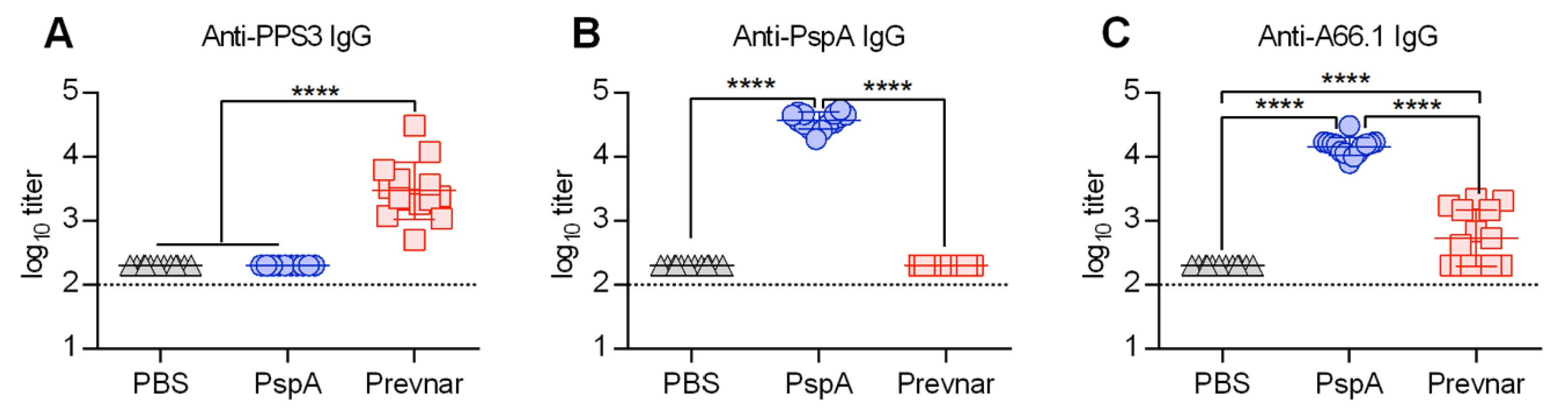
3.1. PspA Protein-Based Vaccination Generates Greater Anti-Pneumococcal IgG Antibody Levels Compared to Prevnar

Previously, we have shown that vaccination with Prevnar, the polysaccharide conjugate vaccine, only partially protects mice against secondary pneumococcal infection [9]. To determine if an S. pneumoniae protein-based vaccine can provide improved protection, we intramuscularly vaccinated mice with PspA. To quantify the antibody response following Prevnar and PspA vaccination, sera were harvested on day 7 post-boost and analyzed by antibody enzyme-linked immunosorbent assay (ELISA) (Figure 1A–C). Mice vaccinated with Prevnar or PspA had a 1.5 to 2 log increase in antigen specific IgG titer compared with PBS mice (Figure 1A,B). To directly compare PspA to the Prevnar vaccine response, ELISA plates were coated with heat-killed S. pneumoniae A66.1 serotype 3. The IgG response against the whole killed bacteria was significantly higher in PspA compared to Prevnar vaccinated mice (Figure 1C).

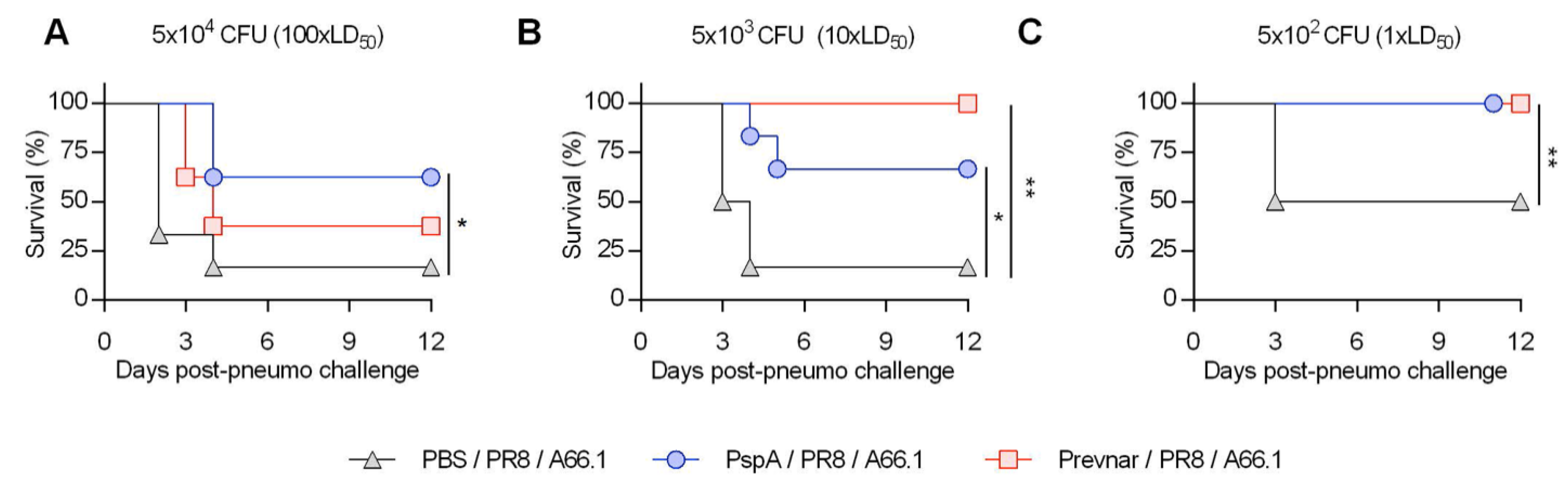
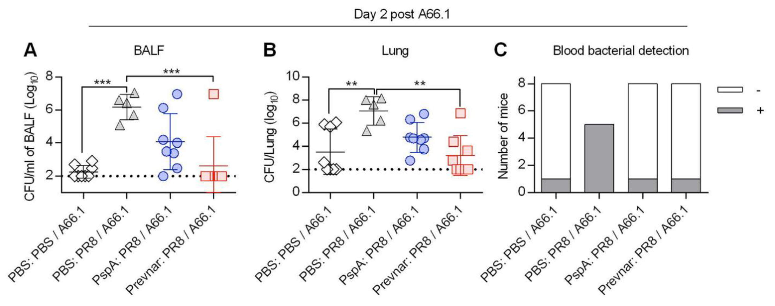
3.2. Vaccine-Induced Protection against Co-Infection Is Bacterial Dose Dependent

To determine if the enhanced anti-pneumococcal antibody response correlates with protection against secondary bacterial infection, vaccinated mice were challenged with a sub-lethal dose (10 PFU) of PR8 and co-infected with 5 × 10⁴ CFU (a dose equivalent to 100 × LD50 for co-infection) of S. pneumoniae A66.1 on day 9 post-influenza (Figure 2A). As expected, Prevnar-vaccinated (Prevnar/PR8/A66.1) mice were only partially protected, with 40% of the Prevnar vaccinated mice succumbing to the secondary infection within 3 days of the S. pneumoniae A66.1 challenge compared to 90% of unvaccinated co-infected mice (PBS/PR8/A66.1). Similarly, 66% of PspA vaccinated mice succumbed to co-infection. These results demonstrate that the PspA vaccination, like Prevnar, cannot fully overcome the defect in antibacterial protective mechanisms following influenza infection. The lack of protection was not due to a potential failure of vaccination, as both PspA and Prevnar vaccinated mice that were challenged with bacteria only were fully protected (Figure S1). In fact, vaccination protected 100% of mice challenged with a 20 × LD50 of single S. pneumoniae A66.1 infection (Figure S1).
Next, we infected mice with lower doses of bacteria to determine whether we had overwhelmed the protective efficacy of the vaccine (Figure 2B,C). Influenza-infected mice were challenged with 5 × 10² bacterial CFU (1 × LD50 for co-infection) (Figure 2C). All the PspA and Prevnar vaccinated mice survived the 1 × LD50 secondary A66.1 challenge. In contrast, only 50% of unvaccinated co-infected mice survived (Figure 2C). No deaths were observed in the vaccinated or unvaccinated singly infected groups (Figure S1). Similar effects were observed in mice challenged with 5 × 10³ bacterial CFU (10 × LD50) (Figure 2B). These results suggest that vaccine-induced protection against co-infection is dependent on the bacterial challenge dose.
To assess whether the increases in survival were associated with enhanced bacterial clearance, bacterial burden in the bronchoalveolar lavage fluid (BALF), lung tissue, and blood was enumerated in mice immunized and challenged with 5 × 10³ CFU of S. pneumoniae A66.1 following influenza infection (Figure 3). As expected, unimmunized co-infected mice had significantly more bacteria in their BALF and lung tissue compared to unimmunized mice singly infected with bacteria. Both PspA and Prevnar immunized co-infected mice had fewer recoverable bacteria compared to the unimmunized co-infected mice (Figure 3A,B). Finally, all five unimmunized co-infected mice had bacteria in their blood, indicative of dissemination, compared to one out of eight mice in the bacteria-only and immunized groups (Figure 3C). These results suggest that immunization with PspA controls pulmonary bacterial outgrowth and bacteremia following a low-dose bacterial challenge in influenza-infected mice.

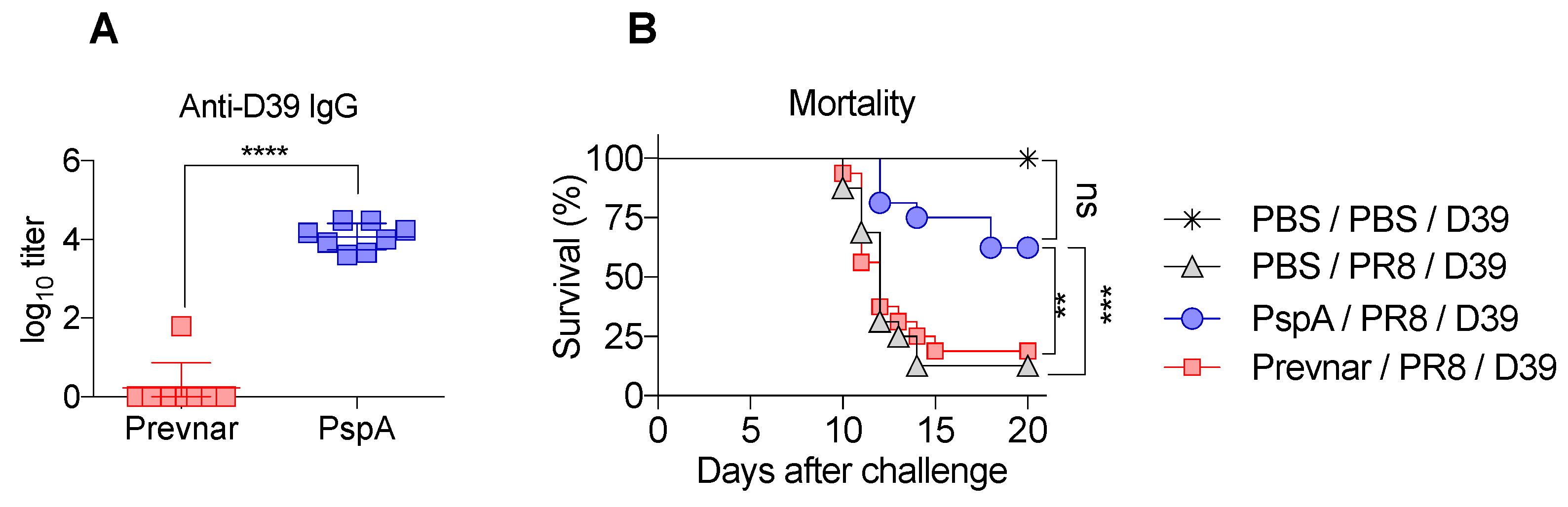
3.3. PspA Immunization Is Protective against Serotype 2 Bacterial Challenge during Co-Infection

To determine whether PspA vaccination can provide heterologous protection in a serotype independent manner during co-infection, we next used S. pneumoniae serotype 2 (strain D39), whose polysaccharide is not included in the Prevnar vaccine. We first measured anti-D39 antibody responses in mice immunized with PspA or Prevnar. As expected, mice immunized with Prevnar did not have detectable levels of anti-D39 antibody in contrast to PspA-immunized mice (Figure 4A). To see if this response was protective, we challenged these mice with a sublethal dose of D39 on day 9 post-influenza and monitored survival. PspA-immunized mice had significantly fewer deaths compared to unimmunized and Prevnar-immunized co-infected mice (Figure 4B). These results suggest that inclusion of PspA in the current Prevnar formulation would increase the breadth of protection.

4. Discussion

During seasonal and pandemic influenza infection, commensal and/or opportunistic bacteria can invade the lower respiratory tract and cause pneumonia that can result in severe morbidity and/or death. In this study, we have shown that PspA vaccination can protect influenza-infected mice against secondary S. pneumoniae challenge. This protection was, however, dose-dependent; PspA vaccinated mice were fully protected against low-dose challenges, which were lethal for unvaccinated co-infected mice. This protection was strongly correlated with enhanced antibody production and bacterial clearance.
This report extends our previous finding that Prevnar, a polysaccharide conjugate vaccine, only partially protects mice against co-infection [9]. Following that study, we hypothesized that the protection against co-infection was incomplete because polysaccharides have reduced immunogenicity compared to protein antigens and the response that is generated is not sufficient to overcome the influenza-induced defective antibacterial clearance mechanism(s). Our data now show that immunization with PspA, like polysaccharide-based vaccines, only partially protects influenza-infected mice against high doses of secondary bacterial pneumonia. These results support the idea that the suboptimal protection induced by vaccination is not a polysaccharide-specific phenomenon. Thus, a question remains as to why vaccine-induced anti-bacterial adaptive immunity cannot provide complete protection against post-influenza secondary S. pneumoniae infection. One of the dominating concepts in the influenza-bacterial co-infection field is centered on cytokine-mediated suppression of anti-bacterial immunity, including downregulation of phagocytic functions of innate immune cells [5,6,24,25,26,27,28,29,30,31]. Since PspA vaccination should not interfere with virus-induced cytokine responses, primary influenza infection is expected to impair anti-bacterial immunity in PspA vaccinated mice. Therefore, it is plausible that the observed suboptimal protection is due to an impaired opsonophagocytic killing of antibody-coated S. pneumoniae. This hypothesis is currently under investigation.
Several recent studies reported that PspA vaccination can induce a protective antibody response and enhance bacterial clearance during influenza-S. pneumoniae co-infection [21,32,33]. The distinctions between our studies and earlier works published by others are that (1) we compared our immunization to Prevnar13, the FDA approved anti-pneumococcal vaccine, and showed that PspA is equally as protective; (2) we showed that PspA can protect against more than one pneumococcal strain, specifically serotype 2 D39, which is a strain not included in Prevnar13; and (3) we showed that protection is dependent on the bacterial challenge dose. The dose escalation studies we conducted showed that 100% of mice immunized with PspA and subsequently challenged with 1 × LD50 of S. pneumoniae in a co-infection model survived. Under the same condition, only ~70% of PspA vaccinated mice survived the challenge of 100 × LD50. Nevertheless, our results agree with the other studies in that PspA vaccination can provide protection against secondary S. pneumoniae infection. How influenza A virus infection impairs anti-bacterial immunity is an area that is under active investigation. This is important because defective bacterial clearance is not due to insufficient antibody production or downregulation of Fcγ receptors needed to mediate antibody-dependent cellular phagocytosis [9].
It is important to note that PspA vaccination has been evaluated in phase 1 clinical trial and PspA was found to be safe and immunogenic—i.e., high levels of cross-clade antibodies to heterologous PspA molecules were detected in vaccinated individuals [17]. However, a theoretical issue was raised with the use of PspA as a vaccine antigen, due to a low sequence homology between PspA and human cardiac myosin [34]. Thus, there is a concern that PspA vaccination could lead to autoantibodies that may cause autoimmune conditions. However, such concerns can be alleviated by the absence of clinical evidence linking anti-PspA antibodies and cardiac injuries. Nonetheless, future efforts regarding the PspA vaccination approach should focus on PspA peptide-based vaccines that do not contain the homology with human cardiac myosin.
In this study, we show for the first time, by focusing primarily on survival, that vaccine-induced protection against influenza-S. pneumoniae co-infection is bacterial dose-dependent. We further show that the protective efficacy of PspA immunization is comparable to Prevnar13-induced protection against secondary serotype 3 pneumococcal (A66.1) challenge. However, PspA, unlike Prevnar, was protective against D39, a serotype not covered in Prevnar. This was not unexpected since, although three families of PspA exist, antibodies against PspA are cross-reactive within families and are cross-protective in a single S. pneumoniae infection model [14,16]. Thus, we conclude that there may be benefits to using PspA as a vaccine antigen over a polysaccharide-based vaccine or as an additional antigen in the current vaccine formulation. However, it needs to be stressed that our PspA vaccination approach was tested on adult mice. We believe that PspA vaccination would also provide some level of protection in susceptible individuals such as infants and older adults. Additional studies assessing the ability of PspA vaccination approach to provide protection against co-infection in other age groups would be of interest.

Supplementary Materials

The following are available online at https://www.mdpi.com/2076-393X/7/4/146/s1. Figure S1: Survival of vaccinated mice following single S. pneumoniae infection.

Author Contributions

Conceptualization, S.R., D.W.M., and Y.F.; methodology, S.R., D.W.M., and Y.F.; investigation and formal analysis, S.R., C.M.W., S.L.S., J.L.B. and Y.F.; writing—original draft preparation, S.R.; writing—review and editing, S.R., D.W.M., and Y.F.; supervision, D.W.M. and Y.F.; funding acquisition, D.W.M. and Y.F.

Funding

This work was supported by the National Institute of Health grant RO1 AI075312 (D.W.M.) and by the American Heart Association grant 17SDG33630188 (Y.F.).

Acknowledgments

The following reagent was obtained through BEI Resources, NIAID, NIH: Streptococcus pneumoniae Family 1, Clade 2 Pneumococcal Surface Protein A (PspA UAB055) with C-Terminal Histidine Tag, Recombinant from Escherichia coli, NR-33178.

Conflicts of Interest

The authors declare no competing financial interests. The funders had no role in the design of the study; in the collection, analyses, or interpretation of data; in the writing of the manuscript, or in the decision to publish the results.

References

  1. Morens, D.M.; Fauci, A.S. The 1918 influenza pandemic: Insights for the 21st century. J. Infect. Dis. 2007, 195, 1018–1028. [Google Scholar] [CrossRef] [PubMed]
  2. Cilloniz, C.; Ewig, S.; Menendez, R.; Ferrer, M.; Polverino, E.; Reyes, S.; Gabarrus, A.; Marcos, M.A.; Cordoba, J.; Mensa, J.; et al. Bacterial co-infection with H1N1 infection in patients admitted with community acquired pneumonia. J. Infect. 2012, 65, 223–230. [Google Scholar] [CrossRef] [PubMed]
  3. Koon, K.; Sanders, C.M.; Green, J.; Malone, L.; White, H.; Zayas, D.; Miller, R.; Lu, S.; Han, J. Co-detection of pandemic (H1N1) 2009 virus and other respiratory pathogens. Emerg. Infect. Dis. 2010, 16, 1976–1978. [Google Scholar] [CrossRef] [PubMed]
  4. Sun, K.; Metzger, D.W. Inhibition of pulmonary antibacterial defense by interferon-gamma during recovery from influenza infection. Nat. Med. 2008, 14, 558–564. [Google Scholar] [CrossRef] [PubMed]
  5. Shahangian, A.; Chow, E.K.; Tian, X.; Kang, J.R.; Ghaffari, A.; Liu, S.Y.; Belperio, J.A.; Cheng, G.; Deng, J.C. Type I IFNs mediate development of postinfluenza bacterial pneumonia in mice. J. Clin. Investig. 2009, 119, 1910–1920. [Google Scholar] [CrossRef] [PubMed]
  6. Kudva, A.; Scheller, E.V.; Robinson, K.M.; Crowe, C.R.; Choi, S.M.; Slight, S.R.; Khader, S.A.; Dubin, P.J.; Enelow, R.I.; Kolls, J.K.; et al. Influenza A inhibits Th17-mediated host defense against bacterial pneumonia in mice. J. Immunol. 2011, 186, 1666–1674. [Google Scholar] [CrossRef]
  7. McCullers, J.A.; Rehg, J.E. Lethal synergism between influenza virus and Streptococcus pneumoniae: Characterization of a mouse model and the role of platelet-activating factor receptor. J. Infect. Dis. 2002, 186, 341–350. [Google Scholar] [CrossRef]
  8. Esposito, S.; Principi, N. Impacts of the 13-Valent Pneumococcal Conjugate Vaccine in Children. J. Immunol. Res. 2015, 2015, 591580. [Google Scholar] [CrossRef]
  9. Sun, K. Limited Efficacy of Antibacterial Vaccination Against Secondary Serotype 3 Pneumococcal Pneumonia Following Influenza Infection. J. Infect. Dis. 2015, 212, 445–452. [Google Scholar]
  10. Madhi, S.A.; Klugman, K.P.; Vaccine Trialist, G. A role for Streptococcus pneumoniae in virus-associated pneumonia. Nat. Med. 2004, 10, 811–813. [Google Scholar] [CrossRef]
  11. Dominguez, A.; Castilla, J.; Godoy, P.; Delgado-Rodriguez, M.; Saez, M.; Soldevila, N.; Astray, J.; Mayoral, J.M.; Martin, V.; Quintana, J.M.; et al. Effectiveness of vaccination with 23-valent pneumococcal polysaccharide vaccine in preventing hospitalization with laboratory confirmed influenza during the 2009–2010 and 2010–2011 seasons. Hum. Vaccin. Immunother. 2013, 9, 865–873. [Google Scholar] [CrossRef] [PubMed]
  12. Croney, C.M.; Coats, M.T.; Nahm, M.H.; Briles, D.E.; Crain, M.J. PspA family distribution, unlike capsular serotype, remains unaltered following introduction of the heptavalent pneumococcal conjugate vaccine. Clin. Vaccine Immunol. 2012, 19, 891–896. [Google Scholar] [CrossRef] [PubMed]
  13. Hotomi, M.; Togawa, A.; Kono, M.; Ikeda, Y.; Takei, S.; Hollingshead, S.K.; Briles, D.E.; Suzuki, K.; Yamanaka, N. PspA family distribution, antimicrobial resistance and serotype of Streptococcus pneumoniae isolated from upper respiratory tract infections in Japan. PLoS ONE 2013, 8, e58124. [Google Scholar] [CrossRef] [PubMed]
  14. Hollingshead, S.K.; Becker, R.; Briles, D.E. Diversity of PspA: Mosaic genes and evidence for past recombination in Streptococcus pneumoniae. Infect. Immun. 2000, 68, 5889–5900. [Google Scholar] [CrossRef] [PubMed]
  15. McDaniel, L.S.; Sheffield, J.S.; Delucchi, P.; Briles, D.E. PspA, a surface protein of Streptococcus pneumoniae, is capable of eliciting protection against pneumococci of more than one capsular type. Infect. Immun. 1991, 59, 222–228. [Google Scholar] [PubMed]
  16. Briles, D.E.; Hollingshead, S.K.; King, J.; Swift, A.; Braun, P.A.; Park, M.K.; Ferguson, L.M.; Nahm, M.H.; Nabors, G.S. Immunization of humans with recombinant pneumococcal surface protein A (rPspA) elicits antibodies that passively protect mice from fatal infection with Streptococcus pneumoniae bearing heterologous PspA. J. Infect. Dis. 2000, 182, 1694–1701. [Google Scholar] [CrossRef] [PubMed]
  17. Nabors, G.S.; Braun, P.A.; Herrmann, D.J.; Heise, M.L.; Pyle, D.J.; Gravenstein, S.; Schilling, M.; Ferguson, L.M.; Hollingshead, S.K.; Briles, D.E.; et al. Immunization of healthy adults with a single recombinant pneumococcal surface protein A (PspA) variant stimulates broadly cross-reactive antibodies to heterologous PspA molecules. Vaccine 2000, 18, 1743–1754. [Google Scholar] [CrossRef]
  18. Arulanandam, B.P.; Lynch, J.M.; Briles, D.E.; Hollingshead, S.; Metzger, D.W. Intranasal vaccination with pneumococcal surface protein A and interleukin-12 augments antibody-mediated opsonization and protective immunity against Streptococcus pneumoniae infection. Infect. Immun. 2001, 69, 6718–6724. [Google Scholar] [CrossRef]
  19. Fukuyama, Y.; Yuki, Y.; Katakai, Y.; Harada, N.; Takahashi, H.; Takeda, S.; Mejima, M.; Joo, S.; Kurokawa, S.; Sawada, S.; et al. Nanogel-based pneumococcal surface protein A nasal vaccine induces microRNA-associated Th17 cell responses with neutralizing antibodies against Streptococcus pneumoniae in macaques. Mucosal Immunol. 2015, 8, 1144–1153. [Google Scholar] [CrossRef] [PubMed] [Green Version]
  20. Piao, Z.; Akeda, Y.; Takeuchi, D.; Ishii, K.J.; Ubukata, K.; Briles, D.E.; Tomono, K.; Oishi, K. Protective properties of a fusion pneumococcal surface protein A (PspA) vaccine against pneumococcal challenge by five different PspA clades in mice. Vaccine 2014, 32, 5607–5613. [Google Scholar] [CrossRef]
  21. Kong, I.G.; Sato, A.; Yuki, Y.; Nochi, T.; Takahashi, H.; Sawada, S.; Mejima, M.; Kurokawa, S.; Okada, K.; Sato, S.; et al. Nanogel-based PspA intranasal vaccine prevents invasive disease and nasal colonization by Streptococcus pneumoniae. Infect. Immun. 2013, 81, 1625–1634. [Google Scholar] [CrossRef] [PubMed]
  22. Winter, C.; Herbold, W.; Maus, R.; Langer, F.; Briles, D.E.; Paton, J.C.; Welte, T.; Maus, U.A. Important role for CC chemokine ligand 2-dependent lung mononuclear phagocyte recruitment to inhibit sepsis in mice infected with Streptococcus pneumoniae. J. Immunol. 2009, 182, 4931–4937. [Google Scholar] [CrossRef] [PubMed]
  23. Bai, Y.; Yang, J.; Eisele, L.E.; Underwood, A.J.; Koestler, B.J.; Waters, C.M.; Metzger, D.W.; Bai, G. Two DHH subfamily 1 proteins in Streptococcus pneumoniae possess cyclic di-AMP phosphodiesterase activity and affect bacterial growth and virulence. J. Bacteriol. 2013, 195, 5123–5132. [Google Scholar] [CrossRef] [PubMed]
  24. Van der Sluijs, K.F.; van Elden, L.J.; Nijhuis, M.; Schuurman, R.; Pater, J.M.; Florquin, S.; Goldman, M.; Jansen, H.M.; Lutter, R.; van der Poll, T. IL-10 is an important mediator of the enhanced susceptibility to pneumococcal pneumonia after influenza infection. J. Immunol. 2004, 172, 7603–7609. [Google Scholar] [CrossRef] [PubMed]
  25. Nakamura, S.; Davis, K.M.; Weiser, J.N. Synergistic stimulation of type I interferons during influenza virus coinfection promotes Streptococcus pneumoniae colonization in mice. J. Clin. Investig. 2011, 121, 3657–3665. [Google Scholar] [CrossRef] [PubMed] [Green Version]
  26. Li, W.; Moltedo, B.; Moran, T.M. Type I interferon induction during influenza virus infection increases susceptibility to secondary Streptococcus pneumoniae infection by negative regulation of gammadelta T cells. J. virology 2012, 86, 12304–12312. [Google Scholar] [CrossRef]
  27. Jakab, G.J. Immune impairment of alveolar macrophage phagocytosis during influenza virus pneumonia. Am. Rev. Respir. Dis. 1982, 126, 778–782. [Google Scholar]
  28. Kodihalli, S.; Sivanandan, V.; Nagaraja, K.V.; Shaw, D.; Halvorson, D.A. Effect of avian influenza virus infection on the phagocytic function of systemic phagocytes and pulmonary macrophages of turkeys. Avian Dis. 1994, 38, 93–102. [Google Scholar] [CrossRef]
  29. Rynda-Apple, A.; Harmsen, A.; Erickson, A.S.; Larson, K.; Morton, R.V.; Richert, L.E.; Harmsen, A.G. Regulation of IFN-gamma by IL-13 dictates susceptibility to secondary postinfluenza MRSA pneumonia. Eur. J. Immunol. 2014, 44, 3263–3272. [Google Scholar] [CrossRef]
  30. Lee, B.; Robinson, K.M.; McHugh, K.J.; Scheller, E.V.; Mandalapu, S.; Chen, C.; Di, Y.P.; Clay, M.E.; Enelow, R.I.; Dubin, P.J.; et al. Influenza-induced type I interferon enhances susceptibility to gram-negative and gram-positive bacterial pneumonia in mice. Am. J. Physiol. Lung Cell. Mol. Physiol. 2015, 309, L158–L167. [Google Scholar] [CrossRef]
  31. Robinson, K.M.; Lee, B.; Scheller, E.V.; Mandalapu, S.; Enelow, R.I.; Kolls, J.K.; Alcorn, J.F. The role of IL-27 in susceptibility to post-influenza Staphylococcus aureus pneumonia. Respiratory research 2015, 16, 10. [Google Scholar] [CrossRef] [PubMed]
  32. King, Q.O.; Lei, B.; Harmsen, A.G. Pneumococcal surface protein A contributes to secondary Streptococcus pneumoniae infection after influenza virus infection. J. Infect. Dis. 2009, 200, 537–545. [Google Scholar] [CrossRef] [PubMed]
  33. Greene, C.J.; Marks, L.R.; Hu, J.C.; Reddinger, R.; Mandell, L.; Roche-Hakansson, H.; King-Lyons, N.D.; Connell, T.D.; Hakansson, A.P. Novel Strategy To Protect against Influenza Virus-Induced Pneumococcal Disease without Interfering with Commensal Colonization. Infect. Immun. 2016, 84, 1693–1703. [Google Scholar] [CrossRef] [PubMed] [Green Version]
  34. Moffitt, K.; Malley, R. Rationale and prospects for novel pneumococcal vaccines. Hum. Vaccin. Immunother. 2016, 12, 383–392. [Google Scholar] [CrossRef] [PubMed]
Figure 1. Pneumococcal surface protein A (PspA) vaccination induces robust anti-pneumococcal A66.1 antibody production. (AC) PPS3- (A), PspA- (B) and A66.1 (C)-specific antibody titers in the serum day 7 post-vaccination. Mice (n = 12/group) were i.m. vaccinated with either PBS, PspA, or Prevnar and boosted on day 21. Sera were harvested on day 28 and analyzed by enzyme-linked immunosorbent assay (ELISA). Groups were compared using Analysis of variance (ANOVA) with Tukey’s post-test for multiple comparison. ****, p < 0.0001. Data shown are representative of two independent experiments.
Vaccines 07 00146 g001
Figure 2. Survival of vaccinated mice following influenza-pneumococcus co-infection is bacterial dose dependent. (AC) Mice were i.m. vaccinated with either PBS, PspA, or Prevnar13 and boosted on day 21 (n = 6/group). Two weeks post-vaccination mice were i.n. infected with 15 PFU of PR8 and i.n. challenged on day 9 post-PR8 with either 5 × 10⁴ (A), 5 × 10³ (B), 5 × 10² CFU (C) of S. pneumoniae serotype 3 A66.1. * p < 0.01, ** p < 0.001; unvaccinated co-infected mice (PBS/PR8/A66.1) compared to either PspA or Prevnar vaccinated co-infected mice as determined by Log-rank test. High lethal (5 × 10⁴) dose survival data were repeated at least twice (A).
Vaccines 07 00146 g002
Figure 3. Reduced pulmonary and blood bacterial burden in PspA immunized mice following influenza-pneumo co-infection. (AC) Mice were i.m. vaccinated with either PBS, PspA, or Prevnar and boosted on day 21 (n = 5–8/group). Two weeks post-vaccination mice were i.n. infected with 15 PFU of PR8 and i.n. challenged on day 10 post-PR8 with 5 × 10³ of S. pneumoniae serotype 3 A66.1. On day 2 post A66.1 infection, mice were sacrificed, and the bacterial CFU in the BALFs (A), lungs (B), and bloods (C) were enumerated. Statistical significance was determined by one-way ANOVA. **, p < 0.01, ***, p < 0.001. Lung burden data are representative of two independent experiments (B).
Vaccines 07 00146 g003
Figure 4. PspA vaccinated mice are protected against influenza-S. pneumoniae serotype 2 co-infection. Mice were i.m. vaccinated with either PBS, PspA, or Prevnar and boosted on day 21. (A) Sera were harvested on day 28 and analyzed by ELISA (n = 8 mice/group). (B) Two weeks post-vaccination mice were i.n. infected with 15 PFU of PR8 and i.n. challenged on day 9 post-PR8 with 1.5 × 10⁴ CFU S. pneumoniae type 2 strain D39 (n = 16/group). Survival data were combined from two independent experiments. Titer and survival data were analyzed by Student’s t test and Log-rank test, respectively. **, p < 0.01, ***, p < 0.001, ****, p < 0.0001.
Vaccines 07 00146 g004

