Research Progress, Application, and Industrialization Prospects of Circular RNA Vaccines in Viral Diseases


Abstract
1. Introduction
2. Overview of RNA Vaccine Technology
2.1. Technical Characteristics of mRNA Vaccines
2.2. Technical Characteristics of saRNA Vaccines
2.3. Structure and Biosynthesis of circRNA
2.4. Functions of Endogenous Non-Coding circRNAs
2.5. Vaccine-Compatible Properties of circRNA
2.5.1. Industrialization Comparison: circRNA and Linear mRNA
2.5.2. Quality Control Standards and Regulatory Framework
2.5.3. Clinical Translation Progress
- (1)
- Human Vaccines
- (2)
- Veterinary Vaccines

| Vaccine | Cold-Chain Dependence | Stability | Variant Adaptability | Antigen Expression | IgG Relative Titer | Manufacturing | Advantages | Disadvantages | References |
|---|---|---|---|---|---|---|---|---|---|
| Linear mRNA | High dependence | Low | Weak | Transient | 1-fold | Simple & mature | Industrial | Cold-chain dependent | [1,6,11,13,16,21,49] |
| SaRNA | Dependence | Moderate | Moderate | High & prolonged | 1.1–1.3-fold | Complex | Self-amplifying | High cost | [17,18,19] |
| CircRNA | Low dependence (preclinical formulation data) | Relatively high (intrinsic closed-loop property, preclinically validated) | Formidable | Sustained & durable | Approx. 3.8-fold (mouse model, SARS-CoV-2 antigen) | Medium | Thermostable | Low translation | [6,8,21,22,31,34,50] |
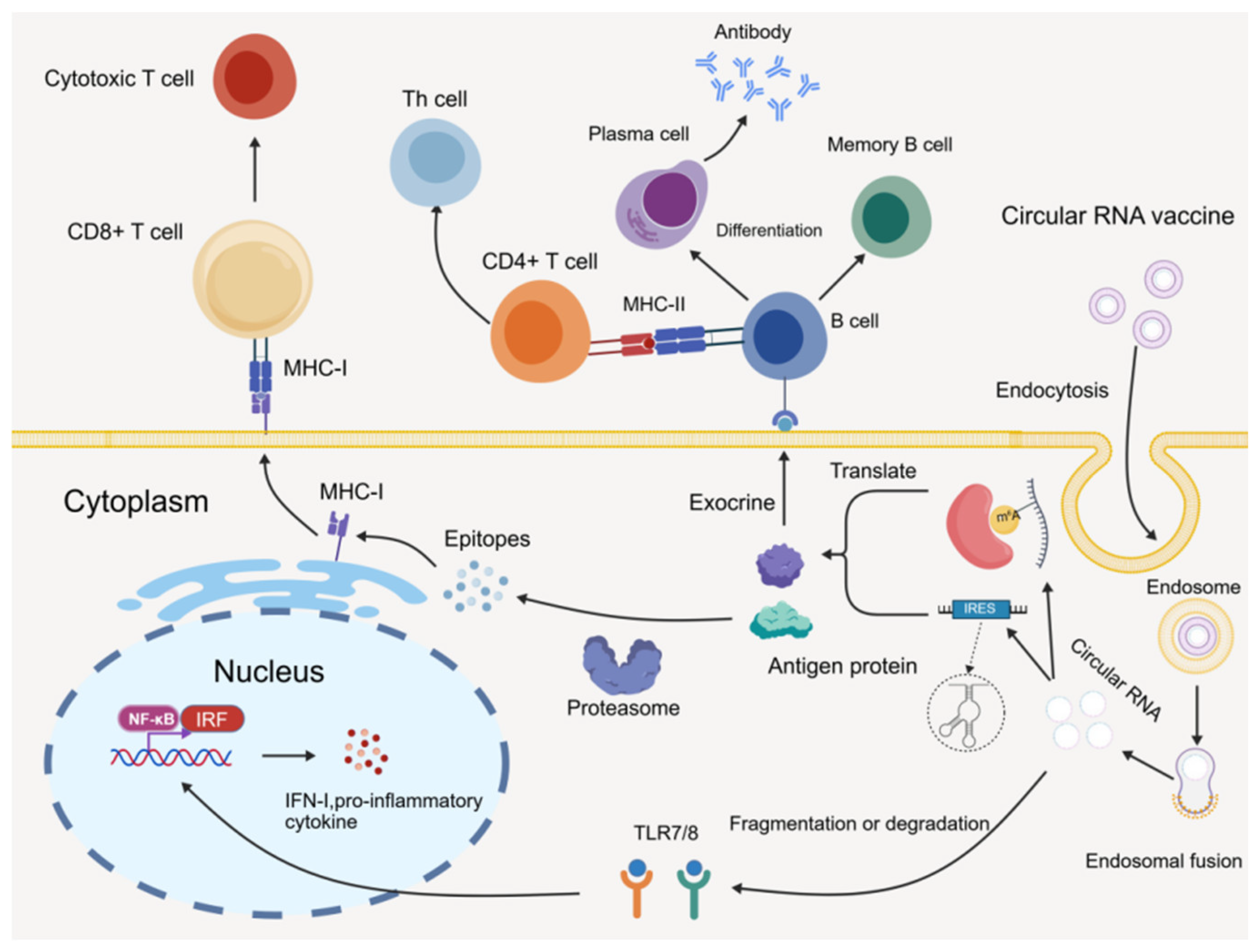
3. Immune Response Mechanism of circRNA Vaccines
4. Synthesis Methods of circRNA Vaccines
4.1. PIE System
- (1)
- Introduction of homology arms and spacer sequence engineering, increasing the circularization efficiency of 5 kb long-chain RNA from 0% to 87%, overcoming the bottleneck of long-fragment circularization [62].
- (2)
- Development of a scarless PIE derivative system based on the Anabaena intron, which requires only the “A, T” dinucleotide to maintain the molecular characteristics of circularization, establishing a PIE-derived system completely free of exogenous sequences [60].
- (3)
- Using cryo-electron microscopy to resolve the high-resolution structure of the rearranged T4 bacteriophage thymidylate synthase gene intron (T4Td), establishing a structure-guided circularization scaffold engineering paradigm that greatly enhances the circularization efficiency of the T4td-PIE system [63].
- (4)
- Repurposing natural complete introns to construct circularization technology and directly utilizing natural complete group I/II introns to achieve circularization without artificial splitting and engineering under mild reaction conditions, enabling the scarless circularization of ultra-large 14 kb RNA fragments with extremely low product immunogenicity [64].
4.2. T4 Rnl2 Enzymatic Ligation System
- (1)
- Using a computational “lock-and-key” design to achieve spontaneous proximity of RNA ends through sequence annealing, relying on T4 Rnl2 for efficient catalytic circularization, with a circularization efficiency exceeding 70% for 74–500 nt RNA. Among them, the efficiency for a 74 nt short sequence can reach 92%, and after optimization, the circularization efficiency for an 1100 nt sequence reaches 74%. The product does not require fine purification by Ribonuclease R (RNase R) or HPLC [45].
- (2)
- Translational co-optimization: avoiding non-specific base pairing between the IRES and the downstream gene of interest, disrupting unfavorable secondary structures through site-directed mutagenesis, relieving inhibition of the IRES functional domain, and simultaneously achieving dual enhancement of circularization efficiency and protein translation efficiency [62].
- (3)
- Splint-free self-assembly strategy: rationally designing linear RNA precursors using RNA secondary structure prediction, allowing the ends to spontaneously form a nicked structure and achieving scarless ligation without exogenous splints. The maximum circularization efficiency reaches 63%, and the IRES translation activity is fully restored after circularization, with translation efficiency being enhanced by up to 26-fold [66,67,68,69].
4.3. LRC System
- (1)
- Bioinformatics-assisted sequence design: utilizing RNA secondary structure simulation algorithms to perform site-directed optimization of the base composition in terminal complementary regions, weakening non-specific folding, strengthening directional terminal pairing capability, and enhancing sequence universality and circularization stability [71].
- (2)
- Optimization of buffer systems and ionic environment: fine-tuning Mg2+ concentration, reaction temperature, and pH to modulate RNA folding kinetics, reducing the formation of incorrect conformations, and significantly improving the self-circularization efficiency of long-fragment LRC [72].
- (3)
- Fusion design with functional elements: modular integration of IRES, spacer sequences, and the LRC scaffold, achieving the assembly of the circularization scaffold and translation functional elements in one step, thereby ensuring efficient self-circularization while safeguarding subsequent antigen translation activity and meeting the needs of integrated molecular design for circRNA vaccines [73].
4.4. CIRC Self-Splicing Intron System
- (1)
- Screening for highly efficient self-splicing introns and implementing structure-guided site-directed mutagenesis to enhance circularization efficiency and sequence adaptability [63].
- (2)
- Fine-tuning parameters such as Mg2+ concentration, pH, and NTP ratios in the co-transcription reaction system to balance transcription and circularization kinetics, thereby suppressing non-specific by-products [22].
- (3)
- Utilizing bioinformatics tools to predict the secondary structure of the fusion RNA, and introducing spacer sequences or synonymous mutations to disrupt competitive stem-loops, restoring the native intron conformation and ensuring circularization efficiency [74].
| Comparison Item | Permuted Intron–Exon | T4 RNA Ligase 2 Mediated Ligation | Linear RNA Self-Circularization | Complete Self-Splicing Intron for RNA Circularization | References |
|---|---|---|---|---|---|
| Circularization principle | Two-step transesterification self-splicing circularization of group I introns | Enzyme-catalyzed ligation of RNA 3′-OH and 5′-phosphate ends | Spontaneous folding of complementary sequences at RNA molecule ends | Based on the second-step splicing reaction of natural complete group I or group II introns | [27,64] |
| Template design features | Intron permutation and splitting, with the target gene inserted between exon regions (E1/E2) | Design linear RNA precursor, using terminal secondary structure self-annealing or a splint to bring the ends into proximity | Adopt a computationally optimized “lock-and-key” terminal complementary structure, bringing the 5′ and 3′ ends into spontaneous proximity through annealing | Use natural complete intron, without splitting, and insert the target gene inside the intron | [27,43,45,64] |
| Key conditions | GTP, Mg2+, high-temperature circularization | T4 Rnl2, ATP, mild conditions | Enzyme-free or with T4 Rnl2, mild conditions | Basic transcription buffer, mild conditions | [27,64] |
| Cyclization efficiency | 87–95% | 70–95% | 70–73% | 80–100% | [35,45,64,75] |
| Applicable RNA length | Long-sequence RNA | Small- to medium-length RNA | Tens to thousands of nt | Long-sequence RNA | [35,45,64] |
| Advantages | Wide application; relatively simple reaction conditions | Precise product; no exogenous sequences | Streamlined process; low cost | High efficiency; low immunogenicity | [40,64,67] |
| Limitations | Multiple steps; potential immunogenicity | Low circularization efficiency for large RNA fragments; splint assistance needed in some cases | Highly dependent on RNA folding properties and structural design precision | Relatively new technology; incomplete system validation | [40,66,76] |
| Main by-products | Uncircularized linear precursors, excised intron fragments, scar sequences, nicked circRNA, concatemers | Linear RNA precursors, splint-DNA residuals, mis-ligated concatemers, nicked circRNA | Misfolded concatemers, partial nicked circRNA, linear side-products | Excised intron debris, minor fraction of nicked circRNA | [12,35,64] |
| Vaccine development readiness | High; pre-clinical immunization-challenge studies in mice, non-human primates for SARS-CoV-2, monkeypox, and other viruses | Moderate–low; mostly cellular-level validation for short-to-medium antigens; scarce animal-challenge reports for full-length viral antigens | Low; only in vitro molecular and cellular data | Moderate; limited proof-of-concept mouse challenge data for viral vaccines; GMP-scale animal efficacy data still absent | [7,29,64,65,70] |
4.5. Quantitative Comparison of Circularization Platforms and Linear mRNA for circRNA Vaccines
4.6. Critical Quality Attributes and Quality Control Status of circRNA Vaccines
5. Molecular Design and Antiviral Immune Mechanisms of circRNA Vaccines
5.1. Vaccine Target Design and Optimization
5.2. Strategies to Improve Translation Efficiency
- (1)
- Selecting and engineering potent IRES: Studies have shown that screening the enterovirus A (EV-A) IRES with excellent universality and truncating it to retain the core functional domain can increase translation efficiency by approximately 50%. Mutagenesis screening of the coxsackievirus B3 (CVB3) IRES yielded mutants such as A62G and U287A that could double protein yield [57]. Inserting eukaryotic translation initiation factor 4G (eIF4G) recruitment aptamers into specific loop regions of strong IRES or adopting dual IRES combinations can also significantly enhance translation [73,79].
- (2)
- Optimizing sequences upstream and downstream of the IRES-ORF expression cassette: Introducing binding motifs for RNA-binding proteins such as polyA binding protein (PABP) and PolyC Binding Protein (PCBP) upstream and downstream of IRES-ORF promotes the formation of the translation initiation complex. Adding spacer sequences such as polyA50 between the IRES, coding region, and residual sequences can alleviate structural interference [22]. Adding the woodchuck hepatitis virus posttranscriptional regulatory element (WPRE) and a poly(A) stretch at the 3′ end can increase translation by more than 5-fold, and removing specific short sequences within WPRE can further restore expression by approximately 2-fold [73].
- (3)
- Introducing epigenetic modifications and purification optimization: Supplementing N6-methyladenosine Triphosphate (N6-methyl-ATP) during IVT to introduce m6A modification can enhance cap-independent translation by recruiting YTH Domain Family Protein 3 (YTHDF3), while reducing immunogenicity and maintaining RNA stability. Thoroughly purifying circRNA to reduce linear impurities can prevent innate immune activation from interfering with translation [71].
- (4)
- Employing circRNA technology to covalently attach a 7-methylguanosine (m7G) cap via a branched structure to form capped circular RNA, which can increase protein expression by approximately 7-fold compared to traditional IRES-circRNA: Non-covalent capping can enhance translation by over 50-fold and supports a rolling circle-like translation mode [82].
5.3. Future Design Directions for circRNA Vaccines
- (1)
- AI-assisted design: In the field of bioinformatics, deep learning models such as convolutional neural networks powered by AI can enhance the performance of antigen prediction, RNA structure modeling, and lipid nanoparticle delivery system optimization, surpassing traditional bioinformatics methods. Integrating AI-driven tools with conventional bioinformatics and experimental validation can elevate the level of vaccine development while ensuring computational efficiency and biological reliability [83].
- (2)
- Virus-specific design: For pan-variant protection, Qu et al. designed a circRNA vaccine targeting the RBD region of the SARS-CoV-2 Delta variant that induced broadly neutralizing antibodies in the body, effectively neutralizing multiple SARS-CoV-2 variants including Omicron [6]. For viral infectious diseases in livestock and poultry, Liu et al. designed a circular mRNA-LNP vaccine with E2-TMD-mi3 self-assembling nanoparticles, which significantly enhanced specific immunity against classical swine fever virus compared to commercially available subunit vaccines [48]. Tian et al. prepared a circRNA nanovaccine encoding the sigma C protein coated with chitosan oligosaccharide-polyethyleneimine, which induced protective immunity against novel duck reovirus in ducks [47].
- (3)
- Safety evaluation: Before newly developed circRNA vaccines can be officially launched on the market, a series of rigorous safety evaluations must be completed, which is a key prerequisite to ensure their widespread use. The four major hidden dangers—purification impurities of circRNA vaccine preparations, toxicity of the delivery vehicle, delayed side effects of long-term administration, and antibody-dependent enhancement—must be individually verified through a full suite of in vivo and in vitro safety tests. Currently, only preclinical basic safety screening can be completed, and a complete clinical safety evaluation system has not yet been established, which is the core bottleneck for the clinical application of circRNA vaccines against emerging infectious diseases [42].
- (4)
- Combined immunotherapy: Liu et al. developed a dual-antigen circRNA vaccine based on a dimeric EDIII fused with the human IgG1 Fc fragment and the Zika virus non-structural protein NS1, which effectively protected mice against Zika virus infection without any observed antibody-dependent enhancement [79]. Yue et al. demonstrated that a trivalent circRNA vaccine effectively protected mice from infection by multiple influenza A H1N1, influenza A H3N2, and influenza B viruses [84]. Qu et al. prepared a circRNARBD-Delta vaccine that can be used as a booster after two doses of a Wuhan-Hu-1 or Delta-specific vaccine against SARS-CoV-2 [6].
6. Vaccine Delivery Systems
6.1. Oral, Intranasal, Aerosol, and Needle-Free Administration Routes
6.1.1. Mucosal Immunization
6.1.2. Cutaneous Immunization
6.2. Delivery Vectors
6.2.1. Advances in LNP Research
6.2.2. Other Delivery Vectors
| Delivery Vector | Key Component | Maximum Cargo Capacity and Delivery Efficiency | Route of Administration | Advantage | Limitation | References |
|---|---|---|---|---|---|---|
| Lipid nanoparticle | Ionizable lipids, cholesterol, helper lipids, and PEGylated lipids | mRNA encapsulation efficiency >35%, endosomal escape rate 25–80% | Intramuscular injection | High clinical maturity; relatively high delivery efficiency; strong applicability | High cytotoxicity; overly broad and non-specific delivery targets; large variability in efficacy for different RNA types | [111,112,113] |
| Exosome | Lipid bilayer structure, surface-specific marker proteins | mRNA encapsulation efficiency ~90%, high delivery efficiency | Intranasal, intravenous injection | High biocompatibility; low immunogenicity; natural cell targeting | Lack of purification methods; low loading efficiency for large RNA molecules | [114,115,116] |
| Bacteriophage T4 | Capsid and nucleic acid | 171 kb, up to ~100% for the natural host E. coli, but low delivery efficiency to other cells | Intranasal administration | Extremely strong loading capacity; non-infectious to animal cells; low cost; rapid production | Low efficiency of entry into animal cells; complicated genetic engineering procedures; potential immunogenicity | [117,118,119,120,121] |
| Virus-like particles | Capsid; enveloped or non-enveloped | mRNA of 500 bp to 10 kb, delivery efficiency ranging from 15% in natural killer cells to 65% in T cells | Intravenous injection, intramuscular injection | Lack infectivity and replication capacity; high safety; uniform size, easy aggregation | Limited targeting; difficult high-purity, low-cost industrial production; limited in vivo data; high variability in delivery efficiency across different cells | [122,123,124,125,126,127,128,129] |
6.3. Strategies to Improve Targeted Delivery Efficiency
6.4. Practical Implementation of Needle-Free Technology
7. Application of circRNA Vaccines in Viral Infectious Diseases
7.1. Advantages of circRNA Vaccines
7.2. Current Status and Gaps in circRNA Vaccine Research
8. Future Directions
Author Contributions
Funding
Institutional Review Board Statement
Informed Consent Statement
Data Availability Statement
Conflicts of Interest
Abbreviations
| 2A | 2A peptide |
| 5′-UTR | 5′ untranslated region |
| 3′-UTR | 3′ untranslated region |
| APCs | Antigen-presenting cells |
| cDNA | Complementary DNA |
| CD4+ | CD4-positive |
| CD8+ | CD8-positive |
| ceRNA | Competing endogenous RNA |
| circRNA | Circular RNA |
| CIRC | Complete self-splicing intron |
| CVB3 | Coxsackievirus B3 |
| DCs | Dendritic cells |
| EDIII-Fc | Envelope domain III-Fc fusion protein |
| eIF4G | Eukaryotic translation initiation factor 4G |
| EV-A | Enterovirus A |
| HPLC | High-performance liquid chromatography |
| HSV-2 | Herpes simplex virus type 2 |
| HTNV | Hantaan virus |
| IgG | Immunoglobulin G |
| IRES | Internal ribosome entry site |
| IRF3 | Interferon regulatory factor 3 |
| IVT | In vitro transcription |
| LNP | Lipid nanoparticle |
| LRC | Linear RNA self-circularization |
| M cells | Microfold cells |
| MALT | Mucosa-associated lymphoid tissue |
| MAVS | Mitochondrial antiviral signaling protein |
| MDA5 | Melanoma differentiation-associated gene 5 |
| m6A | N6-methyladenosine |
| m7G | 7-methylguanosine |
| MHC-I | Major histocompatibility complex class I |
| MHC-II | Major histocompatibility complex class II |
| miRNA | MicroRNA |
| N6-methyl-ATP | N6-methyladenosine triphosphate |
| NF-κB | Nuclear factor kappa-light-chain-enhancer of activated B cells |
| NLRs | NOD-like receptors |
| NS1 | Non-structural protein 1 |
| nsP1-nsP4 | Non-structural proteins 1–4 |
| NTP | Nucleoside triphosphate |
| ORF | Open reading frame |
| PABP | PolyA binding protein |
| PCBP | PolyC Binding Protein |
| PEG | Polyethylene glycol |
| PIE | Permuted intron–exon |
| PKR | Protein kinase R |
| poly(A) | Polyadenosine tail |
| RdRP | RNA-dependent RNA polymerase |
| RBD | Receptor binding domain |
| RIG-I | Retinoic acid-inducible gene I |
| RNase R | Ribonuclease R |
| saRNA | Self-amplifying RNA |
| SIgA | Secretory immunoglobulin A |
| ssRNA | Single-stranded RNA |
| T4 Rnl2 | T4 RNA ligase 2 |
| T4Td | T4 bacteriophage thymidylate synthase gene intron |
| TFF | Tangential flow filtration |
| TLR3 | Toll-like receptor 3 |
| TLR7/8 | Toll-like receptor 7/8 |
| tPA | Tissue-type plasminogen activator |
| WPRE | Woodchuck hepatitis virus posttranscriptional regulatory element |
| YTHDF3 | YTH Domain Family Protein 3 |
References
- Pardi, N.; Krammer, F. mRNA Vaccines for Infectious Diseases—Advances, Challenges and Opportunities. Nat. Rev. Drug Discov. 2024, 23, 838–861. [Google Scholar] [CrossRef] [Scilit] [PubMed]
- Brown, B.D.; Fauci, A.S.; Belkaid, Y.; Merad, M. RNA Vaccines: A Transformational Advance. Immunity 2023, 56, 2665–2669. [Google Scholar] [CrossRef] [Scilit] [PubMed]
- Yakovlev, V.A.; Litvinova, V.R.; Rudometova, N.B.; Borgoyakova, M.B.; Tigeeva, E.V.; Starostina, E.V.; Ivanova, K.I.; Gudymo, A.S.; Danilchenko, N.V.; Perfilyeva, O.N.; et al. Immunogenic and Protective Properties of mRNA Vaccine Encoding Hemagglutinin of Avian Influenza A/H5N8 Virus, Delivered by Lipid Nanoparticles and Needle-Free Jet Injection. Vaccines 2025, 13, 883. [Google Scholar] [CrossRef] [Scilit] [PubMed]
- Wang, C.; Tang, X.; Jiang, C.; Zhang, Y.; Han, B.; Sun, Y.; Guo, J.; Peng, H.; Wang, Z.; Wang, Y.; et al. Intradermal Delivery of SARS-CoV-2 RBD3-Fc mRNA Vaccines via a Needle-Free Injection System Induces Robust Immune Responses in Rats. Front. Immunol. 2025, 16, 1530736. [Google Scholar] [CrossRef] [Scilit] [PubMed]
- Saw, P.E.; Song, E. Advancements in Clinical RNA Therapeutics: Present Developments and Prospective Outlooks. Cell Rep. Med. 2024, 5, 101555. [Google Scholar] [CrossRef] [Scilit] [PubMed]
- Qu, L.; Yi, Z.; Shen, Y.; Lin, L.; Chen, F.; Xu, Y.; Wu, Z.; Tang, H.; Zhang, X.; Tian, F.; et al. Circular RNA Vaccines against SARS-CoV-2 and Emerging Variants. Cell 2022, 185, 1728–1744.e16. [Google Scholar] [CrossRef] [Scilit] [PubMed]
- Hsiung, K.-C.; Chiang, H.-J.; Reinig, S.; Shih, S.-R. Vaccine Strategies Against RNA Viruses: Current Advances and Future Directions. Vaccines 2024, 12, 1345. [Google Scholar] [CrossRef] [Scilit] [PubMed]
- Wang, L.; Wang, L.; Dong, C.; Liu, J.; Cui, G.; Gao, S.; Liu, Z. Exploring the Potential and Advancements of Circular RNA Therapeutics. Exploration 2025, 5, e20240044. [Google Scholar] [CrossRef] [Scilit] [PubMed]
- Ahmadivand, S. Innovation in mRNA Vaccines and RNAi via Protein Nanocages. Vaccines 2025, 13, 653. [Google Scholar] [CrossRef] [Scilit] [PubMed]
- Le, T.; Sun, C.; Chang, J.; Zhang, G.; Yin, X. mRNA Vaccine Development for Emerging Animal and Zoonotic Diseases. Viruses 2022, 14, 401. [Google Scholar] [CrossRef] [Scilit] [PubMed]
- Vogel, A.B.; Lambert, L.; Kinnear, E.; Busse, D.; Erbar, S.; Reuter, K.C.; Wicke, L.; Perkovic, M.; Beissert, T.; Haas, H.; et al. Self-Amplifying RNA Vaccines Give Equivalent Protection against Influenza to mRNA Vaccines but at Much Lower Doses. Mol. Ther. 2018, 26, 446–455. [Google Scholar] [CrossRef] [Scilit] [PubMed]
- Gote, V.; Bolla, P.K.; Kommineni, N.; Butreddy, A.; Nukala, P.K.; Palakurthi, S.S.; Khan, W. A Comprehensive Review of mRNA Vaccines. Int. J. Mol. Sci. 2023, 24, 2700. [Google Scholar] [CrossRef] [Scilit] [PubMed]
- Heine, A.; Juranek, S.; Brossart, P. Clinical and Immunological Effects of mRNA Vaccines in Malignant Diseases. Mol. Cancer 2021, 20, 52. [Google Scholar] [CrossRef] [Scilit] [PubMed]
- Jin, L.; Zhou, Y.; Zhang, S.; Chen, S.-J. mRNA Vaccine Sequence and Structure Design and Optimization: Advances and Challenges. J. Biol. Chem. 2025, 301, 108015. [Google Scholar] [CrossRef] [Scilit] [PubMed]
- Wang, Y.; Zhang, Z.; Luo, J.; Han, X.; Wei, Y.; Wei, X. mRNA Vaccine: A Potential Therapeutic Strategy. Mol. Cancer 2021, 20, 33. [Google Scholar] [CrossRef] [Scilit] [PubMed]
- Fang, E.; Liu, X.; Li, M.; Zhang, Z.; Song, L.; Zhu, B.; Wu, X.; Liu, J.; Zhao, D.; Li, Y. Advances in COVID-19 mRNA Vaccine Development. Signal Transduct. Target. Ther. 2022, 7, 94. [Google Scholar] [CrossRef] [Scilit] [PubMed]
- Bloom, K.; Van Den Berg, F.; Arbuthnot, P. Self-Amplifying RNA Vaccines for Infectious Diseases. Gene Ther. 2021, 28, 117–129. [Google Scholar] [CrossRef] [Scilit] [PubMed]
- Blakney, A.K.; Ip, S.; Geall, A.J. An Update on Self-Amplifying mRNA Vaccine Development. Vaccines 2021, 9, 97. [Google Scholar] [CrossRef] [Scilit] [PubMed]
- Ljungberg, K.; Liljeström, P. Self-Replicating Alphavirus RNA Vaccines. Expert Rev. Vaccines 2015, 14, 177–194. [Google Scholar] [CrossRef] [Scilit] [PubMed]
- Vallet, T.; Vignuzzi, M. Self-Amplifying RNA: Advantages and Challenges of a Versatile Platform for Vaccine Development. Viruses 2025, 17, 566. [Google Scholar] [CrossRef] [Scilit] [PubMed]
- Li, Z.; Cheng, Y.; Wu, F.; Wu, L.; Cao, H.; Wang, Q.; Tang, W. The Emerging Landscape of Circular RNAs in Immunity: Breakthroughs and Challenges. Biomark. Res. 2020, 8, 25. [Google Scholar] [CrossRef] [Scilit] [PubMed]
- Choi, S.-W.; Nam, J.-W. Optimal Design of Synthetic Circular RNAs. Exp. Mol. Med. 2024, 56, 1281–1292. [Google Scholar] [CrossRef] [Scilit] [PubMed]
- Pang, Y.; Mao, C.; Liu, S. Encoding Activities of Non-Coding RNAs. Theranostics 2018, 8, 2496–2507. [Google Scholar] [CrossRef] [Scilit] [PubMed]
- Guria, A.; Sharma, P.; Natesan, S.; Pandi, G. Circular RNAs—The Road Less Traveled. Front. Mol. Biosci. 2020, 6, 146. [Google Scholar] [CrossRef] [Scilit] [PubMed]
- Obi, P.; Chen, Y.G. The Design and Synthesis of Circular RNAs. Methods 2021, 196, 85–103. [Google Scholar] [CrossRef] [Scilit] [PubMed]
- Robic, A.; Kühn, C. Beyond Back Splicing, a Still Poorly Explored World: Non-Canonical Circular RNAs. Genes 2020, 11, 1111. [Google Scholar] [CrossRef] [Scilit] [PubMed]
- Chen, X.; Lu, Y. Circular RNA: Biosynthesis in Vitro. Front. Bioeng. Biotechnol. 2021, 9, 787881. [Google Scholar] [CrossRef] [Scilit] [PubMed]
- Nielsen, A.F.; Bindereif, A.; Bozzoni, I.; Hanan, M.; Hansen, T.B.; Irimia, M.; Kadener, S.; Kristensen, L.S.; Legnini, I.; Morlando, M.; et al. Best Practice Standards for Circular RNA Research. Nat. Methods 2022, 19, 1208–1220. [Google Scholar] [CrossRef] [Scilit] [PubMed]
- Xu, X.; Zhang, J.; Tian, Y.; Gao, Y.; Dong, X.; Chen, W.; Yuan, X.; Yin, W.; Xu, J.; Chen, K.; et al. CircRNA Inhibits DNA Damage Repair by Interacting with Host Gene. Mol. Cancer 2020, 19, 128. [Google Scholar] [CrossRef] [Scilit] [PubMed]
- Zhou, W.-Y.; Cai, Z.-R.; Liu, J.; Wang, D.-S.; Ju, H.-Q.; Xu, R.-H. Circular RNA: Metabolism, Functions and Interactions with Proteins. Mol. Cancer 2020, 19, 172. [Google Scholar] [CrossRef] [Scilit] [PubMed]
- Di Timoteo, G.; Dattilo, D.; Centrón-Broco, A.; Colantoni, A.; Guarnacci, M.; Rossi, F.; Incarnato, D.; Oliviero, S.; Fatica, A.; Morlando, M.; et al. Modulation of circRNA Metabolism by m6A Modification. Cell Rep. 2020, 31, 107641. [Google Scholar] [CrossRef] [Scilit] [PubMed]
- Chen, Y.G.; Chen, R.; Ahmad, S.; Verma, R.; Kasturi, S.P.; Amaya, L.; Broughton, J.P.; Kim, J.; Cadena, C.; Pulendran, B.; et al. N6-Methyladenosine Modification Controls Circular RNA Immunity. Mol. Cell 2019, 76, 96–109.e9. [Google Scholar] [CrossRef] [Scilit] [PubMed]
- Amaya, L.; Grigoryan, L.; Li, Z.; Lee, A.; Wender, P.A.; Pulendran, B.; Chang, H.Y. Circular RNA Vaccine Induces Potent T Cell Responses. Proc. Natl. Acad. Sci. USA 2023, 120, e2302191120. [Google Scholar] [CrossRef] [Scilit] [PubMed]
- Zhang, Y.; Liu, X.; Shen, T.; Wang, Q.; Zhou, S.; Yang, S.; Liao, S.; Su, T.; Mei, L.; Zhang, B.; et al. Small Circular RNAs as Vaccines for Cancer Immunotherapy. Nat. Biomed. Eng. 2025, 9, 249–267. [Google Scholar] [CrossRef] [Scilit] [PubMed]
- Chen, R.; Wang, S.K.; Belk, J.A.; Amaya, L.; Li, Z.; Cardenas, A.; Abe, B.T.; Chen, C.-K.; Wender, P.A.; Chang, H.Y. Engineering Circular RNA for Enhanced Protein Production. Nat. Biotechnol. 2023, 41, 262–272. [Google Scholar] [CrossRef] [Scilit] [PubMed]
- Liu, C.-X.; Chen, L.-L. Circular RNAs: Characterization, Cellular Roles, and Applications. Cell 2022, 185, 2016–2034. [Google Scholar] [CrossRef] [Scilit] [PubMed]
- Zhao, X.; Zhong, Y.; Wang, X.; Shen, J.; An, W. Advances in Circular RNA and Its Applications. Int. J. Med. Sci. 2022, 19, 975–985. [Google Scholar] [CrossRef] [Scilit] [PubMed]
- Rosa, S.S.; Prazeres, D.M.F.; Azevedo, A.M.; Marques, M.P.C. mRNA Vaccines Manufacturing: Challenges and Bottlenecks. Vaccine 2021, 39, 2190–2200. [Google Scholar] [CrossRef] [Scilit] [PubMed]
- Nowak, C.J.A.; Liu, S.; Falconer, R.J.; Gerstweiler, L. Process and Analytical Strategies for the Safe Production of mRNA Vaccines and Therapeutics. Mol. Biol. Rep. 2026, 53, 306. [Google Scholar] [CrossRef] [Scilit] [PubMed]
- Zhang, Z.; Fu, Y.; Ju, X.; Zhang, F.; Zhang, P.; He, M. Advances in Engineering Circular RNA Vaccines. Pathogens 2024, 13, 692. [Google Scholar] [CrossRef] [Scilit] [PubMed]
- Guillen-Cuevas, K.; Lu, X.; Birtwistle, M.R.; Husson, S.M. Purifying Circular RNA by Ultrafiltration. Sep. Purif. Technol. 2024, 366, 132809. [Google Scholar] [CrossRef] [Scilit] [PubMed]
- Chen, K.; Xu, Y.; Li, J.; Gu, S.; Wang, Z.; Li, J.; Zhang, Y. The Potential and Challenges of Circular RNA in the Development of Vaccines and Drugs for Emerging Infectious Diseases. Mol. Ther. Nucleic Acids 2025, 36, 102687. [Google Scholar] [CrossRef] [Scilit] [PubMed]
- Okechukwu Paul-Chima, U.; Michael Ben, O.; Fabian, C.O.; Jovita Nnenna, U.; Chinyere, N.U. Self-Amplifying RNA (saRNA) and Circular RNA (circRNA) Vaccines: Progress, Evidence Gaps, and Translational Pathways for Durable and Scalable Immunization. Hum. Vaccines Immunother. 2026, 22, 2661120. [Google Scholar] [CrossRef] [Scilit] [PubMed]
- Liu, M.; Zhou, T.; Sheets, R. WHO Informal Consultation on Regulatory Considerations for Evaluation of the Quality, Safety and Efficacy of RNA-based Prophylactic Vaccines for Infectious Diseases; World Health Organization (WHO): Geneva, Switzerland, 2021; p. 9. [Google Scholar]
- Chen, R.; Zhuang, Y.; Zhang, L.; Wu, Y.; Luo, L.; Pan, Y.; Li, Y.; Zhu, C.; Xu, R.; Jiang, Y.; et al. A Computationally Optimized Ribonucleic Acid Circularization Strategy without Byproducts. J. Am. Chem. Soc. 2025, 147, 32932–32940. [Google Scholar] [CrossRef] [Scilit] [PubMed]
- Niu, D.; Wu, Y.; Lian, J. Circular RNA Vaccine in Disease Prevention and Treatment. Signal Transduct. Target. Ther. 2023, 8, 341. [Google Scholar] [CrossRef] [Scilit] [PubMed]
- Tian, B.; Cai, Q.; Xiao, Z.; Cai, D.; Ou, Y.; Wang, M.; Jia, R.; Chen, S.; Zhu, D.; Liu, M.; et al. Nanovaccine Based on Chitosan Oligosaccharide-Polyethyleneimine Packaged circRNA Encoding Sigma C Proteins Elicits Protective Immunity against Novel Duck Reovirus in Ducks. Int. J. Biol. Macromol. 2026, 351, 151095. [Google Scholar] [CrossRef] [Scilit] [PubMed]
- Liu, C.; Zhai, W.; Nie, W.; Zhong, C.; Diao, F.; Yin, B. Circular mRNA-LNP Vaccine Encoding Self-Assembled E2-TMD-Mi3 Nanoparticles Licit Enhanced CSFV-Specific Immunity over Commercial Subunit Vaccine. Front. Immunol. 2025, 16, 1604677. [Google Scholar] [CrossRef] [Scilit] [PubMed]
- Lokras, A.G.; Bobak, T.R.; Baghel, S.S.; Sebastiani, F.; Foged, C. Advances in the Design and Delivery of RNA Vaccines for Infectious Diseases. Adv. Drug Deliv. Rev. 2024, 213, 115419. [Google Scholar] [CrossRef] [Scilit] [PubMed]
- Meganck, R.M.; Liu, J.; Hale, A.E.; Simon, K.E.; Fanous, M.M.; Vincent, H.A.; Wilusz, J.E.; Moorman, N.J.; Marzluff, W.F.; Asokan, A. Engineering Highly Efficient Backsplicing and Translation of Synthetic circRNAs. Mol. Ther. Nucleic Acids 2021, 23, 821–834. [Google Scholar] [CrossRef] [Scilit] [PubMed]
- Li, X.; Li, J.; Wei, J.; Du, W.; Su, C.; Shen, X.; Zhao, A.; Xu, M. Design Strategies for Novel Lipid Nanoparticle for mRNA Vaccine and Therapeutics: Current Understandings and Future Perspectives. MedComm 2025, 6, e70414. [Google Scholar] [CrossRef] [Scilit] [PubMed]
- Laila, U.E.; An, W.; Xu, Z.-X. Emerging Prospects of mRNA Cancer Vaccines: Mechanisms, Formulations, and Challenges in Cancer Immunotherapy. Front. Immunol. 2024, 15, 1448489. [Google Scholar] [CrossRef] [Scilit] [PubMed]
- Xie, J.; Ye, F.; Deng, X.; Tang, Y.; Liang, J.-Y.; Huang, X.; Sun, Y.; Tang, H.; Lei, J.; Zheng, S.; et al. Circular RNA: A Promising New Star of Vaccine. J. Transl. Intern. Med. 2023, 11, 372–381. [Google Scholar] [CrossRef] [Scilit] [PubMed]
- Zhang, Y.; Jin, S.; Zuo, Z.; Liu, S.; Xu, J.; Yang, C.; Wan, P.; Xun, L.; Luo, M.; Yang, F.; et al. Rational Design and Immunological Mechanisms of Circular RNA-Based Vaccines: Emerging Frontiers in Combating Pathogen Infection. Vaccines 2025, 13, 563. [Google Scholar] [CrossRef] [Scilit] [PubMed]
- Bu, T.; Yang, Z.; Zhao, J.; Gao, Y.; Li, F.; Yang, R. Expanding the Potential of Circular RNA (CircRNA) Vaccines: A Promising Therapeutic Approach. Int. J. Mol. Sci. 2025, 26, 379. [Google Scholar] [CrossRef] [Scilit] [PubMed]
- Fontanellas, A.; Berraondo, P.; Urigo, F.; Jericó, D.; Martini, P.G.V.; Pastor, F.; Avila, M.A. RNA-Based Therapies in Liver Metabolic Diseases. Gut 2025, 74, 1514–1527. [Google Scholar] [CrossRef] [Scilit] [PubMed]
- Yu, H.; Wen, Y.; Yu, W.; Lu, L.; Yang, Y.; Liu, C.; Hu, Z.; Fang, Z.; Huang, S. Optimized Circular RNA Vaccines for Superior Cancer Immunotherapy. Theranostics 2025, 15, 1420–1438. [Google Scholar] [CrossRef] [Scilit] [PubMed]
- Raziq, K.; Xue, T.; Sun, D. The Shift toward Nanovaccination: A Comprehensive Review of Advancing Nanovaccination for Combinatory Immune Regulation Therapies to Treat Infectious Diseases and Cancer. Int. Immunopharmacol. 2025, 161, 115090. [Google Scholar] [CrossRef] [Scilit] [PubMed]
- Zhou, Y.; Chen, Y.; Xu, M.; Zhang, Y.; Wan, X.; Xia, Y.; Wang, H.; Zeng, H. The Effect of Proteasome in Heart Transplantation: From Mechanisms to Therapeutic Potential. Life Sci. 2025, 364, 123446. [Google Scholar] [CrossRef] [Scilit] [PubMed]
- Fan, B.; Liu, S.; Xu, Y.; Ma, X.; Qi, W.; Miao, L.; Liu, L.; Du, S.; Lin, J. Mechanistic Insights into Circularization via Anabaena Group I Intron-Based Scarless Circular RNA. Mol. Ther. Nucleic Acids 2025, 36, 102626. [Google Scholar] [CrossRef] [Scilit] [PubMed]
- Vaidya, A.J.; Rammohan, M.; Lee, Y.-H.; Lee, K.Z.; Chou, C.; Hartley, Z.; Scott, C.A.; Susler, R.G.; Wang, L.; Loesch-Fries, L.S.; et al. Engineering Alkaline-Stable Barley Stripe Mosaic Virus-like Particles for Efficient Surface Modification. Biochem. Eng. J. 2023, 199, 109062. [Google Scholar] [CrossRef] [Scilit] [PubMed]
- Correa, V.A.; Portilho, A.I.; De Gaspari, E. Vaccines, Adjuvants and Key Factors for Mucosal Immune Response. Immunology 2022, 167, 124–138. [Google Scholar] [CrossRef] [Scilit] [PubMed]
- An, L.; Li, S.; Zhang, K. Structural Insights and Engineering of the T4 Td Intron for Improved RNA Circularization. Nat. Catal. 2025, 8, 1281–1294. [Google Scholar] [CrossRef] [Scilit]
- Shen, Y.; Li, B.; Dong, L.; Tang, W.; Ren, J.; Chen, F.; Zheng, W.; Yu, Y.; Gao, L.; Wei, W. Self-Splicing RNA Circularization Facilitated by Intact Group I and II Introns. Nat. Commun. 2025, 16, 7376. [Google Scholar] [CrossRef] [Scilit] [PubMed]
- Chuai, X.; Zhao, B.; Wu, Y.; Zhou, J.; Ye, T.; Yang, Y.; Wang, C.; Li, F.; Zhang, K.; Hu, J.; et al. Circular RNA Vaccines Encoding Fusion Proteins of Mpox Virus A35R-M1R and B6R-A29L Induce Robust and Durable Protective Immunity in Mice. J. Nanobiotechnol. 2026, 24, 695. [Google Scholar] [CrossRef] [Scilit] [PubMed]
- Chen, H.; Cheng, K.; Liu, X.; An, R.; Komiyama, M.; Liang, X. Preferential Production of RNA Rings by T4 RNA Ligase 2 without Any Splint through Rational Design of Precursor Strand. Nucleic Acids Res. 2020, 48, e54. [Google Scholar] [CrossRef] [Scilit] [PubMed]
- Kim, Y.-C.; Kang, D.H.; Choi, K.; Yoo, Y.-J.; Kim, H.J.; Choi, S.-S.; Kang, S.-H.; Kim, E.-S. Splint-Free Circular RNA Synthesis via RNA Secondary Structure–Guided Ligation. New Biotechnol. 2026, 91, 120–126. [Google Scholar] [CrossRef] [Scilit] [PubMed]
- Huang, Y.; Chen, Y.-Q.; Lou, S.-Y.; Gao, X.; Liu, Y.-X.; Zhang, Y.-L.; Nan, F.; Chen, L.-L.; Yang, L. IRES–Cargo Interplay Structurally Modulates Circular RNA Translation. Cell Res. 2026, 36, 377–380. [Google Scholar] [CrossRef] [Scilit] [PubMed]
- Filbin, M.E.; Vollmar, B.S.; Shi, D.; Gonen, T.; Kieft, J.S. HCV IRES Manipulates the Ribosome to Promote the Switch from Translation Initiation to Elongation. Nat. Struct. Mol. Biol. 2013, 20, 150–158. [Google Scholar] [CrossRef] [Scilit] [PubMed]
- Chen, L.; Song, L.; Yang, J.; Li, T.; Ju, R.; Sun, C.; Xie, Z. Development and Comprehensive Evaluation of Scarless Circularization Systems for Circular RNA Therapeutics. Mol. Ther. Nucleic Acids 2025, 36, 102587. [Google Scholar] [CrossRef] [Scilit] [PubMed]
- Liu, X.; Wang, S.; Sun, Y.; Liao, Y.; Jiang, G.; Sun, B.-Y.; Yu, J.; Zhao, D. Unlocking the Potential of Circular RNA Vaccines: A Bioinformatics and Computational Biology Perspective. eBioMedicine 2025, 114, 105638. [Google Scholar] [CrossRef] [Scilit] [PubMed]
- Lo, M.K.; Spengler, J.R.; Welch, S.R.; Harmon, J.R.; Coleman-McCray, J.D.; Scholte, F.E.M.; Shrivastava-Ranjan, P.; Montgomery, J.M.; Nichol, S.T.; Weissman, D.; et al. Evaluation of a Single-Dose Nucleoside-Modified Messenger RNA Vaccine Encoding Hendra Virus-Soluble Glycoprotein Against Lethal Nipah Virus Challenge in Syrian Hamsters. J. Infect. Dis. 2020, 221, S493–S498. [Google Scholar] [CrossRef] [Scilit] [PubMed]
- Tong, M.; Palmer, N.; Dailamy, A.; Kumar, A.; Khaliq, H.; Han, S.; Finburgh, E.; Wing, M.; Hong, C.; Xiang, Y.; et al. Robust Genome and Cell Engineering via in Vitro and in Situ Circularized RNAs. Nat. Biomed. Eng. 2025, 9, 109–126. [Google Scholar] [CrossRef] [Scilit] [PubMed]
- Wang, F.; Li, W.; Li, B.; Xie, L.; Tong, Y.; Xu, X. cRNAsp12 Web Server for the Prediction of Circular RNA Secondary Structures and Stabilities. Int. J. Mol. Sci. 2023, 24, 3822. [Google Scholar] [CrossRef] [Scilit] [PubMed]
- Zhang, J.; Xi, Z.; Zhao, M.; Bai, J.; Li, Z.; Zhan, X.; Zhang, Y.; Liu, Y. Ribozyme Biotechnology: Emerging Opportunities for Medical Applications in the RNA Therapeutics Era. Med. Rev. 2026, 6, 79–82. [Google Scholar] [CrossRef] [Scilit] [PubMed]
- Lee, K.H.; Lee, J.; Lee, S.-W. Circular RNA as a New Vaccine Platform: Considerations, Challenges, and Perspectives. Vaccines 2026, 14, 221. [Google Scholar] [CrossRef] [Scilit] [PubMed]
- Bai, Y.; Liu, D.; He, Q.; Liu, J.; Mao, Q.; Liang, Z. Research Progress on Circular RNA Vaccines. Front. Immunol. 2023, 13, 1091797. [Google Scholar] [CrossRef] [Scilit] [PubMed]
- Cheng, F.; Li, J.; Hu, C.; Bai, Y.; Liu, J.; Liu, D.; He, Q.; Jin, Q.; Mao, Q.; Liang, Z.; et al. Study on the Characterization and Degradation Pattern of Circular RNA Vaccines Using an HPLC Method. Chemosensors 2024, 12, 120. [Google Scholar] [CrossRef] [Scilit]
- Liu, X.; Li, Z.; Li, X.; Wu, W.; Jiang, H.; Zheng, Y.; Zhou, J.; Ye, X.; Lu, J.; Wang, W.; et al. A Single-Dose Circular RNA Vaccine Prevents Zika Virus Infection without Enhancing Dengue Severity in Mice. Nat. Commun. 2024, 15, 8932. [Google Scholar] [CrossRef] [Scilit] [PubMed]
- Su, P.; Zhang, L.; Zhou, F.; Zhang, L. Circular RNA Vaccine, a Novel mRNA Vaccine Design Strategy for SARS-CoV-2 and Variants. MedComm 2022, 3, e153. [Google Scholar] [CrossRef] [Scilit] [PubMed]
- Wan, J.; Wang, Z.; Wang, L.; Wu, L.; Zhang, C.; Zhou, M.; Fu, Z.F.; Zhao, L. Circular RNA Vaccines with Long-Term Lymph Node-Targeting Delivery Stability after Lyophilization Induce Potent and Persistent Immune Responses. mBio 2024, 15, e01775-23. [Google Scholar] [CrossRef] [Scilit] [PubMed]
- Fukuchi, K.; Nakashima, Y.; Abe, N.; Kimura, S.; Hashiya, F.; Shichino, Y.; Liu, Y.; Ogisu, R.; Sugiyama, S.; Kawaguchi, D.; et al. Internal Cap-Initiated Translation for Efficient Protein Production from Circular mRNA. Nat. Biotechnol. 2026, 44, 120–132. [Google Scholar] [CrossRef] [Scilit] [PubMed]
- Zhao, Y.; Wang, H. Artificial Intelligence-Driven circRNA Vaccine Development: Multimodal Collaborative Optimization and a New Paradigm for Biomedical Applications. Brief. Bioinform. 2025, 26, bbaf263. [Google Scholar] [CrossRef] [Scilit] [PubMed]
- Yue, X.; Zhong, C.; Cao, R.; Liu, S.; Qin, Z.; Liu, L.; Zhai, Y.; Luo, W.; Lian, Y.; Zhang, M.; et al. CircRNA Based Multivalent Neuraminidase Vaccine Induces Broad Protection against Influenza Viruses in Mice. npj Vaccines 2024, 9, 170. [Google Scholar] [CrossRef] [Scilit] [PubMed]
- Barolet, D.; Benohanian, A. Current Trends in Needle-Free Jet Injection: An Update. Clin. Cosmet. Investig. Dermatol. 2018, 11, 231–238. [Google Scholar] [CrossRef] [Scilit] [PubMed]
- Ma, W.; Hu, Z.-B.; Zhu, N.; Yu, Y.; Shen, W.; Fang, W.-J. An Overview of Needle-Free Injection Technology in Human Vaccines. Int. J. Pharm. 2025, 685, 126287. [Google Scholar] [CrossRef] [Scilit] [PubMed]
- Zhang, Z.; Hong, W.; Zhang, Y.; Li, X.; Que, H.; Wei, X. Mucosal Immunity and Vaccination Strategies: Current Insights and Future Perspectives. Mol. Biomed. 2025, 6, 57. [Google Scholar] [CrossRef] [Scilit] [PubMed]
- Dotiwala, F.; Upadhyay, A.K. Next Generation Mucosal Vaccine Strategy for Respiratory Pathogens. Vaccines 2023, 11, 1585. [Google Scholar] [CrossRef] [Scilit] [PubMed]
- Burn, T.N.; Mackay, L.K. Spatial Organization of Tissue-Resident Memory T Cells. Immunity 2026, 59, 863–877. [Google Scholar] [CrossRef] [Scilit] [PubMed]
- Knisely, J.M.; Buyon, L.E.; Mandt, R.; Farkas, R.; Balasingam, S.; Bok, K.; Buchholz, U.J.; D’Souza, M.P.; Gordon, J.L.; King, D.F.L.; et al. Mucosal Vaccines for SARS-CoV-2: Scientific Gaps and Opportunities—Workshop Report. npj Vaccines 2023, 8, 53. [Google Scholar] [CrossRef] [Scilit] [PubMed]
- Jung, J.M.; Lee, M.S.; Seo, Y.K.; Lee, J.E.; Lim, S.Y.; Kim, D.; Lyu, S.; Park, C.; Kim, B.D.; Shin, J.H.; et al. Bioengineered Metastatic Cancer Nanovaccine with a TLR7/8 Agonist for Needle-Free Intranasal Immunization. Biomaterials 2025, 321, 123331. [Google Scholar] [CrossRef] [Scilit] [PubMed]
- Kwon, D.; Bhagchandani, S.H.; Ehrenzeller, S.A.; Iwasaki, A. Harnessing Mucosal Immunity for Protective Vaccines. Nat. Rev. Immunol. 2026, 26, 507–524. [Google Scholar] [CrossRef] [Scilit] [PubMed]
- Mao, S.; Li, S.; Zhang, Y.; Long, L.; Peng, J.; Cao, Y.; Mao, J.Z.; Qi, X.; Xin, Q.; San, G.; et al. A Highly Efficient Needle-Free-Injection Delivery System for mRNA-LNP Vaccination against SARS-CoV-2. Nano Today 2023, 48, 101730. [Google Scholar] [CrossRef] [Scilit] [PubMed]
- Inoue, S.; Mizoguchi, I.; Sonoda, J.; Sakamoto, E.; Katahira, Y.; Hasegawa, H.; Watanabe, A.; Furusaka, Y.; Xu, M.; Yoneto, T.; et al. Induction of Potent Antitumor Immunity by Intradermal DNA Injection Using a Novel Needle-free Pyro-drive Jet Injector. Cancer Sci. 2023, 114, 34–47. [Google Scholar] [CrossRef] [Scilit] [PubMed]
- Salman, M.; Lin, H.; Suntisukwattana, R.; Watcharavongtip, P.; Jermsutjarit, P.; Tantituvanont, A.; Nilubol, D. Intradermal Needle-Free Injection Prevents African Swine Fever Transmission, While Intramuscular Needle Injection Does Not. Sci. Rep. 2023, 13, 4600. [Google Scholar] [CrossRef] [Scilit] [PubMed]
- Suschak, J.J.; Bixler, S.L.; Badger, C.V.; Spik, K.W.; Kwilas, S.A.; Rossi, F.D.; Twenhafel, N.; Adams, M.L.; Shoemaker, C.J.; Spiegel, E.; et al. A DNA Vaccine Targeting VEE Virus Delivered by Needle-Free Jet-Injection Protects Macaques against Aerosol Challenge. npj Vaccines 2022, 7, 46. [Google Scholar] [CrossRef] [Scilit] [PubMed]
- Barbier, A.J.; Jiang, A.Y.; Zhang, P.; Wooster, R.; Anderson, D.G. The Clinical Progress of mRNA Vaccines and Immunotherapies. Nat. Biotechnol. 2022, 40, 840–854. [Google Scholar] [CrossRef] [Scilit] [PubMed]
- Kowalski, P.S.; Rudra, A.; Miao, L.; Anderson, D.G. Delivering the Messenger: Advances in Technologies for Therapeutic mRNA Delivery. Mol. Ther. 2019, 27, 710–728. [Google Scholar] [CrossRef] [Scilit] [PubMed]
- Schoenmaker, L.; Witzigmann, D.; Kulkarni, J.A.; Verbeke, R.; Kersten, G.; Jiskoot, W.; Crommelin, D.J.A. mRNA-Lipid Nanoparticle COVID-19 Vaccines: Structure and Stability. Int. J. Pharm. 2021, 601, 120586. [Google Scholar] [CrossRef] [Scilit] [PubMed]
- Hou, X.; Zaks, T.; Langer, R.; Dong, Y. Lipid Nanoparticles for mRNA Delivery. Nat. Rev. Mater. 2021, 6, 1078–1094. [Google Scholar] [CrossRef] [Scilit] [PubMed]
- Maugeri, M.; Nawaz, M.; Papadimitriou, A.; Angerfors, A.; Camponeschi, A.; Na, M.; Hölttä, M.; Skantze, P.; Johansson, S.; Sundqvist, M.; et al. Linkage between Endosomal Escape of LNP-mRNA and Loading into EVs for Transport to Other Cells. Nat. Commun. 2019, 10, 4333. [Google Scholar] [CrossRef] [Scilit] [PubMed]
- Chatterjee, S.; Kon, E.; Sharma, P.; Peer, D. Endosomal Escape: A Bottleneck for LNP-Mediated Therapeutics. Proc. Natl. Acad. Sci. USA 2024, 121, e2307800120. [Google Scholar] [CrossRef] [Scilit] [PubMed]
- Hosseini-Kharat, M.; Bremmell, K.E.; Prestidge, C.A. Why Do Lipid Nanoparticles Target the Liver? Understanding of Biodistribution and Liver-Specific Tropism. Mol. Ther. Methods Clin. Dev. 2025, 33, 101436. [Google Scholar] [CrossRef] [Scilit] [PubMed]
- Alshehry, Y.; Liu, X.; Zhang, Y.; Zhu, G. Investigation of the Impact of Lipid Nanoparticle Compositions on the Delivery and T Cell Response of circRNA Vaccine. J. Control. Release 2025, 381, 113617. [Google Scholar] [CrossRef] [Scilit] [PubMed]
- Chen, J.; Ye, Z.; Huang, C.; Qiu, M.; Song, D.; Li, Y.; Xu, Q. Lipid Nanoparticle-Mediated Lymph Node–Targeting Delivery of mRNA Cancer Vaccine Elicits Robust CD8+ T Cell Response. Proc. Natl. Acad. Sci. USA 2022, 119, e2207841119. [Google Scholar] [CrossRef] [Scilit] [PubMed]
- Aldén, M.; Olofsson Falla, F.; Yang, D.; Barghouth, M.; Luan, C.; Rasmussen, M.; De Marinis, Y. Intracellular Reverse Transcription of Pfizer BioNTech COVID-19 mRNA Vaccine BNT162b2 In Vitro in Human Liver Cell Line. Curr. Issues Mol. Biol. 2022, 44, 1115–1126. [Google Scholar] [CrossRef] [Scilit] [PubMed]
- Zhu, J.; Tao, P.; Chopra, A.K.; Rao, V.B. Bacteriophage T4 as a Protein-Based, Adjuvant- and Needle-Free, Mucosal Pandemic Vaccine Design Platform. Annu. Rev. Virol. 2024, 11, 395–420. [Google Scholar] [CrossRef] [Scilit] [PubMed]
- Sabzi, S.; Habibi, M.; Badmasti, F.; Shahbazi, S.; Asadi Karam, M.R.; Farokhi, M. Polydopamine-Based Nano Adjuvant as a Promising Vaccine Carrier Induces Significant Immune Responses against Acinetobacter Baumannii-Associated Pneumonia. Int. J. Pharm. 2024, 654, 123961. [Google Scholar] [CrossRef] [Scilit] [PubMed]
- Bian, X.; Chen, J.; Chen, X.; Liu, C.; Ding, J.; Li, M.; Zhang, X.; Liu, Q.; Kong, Q. Construction and Evaluation of an Efficient Live Attenuated Salmonella Choleraesuis Vaccine and Its Ability as a Vaccine Carrier to Deliver Heterologous Antigens. Vaccines 2024, 12, 249. [Google Scholar] [CrossRef] [Scilit] [PubMed]
- Yuan, C.; Ji, X.; Zhang, Y.; Liu, X.; Ding, L.; Li, J.; Ren, S.; Liu, F.; Chen, Z.; Zhang, L.; et al. Important Role of Bacillus Subtilis as a Probiotic and Vaccine Carrier in Animal Health Maintenance. World J. Microbiol. Biotechnol. 2024, 40, 268. [Google Scholar] [CrossRef] [Scilit] [PubMed]
- Zhao, F.; Luppi, B.; Chao, P.-H.; Yang, J.; Zhang, Y.; Feng, R.; Chan, V.; Kannan, R.; Dong, S.; Gogoulis, A.; et al. Biodegradable Polymers with Tertiary Amines Enhance mRNA Delivery of Lipid Nanoparticles via Improved Endosomal Escape. Biomaterials 2026, 324, 123541. [Google Scholar] [CrossRef] [Scilit] [PubMed]
- Eygeris, Y.; Gupta, M.; Kim, J.; Sahay, G. Chemistry of Lipid Nanoparticles for RNA Delivery. Acc. Chem. Res. 2022, 55, 2–12. [Google Scholar] [CrossRef] [Scilit] [PubMed]
- Casmil, I.C.; Friesen, J.J.; Bathula, N.V.; Strumpel, A.; Ho, C.H.; Guez, I.; Kong, K.Y.S.; Varley, A.J.; Miyake-Stoner, S.J.; Aliahmad, P.; et al. Divergent Delivery and Expression Kinetics of Lipid and Polymeric Nanoparticles across mRNA Modalities. Adv. Sci. 2025, 12, e08907. [Google Scholar] [CrossRef] [Scilit] [PubMed]
- Iqbal, Z.; Rehman, K.; Mahmood, A.; Shabbir, M.; Liang, Y.; Duan, L.; Zeng, H. Exosome for mRNA Delivery: Strategies and Therapeutic Applications. J. Nanobiotechnol. 2024, 22, 395. [Google Scholar] [CrossRef] [Scilit] [PubMed]
- Tsai, S.J.; Atai, N.A.; Cacciottolo, M.; Nice, J.; Salehi, A.; Guo, C.; Sedgwick, A.; Kanagavelu, S.; Gould, S.J. Exosome-Mediated mRNA Delivery in Vivo Is Safe and Can Be Used to Induce SARS-CoV-2 Immunity. J. Biol. Chem. 2021, 297, 101266. [Google Scholar] [CrossRef] [Scilit] [PubMed]
- Balaraman, A.K.; Arockia Babu, M.; Afzal, M.; Rekha, M.M.; Gupta, S.; Rana, M.; Ali, H.; Goyal, K.; Subramaniyan, V.; Kumarasamy, V.; et al. Exosome-Based miRNA Delivery: Transforming Cancer Treatment with Mesenchymal Stem Cells. Regen. Ther. 2025, 28, 558–572. [Google Scholar] [CrossRef] [Scilit] [PubMed]
- Zhu, J.; Sha, J.; Batra, H.; Jain, S.; Wu, X.; Hendrix, E.K.; Kilgore, P.B.; Sun, K.; Plante, K.S.; Plante, J.A.; et al. A Modular Bacteriophage T4 Nanoparticle Platform Enables Rapid Design of Dual COVID-19-Flu Mucosal Vaccines. Small Sci. 2025, 5, 2400580. [Google Scholar] [CrossRef] [Scilit] [PubMed]
- Zhu, J.; Batra, H.; Ananthaswamy, N.; Mahalingam, M.; Tao, P.; Wu, X.; Guo, W.; Fokine, A.; Rao, V.B. Design of Bacteriophage T4-Based Artificial Viral Vectors for Human Genome Remodeling. Nat. Commun. 2023, 14, 2928. [Google Scholar] [CrossRef] [Scilit] [PubMed]
- Li, M.; Chen, C.; Wang, X.; Guo, P.; Feng, H.; Zhang, X.; Zhang, W.; Gu, C.; Zhu, J.; Wen, G.; et al. T4 Bacteriophage Nanoparticles Engineered through CRISPR Provide a Versatile Platform for Rapid Development of Flu Mucosal Vaccines. Antivir. Res. 2023, 217, 105688. [Google Scholar] [CrossRef] [Scilit] [PubMed]
- Rao, V.B.; Zhu, J. Bacteriophage T4 as a Nanovehicle for Delivery of Genes and Therapeutics into Human Cells. Curr. Opin. Virol. 2022, 55, 101255. [Google Scholar] [CrossRef] [Scilit] [PubMed]
- Anand, T.; Virmani, N.; Bera, B.C.; Vaid, R.K.; Vashisth, M.; Bardajatya, P.; Kumar, A.; Tripathi, B.N. Phage Display Technique as a Tool for Diagnosis and Antibody Selection for Coronaviruses. Curr. Microbiol. 2021, 78, 1124–1134. [Google Scholar] [CrossRef] [Scilit] [PubMed]
- Beltran-Garcia, J.; Han, S.; Perez, B.S.; Gunn, J.; Kwon, E.; Kaufman, D. Development of Novel Lipid Nanoparticles and Virus-like Particles for In Vivo Engineering of Immune Cells for Targeted Cancer Therapy. Blood 2023, 142, 3632. [Google Scholar] [CrossRef] [Scilit]
- Tan, J.S.; Bin Jaffar Ali, M.N.; Gan, B.K.; Tan, W.S. Next-Generation Viral Nanoparticles for Targeted Delivery of Therapeutics: Fundamentals, Methods, Biomedical Applications, and Challenges. Expert. Opin. Drug Deliv. 2023, 20, 955–978. [Google Scholar] [CrossRef] [Scilit] [PubMed]
- Raikar, A.S.; Pandey, S.K.; Kulshreshtha, A.; Somnache, S.; Gajare, P.; Dessai, P.P.; Prabhu, S.M.; Srivastava, P. VLP-Based Cancer Therapy: Past, Present, and Future Prospects. Curr. Pharm. Des. 2025, 32, 1–15. [Google Scholar] [CrossRef] [Scilit] [PubMed]
- Ikwuagwu, B.; Tullman-Ercek, D. Virus-like Particles for Drug Delivery: A Review of Methods and Applications. Curr. Opin. Biotechnol. 2022, 78, 102785. [Google Scholar] [CrossRef] [Scilit] [PubMed]
- Lotfi, M.S.; Jafari, S.; Razmyeki, M.; Hashemzadeh, M.S. Virus-Like Particles: From Fundamental Concepts to Cutting-Edge Applications—A Comprehensive Review of Production, Engineering, and Clinical Translation. Methods 2026, 253, 85–94. [Google Scholar] [CrossRef] [Scilit] [PubMed]
- Wang, C.; Feng, W.; Li, J.; Wang, J.; Liu, L.; Ye, S.; Zhang, Y.; Fu, J.; Zheng, H.; Chen, E.; et al. Enhanced Nano-Vaccine Utilizing Biomineralized Virus-like Particles for Efficient Glioblastoma Immunotherapy via the Nose-To-Brain Delivery Pathway. ACS Nano 2025, 19, 21154–21168. [Google Scholar] [CrossRef] [Scilit] [PubMed]
- Han, J.; Bai, H.; Li, F.; Zhang, Y.; Zhou, Q.; Li, W. Engineering a Streamlined Virus-like Particle for Programmable Tissue-Specific Gene Delivery. Nat. Commun. 2025, 16, 9157. [Google Scholar] [CrossRef] [Scilit] [PubMed]
- Banskota, S.; Raguram, A.; Suh, S.; Du, S.W.; Davis, J.R.; Choi, E.H.; Wang, X.; Nielsen, S.C.; Newby, G.A.; Randolph, P.B.; et al. Engineered Virus-like Particles for Efficient in Vivo Delivery of Therapeutic Proteins. Cell 2022, 185, 250–265.e16. [Google Scholar] [CrossRef] [Scilit] [PubMed]
- Tang, Y.; Liu, B.; Zhang, Y.; Liu, Y.; Huang, Y.; Fan, W. Interactions between Nanoparticles and Lymphatic Systems: Mechanisms and Applications in Drug Delivery. Adv. Drug Deliv. Rev. 2024, 209, 115304. [Google Scholar] [CrossRef] [Scilit] [PubMed]
- Selmani, A.; Kovačević, D.; Bohinc, K. Nanoparticles: From Synthesis to Applications and Beyond. Adv. Colloid Interface Sci. 2022, 303, 102640. [Google Scholar] [CrossRef] [Scilit] [PubMed]
- Lee, J.; Kang, S.; Park, H.; Sun, J.G.; Kim, E.C.; Shim, G. Nanoparticles for Lymph Node-Directed Delivery. Pharmaceutics 2023, 15, 565. [Google Scholar] [CrossRef] [Scilit] [PubMed]
- Yang, M.; Li, J.; Gu, P.; Fan, X. The Application of Nanoparticles in Cancer Immunotherapy: Targeting Tumor Microenvironment. Bioact. Mater. 2021, 6, 1973–1987. [Google Scholar] [CrossRef] [Scilit] [PubMed]
- Wang, Y.; Guo, X.; Qin, J.; Xue, Y.; Zhang, P.; Liu, Y.; Chen, M.; Zhu, G.; Song, X.; Cheng, L.; et al. Locoregional Immune Checkpoint Blockade and Remodeling of Lymph Nodes by Engineered Dendritic Cell-Derived Exosomes for Suppressing Tumor Progression and Metastasis. Adv. Sci. 2025, 12, 2500139. [Google Scholar] [CrossRef] [Scilit] [PubMed]
- Cho, H.; Ahn, Y.; Oh, T.; Suh, J.; Chae, C. Non-Inferiority Field Study Comparing the Administrations by Conventional Needle-Syringe and Needle-Free Injectors of a Trivalent Vaccine Containing Porcine Circovirus Types 2a/2b and Mycoplasma Hyopneumoniae. Vaccines 2022, 10, 358. [Google Scholar] [CrossRef] [Scilit] [PubMed]
- Wu, Y.; Ren, B.; Gao, X.; Li, N.; Lin, L.; Li, Q. Evaluation of the Immunization Effects of Needle-Free Injection for Four Common Porcine Vaccines. Vaccine 2025, 68, 127943. [Google Scholar] [CrossRef] [Scilit] [PubMed]
- Wang, M.M.; Wappelhorst, C.N.; Jensen, E.L.; Chi, Y.-C.T.; Rouse, J.C.; Zou, Q. Elucidation of Lipid Nanoparticle Surface Structure in mRNA Vaccines. Sci. Rep. 2023, 13, 16744. [Google Scholar] [CrossRef] [Scilit] [PubMed]
- Pfeiffer, M.M.; Mafi, G.G.; Ramanathan, R.; Neilson, T.M.; VanOverbeke, D.L. Frequencies and Severity of Injection-Site Lesions in Muscles from Rounds of Cow Carcasses. Transl. Anim. Sci. 2019, 3, 130–134. [Google Scholar] [CrossRef] [Scilit] [PubMed]
- Ikechukwu, P.; Agu, R. Advancing Needle-Free Jet Injectors for Global Vaccine Delivery. Pharmaceutics 2026, 18, 417. [Google Scholar] [CrossRef] [Scilit]
- Saman, S.; Chauhan, I.; Srivastava, N. Vaccines: An Important Tool for Infectious Disease. Recent Adv. Anti-Infect. Drug Discov. 2023, 18, 88–109. [Google Scholar] [CrossRef] [Scilit] [PubMed]
- Pathan, A.; Shetty Narasimha, K.; Naik, A.; Ranade, A. A Pilot Open-Label Randomized Study to Evaluate the Safety, Tolerability, and Acceptability of the IntegriMedical® Needle Free Injection System versus a Conventional Needle-Based System in Healthy Volunteers, Using Normal Saline as a Placebo. Med. Devices Evid. Res. 2024, 17, 525–536. [Google Scholar] [CrossRef] [Scilit]
- Ji, L.; Gao, L.; Chen, L.; Wang, Y.; Ma, Z.; Ran, X.; Sun, Z.; Xu, X.; Wang, G.; Guo, L.; et al. Insulin Delivery with a Needle-Free Insulin Injector versus a Conventional Insulin Pen in Chinese Patients with Type 2 Diabetes Mellitus: A 16-Week, Multicenter, Randomized Clinical Trial (the FREE Study). eClinicalMedicine 2020, 23, 100368. [Google Scholar] [CrossRef] [Scilit]
- Zhang, S.; Zheng, X.; Ni, P.; Wang, Z.; Liu, B.; Wang, Y.; Hu, H.; Liu, B. Preparation and efficacy of a circRNA vaccine with herpes simplex virus type II gD as immunogen. Chin. J. Biotechnol. 2025, 41, 1354–1371. [Google Scholar] [CrossRef] [Scilit]
- Wu, J.; Rou, W.; Gao, Z.; Ma, X.; Ding, H.; Zheng, T.; Wang, L.; Zhao, L.; Yang, K.; Li, X.; et al. CircRNA Vaccine Encoding a Chimeric Immunogen of B6 and M1 Demonstrates Robust Immune Responses against MPXV. Cell Rep. 2025, 44, 116432. [Google Scholar] [CrossRef] [Scilit]
- Zhao, Y.-Y.; Zhu, F.-M.; Zhang, Y.-J.; Wei, H.Y. Harnessing the Loop: The Perspective of Circular RNA in Modern Therapeutics. Vaccines 2025, 13, 821. [Google Scholar] [CrossRef] [Scilit] [PubMed]
- Pfeiffer, M.; Krammer, L.; Zöhrer, J.; Breinbauer, R.; Nidetzky, B. Integrated Chemoenzymatic Synthesis of the mRNA Vaccine Building Block N1-Methylpseudouridine Triphosphate. Angew. Chem. Int. Ed. 2025, 64, e202506330. [Google Scholar] [CrossRef] [Scilit]
- Zhao, D.; Chen, G.; Wang, S.; Suo, Y.; Ye, Q.; Xing, R.; Yang, Y.; Li, Z.; Wang, X.; Zhou, R.; et al. A Single Ultra-Low Dose Self-Amplifying RNA Rabies Vaccine Confers Rapid and Complete Protection in Mice. Vaccine 2026, 73, 128150. [Google Scholar] [CrossRef] [Scilit]
- Rausch, J.W.; Heinz, W.F.; Payea, M.J.; Sherpa, C.; Gorospe, M.; Le Grice, S.F.J. Characterizing and Circumventing Sequence Restrictions for Synthesis of Circular RNA in Vitro. Nucleic Acids Res. 2021, 49, e35. [Google Scholar] [CrossRef] [Scilit] [PubMed]
- Jiang, Y.; Li, Y.; Wu, T.; Cao, L.; Xu, G.; Liu, J.; Hua, C.; Ding, C.; Yang, B.; Tong, R.; et al. GPC3-Based Circular RNA Vaccine Suppresses Hepatocellular Carcinoma Progression by Activating Adaptive Immune Responses. Hepatology 2025, 84, 672–689. [Google Scholar] [CrossRef] [Scilit] [PubMed]
- Sasikala, A.; Tran, D.T.; Zhang, J.; Nguyen, N.-T. Transdermal Needle-Free Drug Delivery Approaches and Activation Mechanism. Mater. Today Bio 2026, 38, 103002. [Google Scholar] [CrossRef] [Scilit] [PubMed]
- Extance, A. mRNA Vaccines: Hope beneath the Hype. BMJ-Br. Med. J. 2021, 375, n2744. [Google Scholar] [CrossRef] [Scilit] [PubMed]

| Vaccine | Target Antigen | Delivery Vector | Immunological Efficacy | Practical Production Status | References |
|---|---|---|---|---|---|
| circRNARBD | Encodes the RBD of the SARS-CoV-2 spike protein | LNP | Induces sustained humoral immune responses and high titers of neutralizing antibodies | Not yet applied in practical production | [6] |
| circRNAB6M1 | Envelope antigen B6 (extracellular virion) and matrix antigen M1 (intracellular mature virion) of monkeypox virus (MPXV) | LNP | Effectively induces neutralizing antibodies against MPXV and exhibits cross-reactivity against vaccinia virus (VACV) | Not yet applied in practical production | [144] |
| EDIII-Fc circRNA EDIII-Fd circRNA EDIII circRNA | EDIII-Fc: fusion protein of Zika virus envelope domain III (EDIII) and human IgG1 Fc fragmentNS1: non-structural protein 1 of Zika virus | LNP | A single vaccination confers effective protection; the immunogenicity of EDIII-Fc circRNA is superior to that of EDIII-Fd or EDIII circRNA | Not yet applied in practical production | [79] |
| pUC57-circ-gD mRNA | Extracellular domain of glycoprotein D (gD) from herpes simplex virus type 2 (HSV-2) | LNP | Delivers durable, effective and safe immunogenicity | Not yet applied in practical production | [143] |
Disclaimer/Publisher’s Note: The statements, opinions and data contained in all publications are solely those of the individual author(s) and contributor(s) and not of MDPI and/or the editor(s). MDPI and/or the editor(s) disclaim responsibility for any injury to people or property resulting from any ideas, methods, instructions or products referred to in the content. |
© 2026 by the authors. Licensee MDPI, Basel, Switzerland. This article is an open access article distributed under the terms and conditions of the Creative Commons Attribution (CC BY) license.
Share and Cite
Cai, D.; Li, X.; Wang, R.; Lin, C.; Wen, J.; Tian, B. Research Progress, Application, and Industrialization Prospects of Circular RNA Vaccines in Viral Diseases. Vaccines 2026, 14, 781. https://doi.org/10.3390/vaccines14090781
Cai D, Li X, Wang R, Lin C, Wen J, Tian B. Research Progress, Application, and Industrialization Prospects of Circular RNA Vaccines in Viral Diseases. Vaccines. 2026; 14(9):781. https://doi.org/10.3390/vaccines14090781
Chicago/Turabian StyleCai, Dongjie, Xingling Li, Ruoxu Wang, Chen Lin, Jing Wen, and Bin Tian. 2026. "Research Progress, Application, and Industrialization Prospects of Circular RNA Vaccines in Viral Diseases" Vaccines 14, no. 9: 781. https://doi.org/10.3390/vaccines14090781
APA StyleCai, D., Li, X., Wang, R., Lin, C., Wen, J., & Tian, B. (2026). Research Progress, Application, and Industrialization Prospects of Circular RNA Vaccines in Viral Diseases. Vaccines, 14(9), 781. https://doi.org/10.3390/vaccines14090781