Article

Protection Elicited by Glutamine Auxotroph of Yersinia pestis

by
Svetlana V. Dentovskaya
1,*,
Anastasia S. Vagaiskaya
1,
Mikhail E. Platonov
1,
Alexandra S. Trunyakova
1,
Ekaterina A. Krasil’nikova
1,
Elizaveta M. Mazurina
1,
Tat’yana V. Gapel’chenkova
1,
Nadezhda A. Lipatnikova
1,
Rima Z. Shaikhutdinova
1,
Sergei A. Ivanov
1,
Tat’yana I. Kombarova
2,
Florent Sebbane
3 and
Andrey P. Anisimov
1
1
Laboratory for Plague Microbiology, Especially Dangerous Infections Department, State Research Center for Applied Microbiology and Biotechnology, 142279 Obolensk, Russia
2
Laboratory of Biomodels, State Research Center for Applied Microbiology and Biotechnology, 142279 Obolensk, Russia
3
Univ. Lille, CNRS, Inserm, CHU Lille, Institut Pasteur Lille, U1019—UMR 9017—CIIL—Center for Infection and Immunityof Lille, F-59000 Lille, France
*
Author to whom correspondence should be addressed.
Vaccines 2025, 13(4), 353; https://doi.org/10.3390/vaccines13040353
Submission received: 20 February 2025 / Revised: 20 March 2025 / Accepted: 25 March 2025 / Published: 26 March 2025
(This article belongs to the Section Attenuated/Inactivated/Live and Vectored Vaccines)

Abstract

:
Background/Objectives: Yersinia pestis is an important zoonotic pathogen responsible for the rare but deadly disease of people with bubonic, septic, or pneumonic forms of plague. The emergence of multidrug-resistant Y. pestis strains has attracted more and more researchers’ attention to the search for molecular targets for antivirulence therapy, including anti-nutritional-virulence therapy. The glnALG operon plays a crucial role in regulating the nitrogen content within a bacterial cell. This operon codes for three genes: the structural gene glnA and the two regulatory genes glnL and glnG. In this study, we tested the effect of the deletion of glnA and glnALG on the pathogenic properties of Y. pestis. Methods: To assess the contribution of nitrogen metabolism to Y. pestis virulence, knockout mutants ΔglnA and ΔglnALG were constructed. The former was unable to synthesize glutamine, while the latter was not only defective in glutamine synthesis but also lacked the two-component sensor–transcriptional activator pair GlnL and GlnG, which could partially compensate for the decrease in intracellular glutamine concentrations by transporting it from the host or by catabolic reactions. For vaccine studies, immunized mice and guinea pigs were injected s.c. with 200 LD100 of the wild-type Y. pestis strain. Results: A single knockout mutation in the glnA gene did not affect the virulence of Y. pestis in mice and guinea pigs. Knockout of the entire glnALG gene cluster was required for attenuation in these animals. The ΔglnALG strain of Y. pestis did not cause death in mice (LD50 > 105 CFU) and guinea pigs (LD50 > 107 CFU) when administered subcutaneously and provided 100% protection of animals when subsequently infected with 200 LD100 of the Y. pestis virulent wild-type strain 231. Conclusions: Y. pestis, defective in both the glutamine synthetase GlnA and the two-component sensor–transcriptional activator pair GlnL-GlnG, completely lost virulence and provided potent protective immunity to mice and guinea pigs subsequently challenged with a wild-type Y. pestis strain, demonstrating the potential use of the glnALG operon as a new molecular target for developing a safe and efficient live plague vaccine.

1. Introduction

The continued existence and multiplication of bacteria depend on their ability to adjust to variations in the availability of major, minor, and trace elemental nutrients. Nitrogen is especially important because it is a key ingredient in the building of the majority of biomolecules essential for bacterial cells [1]. The central molecules in nitrogen metabolism are glutamine and glutamate. Glutamine serves as a nitrogen donor for many nitrogen-containing molecules in the cell and is a part of several metabolic processes that promote cell growth and proliferation. Glutamine is considered the main intracellular signal for nitrogen availability in different bacteria [2].
The key enzyme in the nitrogen metabolic pathway is glutamine synthetase (GlnA), which utilizes the substrate L-glutamate and adenosine triphosphate (ATP) to produce glutamine [1]. Under conditions of low external nitrogen availability, glutamine synthetase is the enzyme responsible for ammonia assimilation in its entirety. Glutamine synthetase’s synthesis and activity are controlled in accordance with the levels of available nitrogen. The adaptive response to changes in the extracellular nitrogen content in bacteria is coordinated by a two-component GlnLG system (also called NtrBC) [3]. GlnL (NtrB) is a sensory histidine kinase that perceives and then transduces a nitrogen starvation signal, during which its phosphorylation and subsequent activation of the DNA-binding transcription factor GlnG (NtrC) occurs. The glnA, glnL, and glnG genes form an operon, which is central to nitrogen metabolism. In addition to synthesizing glutamine, enterobacteria can meet their needs for this nutrient by absorbing it from the environment with the help of the ABC glutamine transporter encoded by the gene cluster glnHPQ that is also regulated by the two-component GlnLG system together with the alternate sigma factor δ54 [4].
GlnA is essential for antimicrobial resistance in many pathogens [5,6,7,8,9]. In Mycobacterium tuberculosis, glnA was predicted in silico as a possible target for antibacterial substances [10]. In addition, GlnA exhibits moonlighting functionality in Bacillus subtilis [11]. The glnALG operon deletion [12] or even gene glnA-1 knockout [13] caused significant attenuation of several other bacterial pathogens. P. Aurass et al. [14] demonstrated that the glnA gene is crucial for the growth of Salmonella Typhimurium. Furthermore, it regulates significant factors contributing to bacterial virulence, which is contingent upon the presence of glutamine.
Y. pestis is an important zoonotic pathogen responsible for the rare but deadly disease of people with bubonic, septic, or pneumonic forms of plague. The emergence of multidrug-resistant Y. pestis strains has attracted the attention of more and more researchers to the search for molecular targets for vaccination [15,16], including anti-nutritional-virulence therapy [17]. Our understanding of the specific metabolic and nutritional processes utilized by Y. pestis during infection remains limited. Developing more vaccine candidates increases the chances of generating a safe, highly protective, and long-acting vaccine. Recently, it was shown that PhoP-PhoQ and OmpR-EnvZ systems were the only 2 of 23 Y. pestis’ two-component regulatory systems (2CSs) required for the development of lethal plague infection. Knockout of glnLG genes did not reduce the virulence of Y. pestis mutants [18]. Our interest in Y. pestis glutamine synthetase GlnA arose after several passages of an initially avirulent Y. pestis strain I-3189 (subsp. microti bv. Ulegeica) for guinea pigs resulted in a 4-fold increase in the level of glnA gene expression, which was accompanied by a 1.5 × 10⁴-fold increase in virulence [19] and the appearance of a GlnA protein spot on two-dimensional protein electropherograms [20].
In this study, we generated ΔglnA and ΔglnALG Y. pestis isogenic mutants to examine the degree of their attenuation and to conduct a comparative assessment of their safety and protective potency. This research demonstrates that deleting both the glutamine synthetase GlnA and the two-component sensor-transcriptional activator pair GlnL-GlnG from Y. pestis dramatically reduces the mutant’s virulence. Furthermore, animals immunized with this attenuated strain were strongly protected against death after subsequent infection with a wild-type Y. pestis strain. These findings suggest that the glnALG operon could be a promising target for the development of a safe and effective live plague vaccine.

2. Materials and Methods

2.1. Bacterial Strains, Plasmids, and Culture Conditions

The wild-type virulent Y. pestis 231 and its derivative mutant strains listed in Table 1 were obtained from the State Collection of Pathogenic Microbes and Cell Cultures at the State Research Center for Applied Microbiology and Biotechnology (SCPM-O). Escherichia coli cultures were maintained in a Lysogeny Broth (LB) medium, consisting of 1% peptone, 0.5% yeast extract, and 0.5% NaCl, and incubated at a temperature of 37 °C, and Y. pestis strains were grown in BHI (Brain Heart Infusion, HiMedia, Maharashtra, India) at pH 7.2. When needed, the media were supplemented with ampicillin (100 μg/mL, Ap), chloramphenicol (20 μg/mL, Cm), polymyxin B (50 μg/mL, Pol), or L-Glutamine (20 mM; product no. BCBC6452V; Sigma, Saint-Louis, MO, USA).

2.2. Animals

The study utilized a cohort of 132 six-week-old outbred mice obtained from the SCRAMB breeding unit in the Moscow Region, Russia. Additionally, 126 four-week-old guinea pigs were procured from the Lab Animals Breeding Center (Russian Academy of Medical Sciences, Stolbovaya, Moscow Region, Russia).
This research adhered to all ethical guidelines regarding animal welfare. It was conducted in accordance with the National Institutes of Health Animal Welfare Assurance #A5476-01, granted on 7 February 2007, as well as the regulations and directives set forth by the European Union for the responsible handling, care, and protection of laboratory animals (https://environment.ec.europa.eu/topics/chemicals/animals-science_en, accessed on 29 January 2024).

2.3. Mutagenesis

The glnA or glnALG gene deletions were generated in the Y. pestis EV strain by λRed-mediated mutagenesis [20] and confirmed by PCR (Table 2). The Y. pestis DNA fragment containing the respective deletion was then subcloned into the pCVD442 plasmid. The pCVD442-ΔglnA::cat or pCVD442-ΔglnALG::cat plasmid was transferred from an E. coli S17-1 λpir donor strain into a recipient wild-type Y. pestis strain 231 with the help of conjugation. The elimination of the suicide vector and isolation of Y. pestis clones of interest were achieved by culturing them in a medium containing sucrose and chloramphenicol, which acted as selective agents [24]. The resistance cassette was eliminated to produce FRT scar mutants by introducing pCP20 [23]. The presence of all Y. pestis virulence plasmids was confirmed via PCR amplification.
To construct the complemented strains, Y. pestis DNA was digested with BamHI and BglII (Thermo Fisher Scientific, Waltham, MA, USA). DNA fragments of an appropriate size were isolated from the gel, ligated with the pEYlpp vector treated with BamHI and FastAP alkaline phosphatase, and then transferred into DH5α cells. After DNA sequence verification, the recombinant plasmid pEYlpp-glnALG expressing glnALG was introduced into 231ΔglnALG, thus generating the complemented mutant strains C-231ΔglnALG.

2.4. Animal Challenges

Y. pestis 231 mutant strains were grown at 28 °C for 48 h on BHI plus 20 mM glutamine, which was diluted to an appropriate concentration in PBS. To demonstrate the loss of virulence and complementation of the mutant, groups of 6 outbred mice or 6 guinea pigs were challenged subcutaneously (s.c.) with serial tenfold dilutions of 231ΔglnA (10–104 CFU for mice and 10–107 CFU for guinea pigs), 231ΔglnALG (102–105 CFU for mice and 104–107 CFU for guinea pigs), C-231ΔglnALG (1–103 CFU for mice and 10–104 CFU for guinea pigs), and 231 (10–104 CFU for mice and 10–104 CFU for guinea pigs) at 0.2 mL aliquots. Survival was measured for 28 days post-inoculation. For vaccine studies, groups of 6 outbred mice or 6 guinea pigs were vaccinated via the s.c. route with serial tenfold dilutions of the ΔglnALGY. pestis mutant or only with a PBS buffer as a placebo. Four weeks after vaccination, the animals were injected s.c. with 200 LD100 (400 CFU for mice; 6 × 10³ CFU for guinea pigs) of the wild-type Y. pestis strain 231. All animals were observed over a 30-day period.

2.5. Antibody Titers

This study employed an indirect enzyme-linked immunosorbent assay (ELISA) to quantify the levels of immunoglobulin G (IgG) antibodies against Y. pestis antigens F1 [26,27] and LcrV [27,28] in mouse and guinea pig serum samples. Microtiter plates were coated with Y. pestis antigens at a concentration of 0.006 mg/mL and incubated overnight at 4 °C. Serum samples from mice and guinea pigs were then serially diluted and added to the wells. The endpoint titer, representing the highest dilution of serum yielding an absorbance reading 0.2 units above the background, was determined for each sample. Goat anti-mouse IgG conjugated with horseradish peroxidase (1:5000, Sigma, Saint-Louis, MO, USA) and goat anti-guinea pig IgG-HRP (1:5000, Sigma, Saint-Louis, MO, USA) were used as detection antibodies. After incubation with the detection antibodies, the reaction was developed using a TMB (3,3′,5,5′-Tetramethylbenzidine) substrate and stopped with sulfuric acid. Absorbance readings were measured at 450 nm. Background absorbance values were determined from serum samples obtained from animals injected with PBS alone.

2.6. Statistics

LD50 and the 95% confidence intervals of both mutated and virulent strains were determined for mice and guinea pigs using the Kärber method [29]. The graphs were generated using GraphPad Prism version 8.0.0 software for Windows (GraphPad Software, San Diego, CA, USA). Statistical analysis involved unpaired t-tests, ANOVA, and the Log-rank (Mantel–Cox) test. A significance level of p < 0.05 was established for all analyses.

3. Results

3.1. Genetic Organization of the glnALG Region

The genetic locus glnALG (Figure 1A) is located in Y. pestis, similar to that in E. coli and Salmonella enterica, between the typA and hemN genes. It was found to contain three open reading frames having the same transcriptional direction from typA to hemN and to be related to the gene clusters of E. coli and S. enterica (Figure 1B).
A high degree of similarity (91.5% identity) was shown between the predicted glnA gene sequence and the known glnA gene from E. coli strain K-12. Based on this strong homology, we hypothesized that the predicted glnA gene encodes glutamine synthetase. This enzyme plays a crucial role in nitrogen metabolism by catalyzing the conversion of glutamate and ammonia into glutamine, thus regulating intracellular nitrogen levels [30]. Two proteins encoded by glnL and glnG share high-level identity (86.5% and 90.0%) with the known sensor histidine kinase GlnL that senses and then transduces the nitrogen starvation signal and the DNA-binding transcription factor GlnG of E. coli strain K-12, respectively, which are two-component regulatory systems.

3.2. Effect of glnA and glnALG Deletion on the Growth of Mutant Strains

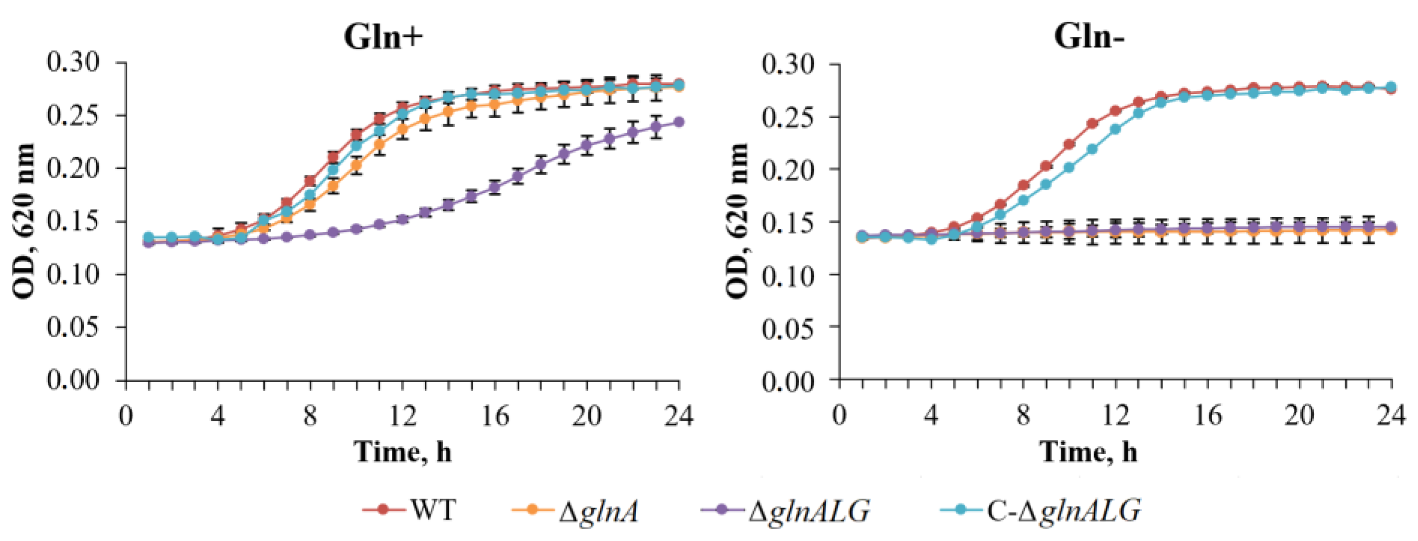
In enterobacteria, glutamine synthetase is the sole enzyme that can synthesize glutamine [31]. The study examined the development patterns of both the wild-type and mutant strains, ΔglnA, ΔglnALG, and C-ΔglnALG, on BHI agar and in BHI broth with and without glutamine. The ΔglnA and ΔglnALG mutants grew comparable to the wild-type strain when glutamine was added to the BHI agar. However, in BHI broth with glutamine, the ΔglnALG mutant displays a slight but obvious growth defect. No growth was observed without glutamine (Figure 2 and Figure 3).
Glutamine synthetase is the exclusive enzyme responsible for glutamine production in enterobacteria [31]. This study investigated the growth patterns of both wild-type and mutant strains (ΔglnA, ΔglnALG, and C-ΔglnALG) when cultured on BHI agar and in BHI broth, with and without the addition of exogenous glutamine.
These results indicate that the limited availability of glutamine caused by the absence of a functional glutamine synthetase explains the auxotrophy for this amino acid in ΔglnA and ΔglnALG Y. pestis strains.

3.3. Loss of the Entire glnALG Operon, Not Just the Single glnA Gene, Reduces Mutant Virulence

Glutamine is one of the two central products in the nitrogen assimilation process, functioning as an amide donor in most biosynthetic reactions [32]. Thus, a defect in glutamine biosynthesis and the potentially associated disturbance in nitrogen metabolism may significantly alter pathogen–host relationships.
The virulence of the constructed Y. pestis 231ΔglnA, 231ΔglnALG mutants, complemented mutant C-231ΔglnALG, and the parent fully virulent Y. pestis strain 231 was determined after subcutaneous inoculation in mice and guinea pigs (Table 3).
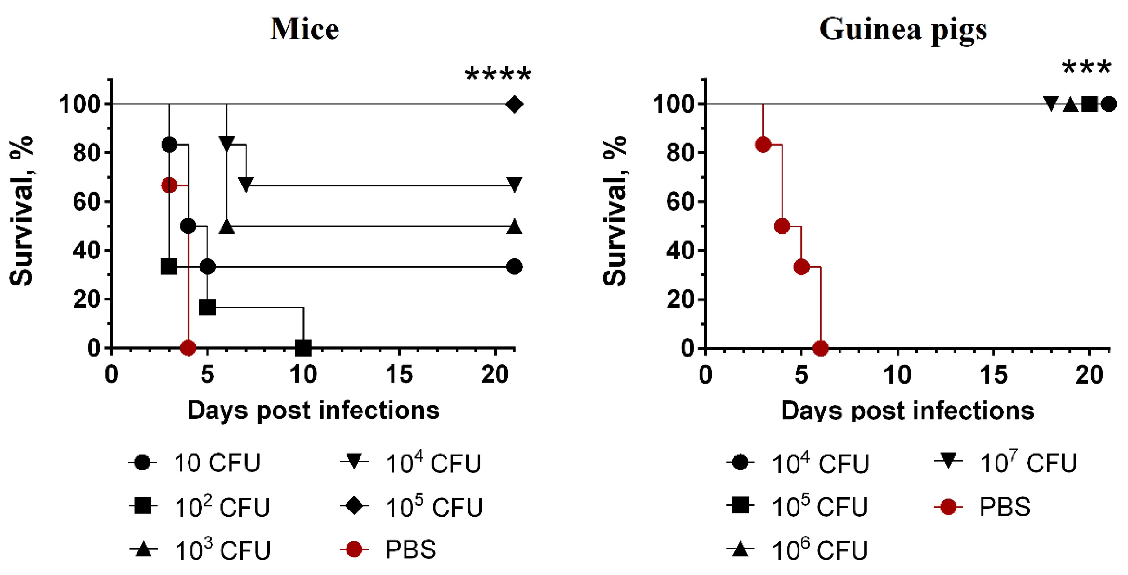
A comparative assessment of virulence in outbred mice and guinea pigs did not reveal any significant differences in the LD50 values of the wild-type strain and the GlnA mutant (Table 3). The strain with the deleted glnALG genes was avirulent for mice at a dose of 105, and for guinea pigs, it was avirulent at a dose of 107 CFU (the greatest administered doses) when injected subcutaneously (Figure 4). All animals showed no signs of disease during the observation period (21 days). After infection with the wild-type strain 231 in doses of 10–103 CFU, mice died by days 4–6, and guinea pigs infected with 102–103 CFU died by days 10–12 of observation (Figure 4). The introduction of the recombinant plasmid pEYlpp-glnALG restored the virulence of the mutant strain 231ΔglnALG in the subcutaneous infection of outbred mice and guinea pigs (Table 3, Figure 4).

3.4. Humoral Immune Responses

The serum antibody responses to the two main immunodominant Y. pestis protective antigens (F1 and LcrV) [28] were determined via ELISA. The level of IgG antibodies to F1 and LcrV antigens in the blood of mice and guinea pigs was assessed on the 28th day after subcutaneous immunization with the studied Y. pestis 231∆glnALG strain (Figure 5). Both anti-F1 (p < 0.0001) and anti-LcrV (p < 0.05) IgG titers in mice increased in a dose-dependent manner when the levels of IgG induced by doses of 10, 102, 103, 104, and 105 CFU were compared. The differences in the levels of antibody responses to Y. pestis LcrV in guinea pig sera were insignificant (p > 0.05). A dose-dependent increase in the levels of antibody titers in guinea pigs was observed to the F1 antigen (p < 0.005).

3.5. Strain with Deletion of the Entire glnALG Operon Provides Protective Immunity Against Fatal Plague

Subcutaneous administration of 105 CFUs of the 231ΔglnALG mutant strain did not cause death of mice for as long as 28 days post-inoculation, and all of the mice that survived such a challenge were fully protected against a subcutaneous inoculation of 200 LD100 of the wild-type strain (Figure 6). This indicated that the 231ΔglnALG mutant strain behaved like a live vaccine strain. When we inoculated lower doses (102–104) of the avirulent strain 231ΔglnALG, the survival of vaccinated animals after subcutaneous administration of the wild-type strain 231 decreased to 80–40%, demonstrating a dose-dependent protective effect. All PBS-treated mice in the control group died by day 4 post-infection. As in the case of mice, all guinea pigs treated with the 231ΔglnALG strain at doses of 104–107 survived subsequent infection with the virulent Y. pestis strain 231, which was not the case for guinea pigs treated with PBS (Figure 6).

4. Discussion

The main processes of life of any organism are growth and reproduction, which require energy sources and mineral substances. Pathogenic microbes use the host’s nutrients for these purposes, and the host resists infection by limiting the pathogen’s access to nutrients [33,34]. In turn, pathogens have developed strategies that combine efficient systems for absorbing nutrients synthesized in the host’s body with the ability to synthesize energy sources and plastic substances that are absent from their host. This area of host–pathogen interaction has been called nutritional virulence and has been the focus of increasing attention in the development of candidate vaccine strains [33].
Bacterial auxotrophs lack the metabolic capability to produce essential organic compounds. Consequently, they rely on their host organisms for these nutrients. Glutamine synthetase plays a crucial role in nitrogen assimilation by catalyzing the conversion of glutamate and ammonia into glutamine. This process is energy-dependent, requiring ATP as the fuel [35]. Glutamine plays a crucial role in the metabolic processing of nitrogen within bacteria, acting as a marker for intracellular nitrogen availability. When external nitrogen sources become scarce, the concentration of glutamine within bacterial cells diminishes, whereas the level of glutamate remains relatively [2]. ΔglnA mutant of S. Typhimurium mimics intracellular nitrogen limitation even when nitrogen is available in excess from the outside. Reduction in the glutamine pool under nitrogen limitation conditions is responsible for the slow growth of S. enterica [2]. The data above are consistent with our observations that Y. pestis strains lacking the glnA or glnALG genes do not grow on solid or liquid nutrient media, not even in those of a rich composition (BHI agar and broth) without the addition of glutamine. The addition of glutamine to the culture medium caused the ability of ΔglnA or ΔglnALG mutants to grow in vitro. Based on the growth data on nutrient media, it can be assumed that the ABC glutamine transporter encoded by the glnHPQ gene group functions in glnA or glnALG knockout mutants at a level necessary to ensure their nutritional needs at all stages of the plague pathogen life cycle in the host organism.
To definitively prove the role of a specific factor in causing the plague, we would need to show that removing this factor from a wild-type virulent strain of the bacteria reduces its ability to cause the disease. Furthermore, reintroducing the removed factor should then restore the bacterial virulence to its original level. We constructed a complementation plasmid pEYlpp-glnALG, which confirmed that the ablation of glutamine synthesis, along with the removal of the two-component regulatory system GlnL-GlnG, ensures significant attenuation of the mutant [12]. To demonstrate that a specific factor has a minimal impact on pathogenicity, it is sufficient to prove that removing this factor does not diminish the pathogen’s virulence [36]. In line with these considerations, we did not complement the knocked-out gene glnA because such a mutation did not decrease virulence. It was precisely this lack of effect on virulence that characterized the ΔglnA mutant.
The amide group present within the glutamine molecule serves as a nitrogen donor during the initial steps of synthesizing the fundamental units that compose essential cellular components such as proteins and nucleic acids. Therefore, it is expected that the observable characteristics of organisms with deletions in the glnA and glnALG genes will show notable distinctions when compared to organisms with the unaltered gene set. For example, according to Aurass et al. [14], the ΔglnA mutant of S. enterica serovar Typhimurium had a reduced ability to penetrate macrophages. It was shown that single ΔglnA mutants of S. enterica serovar Typhimurium did not have reduced virulence in intraperitoneally infected BALB/c mice [12]. While the deletion of the glnA gene alone did not significantly impact virulence, the simultaneous removal of both glnA and genes involved in glutamine transport (glnH and glnQ) or nitrogen regulation (glnLG) led to a notable decrease in the spread of these mutant strains within mice. Furthermore, these double knockouts also exhibited diminished survival rates within the J774 macrophages. This suggests that the degree of glutamine uptake by the host plays an important role in the development of the infectious process caused by S. enterica [12]. We also found that the single ΔglnA mutant of Y. pestis was not attenuated by the subcutaneous infection of mice and guinea pigs. However, the mutant with both the deletion of the glutamine synthetase gene (glnA) and the genes of the two-component nitrogen regulatory system (glnLG) dramatically reduced virulence during the subcutaneous infection of mice and guinea pigs. The obtained results confirm that the availability of glutamine for Y. pestis in the host organism depends on the two-component regulatory system glnLG, which, similarly for S. enterica, is apparently necessary for the transcription of glutamine transport genes [12]. ΔglnA bacteria also remain virulent because their genes that ensure the transport of glutamine into the cell and the genes that regulate this transport are not damaged. The glutamine transport system in ΔglnAGL mutants copes with ensuring their growth on nutrient media with a high glutamine content, but the damaged transport regulation system cannot cope with a full supply of glutamine in the host.
Previous research indicated that the absence of the glnGL operon in the Y. pestis CO92 Orientalis strain did not diminish the bacterium’s ability to cause the disease [18]. This discrepancy in findings could be attributed to several factors, including differences in the bacterial strain (Orientalis versus Antiqua), the mouse model used (OF-1 versus a specific breeding line), and the method of infection (intradermal versus subcutaneous). These variables impact bacterial spread, immune responses, and ultimately, the severity of the disease. Further research involving controlled comparisons of these factors is necessary to elucidate the underlying reasons for the observed discrepancies.
Targeting genes essential for the production or transportation of crucial substances within microorganisms presents a viable strategy for developing vaccine strains effective against a wide range of infectious diseases. Knockout of genes responsible for the transport or synthesis of substances necessary for the vital activity of a microorganism is a promising approach in the creation of vaccine strains against many infectious diseases. We preliminarily assessed the immunogenicity of the constructed Y. pestis 231ΔglnALG strain in mice and guinea pigs. The ΔglnALG mutant did not cause death in mice when administered subcutaneously at 10–105 CFU or in guinea pigs when administered subcutaneously at 104–107 CFU, and at doses of 105 CFU for mice and 104–107 CFU for guinea pigs, it provided 100% protection of animals when subsequently administered subcutaneously with 200 LD100 of the virulent Y. pestis 231 strain. The immunogenicity of the 231ΔglnALG strain was more pronounced in the guinea pig model. Thus, the attenuated Y. pestis strain 231ΔglnALG can be considered a promising vaccine strain candidate. To make a final conclusion about the safety of the strain 231ΔglnALG, it is necessary to infect laboratory animals with large doses, 107 CFU for mice and 2 × 10⁹ CFU and 1,5 × 10¹⁰ CFU for guinea pigs [37].
This type of attenuation of Y. pestis, associated with interference in the metabolism of the microorganism, may provide additional advantages in the construction of a vaccine strain, since the expression of the plague microbe genes associated with pathogenicity is not damaged and the complete antigen composition is available for recognition by the immune system and the formation of an immune response in the host.

5. Conclusions

In summary, the operon glnALG is functionally important in Y. pestis, and thus, its viability as a drug target should be explored.
In addition, a direction for further research may be to study the influence of mutations in genes whose products are responsible for the synthesis and high-affinity transport of nutrients on the pathogenicity of both intracellular and extracellular microorganisms. This requires constructing paired mutations in genes responsible for nutrient biosynthesis and transport or, alternatively, deleting genes of transport systems in auxotrophic pathogens that rely on the uptake of nutrients from the host organism.
Elucidation of host–pathogen interactions at the level of their metabolism should ultimately lead to a deeper understanding of the molecular mechanisms of the pathogenesis of bacterial infections and will allow the selection of optimal molecular targets for vaccine prophylaxis, as well as new pathogen-specific antimicrobial therapy strategies.

Author Contributions

Conceptualization: A.P.A.; data curation: A.P.A., F.S. and S.V.D.; formal analysis: A.S.T., A.S.V., E.M.M., T.V.G., N.A.L., R.Z.S., S.A.I., T.I.K. and E.A.K.; funding acquisition: A.P.A. and S.V.D.; investigation: A.S.V., M.E.P., A.S.T., E.A.K., E.M.M., T.V.G., N.A.L., R.Z.S., S.A.I. and T.I.K.; methodology: S.V.D., T.I.K., M.E.P. and R.Z.S.; project administration: A.P.A. and S.V.D.; resources: A.P.A., S.V.D. and T.I.K.; software: A.S.V., A.S.T., E.M.M. and M.E.P.; supervision: S.V.D., A.S.V. and A.S.T.; validation: S.V.D., A.S.V., A.S.T., E.A.K., M.E.P., E.M.M., T.V.G., N.A.L., R.Z.S., S.A.I. and F.S.; visualization: A.S.V., A.S.T., T.V.G. and S.A.I.; writing—original draft preparation: A.P.A., S.V.D., A.S.V., A.S.T., E.A.K., N.A.L. and F.S.; writing—review and editing: A.P.A., S.V.D. and F.S. All authors have read and agreed to the published version of the manuscript.

Funding

This work was supported by the Russian Science Foundation (grant number 23-15-00132).

Institutional Review Board Statement

The animal study protocol was approved by the Bioethics Committee of the State Research Center for Applied Microbiology and Biotechnology (Permit No: VP-2024/4, 30 August 2024).

Informed Consent Statement

Not applicable.

Data Availability Statement

All data will be provided upon reasonable request.

Conflicts of Interest

All authors declare that they have no conflicts of interest.

References

  1. Reitzer, L.J.; Neidhardt, F.; Ingraham, J.L.; Low, K.B.; Magasanik, B.; Schaechter, M.; Umbarger, H.E. Ammonia Assimilation and the Biosynthesis of Glutamine, Glutamate, Aspartate, Asparagine, L-alanine, and D-alanine. In Escherichia coli and Salmonella Typhimurium: Cellular and Molecular Biology; ASM Press: Washington, DC, USA, 1987; pp. 302–320. [Google Scholar]
  2. Ikeda, T.P.; Shauger, A.E.; Kustu, S. Salmonella Typhimurium Apparently Perceives External Nitrogen Limitation as Internal Glutamine Limitation. J. Mol. Biol. 1996, 259, 589–607. [Google Scholar] [CrossRef] [PubMed]
  3. Zimmer, D.P.; Soupene, E.; Lee, H.L.; Wendisch, V.F.; Khodursky, A.B.; Peter, B.J.; Bender, R.A.; Kustu, S. Nitrogen Regulatory Protein C-Controlled Genes of Escherichia coli: Scavenging as a Defense against Nitrogen Limitation. Proc. Natl. Acad. Sci. USA 2000, 97, 14674–14679. [Google Scholar] [CrossRef] [PubMed]
  4. Nohno, T.; Saito, T.; Hong, J. Cloning and Complete Nucleotide Sequence of the Escherichia coli Glutamine Permease Operon (glnHPQ). Molec. Gen. Genet. 1986, 205, 260–269. [Google Scholar] [CrossRef] [PubMed]
  5. Maria-Neto, S.; Cândido, E.D.S.; Rodrigues, D.R.; De Sousa, D.A.; Da Silva, E.M.; De Moraes, L.M.P.; Otero-Gonzalez, A.D.J.; Magalhães, B.S.; Dias, S.C.; Franco, O.L. Deciphering the Magainin Resistance Process of Escherichia coli Strains in Light of the Cytosolic Proteome. Antimicrob. Agents Chemother. 2012, 56, 1714–1724. [Google Scholar] [CrossRef]
  6. El Khoury, J.Y.; Boucher, N.; Bergeron, M.G.; Leprohon, P.; Ouellette, M. Penicillin Induces Alterations in Glutamine Metabolism in Streptococcus pneumoniae. Sci. Rep. 2017, 7, 14587. [Google Scholar] [CrossRef]
  7. Gustafson, J.; Strässle, A.; Hächler, H.; Kayser, F.H.; Berger-Bächi, B. The femC Locus of Staphylococcus aureus Required for Methicillin Resistance Includes the Glutamine Synthetase Operon. J. Bacteriol. 1994, 176, 1460–1467. [Google Scholar] [CrossRef]
  8. Millanao, A.R.; Mora, A.Y.; Saavedra, C.P.; Villagra, N.A.; Mora, G.C.; Hidalgo, A.A. Inactivation of Glutamine Synthetase-Coding Gene glnA Increases Susceptibility to Quinolones Through Increasing Outer Membrane Protein F in Salmonella enterica Serovar Typhi. Front. Microbiol. 2020, 11, 428. [Google Scholar] [CrossRef]
  9. Harth, G.; Horwitz, M.A. Inhibition of Mycobacterium tuberculosis Glutamine Synthetase as a Novel Antibiotic Strategy against Tuberculosis: Demonstration of Efficacy In Vivo. Infect. Immun. 2003, 71, 456–464. [Google Scholar] [CrossRef]
  10. Crowther, G.J.; Shanmugam, D.; Carmona, S.J.; Doyle, M.A.; Hertz-Fowler, C.; Berriman, M.; Nwaka, S.; Ralph, S.A.; Roos, D.S.; Van Voorhis, W.C.; et al. Identification of Attractive Drug Targets in Neglected-Disease Pathogens Using an In Silico Approach. PLoS Negl. Trop. Dis. 2010, 4, e804. [Google Scholar] [CrossRef]
  11. Fisher, S.H. Regulation of Nitrogen Metabolism in Bacillus subtilis: Vive La Différence! Mol. Microbiol. 1999, 32, 223–232. [Google Scholar] [CrossRef]
  12. Klose, K.E.; Mekalanos, J.J. Simultaneous Prevention of Glutamine Synthesis and High-Affinity Transport Attenuates Salmonella Typhimurium Virulence. Infect. Immun. 1997, 65, 587–596. [Google Scholar] [CrossRef] [PubMed]
  13. Chandra, H.; Basir, S.F.; Gupta, M.; Banerjee, N. Glutamine Synthetase Encoded by glnA-1 Is Necessary for Cell Wall Resistance and Pathogenicity of Mycobacterium bovis. Microbiology 2010, 156, 3669–3677. [Google Scholar] [CrossRef] [PubMed]
  14. Aurass, P.; Düvel, J.; Karste, S.; Nübel, U.; Rabsch, W.; Flieger, A. glnA Truncation in Salmonella Enterica Results in a Small Colony Variant Phenotype, Attenuated Host Cell Entry, and Reduced Expression of Flagellin and SPI-1-Associated Effector Genes. Appl. Environ. Microbiol. 2018, 84, e01838-17. [Google Scholar] [CrossRef] [PubMed]
  15. Anisimov, A.P.; Vagaiskaya, A.S.; Trunyakova, A.S.; Dentovskaya, S.V. Live Plague Vaccine Development: Past, Present, and Future. Vaccines 2025, 13, 66. [Google Scholar] [CrossRef]
  16. Hadjifrangiskou, M.; Brannon, J. The Arsenal of Pathogens and Antivirulence Therapeutic Strategies for Disarming Them. DDDT 2016, 2016, 1795–1806. [Google Scholar] [CrossRef]
  17. Asha, I.J.; Gupta, S.D.; Hossain, M.M.; Islam, M.N.; Akter, N.N.; Islam, M.M.; Das, S.C.; Barman, D.N. In Silico Characterization of a Hypothetical Protein (PBJ89160.1) from Neisseria meningitidis Exhibits a New Insight on Nutritional Virulence and Molecular Docking to Uncover a Therapeutic Target. Evol. Bioinform. Online 2024, 20, 11769343241298307. [Google Scholar] [CrossRef]
  18. Reboul, A.; Lemaître, N.; Titecat, M.; Merchez, M.; Deloison, G.; Ricard, I.; Pradel, E.; Marceau, M.; Sebbane, F. Yersinia pestis Requires the 2-Component Regulatory System OmpR-EnvZ to Resist Innate Immunity during the Early and Late Stages of Plague. J. Infect. Dis. 2014, 210, 1367–1375. [Google Scholar] [CrossRef]
  19. Solomentsev, V.I.; Kadnikova, L.A.; Kislichkina, A.A.; Mayskaya, N.V.; Kombarova, T.I.; Platonov, M.E.; Bogun, A.G.; Anisimov, A.P. Comparative Sequencing of Transcriptomes of Yersinia pestis subsp. microti bv. Ulegeica Different by Virulence for Guinea Pigs. Russ. J. Bacteriol. 2017, 2, 30–35. (In Russian) [Google Scholar]
  20. Anisimov, A.P.; Kislichkina, A.A.; Kopylov PKh Krasilnikova, E.A.; Bogun, A.G.; Dentovskaya, S.V. Search for Factors of Selective Virulence of Rhamnose-Positive Strains of Yersinia pestis. In Proceedings of the 22nd International Scientific Conference “Current Issues on Zoonotic Diseases”, Ulaanbaatar, Mongolia, 5 July 2017; Volume 22, pp. 88–95. Available online: https://www.researchgate.net/publication/320138653 (accessed on 19 February 2025). (In Russian).
  21. Kislichkina, A.A.; Krasil’nikova, E.A.; Platonov, M.E.; Skryabin, Y.P.; Sizova, A.A.; Solomentsev, V.I.; Gapel’chenkova, T.V.; Dentovskaya, S.V.; Bogun, A.G.; Anisimov, A.P. Whole-Genome Assembly of Yersinia pestis 231, the Russian Reference Strain for Testing Plague Vaccine Protection. Microbiol. Resour. Announc. 2021, 10, e01373-20. [Google Scholar] [CrossRef]
  22. Datsenko, K.A.; Wanner, B.L. One-Step Inactivation of Chromosomal Genes in Escherichia coli K-12 Using PCR Products. Proc. Natl. Acad. Sci. USA 2000, 97, 6640–6645. [Google Scholar] [CrossRef]
  23. Cherepanov, P.P.; Wackernagel, W. Gene Disruption in Escherichia coli: TcR and KmR Cassettes with the Option of Flp-Catalyzed Excision of the Antibiotic-Resistance Determinant. Gene 1995, 158, 9–14. [Google Scholar] [CrossRef] [PubMed]
  24. Donnenberg, M.S.; Kaper, J.B. Construction of an eae Deletion Mutant of Enteropathogenic Escherichia coli by Using a Positive-Selection Suicide Vector. Infect. Immun. 1991, 59, 4310–4317. [Google Scholar] [CrossRef]
  25. Dentovskaya, S.V.; Vagaiskaya, A.S.; Platonov, M.E.; Trunyakova, A.S.; Kotov, S.A.; Krasil’nikova, E.A.; Titareva, G.M.; Mazurina, E.M.; Gapel’chenkova, T.V.; Shaikhutdinova, R.Z.; et al. Peptidoglycan-Free Bacterial Ghosts Confer Enhanced Protection against Yersinia pestis Infection. Vaccines 2021, 10, 51. [Google Scholar] [CrossRef] [PubMed]
  26. Trunyakova, A.S.; Platonov, M.E.; Ivanov, S.A.; Kopylov, P.K.; Dentovskaya, S.V.; Anisimov, A.P. A Recombinant Low-Endotoxic Yersinia pseudotuberculosis Strain—over-Producer of Yersinia pestis F1 Antigen. Mol. Gen. Microbio. Viro. 2024, 42, 10. [Google Scholar] [CrossRef]
  27. Kopylov, P.K.; Svetoch, T.E.; Ivanov, S.A.; Kombarova, T.I.; Perovskaya, O.N.; Titareva, G.M.; Anisimov, A.P. Characteristics of the Chromatographic Cleaning and Protectiveness of the LcrV Isoform of Yersinia pestis. Appl. Biochem. Microbiol. 2019, 55, 524–533. [Google Scholar] [CrossRef]
  28. Titball, R.W.; Williamson, E.D. Yersinia pestis (Plague) Vaccines. Expert Opin. Biol. Ther. 2004, 4, 965–973. [Google Scholar] [CrossRef] [PubMed]
  29. Finney, D. Statistical Method in Biological Assay, 3rd ed.; Charles Griffin: London, UK, 1978; p. 508. [Google Scholar]
  30. Niranda-Rjos, J.; Saàanchez-Pescador, R.; Urdea, M.; Covarrubias, A.A. The Complete Nucleotide Sequence of the ginALG Operon of Escherichia coli K12. Nucl. Acids Res. 1987, 15, 2757–2770. [Google Scholar] [CrossRef]
  31. Li, B.; Zhou, L.; Guo, J.; Wang, X.; Ni, B.; Ke, Y.; Zhu, Z.; Guo, Z.; Yang, R. High-Throughput Identification of New Protective Antigens from a Yersinia pestis Live Vaccine by Enzyme-Linked Immunospot Assay. Infect. Immun. 2009, 77, 4356–4361. [Google Scholar] [CrossRef]
  32. Van Heeswijk, W.C.; Westerhoff, H.V.; Boogerd, F.C. Nitrogen Assimilation in Escherichia coli: Putting Molecular Data into a Systems Perspective. Microbiol. Mol. Biol. Rev. 2013, 77, 628–695. [Google Scholar] [CrossRef]
  33. Abu Kwaik, Y.; Bumann, D. Microbial Quest for Food in Vivo: ‘Nutritional Virulence’ as an Emerging Paradigm. Cell Microbiol. 2013, 15, 882–890. [Google Scholar] [CrossRef]
  34. Eisenreich, W.; Heesemann, J.; Rudel, T.; Goebel, W. Metabolic Host Responses to Infection by Intracellular Bacterial Pathogens. Front. Cell. Infect. Microbiol. 2013, 3. [Google Scholar] [CrossRef]
  35. Merrick, M.J.; Edwards, R.A. Nitrogen Control in Bacteria. Microbiol. Rev. 1995, 59, 604–622. [Google Scholar] [CrossRef]
  36. Burrows, T.W. Virulence of Pasteurella pestis and immunity to plague. In Ergebnisse der Mikrobiologie Immunitätsforschung und Experimentellen Therapie; Henle, W., Kikuth, W., Meyer, K.F., Nauck, E.G., Tomcsik, J., Eds.; Springer: Berlin, Heidelberg, 1963; pp. 59–113. [Google Scholar] [CrossRef]
  37. Anisimova, T.I.; Sayapina, L.V.; Sergeeva, G.M.; Isupov, I.V.; Beloborodov, R.A.; Samoilova, L.V.; Anisimov, A.P.; Ledvanov, M.Y.; Shvedun, G.P.; Zadumina, S.Y.; et al. Main Requirements for Vaccine Strains of the Plague Pathogen: Methodological Guidelines MU 3.3.1.1113-02; Federal Centre of State Epidemic Surveillance of Ministry of Health of Russian Federation: Moscow, Russia, 2002. [Google Scholar] [CrossRef]
Figure 1. Schematic representation of the Y. pestis glnALG region. Comparison of the Y. pestis CO92 (GenBank accession number NZ_CP009973) genetic locus glnALG with those of E. coli strain K-12 substr. MC4100 (GenBank accession number HG738867) and S. enterica subsp. enterica serovar Typhimurium strain ATCC 14028 (GenBank accession number CP102669). The amino acid identities are shown between genes of a conserved gene order (A). Schematic representation of the Y. pestis glnA (P1) and glnLG (P2) promoters (B).
Vaccines 13 00353 g001
Figure 2. Glutamine requirement of the wild-type, ΔglnA, ΔglnALG, and C-ΔglnALG Y. pestis strains in broth culture. Cultures were inoculated into BHI broth or BHI broth supplemented with 20 mM of L-glutamine (Gln+) or not (Gln–). Growth was monitored by assaying the optical density (OD). Data are the means ± standard errors.
Vaccines 13 00353 g002
Figure 3. Growth of the wild-type, ΔglnA, ΔglnALG, and C-ΔglnALG Y. pestis strains on BHI agar supplemented with 20 mM of L-glutamine (Gln+) or not (Gln).
Vaccines 13 00353 g003
Figure 4. Survival of outbred mice (n = 6) and guinea pigs (n = 6) after subcutaneous infection with Y. pestis strains 231∆glnA, ∆glnALG, or C-∆glnALG. Log-rank (Mantel–Cox) test was used. #—p > 0.05; ****—p < 0.0001.
Vaccines 13 00353 g004
Figure 5. Anti-Y. pestis F1 and LcrV antibody titers in mouse and guinea pig sera collected 28 days after s.c. infection with 10, 102, 103, 104, or 105 CFU for mice or 104, 105, 106, or 107 CFU for guinea pigs of the Y. pestis 231 ΔglnALG mutant. Titers from three individual animals are shown; horizontal lines indicate the mean.
Vaccines 13 00353 g005
Figure 6. Survival of outbred mice and guinea pigs immunized subcutaneously with the Y. pestis 231ΔglnALG mutant strain after subcutaneous infection with 200 LD100 of the wild-type strain Y. pestis 231. Log-rank (Mantel–Cox) test was used. ***—p < 0.001; ****—p < 0.0001.
Vaccines 13 00353 g006
Table 1. Bacterial strains and plasmids used in this study.
Strain, PlasmidRelevant AttributesSource
Y. pestis
2310.ANT3 phylogroup, wild-type strain, universally virulent (LD50 for mice ≤ 10 CFU, for guinea pigs ≤ 10 CFU); Pgm+, pMT1+, pPCP1+, pCD+SCPM-O [21]
EV1.ORI3 phylogroup, vaccine strain, Pgm, pMT1+, pPCP1+, pCD+SCPM-O
EVΔglnA::catΔglnA derivative of EV, CmrThis study
EVΔglnALG::catΔglnALG derivative of EV, CmrThis study
231ΔglnAΔglnA derivative of 231This study
231ΔglnALGΔglnALG derivative of 231This study
C-231ΔglnALGΔglnALG containing plasmid pEYlpp-glnALGThis study
E. coli
S17-1 λpirthi pro hsdR hsdM+ recA RP4 2-Tc::Mu-Km::Tn7(TpRSmRPmS)SCPM-O
Plasmids
pKD46bla PBADgam bet exopSC101 oriTS[22]
pKD3bla FRT cat FRT PS1 PS2 oriR6K[22]
pCP20bla cat cI857 λPRflp pSC101 oriTS[23]
pCVD442ori R6K mob RP4 bla sacB[24]
pEYR’ori pA15 cat pR’[25]
pEYlppori pA15 cat plppThis study
pCVD442-glnA::catori R6K mob RP4 bla sacB cat glnAThis study
pCVD442-glnALG::catori R6K mob RP4 bla sacB cat glnALGThis study
pEYlpp-glnALGori pA15 cat Plpp glnALGThis study
Table 2. Primers used in this study.
glnA Primers for Mutant Construction and Screening
glnA1FATGCCTGAACACCATAAATGCAGTAACACACGGTAATCGTTCCACGACGACGACTATGGGAATTAGCCATGGTCC
glnA1RGTGTTGGCTGCTTTCGCTCGCCACCTTCCTACACCTTGAAATCTATTAGGTAAACGTGTAGGCTGGAGCTGCTTC
glnA2FCGGTCGCATCCAGGTTAACG
glnA2RGCGTTACGGGTGATATTCAG
glnALG Primers for Mutant Construction and Screening
glnA1FATGCCTGAACACCATAAATGCAGTAACACACGGTAATCGTTCCACGACGACGACTATGGGAATTAGCCATGGTCC
glnLG3RCTACTCCATCCCCAACTCTTTCAACTTCCGCGTTAATGTATTACGGCCCCAGCCCGTGTAGGCTGGAGCTGCTTC
glnA2RGCGTTACGGGTGATATTCAG
glnLG2RCTTGATTCTATTGCAACGGAAC
Primers for pEYlpp Construction
Plpp-SphICGATGAGCATGCGATAACCAGAAGCAATAAAAAATC
PlppR-NdeICGATGTCATATGTAATACCCTCTAGTTTGAGTTAATC
Screening for pCD1
yscFPlusACACCATATGAGTAACTTCTCTGGATTTACG
yscFMinusATTCTCGAGTGGGAACTTCTGTAGGATG
Screening for pMT
caf1PlusAGTTCCGTTATCGCCATTGC
caf1MinusGGTTAGATACGGTTACGGTTAC
Screening for pPst
PstFCAATCATATGTCAGATACAATGGTAGTG
PstRCTCCTCGAGTTTTAACAATCCACTATC
Table 3. Virulence of Y. pestis strains in subcutaneously infected outbred mice and guinea pigs.
Y. pestis StrainsLD50, CFU
MiceGUINEA PIGS
231115
231ΔglnA53
231ΔglnALG>105>107
C-231ΔglnALG732
Disclaimer/Publisher’s Note: The statements, opinions and data contained in all publications are solely those of the individual author(s) and contributor(s) and not of MDPI and/or the editor(s). MDPI and/or the editor(s) disclaim responsibility for any injury to people or property resulting from any ideas, methods, instructions or products referred to in the content.