Article

Efficacy of Commercial Infectious Bronchitis Vaccines against Canadian Delmarva (DMV/1639) Infectious Bronchitis Virus Infection in Layers

by
Mohamed S. H. Hassan
1,2,
Sabrina M. Buharideen
1,
Ahmed Ali
1,3,
Shahnas M. Najimudeen
1,
Dayna Goldsmith
1,
Carla S. Coffin
4,
Susan C. Cork
1,
Frank van der Meer
1 and
Mohamed Faizal Abdul-Careem
1,*
1
Faculty of Veterinary Medicine, University of Calgary, 3330 Hospital Drive NW, Calgary, AB T2N 4N1, Canada
2
Department of Poultry Diseases, Faculty of Veterinary Medicine, Assiut University, Assiut 71515, Egypt
3
Department of Pathology, Faculty of Veterinary Medicine, Beni-Suef University, Beni Suef 62521, Egypt
4
Department of Medicine, Cumming School of Medicine, University of Calgary, 3330 Hospital Drive NW, Calgary, AB T2N 4N1, Canada
*
Author to whom correspondence should be addressed.
Vaccines 2022, 10(8), 1194; https://doi.org/10.3390/vaccines10081194
Submission received: 26 June 2022 / Revised: 22 July 2022 / Accepted: 25 July 2022 / Published: 27 July 2022
(This article belongs to the Special Issue Immunology and Vaccines against Avian Infectious Diseases)

Abstract

:
Vaccination is the most important way to control infectious bronchitis (IB) in chickens. Since the end of 2015, the Delmarva (DMV)/1639 strain of infectious bronchitis virus (IBV) has caused significant damage to the layer flocks in Eastern Canada. The efficacy of a combination of existing IB vaccines licensed in Canada was assessed against experimental challenge with this IBV strain. The layer pullets were vaccinated during the rearing phase with live attenuated IB vaccines of Massachusetts (Mass) + Connecticut (Conn) types followed by an inactivated IB vaccine of Mass + Arkansas (Ark) types and then challenged with the Canadian IBV DMV/1639 strain at 30 weeks of age. Protection was evaluated based on the egg laying performance, immune responses, viral shedding, and viral genome loads and lesions in IBV target organs. The vaccinated challenged hens were protected from the drop in egg production observed in the non-vaccinated challenged hens. Early (5 dpi) anamnestic serum antibody response was measured in the vaccinated challenged hens as well as a significant level of antibodies was detected in the oviduct washes (14 dpi). In contrast, hens in the non-vaccinated challenged group showed delayed (12 dpi) and significantly lower serum antibody response. Viral RNA loads were reduced in the respiratory, alimentary, and reproductive tissues of the vaccinated challenged hens compared to the non-vaccinated challenged hens. Compared to the control groups, the vaccinated challenged hens had less marked microscopic lesions in the trachea, kidney, magnum, and uterus. Our experimental model demonstrated inconclusive results for cell-mediated immune responses and viral shedding. Overall, the vaccination program used in this study minimized viral replication and histopathological changes in most IBV target organs and protected challenged hens against drop in egg production.

1. Introduction

Avian infectious bronchitis (IB) is a leading cause of economic losses in the poultry industry [1]. The causative agent, infectious bronchitis virus (IBV), is an epitheliotropic virus that primarily affects the respiratory tract, although different strains have different tropisms for other tissues such as the alimentary and the urogenital tracts [1]. The spike (S) glycoprotein of IBV is a determinant of tissue tropism and pathogenicity, and the S1 subunit contains serotype- and neutralization-specific epitopes [2]. Mutations and recombination within the S gene are the driving forces for the emergence of new IBV variants globally [3], which makes the control of the IB through vaccination difficult [4].
Clinical indications of IBV-induced respiratory disease in broilers include coughing, sneezing, tracheal rales, watery eyes, and thickened mucosa of the trachea and nasal passages. In addition to the poor weight gain and lost feed efficiency, some virulent IBV strains cause high mortality in young chickens due to nephritis [5]. In susceptible layers, IBV can cause a variety of clinical manifestations including decline in egg production, deformed eggshells, and poor internal egg quality [6,7]. A series of lesions can be detected in the female reproductive system including shortened oviduct, reduction in the number of hierarchical follicles in the ovary, and yolk peritonitis [8,9]. Microscopic lesions of tubular gland dilatation, inflammatory cell infiltration, edema of the interstitial region, and epithelial sloughing in the oviducts are common in the infected layers [10]. The virus can infect layer pullets at an early age leading to devastating effects on the developing reproductive tract [11]. The problem of false layers with permanent damage of the oviduct was highlighted more recently with the emergence of the QX and DMV/1639-like strains [12,13]. Abnormal oviducts of false layers are characterized by dilatation with serous fluid accumulation (cystic formation) and areas of non-patency [14].
Vaccination has been used to prevent IBV infection and its harmful effects for decades. In young birds, live-attenuated IB vaccines are commonly sprayed over chicks in the hatchery before being placed in the barn. Revaccination with another live IB vaccine of the same or a different serotype could be carried out in the field within two weeks of the first vaccination. In layer-type chickens, multiple live vaccinations are usually followed by a killed vaccine administered before the onset of lay to give long-lasting immunity [4]. Development of a new live-attenuated IB vaccine for every pathogenic IBV strain is an unrealistic objective; therefore, there are no homologous live vaccines available for several IBV variants. However, the protectotype approach, using live attenuated vaccines containing different IBV serotypes, has been shown to induce a broader range of cross-protection [15,16].
In contrast with vaccination-challenge experiments in young chickens, similar trials with laying chickens are very limited [17]. Few experimental studies have investigated a vaccination program of a live priming and subsequent boosting by an inactivated vaccine against the drop in egg production. A positive correlation was previously shown between the level of post-vaccination hemagglutination inhibiting antibodies and the level of protection against egg production drop [18]. Layers exposed to the Ark IBV strain were not protected from negative effects on egg production and quality by a combination of live and inactivated vaccines of the Mass type [6]. On the other hand, a significant level of protection against heterologous challenge was demonstrated in layers receiving two antigenically distinct IBV vaccines. Vaccinated layers had significantly higher averages of egg production with lowered incidence of yolk peritonitis and degenerated ovaries compared to the non-vaccinated layers [19]. With the emergence of a growing number of IBV variants that impact the layer performance, the ability to protect laying hens has become more challenging [20].
The distribution of IBV strains in Canadian layer flocks varies by geographic location. IBV infections in Western Canada are mostly caused by strains of the Mass and Conn types [21], whereas the Delmarva (DMV/1639) strain has been the most prevalent IBV strain in Eastern Canada in recent years [22]. The Canadian DMV/1639 strain is a virulent IBV with a broad tissue tropism and produces significant lesions in the reproductive tract of laying hens [23,24]. Currently, there is no homologous vaccines for the DMV/1639 strain in North America. The approach commonly used to vaccinate the Canadian layer-type flocks involves using live attenuated vaccines (Mass and Conn types) during the rearing phase, followed by the injection of an inactivated vaccine containing the Mass antigen at the point of lay. Apart from the vaccines applied during the rearing pre-lay period, the laying hens are not commonly vaccinated. In this study, we conducted a vaccine challenge experiment to evaluate the efficacy of commercial IB vaccines available in Canada against the Canadian IBV DMV/1639 strain infection in laying hens.

2. Materials and Methods

2.1. Vaccines and Challenge Virus

Two commercial live IB vaccines, Mass type (Bronchitis Vaccine, Boehringer Ingelheim Animal Health, Athens, GA, USA) and Mass + Conn types (Bronchitis Vaccine, Zoetis Inc., Kalamazoo, MI, USA), were used. An inactivated vaccine marketed under the name Galllimune® NC-BR containing Mass and Ark types of IBV in addition to Lasota strain of Newcastle disease virus (Boehringer Ingelheim Animal Health, Athens, GA, USA) was also used.
The challenge virus used was an IBV DMV/1639 strain (designated as IBV/Ck/Can/17–036989) in its fourth serial passage in embryonated chicken eggs [23,24]. This strain was isolated in 2017 from a commercial layer flock in Eastern Canada that had a history of decreased egg production [22]. The 50% chicken embryo infectious dose (EID50) of the virus was calculated with the method of Reed and Muench [25]; the titer was 107 EID50/mL.

2.2. Chickens

Forty specific-pathogen-free (SPF) white leghorn pullets at 1-day of age were purchased from the Canadian Food Inspection Agency (CFIA), Ottawa, Ontario. The day-old pullets were randomly divided into two groups (20 chicks in each) and housed in two separate negative pressure rooms at the Veterinary Science Research Station (VSRS), University of Calgary. The adjustments to the feed and lighting systems were carried out according to the management guidelines recommended for growing pullets [26]. The light stimulation started at 17 weeks of age with a gradual light increases until a light regimen of 16-h light:8-h dark was achieved at 24 weeks of age.

2.3. Experimental Procedures

2.3.1. Vaccination

The live IB vaccines were maintained on ice until reconstitution with chilled sterile distilled water (one recommended dose/30 µL) and administered via the ocular route. Each chicken in the vaccinated group (V) received the live Mass type vaccine at 1-day of age. The live Mass + Conn types of vaccine was used for revaccination at 2, 5 and 9 weeks of age. At 18 weeks of age, each chicken in the vaccinated group was injected intramuscularly with 0.5 mL of the inactivated vaccine as per the manufacturer’s instructions. The second group of chickens was kept as non-vaccinated (NV). All the chickens were bled at 23 weeks of age before the challenge experiment.

2.3.2. Viral Challenge

At 30 weeks of age, the vaccinated and non-vaccinated hens were further divided into 4 groups (10 chickens in each). There were 2 control groups: vaccinated non-challenged (VNC), non-vaccinated non-unchallenged (NVNC), and 2 challenged groups: vaccinated challenged (VC) and non-vaccinated challenged (NVC). The challenged groups received 100 µL of the challenge virus containing 106 EID50 via the oculo-nasal route [6]. The control groups were mock inoculated with 100 μL of phosphate buffered saline (PBS). All the experimental groups were maintained separately in four different negative pressure rooms at VSRS.

2.3.3. Clinical Observations and Sample Collection

The hens in all groups were observed twice daily for the manifestation of clinical signs and egg production for two weeks. The egg production was presented as the percentage of production/group/3-day period starting from 3 days before the challenge until the end of the experiment at 14 dpi. At 5 and 12 dpi, oropharyngeal (OP) and cloacal (CL) swabs, and 3 mL of blood from the wing vein were collected from all hens.
At 14 dpi, all the hens were euthanized by cervical dislocation under isoflurane anesthesia. When a hen had an egg in the oviduct at the postmortem examination, this egg was added to the number of eggs for the next day’s egg production. Portions of trachea, lung, kidney, cecal tonsils, oviduct, and ovary were collected in RNA Save® (Biological Industries, Beit Haemek, Israel) and stored at −80 °C until processing. Portions of trachea, lung, kidney, and oviduct were collected in 10% neutral buffered formalin (VWR International, Edmonton, AB, Canada). Oviduct washes were collected using 10 mL of cold PBS for each chicken [27].

2.3.4. Quantification of Viral Genome Loads from Swabs and Tissues

Viral RNA was extracted from the swabs and tissues samples using Trizol® reagent (Invitrogen Canada Inc., Burlington, ON, Canada), according to manufacturer’s guidelines. Quantification of viral genome loads in 100 ng of RNA was carried out by a SYBR green-based qRT-PCR, using an IBV nucleocapsid (N) gene-specific primers as previously described [28]. Ct values were converted to log 10 copies of viral RNA by a standard curve generated using six ten-fold dilutions of an in-house prepared plasmid [28].

2.3.5. Antibody-Mediated Immune Responses by Indirect ELISA

Anti-IBV antibodies in serum (1:500 dilution) and oviduct washes (1:10 dilution) were analyzed using a commercial IBV ELISA kit (IDEXX Laboratories, Inc., Westbrook, ME, USA) in accordance with the manufacturer’s instructions. Antibody titers were calculated using a formula provided by the manufacturer to convert the sample/positive ratio, where titers >396 (cut-off) were considered positive.

2.3.6. Cell-Mediated Immune Responses by Flow Cytometry

A density gradient media 1.084 g/mL (Ficoll-Paque PREMIUM, Cytiva, Marlborough, MA, USA) was used to isolate mononuclear cells from 2 mL of anticoagulated blood obtained at 5 and 12 dpi, according to the manufacturer’s instructions. For each hen, 106 cells of the obtained peripheral blood mononuclear cells (PBMCs) were washed in a PBS containing 1% bovine serum albumin (BSA) (Sigma-Aldrich, Saint Louis, MO, USA) followed by centrifugation at 211 xg for 5 min at 4 °C. The cells were resuspended in 0.2% chicken serum (diluted in 1% BSA) and incubated for 15 min at 4 °C for Fc blocking. After centrifugating the cells as indicated above, the supernatant was discarded and the cell pellets were resuspended in the dark for 20 min at 4 °C using fluorescein isothiocyanate (FITC)-conjugated mouse anti-chicken CD8 (Southern Biotech, Birmingham, AL, USA) and phycoerythrin (PE)-conjugated mouse anti-chicken CD4 (Southern Biotech, Birmingham, AL, USA). The stained PBMCs were washed twice with 1% BSA before being fixed in 1% paraformaldehyde (Electron Microscopy Sciences, Hatfield, PA, USA). The samples were analyzed at the Flow Cytometry Core Facility, University of Calgary (BD LSR II flow cytometer, BD Bioscience, San Jose, CA, USA). Data acquisition was done using BD FACSDiva 6.1.3 software (BD Bioscience, San Jose, CA, USA).

2.3.7. Histopathology

Tissues collected in 10% neutral buffered formalin were embedded in paraffin wax, cut into 5µm sections, and the sections were stained with hematoxylin and eosin (H&E) at the Diagnostic Services Unit (DSU) of the University of Calgary before they were examined with light microscopy (Olympus BX51, Center Valley, PA, USA). Stained sections of trachea, lung, kidney, and oviduct (magnum, isthmus, and uterus) were examined for IBV-related lesions (Table 1). The lesions were scaled according to a modified scoring system previously described by Benyeda et al. [29] and Chousalkar et al. [10] as no change (−,0), mild (+, 1), moderate (++, 2), or severe (+++, 3).

2.3.8. Statistical Analysis

The proportions of egg production were compared between all groups using Pearson’s chi-squared test. The pre-challenge antibody titers were compared between the vaccinated and non-vaccinated groups using unpaired t test. One-way analysis of variance (ANOVA) followed by Tukey’s multiple comparisons test was used to identify the differences in the IBV genome loads in swabs and tissues, post-challenge antibody titers, and peripheral blood T cells between all groups. The mean lesion scores were compared between all groups using Kruskal–Wallis’s test followed by Dunn’s multiple comparisons test. GraphPad Prism 9.3.1 Software (GraphPad Software, San Diego, CA, USA) was used for the data analysis and to draw graphs.

3. Results

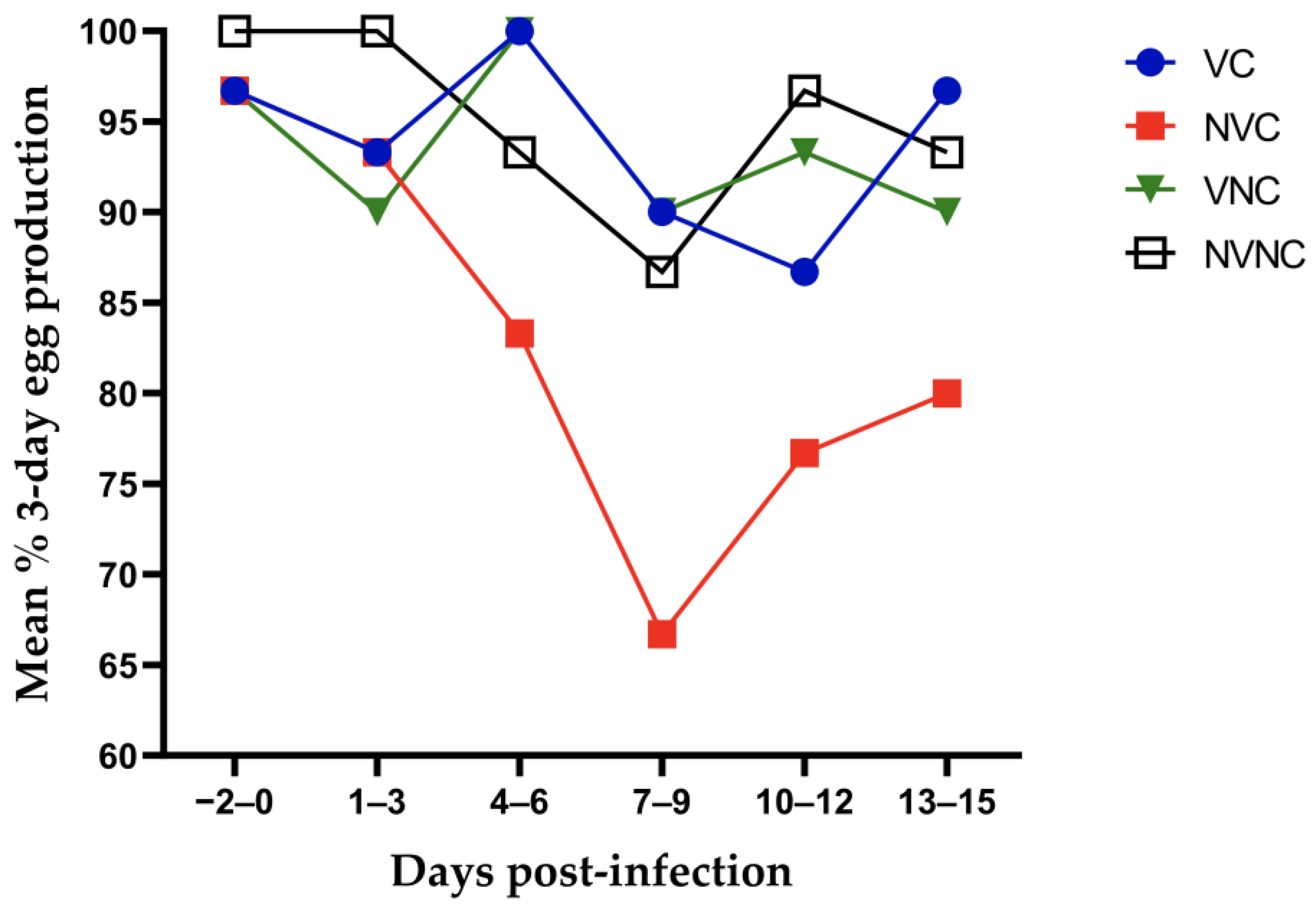
3.1. Clinical Observations and Egg Production
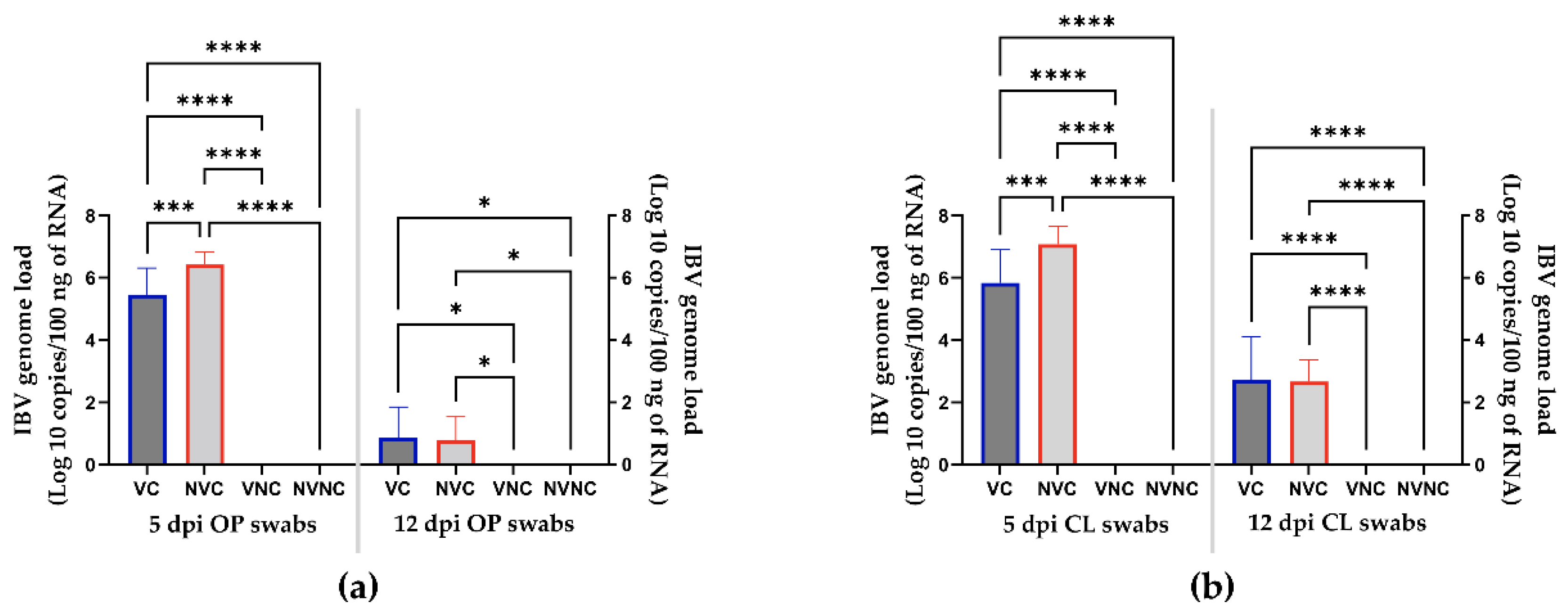
No appreciable clinical manifestations such as coughing, sneezing or conjunctivitis could be detected in any of the groups, except the dullness with ruffled feathers in two NVC hens on 6 dpi. The egg production of all groups is shown in Figure 1. The egg production from 1–15 dpi of the NVC group was significantly lower than that of any other group (p < 0.05). The egg production in the VC group was not significantly different from the control groups (p > 0.05). In comparison with the pre-challenge egg production, the NVC group showed a maximum drop of 30% at 7–9 dpi. At the end of the experiment, the egg production of the NVC group was 16.7% lower than the pre-challenge level. The egg production in the VC group dropped by a 10% at 10–12 dpi, while returned to the pre-challenge level at the end of the experiment.

3.2. Viral Shedding

Viral RNA loads in OP and CL swabs were below the detection limit in the control groups during the experiment. For challenged groups, the VC group demonstrated significantly decreased viral RNA loads in OP and CL swabs collected at 5 dpi compared to the NVC group (p < 0.05; Figure 2a,b). The viral RNA loads in OP and CL swabs did not differ significantly between the challenged groups at 12 dpi (p > 0.05; Figure 2a,b).

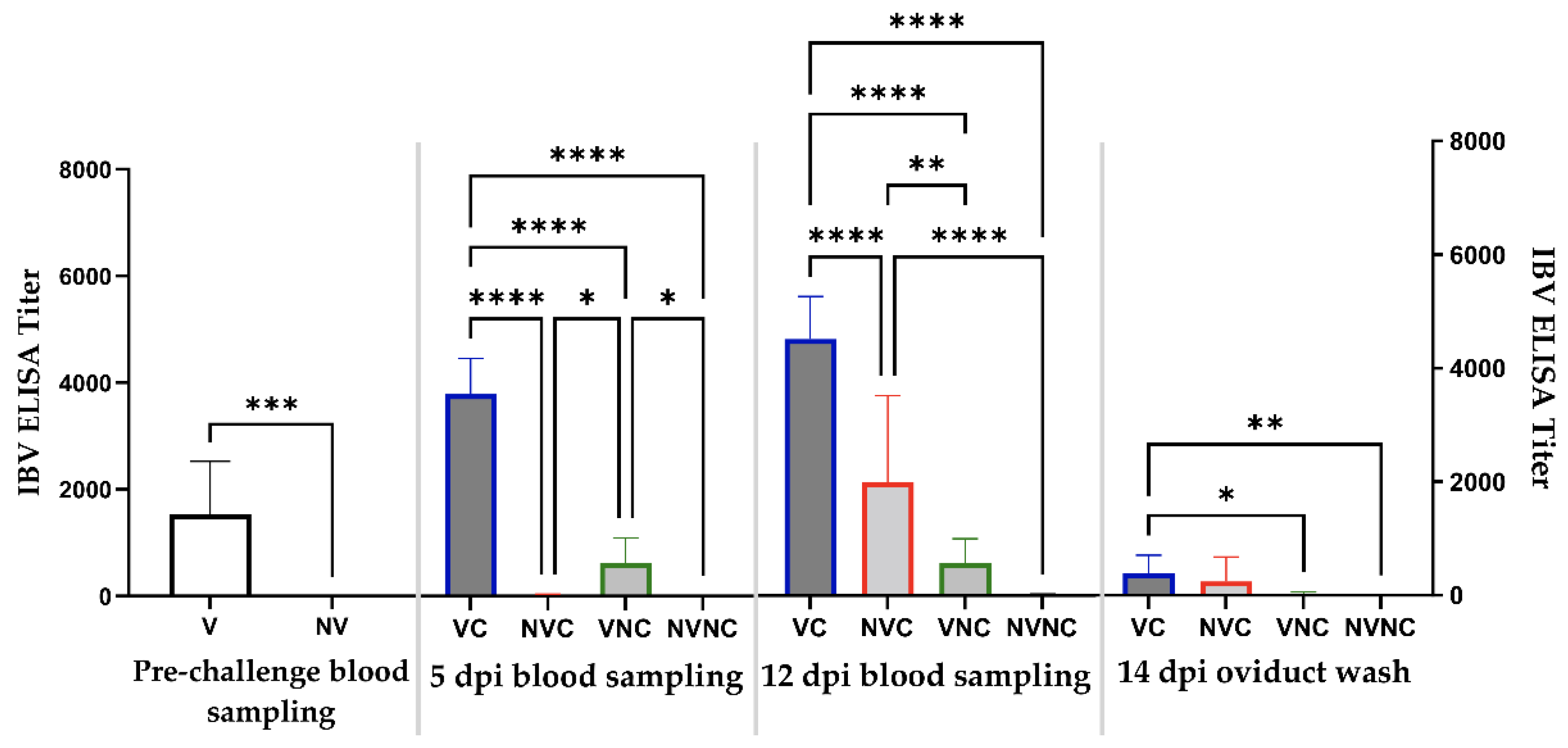
3.3. Anti-IBV Antibody Titer

Hens were bled prior to challenge at approximately 5 weeks following completion of the vaccination regimen. A significantly high level of antibody titer was measurable in the vaccinated group (p < 0.05; Figure 3), while the non-vaccinated group had no detectable antibodies. At challenge, a significantly higher antibody titer was observed in the VC group compared to all other groups at 5 and 12 dpi (p < 0.05). The NVC group was seronegative at 5 dpi, while it had a significantly higher antibody titer compared to the control groups at 12 dpi (p < 0.05). There was no significant difference in the antibody titer in the oviduct washes between the challenged groups (p > 0.05); however, the VC group showed significantly higher antibody titer compared to the control groups (p < 0.05; Figure 3).

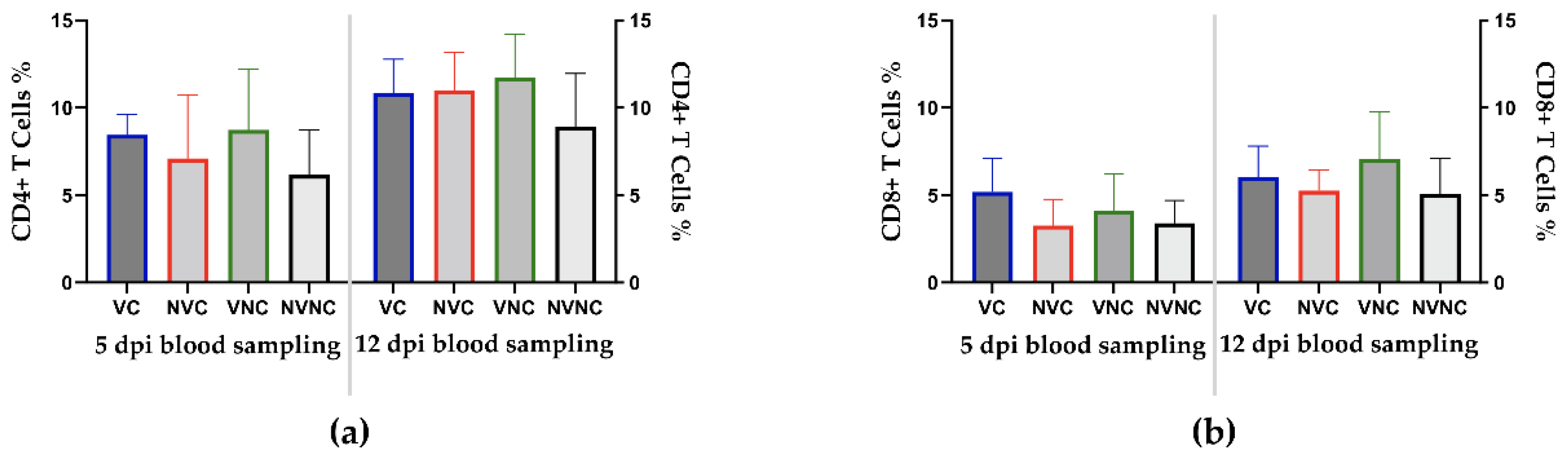
3.4. Peripheral Blood CD4+ and CD8+ T Cells

There were no significant differences in the percentages of CD4+ and CD8+ T cells between all the groups at 5 and 12 dpi (p > 0.05; Figure 4a,b).

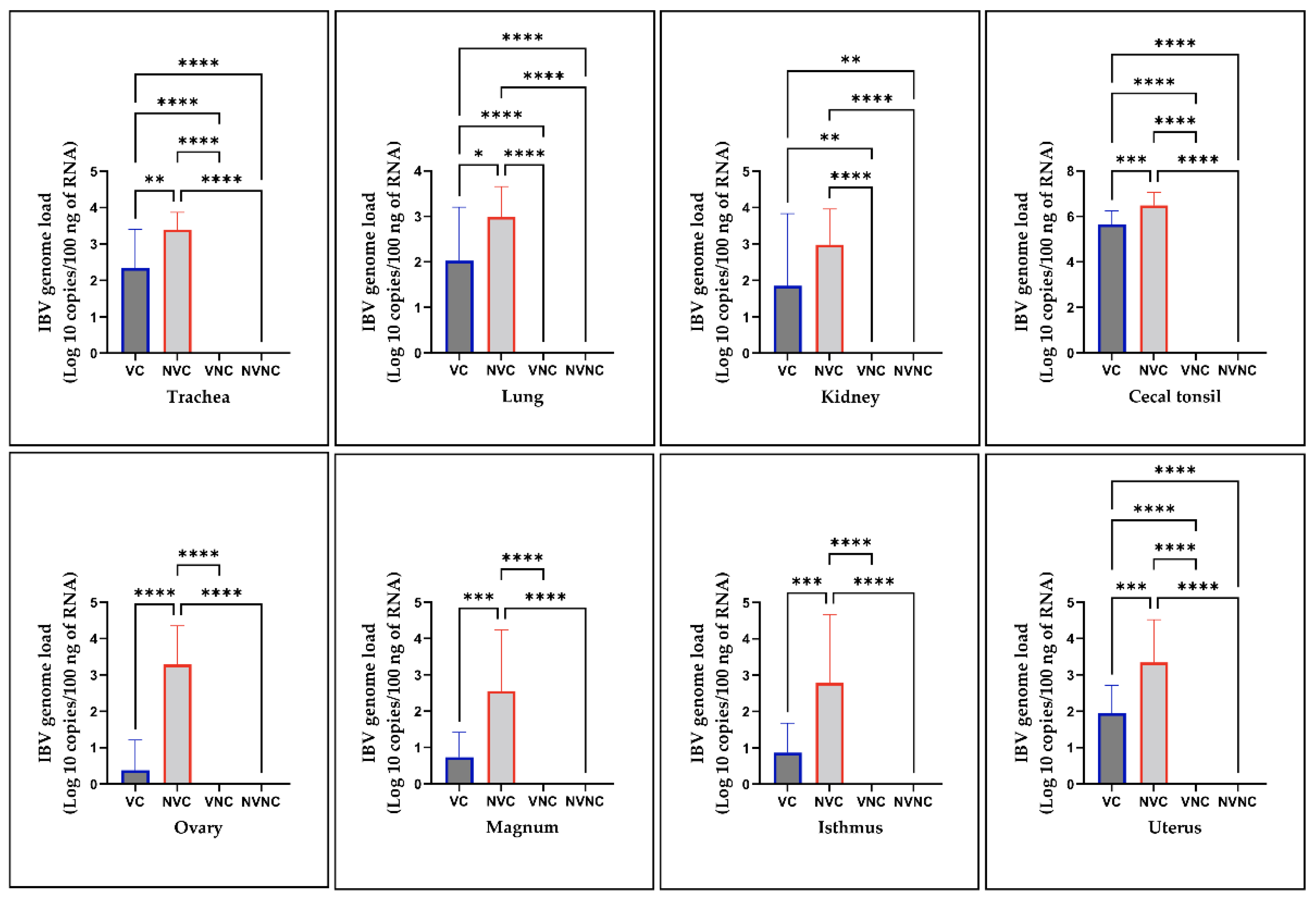
3.5. IBV Genome Loads in Tissues
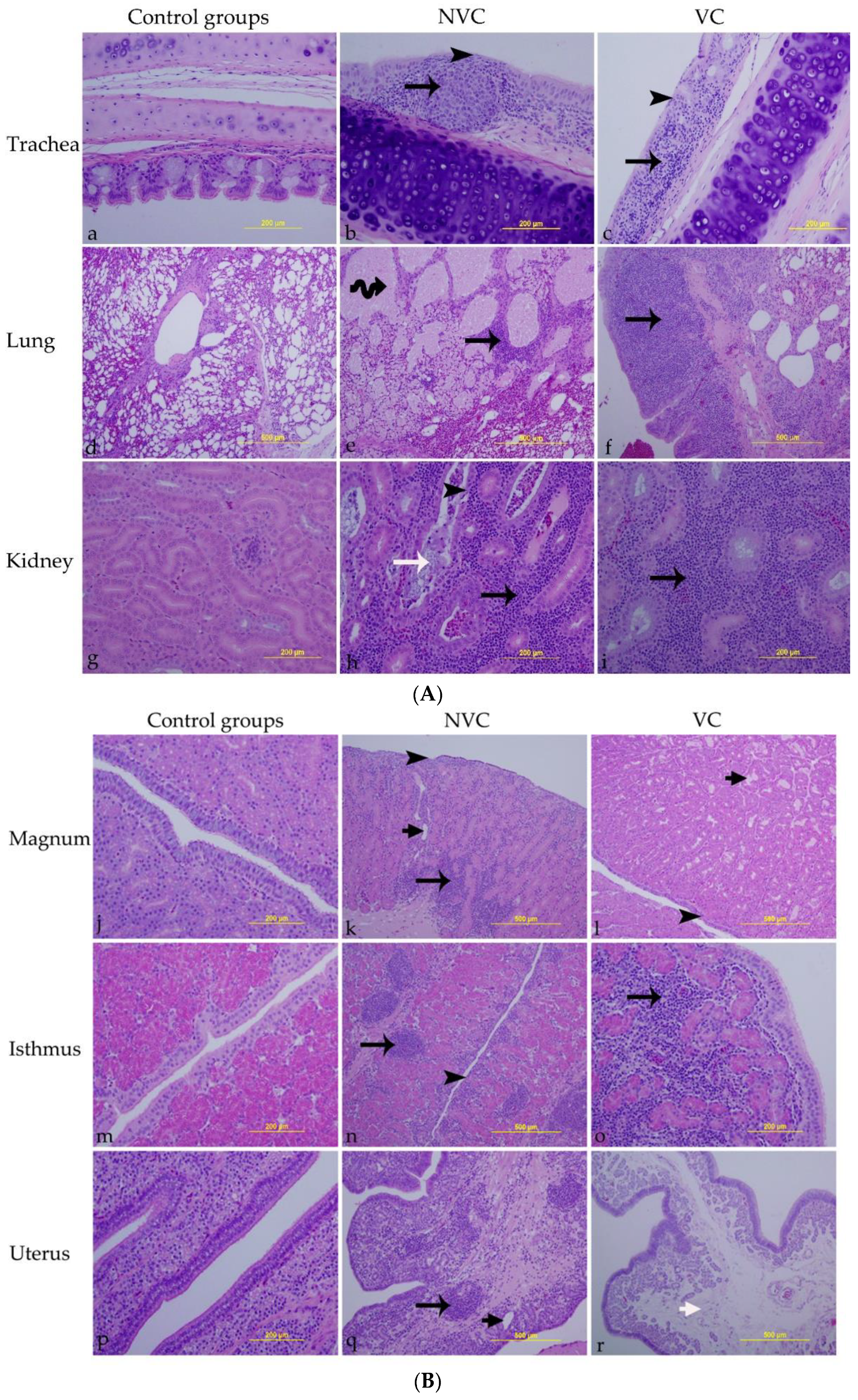
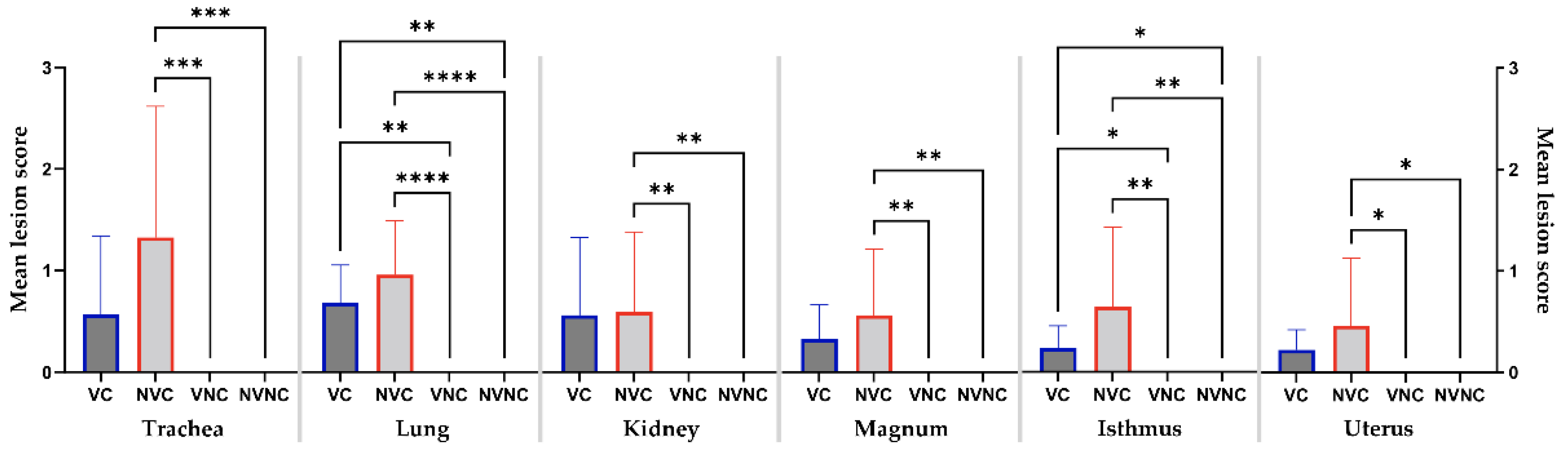
Viral RNA loads were below the detection limit in all IBV target organs of the control groups. The viral RNA was quantifiable in the trachea, lung, kidney, ovary, and oviduct (magnum, isthmus, and uterus) of the challenged groups with the cecal tonsils containing the highest amount of viral RNA. The VC group had significantly lower viral loads in all examined tissues (p < 0.05) except the kidney (p > 0.05) compared to the NVC group (Figure 5).

3.6. Histopathology

There was no significant pathology in the trachea, lung, kidney, or different parts of oviduct of the control groups (Figure 6a,d,g,j,m,p).
In the NVC group, the trachea showed epithelial cell necrosis with deciliation, and the lamina propria was infiltrated with focal lymphoplasmacytic aggregates (Figure 6b). The lung demonstrated hyperplasia of the epithelial cells lining the secondary bronchi together with mononuclear cell infiltrations in the lamina propria. Furthermore, the tertiary bronchi and atria were occupied by homogenous eosinophilic material, and the interstitial connective tissue was infiltrated with mononuclear cell infiltrations (Figure 6e). The kidney showed tubular cell necrosis with cellular exfoliation. Dilated renal tubules were occluded by intra-luminal casts containing a mixture of necrosed epithelia, mucus and disintegrated heterophils. The interstitial connective tissue was predominantly infiltrated with lymphoplasmacytic inflammatory cells, and few heterophils could be observed (Figure 6h). In the oviduct, the surface epithelium varied from areas of attenuation to patchy areas of necrosis and sloughing with ciliary losses. The lamina propria was infiltrated with multi-focal mononuclear cell infiltrations. Moreover, there was glandular dilatations together with edema among some glands particularly in the magnum and uterus (Figure 6k,n,q).
In the VC group, the trachea was characterized by epithelial cell necrosis with cilia and goblet cell losses, and the lamina propria was thickened because of mononuclear cell infiltrations (Figure 6c). The lung revealed diffuse mononuclear cell infiltrations in the lamina propria of the secondary bronchi (Figure 6f). In the kidney, the interstitial connective tissue was thickened due to diffuse lymphoplasmacytic cell infiltrates (Figure 6i). The magnum showed epithelial cell necrosis and deciliation in some areas with some dilated albuminous glands in the lamina propria (Figure 6l). The isthmus was characterized by diffuse mononuclear cell infiltrations among the inter-glandular connective tissue (Figure 6o). The uterus revealed only a widened area of edema in the lamina propria (Figure 6r).
The mean lesion scores in trachea, lung, kidney, magnum, isthmus, and uterus are summarized in Table 2. Although the lesion scores in all tissues did not differ significantly between the challenged groups (p > 0.05), only the NVC group showed significantly higher lesion scores in the trachea, kidney, magnum, and uterus compared to the control groups (p < 0.05; Figure 7). Both the challenged groups had significantly higher lesion scores in the lung and isthmus compared to the control groups (p < 0.05; Figure 7). Comparative histopathology of trachea, lung, kidney, magnum, isthmus, and uterus are shown in Supplementary Tables S1–S6.

4. Discussion

The use of a combined vaccination regimen including two or more antigenically distinct IB vaccines has been demonstrated to improve protection against heterologous IBV challenge [15,30]. The Canadian IBV DMV/1639 strain, which represents a major threat to the egg laying industry in Canada, showed low nucleotide (77.2%) and amino acid (73.2–75.5%) similarities in the S1 part of the S glycoprotein with the live Mass and Conn vaccine strains [22]. In this study, the level of protection against the Canadian IBV DMV/1639 strain experimental challenge in layers was evaluated after a vaccination program of a heterologous live priming by Mass and Conn types of vaccines and boosting with an inactivated vaccine that contained Mass and Ark antigens. Egg production, viral shedding, antibody- and cell-mediated immune responses, viral loads in tissues, and microscopic lesions were investigated.
IBV infection is a well-known cause of decreased egg production, as well as reduced internal and external egg quality [31]. Our earlier work demonstrated the ability of the Canadian DMV/1639 strain to adversely affect the numbers of eggs produced by experimentally challenged layers [24]. In the current study, all groups produced ≥90% from 1 to 3 dpi which excludes that the drop in egg production could have resulted from the stress associated with handling hens during virus/mock challenge. Compared to the other groups, the average egg production in the NVC group over 15 dpi was significantly lower. On the other hand, the VC group demonstrated an egg laying performance comparable to the control groups and to the pre-challenge egg production level.
Despite the genetic differences between the vaccine strains and challenge virus used in this study, an anamnestic IBV serum antibody response was induced in the VC group. Sera of heterologous genotype vaccines have previously demonstrated cross-reaction with field IBV isolates using virus neutralization (VN) test [19,32]. Furthermore, De Wit et al. showed that high levels of NV antibody against heterologous IBVs were associated with higher level of protection to the female reproductive tract [19]. In the current study, the anamnestic IBV serum antibody response measured by ELISA might have also been associated with protecting the VC group against the drop in egg production. Large amounts of ani-IBV antibodies were also detected the oviduct of the VC group, whereas local antibodies may be more effective in protecting the reproductive tract [27]. On the other hand, the serum antibody response in the NVC group appeared later and at a lower level than the VC group. The cell-mediated immune response represents the other arm of the adaptive immune response and helps in controlling acute IBV infection [33]. In the current study, changes in CD4+ and CD8+ T cell populations in the peripheral blood following challenge were not observed. More frequent blood sampling could have possibly helped to examine the differences in T cell responses amongst experimental groups [34].
The Canadian IBV DMV/1639 strain was previously shown to have a wide tissue tropism and can induce severe lesions in the respiratory, urinary, and genital tracts [23]. This study used a qRT-PCR assay to detect and monitor viral shedding and spread of the viral RNA in IBV-susceptible tissues. The viral shedding through OP and CL routes was significantly decreased in the VC group compared to the NVC group at 5 dpi. However, the VC and NVC groups displayed comparable rates of OP and CL viral shedding at 12 dpi, leaving the outcome of this parameter inconclusive. A longer observation period would have possibly shown more solid data regarding viral shedding in vaccinated layers. Previously, when SPF pullets were infected with the Canadian DMV/1639 strain at an early age, persistent cloacal viral shedding continued up to 105 dpi [23]. The viral RNA was detected from all the tissues of the challenged groups; however, a significant decrease in the viral genome loads within the trachea, lung, cecal tonsil, ovary, and oviduct was detected in the VC group compared to the NVC group. Amongst the tissues of the VC group that had decreased viral genome loads, the most pronounced decline was observed in the reproductive organs. A similar finding was reported by Chousalkar et al., whereas heterologous vaccination reduced the viral RNA of the N1/88 strain to below the detectable levels in the oviduct of 83% of the vaccinated hens examined between 6 and 16 dpi [35]. Conversely, the highest number of copies of viral RNA was detected in the cecal tonsils, which commonly represent the site of virus persistence [23,36].
Histopathological observations of the trachea, lung, kidney, and oviduct revealed that the mean scores obtained in the challenged groups were not significantly different. However, the trachea, kidney, magnum, and uterus of the VC group showed less marked lesion scores (scores not statistically different from the control groups). In addition, a higher number of birds in the VC group had no histopathological changes in the trachea, kidney, and magnum compared to the NVC group. Except for the kidney, no severe microscopic lesions were detected in other tissues of the VC group (Table 2), which is consistent with the viral genome load data. Similarly, the potential for DMV/1639 strain to induce renal lesions in vaccinated chickens was previously shown [37].
Assessment of protection against IBV challenge is most commonly performed by quantification of ciliostasis or virus re-isolation from the trachea, particularly as part of the licensing procedure for IB vaccines [17]. However, egg laying performance and health of the reproductive tract are critical factors when vaccine protection in layers is evaluated. The vaccination program used in this study protected the challenged layers from the negative effects on egg production by minimizing virus replication leading to only mild lesions in the reproductive organs. Early and recent studies have indicated the importance of incorporating an inactivated vaccine in an IBV vaccination program for layers [18,19,38]. Interestingly, a prime-boost vaccination program utilizing the same IBV types used in this study was previously demonstrated to improve protection of the kidney in chickens challenged with nephropathogenic IBV strain, PA/Wolgemuth/98 [39]. A recent epidemiological and molecular study concluded that DMV/1639 strain likely evolved from PA/Wolgemuth/98-like IBVs [37]. DMV/1639 and PA/Wolgemuth/98 strains were clustered within the same genetic lineage (GI-17) using phylogenetic analysis based on the complete S1 gene [22]. The present results confirm the improved protection conferred by a similar vaccination program against IBV strains of the GI-17 lineage. Gelb et al. was also able to demonstrate cross-protection against challenge with other IBV field strains using commercial bivalent IB vaccine containing the Mass and Ark strains [40]. This phenomenon of IBV cross-protection could be due to immunity induced against less variable virus proteins other than the S protein [41]. It is worth mentioning that apart from the S gene, other genes within the Canadian IBV DMV/1639 strain showed high sequence similarity with a Conn-like vaccine strain [22], whereby the Conn strain was the main component of our vaccination program.
In summary, a vaccination program of live priming and inactivated boosting utilizing the commercial IB vaccines in Canada demonstrated reasonable protection against challenge with the Canadian DMV/1639 strain in layers. Further investigations are warranted to elucidate the role of the maternal antibodies derived from the current vaccination regimen in protecting newly hatched chicks against DMV/1639-induced reproductive tract anomalies (false layer syndrome). Moreover, controlled field or experimental trials using commercial chickens can be conducted to validate the data generated using SPF chickens.

Supplementary Materials

The following supporting information can be downloaded at: https://www.mdpi.com/article/10.3390/vaccines10081194/s1, Table S1: Comparative histopathology of trachea at 14 dpi following infection with the Canadian DMV/1639 strain (IBV/Ck/Can/17–036989); Table S2: Comparative histopathology of lung at 14 dpi following infection with the Canadian DMV/1639 strain (IBV/Ck/Can/17–036989); Table S3: Comparative histopathology of kidney at 14 dpi following infection with the Canadian DMV/1639 strain (IBV/Ck/Can/17–036989); Table S4: Comparative histopathology of magnum at 14 dpi following infection with the Canadian DMV/1639 strain (IBV/Ck/Can/17–036989); Table S5: Comparative histopathology of isthmus at 14 dpi following infection with the Canadian DMV/1639 strain (IBV/Ck/Can/17–036989); Table S6: Comparative histopathology of uterus at 14 dpi following infection with the Canadian DMV/1639 strain (IBV/Ck/Can/17–036989).

Author Contributions

Conceptualization, M.F.A.-C. and S.C.C.; methodology, M.F.A.-C. and M.S.H.H.; software, M.F.A.-C.; validation, M.F.A.-C., S.C.C., F.v.d.M., D.G. and C.S.C.; formal analysis, M.S.H.H. and A.A.; investigation, M.S.H.H., S.M.B., S.M.N. and A.A.; resources, M.F.A.-C., C.S.C. and F.v.d.M.; data curation, M.F.A.-C. and M.S.H.H.; writing—original draft preparation, M.S.H.H. and A.A.; writing—review and editing, M.F.A.-C., S.C.C., F.v.d.M., D.G., C.S.C., M.S.H.H., A.A., S.M.B. and S.M.N.; visualization, M.S.H.H. and A.A.; supervision, M.F.A.-C., S.C.C., F.v.d.M., D.G. and C.S.C.; project administration, M.F.A.-C.; funding acquisition, M.F.A.-C. and S.C.C. All authors have read and agreed to the published version of the manuscript.

Funding

This research was funded by Natural Sciences and Engineering Research Council of Canada (NSERC) Alliance grant (grant #: 10032197) and Egg Farmers of Canada (EFC, grant number 10025236). PhD studies of M.S.H.H. and A.A. are funded by the Ministry of Higher Education of the Arab Republic of Egypt.

Institutional Review Board Statement

The study was conducted in accordance with the Declaration of Helsinki, and approved by the Health Science Animal Care Committee (HSACC) of the University of Calgary (Protocol number AC19-0011, approved on 19 March 2019).

Informed Consent Statement

Not applicable.

Data Availability Statement

The datasets used and/or analyzed within the frame of the study can be provided by the corresponding author upon reasonable request.

Acknowledgments

We want to thank the University of Guelph for kindly providing us the IBV-infected samples from which the virus was isolated. We acknowledge the help of the staff including Greg Boorman at the Veterinary Science Research Station at Spyhill campus, University of Calgary for the animal management.

Conflicts of Interest

The authors declare no conflict of interest.

References

  1. Cavanagh, D. Coronavirus avian infectious bronchitis virus. Vet. Res. 2007, 38, 281–297. [Google Scholar] [CrossRef] [PubMed] [Green Version]
  2. Wickramasinghe, I.N.; van Beurden, S.J.; Weerts, E.A.; Verheije, M.H. The avian coronavirus spike protein. Virus Res. 2014, 194, 37–48. [Google Scholar] [CrossRef] [PubMed]
  3. Lin, S.Y.; Chen, H.W. Infectious Bronchitis Virus Variants: Molecular Analysis and Pathogenicity Investigation. Int. J. Mol. Sci. 2017, 18, 2030. [Google Scholar] [CrossRef] [PubMed] [Green Version]
  4. Jordan, B. Vaccination against infectious bronchitis virus: A continuous challenge. Vet. Microbiol. 2017, 206, 137–143. [Google Scholar] [CrossRef]
  5. Raj, G.D.; Jones, R.C. Infectious bronchitis virus: Immunopathogenesis of infection in the chicken. Avian Pathol. 1997, 26, 677–706. [Google Scholar] [CrossRef]
  6. Muneer, M.A.; Newman, J.A.; Halvorson, D.A.; Sivanandan, V.; Coon, C.N. Effects of avian infectious bronchitis virus (Arkansas strain) on vaccinated laying chickens. Avian Dis. 1987, 31, 820–828. [Google Scholar] [CrossRef]
  7. Chousalkar, K.K.; Roberts, J.R. Effects of Australian strains of infectious bronchitis virus on internal and external quality of hen eggs. Anim. Prod. Sci. 2009, 49, 162–169. [Google Scholar] [CrossRef]
  8. Hong, S.M.; Kwon, H.J.; Kim, I.H.; Mo, M.L.; Kim, J.H. Comparative genomics of Korean infectious bronchitis viruses (IBVs) and an animal model to evaluate pathogenicity of IBVs to the reproductive organs. Viruses 2012, 4, 2670–2683. [Google Scholar] [CrossRef] [Green Version]
  9. Sevoian, M.; Levine, P.P. Effects of Infectious Bronchitis on the Reproductive Tracts, Egg Production, and Egg Quality of Laying Chickens. Avian Dis. 1957, 1, 136–164. [Google Scholar] [CrossRef]
  10. Chousalkar, K.K.; Roberts, J.R.; Reece, R. Comparative histopathology of two serotypes of infectious bronchitis virus (T and n1/88) in laying hens and cockerels. Poult. Sci. 2007, 86, 50–58. [Google Scholar] [CrossRef]
  11. Crinion, R.A.; Ball, R.A.; Hofstad, M.S. Abnormalities in laying chickens following exposure to infectious bronchitis virus at one day old. Avian Dis. 1971, 15, 42–48. [Google Scholar] [CrossRef] [PubMed]
  12. Benyeda, Z.; Mato, T.; Suveges, T.; Szabo, E.; Kardi, V.; Abonyi-Toth, Z.; Rusvai, M.; Palya, V. Comparison of the pathogenicity of QX-like, M41 and 793/B infectious bronchitis strains from different pathological conditions. Avian Pathol. 2009, 38, 449–456. [Google Scholar] [CrossRef] [PubMed] [Green Version]
  13. Parent, E.; Gagnon-Francoeur, A.; Lanthier, B.; Hébert, G.; Buczinski, S.; Boulianne, M. Diagnostic Accuracy of Ultrasonography to Detect False Layers in a Commercial Laying Flock Infected by an Infectious Bronchitis Virus Delmarva Genotype Causing Cystic Oviducts. Avian Dis. 2020, 64, 149–156. [Google Scholar] [CrossRef]
  14. Zhong, Q.; Hu, Y.X.; Jin, J.H.; Zhao, Y.; Zhao, J.; Zhang, G.Z. Pathogenicity of virulent infectious bronchitis virus isolate YN on hen ovary and oviduct. Vet. Microbiol. 2016, 193, 100–105. [Google Scholar] [CrossRef]
  15. Cook, J.K.; Orbell, S.J.; Woods, M.A.; Huggins, M.B. Breadth of protection of the respiratory tract provided by different live-attenuated infectious bronchitis vaccines against challenge with infectious bronchitis viruses of heterologous serotypes. Avian Pathol. 1999, 28, 477–485. [Google Scholar] [CrossRef] [Green Version]
  16. Cook, J.K.; Chesher, J.; Baxendale, W.; Greenwood, N.; Huggins, M.B.; Orbell, S.J. Protection of chickens against renal damage caused by a nephropathogenic infectious bronchitis virus. Avian Pathol. 2001, 30, 423–426. [Google Scholar] [CrossRef] [PubMed] [Green Version]
  17. de Wit, J.J.; Cook, J.K. Factors influencing the outcome of infectious bronchitis vaccination and challenge experiments. Avian Pathol. 2014, 43, 485–497. [Google Scholar] [CrossRef] [Green Version]
  18. Box, P.G.; Holmes, H.C.; Finney, P.M.; Froymann, R. Infectious bronchitis in laying hens: The relationship between haemagglutination inhibition antibody levels and resistance to experimental challenge. Avian Pathol. 1988, 17, 349–361. [Google Scholar] [CrossRef]
  19. de Wit, J.J.S.; Malo, A.; Cook, J.K.A. Induction of IBV strain-specific neutralizing antibodies and broad spectrum protection in layer pullets primed with IBV Massachusetts (Mass) and 793B vaccines prior to injection of inactivated vaccine containing Mass antigen. Avian Pathol. 2019, 48, 135–147. [Google Scholar] [CrossRef]
  20. de Wit, J.J.; Dijkman, R.; Guerrero, P.; Calvo, J.; Gonzalez, A.; Hidalgo, H. Variability in biological behaviour, pathogenicity, protectotype and induction of virus neutralizing antibodies by different vaccination programmes to infectious bronchitis virus genotype Q1 strains from Chile. Avian Pathol. 2017, 46, 666–675. [Google Scholar] [CrossRef] [Green Version]
  21. Amarasinghe, A.; Popowich, S.; De Silva Senapathi, U.; Abdul-Cader, M.S.; Marshall, F.; van der Meer, F.; Cork, S.C.; Gomis, S.; Abdul-Careem, M.F. Shell-Less Egg Syndrome (SES) Widespread in Western Canadian Layer Operations Is Linked to a Massachusetts (Mass) Type Infectious Bronchitis Virus (IBV) Isolate. Viruses 2018, 10, 437. [Google Scholar] [CrossRef] [PubMed] [Green Version]
  22. Hassan, M.S.H.; Ojkic, D.; Coffin, C.S.; Cork, S.C.; van der Meer, F.; Abdul-Careem, M.F. Delmarva (DMV/1639) Infectious Bronchitis Virus (IBV) Variants Isolated in Eastern Canada Show Evidence of Recombination. Viruses 2019, 11, 1054. [Google Scholar] [CrossRef] [Green Version]
  23. Hassan, M.S.H.; Ali, A.; Buharideen, S.M.; Goldsmith, D.; Coffin, C.S.; Cork, S.C.; van der Meer, F.; Boulianne, M.; Abdul-Careem, M.F. Pathogenicity of the Canadian Delmarva (DMV/1639) Infectious Bronchitis Virus (IBV) on Female Reproductive Tract of Chickens. Viruses 2021, 13, 2488. [Google Scholar] [CrossRef]
  24. Hassan, M.S.H.; Najimudeen, S.M.; Ali, A.; Altakrouni, D.; Goldsmith, D.; Coffin, C.S.; Cork, S.C.; van der Meer, F.; Abdul-Careem, M.F. Immunopathogenesis of the Canadian Delmarva (DMV/1639) infectious bronchitis virus (IBV): Impact on the reproductive tract in layers. Microb. Pathog. 2022, 166, 105513. [Google Scholar] [CrossRef] [PubMed]
  25. Reed, L.J.; Muench, H. A simple method of estimating fifty per cent endpoints. Am. J. Epidemiol. 1938, 27, 493–497. [Google Scholar] [CrossRef]
  26. Hy-Line W36 Commercial Layers Management Guide. Available online: https://www.hyline.com/literature/W-36 (accessed on 17 February 2020).
  27. Raj, G.D.; Jones, R.C. Local antibody production in the oviduct and gut of hens infected with a variant strain of infectious bronchitis virus. Vet. Immunol. Immunopathol. 1996, 53, 147–161. [Google Scholar] [CrossRef]
  28. Kameka, A.M.; Haddadi, S.; Kim, D.S.; Cork, S.C.; Abdul-Careem, M.F. Induction of innate immune response following infectious bronchitis corona virus infection in the respiratory tract of chickens. Virology 2014, 450–451, 114–121. [Google Scholar] [CrossRef] [Green Version]
  29. Benyeda, Z.; Szeredi, L.; Mató, T.; Süveges, T.; Balka, G.; Abonyi-Tóth, Z.; Rusvai, M.; Palya, V. Comparative histopathology and immunohistochemistry of QX-like, Massachusetts and 793/B serotypes of infectious bronchitis virus infection in chickens. J. Comp. Pathol. 2010, 143, 276–283. [Google Scholar] [CrossRef]
  30. de Wit, J.J.; Nieuwenhuisen-van Wilgen, J.; Hoogkamer, A.; van de Sande, H.; Zuidam, G.J.; Fabri, T.H. Induction of cystic oviducts and protection against early challenge with infectious bronchitis virus serotype D388 (genotype QX) by maternally derived antibodies and by early vaccination. Avian Pathol. 2011, 40, 463–471. [Google Scholar] [CrossRef] [Green Version]
  31. Hassan, M.S.H.; Abdul-Careem, M.F. Avian Viruses that Impact Table Egg Production. Animals 2020, 10, 1747. [Google Scholar] [CrossRef]
  32. Shimazaki, Y.; Harada, M.; Horiuchi, T.; Yoshida, K.; Tanimura, C.; Nakamura, S.; Mase, M.; Suzuki, S. Serological studies of infectious bronchitis vaccines against Japanese field isolates of homologous and heterologous genotypes. J. Vet. Med. Sci. 2009, 71, 891–896. [Google Scholar] [CrossRef] [Green Version]
  33. Collisson, E.W.; Pei, J.; Dzielawa, J.; Seo, S.H. Cytotoxic T lymphocytes are critical in the control of infectious bronchitis virus in poultry. Dev. Comp. Immunol. 2000, 24, 187–200. [Google Scholar] [CrossRef]
  34. Seo, S.H.; Collisson, E.W. Specific cytotoxic T lymphocytes are involved in in vivo clearance of infectious bronchitis virus. J. Virol. 1997, 71, 5173–5177. [Google Scholar] [CrossRef] [PubMed] [Green Version]
  35. Chousalkar, K.K.; Cheetham, B.F.; Roberts, J.R. Detection of infectious bronchitis virus strain N1/88 from the oviduct and feces of experimentally infected vaccinated and unvaccinated hens. Poult. Sci. 2010, 89, 1603–1608. [Google Scholar] [CrossRef]
  36. De Wit, J.J. Detection of infectious bronchitis virus. Avian Pathol. 2000, 29, 71–93. [Google Scholar] [CrossRef] [PubMed] [Green Version]
  37. Gelb, J., Jr.; Ladman, B.S.; Pope, C.R.; Ruano, J.M.; Brannick, E.M.; Bautista, D.A.; Coughlin, C.M.; Preskenis, L.A. Characterization of nephropathogenic infectious bronchitis virus DMV/1639/11 recovered from Delmarva broiler chickens in 2011. Avian Dis. 2013, 57, 65–70. [Google Scholar] [CrossRef] [Green Version]
  38. Buharideen, S.M.; Hassan, M.S.H.; Najimudeen, S.M.; Niu, D.; Czub, M.; Gomis, S.; Abdul-Careem, M.F. Immune Responses in Laying Hens after an Infectious Bronchitis Vaccination of Pullets: A Comparison of Two Vaccination Strategies. Vaccines 2021, 9, 531. [Google Scholar] [CrossRef]
  39. Ladman, B.S.; Pope, C.R.; Ziegler, A.F.; Swieczkowski, T.; Callahan, C.J.; Davison, S.; Gelb, J., Jr. Protection of chickens after live and inactivated virus vaccination against challenge with nephropathogenic infectious bronchitis virus PA/Wolgemuth/98. Avian Dis. 2002, 46, 938–944. [Google Scholar] [CrossRef] [Green Version]
  40. Gelb, J., Jr.; Wolff, J.B.; Moran, C.A. Variant serotypes of infectious bronchitis virus isolated from commercial layer and broiler chickens. Avian Dis. 1991, 35, 82–87. [Google Scholar] [CrossRef]
  41. Cavanagh, D.; Elus, M.M.; Cook, J.K. Relationship between sequence variation in the S1 spike protein of infectious bronchitis virus and the extent of cross-protection in vivo. Avian Pathol. 1997, 26, 63–74. [Google Scholar] [CrossRef] [Green Version]
Figure 1. Three-day interval mean percentage of egg production starting from 3 days before the challenge until 15 dpi following infection with the Canadian DMV/1639 strain (IBV/Ck/Can/17–036989).
Vaccines 10 01194 g001
Figure 2. IBV genome loads in OP (a) and CL (b) swabs collected at 5 and 12 dpi following infection with the Canadian DMV/1639 strain (IBV/Ck/Can/17–036989). The average starting IBV copies was quantified per 100 ng of the extracted RNA and differences between groups were identified using one-way ANOVA followed by Tukey’s multiple comparisons test, and the error bars represent the SD. Statistical significance: * p < 0.05, *** p < 0.001, **** p < 0.0001.
Vaccines 10 01194 g002
Figure 3. IBV ELISA titers in pre-challenge serum (23 weeks of age) and post-challenge serum (5 and 12 dpi) and oviduct wash (14 dpi). Pre-challenge mean titers were compared using unpaired t test. Post-challenge mean titers were compared using one-way ANOVA followed by Tukey’s multiple comparisons test. The error bars represent the SD. Statistical significance: * p < 0.05, ** p < 0.01, *** p < 0.001, **** p < 0.0001.
Vaccines 10 01194 g003
Figure 4. Percentages of CD4+ (a) and CD8+ (b) T cells in peripheral blood mononuclear cells (PBMCs) at 5 and 12 dpi following infection with the Canadian DMV/1639 strain (IBV/Ck/Can/17–036989). Mean percentages of CD4+ and CD8+ T cells were compared between groups using one-way ANOVA test followed by Tukey’s multiple comparisons test, and the error bars represent the SD.
Vaccines 10 01194 g004
Figure 5. IBV genome loads in trachea, lung, kidney, cecal tonsil, ovary, magnum, isthmus, and uterus collected at 14 dpi following infection with the Canadian DMV/1639 strain (IBV/Ck/Can/17–036989). The average starting IBV copies was quantified per 100 ng of the extracted RNA and differences between groups were identified using one-way ANOVA followed by Tukey’s multiple comparisons test, and the error bars represent the SD. Statistical significance: * p < 0.05, ** p < 0.01, *** p < 0.001, **** p < 0.0001.
Vaccines 10 01194 g005
Figure 6. (A) Microscopic lesions detected in trachea, lung, and kidney; (B) Microscopic lesions detected in magnum, isthmus, and uterus. Tissue sections were examined at 14 dpi following infection with the Canadian DMV/1639 strain (IBV/Ck/Can/17–036989). (a,d,g,j,m,p) are control groups (VNC and NVNC). (b,e,h,k,n,q) are vaccinated challenged group (VC). (c,f,i,l,o,r) are non-vaccinated challenged group (NVC). Black arrowheads refer to epithelial necrosis; long black arrows refer to mononuclear cell infiltrations; coiled black arrow indicates homogenous eosinophilic materials filling the lumen of tertiary bronchi; long white arrow refers to cellular casts inside the renal tubule; short black arrows show glandular dilatation; short white arrow reveals edema in lamina propria.
Vaccines 10 01194 g006
Figure 7. Mean lesion scores in trachea, lung, kidney, magnum, isthmus, and uterus at 14 dpi following infection with the Canadian DMV/1639 strain (IBV/Ck/Can/17–036989). Mean lesion scores were calculated according to the severity of the observed lesions in ten hens per group and differences between groups were compared using Kruskal–Wallis’ test followed by Dunn’s multiple comparisons test, and the error bars represent the SD. Statistical significance: * p < 0.05, ** p < 0.01, *** p < 0.001, **** p < 0.0001.
Vaccines 10 01194 g007
Table 1. Lesions scored for each tissue examined at 14 dpi following infection with the Canadian DMV/1639 strain (IBV/Ck/Can/17–036989).
Tissue Lesions
Trachea Loss of epithelial lining
Loss of cilia
Necrosis of epithelial lining
Inflammatory cell infiltrations in the lamina propria
Lung Peri-bronchitis
Inflammatory cell infiltrations in the interstitial tissue
Circulatory disturbances (hyperemia, edema, and hemorrhage in the interstitial tissue
Kidney Necrosis of ducto-tubular epithelium
Inflammatory cell infiltrations in the interstitial tissue
Renal tubular dilatation
Oviduct (magnum, isthmus, and uterus) Epithelial cell necrosis
Loss of cilia
Tubular gland dilatation
Lymphocyte infiltrations in lamina propria
Edema in lamina propria
Table 2. Mean lesion scores in trachea, lung, kidney, magnum, isthmus, and uterus at 14 dpi following infection with the Canadian DMV/1639 strain (IBV/Ck/Can/17–036989).
GroupsVCNVCControl Groups
No Change
(−)
Mild
(+)
Moderate
(++)
Severe
(+++)
No Change
(−)
Mild
(+)
Moderate
(++)
Severe
(+++)
No Change
(−)
Mild
(+)
Moderate
(++)
Severe
(+++)
Trachea6/10a1/103/100/101/104/102/103/1010/100/100/100/10
Lung0/108/102/100/100105/104/101/1010/100/100/100/10
Kidney6/100/103/101/103/104/102/101/1010/100/100/100/10
Magnum4/106/100/100/102/107/100/101/1010/100/100/100/10
Isthmus2/86/80/80/81/75/70/71/710/100/100/100/10
Uterus4/106/100/100/104/105/100/101/1010/100/100/100/10
a = Mean severity score of each tissue: No change (−) = 0; mild (+) = > 0 to 1; moderate (++) = > 1 to 2; severe (+++) = > 2 to 3.
Publisher’s Note: MDPI stays neutral with regard to jurisdictional claims in published maps and institutional affiliations.

