Review

From Concept to Clinical Product: A Brief History of the Novel Shigella Invaplex Vaccine’s Refinement and Evolution

by
K. Ross Turbyfill
1,
Kristen A. Clarkson
1,
Edwin V. Oaks
2 and
Robert W. Kaminski
1,*
1
Department of Diarrheal Disease Research, Bacterial Diseases Branch, Walter Reed Army Institute of Research, Silver Spring, Silver Spring, MD 20910, USA
2
Patuxent Research and Consulting Group, Gambrills, Gambrills, MD 21054, USA
*
Author to whom correspondence should be addressed.
Vaccines 2022, 10(4), 548; https://doi.org/10.3390/vaccines10040548
Submission received: 19 January 2022 / Revised: 12 March 2022 / Accepted: 23 March 2022 / Published: 1 April 2022
(This article belongs to the Special Issue Frontiers in Shigella Vaccine Development)

Abstract

:
The Shigella invasin complex or Invaplex vaccine is a unique subunit approach to generate a protective immune response. Invaplex is a large, macromolecular complex consisting of the major Shigella antigens: lipopolysaccharide (LPS) and the invasion plasmid antigen (Ipa) proteins B and C. Over the past several decades, the vaccine has progressed from initial observations through pre-clinical studies to cGMP manufacture and clinical evaluations. The Invaplex product maintains unique biological properties associated with the invasiveness of virulent shigellae and also presents both serotype-specific epitopes, as well as highly conserved invasin protein epitopes, to the immunized host. The vaccine product has evolved from a native product isolated from wild-type shigellae (native Invaplex) to a more defined vaccine produced from purified LPS and recombinant IpaB and IpaC (artificial Invaplex). Each successive “generation” of the vaccine is derived from earlier versions, resulting in improved immunogenicity, homogeneity and effectiveness. The current vaccine, detoxified artificial Invaplex (InvaplexAR-Detox), was developed for parenteral administration by incorporating LPS with under-acylated lipid A. InvaplexAR-Detox has demonstrated an excellent safety and immunogenicity profile in initial clinical studies and is advancing toward evaluations in the target populations of children and travelers to endemic countries.

1. Introduction

The development and evolution of the Shigella invasin complex (Invaplex) family of products has occurred over the past 25-plus years (Figure 1) and represents stepwise, progressive discoveries and improvements that often reflect the current state of knowledge on Shigella proteins, lipopolysaccharide (LPS), and immunology. The progress not only involves research on the unique properties of the invasin complex but also critical improvements in product manufacturing, assay development and necessary adjustments to satisfy current regulatory standards and potential commercial development. The Invaplex product today (called artificial detoxified Invaplex or InvaplexAR-Detox) represents the latest improvement and is a candidate vaccine that stimulates pronounced systemic and mucosal immune responses in humans that most reflect the immunity observed after natural infection.

2. Shigella Antigens

After natural infection, the human host responds to several antigens of the Shigella pathogen. The most well characterized antigen is LPS, more specifically the O-specific polysaccharide (O-SP) of the LPS, which is responsible for serotype-specific immunity and protective immunity [1,2]. Conjugate vaccines using O-SP coupled to recombinant Pseudomonas aeruginosa exoprotein A protected immunized individuals against future disease in studies conducted in the early 1990s [3,4]. Although the O-SP conjugate vaccine approach was very promising and is still being pursued today [5,6,7], it was becoming clear in the late 1980s and early 1990s that a pronounced immune response to the virulence-specific proteins (primarily invasion plasmid antigen (Ipa) proteins IpaB, IpaC and IpaD) also occurred in humans after infection [8]. At the time, the only vaccines capable of stimulating an immune response to both LPS and the Ipa proteins were the live, attenuated vaccines, although the responses to the Ipa proteins were only secondarily evaluated [9,10,11]. More recent CHIM data have complemented field studies and pre-clinical animal studies to make a strong case for immune response to the Ipa proteins playing an important role in protection against shigellosis [12,13,14,15,16].

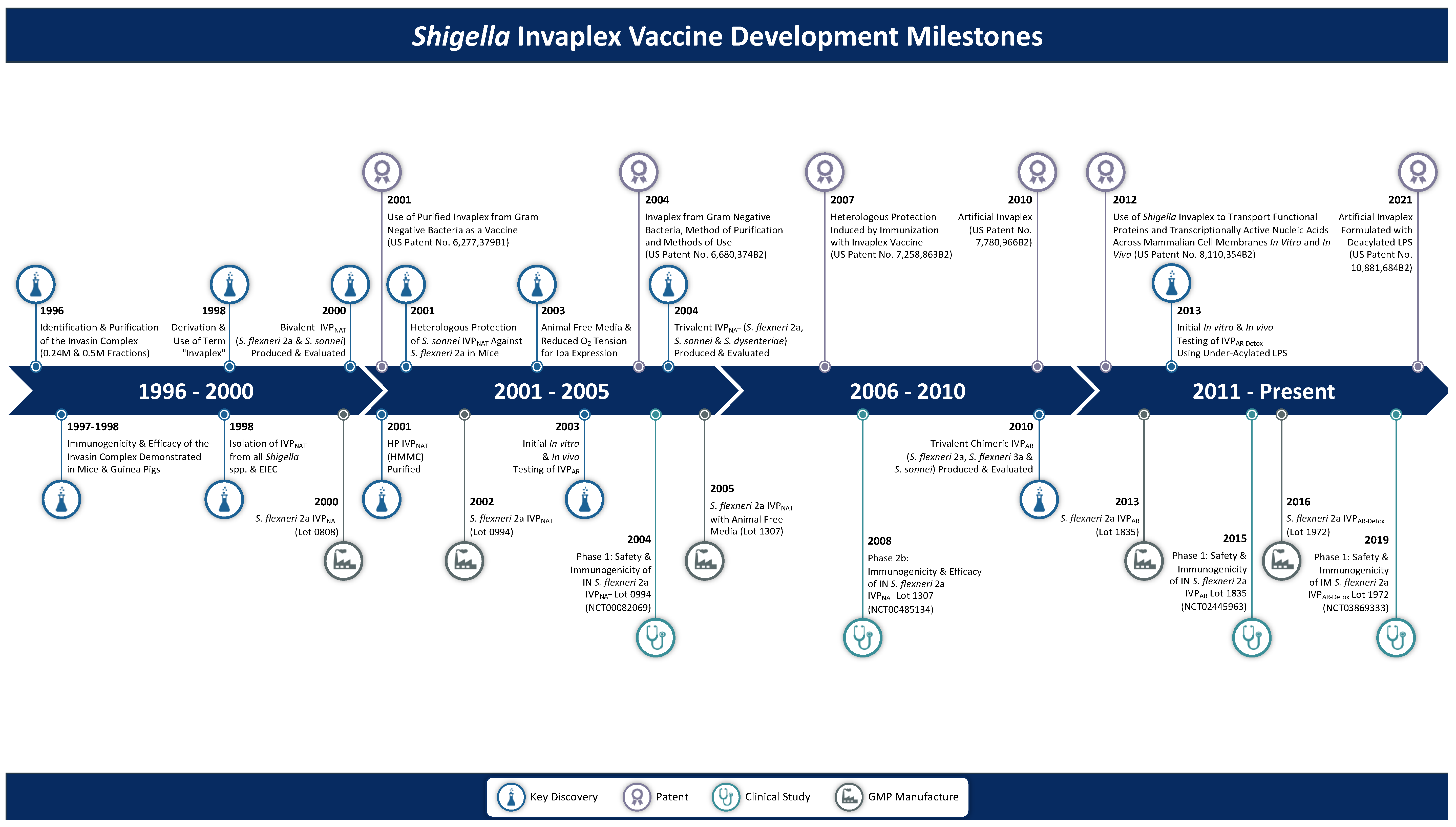
3. Native Invaplex (InvaplexNAT)

3.1. Discovery of Invaplex

In the mid-1990s, significant advances were underway in the mechanisms by which the Ipa proteins interacted with host cells. Initial reports described the controlled excretion/release of the Ipa proteins as either IpaB/IpaC or IpaB/IpaC/IpaD complexes and established the foundation of the Type III secretion system (TTSS) [17,18,19]. Coupled with this early research on the biomechanics of the Ipa proteins’ secretion and function were the first observations that IpaB, IpaC and IpaD were prominent, conserved antigens recognized by most Shigella-infected individuals [8]. Although antigen-specific assays for the Ipa proteins would come later, it was clear that the infected host responded vigorously to virulence-specific antigens found in the water extracts of virulent Shigella [8,14,20,21]. It was unclear what role, if any, the host immune response to the highly conserved Ipa proteins had in protection against disease, since dogma clearly identified LPS as the antigen responsible for protective, serotype-specific immunity [1,2,3,22]. Even so, one hypothetical outcome of the response to the conserved Ipa proteins would be the potential for broad protection against all Shigella serotypes and enteroinvasive Escherichia coli (EIEC). Researchers at the Walter Reed Army Institute of Research (WRAIR) investigated methods to identify and measure the host response to protein antigens. Enzyme-linked immunosorbent assays (ELISA) that could measure the virulence-specific immune response utilized water extracts from wild-type shigellae, which contained the Ipa proteins, VirG (IcsA), LPS and several other undefined proteins [8]. To prepare more specific reagents, attempts to isolate the native Ipa proteins or Ipa complexes from the water extract by various chromatographic methods always resulted in IpaB and IpaC-containing fractions that copurified with LPS [23]. Although the LPS was initially viewed as a “contaminant”, an alternative explanation was that a naturally occurring complex of Ipa proteins and LPS had been isolated. Ultimately, the isolated invasion protein–LPS complex became known as Invaplex and more specifically as native Invaplex (InvaplexNAT), the first generation of Invaplex isolated from virulent wild-type shigellae using ion-exchange chromatography [24].
InvaplexNAT could be isolated in two anion-exchange fractions called InvaplexNAT 24 (0.24 M sodium chloride (NaCl) step) and InvaplexNAT 50 (0.50 M NaCl step) [24]. Each Invaplex preparation contained the major Shigella antigens IpaB, IpaC, IpaD and LPS and was the formulation for a possible vaccine. Other proteins were also found in InvaplexNAT 24 and InvaplexNAT 50, but neither contained any major outer membrane proteins. The universality of InvaplexNAT was demonstrated by isolating complexes from all Shigella species and EIEC [25], which were immunogenic and provided homologous protection in mice and guinea pigs after intranasal immunization [24,25]. Subsequent evaluation of bivalent (S. flexneri 2a and S. sonnei) and trivalent (S. flexneri 2a, S. dysenteriae 1 and S. sonnei) iterations of the InvaplexNAT product were also successful against homologous challenges [26,27].

3.2. Biological Properties of InvaplexNAT

The innate Shigella virulence attributes of inducing phagocytosis by the host cells and subsequent escape of the bacterium from the phagosome are dependent on functional Ipa proteins [28]. Maintenance of Ipa protein functionality in InvaplexNAT was evaluated in a series of in vitro studies on InvaplexNAT and its ability to interact with non-phagocytic eukaryotic cells. After a brief incubation with host cells InvaplexNAT rapidly decorated the host cell surface, followed by an induced, actin-dependent endocytosis similar to the process utilized by wild-type shigellae. Internalized InvaplexNAT migrated through the retrograde endosomal pathway (early endosome to late endosome to Golgi apparatus) and was ultimately localized free in the cytoplasm [29]. The uptake of Invaplex could be inhibited with antibodies specific for IpaB and IpaC, but antibodies to LPS had no effect on the uptake event. The property of inducing phagocytosis by host cells is considered an essential biological property of Invaplex found in all generations of the product (Table 1) and may be a possible mechanism by which LPS and the Ipa proteins are presented to the host immune system via antigen-presenting cells. In fact, separate studies exploited the native biological activity of Invaplex to deliver and enhance the immune response to otherwise non-immunogenic antigens in which InvaplexNAT was serving as an adjuvant and/or transporter of heterologous, functional cargos (proteins, nucleic acids) into host cells [12,13,15,29,30,31,32]. Furthermore, maintenance of the biological activity suggests that a native structure similar to that found in wild-type shigellae is present in the native complex, making it not only a good vaccine candidate but also a good antigen for use in immunological assays to measure responses to the native invasin complex.

3.3. Intranasal Delivery of Invaplex without an Adjuvant

One of the striking features after infection with Shigella spp. is the dominant immune response to IpaB, IpaC and IpaD, with a large proportion of infected individuals responding to IpaB and IpaC and a lower proportion responding to IpaD [33,34]. This strong response to the Ipa proteins likely reflects their inherent immunogenicity and critical interaction with the host. Although other Shigella proteins are recognized by the immune system, no other antigens stimulate such a dominant, virulence-specific response post-infection.
Unfortunately, it was unclear how to immunize and stimulate a good mucosal immune response in the intestine other than by oral delivery of live or killed whole bacteria. For subunit vaccines and some inactivated whole-cell vaccines, the use of a strong mucosal adjuvant, such as mutant E. coli heat-labile toxin [35], was often necessary to drive a mucosal response. In contrast, immunization in the nasal cavity and exploitation of the “common mucosal pathway” to stimulate migration of mature lymphocytes and production of secretory IgA at distant sites [36,37], such as the intestinal tract, would provide a simple way to stimulate intestinal immunity [38,39]. Utilization of the non-injectable, intranasal route would alleviate many of the clinical concerns associated with administering the wild-type LPS (endotoxin) found in InvaplexNAT and would overcome many limitations of oral immunization with a subunit vaccine, such as antigen quantity, the need for an adjuvant and stability upon delivery into the gastrointestinal tract.
Pre-clinical experiments clearly demonstrated that animals intranasally immunized with InvaplexNAT produced antibodies to IpaB, IpaC and LPS, suggesting that InvaplexNAT circumvented the poor immunogenicity of LPS delivered alone [40,41]. The intranasal route was used for all animal and human immunizations with InvaplexNAT. Pipets were used for all animal studies, but evaluation of different nasal delivery devices was required for humans. Over the course of clinical development, three devices were used for human intranasal immunization: a micropipette, the Dolphin device (Valois of America, Inc., Congers, NY, USA) and the VaxINator™ device (Teleflex, Wayne, PA, USA). Each device was able to deliver the targeted amount of vaccine, with the Dolphin and VaxINator™ devices providing optimal spray distribution to the nasal mucosa. Results from the human studies clearly showed that intranasal immunization led to a significant mucosal immune response in the gut [42,43], which was one of the first direct demonstrations in humans that local intranasal immunizations led to an immune response in the gut. Subsequent studies with the Invaplex vaccine would show that circulating immune lymphocytes carrying the gut mucosal homing marker (α4β7 + B cells) were prominent after intranasal immunization (InvaplexAR) and after parenteral immunization (InvaplexAR-Detox) (see below).

3.4. Immunogenicity of InvaplexNAT: Assay Development and Results

Measurement of the specific immune response to the novel Invaplex vaccine has evolved from using traditional ELISAs and other antibody-based assays, such as antibody-secreting cell (ASC) assays to the current use of antigen-specific assays, along with functional assays like the serum bactericidal assay [44,45,46]. The antigens used in early studies consisted of purified LPS to detect serotype-specific responses, the water extract preparations from virulent (Vir+) and avirulent (Vir−) Shigella [24] and, later, the InvaplexNAT product. Purified IpaB and IpaC were prominently used in more recent studies once they became readily available and were used to measure a response that would potentially react with all Shigella species and EIEC. One antigen frequently used was InvaplexNAT prepared from the same Shigella serotype used to produce the vaccine. Studies using sera from animals or humans immunized with the live attenuated vaccine S. flexneri 2a SC602 [9,47] or S. sonnei WRSS2 and WRSS3 vaccines [48] or InvaplexNAT [24,42,43] consistently demonstrated that there were more seroconversions measured with InvaplexNAT than with any other antigen. Although not fully understood, InvaplexNAT, when used as an immunological assay antigen reagent, seems to maintain a native conformation not found in purified LPS, IpaB or IpaC [32]. For this reason, InvaplexNAT has been used as an ELISA antigen to measure the immunogenicity of all Invaplex products.
The potent immunogenicity of InvaplexNAT after intranasal immunization minimized any need for a mucosal adjuvant. Dosing studies in mice and guinea pigs clearly showed that three biweekly intranasal immunizations of relatively low doses were superior to a two-dose regimen for stimulating a strong, protective immune response. If higher dose amounts were used, a two-dose schedule could be used. In mice, the dose amount ranged from 1 µg to 25 µg, with 5 µg used routinely; in guinea pigs, the routinely used dose amount was 25 µg. The three-dose schedule has been maintained for all Invaplex products, although more recent efforts using parenteral immunizations have spaced out the time schedule to three-week intervals between doses.
After immunization with S. flexneri 2a InvaplexNAT 24 or 50, small animals responded with pronounced serum IgA and IgG specific to LPS and the Vir+ water extract [24]. Western blot analysis of guinea pig immune sera indicated that IpaB and IpaC were dominant immunogens in InvaplexNAT 24- and 50-immunized animals, but antibodies to several other non-virulence-specific proteins were found in animals immunized with InvaplexNAT 50. In both guinea pigs and mice immunized with either InvaplexNAT 24 or 50, significant protection was observed upon challenge with homologous, virulent shigellae [24]. Similar immune responses were observed with InvaplexNAT prepared from all Shigella species and EIEC. The one notable difference was for S. sonnei InvaplexNAT 24, which had much lower quantities of IpaB and undetectable LPS and was not protective in mice or guinea pigs [31,32,49]. These results suggested that either IpaB or LPS or both antigens were crucial for protection in these models. The S. sonnei InvaplexNAT 50 product stimulated antibodies to LPS, IpaB, IpaC and other proteins and was fully protective.

3.5. Heterologous Immunity Induced by Invaplex

One of the unique features of the Invaplex vaccine products is the ability to deliver, in controlled amounts, a combination of the serotype-specific LPS antigens with highly conserved IpaB and IpaC proteins. The sequence identity between the IpaB and IpaC proteins of different Shigella spp. and EIEC is greater than 98% [50,51]. Studies with epitope-defined monoclonal antibodies have demonstrated that several epitopes for IpaB and IpaC are conserved on Ipa proteins from all Shigella spp. [16,51]. The high degree of sequence and epitope conservation suggests that a vaccine containing IpaB and IpaC, such as Invaplex, has the potential to stimulate immunity to multiple Shigella spp. Using InvaplexNAT 50 prepared from S. sonnei, it was possible to protect mice from a lethal challenge with not only the homologous S. sonnei but also the heterologous S. flexneri 2a [49,52]. The level of protection was highly significant against both challenge organisms. None of the animals immunized with the S. sonnei InvaplexNAT 50 produced antibodies to S. flexneri 2a LPS, but antibodies to several cross-reactive proteins, including IpaB, IpaC, elongation factor G (84 kDa) and DnaK (72 kDa), were produced after immunization with InvaplexNAT 50 [49,52].
The demonstration that cross protection against a heterologous Shigella species was possible in the mouse pulmonary model challenged the long-standing concept in humans that LPS was the key protective antigen and that immunity was serotype-specific. Once InvaplexAR was developed, along with its associated purified components, it was demonstrated that heterologous immunity was maintained with a vaccine consisting of only LPS, IpaB and IpaC [53]. Studies in other labs have also demonstrated heterologous immunity in mice with purified IpaB and IpaD and an IpaB/IpaD fusion protein [54,55,56]. However, the broad-based protection identified in the mouse model has not been replicated by this team in the guinea pig model [57]. The mechanism by which conserved protein targeted immunity is involved in protection in mice is unknown, and furthermore, it is not clear what role, if any, it has in humans exposed to shigellae. Even so, as the immune response to the Ipa proteins is dominant post-infection in humans, it is likely the anti-Ipa immune responses are key components of protective immunity [33] and would enhance any vaccine’s effectiveness against the entire Shigella genus. As clinical trials progress, investigations on cross-protective immunity will be a key component of the studies to demonstrate a role of anti-Ipa protein immune responses [34].

3.6. cGMP Manufacture of InvaplexNAT

The encouraging immunogenicity, protection and lack of noticeable side effects in immunized animals led to the initial stages of clinical development including process development for transition to cGMP; development of lot-release criteria that reflected the quality, reproducibility and identity of the product; and adjustments to more readily comply with regulatory guidelines. The first cGMP pilot-scale (30 L) lot of S. flexneri 2a InvaplexNAT 50 (Lot 0808) was produced at the WRAIR Pilot Bioproduction Facility (PBF) and released in 2002. S. flexneri 2a InvaplexNAT 50 (Lot 0808) was introduced in the initial investigational new drug (IND) application submitted to the US Food and Drug Administration (FDA) in 2004. In subsequent cGMP lots, improved and more reliable expression of the Ipa proteins was achieved by growing the virulent S. flexneri 2a strain 2457T with reduced agitation and aeration during the late stages of fermentation (Lot 0994) [58,59] and growth in a WRAIR-developed medium using non-animal-sourced components to remove the concern of bovine spongiform encephalopathy-causing prions in the final product (Lot 1307). Lot 0994 which was used in the first-in-human trials conducted in 2004 [42], and Lot 1307 was used in phase 1 and phase 2b clinical trials from 2007 to 2009 [43,60,61] (see Table 2).

3.7. Clinical Evaluation of S. flexneri 2a InvaplexNAT

The novelty of the InvaplexNAT product required new clinical methods and evaluations that permitted the safety and immunogenicity of the S. flexneri 2a InvaplexNAT to be thoroughly tested in humans (Table 3). Modeled after experiments in small animals, the three-dose, two-week-interval immunization schedule and the immunological assays were maintained. The first two human trials focused on safety and used a gradual dose escalation (10, 50, 240, 480 and 690 µg) delivered with a micropipette (Lot 0994) [42] or the Dolphin intranasal spray device (Lot 1307) [43]. The InvaplexNAT product was well-tolerated after three immunizations, with no serious adverse events (SAE) and the majority of adverse events (AE) reported as mild. Individuals immunized with the Dolphin device had higher immune responses. The mucosal immune response (ASC levels and fecal IgA), particularly with the high dose (690 µg), were of significant magnitude and frequency to warrant moving the InvaplexNAT vaccine product to a phase 2b inpatient challenge study. An expanded phase 1/2b study was conducted with Lot 1307 in 2008. Unlike the initial phase 1 study with Lot 1307, the second study using only the 690 µg dose amount did not achieve the magnitude or frequency of positive immune responses seen in the earlier studies, and upon challenge, the subjects were not protected, although the challenge study was compromised by low volunteer numbers and a suboptimal level of disease in the unvaccinated controls [60].
Analysis of all clinical study results with InvaplexNAT suggested that a more robust immune response to the three key antigen components (LPS, IpaB and IpaC) would be required to achieve immune responses comparable to natural infection and vaccine efficacy. It was likely that the quantity of specific antigens delivered by the intranasal route in InvaplexNAT needed to be increased to improve the magnitude and frequency of positive immune responses. Furthermore, comments from the FDA clearly signaled that a more defined product would be needed as the vaccine approach matured.
Table 3. Clinical evaluations of Shigella Invaplex vaccine products.
Study TitlePhaseClinicalTrials.gov IdentifierStudy
Start Date
Product
(cGMP Lot No.)
Reference
Invaplex 50 Vaccine Dose-Ranging1NCT00082069April 2004S. flexneri 2a InvaplexNAT
(0994)
[42]
Shigella flexneri 2a Invaplex 50 Vaccine Dose Finding and Assessment of Protection1NCT00485134August 2007S. flexneri 2a InvaplexNAT
(0994 and 1307)
[7]
2b 1January 2008S. flexneri 2a InvaplexNAT
(1307)
[60]
2bNovember 2008S. flexneri 2a InvaplexNAT
(1307)
Safety and Immunogenicity of Artificial Invaplex (Shigella flexneri 2a InvaplexAR) Administered Intranasally to Healthy, Adult Volunteers1NCT02445963October 2015S. flexneri 2a InvaplexAR
(1835)
[63]
A Phase 1 Double-blind, Placebo-controlled, Dose Escalating Study of Intramuscular Detoxified Shigella flexneri 2a Artificial Invasin Complex (InvaplexAR-Detox) Vaccine1NCT03869333March 2019S. flexneri 2a
InvaplexAR-Detox
(1972)
[64]
1 A phase 2b (safety, immunogenicity and efficacy) study was started in January 2008. The study plan included an outpatient immunization phase (WRAIR Clinical Trials Center; Silver Spring, MD, USA), followed by an inpatient challenge phase (Johns Hopkins Bayview; Baltimore, MD, USA). Due to insufficient recruitment for the inpatient phase, the study did not progress to the study challenge phase.

3.8. Transition to an Invaplex with Greater Effectiveness

During the process of development and cGMP manufacture of S. flexneri 2a InvaplexNAT, several observations led to an alternative development plan for the product. It was clear that the InvaplexNAT product had many uncharacterized proteins in the final product, although the immune response stimulated by InvaplexNAT was primarily against IpaB, IpaC and LPS. In addition, the yields of the final InvaplexNAT product would require significant improvement for advanced, larger-scale clinical studies. Further characterization of the InvaplexNAT product focused on the identification and isolation of the “active” component of InvaplexNAT. Using size-exclusion chromatography (SEC-FPLC), a high-molecular-mass complex (HMMC) of 1–2 MDa containing only IpaB, IpaC and LPS was isolated from both S. flexneri InvaplexNAT 24 and 50 that stimulated strong immune responses to the three antigens and solid protection in mice and guinea pigs. Other SEC-FPLC fractions containing only Ipa proteins and/or other proteins with minimal or no LPS were either not protective or only partially protective [49]. Additionally, the absence of an HMMC in S. sonnei InvaplexNAT 24 coinciding with the previously noted lack of IpaB and LPS in S. sonnei InvaplexNAT 24 and its lack of protection in small animals suggested that the HMMC is the effector of Invaplex potency and efficacy. The ability to isolate the active component (highly purified Invaplex or HP Invaplex) of InvaplexNAT consisting of only IpaB, IpaC and LPS in an HMMC provided the foundation for the next generation of Invaplex (called artificial Invaplex or InvaplexAR) in which purified components were assembled into an HMMC.

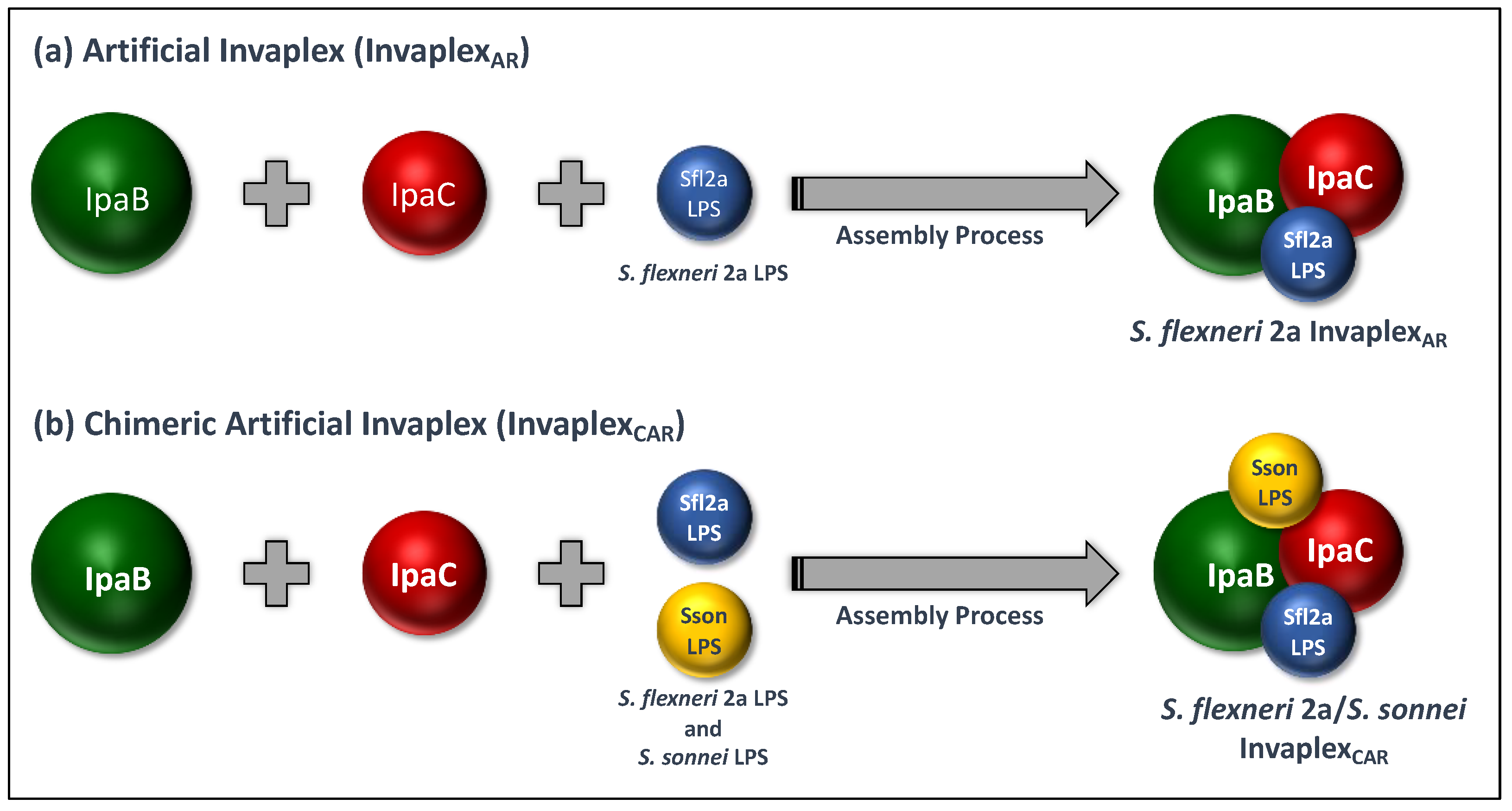
4. Artificial Invaplex (InvaplexAR)

4.1. Development of InvaplexAR

The isolation of the HP Invaplex established the approximate molar ratio of 8 IpaC:1 IpaB in the native complex and a weight ratio of 0.56 for LPS/total protein. Although the exact origin and mechanism by which InvaplexNAT is formed was not known, assembly experiments to produce InvaplexAR followed, in general, the process by which the water extraction was performed to produce InvaplexNAT. The assembly process of InvaplexAR required large quantities of purified IpaB, IpaC and LPS (Table 1). Existing methods to purify each antigen were available but, for the most part, yielded low quantities of the purified component. Therefore, development of improved purification methods for each component were necessary.
The Westphal LPS purification procedure yields a highly pure LPS with representation of long-chained O-SP and minimal protein contamination (1–2% protein) [65]. By increasing the number of extractions, the yield was significantly improved to produce gram quantities of LPS for each kg of cell paste. The modified procedure was applicable to all Shigella species with minor changes and has provided large quantities of LPS not only for InvaplexAR studies but for use as a key immunological reagent for vaccine evaluations used by WRAIR and global collaborators. The improved procedure was transitioned to cGMP manufacture and has been successfully used to produce large quantities of S. flexneri 2a LPS.
Expression of full-size IpaB and IpaC in heterologous hosts such as E. coli was initially described in the mid-1990s [66]. The early recombinants produced low yields of the polyhistidine-tagged protein, and the purified proteins were often characterized by poor solubility, largely due to their inherent hydrophobicity [66,67,68]. Using coexpression of IpaB or IpaC with their cognate chaperone IpgC [68] and maintaining solubility of the purified proteins in mild detergents, working quantities of the proteins were produced, allowing research-scale experimentation on structure and function of the proteins [69,70,71,72].
Initial studies attempting to produce an InvaplexAR used purified LPS from S. flexneri 2a and purified IpaB and his-tagged IpaC produced at WRAIR from recombinants kindly provided by the Picking lab. The first artificial Invaplex was produced in 2003 (Figure 2A) with an IpaC:IpaB molar ratio of approximately 5, contained about 1.2 mg LPS/mg protein and had an approximate mass of 1 MDa. The InvaplexAR was soluble, without any added detergent, and was stable frozen or at room temperature (Table 2). Mice or guinea pigs immunized with InvaplexAR had superior immune responses to IpaB, IpaC and LPS as compared to InvaplexNAT; InvaplexAR induced solid protection in both animal models [53]. This initial research successfully produced InvaplexAR products for S. flexneri 1a, S. flexneri 2a, S. sonnei and S. dysenteriae 1, as well as a construct containing LPS of both S. flexneri 2a and S. sonnei. The Invaplex product containing LPS from two Shigella serotypes complexed with IpaB and IpaC was termed chimeric InvaplexAR or InvaplexCAR (Figure 2B). Although the success of the prototype InvaplexAR was very promising, it was clear that significant improvements in production/purification were necessary prior to cGMP manufacture and transition to clinical evaluation. Critical improvements in the systems used to produce the recombinant IpaB and IpaC were necessary. By using recombinant expression systems with higher plasmid copy numbers, antibiotic selection markers compatible with clinical studies and purification strategies that did not depend on affinity-tagged IpaB or IpaC, significantly increased yields of the target proteins became possible.
Although the prototype InvaplexAR was protective in mice and guinea pigs, studies on the InvaplexAR produced with non-his-tagged IpaC and IpaB sought to maximize immune responses to all antigens, with emphasis on LPS. Enhanced immune responses to all Invaplex antigens were considered key to the vaccine’s effectiveness in humans. By modifying the InvaplexAR assembly and purification protocols to include different amounts and ratios of each reactant/antigen, it was possible to produce an InvaplexAR that contained greater quantities of LPS, achieved an IpaC:IpaB ratio approaching the ratio identified with HP Invaplex, had a high molecular mass and, most importantly, stimulated a solid immune response to all three components. The modified assembly strategy has also been successfully used to produce InvaplexAR for S. flexneri 3a, S. flexneri 6 and S. sonnei (Table 4), which are the leading target serotypes for a multivalent Shigella vaccine [73,74].

4.2. cGMP Manufacture of InvaplexAR

The first InvaplexAR cGMP lot was manufactured at the WRAIR PBF in 2013 (Lot 1835). Prior to the assembly phase of InvaplexAR, cGMP lots of purified S. flexneri 2a LPS, IpaB and IpaC were manufactured using protocols and master cell banks developed at WRAIR. Gram quantities of each antigen were produced and stored frozen. The cGMP lots of IpaB, IpaC and S. flexneri 2a LPS were used to assemble the final InvaplexAR product. Lot 1835 characteristics are outlined in Table 2, which compares multiple generations of cGMP Invaplex lots. Noteworthy characteristics include purity (well-defined composition), immunogenicity, protective efficacy and lack of adverse side effects in small animals.

4.3. Clinical Evaluation of S. flexneri 2a InvaplexAR

The initial Phase 1 trial for InvaplexAR Lot 1835 was conducted in 2015 (Table 3). Volunteers were immunized intranasally with four dose amounts of InvaplexAR (10, 50, 250 and 500 µg) on days 0, 14 and 28 [75]. The primary goals related to safety and effective delivery of the vaccine with the VaxINator™ nasal spray device were achieved. The vaccine was safe and well tolerated at all doses tested, and no AEs met stopping criteria. The most common AEs were nasal congestion, rhinorrhea and postnasal drip. The AE rates were consistent with prior intranasal immunizations with S. flexneri 2a InvaplexNAT [42,43] and resolved within 72 h. Intranasal immunization with ≥50 µg of InvaplexAR induced serum IgG and IgA directed to InvaplexNAT and LPS antigens, with no clear dose response and no significant increase in titer after the third immunization. ASC or antibody in lymphocyte supernatant (ALS) assays detected IgA- and IgG-secreting cells specific to InvaplexNAT after immunization with ≥50 µg of InvaplexAR. Antibodies to LPS and InvaplexNAT were also detected in fecal and saliva samples in 38% to 67% of the volunteers. Functional bactericidal serum antibodies were detected in ≥50% of individuals in the 50 µg and 500 µg dose cohorts. Overall, the phase 1 trial with InvaplexAR showed a safety profile consistent with that of previous studies with InvaplexNAT and demonstrated improved immunogenicity to key Shigella antigens. Despite these encouraging results, a robust immune response was not present in a high proportion of individuals, suggesting that additional adjustments were necessary.

5. Artificial Detoxified Invaplex (InvaplexAR-Detox)

Although the safety profile and immunogenicity of Invaplex vaccines delivered intranasally were encouraging, the potential for combining Invaplex with other licensed vaccines and uptake into existing immunization programs suggested that parenteral immunization may be preferable. Therefore, the current generation of InvaplexAR is formulated with LPS containing an under-acylated lipid A moiety (artificial detoxified Invaplex or InvaplexAR-Detox) instead of fully-acylated lipid A. Transition to the under-acylated LPS widened the vaccine-associated safety window to permit parenteral administration by reducing but not completely abrogating the danger signal associated with endotoxin, resulting in a highly immunogenic vaccine.

5.1. Development and Pre-Clinical Testing of InvaplexAR-Detox

The inflammatory activity of lipid A (endotoxicity) is dependent on interactions with the Toll-like receptor (TLR)-4 complex (MD-2, CD14 and TLR-4). The number and position of acyl chains contribute to binding between lipid A and the TLR-4 complex, with the hexa-acylated lipid A moiety having the highest biological activity. Both penta- and tetraacylated lipid A have been shown to have reduced or attenuated endotoxic activity [76]. Therefore, studies were conducted to evaluate the inflammatory potential of LPS isolated from mutant Shigella strains with single and double deletions of the msbB gene, which is a late acyl transferase. Although LPS from the single msbB mutants were found to be less proinflammatory as compared to fully acylated lipid A, LPS from the double msbB mutant strain (WR30) was utilized for downstream development [77]. Furthermore, when pilot batches of InvaplexAR-Detox assembled with S. flexneri 2a LPS from the various msbB Shigella mutants were utilized in monocyte activation experiments, the level of proinflammatory cytokines (TNF-alpha and IL-6) released was significantly lower as compared to the cytokines released after monocyte incubation with similar amounts of LPS from the mutated Shigella strains, indicating that complexing the LPS with proteins further reduced the proinflammatory potential of the vaccine [78].

5.2. Pre-Clinical Studies with InvaplexAR-Detox

The impact of InvaplexAR-Detox’s exceptional immunogenicity has been demonstrated in several animal models. Guinea pigs immunized parenterally or intranasally with S. flexneri 2a InvaplexAR-Detox were significantly protected in the Sereny model, whereas no animals in the control group were protected. Using a refinement to the guinea pig rectocolitis challenge model [79], animals immunized with InvaplexAR-Detox were also significantly protected from disease (protective efficacy = 67%; p-value = 0.008). In this experiment, myeloperoxidase (MPO) concentrations, an indicator of gut inflammation, were determined in stool samples collected pre-challenge and 1, 2 and 3 days post-challenge. High levels of MPO (≥1500 ng/mL), indicating inflammation in the gut, were detected post-challenge in stool of the saline-treated animals; however, MPO was reduced to negligible levels (≤250 ng/mL) in the animals vaccinated intramuscularly with InvaplexAR-Detox. Collectively, these studies demonstrate that InvaplexAR-Detox was protective and has the capacity to significantly reduce intestinal inflammation, potentially offering added benefits to children in endemic settings because elevated MPO levels have been considered a marker for increased risk for developing stunting and gut enteropathy [80]. The encouraging results from the in vitro and in vivo assessments of S. flexneri 2a InvaplexAR-Detox led to the cGMP manufacture of the product at the WRAIR PBF, which was completed in 2015. A double msbB mutant of wild-type S. flexneri serotype 2a strain 2457T WR30 in which both msbB1 and msbB2 were deleted was used in combination with the same cGMP manufactured lots of IpaB and IpaC used to manufacture InvaplexAR (Lot 1835), as described previously (Table 2).

5.3. Clinical Evaluation of InvaplexAR-Detox

cGMP InvaplexAR-Detox Lot 1972 was evaluated in a first-in-human study during 2019 (Table 3) in which the vaccine was delivered to three cohorts with intramuscular doses of 2.5, 10 and 25 µg delivered on days 0, 21 and 42. The vaccine was well tolerated, with no SAEs and more than 90% of the recorded AEs scored as mild. AEs were dose-dependent and reduced with frequency of administration. The most frequent AEs were pain and tenderness at the injection site and occurred most frequently after the first injection. All three dose levels of InvaplexAR-Detox were highly immunogenic, inducing strong serum antibody responses to all Shigella antigens (LPS, IpaB and IpaC) contained within the vaccine. The magnitude of the LPS-specific serum IgG responses were comparable to or exceeded antibody levels observed after intranasal immunization with InvaplexNAT [42,43] and InvaplexAR [75], infection with S. flexneri 2a or after immunization with other vaccine candidates [6]. In addition to robust systemic responses, the InvaplexAR-Detox vaccine elicited significant mucosal IgG and IgA responses, with an average of 93% of vaccinated subjects demonstrating a mucosal antibody response across all dose levels [78]. The InvaplexAR-Detox vaccine also induced robust serum bactericidal antibodies capable of killing S. flexneri 2a (the targeted organism) that were comparable to or exceeded those induced after infection with wild-type S. flexneri 2a [81]. The high level of functional antibodies induced after vaccination were maintained ≥1.4 years after the primary immunization. More importantly, immunization with InvaplexAR-Detox induced a broad bactericidal antibody response capable of killing other Shigella serotypes that also contribute to global morbidity and mortality (S. flexneri 3a and 6). These findings may be associated with the fact that the InvaplexAR-Detox used in the study was the first parenteral Shigella vaccine that delivered controlled amounts of Ipa proteins, which are broadly conserved and surface-expressed in all Shigella serotypes. InvaplexAR-Detox also induced LPS and Ipa protein-specific immunological memory B cell responses, which are key components of a protective immune response after immunization.

6. Next Steps and Future Directions

The InvaplexAR and InvaplexAR-Detox vaccines represent the first subunit products to deliver controlled amounts of the Ipa proteins and serotype-specific LPS to induce robust immune responses in humans. The customization afforded by the Invaplex approach greatly facilitates expansion to other Shigella serotypes. Moreover, the inherent adjuvant properties of Invaplex also hold the promise of enhancing immune response to codelivered vaccines in the absence of an exogenous adjuvant, paving the way for less expensive combination vaccine approaches.
The excellent safety profile, tolerability and immunogenicity of the InvaplexAR-Detox product warrant further evaluations and development. Several activities on the critical product development pathway will occur in parallel over the coming years. Efforts are underway to cGMP manufacture an S. sonnei InvaplexAR-Detox vaccine. The new product will be evaluated alone and in combination with the S. flexneri 2a InvaplexAR-Detox vaccine to establish safety and preliminary dosing levels of the multivalent vaccine. Given the cross reactivity of functional antibody responses generated in the phase 1 study with S. flexneri 2a InvaplexAR-Detox, it may be possible to provide sufficient coverage against the four major Shigella serotypes with a bivalent InvaplexAR-Detox product. In parallel, further efforts towards a stable InvaplexAR-Detox formulation that will not be as dependent on the cold chain are underway. It is also anticipated that the InvaplexAR-Detox product will enter age-descending and dose-finding studies in adults, children and infants living in endemic settings to determine immunogenicity in target populations.

Author Contributions

K.R.T., K.A.C., E.V.O. and R.W.K. contributed equally to writing—original draft preparation, writing—review and editing and visualization of the manuscript. All authors have read and agreed to the published version of the manuscript.

Funding

This research received no external funding.

Institutional Review Board Statement

The studies summarized in this manuscript were conducted according to the guidelines of the Declaration of Helsinki and approved by the Institutional Review Board of WRAIR.

Informed Consent Statement

Not applicable.

Data Availability Statement

Not applicable.

Acknowledgments

The Department of Defense Diarrheal Disease Program is a joint program between Army and Navy personnel that has successfully evaluated the safety, immunogenicity and efficacy of enteric vaccine candidates, including all Invaplex products. Much of the work described above is a direct reflection on the excellence and dedication of the Army/Navy collaboration. Additionally, the WRAIR Pilot Bioproduction Facility significantly contributed to the successes of the Invaplex vaccine program through their expertise, experience and dedication to the GMP manufacture of all Invaplex products described in the manuscript. Finally, the authors would like to thank A. Lou Bourgeois for his critical revisions to the manuscript and mentorship to RWK and KAC.

Conflicts of Interest

The authors declare no conflict of interest.

Disclaimer

Some authors (K.R. Turbyfill, K.A. Clarkson, R.W. Kaminski) are employees of the U.S. Government. Material has been reviewed by the Walter Reed Army Institute of Research. There is no objection to its presentation and/or publication. The opinions or assertions contained herein are the private views of the author and are not to be construed as official or as reflecting true views of the Department of the Army, nor the Department of Defense, nor the U.S. Government. The investigators have adhered to the policies for protection of human subjects as prescribed in AR 70–25.

Copyright Statement

Some authors (K.R. Turbyfill, K.A. Clarkson, R.W. Kaminski) are employees of the U.S. Government. This work was prepared as part of their official duties. Title 17 U.S.C. §105 provides that “Copyright protection under this title is not available for any work of the United States Government.” Title 17 U.S.C. §101 defines a U.S. Government work as a work prepared by a military service member or employee of the U.S. Government as part of that person’s official duties.

References

  1. Ferreccio, C.; Prado, V.; Ojeda, A.; Cayyazo, M.; Abrego, P.; Guers, L.; Levine, M.M. Epidemiologic patterns of acute diarrhea and endemic Shigella infections in children in a poor periurban setting in Santiago, Chile. Am. J. Epidemiol. 1991, 134, 614–627. [Google Scholar] [CrossRef] [PubMed] [Green Version]
  2. Formal, S.B.; Oaks, E.V.; Olsen, R.E.; Wingfield-Eggleston, M.; Snoy, P.J.; Cogan, J.P. Effect of prior infection with virulent Shigella flexneri 2a on the resistance of monkeys to subsequent infection with Shigella sonnei. J. Infect. Dis. 1991, 164, 533–537. [Google Scholar] [CrossRef] [PubMed]
  3. Cohen, D.; Ashkenazi, S.; Green, M.S.; Gdalevich, M.; Robin, G.; Slepon, R.; Yavzori, M.; Orr, N.; Block, C.; Ashkenazi, I.; et al. Double-blind vaccine-controlled randomised efficacy trial of an investigational Shigella sonnei conjugate vaccine in young adults. Lancet 1997, 349, 155–159. [Google Scholar] [CrossRef]
  4. Taylor, D.N.; Trofa, A.C.; Sadoff, J.; Chu, C.; Bryla, D.; Shiloach, J.; Cohen, D.; Ashkenazi, S.; Lerman, Y.; Egan, W.; et al. Synthesis, characterization, and clinical evaluation of conjugate vaccines composed of the O-specific polysaccharides of Shigella dysenteriae type 1, Shigella flexneri type 2a, and Shigella sonnei (Plesiomonas shigelloides) bound to bacterial toxoids. Infect. Immun. 1993, 61, 3678–3687. [Google Scholar] [CrossRef] [Green Version]
  5. Farzam, N.; Ramon-Saraf, R.; Banet-Levi, Y.; Lerner-Geva, L.; Ashkenazi, S.; Kubler-Kielb, J.; Vinogradov, E.; Robbins, J.B.; Schneerson, R. Vaccination with Shigella flexneri 2a conjugate induces type 2a and cross-reactive type 6 antibodies in humans but not in mice. Vaccine 2017, 35, 4990–4996. [Google Scholar] [CrossRef]
  6. Talaat, K.R.; Alaimo, C.; Martin, P.; Bourgeois, A.L.; Dreyer, A.M.; Kaminski, R.W.; Porter, C.K.; Chakraborty, S.; Clarkson, K.A.; Brubaker, J.; et al. Human challenge study with a Shigella bioconjugate vaccine: Analyses of clinical efficacy and correlate of protection. EBioMedicine 2021, 66, 103310. [Google Scholar] [CrossRef]
  7. Riddle, M.S.; Kaminski, R.W.; Di Paolo, C.; Porter, C.K.; Gutierrez, R.L.; Clarkson, K.A.; Weerts, H.E.; Duplessis, C.; Castellano, A.; Alaimo, C.; et al. Safety and Immunogenicity of a Candidate Bioconjugate Vaccine against Shigella flexneri 2a Administered to Healthy Adults: A Single-Blind, Randomized Phase I Study. Clin. Vaccine Immunol. 2016, 23, 908–917. [Google Scholar] [CrossRef] [Green Version]
  8. Oaks, E.V.; Hale, T.L.; Formal, S.B. Serum immune response to Shigella protein antigens in rhesus monkeys and humans infected with Shigella spp. Infect. Immun. 1986, 53, 57–63. [Google Scholar] [CrossRef] [Green Version]
  9. Coster, T.S.; Hoge, C.W.; VanDeVerg, L.L.; Hartman, A.B.; Oaks, E.V.; Venkatesan, M.M.; Cohen, D.; Robin, G.; Fontaine-Thompson, A.; Sansonetti, P.J.; et al. Vaccination against shigellosis with attenuated Shigella flexneri 2a strain SC602. Infect. Immun. 1999, 67, 3437–3443. [Google Scholar] [CrossRef] [Green Version]
  10. Kotloff, K.L.; Taylor, D.N.; Sztein, M.B.; Wasserman, S.S.; Losonsky, G.A.; Nataro, J.P.; Venkatesan, M.; Hartman, A.; Picking, W.D.; Katz, D.E.; et al. Phase I evaluation of delta virG Shigella sonnei live, attenuated, oral vaccine strain WRSS1 in healthy adults. Infect. Immun. 2002, 70, 2016–2021. [Google Scholar] [CrossRef] [Green Version]
  11. Kotloff, K.L.; Noriega, F.; Losonsky, G.A.; Sztein, M.B.; Wasserman, S.S.; Nataro, J.P.; Levine, M.M. Safety, immunogenicity, and transmissibility in humans of CVD 1203, a live oral Shigella flexneri 2a vaccine candidate attenuated by deletions in aroA and virG. Infect. Immun. 1996, 64, 4542–4548. [Google Scholar] [CrossRef] [PubMed] [Green Version]
  12. Oaks, E.; Kaminski, R. Use of Shigella Invaplex to Transport Functional Proteins and Transcriptionally Active Nucleic Acids Across Mammalian Cell Membranes In Vitro and In Vivo. U.S. Patent 7,632,659, 15 December 2009. [Google Scholar]
  13. Oaks, E.; Kaminski, R. Use of Shigella Invaplex to Transport Functional Proteins and Transcriptionally Active Nucleic Acids Across Mammalian Cell Membranes In Vitro and In Vivo. U.S. Patent 8,110,354, 7 February 2012. [Google Scholar]
  14. Van de Verg, L.L.; Herrington, D.A.; Boslego, J.; Lindberg, A.A.; Levine, M.M. Age-specific prevalence of serum antibodies to the invasion plasmid and lipopolysaccharide antigens of Shigella species in Chilean and North American populations. J. Infect. Dis. 1992, 166, 158–161. [Google Scholar] [CrossRef] [PubMed]
  15. Camp, J.V.; Wilson, R.L.; Singletary, M.; Blanchard, J.L.; Aldovini, A.; Kaminski, R.W.; Oaks, E.V.; Kozlowski, P.A. Invaplex functions as an intranasal adjuvant for subunit and DNA vaccines co-delivered in the nasal cavity of nonhuman primates. Vaccine X 2021, 8, 100105. [Google Scholar] [CrossRef] [PubMed]
  16. Mills, J.A.; Buysse, J.M.; Oaks, E.V. Shigella flexneri invasion plasmid antigens B and C: Epitope location and characterization with monoclonal antibodies. Infect. Immun. 1988, 56, 2933–2941. [Google Scholar] [CrossRef] [PubMed] [Green Version]
  17. Menard, R.; Sansonetti, P.; Parsot, C. The secretion of the Shigella flexneri Ipa invasins is activated by epithelial cells and controlled by IpaB and IpaD. EMBO J. 1994, 13, 5293–5302. [Google Scholar] [CrossRef] [PubMed]
  18. Menard, R.; Prevost, M.C.; Gounon, P.; Sansonetti, P.; Dehio, C. The secreted Ipa complex of Shigella flexneri promotes entry into mammalian cells. Proc. Natl. Acad. Sci. USA 1996, 93, 1254–1258. [Google Scholar] [CrossRef] [PubMed] [Green Version]
  19. Watarai, M.; Tobe, T.; Yoshikawa, M.; Sasakawa, C. Contact of Shigella with host cells triggers release of Ipa invasins and is an essential function of invasiveness. EMBO J. 1995, 14, 2461–2470. [Google Scholar] [CrossRef] [PubMed]
  20. Li, A.; Rong, Z.C.; Ekwall, E.; Forsum, U.; Lindberg, A.A. Serum antibody responses against shigella lipopolysaccharides and invasion plasmid-coded antigens in shigella infected Swedish patients. Scand. J. Infect. Dis. 1993, 25, 569–577. [Google Scholar] [CrossRef] [PubMed]
  21. Samandari, T.; Kotloff, K.L.; Losonsky, G.A.; Picking, W.D.; Sansonetti, P.J.; Levine, M.M.; Sztein, M.B. Production of IFN-gamma and IL-10 to Shigella invasins by mononuclear cells from volunteers orally inoculated with a Shiga toxin-deleted Shigella dysenteriae type 1 strain. J. Immunol. 2000, 164, 2221–2232. [Google Scholar] [CrossRef] [Green Version]
  22. Cohen, D.; Meron-Sudai, S.; Bialik, A.; Asato, V.; Goren, S.; Ariel-Cohen, O.; Reizis, A.; Hochberg, A.; Ashkenazi, S. Serum IgG antibodies to Shigella lipopolysaccharide antigens—A correlate of protection against shigellosis. Hum. Vaccines Immunother. 2019, 15, 1401–1408. [Google Scholar] [CrossRef] [PubMed] [Green Version]
  23. Turbyfill, K.R.; Oaks, E.; (Walter Reed Army Institute of Research, Silver Spring, MD, USA). Unpublished Observations. 1996.
  24. Turbyfill, K.R.; Hartman, A.B.; Oaks, E.V. Isolation and characterization of a Shigella flexneri invasin complex subunit vaccine. Infect. Immun. 2000, 68, 6624–6632. [Google Scholar] [CrossRef] [PubMed] [Green Version]
  25. Oaks, E.; Turbyfill, K.R.; Hartman, A. Use of Purified Invaplex from Gram Negative Bacteria as a Vaccine. U.S. Patent 6,277,379, 21 August 2001. [Google Scholar]
  26. Oaks, E.V.; Turbyfill, K.R. Development and evaluation of a Shigella flexneri 2a and S. sonnei bivalent invasin complex (Invaplex) vaccine. Vaccine 2006, 24, 2290–2301. [Google Scholar] [CrossRef] [PubMed]
  27. Oaks, E.; Turbyfill, K.R.; (Walter Reed Army Institute of Research, Silver Spring, MD, USA). Unpublished Observations. 2011.
  28. Menard, R.; Sansonetti, P.J.; Parsot, C. Nonpolar mutagenesis of the ipa genes defines IpaB, IpaC, and IpaD as effectors of Shigella flexneri entry into epithelial cells. J. Bacteriol. 1993, 175, 5899–5906. [Google Scholar] [CrossRef] [PubMed] [Green Version]
  29. Kaminski, R.W.; Turbyfill, K.R.; Oaks, E.V. Mucosal adjuvant properties of the Shigella invasin complex. Infect. Immun. 2006, 74, 2856–2866. [Google Scholar] [CrossRef] [PubMed] [Green Version]
  30. Kaminski, R.W.; Turbyfill, K.R.; Chao, C.; Ching, W.M.; Oaks, E.V. Mucosal adjuvanticity of a Shigella invasin complex with dna-based vaccines. Clin. Vaccine Immunol. 2009, 16, 574–586. [Google Scholar] [CrossRef] [PubMed] [Green Version]
  31. Oaks, E.; Turbyfill, K.R. Invaplex from Gram Negative Bacteria, Method of Purification and Methods of Use. U.S. Patent 6,245,892, 12 June 2001. [Google Scholar]
  32. Oaks, E.; Turbyfill, K.R. Invaplex from Gram Negative Bacteria, Method of Purification and Methods of Use. U.S. Patent 6,680,374, 20 January 2004. [Google Scholar]
  33. Clarkson, K.A.; Frenck, R.W., Jr.; Dickey, M.; Suvarnapunya, A.E.; Chandrasekaran, L.; Weerts, H.P.; Heaney, C.D.; McNeal, M.; Detizio, K.; Parker, S.; et al. Immune Response Characterization after Controlled Infection with Lyophilized Shigella sonnei 53G. mSphere 2020, 5, e00988-19. [Google Scholar] [CrossRef] [PubMed]
  34. Ndungo, E.; Randall, A.; Hazen, T.H.; Kania, D.A.; Trappl-Kimmons, K.; Liang, X.; Barry, E.M.; Kotloff, K.L.; Chakraborty, S.; Mani, S.; et al. A Novel Shigella Proteome Microarray Discriminates Targets of Human Antibody Reactivity following Oral Vaccination and Experimental Challenge. mSphere 2018, 3, e00260-18. [Google Scholar] [CrossRef] [Green Version]
  35. Kotloff, K.L.; Sztein, M.B.; Wasserman, S.S.; Losonsky, G.A.; DiLorenzo, S.C.; Walker, R.I. Safety and immunogenicity of oral inactivated whole-cell Helicobacter pylori vaccine with adjuvant among volunteers with or without subclinical infection. Infect. Immun. 2001, 69, 3581–3590. [Google Scholar] [CrossRef] [PubMed] [Green Version]
  36. Mestecky, J. The common mucosal immune system and current strategies for induction of immune responses in external secretions. J. Clin. Immunol. 1987, 7, 265–276. [Google Scholar] [CrossRef] [PubMed]
  37. Moldoveanu, Z.; Russell, M.W.; Wu, H.Y.; Huang, W.Q.; Compans, R.W.; Mestecky, J. Compartmentalization within the common mucosal immune system. Adv. Exp. Med. Biol. 1995, 371A, 97–101. [Google Scholar] [CrossRef]
  38. Kozlowski, P.A.; Williams, S.B.; Lynch, R.M.; Flanigan, T.P.; Patterson, R.R.; Cu-Uvin, S.; Neutra, M.R. Differential induction of mucosal and systemic antibody responses in women after nasal, rectal, or vaginal immunization: Influence of the menstrual cycle. J. Immunol. 2002, 169, 566–574. [Google Scholar] [CrossRef] [PubMed]
  39. Fries, L.F.; Montemarano, A.D.; Mallett, C.P.; Taylor, D.N.; Hale, T.L.; Lowell, G.H. Safety and immunogenicity of a proteosome-Shigella flexneri 2a lipopolysaccharide vaccine administered intranasally to healthy adults. Infect. Immun. 2001, 69, 4545–4553. [Google Scholar] [CrossRef] [PubMed] [Green Version]
  40. Adamus, G.; Mulczyk, M.; Witkowska, D.; Romanowska, E. Protection against keratoconjunctivitis shigellosa induced by immunization with outer membrane proteins of Shigella spp. Infect. Immun. 1980, 30, 321–324. [Google Scholar] [CrossRef] [PubMed]
  41. Mallett, C.P.; Hale, T.L.; Kaminski, R.W.; Larsen, T.; Orr, N.; Cohen, D.; Lowell, G.H. Intransal or intragastric immunization with proteosome-Shigella lipopolysaccharide vaccines protects against lethal pneumonia in a murine model of Shigella infection. Infect. Immun. 1995, 63, 2382–2386. [Google Scholar] [CrossRef] [Green Version]
  42. Tribble, D.; Kaminski, R.; Cantrell, J.; Nelson, M.; Porter, C.; Baqar, S.; Williams, C.; Arora, R.; Saunders, J.; Ananthakrishnan, M.; et al. Safety and immunogenicity of a Shigella flexneri 2a Invaplex 50 intranasal vaccine in adult volunteers. Vaccine 2010, 28, 6076–6085. [Google Scholar] [CrossRef]
  43. Riddle, M.S.; Kaminski, R.W.; Williams, C.; Porter, C.; Baqar, S.; Kordis, A.; Gilliland, T.; Lapa, J.; Coughlin, M.; Soltis, C.; et al. Safety and immunogenicity of an intranasal Shigella flexneri 2a Invaplex 50 vaccine. Vaccine 2011, 29, 7009–7019. [Google Scholar] [CrossRef] [Green Version]
  44. Weerts, H.P.; Yu, J.; Kaminski, R.W.; Nahm, M.H. A High-throughput Shigella-specific Bactericidal Assay. J. Vis. Exp. 2019, 144, e59164. [Google Scholar] [CrossRef] [PubMed] [Green Version]
  45. Nahm, M.H.; Yu, J.; Weerts, H.P.; Wenzel, H.; Tamilselvi, C.S.; Chandrasekaran, L.; Pasetti, M.F.; Mani, S.; Kaminski, R.W. Development, Interlaboratory Evaluations, and Application of a Simple, High-Throughput Shigella Serum Bactericidal Assay. mSphere 2018, 3, e00146-18. [Google Scholar] [CrossRef] [PubMed] [Green Version]
  46. Rossi, O.; Molesti, E.; Saul, A.; Giannelli, C.; Micoli, F.; Necchi, F. Intra-Laboratory Evaluation of Luminescence Based High-Throughput Serum Bactericidal Assay (L-SBA) to Determine Bactericidal Activity of Human Sera against Shigella. High Throughput 2020, 9, 14. [Google Scholar] [CrossRef]
  47. Oaks, E.; (Walter Reed Army Institute of Research, Silver Spring, MD, USA). Unpublished Observations. 2004.
  48. Frenck, R.W., Jr.; Baqar, S.; Alexander, W.; Dickey, M.; McNeal, M.; El-Khorazaty, J.; Baughman, H.; Hoeper, A.; Barnoy, S.; Suvarnapunya, A.E.; et al. A Phase I trial to evaluate the safety and immunogenicity of WRSs2 and WRSs3; two live oral candidate vaccines against Shigella sonnei. Vaccine 2018, 36, 4880–4889. [Google Scholar] [CrossRef]
  49. Turbyfill, K.R.; Kaminski, R.W.; Oaks, E.V. Immunogenicity and efficacy of highly purified invasin complex vaccine from Shigella flexneri 2a. Vaccine 2008, 26, 1353–1364. [Google Scholar] [CrossRef] [PubMed]
  50. Yao, R.; Palchaudhuri, S. Nucleotide sequence of the ipaBCD structural genes of Shigella dysenteriae. Mol. Microbiol. 1991, 5, 2217–2221. [Google Scholar] [CrossRef] [PubMed]
  51. Pierson, T.; Islam, D.; Odundo, E.; Kaminski, R.; Hang, J.; Ruamsap, N.; Sakpaisal, P.; Silapong, S.; Oransathid, W.; Lertsethtakarn, P.; et al. Shigella IpaB/C/D Invasion Plasmid Sequence Conservation in a Wide Range of Clinical Isolates. In Proceedings of the Military Health System Research Symposium, Fort Lauderdale, FL, USA, 18 August 2014. [Google Scholar]
  52. Oaks, E.; Turbyfill, K.R. Heterologous Protection Induced by Immunization with Invaplex Vaccine. U.S. Patent 7,258,863, 21 August 2007. [Google Scholar]
  53. Turbyfill, K.R.; Clarkson, K.A.; Vortherms, A.R.; Oaks, E.V.; Kaminski, R.W. Assembly, Biochemical Characterization, Immunogenicity, Adjuvanticity, and Efficacy of Shigella Artificial Invaplex. mSphere 2018, 3, e00583-17. [Google Scholar] [CrossRef] [PubMed] [Green Version]
  54. Chitradevi, S.T.; Kaur, G.; Uppalapati, S.; Yadav, A.; Singh, D.; Bansal, A. Co-administration of rIpaB domain of Shigella with rGroEL of S. Typhi enhances the immune responses and protective efficacy against Shigella infection. Cell. Mol. Immunol. 2015, 12, 757–767. [Google Scholar] [CrossRef] [PubMed] [Green Version]
  55. Martinez-Becerra, F.J.; Chen, X.; Dickenson, N.E.; Choudhari, S.P.; Harrison, K.; Clements, J.D.; Picking, W.D.; Van De Verg, L.L.; Walker, R.I.; Picking, W.L. Characterization of a novel fusion protein from IpaB and IpaD of Shigella spp. and its potential as a pan-Shigella vaccine. Infect. Immun. 2013, 81, 4470–4477. [Google Scholar] [CrossRef] [PubMed] [Green Version]
  56. Martinez-Becerra, F.J.; Kissmann, J.M.; Diaz-McNair, J.; Choudhari, S.P.; Quick, A.M.; Mellado-Sanchez, G.; Clements, J.D.; Pasetti, M.F.; Picking, W.L. Broadly protective Shigella vaccine based on type III secretion apparatus proteins. Infect. Immun. 2012, 80, 1222–1231. [Google Scholar] [CrossRef] [PubMed] [Green Version]
  57. Kaminski, R.; (Walter Reed Army Institute of Research, Silver Spring, MD, USA). Unpublished Observations. 2018.
  58. Turbyfill, K.R.; Oaks, E.; (Walter Reed Army Institute of Research, Silver Spring, MD, USA). Unpublished Observations. 2001.
  59. Turbyfill, K.R.; Karwoski, J.; Pradier, A.; Ash, L.; Kaminski, R.; Oaks, E. Fermentation conditions for production of large quantities of the subcellular invasin complex (Invaplex) vaccine from Shigella flexneri. In Proceedings of the American Society for Microbiology, Annual Meeting, Orlando, FL, USA, 21–25 May 2006. [Google Scholar]
  60. Harro, C.; Riddle, M.S.; Kaminski, R.; Turbyfill, K.R.; Gormley, R.; Porter, C.; Ranallo, R.T.; Kordis, A.; Buck, M.; Jones, A.; et al. Shigella flexneri 2a Invaplex 50 intranasal vaccine phase 2b challenge study. In Proceedings of the Vaccines for Enteric Diseases (VED), Malaga, Spain, 9–11 September 2009. [Google Scholar]
  61. Turbyfill, K.R.; Oaks, E.; (Walter Reed Army Institute of Research, Silver Spring, MD, USA). Unpublished Observations. 2006.
  62. Hartman, A.B.; Powell, C.J.; Schultz, C.L.; Oaks, E.V.; Eckels, K.H. Small-animal model to measure efficacy and immunogenicity of Shigella vaccine strains. Infect. Immun. 1991, 59, 4075–4083. [Google Scholar] [CrossRef] [PubMed] [Green Version]
  63. Clarkson, K.A.; Duplessis, C.; Turbyfill, K.R.; Porter, C.; Gutierrez, R.L.; Riddle, M.S.; Lee, T.; Weerts, H.; Sumlin, S.C.; Lynen, A.; et al. A Phase 1 Open-label, Dose Escalating Study of Shigella flexneri 2a Artificial Invaplex administered intranasally to healthy, adult volunteers. In Proceedings of the Vaccines for Enteric Diseases (VED), Albufeira, Portugal, 9–11 October 2017. [Google Scholar]
  64. Clarkson, K.A.; Gutierrez, R.L.; Turbyfill, K.R.; Vortherms, A.R.; Lynen, A.; Barnard, B.A.; Weerts, H.; Porter, C.; Maier, N.; Erdem, R.; et al. Clinical Evaluation of Shigella flexneri 2a Detoxified Artificial Invaplex. In Proceedings of the Vaccines Against Shigella and ETEC (VASE), Virtual Symposium, 28–30 September 2021. [Google Scholar]
  65. Westphal, O.; Jann, K. Bacterial Lipopolysaccharides: Extraction with Phenol-Water and Further Applications of the Procedure; Academic Press: Cambridge, MA, USA, 1965. [Google Scholar]
  66. Picking, W.L.; Mertz, J.A.; Marquart, M.E.; Picking, W.D. Cloning, expression, and affinity purification of recombinant Shigella flexneri invasion plasmid antigens IpaB and IpaC. Protein Expr. Purif. 1996, 8, 401–408. [Google Scholar] [CrossRef] [PubMed]
  67. Birket, S.E.; Harrington, A.T.; Espina, M.; Smith, N.D.; Terry, C.M.; Darboe, N.; Markham, A.P.; Middaugh, C.R.; Picking, W.L.; Picking, W.D. Preparation and characterization of translocator/chaperone complexes and their component proteins from Shigella flexneri. Biochemistry 2007, 46, 8128–8137. [Google Scholar] [CrossRef] [PubMed]
  68. Menard, R.; Sansonetti, P.; Parsot, C.; Vasselon, T. Extracellular association and cytoplasmic partitioning of the IpaB and IpaC invasins of S. flexneri. Cell 1994, 79, 515–525. [Google Scholar] [CrossRef]
  69. Dickenson, N.E.; Choudhari, S.P.; Adam, P.R.; Kramer, R.M.; Joshi, S.B.; Middaugh, C.R.; Picking, W.L.; Picking, W.D. Oligomeric states of the Shigella translocator protein IpaB provide structural insights into formation of the type III secretion translocon. Protein Sci. 2013, 22, 614–627. [Google Scholar] [CrossRef] [PubMed] [Green Version]
  70. Hume, P.J.; McGhie, E.J.; Hayward, R.D.; Koronakis, V. The purified Shigella IpaB and Salmonella SipB translocators share biochemical properties and membrane topology. Mol. Microbiol. 2003, 49, 425–439. [Google Scholar] [CrossRef] [PubMed]
  71. Mounier, J.; Boncompain, G.; Senerovic, L.; Lagache, T.; Chretien, F.; Perez, F.; Kolbe, M.; Olivo-Marin, J.C.; Sansonetti, P.J.; Sauvonnet, N. Shigella effector IpaB-induced cholesterol relocation disrupts the Golgi complex and recycling network to inhibit host cell secretion. Cell Host Microbe 2012, 12, 381–389. [Google Scholar] [CrossRef] [PubMed] [Green Version]
  72. Senerovic, L.; Tsunoda, S.P.; Goosmann, C.; Brinkmann, V.; Zychlinsky, A.; Meissner, F.; Kolbe, M. Spontaneous formation of IpaB ion channels in host cell membranes reveals how Shigella induces pyroptosis in macrophages. Cell Death Dis. 2012, 3, e384. [Google Scholar] [CrossRef] [PubMed]
  73. Livio, S.; Strockbine, N.A.; Panchalingam, S.; Tennant, S.M.; Barry, E.M.; Marohn, M.E.; Antonio, M.; Hossain, A.; Mandomando, I.; Ochieng, J.B.; et al. Shigella isolates from the global enteric multicenter study inform vaccine development. Clin. Infect. Dis. 2014, 59, 933–941. [Google Scholar] [CrossRef] [PubMed]
  74. Turbyfill, K.R.; Clarkson, K.A.; Vortherms, A.R.; Oaks, E.; Kaminski, R.; (Walter Reed Army Institute of Research, Silver Spring, MD, USA). Manuscript in Preparation. 2022.
  75. Clarkson, K.A.; Duplessis, C.; Turbyfill, K.R.; Porter, C.; Weerts, H.; Gutierrez, R.L.; Riddle, M.S.; Lynen, A.; Oaks, E.; Kaminski, R.; Walter Reed Army Institute of Research, Silver Spring, MD, USA. A Phase 1 Open-label, Dose Escalating Study of Shigella flexneri 2a Artificial Invaplex (InvaplexAR) Administered Intranasally to Healthy, Adult Volunteers. 2022; Manuscript in Preparation. [Google Scholar]
  76. D’Hauteville, H.; Khan, S.; Maskell, D.J.; Kussak, A.; Weintraub, A.; Mathison, J.; Ulevitch, R.J.; Wuscher, N.; Parsot, C.; Sansonetti, P.J. Two msbB genes encoding maximal acylation of lipid A are required for invasive Shigella flexneri to mediate inflammatory rupture and destruction of the intestinal epithelium. J. Immunol. 2002, 168, 5240–5251. [Google Scholar] [CrossRef] [PubMed] [Green Version]
  77. Ranallo, R.T.; Kaminski, R.W.; George, T.; Kordis, A.A.; Chen, Q.; Szabo, K.; Venkatesan, M.M. Virulence, inflammatory potential, and adaptive immunity induced by Shigella flexneri msbB mutants. Infect. Immun. 2010, 78, 400–412. [Google Scholar] [CrossRef] [PubMed] [Green Version]
  78. Clarkson, K.A.; Gutierrez, R.L.; Turbyfill, K.R.; Detizio, K.; Vortherms, A.R.; Lynen, A.; Barnard, B.A.; Weerts, H.; Porter, C.; Maier, N.; et al.; Walter Reed Army Institute of Research, Silver Spring, MD, USA GMP manufacture, characterization and clinical evaluation of Shigella flexneri 2a detoxified artificial invaplex. 2022; Manuscript in Preparation. [Google Scholar]
  79. Shim, D.H.; Suzuki, T.; Chang, S.Y.; Park, S.M.; Sansonetti, P.J.; Sasakawa, C.; Kweon, M.N. New animal model of shigellosis in the Guinea pig: Its usefulness for protective efficacy studies. J. Immunol. 2007, 178, 2476–2482. [Google Scholar] [CrossRef] [PubMed] [Green Version]
  80. Guerrant, R.L.; Bolick, D.T.; Swann, J.R. Modeling Enteropathy or Diarrhea with the Top Bacterial and Protozoal Pathogens: Differential Determinants of Outcomes. ACS Infect. Dis. 2021, 7, 1020–1031. [Google Scholar] [CrossRef]
  81. Clarkson, K.A.; Talaat, K.R.; Alaimo, C.; Martin, P.; Bourgeois, A.L.; Dreyer, A.; Porter, C.K.; Chakraborty, S.; Brubaker, J.; Elwood, D.; et al. Immune response characterization in a human challenge study with a Shigella flexneri 2a bioconjugate vaccine. EBioMedicine 2021, 66, 103308. [Google Scholar] [CrossRef] [PubMed]
Figure 1. Milestones of Invaplex vaccine development: timeline of key discoveries, patents, cGMP manufactures, and clinical studies associated with the development and evolution of the Invaplex product.
Vaccines 10 00548 g001
Figure 2. Assembly of artificial Invaplex and chimeric artificial Invaplex: (a) artificial Invaplex is assembled in a self-assembly reaction between IpaB, IpaC and LPS in a controlled manner that facilitates customization and quantities of the key immunogens contained within the complex; (b) multivalent Invaplex vaccines can be produced using admixtures of artificial Invaplex specific for different serotypes or manufactured as a “chimera” with LPS from multiple Shigella serotypes (example shown is a bivalent, chimeric Invaplex) with the complex containing IpaB, IpaC and LPS from more than one serotype.
Vaccines 10 00548 g002
Table 1. Summary of the Invaplex product family.
Attribute/Property1st Generation2nd Generation3rd Generation
NameNative Invaplex
(InvaplexNAT)
Artificial Invaplex
(InvaplexAR)
Detoxified Artificial Invaplex
(InvaplexAR-Detox)
Date of Discovery199620032013
SourceIsolated from wild-type shigellaeAssembled from recombinant proteins and wild-type LPSAssembled from recombinant proteins and under-acylated LPS
Complex Composition
  • IpaB
  • IpaC
  • IpaD
  • Wild-type LPS
  • Unidentified proteins
  • IpaB
  • IpaC
  • Wild-type LPS
  • IpaB
  • IpaC
  • Under-acylated LPS
First cGMP Manufacture Date200020132016
Adjuvant Activity
Induces Uptake
(Non-Phagocytic Cells)
Produced for Multiple
Shigella Serotypes
S. flexneri 2a
S. sonnei
S. dysenteriae
S. boydii
S. flexneri 2a
S. flexneri 3a
S. flexneri 6
S. flexneri 1b
S. dysenteriae
S. sonnei
S. flexneri 2a
S. sonnei
Route of ImmunizationIntranasalIntranasalParenteral
Efficacious in Mice
Efficacious in Guinea Pigs
Heterologous Protection (Mice)Not Tested
Table 2. Comparison of cGMP S. flexneri 2a Invaplex products.
AnalysisInvaplexNAT
(cGMP Lot 1307)
InvaplexAR
(cGMP Lot 1835)
InvaplexAR-Detox
(cGMP Lot 1972)
Endotoxin (EU/mL) 10.6 × 1075.3 × 1074.6 × 107
Protein Concentration (mg/mL) 23.52.92.8
IpaC: IpaB Ratio 31.5:16.8:15.4:1
LPS: Total Protein Ratio (EU/mg)0.17 × 1071.8 × 1071.6 × 107
SEC Retention Time (mins) 417.4
Multiple Peaks
15.7
Single Peak
16.2
Single Peak
Size, DH (nm ± standard deviation) 510 ± 324 ± 324 ± 4
Immunogenicity 62036 (100%)
p < 0.001
8146 (100%)
p < 0.0001
146,295 (100%)
p < 0.0001
Protective Efficacy 783%
p < 0.001
100%
p < 0.0001
75%
p = 0.0003
GLP ToxicologyNo significant histopathology in mice after
IN immunization
No significant histopathology in mice after
IN immunization
No significant adverse findings in rabbits after
IM immunization
PyrogenicityNegative in rabbits
(IN immunization)
Negative in rabbits
(IN immunization)
Monocyte Activation
Test—PASS
Stability at −80 °C (no loss of antigen content or immunogenicity)≥3 years≥5 years≥5 years
Stage of Clinical DevelopmentPhase 1: Safe and immunogenic
P2b: Less immunogenic and not protective
Phase 1: Safe and immunogenic Phase 1: Safe and highly immunogenic
Lowest Immunogenic Dose for ≥50% Seroconversion in HumansAnti-InvaplexNAT
IgG
690 µg50 µg 2.5 µg
Anti-LPS IgG690 µg250 µg2.5 µg
1 Endotoxin was measured by Limulus amebocyte lysate assay. 2 Final total protein concentration was determined by Pierce BCA total protein assay against a BSA standard curve. 3 The IpaC:IpaB ratio was determined by quantitative ELISA for Lot 1307. For Lots 1835 and 1972, IpaC:IpaB ratios were determined by scanning Coomassie-stained SDS-PAGE gels using a Bio-Rad GS900 calibrated flatbed scanner, and the pixel densitometric ratios were calculated using Bio-Rad Image Lab software. 4 Size-exclusion chromatography (SEC-HPLC) was performed using a TSK-GEL G5000PWXL 7.8 mm × 30 cm column (TOSOH Bioscience) with a 10 µm particle size and an exclusion limit of 1 × 10⁷ Daltons connected to a Dionex UltiMate™ 3000 UHPLC with a diode array detector reading at a 215 nm wavelength. 5 Dynamic light scattering (DLS) measurements of hydrodynamic diameter (DH) in nm were performed using a Malvern zetasizer µV DLS/SLS detector. 6 Guinea pigs were immunized intranasally or intramuscularly three times at two-week intervals (days 0, 14, 28) with either 25 µg of the cGMP Invaplex product or saline (not shown). Blood was collected at baseline (day 0) and two weeks after the last immunization (day 42) to determine vaccine-induced serum IgG responses by ELISA. Outlined above are InvaplexNAT-specific geometric mean serum IgG ELISA endpoint titers on day 42 with the percentage of responders in parentheses (defined as a ≥4-fold rise over baseline). p-values were determined by two-way ANOVA with Bonferroni post hoc test comparing the immunized group to the saline control group. 7 Guinea pigs were ocularly challenged 3 weeks after the last immunization with ~2 × 10⁸ cfu/eye of S. flexneri 2a, 2457T. The eyes of each animal were monitored daily for inflammation and keratoconjunctivitis. Disease was graded as previously described [62], with scores ≥ 2 considered positive for disease or unprotected. Results 5 days post-challenge were used to determine protective efficacy, calculated as ((% disease in control animals—% disease in vaccine group)/% disease in control animals) × 100. p-values were determined by Fisher’s exact test, comparing the immunized group to the saline control group.
Table 4. InvaplexAR products for the four most globally prevalent Shigella serotypes.
Shigella
Species
Endotoxin
(×106 EU/mL) 1
IpaC:IpaB
Ratio 2
SEC-HPLC
Retention Time (min) 3
DLS DH
(nm) 4
Immunogenicity
(Responses Directed to IpaB, IpaC and LPS) 5
Efficacy
(p Value) 6
S. flexneri 2a637.8:115.619.5 ± 7.7Yes75% (p = 0.0005)
S. flexneri 3a163.8:116.219.5 ± 4.8Yes70% (p = 0.012)
S. flexneri 62203.9:116.028.2 ± 0.4Yes71% (p = 0.0213)
S. sonnei5.72.6:116.119.5 ± 4.0Yes92% (p < 0.0001)
1 Endotoxin was measured using the Charles River Laboratories EndoSafe®-PTS endotoxin reader and FDA-licensed cartridges with a sensitivity of 0.001 EU/mL. 2 The IpaC:IpaB ratio was determined by scanning Coomassie-stained SDS-PAGE gels using a Bio-Rad GS900 calibrated flatbed scanner and the pixel densitometric ratios calculated using Bio-Rad Image Lab v software. 3 Size-exclusion chromatography (SEC-HPLC) was performed using a TSK-GEL G5000PWXL 7.8 mm × 30 cm column (TOSOH Bioscience) with a 10 µm particle size and an exclusion limit of 1 × 10⁷ Daltons connected to a Dionex UltiMate™ 3000 UHPLC with a diode array detector reading at a 215 nm wavelength. 4 Dynamic light scattering (DLS) measurements of hydrodynamic diameter (DH) in nanometers were performed using a Malvern zetasizer µV DLS/SLS detector. 5 Serum IgG specific for IpaB, IpaC and homologous serotype-specific LPS was determined by ELISA. Responders were defined as ≥4-fold increase in antigen-specific titers over baseline. 6 Guinea pigs were ocularly challenged 21 days after the final immunization with Shigella spp. (~2 × 10⁸ cfu/eye) homologous to the Shigella serotype targeted by the monovalent Invaplex vaccine product. The eyes of each animal were monitored daily for inflammation and keratoconjunctivitis. Disease was graded as previously described [62], with scores ≥ 2 considered positive for disease or unprotected. Results 5 days post-challenge were used to determine protective efficacy, calculated as ((% disease in control animals—% disease in vaccine group)/% disease in control animals) × 100. p-values were determined by Fisher’s exact test, comparing the immunized group to the saline control group.
Publisher’s Note: MDPI stays neutral with regard to jurisdictional claims in published maps and institutional affiliations.

