Article

Development of a Subunit Vaccine against Duck Hepatitis A Virus Serotype 3

1
International Degree Program of Animal Vaccine Technology, International College, National Pingtung University of Science and Technology, 1, Shuefu Road, Neipu, Pingtung 91201, Taiwan
2
Institute of Veterinary Research and Development in Central Vietnam, Km 4, Road 2/4, Vinh Hoa, Nha Trang City 57000, Vietnam
3
Graduate Institute of Animal Vaccine Technology, College of Veterinary Medicine, National Pingtung University of Science and Technology, 1, Shuefu Road, Neipu, Pingtung 91201, Taiwan
*
Author to whom correspondence should be addressed.
Vaccines 2022, 10(4), 523; https://doi.org/10.3390/vaccines10040523
Submission received: 8 February 2022 / Revised: 24 March 2022 / Accepted: 25 March 2022 / Published: 28 March 2022

Abstract

:
In this study, we sought to develop a subunit vaccine against the increasingly prevalent Duck hepatitis A virus serotype 3 (DHAV-3). The VP1 protein of DHAV-3 and a truncated version containing the C-terminal region of VP1, termed VP1-C, were expressed recombinantly in Escherichia coli as vaccine antigens. For enhanced immune response, a truncated version of flagellin, nFliC, was included as vaccine adjuvant. Ducklings were vaccinated once for immune response analysis and challenge test. Results showed that VP1-C elicited a higher level of virus-specific antibody response and neutralization titer than VP1. The addition of nFliC further enhanced the antibody response. In terms of cellular immune response, the VP1-C + nFliC vaccine elicited the highest level of T cell proliferation among the vaccine formulations tested. Examination of the cytokine expression profile showed that peripheral blood mononuclear cells from the VP1-C + nFliC vaccine group expressed the highest levels of pro-inflammatory (IL-6) and TH-1 type (IL-12 and IFN-γ) cytokines. Finally, in a DHAV-3 challenge test, the VP1-C + nFliC vaccine provided a 75% protection rate (n = 8), in contrast to 25% for the VP1 vaccine. In conclusion, E. coli-expressed VP1-C has been shown to be a promising antigen when combined with nFliC and may be further developed as a single-dose subunit vaccine against DHAV-3.

1. Introduction

Duck hepatitis A virus (DHAV) infection of ducklings results in liver necrosis and ecchymotic hemorrhage, with mortality rates reaching 95% [1,2,3]. There are currently three antigenically unrelated genotypes in circulation, DHAV-1, DHAV-2, and DHAV-3 [4,5,6,7]. DHAV-1 is the most prevalent worldwide and DHAV-2 is limited to Taiwan [1,3,8,9].
DHAV-3 has been reported in Korea, China, and recently Vietnam [1,3,9]. Since DHAV-3 is becoming increasing prevalent in Vietnam, we aimed to develop a safe and efficacious subunit vaccine against DHAV-3.
As the only member of the Avihepatovirus genus of the Picornaviridae family, DHAV is a nonenveloped virus with a single-stranded, positive-sense RNA genome [10,11,12,13]. The genome encodes a long open reading frame that is translated into a precursor polyprotein [10,14]. While being translated, the precursor polyprotein is also processed by nascent viral proteinases [15,16,17]. The polyprotein can be divided into three regions: P1, P2, and P3 [12,17,18]. The P1 region encodes the capsid proteins, while the P2 and P3 regions encode proteins for genome replication and protein processing [11,12,18,19]. For virus capsid assembly, the P1 precursor protein folds nascently and is cleaved into capsid proteins VP0, VP3, and VP1, forming the first intermediate structural unit, the protomer [10,19,20,21]. Five protomers then assemble into a pentamer and finally 14 pentamers pack together to form an icosahedral capsid [22,23,24,25]. The three viral capsid proteins do not have sequence homology, but nonetheless, all fold similarly into a wedge-shaped, eight-stranded β-barrel [19,20,26,27]. The eight antiparallel β-strands form two β-sheets that comprise the wedge structure whereas the connecting loops and the C-terminus are surface-exposed and contain important neutralizing antigenic sites [16,18,19,20,26]. The N-terminus, on the other hand, resides on the inside of the virion and forms a network of protein–protein interactions important for pentamer formation and capsid stability [18,20].
In a previous study, the VP1 protein of DHAV-1 has been shown to elicit neutralizing antibodies that may interfere with receptor-binding [1]. We therefore aimed to evaluate the protective efficacy of DHAV-3 VP1 in ducklings. In addition to expressing the full VP1 recombinantly in Escherichia coli as the antigen, we also expressed a shorter recombinant protein containing only the C-terminal portion (residues 149 to 218) of VP1, termed VP1-C. The shorter VP1-C may allow better protein-folding and also contains the antigenically important, surface-exposed C-terminus. Furthermore, since purified protein antigens usually do not elicit a strong immune response, we evaluated the benefit of adding a biological adjuvant, flagellin. Flagellin is a pathogen-associated molecular pattern that activates Toll-like receptor 5, leading to inflammatory immune response and adaptive immunity [28,29]. In our previous work, the N-terminus of flagellin, termed nFliC, was shown to be sufficient for adjuvancy [28]. Therefore, nFliC was employed as an adjuvant in this study.
Overall, this study aimed to evaluate the protective efficacy of DHAV-3 VP1 and VP1-C when adjuvanted with nFliC. Ducklings were immunized once with formulated vaccines for immune response analysis and challenge test.

2. Materials and Methods

2.1. DHAV-3 Virus Culture

A field strain of DHAV, named NT01 (OK631673), was isolated from a duck farm in Ninh Thuan province and confirmed to be DHAV-3 using primers for the VP1 gene (Table 1). Briefly, liver samples were collected from ducklings showing opisthotonus posture and homogenized by grinding through a mesh in 0.9% saline. Samples were filtered through 0.2 µm Millex-FG syringe filters (Merk KGaA, Darmstadt, Germany) and the filtrates were stored at −80 °C as stock samples. The virus was propagated successfully, with visible cytopathic effect (Figure S3 in Supplementary Materials), in the Leghorn Male Hepatoma cell line (ATCC ® CRL-2118TM) at 37 °C, 5% CO2, in Waymouth’s Medium (Gibco Invitrogen, Carlsbad, CA, USA) supplemented with 10% FBS (Fetal Bovine Serum, Gibco Invitrogen, Carlsbad, CA, USA). Virus presence in the cell culture (Figure S4 in Supplementary Materials) was reconfirmed with reverse transcription quantitative PCR using primers qVP1-F and qVP1-R (Table 1) designed based on the VP1 gene, indirect immunofluorescence antibody (IFA), as well as independent challenge tests (Figures S3–S5 in Supplementary Materials). TCID50 of the cultured virus was determined by end-point dilution assay and reached 104.68.
For VP1 gene cloning and sequencing, total RNA extraction of the cultured virus was performed with the Total RNA Extraction Miniprep System (Viogen, Taipei, Taiwan), followed by reverse transcription PCR using the High-Capacity cDNA Reverse Transcriptase Kit (Applied Biosystems, Foster, CA, USA) with VP1 primers listed in Table 1. The resulting PCR product was digested with the restriction enzymes EcoRI and HindIII, ligated into the plasmid vector pET32a (Novagen, Darmstadt, Germany), and sent for sequencing.

2.2. Recombinant Protein Expression of VP1, VP1-C, and nFliC

VP1, VP1-C, and nFliC genes were cloned and inserted into pET32a for recombinant protein expression in E. coli. Full-length VP1 gene was obtained as described in Section 2.1. For VP1-C (residues 149 to 218), PCR was performed using the VP1 gene as the template along with a pair of primers, VP1-C F and VP1-C R (Table 1). For the cloning of nFliC, a pair of primers (Table 1) was employed for PCR, with full-length FliC as the template [2]. PCR products were inserted into pET32a, and the resulting plasmid constructs were propagated in DH5α (Yeastern Biotech, Taipei, Taiwan). Sequence of the inserted genes were reconfirmed.
For recombinant protein expression, BL21 (DE3) E. coli cells (Yeastern Biotech, Taipei, Taiwan) were transformed with plasmid constructs according to the manufacturer’s instructions. Protein expression was induced with 1-mM isopropyl-b-D-galactopyranoside (IPTG; Sigma, Darmstadt, Germany) at 37 °C for 4 h. Subsequently, cells were lysed in denaturing lysis buffer (6 M Urea, 300 mM KCl, 50 mM KH2PO4 and 5 mM Imidazole) and sonicated. Protein expression was verified with 12% sodium dodecyl sulfate polyacrylamide gel electrophoresis (SDS-PAGE) analysis, using BSA (KPL, Gaithersburg, MD, USA) standards for protein quantitation. Western blotting was also performed to reconfirm the identity of the recombinant proteins. 6× His tag antibody (GeneTex, Hsinchu, Taiwan) at 1:5000 dilution was used as the primary antibody, with goat anti-mouse IgG antibody (HRP) (GeneTex, Hsinchu, Taiwan) at 1:5000 dilution as the secondary antibody. Western Lightning PLUS (PerkinElmer, Waltham, MA, USA) was used for color development. Finally, recombinant proteins were purified through the His tag using Bio-scale Mini Profinity IMAC cartridges (1 mL) (Bio-Rad, Hercules, CA, USA) before dialyzing against diminishing concentrations of urea (3 M, 1 M, 0.5 M, and ddH2O) at 4 °C. Endotoxin levels of the purified proteins were confirmed to be less than 0.125 EU/mL with ToxinSensorTM Chromogenic LAL Endotoxin Assay Kit (GenScript, Piscataway, NJ, USA).

2.3. Vaccine Preparation and Immunization of Ducklings

Five vaccine formulations were prepared: (1) VP1, (2) VP1 + nFliC, (3) VP1-C, (4) VP1-C + nFliC, and (5) PBS as the vehicle control. Recombinant proteins were emulsified with the water-in-oil adjuvant Summit-P101 (Country Best Biotech, Taipei, Taiwan) at a 40:60 (water:oil) ratio. Each vaccination dose was 0.5 mL, containing 80 µg of recombinant protein. The vaccine formulations of VP1/VP1-C + nFliC contain 40 µg of antigen and 40 µg of nFliC.
Day-old Tsiay ducklings from a regional farm in Khanh Hoa province were assigned to five groups of 11 (three ducklings for immune response analysis and eight for the challenge test) and kept at the Institute Veterinary of Research and Development in Central Vietnam.
Ducklings were vaccinated once intramuscularly with the five vaccine formulations on Day 0. For immune response analysis, whole blood was collected weekly from three ducklings per vaccine group. All animal trial procedures followed the Ethical Rules and Law of the Animal Ethics Committee, and the protocol (AEC-NLU-20190905) was approved by the Animal Ethic Committee of Nong Lam University of Ho Chi Minh City (NLU), Vietnam.

2.4. Analysis of Humoral Immune Response

Levels of virus-specific antibody of the vaccinated ducklings were determined. Whole blood samples were permitted to coagulated and then centrifuged at 700× g for 5 min for serum collection. Indirect enzyme-linked immunosorbent assay (ELISA) was carried out by coating plates with 100 µL of 100 TCID50 NT01 overnight at 4 °C. After washing and blocking, serum samples at 1:10,000 dilution (0.5% skim milk in PBS) were added as the primary antibody. Goat HRP-conjugated anti-duck IgG (Sigma, Carlsbad, CA, USA) at a 1:5000 dilution was used as the secondary antibody. The Peroxidase kit (KPL, Gaitherburg, MD, USA) was used for color development, and optical density was read at 450 nm on the MultiskanTM FC microplate photometer (Thermo Fisher Scientific, Vantaa, Finland).
To determine serum neutralization titers, sera were heated at 56 °C for 30 min to inactivate the complement. Sera were diluted two-fold serially, mixed with 100 TCID50 of NT01 and incubated for 1 h at 37 °C before applying to LMH cells (5 × 10⁴ cell/well) in 96-well plates. The cells were observed for six days for the appearance of CPE to calculate serum neutralization titers using the Reed and Muench method [30].

2.5. Analysis of Cellular Immune Response

To assess the cellular immunity elicited by the vaccines, T cell proliferation assay was performed. Peripheral blood mononuclear cells (PBMCs) were collected from immunized ducklings. Briefly, whole blood was collected using BD Vacutainer™ EDTA Blood Collection Tubes (BD Biosciences, Franklin Lakes, NJ, USA) and layered onto equal volume of Ficoll-Paque (Amersham Biosciences, Piscataway, NJ, USA) for centrifugation at 525× g for 45 min. The buffy coat layer containing PBMCs was collected, and the cells were rinsed twice with PBS before resuspension at 2 × 10⁶ cells/mL in RPMI-1640 (Gibco Invitrogen, Carlsbad, CA, USA) supplemented with 10% fetal bovine serum (Gibco Invitrogen, Carlsbad, CA, USA). For antigen stimulation, prepared PBMCs (4 × 10⁵ cells/well in 96-well plates) were incubated with 10 µg/mL of purified recombinant VP1 for 48 h at 37 °C and 5% CO2. Concanavalin A (Thermo Fishier Scientific, Ward Hill, MA, USA) at 5 µg/mL was used as the positive control for cell stimulation, with cell-only as the negative control and medium-only as background. The EZcountTM MTT Cell Assay Kit (Himedia, Dubai, India) was used then to measure the extents of cell proliferation and the results were read at 560 nm. Stimulation index (SI) = (OD of treatment − OD of background)/(OD of the negative control − OD of background).

2.6. Analysis of Cytokine mRNA Levels

To examine the cytokine expression profile of lymphocytes from vaccinated ducklings, isolated PBMCs (2 × 10⁶ cells/well in 24-well plates) were stimulated with 10 µg/mL of purified recombinant VP1 for 3 h at 37 °C and 5% CO2. Total RNA was then extracted with the Total RNA Extraction Miniprep System (Viogene, Taipei, Taiwan) and cDNA synthesis was carried out using the High-Capacity cDNA Reverse Transcriptase Kit (Applied Biosystems, Foster, CA, USA).
Real-time PCR was set up in the SmartCycler I (Cepheid, Sunnyvale, CA, USA) with primers (Table 1) for cytokines and the housekeeping gene glyceraldehyde-3-phosphate dehydrogenase (GAPDH), which was used as an internal control for gene expression [31,32]. Cytokine expression levels were expressed as N-fold increase or decrease relative to that of the PBS control group, as calculated by the 2−ΔΔCT method [33], assuming amplification efficiency approaches 100%.

2.7. Duck Hepatitis A Virus Serotype 3 Challenge Test

On Day 14, vaccinated ducklings (n = 8) were challenged intramuscularly with 100 TCID50 of NT01. Ducklings were monitored for clinical signs of DHAV infection for humane sacrifice: ruffled feather, wing drooping, ataxia, anorexia, watery diarrhea (green manure, dehydration) and depression. Surviving ducklings were sacrificed at the end of the 14-day observation period. Animal experimental protocols (AEC-NLU Approval No. 20190905) were approved by the Animal Ethic Committee, Nong Lam University of Ho Chi Minh City (NLU), Vietnam.

2.8. Statistical Analysis

Statistical analysis was performed using the Graphpad Prism software version 8.4.2 (San Jose, CA, USA). After confirming that data points follow a normal distribution by the Shapiro-Wilk test, one-way analysis of variance (ANOVA) and Tukey’s post-hoc test were used for mean comparison for data from antibody response, cytokine mRNA levels and T cell proliferation. All data are expressed as mean ± SD (standard deviation). In all cases, p < 0.05 was considered to be statistically significant.

3. Results

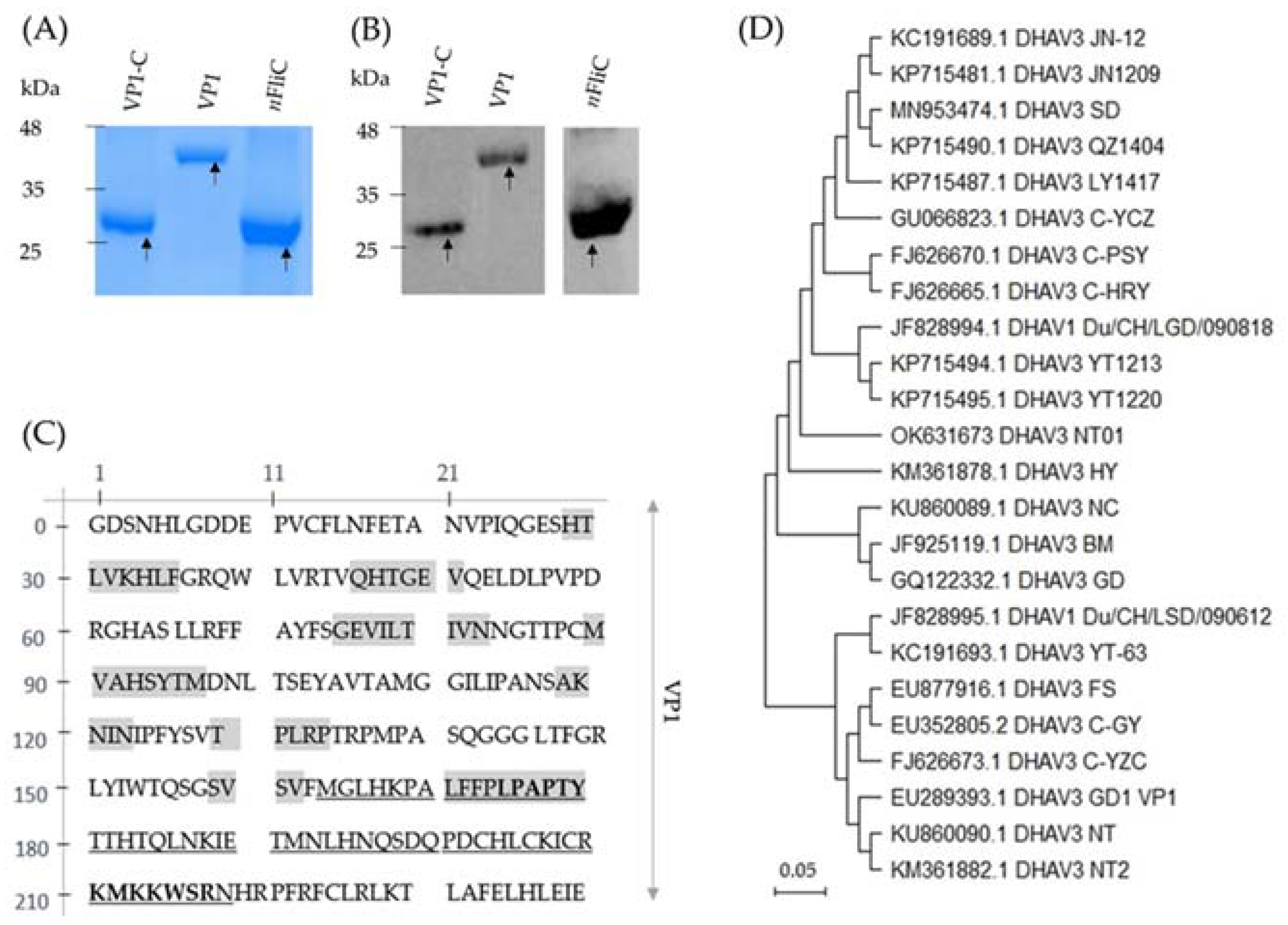
3.1. Recombinant VP1 and VP1-C Were Formulated as Vaccines

For the development of a subunit vaccine against DHAV-3, the VP1 gene was cloned from a DHAV-3 field isolate and expressed in E. coli. Purified recombinant VP1, along with VP1-C and nFliC have expected molecular weights of 46, 28, and 31 kDa, respectively (pET32a vector inserts a 20-kDa tag), in SDS-PAGE analysis (Figure 1A). Identities of the proteins were reconfirmed in Western blot analysis using anti-His antibody (Figure 1B, see Figures S1 and S2 in Supplementary Materials for full gel photos). Expression quantity of the recombinant proteins were: 152 mg/L for VP1, 213 mg/L for VP1-C, and 814 mg/L for nFliC. The amino acid sequence of VP1 is shown in Figure 1C. Furthermore, amino acid sequence comparison showed that the VP1 gene is 97.5% and 97.92% similar with that of DHAV-3 strains from China (KP715494.1, YT1213, China) and Vietnam (KU860089.1, NC, Vietnam), respectively (Figure 1D). Five vaccine formulations were prepared: (1) VP1, (2) VP1 + nFliC, (3) VP1-C, (4) VP1-C + nFliC, and (5) PBS as the vehicle control. Ducklings were vaccinated once intramuscularly for immune response analysis and challenge test.

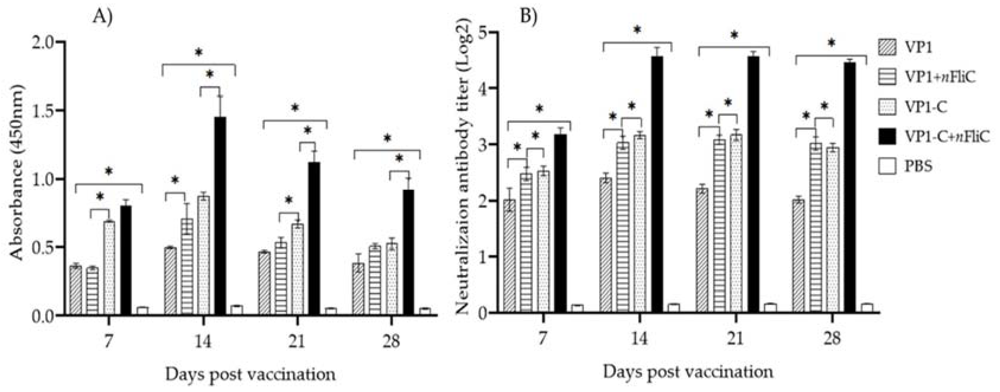
3.2. VP-1C + nFliC Elicited the Highest Level of Virus-Specific Antibody and Neutralization Titer

To evaluate the humoral immunity induced by the vaccines, DHAV-3-specific antibody levels of vaccinated ducklings were determined using indirect ELISA. Results showed that, for all of the vaccine groups, antibody levels peaked on Day 14 after vaccination and decreased thereafter (Figure 2A). Comparing between the vaccine groups on Day 14, the VP1-C group showed a higher antibody level than the full-length VP1, indicating that the C-terminus of VP1 may serve as a better antigen. Furthermore, the addition of nFliC enhanced antibody levels for both VP1 and VP1-C. For end-pint dilution tittering of antibody levels, see Figure S6 in Supplementary Materials. Overall, the VP-1C + nFliC vaccine elicited the highest level of virus-specific antibody.
Sera from vaccinated ducklings were also evaluated for neutralizing antibody titer against DHAV-3. Neutralizing antibody was detected as early as Day 7 after vaccination and reached the highest levels on Day 14 for the vaccine groups (Figure 2B). On Day 14, the VP1-C group showed a higher neutralizing titer than the VP1 group. Furthermore, the addition of nFliC enhanced neutralizing titer for both VP1 and VP1-C. These neutralizing antibody results correlate well with that of virus-specific antibody. In summary, analysis of humoral immune response showed that VP1-C may be a superior antigen compared to full-length VP1 and that nFliC can enhance antibody titers.

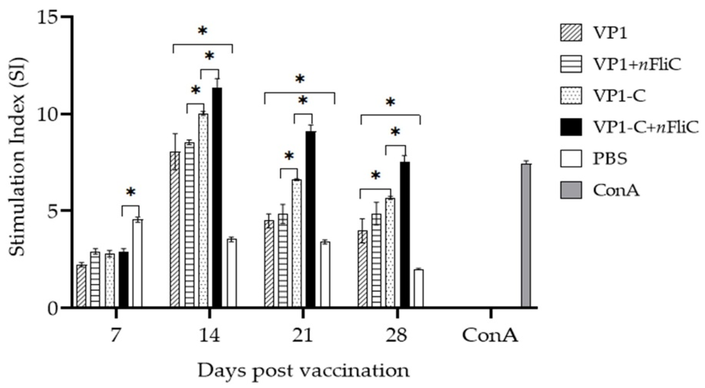
3.3. VP-1C + nFliC Elicited the Highest Level of T Cell Immune Response

An antigen-specific T cell proliferation experiment was performed to evaluate the cellular immunity elicited by the vaccines. Results showed that on Day 14 after vaccination, VP1-C induced a higher level of T cell proliferation than VP1 (Figure 3). The addition of nFliC provided a boost to both VP1 and VP1-C. Consistent with the result of humoral immunity, the VP1-C + nFliC vaccine also induced the highest level of cellular immunity among the various formulations.

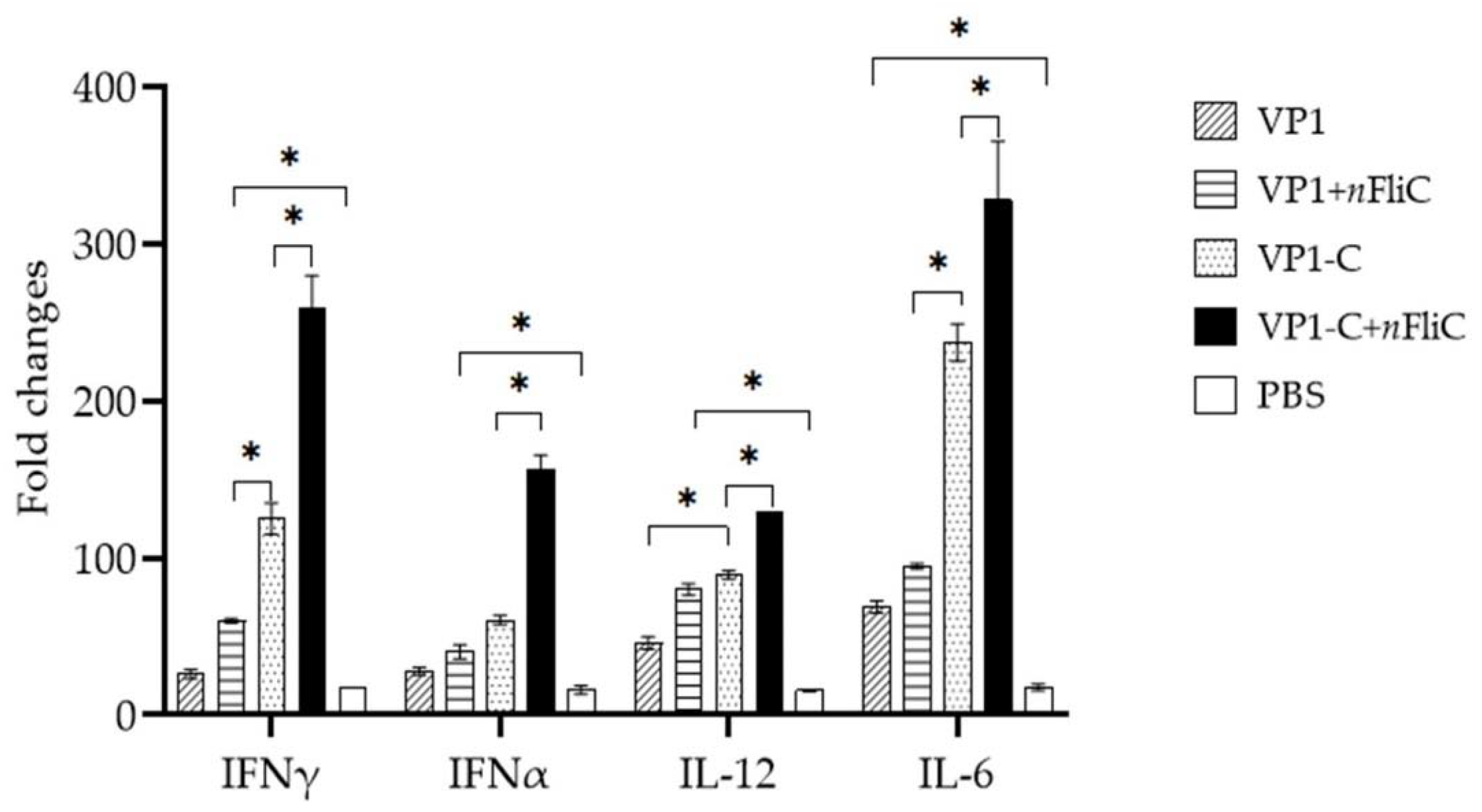
3.4. VP1-C + nFliC Elicited the Highest Level of ProInflammatory Cytokine Expression

To obtain a more detailed profile of the immune response elicited by the vaccines, cytokine expression of PBMCs from immunized ducklings (Day 14) was analyzed. Overall, the VP1-C + nFliC vaccine group showed the highest cytokine expression levels compared to other groups (Figure 4). In terms of the level of the pro-inflammatory cytokine IL-6, a significant difference can be seen between the VP1 and VP1-C vaccines. For the TH-1 type cytokines, IL-12 and IFN-γ, VP1-C appeared to provide a stronger boost than VP1, and nFliC also contributed to enhanced cytokine expression.

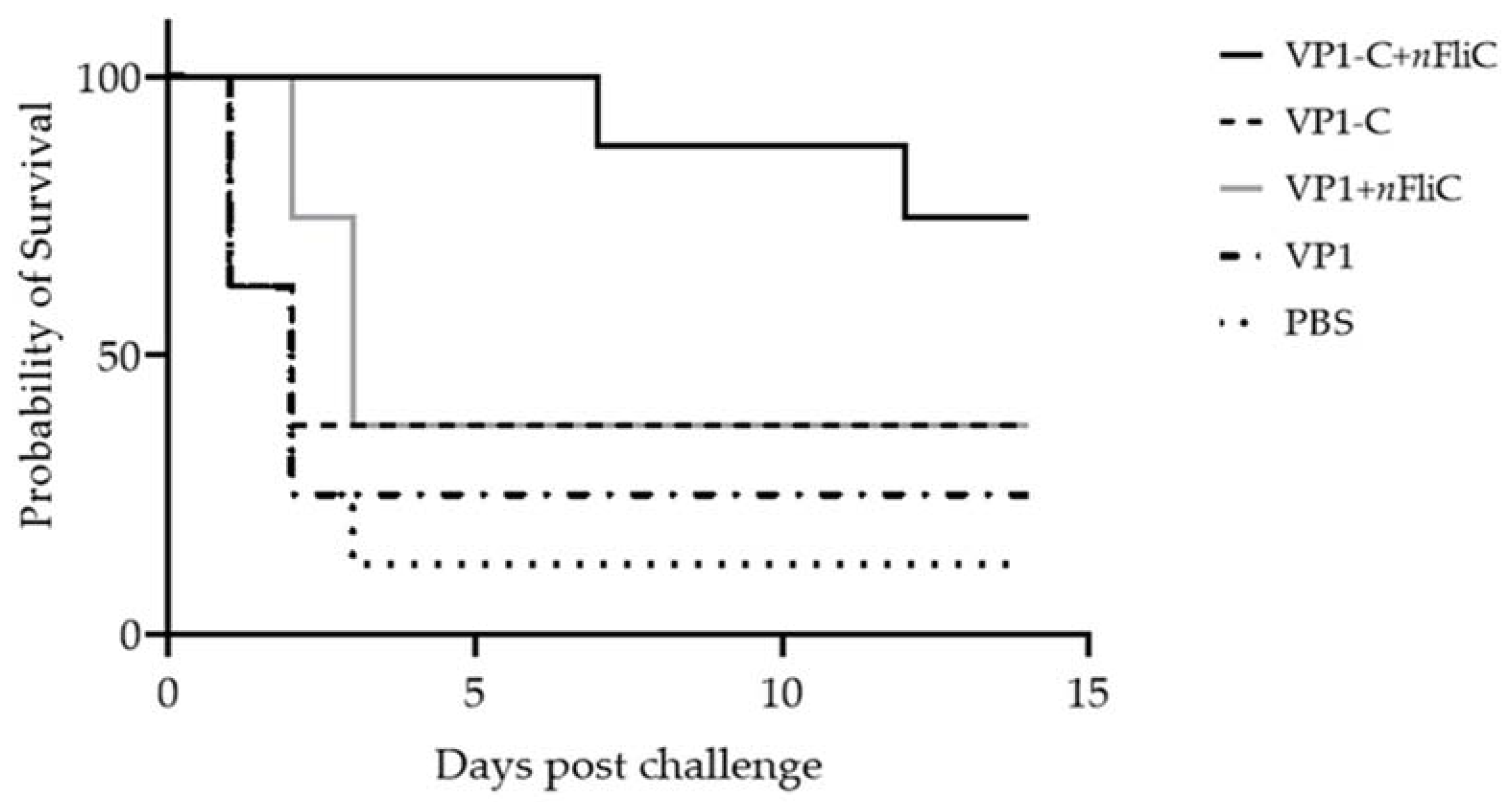
3.5. VP1-C + nFliC Provided 75% Protection Rate in a Challenge Experiment versus 25% for VP1

For protective efficacy evaluation, immunized ducklings were challenged with 100 TCID50 of the DHAV-3 isolate NT01 two weeks after vaccination. Survival rate for the VP1 vaccine group was 25%, with the VP1-C group slightly higher at 37.5% (Figure 5). With the addition of nFliC in the vaccine formulation, survival rate was significantly enhanced for the VP1-C group, rising to 75%. Survival rate of the VP1 group, however, showed only slight improvement with the addition of nFliC. In summary, VP1-C enhanced by nFliC provided a promising level of protection against DHAV-3 challenge.

4. Discussion

Our study showed that a single-dose vaccination of ducklings with the VP1-C + nFliC vaccine formulation provided a promising level of protection against DHAV-3 challenge. Analysis of humoral and cellular immunity confirmed the immunogenicity of VP1-C and demonstrated that the inclusion of nFliC provided a significant boost to immune response for VP1-C.
The results obtained in our study are quite similar to those observed for E. coli-expressed VP1 of enterovirus 71 [34]. Zhang et. al. showed that full-length VP1 of EV71 failed to elicit neutralizing serum in mice. Neutralizing activity was only observed when mice were vaccinated with truncated C-terminal portions of VP1: VP1202–297 or VP1202–248, both of which reside toward the C-terminus of VP1. Other works on EV71 also identified positions 163–177 and 208–222 (near the C-terminus) as linear neutralizing epitopes [30,35,36]. The result from our study correlates well with the observation that a truncated VP1 containing the C-terminal region elicits better protective immunity than full-length VP1. It remains an interesting question, though, as to why full-length VP1 fails to elicit protective immunity since it contains all the neutralizing epitopes. A hypothesis could be that protein folding of E. coli-expressed VP1 may result in the masking of neutralizing epitopes.
While E. coli-expressed VP1 appears ineffective as a vaccine antigen, baculovirus-expressed VP1 seems to elicit better neutralizing immunity. In one study, rabbit sera raised against baculovirus-expressed VP1 of DHAV-1 neutralized virus infection in vitro and in vivo [1]. In another study, baculovirus-expressed virus-like particle composed of DHAV-1 VP0, VP3, and VP1 was found to induce strong humoral immune response and provided strong protection [37]. The baculovirus expression system may lend the superior immunogenicity through post-translational modifications.
Various protein expression systems confer different advantages in terms of vaccine efficacy and costs. By showing that E. coli-expressed VP1-C may serve as an effective antigen, we have demonstrated that the E. coli expression system remains a viable option for DHAV antigen production.

5. Conclusions

In conclusion, we have shown E. coli-expressed VP1-C to be a promising antigen when combined with nFliC, and it may be further developed as a single-dose subunit vaccine against DHAV-3.

Supplementary Materials

The following supporting information can be downloaded at: https://www.mdpi.com/article/10.3390/vaccines10040523/s1. Figure S1: Full gel photo of SDS-PAGE of the recombinant proteins. Figure S2: Full gel photo of western blot analysis of the recombinant proteins. Figure S3: Cytopathic effect of DHAV-3 on the Leghorn Male Hepatoma cell line. Figure S4: Viral copy numbers (log10) of DHAV-3 in the LMH confluent cell line as determined by RT-qPCR. Figure S5: Gross lesions of ducklings infected with the DHAV-3 isolate, NT01. Figure S6: Total IgG titers of vaccinated ducklings against DHAV-3 were determined by indirect ELISA.

Author Contributions

T.-N.T. designed the study, carried out the laboratory work, performed data analysis and drafted the manuscript; L.-T.C. acquired funding, supervised the study, contributed to data analysis, and wrote the manuscript. All authors have read and agreed to the published version of the manuscript.

Funding

This work was supported in part by an internal grant from NPUST, Taiwan (No. 1085702–63).

Data Availability Statement

The data displayed in this study is available on request from the corresponding author.

Acknowledgments

We thank Duc-Tan Nguyen and Khac-Hung Vu of the Institute of Veterinary Research and Development in Central Vietnam, Vietnam for providing the facility for animal testing. We are grateful to Van-Khien Do for supplying field samples of DHAV-3.

Conflicts of Interest

All authors have no financial or personal relationships with other people or organizations that could inappropriately influence or bias their work.

References

  1. Li, X.; Zhao, R.; Lin, W.; Li, C.; Zhang, T.; Meng, F.; Liu, M.; Zhang, Y. Evidence of VP1 of Duck Hepatitis A Type 1 Virus as a Target of Neutralizing Antibodies and Involving Receptor-Binding Activity. Virus Res. 2017, 227, 240–244. [Google Scholar] [CrossRef] [PubMed]
  2. Yugo, D.M.; Hauck, R.; Shivaprasad, H.L.; Meng, X.-J. Hepatitis Virus Infections in Poultry. Avian Dis. 2016, 60, 576–588. [Google Scholar] [CrossRef] [PubMed]
  3. Zhang, R.; Yang, Y.; Lan, J.; Lin, S.; Xie, Z.; Zhang, X.; Jiang, S. A Novel Peptide Isolated from a Phage Display Peptide Library Modeling Antigenic Epitope of DHAV-1 and DHAV-3. Vaccines 2020, 8, 121. [Google Scholar] [CrossRef] [PubMed] [Green Version]
  4. Wu, X.; Li, X.; Zhang, Q.; Wulin, S.; Bai, X.; Zhang, T.; Wang, Y.; Liu, M.; Zhang, Y. Identification of a Conserved B-Cell Epitope on Duck Hepatitis a Type 1 Virus VP1 Proteine. PLoS ONE 2015, 10, e0118041. [Google Scholar]
  5. Kim, M.C.; Kim, M.J.; Kwon, Y.K.; Lindberg, A.M.; Joh, S.J.; Kwon, H.M.; Lee, Y.J.; Kwon, J.H. Development of Duck Hepatitis A Virus Type 3 Vaccine and Its Use to Protect Ducklings against Infections. Vaccine 2009, 27, 6688–6694. [Google Scholar] [CrossRef] [PubMed]
  6. Fu, Y.; Pan, M.; Wang, X.; Xu, Y.; Yang, H.; Zhang, D. Molecular Detection and Typing of Duck Hepatitis A Virus Directly from Clinical Specimens. Vet. Microbiol. 2008, 131, 247–257. [Google Scholar] [CrossRef]
  7. Wen, X.; Guo, J.; Sun, D.; Wang, M.; Cao, D.; Cheng, A.; Zhu, D.; Liu, M.; Zhao, X.; Yang, Q.; et al. Mutations in VP0 and 2C Proteins of Duck Hepatitis a Virus Type 3 Attenuate Viral Infection and Virulence. Vaccines 2019, 7, 111. [Google Scholar] [CrossRef] [Green Version]
  8. Pan, M.; Yang, X.; Du, J.; Zhou, L.; Ge, X.; Guo, X.; Liu, J.; Zhang, D.; Yang, H. Recovery of Duck Hepatitis A Virus 3 from a Stable Full-Length Infectious CDNA Clone. Virus Res. 2011, 160, 439–443. [Google Scholar] [CrossRef]
  9. Thi, H.; Doan, T.; Thi, X.; Le, K.; Thi, R.; Thi, K.; Thanh, N.; Le, H. Sequencing and Phylogenetic Analysis Reveal the Prevalence of Duck Hepatitis A Virus Genotype-3 in Vietnam. Kafkas Univ. Vet. Fak. Derg. 2017, 23, 369–376. [Google Scholar]
  10. Li, C.; Chen, Z.; Meng, C.; Li, L.; Liu, G. High Yield Expression of Duck Hepatitis A Virus VP1 Protein in Escherichia Coli, and Production and Characterization of Polyclonal Antibody. J. Virol. Methods 2013, 191, 69–75. [Google Scholar] [CrossRef]
  11. Tseng, C.H.; Knowles, N.J.; Tsai, H.J. Molecular Analysis of Duck Hepatitis Virus Type 1 Indicates That It Should Be Assigned to a New Genus. Virus Res. 2007, 123, 190–203. [Google Scholar] [CrossRef] [PubMed]
  12. Kim, M.C.; Kwon, Y.K.; Joh, S.J.; Kim, S.J.; Tolf, C.; Kim, J.H.; Sung, H.W.; Lindberg, A.M.; Kwon, J.H. Recent Korean Isolates of Duck Hepatitis Virus Reveal the Presence of a New Geno- and Serotype When Compared to Duck Hepatitis Virus Type 1 Type Strains. Arch. Virol. 2007, 152, 2059–2072. [Google Scholar] [CrossRef] [PubMed]
  13. Fowler, V.; Bashiruddin, J.B.; Belsham, G.J.; Stenfeldt, C.; Bøtner, A.; Knowles, N.J.; Bankowski, B.; Parida, S.; Barnett, P. Characteristics of a Foot-and-Mouth Disease Virus with a Partial VP1 G-H Loop Deletion in Experimentally Infected Cattle. Vet. Microbiol. 2014, 169, 58–66. [Google Scholar] [CrossRef] [PubMed]
  14. Kitamura, N.; Semler, B.L.; Rothberg, P.G.; Larsen, G.R.; Adler, C.J.; Dorner, A.J.; Emini, E.A.; Hanecak, R.; Lee, J.J.; Van Der Werf, S.; et al. Primary Structure, Gene Organization and Polypeptide Expression of Poliovirus RNA. Nature 1981, 291, 547–553. [Google Scholar] [CrossRef] [PubMed]
  15. Sun, D.; Wang, M.; Wen, X.; Cheng, A.; Jia, R.; Sun, K.; Yang, Q.; Wu, Y.; Zhu, D.; Chen, S.; et al. Cleavage of poly(A)-binding Protein by Duck Hepatitis A Virus 3C Protease. Sci. Rep. 2017, 7, 16261. [Google Scholar] [CrossRef]
  16. Cho, J.G.; Jo, Y.J.; Sung, J.H.; Hong, J.K.; Hwang, J.H.; Park, J.H.; Lee, K.N.; Park, S.G. Monoclonal and Polyclonal Antibodies Specific for Foot and Mouth Disease Virus Type A and Type O VP1. Hybridoma 2012, 31, 358–363. [Google Scholar] [CrossRef]
  17. Kim, M.C.; Kwon, Y.K.; Joh, S.J.; Lindberg, A.M.; Kwon, J.H.; Kim, J.H.; Kim, S.J. Molecular Analysis of Duck Hepatitis Virus Type 1 Reveals a Novel Lineage Close to the Genus Parechovirus in the Family Picornaviridae. J. Gen. Virol. 2006, 87, 3307–3316. [Google Scholar] [CrossRef]
  18. Kristensen, T.; Belsham, G.J. Identification of Plasticity and Interactions of a Highly Conserved Motif within a Picornavirus Capsid Precursor Required for Virus Infectivity. Sci. Rep. 2019, 9, 11747. [Google Scholar] [CrossRef]
  19. Jiang, P.; Liu, Y.; Ma, H.-C.; Paul, A.V.; Wimmer, E. Picornavirus Morphogenesis. Microbiol. Mol. Biol. Rev. 2014, 78, 418–437. [Google Scholar] [CrossRef] [Green Version]
  20. Curry, S.; Fry, E.; Blakemore, W.; Abu-Ghazaleh, R.; Jackson, T.; King, A.; Lea, S.; Newman, J.; Stuart, D. Dissecting the Roles of VP0 Cleavage and RNA Packaging in Picornavirus Capsid Stabilization: The Structure of Empty Capsids of Foot-and-Mouth Disease Virus. J. Virol. 1997, 71, 9743–9752. [Google Scholar] [CrossRef] [Green Version]
  21. Shakeel, S.; Westerhuis, B.M.; Ora, A.; Koen, G.; Bakker, A.Q.; Claassen, Y.; Wagner, K.; Beaumont, T.; Wolthers, K.C.; Butcher, S.J. Structural Basis of Human Parechovirus Neutralization by Human Monoclonal Antibodies. J. Virol. 2015, 89, 9571–9580. [Google Scholar] [CrossRef] [PubMed] [Green Version]
  22. Sherry, B.; Mosser, A.G.; Colonno, R.J.; Rueckert, R.R. Use of Monoclonal Antibodies to Identify Four Neutralization Immunogens on a Common Cold Picornavirus, Human Rhinovirus 14. J. Virol. 1986, 57, 246–257. [Google Scholar] [CrossRef] [PubMed] [Green Version]
  23. Dill, V.; Eschbaumer, M. Cell Culture Propagation of Foot-and-Mouth Disease Virus: Adaptive Amino Acid Substitutions in Structural Proteins and Their Functional Implications. Virus Genes 2020, 56, 1–15. [Google Scholar] [CrossRef] [PubMed] [Green Version]
  24. Azuma, H.; Yoneda, S. Structure and Dynamics of the GH Loop of the Foot-and-Mouth Disease Virus Capsid. J. Mol. Graph. Model. 2009, 28, 278–286. [Google Scholar] [CrossRef] [PubMed]
  25. Rossmann, M.G.; Arnold, E.; Erickson, J.W.; Frankenberger, E.A.; Griffith, J.P.; Hecht, H.J.; Johnson, J.E.; Kamer, G.; Luo, M.; Mosser, A.G.; et al. Structure of a Human Common Cold Virus and Functional Relationship to Other Picornaviruses. Nature 1985, 317, 145–153. [Google Scholar] [CrossRef] [PubMed]
  26. Lea, S.; Hernéndez, J.; Blakemore, W.; Brocchi, E.; Curry, S.; Domingo, E.; Fry, E.; Ghazaleh, R.A.; King, A.; Newman, J.; et al. The Structure and Antigenicity of a Type C Foot-and-Mouth Disease Virus. Structure 1994, 2, 123–139. [Google Scholar] [CrossRef] [Green Version]
  27. Mateu, M.G. Antibody Recognition of Picornaviruses and Escape from Neutralization: A Structural View. Virus Res. 1995, 38, 1–24. [Google Scholar] [CrossRef]
  28. Yoon, S.; Kurnasov, O.; Natarajan, V.; Hong, M.; Gudkov, A.V.; Osterman, A.L. Structural Basis of TLR5-Flagellin Recognition and Signaling. Science 2012, 335, 859–864. [Google Scholar] [CrossRef] [Green Version]
  29. Doan, T.; Wang, H.; Ke, G.; Cheng, L. N-Terminus of Flagellin Fused to an Antigen Improves Vaccine Efficacy against Pasteurella Multocida Infection in Chickens. Vaccines 2020, 8, 283. [Google Scholar] [CrossRef]
  30. Reed, L.J.; Muench, H. Simple Method of Estimating Fifty Percent Endpoints. Am. J. Hyg. 1938, 27, 493–497. [Google Scholar]
  31. Zhou, P.; Ma, L.; Rao, Z.; Li, Y.; Zheng, H.; He, Q.; Luo, R. Duck Tembusu Virus Infection Promotes the Expression of Duck Interferon-Induced Protein 35 to Counteract RIG-I Antiviral Signaling in Duck Embryo Fibroblasts. Front. Immunol. 2021, 12 (July), 1–15. [Google Scholar] [CrossRef] [PubMed]
  32. Lee, J.W.; Lin, Y.M.; Yen, T.Y.; Yang, W.J.; Chu, C.Y. CpG Oligodeoxynucleotides Containing GACGTT Motifs Enhance the Immune Responses Elicited by a Goose Parvovirus Vaccine in Ducks. Vaccine 2010, 28, 7956–7962. [Google Scholar] [CrossRef] [PubMed]
  33. Livak, K.J.; Schmittgen, T.D. Analysis of Relative Gene Expression Data Using Real-Time Quantitative PCR and the 2−ΔΔCT Method. Methods 2001, 25, 402–408. [Google Scholar] [CrossRef] [PubMed]
  34. Zhang, J.; Dong, M.; Jiang, B.; Dai, X.; Meng, J. Antigenic Characteristics of the Complete and Truncated Capsid Protein VP1 of Enterovirus 71. Virus Res. 2012, 167, 337–342. [Google Scholar] [CrossRef]
  35. Foo, D.G.W.; Alonso, S.; Chow, V.T.K.; Poh, C.L. Passive Protection against Lethal Enterovirus 71 Infection in Newborn Mice by Neutralizing Antibodies Elicited by a Synthetic Peptide. Microbes Infect. 2007, 9, 1299–1306. [Google Scholar] [CrossRef]
  36. Liu, C.C.; Chou, A.H.; Lien, S.P.; Lin, H.Y.; Liu, S.J.; Chang, J.Y.; Guo, M.S.; Chow, Y.H.; Yang, W.S.; Chang, K.H.W.; et al. Identification and Characterization of a Cross-Neutralization Epitope of Enterovirus 71. Vaccine 2011, 29, 4362–4372. [Google Scholar] [CrossRef]
  37. Wang, A.; Gu, L.; Wu, S.; Zhu, S. Duck Hepatitis a Virus Structural Proteins Expressed in Insect Cells Self-Assemble into Virus-like Particles with Strong Immunogenicity in Ducklings. Vet. Microbiol. 2018, 215, 23–28. [Google Scholar] [CrossRef]
Figure 1. Recombinant protein expression of VP1, VP1-C, and nFliC. (A) SDS-PAGE and (B) Western blot analysis were performed for the purified recombinant proteins. (C) Protein sequence of cloned VP1 from the DHAV-3 strain NT01 used in this study is shown. Predicted β-strands (highlighted in grey), potential conserved B cell epitope, heparan-binding site (bold font), and VP1-C (underlined) are indicated. Prediction of β-strand locations is based on sequence comparison with the VP1 gene of the Human Parechovirus 1 strain, Harris (protein data bank ID: 5MJV). (D) Phylogenetic analysis of NT01 VP1 amino acid was performed with 23 DHAV isolates of two serotypes (DHAV1 and DHAV3). Phylogenetic analysis was performed using the maximum likelihood method (in the MEGA 11 software).
Vaccines 10 00523 g001
Figure 2. Virus-specific antibody levels of vaccinated ducklings. Ducklings (n = 3 per group) were vaccinated once with five different vaccine formulations: (1) VP1, (2) VP1 + nFliC, (3) VP1-C, (4) VP1-C + nFliC, and (5) PBS, and virus-specific antibody levels were determined by indirect ELISA. (A) IgG antibody level of vaccinated ducklings; (B) Neutralization antibody titer of vaccinated ducklings. Data are presented as mean ± SD. An asterisk indicates statistically significant difference (p < 0.05).
Vaccines 10 00523 g002
Figure 3. T cell proliferation of PBMCs from vaccinated ducklings. Ducklings (n = 3 per group) were vaccinated once with five different vaccine formulations: (1) VP1, (2) VP1 + nFliC, (3) VP1-C, (4) VP1-C + nFliC, and (5) PBS. Isolated PBMCs were stimulated with VP1, and proliferation extent was measured using the MTT assay. ConA was used as the positive control. Data are presented as mean ± SD. An asterisk indicates statistically significant difference (p < 0.05).
Vaccines 10 00523 g003
Figure 4. Cytokine expression profile of PBMCs from vaccinated ducklings. Ducklings (n = 3 per group) were vaccinated once with five different vaccine formulations: (1) VP1, (2) VP1 + nFliC, (3) VP1-C, (4) VP1-C + nFliC, and (5) PBS. Isolated PBMCs (Day 14) were stimulated with VP1, and RT-PCR was performed to determine mRNA levels of pro-inflammatory cytokines (IFNγ, IFNα, IL-12, and IL-6). Data are presented as mean ± SD. An asterisk indicates statistically significant difference (p < 0.05).
Vaccines 10 00523 g004
Figure 5. Survival rate of vaccinated ducklings after challenge with Duck hepatitis A serotype 3, NT01. Ducklings (n = 8 per group) were vaccinated once with five different vaccine formulations: (1) VP1, (2) VP1 + nFliC, (3) VP1-C, (4) VP1-C + nFliC, and (5) PBS. Two weeks after vaccination, ducklings were challenged with 100 TCID50 NT01. Survival rate was recorded for 14 days.
Vaccines 10 00523 g005
Table 1. Primers for gene cloning and cytokine genes.
Target Gene Sequence (5′-3′)Gene-Length (bp)Annealing Temp. (°C)GenBank
VP1F1 gaattcatgggtgattctaatcatctt73253OK631673
Raagcttttcaatttccaaatggagct
VP1-CFgaattcatgggcaggttgtatatctgg22253OK631673
Raagcttattgcgagaccatttctt
nFliCFgaattcatgaactgcactaaacaaact30952[29]
Raagcttttcagcctggatggagtc
GAPDHFatcacaccacacatggcgt20759.5GCA_015476345.1
Rtttatagccgccgaggctg
IFN-γFcagacctactgcttgtttgt19253.5AJ012254
Rttcatttctctctgtccagt
IFN-αFcagccatctacagcgccc13860AY879230.1
Rggctgggagccatgttgc
IL-12Fctggaggtcattgatgaggtg16856AJ564201
Rgaaagtcaaagggaagtaggac
IL-6Faccgtgtgcgagaacagc13560AB191038
Rgaaaagcccgctggagagt
DHAV-1Fgccccactctatggaaatttg76753[9]
Ratttggtcagattcaatttcc
DHAV-3Fatgcgagttggtaaggattttcag88156[9]
Rgatcctgatttaccaacaaccat
qVP1Fatgaaccagtgtgttttctc17553OK631673
Raacagagatgcatgaccc
1 Bold font represents restriction enzyme (RE) sites. IFN-α, γ: interferon alpha, gamma; IL-6, 12: interleukin-6, 12; GAPDH: glyceraldehyde-3-phosphate dehydrogenase.
Publisher’s Note: MDPI stays neutral with regard to jurisdictional claims in published maps and institutional affiliations.

