Protective Effects of SPA0355, a Thiourea Analogue, Against Lipopolysaccharide-Induced Acute Kidney Injury in Mice


Abstract
1. Introduction
2. Materials and Methods
2.1. Animal Procedures
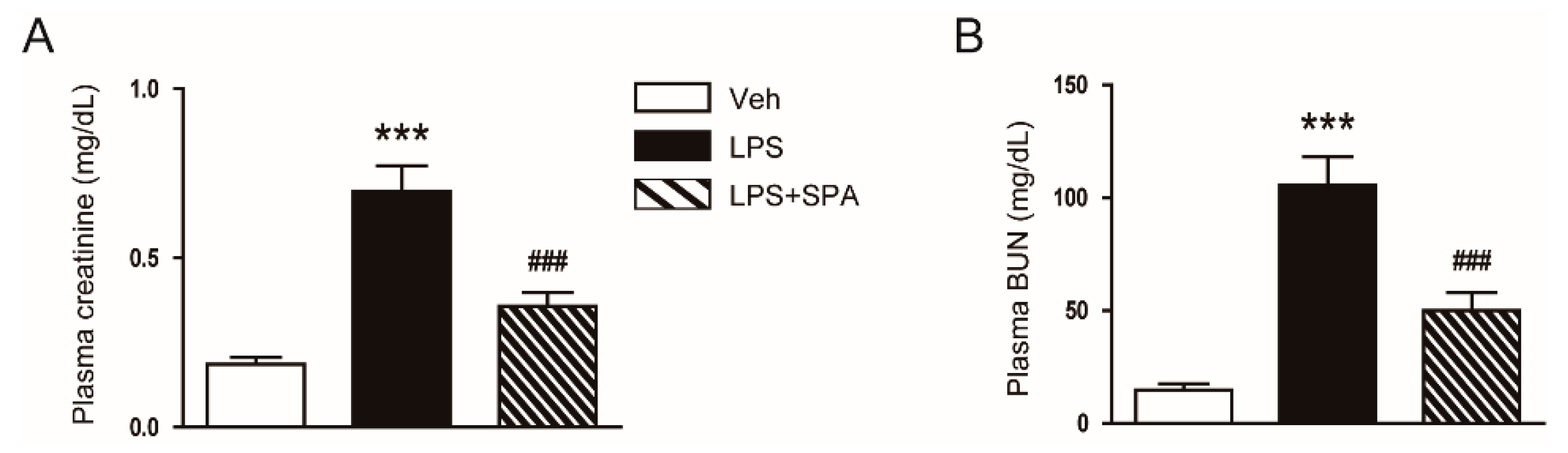
2.2. Biochemical Analysis
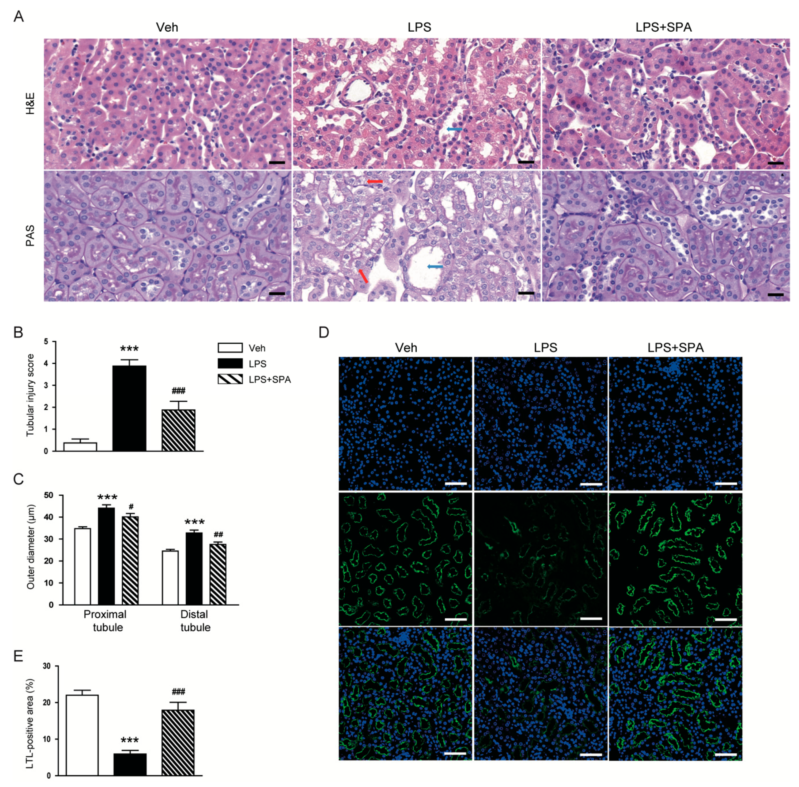
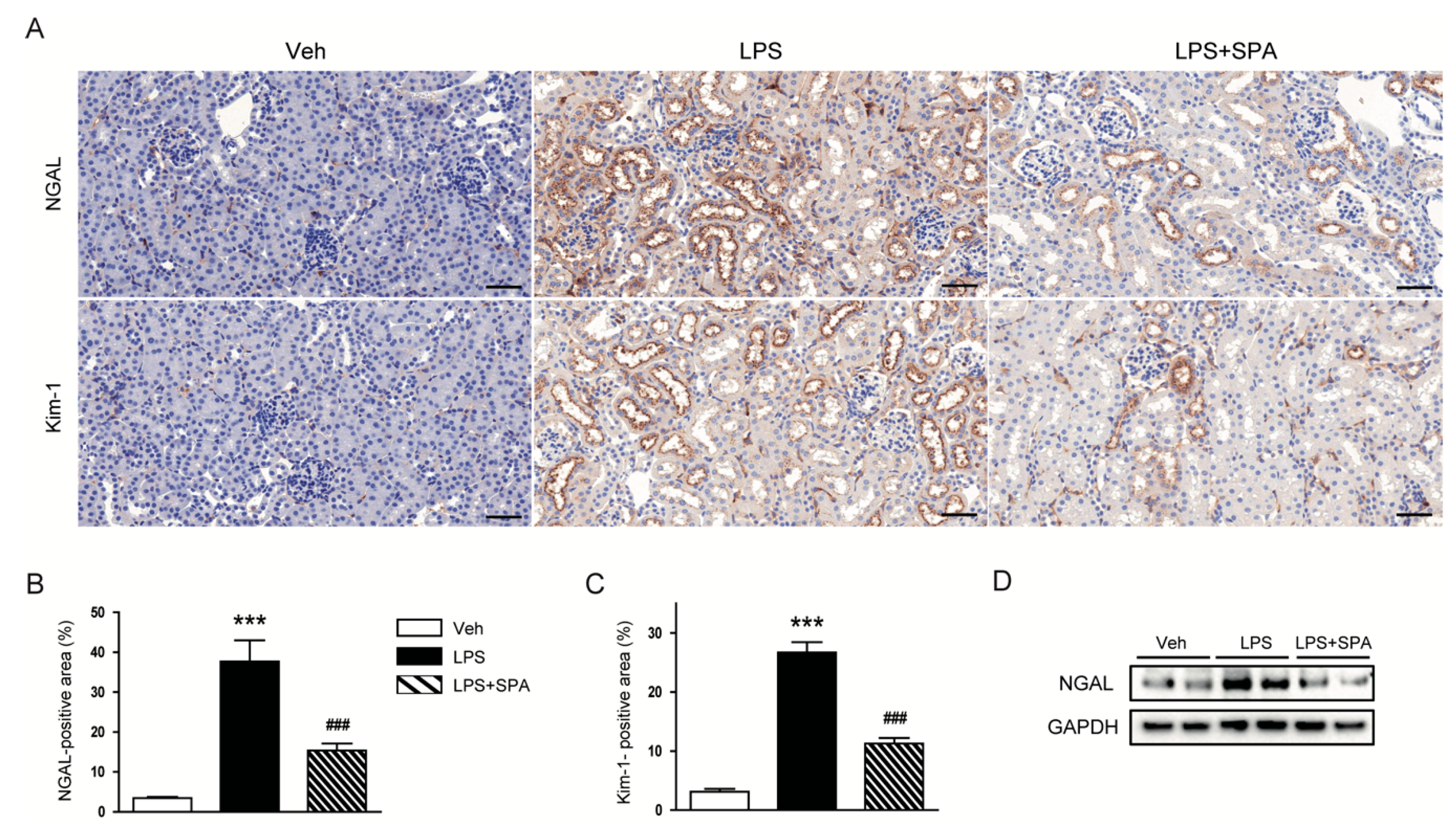
2.3. Histological Analysis
2.4. Western Blot Analysis
2.5. Real-Time Reverse Transcription-Polymerase Chain Reaction (RT-PCR)
2.6. TdT-Mediated dUTP Nick End Labeling (TUNEL) Staining
2.7. Statistical Analysis
3. Results
3.1. SPA0355 Ameliorated LPS-Induced Kidney Damage
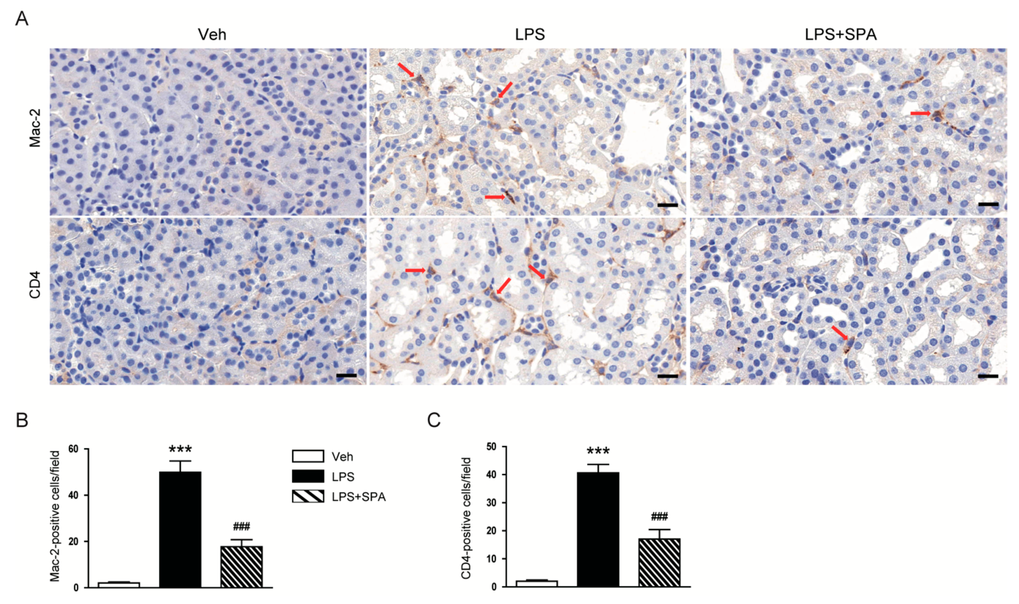
3.2. SPA0355 Suppressed LPS-Induced Inflammatory Responses
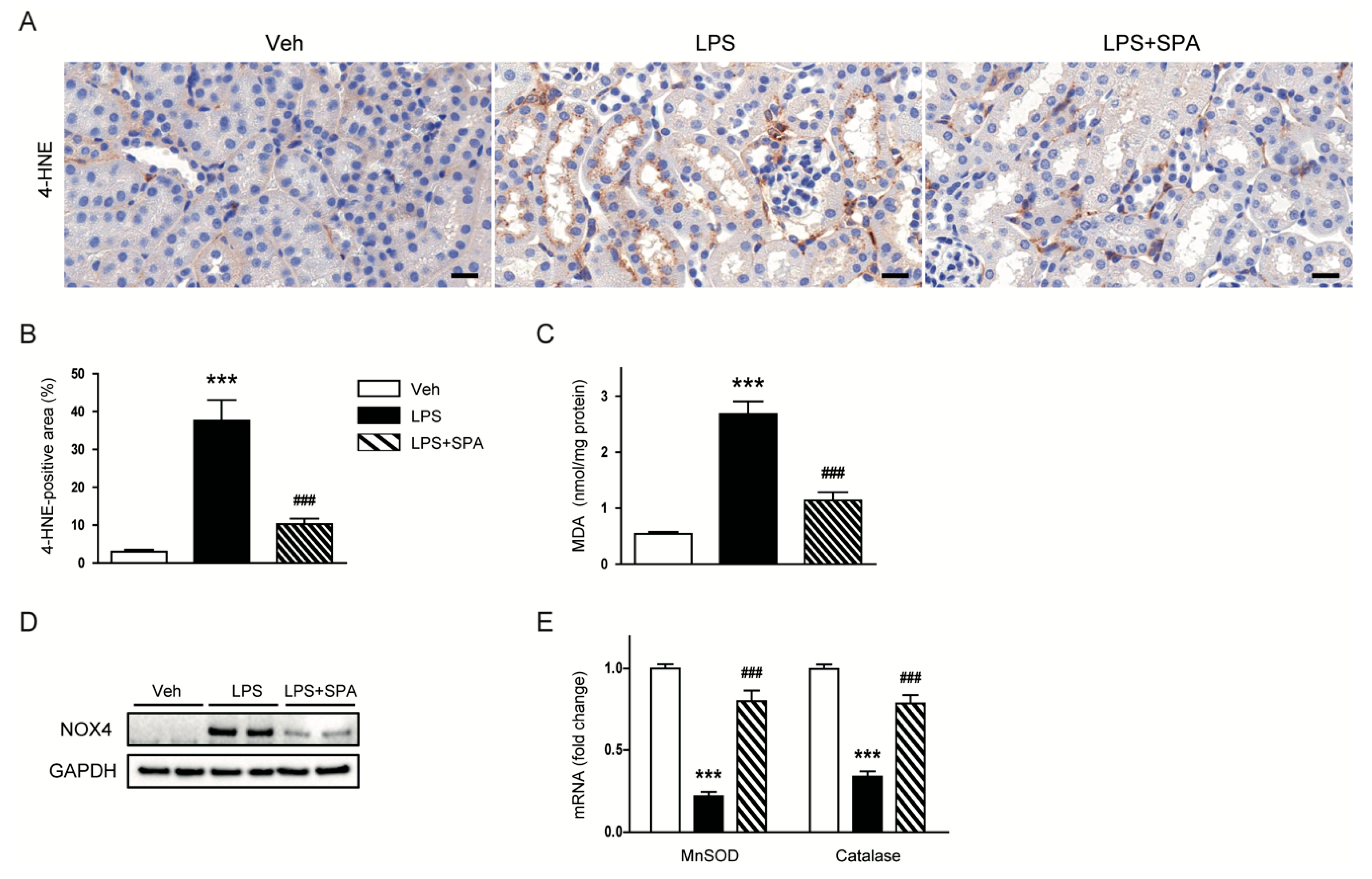
3.3. SPA0355 Attenuated LPS-Induced Oxidative Stress
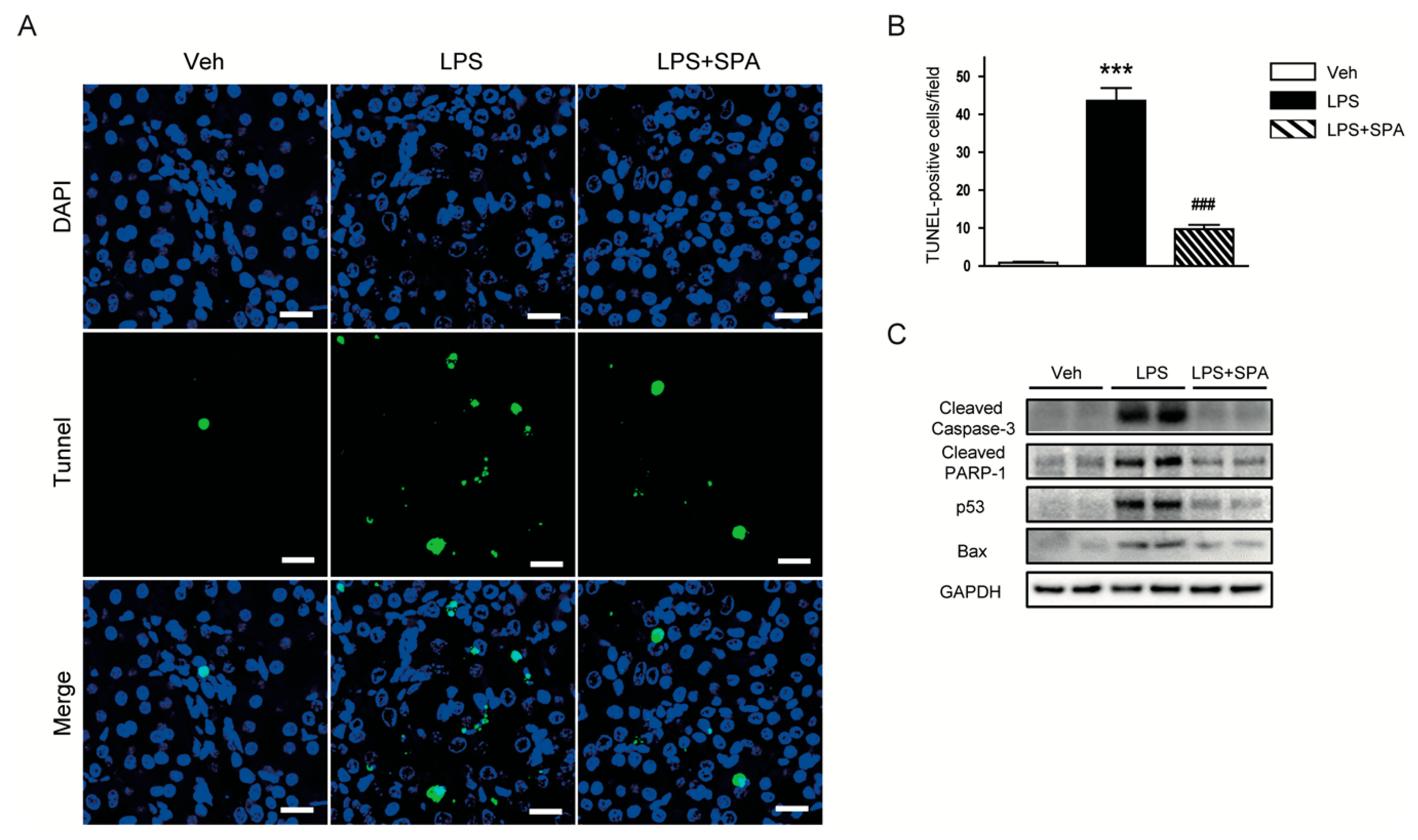
3.4. SPA0355 Inhibited LPS-Induced Tubular Cell Apoptosis
4. Discussion
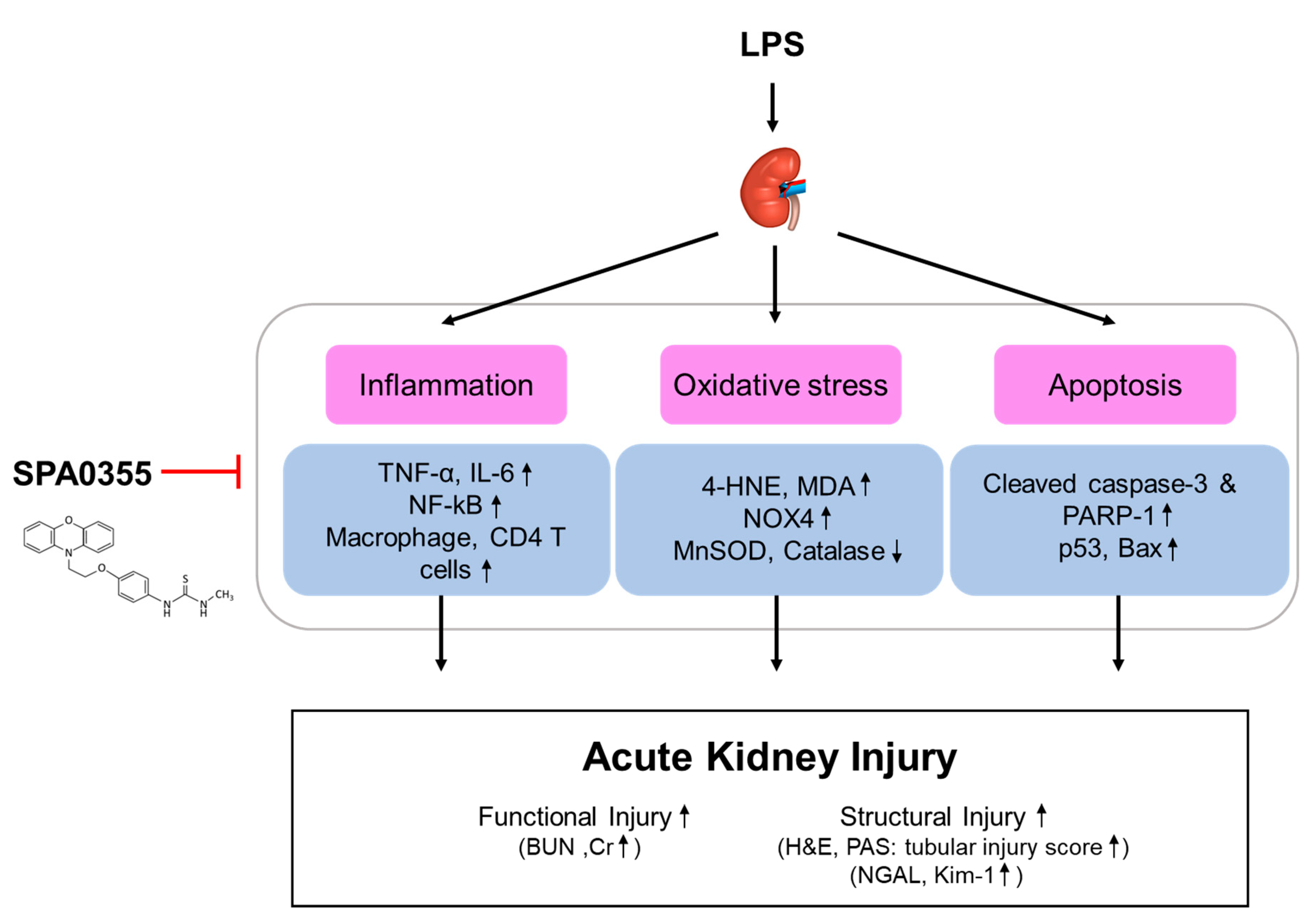
5. Conclusions
Author Contributions
Funding
Conflicts of Interest
References
- Uchino, S.; Kellum, J.A.; Bellomo, R.; Doig, G.S.; Morimatsu, H.; Morgera, S.; Schetz, M.; Tan, I.; Bouman, C.; Macedo, E.; et al. Acute renal failure in critically ill patients: A multinational, multicenter study. JAMA 2005, 294, 813–818. [Google Scholar] [CrossRef] [PubMed]
- Hoste, E.A.; Bagshaw, S.M.; Bellomo, R.; Cely, C.M.; Colman, R.; Cruz, D.N.; Edipidis, K.; Forni, L.G.; Gomersall, C.D.; Govil, D.; et al. Epidemiology of acute kidney injury in critically ill patients: The multinational AKI-EPI study. Intensive Care Med. 2015, 41, 1411–1423. [Google Scholar] [CrossRef]
- Peerapornratana, S.; Manrique-Caballero, C.L.; Gómez, H.; Kellum, J.A. Acute kidney injury from sepsis: Current concepts, epidemiology, pathophysiology, prevention and treatment. Kidney Int. 2019, 96, 1083–1099. [Google Scholar] [CrossRef] [PubMed]
- Shum, H.P.; Yan, W.W.; Chan, T.M. Recent knowledge on the pathophysiology of septic acute kidney injury: A narrative review. J. Crit. Care 2016, 31, 82–89. [Google Scholar] [CrossRef] [PubMed]
- Morrell, E.D.; Kellum, J.A.; Pastor-Soler, N.M.; Hallows, K.R. Septic acute kidney injury: Molecular mechanisms and the importance of stratification and targeting therapy. Crit. Care 2014, 18, 501. [Google Scholar] [CrossRef] [PubMed]
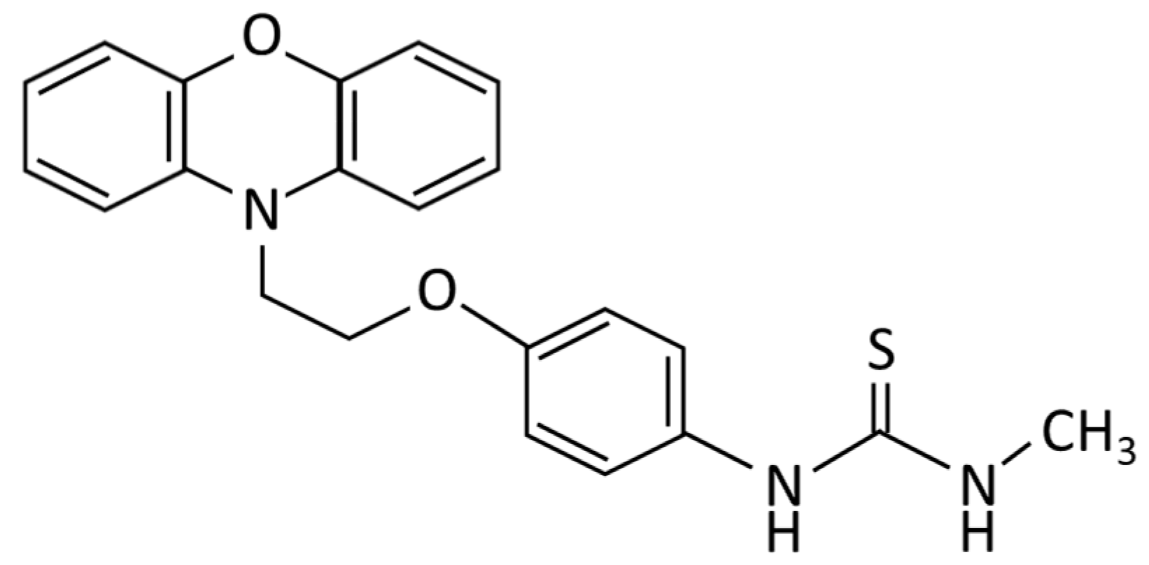
- Lee, Y.R.; Hwang, J.K.; Lee, H.S.; Cheon, Y.J.; Ryu, J.H.; Lee, S.I.; Kwak, H.B.; Lee, S.M.; Kim, J.S.; Park, J.W.; et al. SPA0355, a thiourea analogue, inhibits inflammatory responses and joint destruction in fibroblast-like synoviocytes and mice with collagen-induced arthritis. Br. J. Pharmacol. 2011, 164, 794–806. [Google Scholar] [CrossRef]
- Bae, U.J.; Song, M.Y.; Jang, H.Y.; Gim, H.J.; Ryu, J.H.; Lee, S.M.; Jeon, R.; Park, B.H. The efficacy of SPA0355 in protecting β cells in isolated pancreatic islets and in a murine experimental model of type 1 diabetes. Exp. Mol. Med. 2013, 45, 51. [Google Scholar] [CrossRef]
- Bae, U.J.; Yang, J.D.; Ka, S.O.; Koo, J.H.; Woo, S.J.; Lee, Y.R.; Yu, H.C.; Cho, B.H.; Zhao, H.Y.; Ryu, J.H.; et al. SPA0355 attenuates ischemia/reperfusion-induced liver injury in mice. Exp. Mol. Med. 2014, 46, 109. [Google Scholar] [CrossRef]
- Jang, H.Y.; Jeon, R.; Kang, K.W.; Song, M.Y.; Lim, J.M.; Lee, E.; Ryu, J.H.; Lee, S.M.; Park, B.H. SPA0355 suppresses T-cell responses and reduces airway inflammation in mice. Eur. J. Pharmacol. 2014, 745, 19–28. [Google Scholar] [CrossRef]
- Kim, S.H.; Zhang, Z.; Moon, Y.J.; Park, I.W.; Cho, Y.G.; Jeon, R.; Park, B.H. SPA0355 prevents ovariectomy-induced bone loss in mice. Korean J. Physiol. Pharmacol. 2019, 23, 47–54. [Google Scholar] [CrossRef]
- Doi, K.; Leelahavanichkul, A.; Yuen, P.S.; Star, R.A. Animal models of sepsis and sepsis-induced kidney injury. J. Clin. Investig. 2009, 119, 2868–2978. [Google Scholar] [CrossRef] [PubMed]
- Khajevand-Khazaei, M.R.; Azimi, S.; Sedighnejad, L.; Salari, S.; Ghorbanpour, A.; Baluchnejadmojarad, T.; Mohseni-Moghaddam, P.; Khamse, S.; Roghani, M. S-allyl cysteine protects against lipopolysaccharide-induced acute kidney injury in the C57BL/6 mouse strain: Involvement of oxidative stress and inflammation. Int. Immunopharmacol. 2019, 69, 19–26. [Google Scholar] [CrossRef] [PubMed]
- Ni, J.; Zhao, Y.; Su, J.; Liu, Z.; Fang, S.; Li, L.; Deng, J.; Fan, G. Toddalolactone protects lipopolysaccharide-induced sepsis and attenuates lipopolysaccharide-induced inflammatory response by modulating HMGB1-NF-κB translocation. Front. Pharmacol. 2020, 11, 109. [Google Scholar] [CrossRef] [PubMed]
- Kim, J.W.; Jo, J.; Kim, J.Y.; Choe, M.; Leem, J.; Park, J.H. Melatonin attenuates cisplatin-induced acute kidney injury through dual suppression of apoptosis and necroptosis. Biology 2019, 8, 64. [Google Scholar] [CrossRef] [PubMed]
- Kim, J.Y.; Jo, J.; Kim, K.; An, H.J.; Gwon, M.G.; Gu, H.; Kim, H.J.; Yang, A.Y.; Kim, S.W.; Jeon, E.J.; et al. Pharmacological activation of Sirt1 ameliorates cisplatin-induced acute kidney injury by suppressing apoptosis, oxidative stress, and inflammation in mice. Antioxidants 2019, 8, 322. [Google Scholar] [CrossRef] [PubMed]
- Kim, J.Y.; Park, J.H.; Kim, K.; Jo, J.; Leem, J.; Park, K.K. Pharmacological inhibition of Caspase-1 ameliorates cisplatin-induced nephrotoxicity through suppression of apoptosis, oxidative stress, and inflammation in mice. Mediat. Inflamm. 2018, 2018, 6571676. [Google Scholar] [CrossRef]
- Kim, Y.J.; Ryu, J.H.; Cheon, Y.J.; Lim, H.J.; Jeon, R. Design and synthesis of urea and thiourea derivatives and their inhibitory activities on lipopolysaccharide-induced NO production. Bioorganic Med. Chem. Lett. 2007, 17, 3317–3321. [Google Scholar] [CrossRef]
- Jin, G.H.; Lee, D.Y.; Cheon, Y.J.; Gim, H.J.; Kim, D.H.; Kim, H.D.; Ryu, J.H.; Jeon, R. Synthesis of phenylisothiourea derivatives as inhibitors of NO production in LPS activated macrophages. Bioorganic Med. Chem. Lett. 2009, 19, 3088–3092. [Google Scholar] [CrossRef]
- Cheon, Y.J.; Gim, H.J.; Jang, H.R.; Ryu, J.H.; Jeon, R. Synthesis of heterocyte-linked thioureas and their inhibitory activities of NO production in LPS activated macrophages. Bull. Korean Chem. Soc. 2010, 31, 27–30. [Google Scholar] [CrossRef]
- Li, H.; Chen, W.; Chen, Y.; Zhou, Q.; Xiao, P.; Tang, R.; Xue, J. Neferine attenuates acute kidney injury by inhibiting NF-κB signaling and upregulating Klotho expression. Front. Pharmacol. 2019, 10, 1197. [Google Scholar] [CrossRef]
- Mir, S.M.; Ravuri, H.G.; Pradhan, R.K.; Narra, S.; Kumar, J.M.; Kuncha, M.; Kanjilal, S.; Sistla, R. Ferulic acid protects lipopolysaccharide-induced acute kidney injury by suppressing inflammatory events and upregulating antioxidant defenses in Balb/c mice. Biomed. Pharmacother. 2018, 100, 304–315. [Google Scholar] [CrossRef] [PubMed]
- Tsai, Y.C.; Wang, S.; Wu, M.; Liao, C.; Lin, C.; Chen, J.; Fu, S. Pilloin, a flavonoid isolated from aquilaria sinensis, exhibits anti-inflammatory activity in vitro and in vivo. Molecules 2018, 23, 3177. [Google Scholar] [CrossRef] [PubMed]
- Li, Y.; Zhang, J.; Su, L.; Kellum, J.A.; Peng, Z. Downregulation of TIMP2 attenuates sepsis-induced AKI through the NF-κb pathway. Biochim. Biophys. Acta Mol. Basis Dis. 2019, 1865, 558–569. [Google Scholar] [CrossRef] [PubMed]
- Karin, M.; Ben-Neriah, Y. Phosphorylation meets ubiquitination: The control of NF-[kappa]B activity. Annu. Rev. Immunol. 2000, 18, 621–623. [Google Scholar] [CrossRef]
- Mittal, M.; Siddiqui, M.R.; Tran, K.; Reddy, S.P.; Malik, A.B. Reactive oxygen species in inflammation and tissue injury. Antioxid. Redox Signal. 2014, 20, 1126–1167. [Google Scholar] [CrossRef]
- Huang, Y.; Mao, Z.; Zhang, Z.; Obata, F.; Yang, X.; Zhang, X.; Huang, Y.; Mitsui, T.; Fan, J.; Takeda, M.; et al. Connexin43 contributes to inflammasome activation and lipopolysaccharide-initiated acute renal injury via modulation of intracellular oxidative status. Antioxid. Redox Signal. 2019, 31, 1194–1212. [Google Scholar] [CrossRef]
- He, J.; Huang, T.; Zhao, L. 3,3’-Diindolylmethane mitigates lipopolysaccharide-induced acute kidney injury in mice by inhibiting NOX-mediated oxidative stress and the apoptosis of renal tubular epithelial cells. Mol. Med. Rep. 2019, 19, 5115–5122. [Google Scholar] [CrossRef]
- Hus, B.G.; Lee, R.P.; Yang, F.; Harn, H.J.; Chen, H. Post-treatment with N-acetylcysteine ameliorates endotoxin shock-induced organ damage in conscious rats. Life Sci. 2006, 79, 2010–2016. [Google Scholar]
- Lowes, D.A.; Webster, N.R.; Murphy, M.P.; Galley, H.F. Antioxidants that protect mitochondria reduce interleukin-6 and oxidative stress, improve mitochondrial function, and reduce biochemical markers of organ dysfunction in a rat model of acute sepsis. Br. J. Anaesth. 2013, 110, 472–480. [Google Scholar] [CrossRef]
- Plotnikov, E.Y.; Pevzner, I.B.; Zorova, L.D.; Chernikov, V.P.; Prusov, A.N.; Kireev, I.I.; Silachev, D.N.; Skulachev, V.P.; Zorov, D.B. Mitochondrial damage and mitochondria-targeted antioxidant protection in LPS-induced acute kidney injury. Antioxidants 2019, 8, 176. [Google Scholar] [CrossRef]
- Gloire, G.; Legrand-Poels, S.; Piette, J. NF-kappaB activation by reactive oxygen species: Fifteen years later. Biochem. Pharmacol. 2006, 72, 1493–1505. [Google Scholar] [CrossRef]
- Lingappan, K. NF-κB in oxidative stress. Curr. Opin. Toxicol. 2018, 7, 81–86. [Google Scholar] [CrossRef] [PubMed]
- Chen, Y.; Li, H. Alkannin protects human renal proximal tubular epithelial cells from LPS-induced inflammatory injury by regulation of microRNA-210. Biomed. Pharmacother. 2018, 108, 1679–1685. [Google Scholar] [CrossRef] [PubMed]
- He, J.; Zhang, B.; Gan, H. CIDEC is involved in LPS-induced inflammation and apoptosis in renal tubular epithelial cells. Inflammation 2018, 41, 1912–1921. [Google Scholar] [CrossRef] [PubMed]
- Guo, R.; Wang, Y.; Minto, A.W.; Quigg, R.J.; Cunningham, P.N. Acute renal failure in endotoxemia is dependent on caspase activation. J. Am. Soc. Nephrol. 2004, 15, 3093–3102. [Google Scholar] [CrossRef] [PubMed]


| Gene | Primer Sequence (5′→3′) | Accession No. |
|---|---|---|
| MnSOD 1 | Forward: AACTCAGGTCGCTCTTCAGC Reverse: CTCCAGCAACTCTCCTTTGG | NM_013671.3 |
| Catalase | Forward: CAAGTACAACGCTGAGAAGCCTAAG Reverse: CCCTTCGCAGCCATGTG | NM_009804.2 |
| GAPDH 2 | Forward: ACTCCACTCACGGCAAATTC Reverse: TCTCCATGGTGGTGAAGACA | NM_001289726.1 |
© 2020 by the authors. Licensee MDPI, Basel, Switzerland. This article is an open access article distributed under the terms and conditions of the Creative Commons Attribution (CC BY) license (http://creativecommons.org/licenses/by/4.0/).
Share and Cite
Kim, J.-Y.; Leem, J.; Hong, H.-L. Protective Effects of SPA0355, a Thiourea Analogue, Against Lipopolysaccharide-Induced Acute Kidney Injury in Mice. Antioxidants 2020, 9, 585. https://doi.org/10.3390/antiox9070585
Kim J-Y, Leem J, Hong H-L. Protective Effects of SPA0355, a Thiourea Analogue, Against Lipopolysaccharide-Induced Acute Kidney Injury in Mice. Antioxidants. 2020; 9(7):585. https://doi.org/10.3390/antiox9070585
Chicago/Turabian StyleKim, Jung-Yeon, Jaechan Leem, and Hyo-Lim Hong. 2020. "Protective Effects of SPA0355, a Thiourea Analogue, Against Lipopolysaccharide-Induced Acute Kidney Injury in Mice" Antioxidants 9, no. 7: 585. https://doi.org/10.3390/antiox9070585
APA StyleKim, J.-Y., Leem, J., & Hong, H.-L. (2020). Protective Effects of SPA0355, a Thiourea Analogue, Against Lipopolysaccharide-Induced Acute Kidney Injury in Mice. Antioxidants, 9(7), 585. https://doi.org/10.3390/antiox9070585