Article

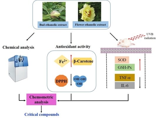
Comparison of Chemical Compositions, Antioxidant, and Anti-Photoaging Activities of Paeonia suffruticosa Flowers at Different Flowering Stages

1
Bioengineering Research Centre, Guangzhou Institute of Advanced Technology, Chinese Academy of Sciences, Guangzhou 511458, China
2
Guangdong Provincial Key Laboratory of New Drug Screening, School of Pharmaceutical Sciences, Southern Medical University, Guangzhou 510515, China
3
CAS Key Laboratory of Tropical Marine Bio-Resources and Ecology, South China Sea Institute of Oceanology, Chinese Academy of Sciences, Guangzhou 510301, China
4
Guangdong Key Laboratory of Marine Materia Medica, South China Sea Institute of Oceanology, Chinese Academy of Sciences, Guangzhou 510301, China
*
Authors to whom correspondence should be addressed.
Antioxidants 2019, 8(9), 345; https://doi.org/10.3390/antiox8090345
Submission received: 15 July 2019 / Revised: 14 August 2019 / Accepted: 20 August 2019 / Published: 1 September 2019
(This article belongs to the Special Issue Phenolic Profiling and Antioxidant Capacity in Plants)

Abstract

:
Paeonia suffruticosa is an ornamental, edible, and medicinal plant. The ethanolic extracts of P. suffruticosa bud and flower were examined for their antioxidant, anti-photoaging, and phytochemical properties prior to chemometric analysis. The results showed that the bud ethanolic extract (BEE) and the flower (the early flowering stage) ethanolic extract (FEE) had better antioxidant activities, and significantly increased the activities of superoxide dismutase (SOD) and glutathione peroxidase (GSH-Px) and reduced the levels of tumor necrosis factor-α (TNF-α) and interleukin-6 (IL-6) in the skin tissues. In total, 68 compounds, including 20 flavonoids, 15 phenolic derivatives, 12 terpenoids, 9 fatty acids, and 12 others were identified or tentatively identified by ultra-fast liquid chromatography quadrupole time-of-flight mass spectrometry (UFLC-Q-TOF-MS). Gallic acid, 1,2,3,4,6-O-pentagalloyl glucose, paeoniflorin, and oxypaeoniflorin were predominant compounds in the extracts. Taken together, P. suffruticosa flowers are a candidate for functional material in food and health related industries, and their optimal time to harvest is before the early flowering stage.

Graphical Abstract

1. Introduction

Ultraviolet-B (UVB) radiation is one of the most effective constituents of solar light, and has become the primary source of oxidative stress to humans. UVB generally impacts the basal layer of epidermal skin and causes premature skin photoaging, local and systemic immunosuppression, cutaneous inflammatory disorders, and photocarcinogenesis [1]. It is concluded that oxidative stress is a problem of skin cells and that endogenous and exogenous antioxidants could play an important role in decreasing it [2]. A large number of phytochemicals obtained from plants could reduce the harmful effects of oxidative stress and help to prevent the photoaging of skin [3,4]. Experimental and epidemiological studies indicated that the consumption of plant foods is related to lower incidence of age-related diseases [5]. Thus, there is increasing interest in the antioxidant properties of phytochemicals found in plant foods.
Paeonia suffruticosa, belonging to Paeoniaceae, is mainly distributed in China, Japan, America, and Europe, and is an ornamental, edible, and medicinal plant in China and Japan [6,7]. Root bark of P. suffruticosa, named Cortex Moutan, is an important Chinese traditional medicine with the functions of lowering blood sugar, lowering blood pressure, anti-inflammatory, anti-bacterial, anti-tumor, and regulating the cardiovascular system [8]. P. suffruticosa flower as a characteristic natural resource that also has a long history of medicinal and edible use in China. Monoterpenoids, flavonoids, and essential oil have been found in P. suffruticosa flower [6,9,10]. The essential oils of P. suffruticosa flower buds possesses an inhibitory effect on common food-borne bacterial pathogens [6]. Moreover, P. suffruticosa flower as medicinal herb is used for the treatment of gynaecological diseases, and has experienced a growing number of applications in the food industry to produce cake, herbal tea, and drink, as well as in the cosmetic industry to produce facial masks and sunscreen creams. It is known that the chemical constituents that are responsible for the bioactivity are affected by the different flowering stages [11,12]. However, the characteristic phytochemicals and the composition change in the flowers are unclear and there is no principle to guide as to which flowering stage is suitable for harvesting when the flower is used in industry. The study on the relationship between phytochemicals and bioactivity during the flowering stages is less documented, which is a limitation to the development of products originated from the P. suffruticosa flower.
In this study, the antioxidant activities of the ethanolic extracts of P. suffruticosa flowers were evaluated using four different methods, including 2,2-diphenyl-1-picrylhydrazyl (DPPH) and hydroxyl radical scavenging activity, inhibition of β-carotene bleaching, and ferric reducing antioxidant power (FRAP) assays. The anti-photoaging activity of the ethanolic extracts of P. suffruticosa flowers was evaluated using a UVB-irradiated mouse model. The characteristic chemicals of the ethanolic extract of P. suffruticosa flowers were investigated prior to the correlation analysis between the multiple ingredients and their bioactivities.

2. Materials and Methods

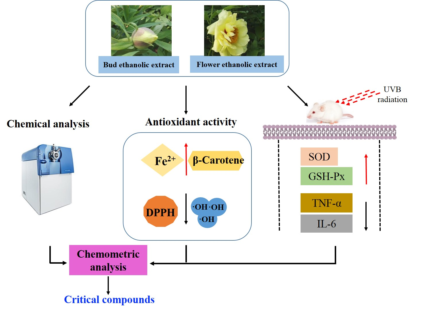
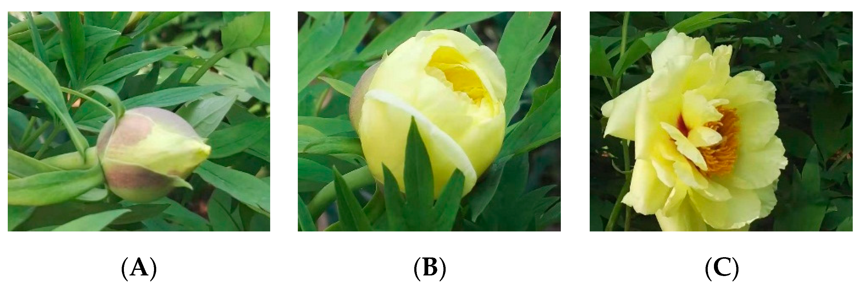
2.1. Plant Materials

P. suffruticosa flowering buds were collected on 30 March 2016, and P. suffruticosa flowers at the early flowering stage were collected ten days later. P. suffruticosa flowers at the full flowering stage was collected on 20 April 2016, and then divided into petal and stamen (Figure 1). All of P. suffruticosa samples were collected in Luoyang, Henan province, China. All voucher specimens were stored at the Guangzhou Institute of Advanced Technology, Chinese Academy of Sciences, Guangdong, China.

2.2. Chemicals and Reagents

Leucine, gallic acid, adenosine, tryptophan, and p-hydroxybenzoic acid were purchased from the National Institute for the Control of Pharmaceutical and Biological Products (Beijing, China). Morroniside, loganin, and geniposide were purchased from Chengdu Biopurify Phytochemicals Ltd. (Chengdu, China). Paeoniflorin, oxypaeoniflorin, benzoylpaeoniflorin, paeonol, 1,2,3,4,6-O-pentagalloyl glucose, luteolin, and apigenin were purchased from Chengdu Pufei De Biotech Co., Ltd. (Chengdu, China). Quercetin-3-O-glucoside and rhoifolin were purchased from Sigma-Aldrich (Shanghai, China). Hexadecanoic acid, 9-octadecenoic acid, octadecanoic acid, and 2,4,6-tris(2-pyridyl)-1,3,5-triazine were purchased from Shanghai Yuan Ye Biotechnology Co., Ltd. (Shanghai, China). 2,2-diphenyl-1-picrylhydrazyl (DPPH), butylated hydroxytoluene (BHT), β-carotene, and vitamin C (VC) were purchased from Shanghai Ekear Bio-Tech Co., Ltd. (Shanghai, China). HPLC-grade acetonitrile and LC/MS grade methanol were purchased from Fisher Scientific (Fair Lawn, NJ, USA). Water was obtained from an ultrapure water system (Purelab Plus, Pall, Port Washington, NY, USA).

2.3. Sample Preparation

The dried and ground sample (100 g) was extracted with 95% (v/v) ethanol (800 mL) in a KQ600DE ultrasonic bath (Kunshan, Jiangsu, China) for 30 min at room temperature. This extraction was repeated after filtration. The combined filtrate was evaporated under vacuum to yield the ethanolic extract. The dried ethanolic extract was weighed accurately and dissolved in 95% ethanol to obtain a series of solutions with different concentrations for the further study.

2.4. Total Phenolic Content Assay

Total phenolic content was determined using the described method [13]. Sample solution (1.0 mL) was mixed with the Folin–Ciocalteu reagent (5.0 mL; 0.1 mmol/L) and allowed to incubate for 5 min. A sodium carbonate solution (4.0 mL; 75 g/L) was added, and the mixture was vortexed and incubated at 25 °C for 30 min in darkness. A standard gallic acid solution was used and the absorbance was determined at 765 nm by spectrophotometer (UV-6100; Shanghai Metash Instrument Co., Ltd., Shanghai, China). Total phenolic content was expressed as mg gallic acid equivalents per gram of extract (mg GAE/g ext.). All measurements were performed in triplicate.

2.5. Total Flavonoid Content Assay

Total flavonoid content was measured using a previously reported method [13]. In this procedure, a mixture of an aliquot (2.0 mL) of sample solution, a 95% ethanol solution (4.0 mL), and sodium nitrite solution (2.0 mL; 50 g/L) was incubated at 25 °C for 5 min, and aluminium nitrate solution (2.0 mL; 100 g/L) was then added. After incubation at 25 °C for 6 min, sodium hydroxide solution (5.0 mL; 4 g/L) was added. Rutin was used to calculate the standard curve, and the absorbance was determined at 510 nm by spectrophotometer. Total flavonoid content was expressed as mg rutin equivalent per gram of extract (mg RE/g ext.). All measurements were performed in triplicate.

2.6. Antioxidant Assay

2.6.1. DPPH Radical Scavenging Activity Assay

DPPH radical scavenging activity was measured according to a previously reported method [14]. Briefly, sample solution (1.0 mL) was mixed with a freshly prepared methanol solution of DPPH (1.0 mL; 80 μg/mL). The absorbance was measured at 517 nm spectrophotometrically after the mixture was incubated for 30 min at 25 °C. VC and BHT were used as two standard antioxidant compounds. All determinations were performed in triplicate. In control experiment, 95% ethanol (2.0 mL) was used. The radical scavenging capability of DPPH was calculated using the following equation: Scavenging effect (%) = (1 − Asample/Acontrol) × 100, where Asample and Acontrol are the absorbance of the sample and the control, respectively.

2.6.2. Hydroxyl Radical Scavenging Activity Assay

The hydroxyl radical scavenging activity was determined according to a previously described method [13]. The hydroxyl radical was produced by the Fenton reaction between ferrous sulphate and hydrogen peroxide. Briefly, a sample solution (2.0 mL), a ferrous sulphate solution (2.0 mL; 5.0 mmol/L), a salicylic acid solution (2.0 mL; 5.0 mmol/L), and a hydrogen peroxide solution (2.0 mL; 5.0 mmol/L) were added to the test tubes. The absorbance was measured spectrophotometrically at 510 nm after the mixture was incubated at 37 °C for 1 h. VC was used as the standard antioxidant compound. In control experiment, 95% ethanol (2.0 mL) was used. All determinations were performed in triplicate. The hydroxyl radical scavenging capability was calculated using the following equation: Scavenging effect (%) = [1 − (AsampleA-hydrogen peroxide)/Acontrol] × 100, where Asample, A-hydrogen peroxide, and Acontrol are the absorbance of the sample, the sample without hydrogen peroxide, and the control, respectively.

2.6.3. Ferric Reducing Antioxidant Power (FRAP) Assay

In this study, FRAP assays were performed according to a previously described method [14]. The antioxidants could restore ferric tripyridyltriazine (Fe3+-TPTZ) to blue Fe2+-TPTZ under acidic conditions. The FRAP solution was prepared by mixing acetate buffer (300 mM, pH 3.6), TPTZ (10 mM) and FeCl3 solution (20 mM) in a ratio of 10:1:1 (v/v/v). An aliquot (0.15 mL) of sample solution was mixed with the FRAP solution (2.85 mL) and incubated at 37 °C for 30 min. The total antioxidant activity of the samples could be evaluated by the absorbance of Fe2+-TPTZ at 593 nm. All measurements were performed in triplicate. The results of the FRAP assay were reported in mM FeSO4.

2.6.4. Inhibition of β-Carotene Bleaching Assay

Inhibitory ability of β-carotene bleaching was assessed according to a previously described method [14]. Briefly, a solution of β-carotene was prepared by dissolving 2 mg in chloroform (10 mL). Two millilitres of this solution was pipetted into a round-bottom flask. After the chloroform was removed at 40 °C under vacuum, linoleic acid (40 mg), Tween-80 emulsifier (400 mg), and distilled water (100 mL) were added to the flask with vigorous shaking. An aliquot (4.8 mL) of this emulsion was transferred to different test tubes containing the sample solution (0.2 mL). A 4.8 mL portion of the emulsion combined with 95% ethanol (0.2 mL) was used as a control. The tubes were shaken and incubated at 50 °C in a water bath. As soon as the emulsion was added to each tube, the absorbance was measured at 470 nm at 0 min and at 15-min intervals over a 120 min period. All measurements were performed in triplicate. The β-carotene bleaching inhibition was calculated using the following equation: Bleaching inhibition (%) = [1 − (Asample–0Asample–t)/(Acontrol–0Acontrol–t)] × 100, where Asample–0 and Acontrol–0 are the initial absorbance (t = 0 min) of the sample and the control, respectively; Asample–t and Acontrol–t are the absorbance (t = 15, 30, 45, 60, 75, 90, 105, and 120 min) of the sample and the control, respectively.

2.7. Animal Treatment and UV Irradiation

Kunming mice (body weights of 18–20 g) were purchased from the Experimental Animal Center of Southern Medical University (Guangzhou, China). Mice were fed on standard laboratory diet and water at libitum, and kept in 12 h dark/light cycle room at 21 ± 3 °C with a relative humidity of 55% ± 10% for one week prior to the ultraviolet treatment. Animal experiments and all procedures were approved by the Animal Care and Use Committee of Southern Medical University (Guangzhou, China). Mice were randomly divided into 7 groups (n = 6), control group (no irradiation), model group (irradiated group with no treatment), model + emulsifier (ME) group (irradiated group treated with emulsifier containing stearic acid 10.0 g, glycerin monostearate 3.0 g, liquid paraffin 5.0 g, vaseline 1.0 g, lanolin 4.0 g, triethanolamine 1.0 g, and distilled water 50 mL), bud ethanolic extract (BEE) group (irradiated group treated with 2.0% (w/w) BEE in emulsifier), flower ethanolic extract (FEE) group (irradiated group treated with 2.0% (w/w) FEE in emulsifier), petal ethanolic extract (PEE) group (irradiated group treated with 2.0% (w/w) PEE in emulsifier), stamen ethanolic extract (SEE) group (irradiated group treated with 2.0% (w/w) SEE in emulsifier). The shaved dorsal skin of the mice was exposed to 120 mJ/cm UVB radiations (emission peak 306 nm; Sankyo Denki Co., Tokyo, Japan) for 20 min, thrice a week. The preparations (0.2 g) were topically administered daily to the back of the mouse skin. Mice were anaesthetized and their dorsal skin tissues were collected at the end of experiment (4 weeks). Dorsal skin tissues were washed with physiological saline (0.9% NaCl) and stored at −80 °C until analyses were performed. Half of dorsal skin tissue was used for histological analysis, and the other half was used for biochemical analysis.

2.8. Histological Analysis

Dorsal mouse skin samples were fixed overnight with 4% paraformaldehyde in phosphate buffered saline (PBS) at 4 °C and then embedded in paraffin. Paraffin sections (4 μm) were mounted on silane-coated slides and stained with Hematoxylin&Eosin (H&E) using H&E staining kit (Sangon Biotech (Shanghai) Co., Ltd., Shanghai, China). The sections were examined, and images were recorded using a microscope and Image-Pro Express 5.1.1.14 Pathology Image Analysis System (Olympus Corporation, Tokyo, Japan) at 400× magnifications. The epidermal thickness was measured at 8 random sites randomly selected locations per slide using the image analysis program.

2.9. Biochemical Analysis

The dorsal skin tissues were homogenized in a glass homogenizer with a buffer containing 1.15% KCl in a 1:10 (w/v) whole homogenate. The homogenates used for the biochemical analysis were obtained by centrifugation at 12,000× g for 30 min. Protein levels in skin homogenates were measured using the Pierce BCA protein assay kit (Beyotime Biotechnology, Shanghai, China). Concentrations of superoxide dismutase (SOD), glutathione peroxidase (GSH-Px), tumor necrosis factor-α (TNF-α), and interleukin-6 (IL-6) in dorsal skin tissues were determined using mouse-specific enzyme-linked immunosorbent assay (ELISA) kits (NeoBioscience, Shenzhen, China). Analysis was performed according to the manufacturer’s instruction.

2.10. UFLC-DAD-Q-TOF-MS Analysis

2.10.1. System and Conditions

Ultra-fast liquid chromatography (UFLC) analysis was performed on a Shimadzu UFLC XR system (Shimadzu Corp., Kyoto, Japan) with an Agilent Eclipse Plus C18 column (2.1 i.d. ×100 mm, 1.8 μm, Agilent Technologies, CA, USA). The mobile phases were composed of methanol (A) and water with 0.1% formic acid (B) using a linear gradient elution of 5–100% A within 30 min at 0.3 mL/min. The injection volume was 5 μL, and the column temperature was set at 25 °C. The identification experiment was performed using AB SCIEX Triple TOF 5600 plus mass spectrometer system (AB SCIEX, Foster City, CA, USA). The system was operated using the Analyst TF 1.6 software (AB SCIEX, Foster City, CA, USA). The parameters of the MS detector were as follows: Ion source gas 155 psi; ion source gas 255 psi; curtain gas 30 psi; source temperature 550 °C; ion spray voltage floating 4500 V; collision energy 35 eV; collision energy spread 15 eV; and declustering potential 80 eV. Spectra were acquired in a scan range from m/z 100–1500. Both positive and negative ion modes were used for compounds ionization. Nitrogen was used as the nebulizer and auxiliary gas.

2.10.2. Establishment of Tentative Peak Assignment

The UFLC-Q-TOF-MS data of samples were extracted and analysed using the PeakView software (AB SCIEX, Foster City, CA, USA), mainly with the XIC manager tool, which provided the quasi-molecular weights, mass errors, and isotope pattern fits. The predicted formula with errors less than ± 5 ppm was searched against the compounds reported in the genus Paeonia to obtain the tentative identification. The identification of the compounds within the samples was further confirmed by determining the possible elemental compositions of the fragment ions and the proposed fragmentation pathways using their MS spectrum.

2.11. HPLC-DAD Analysis

High performance liquid chromatography (HPLC) analysis was performed on an Agilent (CA, USA) 1290 UPLC-DAD system with a reverse-phase Agilent EC-C18 column (3.0 i.d. × 100 mm, 2.7 μm, Agilent Technologies, CA, USA). A gradient elution mobile phase system composed of 0.5% formic acid solution (A) and acetonitrile (B) was applied as follows: 0–3 min, 5–8% B; 3–4 min, 8–13% B; 4–13 min, 13–14% B; 13–14 min, 14–20% B; 14–29 min, 20–30% B; 29–30 min, 30–50% B; 30–33min, 50–95%B. The mobile phase flow rate was 0.3 mL/min, and the column temperature was maintained at 30 °C. The injection volume was 5 μL and the UV wavelength was set at 254 nm.
The developed method was validated by linear range, limit of detection (LOD), limit of quantitation (LOQ), precision, and recovery according to the previous report [14].

2.12. Statistical Analysis

The results were expressed as the mean ± standard deviation (SD) of each experiment. The data obtained were analysed using a one-way analysis of variance (ANOVA) followed by Tukey’s multiple comparison test. A p value < 0.05 was considered to be significant. Pearson’s correlation analysis was used to characterize the correlation between the phytochemicals and the bioactivities. One-way ANOVA and Pearson’s correlation analysis were performed using the software IBM SPSS Statistics (version 19.0).

3. Results and Discussion

3.1. Total Phenolic and Flavonoid Contents

The extract yields of bud, flower, petal, and stamen of P. suffruticosa had obvious differences (Table 1). Total phenolic contents in the BEE, FEE, PEE, and SEE were determined. BEE had the highest total phenolic content (191.80 ± 3.44 mg GAE/g ext.), followed by FEE (113.84 ± 0.84 mg GAE/g ext.), SEE (77.25 ± 0.58 mg GAE/g ext.) and PEE (51.66 ± 0.34 mg GAE/g ext.). Unexpectedly, FEE had the highest total flavonoid content (49.87 ± 0.82 mg RE/g ext.), followed by BEE (30.60 ± 0.94 mg RE/g ext.), PEE (12.98 ± 0.78 mg RE/g ext.) and SEE (8.86 ± 0.98 mg RE/g ext.). Total phenolic content in the flowers had a trend of gradual decrease during the flowering stage of P. suffruticosa. In the other hand, there might be a maximum content of flavonoids in the flowering stage.

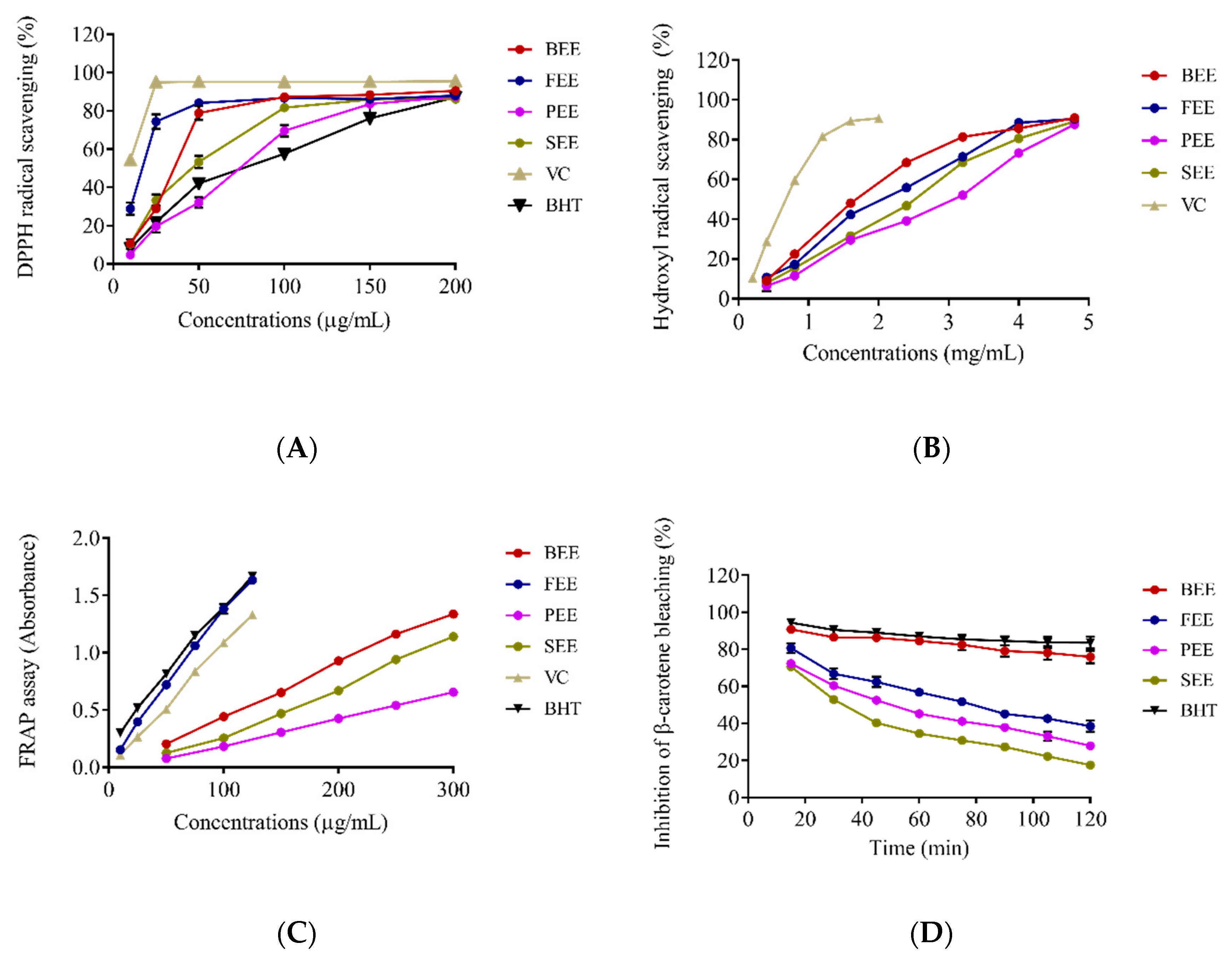
3.2. Antioxidant Activity In Vitro

The antioxidant capabilities of BEE, FEE, PEE, and SEE were tested by the DPPH and hydroxyl radical scavenging, FRAP, and inhibition of β-carotene bleaching assays (Figure 2). The FEE had stronger DPPH radical scavenging activity than the other extracts and BHT below 50 μg/mL, while it was obviously weaker than that of VC at concentration 10–200 μg/mL. The BEE and SEE also had stronger activity than BHT. On the basic of IC50 values, antioxidant activity is defined as the concentration of antioxidant required for 50% scavenging of the radicals (Table 1). The IC50 value of FEE for scavenging DPPH was 14.83 ± 2.03 μg/mL, which was lower than that of BEE (34.44 ± 1.35 μg/mL), PEE (64.00 ± 5.60 μg/mL), and SEE (42.70 ± 0.79 μg/mL). A lower IC50 value suggested that antioxidant activity was better in the four extracts. Unexpectedly, the IC50 value of BEE was 1.57 ± 0.04 mg/mL, which was lower than that of FEE (1.79 ± 0.02 mg/mL), PEE (2.57 ± 0.06 mg/mL), and SEE (2.10 ± 0.004 mg/mL). The BEE had strongest hydroxyl radical scavenging activity than the other extracts, but was weaker than VC. In the FRAP assay, FEE had the strongest activity, followed by BEE, PEE, and SEE. Moreover, FEE had similar activity to BHT, but was much stronger than VC. The activity of BEE in the inhibition of the β-carotene bleaching assay was a little weaker than that of BHT (100 μg/mL) over 120 min, but had stronger and longer lasting inhibitory activity than that of other extracts at 100 μg/mL. Based on the results of the antioxidant evaluation, the antioxidant activities of BEE and FEE were noticeable. BEE had a greater effect on hydroxyl radical scavenging and inhibition of the β-carotene bleaching, whereas FEE had a greater effect on DPPH radical scavenging and FRAP. These results indicated that there was a correlation between bioactivity and the flowers at the different harvest time, due to the change of chemical compositions.

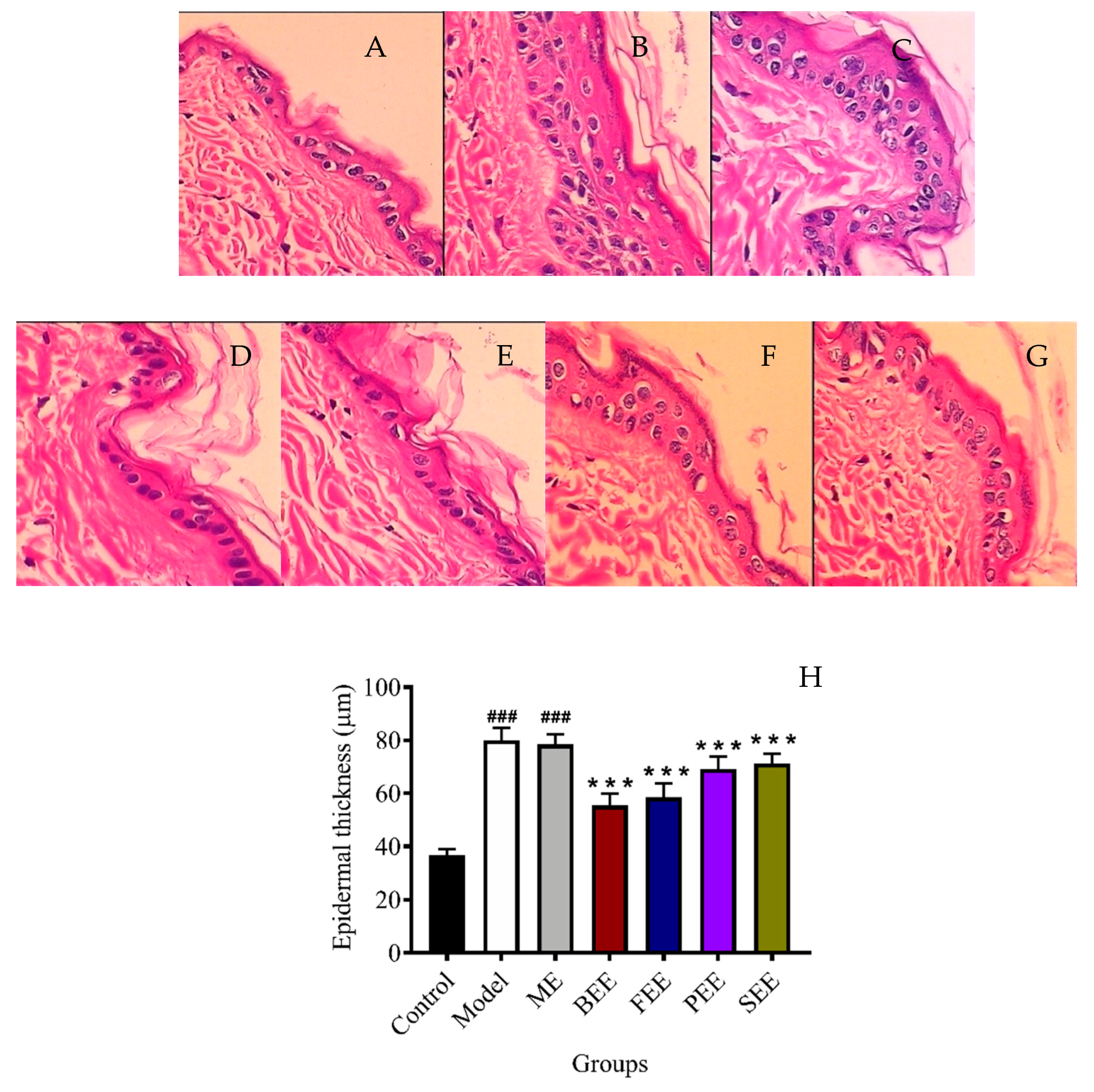
3.3. Effects on the Morphology in Mouse Skin

Skin that is exposed excessively to UVB irradiation is easily damaged. UVB irradiation led to excessive damage to the morphology and integrity of the mouse skin. It is reported that UVB radiation on the skin induces a variety of responses in the epidermis, including keratinocyte proliferation that leads to epidermal hyperplasia and thickening [15]. In this study, the change incurred on the mouse skin was assessed after UVB irradiation. The epidermal thickness of the ME group significantly increased by 1.14 fold when compared to that of the control group. In the case of the extract treatment groups, the epidermal thickness of BEE, FEE, PEE, and SEE groups significantly decreased (p < 0.001) to 70.62%, 74.62%, 88.13%, and 90.74%, respectively, when compared to that of the ME group (Figure 3H). Topical treatment with the four extracts had the effect of inhibiting the epidermal thickening, and of which BEE could best prevent UVB-induced damage to the skin. A lot of studies have reported natural products are the important resources of antioxidants [16,17]. Phenols and flavonoids, the main constituents in flowers, were considered as antioxidants and UV absorbing secondary metabolites [18,19]. It has also been reported that flavonoids help to prevent the skin photoaging by regulating pro-inflammatory cytokines [20,21]. Interestingly, abundant phenols and flavonoids were found in the BEE and FEE, respectively. Phenols and flavonoids in the BEE and FEE appear to be useful against the chronic effects of UV light. More studies on antioxidant and anti-inflammatory activities of the four extracts were performed in the next experiments.

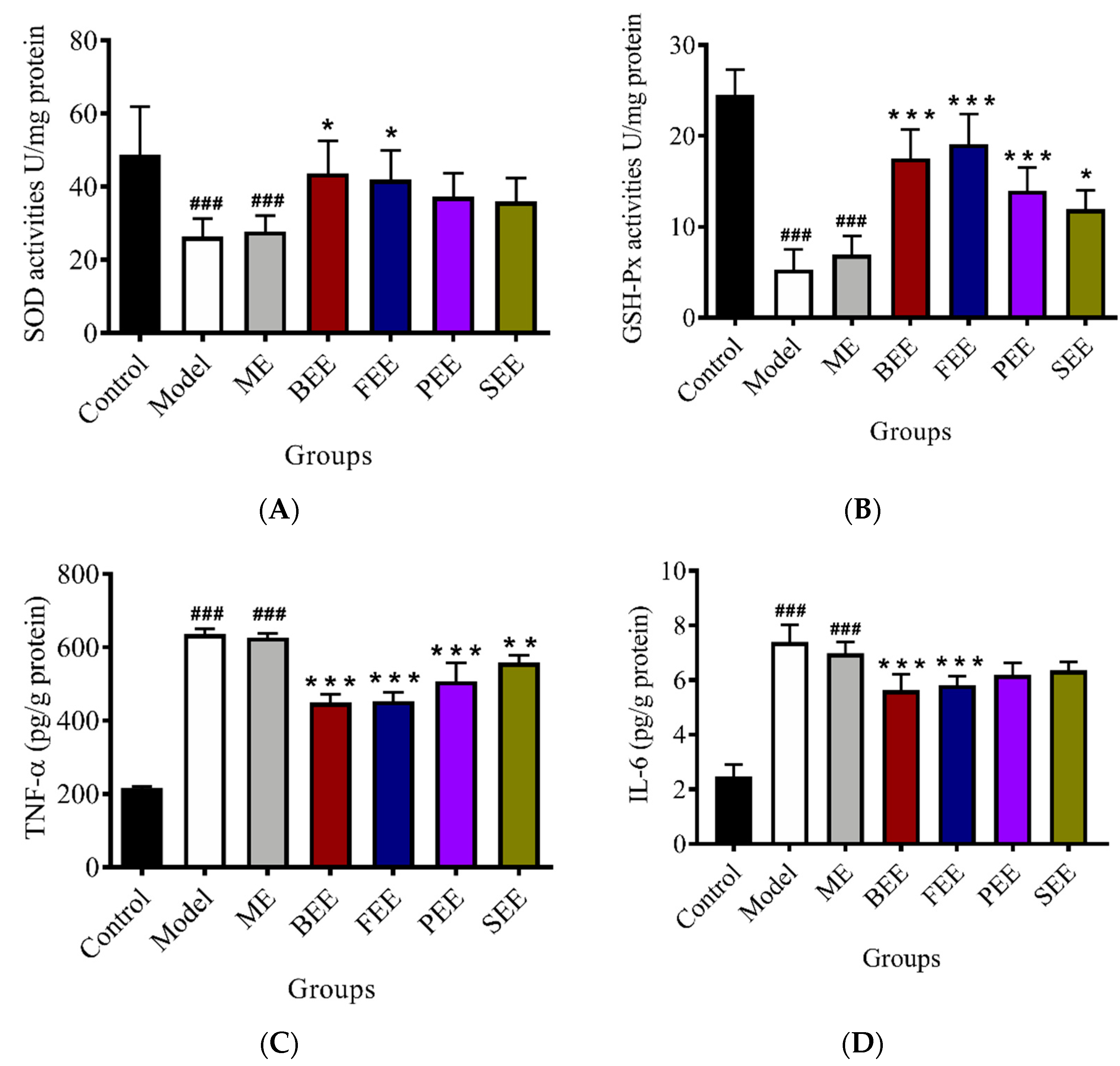
3.4. Effects on the Activities of SOD and GSH-Px in Mouse Skin

The activities of endogenous antioxidants in the skin tissues were further investigated (Figure 4A,B). The activity of SOD in the model group and model + emulsifier groups was significantly decreased when compared with the control group. Using a UVB-irradiated mouse model, topical treatment with BEE and FEE could significantly increase (p < 0.05) the activity of SOD, but topical treatment with PEE and SEE had no significant effect on the activity of SOD. The activity of GSH-Px in the skin tissues was significantly decreased when the mice were treated with UVB irradiation. Topical treatment with BEE (p < 0.001), FEE (p < 0.001), PEE (p < 0.001), and SEE (p < 0.05) significantly increased the activity of GSH-Px in the skin of UVB irradiated mice.
UVB irradiated skin depletes antioxidant defensive capabilities [5]. ROS plays a significant role in UVB-induced skin carcinogenesis. In the body, endogenous antioxidants offset UV-induced oxidative stress by the neutralization of the ROS prior to oxidative changes occuring in the tissues [22]. SOD could alternately catalyze the dismutation of the superoxide radical into either ordinary molecular oxygen or hydrogen peroxide, and thereby is an important antioxidant defense [23]. GSH, a free radical-scavenger and a cofactor for protective enzymes, plays a pivotal role in protecting cells from oxidative damage [24]. Normally, these enzymes are able to scavenge the ROS efficiently, and consequently protect skin from damage. However, excessive and chronic exposure to UVB radiation can overwhelm the cutaneous antioxidant capacity leading to oxidative damage, resulting in skin photoaging [25]. BEE and FEE with excellent antioxidant capability could significantly increase the activities of SOD and GSH-Px in the damaged skin, which was responsible for inhibiting the epidermal thickening, consequently ameliorating the skin’s pathological symptom.

3.5. Effects on the Leveles of TNF-α and IL-6 in Mouse Skin

As Figure 4C,D shows, inflammatory cytokines (TNF-α and IL-6) in the UVB irradiated group were significantly increased when compared with the control group. Topical treatment with BEE (p < 0.001), FEE (p < 0.001), PEE (p < 0.001), and SEE (p < 0.01) significantly decreased the content of TNF-α in the skin tissues of UVB irradiated mice. BEE and FEE could significantly decrease the content of IL-6 in the skin tissues of UVB irradiated mice. Contrarily, no significant effect on the contents of IL-6 was found in PEE and SEE treated groups.
Inflammation response that causes erythema, edema, and an influx of inflammatory cells, is one of the most obvious results of UVB radiation. Epidermal keratinocytes strongly contribute to cutaneous inflammation via their release of pro-inflammatory cytokines [26]. Pro-inflammatory cytokines, such as TNF-α and IL-6, play crucial roles in inflammatory development and are considered to be indicators of the degree of inflammation [27]. Moreover, pro-inflammatory cytokines stimulate the epidermal keratinocytes and dermal fibroblasts, and degrade dermal collagen and elastic fibers, consequently causing the epidermal thickening and the formation of wrinkles [4]. Accordingly, the inhibition of pro-inflammatory cytokines secretion induced by UVB irradiation is important to protect skin from photo-damage. BEE and FEE could significantly decrease the levels of TNF-α and IL-6 in damaged skin and thereby partly compensate for the inflammation induced by UVB radiation. As a result, the anti-inflammatory capability of BEE and FEE was responsible for ameliorating the skin’s pathological symptom. These results suggested that the BEE and FEE, with more excellent antioxidant and anti-inflammatory capabilities, had a greater effect on the prevention of skin photoaging. Their molecular mechanism of anti-photoaging is still unclear and is, thus, required in future study.

3.6. Chemical Compositions of Ethanolic Extracts of P. suffruticosa Flowers

Compounds at the different flowering stages of P. suffruticosa were identified by UPLC-Q-TOF-MS/MS method through comparison retention time (Rt), accurate molecular weight (MW), and fragmentography with the reference standards. When no standards were unavailable, compounds were tentatively identified by the accurate molecular weight and fragmentation pathway, which had been carried out on the same kind of compounds or literature report. As shown in Table 2, a total of 68 compounds, including 12 monoterpenoids, 20 flavonoids, 15 phenols and their derivatives, 9 fatty acids, and 12 others, were identified or tentatively identified in this study.
Monoterpenoids are present as ubiquitous chemical compounds in P. suffruticosa, which are characterized by two isoprene units and have the molecular formula C10H16. Among them, paeoniflorin (28), oxypaeoniflorin (21), and benzoylpaeoniflorin (54) were precisely identified by the comparison to their reference standards, respectively. Analyzing the mass spectrogram, the fragment ions were usually generated by losing the units of glucose residue (C6H10O5, 162 Da), benzoic acid residue (C7H6O2, 122 Da), and hydroxybenzoic acid residue (C7H6O3, 138 Da). Therefore, compound 3 with m/z 375.12986 (calculated to be C16H24O10), compound 17 with m/z 527.14061 (calculated to be C23H28O14), compound 23 with m/z 525.16213 (calculated to be C24H29O13), compound 30 with m/z 647.16060 (calculated to be C30H32O16), compound 36 with m/z 631.16737 (calculated to be C30H32O15), compound 42 with m/z 615.17285 (calculated to be C30H32O14), compound 49 with m/z 599.17976 (calculated to be C30H32O13), compound 51 with m/z 599.18003 (calculated to be C30H32O13), and compound 52 with m/z 629.19181 (calculated to be C31H34O14) in negative mode were tentatively identified as 8-debenzoylpaeoniflorin [28], debenzoygalloypeaoniflorin, mudanpioside E, galloyoxypaeoniflorin, galloylpaeonifflorin, benzoyloxypaeoniflorin, mudanploside H, mudanpioside C, benzoloxypaeoniflorin, and mudanpioside J, respectively [29].
Flavonoids, especially quercetin, kaempferol, isorhamnetin, apigenin and their derivatives, are another set of components also identified in P. suffruticosa [30]. Their fragment behaviors have been investigated very well in positive and negative ion modes. Usually, the [aglycone−H] ion is generated by loss of the linkage of sugars and thereafter followed by Ret–Diels–Alder (RDA) dissociation, subsequently generating m/z 179 and 151 ions [31]. Compounds 46, 53, and 55 were undeniably identified as rhoifolin, luteolin, and apigenin, respectively. According to the fragmentation mechanism, compound 45 were inferred as apigenin-7-O-glucoside with m/z at 431.10019 in negative ion mode [30]. Kaempferol is an isomer of luteolin with molecular formula C15H10O6. Compounds 29, 39, 41, and 43 were inferred as kaempferol-3,7-O-di-glucoside, kaempferol-3-O-glucoside, kaempferol-3-O-rutinoside, and kaempferol-3-O-(2′′-O-galloyl)-glucoside [10,30]. Isorhamnetin with molecular formula C16H12O7, was 30 Da (CH2O) higher than luteolin. Compounds 44 and 48 were isomers with molecular formula of C22H22O12. The fragment ion at m/z 317 was yielded by loss of glucose. Thus they were tentatively identified as isorhamnetin-3-O-glucoside and isorhamnetin-7-O-glucoside, respectively. Compound 34 gave an [M−H] ion at m/z 639.15895 (calculated as C28H31O17) and was inferred as isorhamnetin-3,7-di-O-glucoside [32]. Quercetin was C15H10O7, 16 Da (O) higher than luteolin. Compound 40 was quercetin-3-O-glucoside with an [aglycone−H] ion at m/z 301.0354 by loss of glucose residue [10]. Compounds 26 and 33 were isomers, which gave [M−H] at m/z 625 and were tentatively identified as quercetin-3,7-di-O-glucoside or its isomer [32]. Compound 37 was 30 Da (CH2O) higher than compound 34 and was inferred as 6,3′-dimethoxyquercetin-7-O-di-glucoside with m/z at 669.16952. Compound 22 (eriodictyol-7-O-glucoside) was calculated as C21H22O11 and generated a fragment ion at m/z 287 by loss of one glycoside. Compound 32 (patuletin-3,5-di-O-glucoside) was calculated as C28H32O18, and generated a fragment ion at m/z 331 by loss of two glycosides [32]. Compound 38 was inferred as monoxerutin with a quasi-molecular ion at m/z 653.17535. Diosmin (47) gave a quasi-molecular ion at m/z 607.17007, and subsequently generated an ion of 299 by loss of a rutinoside. Compound 56 has 30 Da (CH2O) more than apigenin, and it was identified as chrysoeriol with an [M−H] ion at m/z 299.05704.
There were 14 phenols and derivatives found in the four extracts of P. suffruticosa flowers. Compounds 5, 15, 35, and 50 were directly identified as gallic acid, p-hydroxybenzoic acid, 1,2,3,4,6-O-pentagalloyl glucose, and paeonol by the comparison of their reference standards, respectively. Compounds 4 and 9 were a pair of isomers, which showed 162 Da more than gallic acid, suggesting the presence of C6H10O5. They were tentatively identified as glucogallin or isomer. Interestingly, compound 10 gave 324 Da more than gallic acid, suggesting the presence of two C6H10O5 [33]. Compound 16 showed CH2 (14 Da) more than gallic acid, and was referred to be methyl gallate [29]. In mass spectrum, compound 12 was determined to be C14H18O9 and yield an ion at m/z 167.0341 [M−H−C6H10O5]. It was tentatively identified as mudanoside A [29]. Compound 14 gave an [M−H] ion at m/z 315.07227, corresponding to C13H16O9. It yielded daughter ions by successive losses of C6H10O5, CO2 and was inferred to be gentisic acid-5-O-glucoside. Compounds 24 and 31 were tentatively identified as trigalloyl glucose and teragalloyl glucose. Compound 20 was an isomer of trigalloyl glucose with a characteristic structure of C7H6O3. Compound 25 gave an [M+H]+ ion at m/z 155.07034, corresponding to C8H10O3, and was tentatively identified as 3,4-dimethoxyphenol. Compound 27 was deduced as paeonolide based on the deprotonated ion at m/z 299.1206 and a fragment ion at m/z 167.0707.
Long chain fatty acid often provides ions by losses of CO2, H2O, and (CH2)n in MS spectrum. Compounds 64, 66, and 68 were accurately identified as hexadecanoic acid, 9-octadecenoic acid, and octadecanoic acid by comparing them with their reference standards. Compound 57 with m/z 327.21889 (calculated to be C18H32O5), compound 58 with m/z 329.23479 (calculated to be C18H34O5), compound 59 with m/z 287.22468 (calculated to be C16H32O4), compound 60 with m/z 293.21325 (calculated to be C18H30O3), compound 61 with m/z 295.22859 (calculated to be C18H32O3), and compound 65 with m/z 355.32323 (calculated to be C22H44O3) were tentatively identified as malyngic acid, 9,12,13-trihydroxyoctadec-10-enoic acid, 3,12-dihydroxyhexadecanoic acid, 9-oxooctadeca-10,12-dienoic acid, 13-hydroxy-9,11-octadecadienoic acid, and 2-hydroxybehenic acid, respectively [34].
Compound 1 showed [M+H]+ and [M+Na]+ at m/z 343.12331 and 365.10508, and gave [M+Na-C6H10O5]+ at m/z 203.0519, suggesting the presence of disaccharide structure. Leuine (2) and tryptophan (13) were two amino acids with typical fragmentation ions by the loss of CO2 and NH3, and further confirmed by comparison with their reference standards. Compounds 7, 18, and 19 were three iridoids, and precisely identified as morroniside, loganin, and geniposide when compared with their reference standards. Guanosine (6) and adenosine (8) were two nucleosides. Compound 8 was directly identified as adenosine by comparison with its reference standard. Compound 6 was tentatively identified with accurate molecular weight and fragment ions in both positive and negative modes. Compound 11 gave an [M−H] ion at m/z 218.1036 and was tentatively identified as N-(tert-Butoxycarbonyl)threonine. Compounds 62 and 63 were both pentacyclic triterpenoids with [M−H] ions at m/z 471.34891 and 455.35374, and were tentatively identified as hederagenin and oleanolic acid, respectively [33]. Compound 67 gave an [M−H] ion at m/z 399.32889, calculated to be C27H44O2, and was inferred as dehydrotocopherol.
It can be shown that the flavonoid and phenolic contents in P. suffruticosa flower changes significantly during the flowering development stage (Figure 5). In this study, all compounds were detectable both in BEE and PEE. p-Hydroxybenzoic acid (15) could not be detected in FEE, and geniposide (19) and benzoylpaeoniflorin (54) could not be detected in the SEE. On the basis of the peak intensities in negative mode, there were 24 compounds with 10-fold intensity change among the four extracts, including 8 terpenoids (morroniside (7), mudanoside A (12), mudanpioside E (23), galloyloxypaeoniflorin (30), mudanploside H (42), mudanpioside C (49), benzoyloxypaeoniflorin (51), and benzoylpaeoniflorin (54)), 10 flavonoids (eriodictyol-7-O-glucoside (22), quercetin-O-di-glucoside or isomer (26, 33), kaempferol-3,7-di-O-glucoside (29), patuletin-3,5-di-O-glucoside (32), quercetin-di-O-glucoside or isomer (33), 6,3′-dimethoxyquercetin-di-O-glucoside (37), monoxerutin (38), isorhamnetin-3-O-glucoside (44), apigenin (55), and chrysoeriol (56)), 4 phenols (gentisic acid-5-O-glucoside (14), p-hydroxybenzoic acid (15), 3,4-dimethoxyphenol (25), and paeonolide (27)), and 2 fatty acids (9,12,13-trihydroxyoctadec-10-enoic acid (58) and 3,12-dihydroxyhexadecanoic acid (59)). Considering the distribution of these largest abundant compounds, there were 4 compounds (7, 12, 14, and 26), one compound (56), 9 compounds (22, 25, 27, 29, 42, 49, 51, 54, and 55), and 10 compounds (15, 23, 30, 32, 33, 37, 38, 44, 58, and 59) in BEE, FEE, PEE, and SEE, respectively. Obvious variation was easily found in the four extracts from the different flowering stages. The harvest stage was of the highest importance for the application of flowers, which affects chemical composition, nutritional value, and bioactivity. Flower harvesting at the correct stage could significantly increase quality, and even reduce the difficulty of product development.

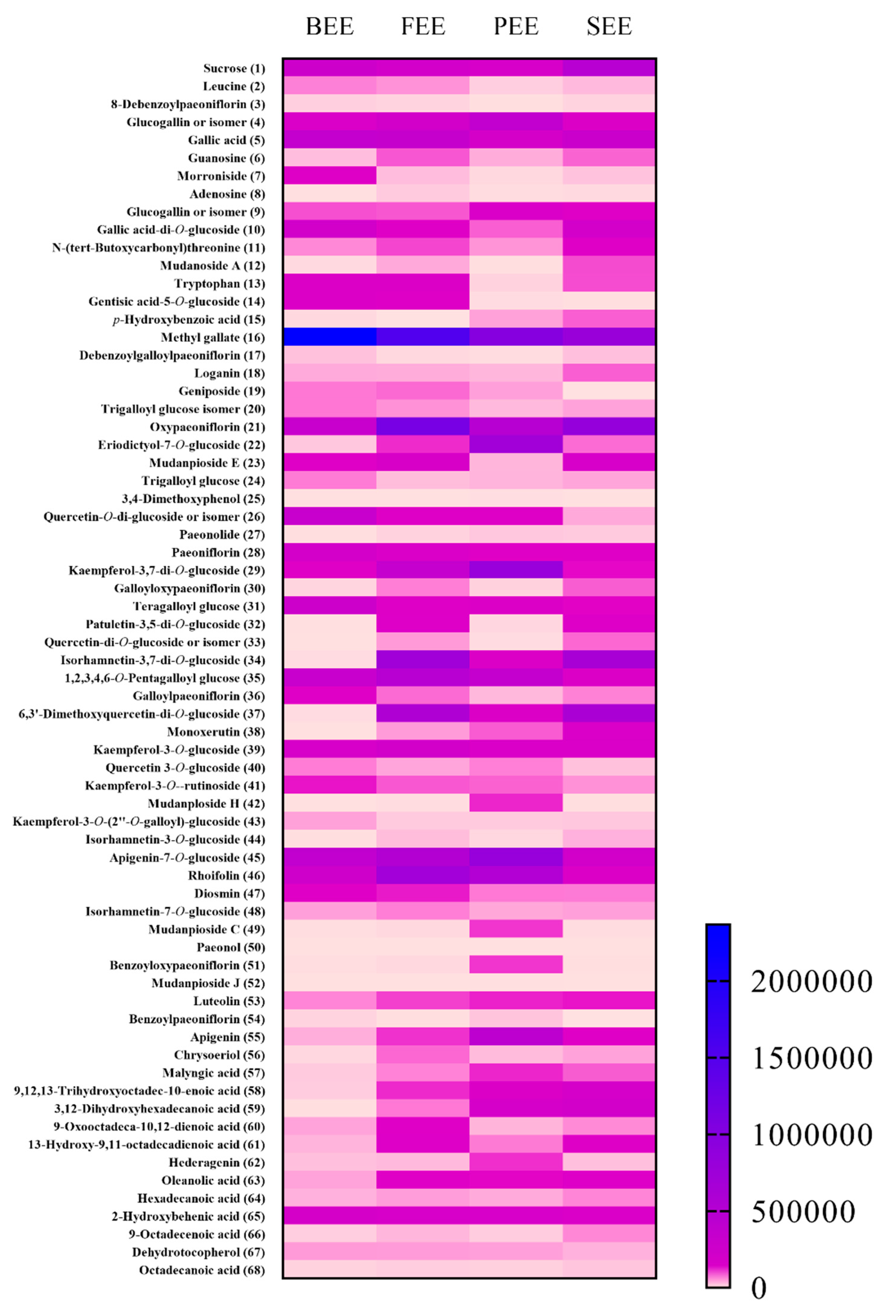
3.7. Determination of Phytochemicals in Ethanolic Extracts of P. suffruticosa Flowers

In this study, though there were 68 compounds found by UFLC-Q-TOF-MS analysis, it was difficult to quantify all the 68 compounds by HPLC-DAD analysis due to the trace contents in the extracts, no/weak UV absorption, and no available standards commercially. Moreover, phenols, monoterpenoids, and flavonoids had multiple bioactivities, and they were predominant constituents in P. suffruticosa flowers. The method developed for the simultaneous determination of 3 phenols, 3 monoterpenoids, 2 flavonoids was validated by HPLC-DAD (Table 3). Gallic acid (5), 1,2,3,4,6-O-pentagalloyl glucose (35), and paeonol (50) were determined in the four extracts (Table 4). In BEE, the content of 1,2,3,4,6-O-pentagalloyl glucose (35) was 197.20 ± 6.24 mg/g ext., which was higher than that of gallic acid (5) (159.99 ± 5.06 mg/g ext.) and paeonol (50) (1.68 ± 0.05 mg/g ext.). Unexpectedly, the content of gallic acid (5) was higher than that of 1,2,3,4,6-O-pentagalloylglucose (35) and paeonol (50) in the other three extracts. Oxypaeoniflorin (21) (6.93 ± 0.17 mg/g ext.), paeoniflorin (28) (16.19 ± 0.40 mg/g ext.), and benzoylpaeoniflorin (54) (0.48 ± 0.01 mg/g ext.) were able to be determined in PEE. The content of paeoniflorin (28) (19.61 ± 0.38 mg/g ext.) was much higher than that of oxypaeoniflorin (21) (10.46 ± 0.20 mg/g ext.) in FEE, but the content of paeoniflorin (28) (10.84 ± 0.49 mg/g ext.) was a little lower than that of oxypaeoniflorin (21) (11.19 ± 0.50 mg/g ext.) in SEE. Benzoylpaeoniflorin (54) was not determined in FEE and SEE. The contents of luteolin (53) and apigenin (55) in PEE were 1.23 ± 0.03 mg/g ext. and 1.48 ± 0.04 mg/g ext., respectively. The contents of luteolin (53) in BEE, FEE, and SEE were lower than 0.16 mg/g ext., approximately one eighth of that in PEE. These results indicated that gallic acid (5) and 1,2,3,4,6-O-pentagalloyl glucose (35) were predominant compounds in ethanolic extracts of P. suffruticosa flowers, especially BEE.

3.8. Pearson’s Correlation Analysis between Phytochemicals and Bioactivities

Pearson’s correlation analysis has been used for the better understanding of the relationship between the phytochemicals and their bioactivities [35]. The correlation coefficient (r) of a single ingredient to each bioactivity is shown in Figure 6.
Total phenolic and total flavonoid contents had a high negative correlation with DPPH and hydroxyl radical scavenging. Total phenolic content had a higher correlation with hydroxyl radical scavenging (r = −0.913) rather than DPPH scavenging (r = −0.549). By contrast, total flavonoid content had a higher correlation with DPPH scavenging (r = −0.862). Total phenolic and total flavonoid contents had a positive correlation with the activities of GSH-Px (rTPC = 0.648; rTFC = 0. 960, p < 0.05) and SOD (rTPC = 0.885; rTFC = 0.811), and a negative correlation with the contents of TNF-α (rTPC = −0.712; rTFC = −0.847) and IL-6 (rTPC = −0.880; rTFC = −0.783).
The correlation of phytochemicals and the antioxidant activity were further investigated. DPPH scavenging had no significant correlation with any compound. Hydroxyl radical scavenging had a significantly negative correlation with leucine (2), 8-debenzoylpaeoniflorin (3), gallic acid (5), tryptophan (13) and trigalloyl glucose isomer (20), and a significantly positive correlation with glucogallin or isomer (9) and malyngic acid (57). The activity of GSH-Px had a significantly positive correlation with geniposide (19) and a significantly negative correlation with p-hydroxybenzoic acid (15) and 3,12-dihydroxyhexadecanoic acid (59). The activity of SOD had a significantly positive correlation with gentisic acid-5-O-glucoside (14), methyl gallate (16), paeoniflorin (28), and diosmin (47), and a negative correlation with 9,12,13-trihydroxyoctadec-10-enoic acid (58) and 3,12-dihydroxyhexadecanoic acid (59). Gallic acid (5) and its derivatives, such as trigalloyl glucose (20), are strong natural antioxidants and can scavenge free radicals [36,37]. It has been reported that administration of geniposide (19), paeoniflorin (28), and diosmin (47) can significantly increase the activities of free radical scavenging enzymes such as SOD and GSH-Px [38,39,40].
It was found that the compounds significantly correlated with the content of IL-6 were identical to those of compounds correlated with the activity of SOD, but their correlations were opposite. It has been reported that paeoniflorin (28) and diosmin (47), with their potential anti-inflammatory capabilities, could significantly down-regulate the mRNA expression levels of IL-6 [39,41]. The content of TNF-α had a significantly positive correlation with glucogallin or isomer (9), and had a significantly negative correlation with leucine (2), gallic acid (5) and tryptophan (13). Interestingly, gallic acid (5) can inhibit mast cell-derived inflammatory allergic reactions via pro-inflammatory cytokine expression [42].
Gallic acid (5), methyl gallate (16), geniposide (19), trigalloyl glucose (20), paeoniflorin (28), and diosmin (47) were the critical compounds to the antioxidant, anti-photoaging and anti-inflammatory activities of P. suffruticosa flowers according to the results of Pearson’s correlation analysis. These compounds should be the biomarkers in the product development and the quality control of P. suffruticosa flowers.

4. Conclusions

This study showed that P. suffruticosa flowers have different antioxidant and anti-photoaging properties at different flowering stages. The phytochemicals were elucidated, and a significant difference was found in terms of abundance. Meanwhile, the results of chemometric analysis proved that the multiple ingredients in P. suffruticosa flowers contributed to their antioxidant and anti-photoaging activities. In order to have the excellent antioxidant and anti-photoaging activities and high levels of bioactive phytochemicals, the optimal time to harvest the P. suffruticosa flower is before the early flowering stage. Taken together, this study provides valuable evidence that the P. suffruticosa flower has a great potential to be developed as functional material in food and health related industries.

Author Contributions

J.H., X.Z. and M.L. conceived and designed the experiments; J.H., Y.D., X.L., Y.W., T.G. and M.L. performed the experiments; J.H., X.Z. and M. L. analyzed the data; J.H. and M.L. wrote the paper.

Funding

This research was funded by Science and Technology Program of Guangzhou (No. 201904010337), National Natural Science Foundation of China (Nos. 31670056 and 31870776) and Guangdong Key Laboratory of Marine Materia Medica (LMM2018-5).

Conflicts of Interest

The authors declare no conflict of interest.

References

  1. Muzaffer, U.; Paul, V.I.; Prasad, N.R.; Karthikeyan, R.; Agilan, B. Protective effect of Juglans regia L. against ultraviolet B radiation induced inflammatory responses in human epidermal keratinocytes. Phytomedicine 2018, 42, 100–111. [Google Scholar] [CrossRef] [PubMed]
  2. Chen, L.; Hu, J.Y.; Wang, S.Q. The role of antioxidants in photoprotection: A critical review. J. Am. Acad. Dermatol. 2012, 67, 1013–1024. [Google Scholar] [CrossRef] [PubMed]
  3. Tiraravesit, N.; Yakaew, S.; Rukchay, R.; Luangbudnark, W.; Viennet, C.; Humbert, P.; Viyoch, J. Artocarpus altilis heartwood extract protects skin against UVB in vitro and in vivo. J. Ethnopharmacol. 2015, 175, 153–162. [Google Scholar] [CrossRef] [PubMed]
  4. Divya, S.P.; Wang, X.; Pratheeshkumar, P.; Son, Y.O.; Roy, R.V.; Kim, D.; Dai, J.; Hitron, J.A.; Wang, L.; Asha, P.; et al. Blackberry extract inhibits UVB-induced oxidative damage and inflammation through MAP kinases and NF-κB signaling pathways in SKH-1 mice skin. Toxicol. Appl. Pharm. 2015, 284, 92–99. [Google Scholar] [CrossRef] [PubMed]
  5. Pandel, R.; Poljšak, B.; Godic, A.; Dahmane, R. Skin photoaging and the role of antioxidants in its prevention. ISRN Dermatol. 2013, 2013, 930163. [Google Scholar] [CrossRef] [PubMed]
  6. Han, C.V.; Bhat, R. In vitro control of food-borne pathogenic bacteria by essential oils and solvent extracts of underutilized flower buds of Paeonia suffruticosa (andr.). Ind. Crop. Prod 2014, 54, 203–208. [Google Scholar] [CrossRef]
  7. Zhu, S.; Shirakawa, A.; Shi, Y.; Yu, X.; Tamura, T.; Shibahara, N.; Yoshimatsu, K.; Komatsu, K. Impact of different post-harvest processing methods on the chemical compositions of peony root. J. Nat. Med. 2018, 72, 757–767. [Google Scholar] [CrossRef] [Green Version]
  8. Wang, Z.; He, C.; Peng, Y.; Chen, F.; Xiao, P. Origins, phytochemistry, pharmacology, analytical methods and safety of Cortex Moutan (Paeonia suffruticosa Andrew): A systematic review. Molecules 2017, 22, 946. [Google Scholar] [CrossRef]
  9. Chen, L.J.; Games, D.E.; Jones, J. Isolation and identification of four flavonoid constituents from the seeds of Oroxylum indicum by high-speed counter-current chromatography. J. Chromatogr. A 2003, 988, 95–105. [Google Scholar] [CrossRef]
  10. Ogawa, K.; Nakamura, S.; Sugimoto, S.; Tsukioka, J.; Hinomaru, F.; Nakashima, S.; Matsumoto, T.; Ohta, T.; Fujimoto, K.; Yoshikawa, M.; et al. Constituents of flowers of Paeoniaceae plants, Paeonia suffruticosa and Paeonia lactiflora. Phytochem. Lett. 2015, 12, 98–104. [Google Scholar] [CrossRef]
  11. Yuan, J.; Hao, L.J.; Wu, G.; Wang, S.; Duan, J.A.; Xie, G.Y.; Qin, M.J. Effects of drying methods on the phytochemicals contents and antioxidant properties of chrysanthemum flower heads harvested at two developmental stages. J. Funct. Foods 2015, 19, 786–795. [Google Scholar] [CrossRef]
  12. Zeng, H.; Su, S.; Xiang, X.; Sha, X.; Zhu, Z.; Wang, Y.; Guo, S.; Yan, H.; Qian, D.; Duan, J. Comparative analysis of the major chemical constituents in Salvia miltiorrhiza roots, stems, leaves and flowers during different growth periods by UPLC-TQ-MS/MS and HPLC-ELSD methods. Molecules 2017, 22, 771. [Google Scholar] [CrossRef] [PubMed]
  13. He, J.Y.; Zhang, Y.H.; Ma, N.; Zhang, X.L.; Liu, M.H.; Fu, W.M. Comparative analysis of multiple ingredients in Rosa roxburghii and R. sterilis fruits and their antioxidant activities. J. Funct. Foods 2016, 27, 29–41. [Google Scholar] [CrossRef]
  14. Liu, M.H.; Ko, C.H.; Ma, N.; Tan, P.W.; Fu, W.M.; He, J.Y. Chemical profiles, antioxidant and anti-obesity effects of extract of Bambusa textilis McClure leaves. J. Funct. Foods 2016, 22, 533–546. [Google Scholar] [CrossRef]
  15. Hassan, S.; Hussein, A.J.; Saeed, A.K. Role of green tea in reducing epidermal thickness upon ultraviolet light-B injury in BALB/c mice. Adv. Biol. 2015, 2015. [Google Scholar] [CrossRef]
  16. Alu’datt, M.H.; Rababah, T.; Alhamad, M.N.; Al-Mahasneh, M.A.; Ereifej, K.; Al-Karaki, G.; Al-Duais, M.; Andrade, J.E.; Tranchant, C.C.; Kubow, S.; et al. Profiles of free and bound phenolics extracted from Citrus fruits and their roles in biological systems: Content, and antioxidant, anti-diabetic and anti-hypertensive properties. Food Funct. 2017, 8, 3187–3197. [Google Scholar] [CrossRef] [PubMed]
  17. Lesjak, M.; Beara, I.; Simin, N.; Pintać, D.; Majkić, T.; Bekvalac, K.; Orčić, D.; Mimica-Dukić, N. Antioxidant and anti-inflammatory activities of quercetin and its derivatives. J. Funct. Foods 2018, 40, 68–75. [Google Scholar] [CrossRef]
  18. Interdonato, R.; Rosa, M.; Nieva, C.B.; González, J.A.; Hilal, M.; Prado, F.E. Effects of low UV-B doses on the accumulation of UV-B absorbing compounds and total phenolics and carbohydrate metabolism in the peel of harvested lemons. Environ. Exp. Bot. 2011, 70, 204–211. [Google Scholar] [CrossRef]
  19. Chen, G.L.; Chen, S.G.; Xie, Y.Q.; Chen, F.; Zhao, Y.Y.; Luo, C.X.; Gao, Y.Q. Total phenolic, flavonoid and antioxidant activity of 23 edible flowers subjected to in vitro digestion. J. Funct. Foods 2015, 17, 243–259. [Google Scholar] [CrossRef]
  20. Lee, H.J.; Im, A.R.; Kim, S.M.; Kang, H.S.; Lee, J.D.; Chae, S. The flavonoid hesperidin exerts anti-photoaging effect by downregulating matrix metalloproteinase (MMP)-9 expression via mitogen activated protein kinase (MAPK)-dependent signaling pathways. Bmc Complement. Altern. Med. 2018, 18, 39. [Google Scholar] [CrossRef]
  21. Huang, C.Y.; Lin, Y.T.; Kuo, H.C.; Chiou, W.F.; Lee, M.H. Compounds isolated from Eriobotrya deflexa leaves protect against ultraviolet radiation B-induced photoaging in human fibroblasts. J. Photochem. Photobiol. B 2017, 175, 244–253. [Google Scholar] [CrossRef] [PubMed]
  22. Lephart, E.D. Skin aging and oxidative stress: Equol’s anti-aging effects via biochemical and molecular mechanisms. Ageing Res. Rev. 2016, 31, 36–54. [Google Scholar] [CrossRef] [PubMed]
  23. Masaki, H. Role of antioxidants in the skin: Anti-aging effects. J. Dermatol. Sci. 2010, 58, 85–90. [Google Scholar] [CrossRef] [PubMed]
  24. Nóbrega-Pereira, S.; Fernandez-Marcos, P.J.; Brioche, T.; Gomez-Cabrera, M.C.; Salvador-Pascual, A.; Flores, J.M.; Viña, J.; Serrano, M. G6PD protects from oxidative damage and improves healthspan in mice. Nat. Commun. 2016, 7, 10894. [Google Scholar] [Green Version]
  25. Wang, X.F.; Huang, Y.F.; Wang, L.; Xu, L.Q.; Yu, X.T.; Liu, Y.H.; Li, C.L.; Zhan, J.Y.; Su, Z.R.; Chen, J.N.; et al. Photo-protective activity of pogostone against UV-induced skin premature aging in mice. Exp. Gerontol. 2016, 77, 76–86. [Google Scholar] [CrossRef] [PubMed]
  26. Martinez, R.M.; Fattori, V.; Saito, P.; Melo, C.B.P.S.; Borghi, M.; Pinto, I.C.; Bussmann, A.J.C.; Baracat, M.M.; Georgetti, S.R.; Verri, W.A., Jr.; et al. Lipoxin A4 inhibits UV radiation-induced skin inflammation and oxidative stress in mice. J. Dermatol. Sci. 2018, 91, 164–174. [Google Scholar] [CrossRef] [PubMed] [Green Version]
  27. He, J.; Lu, X.; Wei, T.; Dong, Y.; Cai, Z.; Tang, L.; Liu, M. Asperuloside and asperulosidic acid exert an anti-Inflammatory effect via suppression of the NF-κB and MAPK signaling pathways in LPS-induced RAW 264.7 macrophages. Int. J. Mol. Sci. 2018, 19, 2027. [Google Scholar] [CrossRef] [PubMed]
  28. Hsu, F.L.; Lai, C.W.; Cheng, J.T. Antihyperglycemic effects of paeoniflorin and 8-debenzoylpaeoniflorin, glucosides from the root of Paeonia lactiflora. Planta Med. 1997, 63, 323–325. [Google Scholar] [CrossRef] [PubMed]
  29. Xu, S.J.; Yang, L.; Zeng, X.; Zhang, M.; Wang, Z.T. Characterization of compounds in the Chinese herbal drug Mu-Dan-Pi by liquid chromatography coupled to electrospray ionization mass spectrometry. Rapid Commun. Mass Spectrom. 2006, 20, 3275–3288. [Google Scholar] [CrossRef] [PubMed]
  30. Zhao, W.; Geng, Y.; Cui, L.; Duan, W.; Wang, X.; Yan, H. Flavonoids from flowers of Paeonia suffruticosa Andr. Mod. Chin. Med. 2016, 18, 303–306. [Google Scholar]
  31. Akimoto, N.; Ara, T.; Nakajima, D.; Suda, K.; Ikeda, C.; Takahashi, S.; Muneto, R.; Yamada, M.; Suzuki, H.; Shibata, D.; et al. Flavonoidsearch: A system for comprehensive flavonoid annotation by mass spectrometry. Sci. Rep. 2017, 7, 1243. [Google Scholar] [CrossRef] [PubMed]
  32. Du, H.; Wu, J.; Ji, K.X.; Zeng, Q.Y.; Wang, L.S. Methylation mediated by an anthocyanin, O-methyltransferase, is involved in purple flower coloration in Paeonia. J. Exp. Bot. 2015, 66, 6563. [Google Scholar] [CrossRef] [PubMed]
  33. Li, J.H.; Zeng, R.; Qu, Y.; Huang, L.F. Rapid identification on chemical constituents in roots of Paeonia delavayi var. Lutea by UPLC-Q-TOF-MSE combined with UNIFI informatics platform. Chin. Tradit. Herb. Drugs 2017, 48, 1529–1536. [Google Scholar]
  34. Yu, S.; Du, S.; Yuan, J.; Hu, Y. Fatty acid profile in the seeds and seed tissues of Paeonia, L. species as new oil plant resources. Sci. Rep. 2016, 6, 26944. [Google Scholar] [CrossRef] [PubMed]
  35. Wang, L.; Luo, Y.; Wu, Y.; Wu, Z. Impact of fermentation degree on phenolic compositions and bioactivities during the fermentation of guava leaves with Monascus anka and Bacillus sp. J. Funct. Foods 2018, 41, 183–190. [Google Scholar] [CrossRef]
  36. Asnaashari, M.; Farhoosh, R.; Sharif, A. Antioxidant activity of gallic acid and methyl gallate in triacylglycerols of Kilka fish oil and its oil-in-water emulsion. Food Chem. 2014, 159, 439–444. [Google Scholar] [CrossRef] [PubMed]
  37. Ngoc, T.M.; Hung, T.M.; Thuong, P.T.; Kim, J.C.; Min, B.S. Antioxidative activities of galloyl glucopyranosides from the stem-bark of Juglans mandshurica. Biosci. Biotechnol. Biochem. 2008, 72, 2158–2163. [Google Scholar] [CrossRef]
  38. Chen, P.; Chen, Y.; Wang, Y.; Cai, S.; Deng, L.; Liu, J.; Zhang, H. Comparative evaluation of hepatoprotective activities of geniposide, crocins and crocetin by CCl4-induced liver injury in mice. Biomol. Ther. 2016, 24, 156. [Google Scholar] [CrossRef]
  39. Zhang, M.H.; Feng, L.; Zhu, M.M.; Gu, J.F.; Wu, C.; Jia, X.B. Antioxidative and anti-inflammatory activities of paeoniflorin and oxypaeoniflora on AGEs-induced mesangial cell damage. Planta Med. 2013, 79, 1319–1323. [Google Scholar] [CrossRef]
  40. Senthamizhselvan, O.; Manivannan, J.; Silambarasan, T.; Raja, B. Diosmin pretreatment improves cardiac function and suppresses oxidative stress in rat heart after ischemia/reperfusion. Eur. J. Pharmacol. 2014, 736, 131–137. [Google Scholar] [CrossRef]
  41. Imam, F.; Al-Harbi, N.O.; Al-Harbi, M.M.; Ansari, M.A.; Zoheir, K.M.; Iqbal, M.; Anwer, M.K.; Al-Hoshani, A.R.; Attia, S.M.; Ahmad, S.F. Diosmin downregulates the expression of T cell receptors, pro-inflammatory cytokines and NF-κB activation against LPS-induced acute lung injury in mice. Pharmacol. Res. 2015, 102, 1–11. [Google Scholar] [CrossRef] [PubMed]
  42. Kim, S.H.; Jun, C.D.; Suk, K.; Choi, B.J.; Lim, H.; Park, S.; Lee, S.H.; Shin, H.Y.; Kim, D.K.; Shin, T.Y. Gallic acid inhibits histamine release and pro-inflammatory cytokine production in mast cells. Toxicol. Sci. 2005, 91, 123–131. [Google Scholar] [CrossRef] [PubMed]
Figure 1. Pictures of Paeonia suffruticosa flowers at the different flowering stages. (A) P. suffruticosa flowering bud; (B) P. suffruticosa flower at the early flowering stage; (C) P. suffruticosa flower at the full flowering stage.
Antioxidants 08 00345 g001
Figure 2. In vitro antioxidant effects of ethanolic extract of Paeonia suffruticosa flowers. Effects of ethanolic extract of P. suffruticosa flowers on DPPH scavenging (A), hydroxyl radical scavenging (B), FRAP assay (C), and inhibition of β-carotene bleaching (D). Data were expressed as mean ± SD (n = 3). VC: vitamin C; BHT: butylated hydroxytoluene; BEE: bud ethanolic extract; FEE: flower ethanolic extract; PEE: petal ethanolic extract; SEE: stamen ethanolic extract.
Antioxidants 08 00345 g002
Figure 3. Effects of ethanolic extract of Paeonia suffruticosa flowers on UVB-induced epidermal thickening in the mice. Representative images of histological observation by H&E staining of mouse dorsal skin (400×). (A) control group: no irradiation; (B) model group: irradiated group with no treatment; (C) model + emulsifier (ME) group: irradiated group treated with emulsifier; (D) bud ethanolic extract (BEE) group: irradiated group treated with 2.0% (w/w) BEE in emulsifier; (E) flower ethanolic extract (FEE) group: irradiated group treated with 2.0% (w/w) FEE in emulsifier; (F) petal ethanolic extract (PEE) group: irradiated group treated with 2.0% (w/w) PEE in emulsifier; (G) stamen ethanolic extract (SEE) group: irradiated group treated with 2.0% (w/w) SEE in emulsifier. (H) Epidermal thickness. Data were expressed as mean ± SD (n = 6). *** p < 0.001 versus ME group; ### p < 0.001 versus control group.
Antioxidants 08 00345 g003
Figure 4. Effects of ethanolic extracts of Paeonia suffruticosa flowers on the activities of antioxidant enzymes and the concentrations of pro-inflammatory cytokines in UVB-irradiated mouse skin. Concentrations of SOD (A), GSH-Px (B), TNF-α (C), and IL-6 (D) in dorsal skin tissues were determined using mouse-specific enzyme-linked immunosorbent assay (ELISA) kits. Control group: no irradiation; Model group: irradiated group with no treatment; Model + emulsifier (ME) group: irradiated group treated with emulsifier; Bud ethanolic extract (BEE) group: irradiated group treated with 2.0% (w/w) BEE in emulsifier; Flower ethanolic extract (FEE) group: irradiated group treated with 2.0% (w/w) FEE in emulsifier; Petal ethanolic extract (PEE) group: irradiated group treated with 2.0% (w/w) PEE in emulsifier; Stamen ethanolic extract (SEE) group: irradiated group treated with 2.0% (w/w) SEE in emulsifier. Data were expressed as mean ± SD (n = 6). * p < 0.05, ** p < 0.01, and *** p < 0.001 versus ME group; ### p < 0.001 versus control group.
Antioxidants 08 00345 g004
Figure 5. The peak intensities of the ethanolic extracts of Paeonia suffruticosa flowers. The peak intensities of the related compound in bud ethanolic extract (BEE), flower ethanolic extract (FEE), petal ethanolic extract (PEE), and stamen ethanolic extract (SEE) were obtained in negative mode. The numbered compounds were consistent with that in Table 2.
Antioxidants 08 00345 g005
Figure 6. Correlation analysis between phytochemicals and bioactivities. The numbered compounds were consistent with that in Table 2. DPPHS: DPPH scavenging; HRS: hydroxyl radical scavenging.
Antioxidants 08 00345 g006
Table 1. Extraction yields, total phenolic, and total flavonoid contents, and antioxidant activities (IC50 values) of Paeonia suffruticosa flowers.
ItemsVCBHTSamples
BEEFEEPEESEE
Extraction yields (%)----35.46 ± 3.1250.76 ± 1.9542.72 ± 2.4825.77 ± 4.54
Total phenolic content (mg GAE/g ext.)----191.80 ± 3.44113.84 ± 0.8451.66 ± 0.3477.25 ± 0.58
Total flavonoid content (mg RE/g ext.)----30.60 ± 0.9449.87 ± 0.8212.98 ± 0.788.86 ± 0.98
DPPH radical scavenging activity (μg/mL)6.16 ± 0.4664.49 ± 4.6834.44 ± 1.3514.83 ± 2.0364.00 ± 5.6042.70 ± 0.79
Hydroxyl radical scavenging activity (mg/mL)0.61 ± 0.02--1.57 ± 0.041.79 ± 0.022.57 ± 0.062.10 ± 0.004
VC: vitamin C; BHT: butylated hydroxytoluene; BEE: bud ethanolic extract; FEE: flower ethanolic extract; PEE: petal ethanolic extract; SEE: stamen ethanolic extract. Data were expressed as mean ± SD (n = 3).
Table 2. The compounds identified in the ethanolic extracts of Paeonia suffruticosa flowers by UFLC-Q-TOF-MS.
No.Rt
(min)
Molecular Formula[M+H]+
(ppm)
[M−H]
(ppm)
Fragments in Positive ModeFragments in Negative ModeIdentification
11.03C12H22O11343.12331
(−0.5)
341.10911
(0.5)
365.10508 [M+Na]+,
203.0519 [M+Na−C6H10O5]+
179.0557 [M−H−C6H10O5]Sucrose
2 *1.69C6H13NO2132.1018
(−0.8)
130.08735
(0.02)
86.0986 [M+H−HCOOH]+,
69.0731 [M+H−HCOOH-NH3]+
112.0398 [M−H−H2O],
86.0338 [M−H−CO2]
Leucine
31.77C16H24O10377.14221
(−5.3)
375.12986
(0.5)
377.14293 [M+NH4]+,
197.0778 [M+H−C6H10O5−H2O]+,
179.0 [M+H−C6H10O5−2H2O]+
195.0581 [M−H−C6H10O5−H2O]8-Debenzoylpaeoniflorin
42.10C13H16O10333.08113
(−1.5)
331.06748
(1.2)
350.10806 [M+NH4]+169.0138 [M−H−C6H10O5],
125.0340 [M−H−C6H10O5−CO2]
Glucogallin or isomer
5 *2.30C7H6O5171.02846
(−2)
169.01513
(5.2)
153.0181 [M+H−H2O]+,
127.0392 [M+H−CO2]+,
109.1294 [M+H−CO2−H2O]+
125.0245 [M−H−CO2]Gallic acid
62.48C10H13N5O5284.09891
(−0.1)
282.08447
(0.3)
152.0260 [M+H−C5H8O4]+,
135.0293 [M+H−C5H8O4−NH3]+
150.0419 [M−H−C5H8O4],
133.0159 [M−H−C5H8O4−NH3]
Guanosine
7 *2.70C17H26O11 405.13989
(−0.8)
424.18247 [M+H+NH4]+359.1325 [M−H−HCOOH],
197.0806 [M−H−HCOOH−C6H10O5]
Morroniside
8 *2.74C10H13N5O4268.10411
(0.3)
266.08843
(−3.9)
136.0616 [M+H−C5H8O4]+,
119.0355 [M+H−C5H8O4−NH3]+
134.0437 [M−H−C5H8O4]Adenosine
93.20C13H16O10331.06755
(1.4)
331.06745
(1.1)
315.0683 [M+H−H2O]+,
153.0172 [M+H−H2O−C6H10O5]+
241.0325 [M−H−C3H6O3],
169.0138 [M−H−C6H10O5],
125.0340 [M−H−C6H10O5−CO2]
Glucogallin or isomer
103.70C19H26O15495.13369
(−1.5)
493.12022
(0.7)
333.0772 [M+H−C6H10O5]+,
315.0711 [M+H−C6H12O6]+,
297.0592 [M+H−C6H12O6−H2O]+,
171.0272 [M+H−2C6H10O5]+,
153.0178 [M+H−C6H12O6−C6H10O5]+
313.0546 [M−H−C6H12O6],
169.0124 [M−H−2C6H10O5]
Gallic acid-di-O-glucoside
114.10C9H17NO5220.11782
(−0.6)
218.1036
(0.9)
202.1071 [M+H−H2O]+,
184.0959 [M+H−2H2O]+,
174.1126 [M+H−HCOOH]+
146.0825 [M−H−C4H10O],
N-(tert-Butoxycarbonyl)threonine
124.60C14H18O9331.10195
(−1.2)
329.08812
(0.9)
169.0427 [M+H−C6H10O5]+167.0341 [M−H−C6H10O5]Mudanoside A
13 *4.86C11H12N2O2205.09701
(−0.7)
203.08298
(1.9)
188.0709 [M+H−NH3]+,
118.0657 [M+H−CO2−C2H5N]+
159.0911 [M−H−CO2],
116.0513 [M−H−CO2−C2H5N]
Tryptophan
145.06C13H16O9317.08639
(−1)
315.07227
(0.4)
155.0321 [M+H−C6H10O5]+,
137.0229 [M+H−C6H10O5−H2O]+
153.0189 [M−H−C6H10O5],
109.0303 [M−H−C6H10O5−CO2]
Gentisic acid-5-O-glucoside
15 *5.91C7H6O3139.03894
(−0.3)
137.02588
(10.7)
121.0279 [M+H−H2O]+,
95.0495 [M+H−CO2]+,
77.0400 [M+H−CO2−H2O]+
93.0363 [M−H−CO2]p-Hydroxybenzoic acid
166.30C8H8O5185.04434
(−0.6)
183.03088
(5.4)
153.0188 [M+H−CH3OH]+,
125.0239 [M+H−CH3COOH]+,
107.0137 [M+H−CH3COOH−H2O]+
168.0065 [M−H−CH3],
124.0174 [M−H−CH3−CO2]
Methyl gallate
176.58C23H28O14529.15249
(−5.1)
527.14061
(0)
493.1146 [M+H−2H2O]+,
315.0648 [M+H−C7H4O5−2H2O]+,
179.0679 [C10H11O3]+
345.1178 [M−H−C8H6O5],
313.0594 [M−H−C7H4O5−2H2O],
271.0502, 211.0201,
169.0146 [C10H11O3]
Debenzoylgalloylpaeoniflorin
18 *6.76C17H26O10391.16024
(0.9)
389.14563
(0.8)
343.1424 [M−H−HCOOH],
181.0874 [M−H−HCOOH−C6H10O5],
163.0764 [M−H−HCOOH−C6H10O5−H2O]
Loganin
19 *7.15C17H24O10 387.12994
(0.7)
341.1357 [M−H−HCOOH],
179.0705 [M−H−HCOOH−C6H10O5]
Geniposide
207.28C27H24O18637.10441
(1.4)
635.08956
(0.9)
467.0798 [M+H−C7H6O3]+,
297.0615 [M+H−2C7H6O3]+,
279.0415 [M+H−2C7H6O3−H2O]+,
153.0171 [C7H5O4]+
465.1075 [M−H−C7H6O3],
295.0506 [M−H−2C7H6O3],
169.1023 [C7H5O5]
Trigalloyl glucose isomer
21 *7.53C23H28O12497.1651
(−0.5)
495.15083
(0.1)
479.1557 [M+H−H2O]+,
335.1123 [M+H−C6H10O5]+,
317.1018 [M+H−H2O−C6H10O6]+,
197.0810 [M+H−C6H10O6−C7H6O3]+,
179.0704 [M+H−C6H10O6−C7H6O3−H2O]+,
151.0752 [M+H−C6H10O6−C7H6O3−2H2O]+,
133.0643 [M+H−C6H10O6−C7H6O3−3H2O]+
465.1446 [M−H−CH2O],
333.0998 [M−H−C6H10O5],
195.0657 [M−H−C6H10O6−C7H6O3],
165.0549 [M−H−C6H10O6−C7H6O3−CH2O],
137.0348 [M−H−C6H10O6−C7H6O3−CH2O−H2O]
Oxypaeoniflorin
228.23C21H22O11451.12391
(0.9)
449.10937
(1)
289.0705 [M+H−C6H10O5]+,
271.0590 [M+H−C6H10O5−H2O]+,
153.0168
287.0562 [M−H−C6H10O5],
259.0618, 151.0034
Eriodictyol-7-O-glucoside
238.45C24H30O13 525.16213
(1.5)
544.202 [M+NH4]+,
365.1232 [M+H−C6H10O5]+,
347.1106 [M+H−C6H10O5−H2O]+,
197.0797 [M+H−C6H12O6−C8H6O3]+,
179.0689 [M+H−C6H12O6−C8H6O3−H2O]+
495.1558 [M−H−CH2O],
345.1044 [M−H−C6H12O6],
195.0652 [M−H−C6H12O6−C8H6O3],
177.0639 [M−H−C6H12O6−C8H6O3−H2O]
Mudanpioside E
248.64C27H24O18 635.08955
(0.9)
654.12968 [M+NH4]+,
619.0922 [M+H−H2O]+,
449.0770 [M+H−H2O−C7H6O5]+,
297.0615 [M+H−H2O−C7H6O5−C7H4O4]+,
279.0476 [M+H−2H2O−C7H6O5−C7H4O4]+
465.0666 [M−H−C7H6O5],
313.0554 [M−H−C7H6O5−C7H4O4],
241.05094 [M−H−C7H6O5−2C7H4O4]
Trigalloyl glucose
258.79C8H10O3155.07034
(0.5)
140.0472 [M+H−CH3]+,
123.0441 [M+H−CH4O]+
3,4-Dimethoxyphenol
269.20C27H30O17627.15612
(0.9)
625.14123
(0.3)
465.1022 [M+H−C6H10O5]+,
303.0497 [M+H−2C6H10O5]+
463.0928 [M−H−C6H10O5],
301.0358 [M−H−2C6H10O5]
Quercetin-O-di-glucoside or isomer
279.67C20H28O12461.16556
(0.5)
459.14963
(−2.6)
299.1206 [M+H−C6H10O5]+,
167.0707 [M+H−C6H10O5−C5H8O4]+
297.0885 [M−H−C9H10O3],
165.0551 [M−H−C6H10O5−C5H8O4]
Paeonolide
28 *10.03C23H28O11481.17026
(−0.4)
479.15559
(−0.6)
319.1260 [M+H−C6H10O5]+,
301.1567 [M+H−C6H10O5−H2O]+,
179.0703 [M+H−C6H10O5−H2O−C7H6O2]+,
449.1520 [M−H−CH2O],
327.1088 [M−H−C7H6O2],
165.0551 [M−H−C7H6O2−C6H10O5]
Paeoniflorin
2910.27C27H30O16611.16221
(2.5)
609.14676
(1.1)
449.1081 [M+H−C6H10O5]+,
287.0552 [M+H−2C6H10O5]+
447.0961 [M−H−C6H10O5],
285.0403 [M−H−2C6H10O5]
Kaempferol-3,7-di-O-glucoside
3010.48C30H32O16649.17332
(−4.6)
647.1606
(−1.8)
511.1395 [M+H−C7H6O3]+,
315.0712 [M+H−C17H18O7]+,
153.0174 [M+H−C17H18O7−C6H10O5]+
509.1335 [M−H−C7H6O3],
313.0559 [M−H−C17H18O7],
Galloyloxypaeoniflorin
3110.78C34H28O22789.11272
(−2.3)
787.10067
(0.9)
771.1010 [M+H−H2O]+,
619.0877 [M+H−C7H4O4−H2O]+,
449.0694 [M+H−2C7H4O4−2H2O]+,
279.0476 [M+H−3C7H4O4−3H2O]+
635.0985 [M−H−C7H4O4],
617.0932 [M−H−C7H4O4−H2O],
465.0734 [M−H−2C7H4O4−H2O],
295.0473 [M−H−3C7H4O4−2H2O],
Teragalloyl glucose
3211.28C28H32O18657.16586
(−0.4)
655.135
(2.9)
495.118 [M+H−C6H10O5]+,
333.0606 [M+H−2C6H10O5]+
331.0490 [M−H−2C6H10O5]−,Patuletin-3,5-di-O-glucoside
3311.69C27H30O17627.1546
(−1.6)
625.1419
(1.4)
465.1038 [M+H− C6H10O5]+,
303.0500 [M+H−2 C6H10O5]+
301.0337 [M−H− C6H10O5],
271.0245 [M−H−2 C6H10O5−CH2O]
Quercetin-di-O-glucoside or isomer
3412.21C28H32O17641.17086
(−0.6)
639.15895
(3.6)
479.1168 [M+H−C6H10O5]+,
317.0652 [M+H−2C6H10O5]+
315.0507 [M−H−2C6H10O5]Isorhamnetin-3,7-di-O-glucoside
35 *12.33C41H32O26941.12626
(0.8)
939.11461
(3.9)
771.110 [M+H−C7H6O5]+,
431.0623 [M+H−3C7H6O5]+,
279.0462 [M+H−3C7H6O5−C6H10O5]+,
769.1070 [M−H−C7H6O5],
617.0931 [M−H− C7H6O5−C7H4O4],
465.07051 [M−H− C7H6O5−2C7H4O4],
295.0430 [M−H−2 C7H6O5−2C7H4O4]
1,2,3,4,6-O-Pentagalloyl glucose
3612.36C30H32O15633.18077
(−1)
631.16737
(0.8)
315.0721 [M+H−C7H6O2−C10H12O10]+,
179.0689 [C10H11O3]+
613.1706 [M−H−H2O],
509.1421 [M−H− C7H6O2],
313.1568 [M−H− C7H6O2− C10H12O10],
271.0539 [M−H− C7H6O2− C10H12O10−C2H2O]
Galloylpaeoniflorin
3712.47C29H34O18671.18123
(−0.8)
669.16952
(3.4)
509.1271 [M+H−C6H10O5]+,
347.0753 [M+H−2C6H10O5]+,
345.0623 [M−H−2C6H10O5],
301.0361 [M−H−2C6H10O5−CO2]
6,3′-Dimethoxyquercetin-di-O-glucoside
3812.98C29H34O17655.18661
(−0.4)
653.17535
(4.6)
509.1293 [M+H−C5H8O4]+,
347.0763 [M+H−C5H8O4−C6H10O5]+
345.0552 [M−H−C5H8O4−C6H10O5],
301.0335 [M−H−C5H8O4−C6H10O5−C2H4O]
Monoxerutin
3913.11C21H20O11449.10826
()
447.09399
(1.6)
287.0555 [M+H−C6H10O5]+,
153.0181
285 [M−H−C6H10O5]Kaempferol-3-O-glucoside
40 *13.48C21H20O12 463.08881
(1.3)
301.0354 [M−H−C6H10O5],
151.0026
Quercetin-3-O-glucoside
4113.59C27H30O15595.16617
(0.7)
593.15348
(3.9)
287.0549 [M+H−C12H20O9]+285.0425 [M−H−C12H20O9]Kaempferol-3-O-rutinoside
4213.66C30H32O14617.18694
(0.7)
615.17285
(1.5)
599.1479 [M+H−H2O]+,
479.1685 [M+H−C7H6O3]+,
443.1294 [M+H−C7H6O3−2H2O]+,
317.1017 [M+H−C7H6O3−C6H10O5]+,
179.0703 [C10H11O3]+
431.1396 [M−H−CH2O2−C7H6O3],
281.06836 [M−H−CH2O2−C7H6O3−C7H6O2−CH2O]
Mudanploside H
4313.94C28H24O15601.11847
(−0.5)
599.10555
(2.2)
287.0544 [M+H−C13H14O9]+285.0413 [M−H−C13H14O9]Kaempferol-3-O-
(2′’-O-galloyl)-glucoside
4414.28C22H22O12479.1188
(0.8)
477.10532
(3.1)
317.0661 [M+H−C6H10O5]+315.0537 [M−H− C6H10O5],
299.0214 [M−H−C6H10O6],
271.0272 [M−H−C6H10O6−H2O],
255.0335, 199.0371, 171.0587
Isorhamnetin-3-O-glucoside
4514.31C21H20O10433.11321
(0.7)
431.10019
(4.2)
271.0600 [M+H−C6H10O5]+,
153.0176
269.0449 [M−H−C6H10O5]Apigenin-7-O-glucoside
46 *14.61C27H30O14579.1711
(0.5)
577.15834
(3.6)
433.1131 [M+H−C5H8O4]+,
271.0606 [M+H−C5H8O4−C6H10O5]+,
153.0177
269.0462 [M+H−C5H8O4−C6H10O5]7Rhoifolin
4714.91C28H32O150.5607.17007
(5.3)
463.1220 [M+H−C6H10O4]+,
301.0703 [M+H−C6H10O4−C6H10O5]+,
286.0461 [M+H−C6H10O4−C6H10O5−CH3]+
443.0939 [M−H−C6H12O5],
299.0546 [M−H−C6H10O4−C6H10O5],
284.0301 [M−H−C5H8O4−C6H10O5−CH3]
Diosmin
4814.95C22H22O12479.11832
(−0.2)
477.10464
(1.7)
317.0668 [M+H−C6H10O5]+285.0372 [M−H−C6H10O5−CH2O]Isorhamnetin-7-O-glucoside
4915.34C30H32O13601.18754
(−6.7)
599.17976
(4.6)
461.1447 [M+H−C7H6O2−H2O]+,
443.1269 [M+H−C7H6O2−2H2O]+,
301.1056 [M+H−C7H4O2−C7H4O3−CO2]+,
283.08136 [M+H−C7H4O2−C7H4O3−CO2−H2O]+,
179.0695 [C10H11O3]+
447.1527 [M−H−CH2O−C7H6O2],
431.1379 [M−H−CH2O−C7H6O3],
281.0682 [M−H−C7H4O2−C7H4O3−CO2−H2O],
179.0329 [C10H11O3]
Mudanpioside C
50 *15.69C9H10O3167.06995
(−1.9)
165.05663
(5.5)
149.0737 [M+H−H2O]+,
121.0616 [M+H−H2O−CO]+
150.0318 [M−H−CH3],
135.0098 [M−H−CH2O],
122.0377 [M−H−CH3−CO]
Paeonol
5115.78C30H32O13 599.18003
(5)
569.1748 [M−H−CH2O],
447.1519 [M−H−CH2O−C7H6O2]
Benzoyloxypaeoniflorin
5216.14C31H34O14631.20136
(−1.2)
629.19181
(6.7)
457.1797 [M+H−C8H6O4−H2O]+,
317.1009 [M+H−C7H6O2−C8H6O4−H2O]+,
297.1009 [M+H−C7H6O2−C8H6O4−2H2O]+,
279.1108 [M+H−C7H6O2−C8H6O4−3H2O]+,
179.0692 [C10H11O3]+
477.1511 [M−H−C8H6O3],
333.1206 [M−H−C7H6O2−C8H6O4]
Mudanpioside J
53 *16.95C15H10O6 285.04116
(2.4)
175.0407, 133.0318151.0031, 133.0278Luteolin
54 *17.47C30H32O12585.19501
(−2.8)
583.1816
(−0.9)
445.1496 [M+H−C7H6O2−H2O]+,
427.1407 [M+H−C7H6O2−2H2O]+,
179.0698 [C10H11O3]+
461.1775 [M−H−C7H6O2],
343.1562 [M−H−C7H4O2−C7H4O3]
Benzoylpaeoniflorin
55 *18.27C15H10O5 269.0465
(3.5)
225.0553, 149.0235, 117.0346Apigenin
5618.49C16H12O6301.07102
(1.2)
299.05704
(3.1)
286.0479 [M+H−CH3]+,
258.0531 [M+H−CH3−CO]+
153.0158
284.0352 [M−H−CH3],
256.0408 [M−H−CH3−CO]
Chrysoeriol
5719.66C18H32O5329.23227
(0.1)
327.21889
(3.7)
293.1959 [M+H−2H2O]+,
275.1959 [M+H−2H2O]+,
225.1488, 185.1171, 161.1375,
119.0830, 105.0700,
309.2062 [M−H−H2O],
291,1961 [M−H−2H2O],
229.1448, 211.1341, 171.1038
Malyngic acid
5820.57C18H34O5331.2482
(0.9)
329.23479
(4.4)
313.2713 [M+H−H2O]+,
295.2205 [M+H−2H2O]+,
277.2121 [M+H−3H2O]+,
213.1447, 195.1343, 171.1285
311.1223 [M−H−H2O],
229.1447, 211.1327, 171.023
9,12,13-Trihydroxyoctadec-10-enoic acid
5921.15C16H32O4289.23763
(1.0)
287.22468
(6.6)
271.2277 [M+H−H2O]+,
253.2155 [M+H−2H2O]+,
235.2064 [M+H−3H2O]+,
217.1897 [M+H−4H2O]+,
161.1364, 135.1159, 111.1162
269.2127 [M−H−H2O],
241.2167 [M−H−HCOOH]
3,12-Dihydroxyhexadecanoic acid
6024.79C18H30O3295.22674
(−0.1)
293.21325
(3.5)
277.2145 [M+H−H2O]+,
231.1648 [M+H−3H2O]+,
207.1426, 171.1093, 147.1158
275.2024 [M−H−H2O],
223.134, 195.1395
9-Oxooctadeca-10,12-dienoic acid
6125.57C18H32O3297.24039
(−6.8)
295.22859
(2.4)
281.0520 [M+H−H2O]+,
191.0009,133.0103
277.21992 [M−H−H2O],
195.1402
13-Hydroxy-9,11-octadecadienoic acid
6226.63C30H48O4473.36163
(−1.9)
471.34891
(2)
427.3607 [M+H−HCOOH]+425.2233 [M−H−HCOOH]Hederagenin
6328.34C30H48O3457.36722
(−0.9)
455.35374
(1.5)
439.3579 [M+H−H2O]+,
411.3613 [M+H−HCOOH]+,
393.3485 [M+H−HCOOH−H2O]+
409.2504 [M−H−HCOOH]Oleanolic acid
64 *29.21C16H32O2257.24755
(0.2)
255.23339
(1.7)
229.2057 [M+H−CO]+237.1230 [M−H−H2O]Hexadecanoic acid
6529.42C22H44O3357.33656
(0.7)
355.32323
(4.1)
339.3238 [M+H−H2O]+,
321.3230 [M+H−2H2O]+,
303.3031 [M+H−3H2O]+
309.3120 [M−H−HCOOH]2-Hydroxybehenic acid
66 *29.50C18H34O2283.26342
(0.9)
281.24868
(0.3)
265.2497 [M+H−H2O]+,
247.2429 [M+H−2H2O]+,
237.5873 [M+H−HCOOH]+
263.0356 [M−H−H2O]9-Octadecenoic acid
6729.38C27H44O2401.34101
(−1)
399.32889
(5.1)
283.3072 [M+H−H2O]+355.3114 [M−H−CO2]Dehydrotocopherol
68 *29.65C18H36O2 283.26451
(0.9)
265.2659 [M−H−H2O]Octadecanoic acid
* The compound was identified by the comparison with reference standard.
Table 3. Method validation of three phenols, three monoterpenoids, and two flavonoids.
CompoundsRegression EquationCorrelation Coefficient (r)Linear Range
(μg/mL)
LOD
(μg/mL)
LOQ
(μg/mL)
Intraday Precision
(RSD%, n = 6)
Interday Precision
(RSD%, n = 6)
Accuracy
(%, n = 6)
Gallic acid (5)y = 9579.15x − 281.130.999381.61–807.00.481.611.772.6097.05 ± 4.64
Oxypaeoniflorin (21)y = 17106.22x − 36.290.999521.44–72.000.431.441.965.4297.79 ± 4.90
Paeoniflorin (28)y = 2380.45x − 11.640.999870.98–246.00.290.981.673.2498.02 ± 4.01
1,2,3,4,6-O-Pentagalloyl glucose (35)y = 10624.06x + 58.480.999571.06–532.00.321.062.574.2296.86 ± 2.27
Luteolin (53)y = 20195.32x − 2.470.999990.76–19.000.230.761.193.9399.14 ± 3.96
Apigenin (55)y = 17107.34x + 12.700.999871.72–43.000.521.721.722.39101.5 ± 3.17
Benzoylpaeoniflorin (54)y = 2765.39x − 0.540.999991.56–39.000.471.561.703.50101.8 ± 5.17
Paeonol (50)y = 17767.49x − 3.550.999781.96–49.000.591.964.393.6397.74 ± 1.58
Table 4. Contents of three phenols, three monoterpenoids, and two flavonoids in the bud, flower, petal, and stamen ethanolic extracts of Paeonia suffruticosa flowers, as determined using HPLC-DAD analysis.
CompoundsBEE
(mg/g ext.)
FEE
(mg/g ext.)
PEE
(mg/g ext.)
SEE
(mg/g ext.)
Gallic acid (5)159.99 ± 5.0646.98 ± 0.9232.31 ± 0.8040.39 ± 1.82
Oxypaeoniflorin (21)nd10.46 ± 0.206.93 ± 0.1711.19 ± 0.50
Paeoniflorin (28)1.76 ± 0.0619.61 ± 0.3816.19 ± 0.4010.84 ± 0.49
1,2,3,4,6-O-Pentagalloyl glucose (35)197.20 ± 6.2438.72 ± 0.7626.25 ± 0.6521.44 ± 0.97
Luteolin (53)0.14 ± 0.0040.16 ± 0.0031.23 ± 0.030.14 ± 0.01
Apigenin (55)ndnd1.48 ± 0.04nd
Benzoylpaeoniflorin (54)1.30 ± 0.04nd0.48 ± 0.01nd
Paeonol (50)1.68 ± 0.051.12 ± 0.022.60 ± 0.060.36 ± 0.02
BEE: bud ethanolic extract; FEE: flower ethanolic extract; PEE: petal ethanolic extract; SEE: stamen ethanolic extract. nd: not detectable. Data were expressed as mean ± SD (n = 3).

