Article

Proteomic Analyses of Thioredoxins f and m Arabidopsis thaliana Mutants Indicate Specific Functions for These Proteins in Plants

by
Juan Fernández-Trijueque
1,
Antonio-Jesús Serrato
2 and
Mariam Sahrawy
2,*
1
Master Diagnóstica, Avenida del Conocimiento, 100. P.T. Ciencias de la Salud, 18016 Granada, Spain
2
Departamento de Bioquímica, Biología Molecular y Celular de Plantas, Estación Experimental del Zaidín, Consejo Superior de Investigaciones Científicas, C/Profesor Albareda 1, 18008 Granada, Spain
*
Author to whom correspondence should be addressed.
Antioxidants 2019, 8(3), 54; https://doi.org/10.3390/antiox8030054
Submission received: 11 January 2019 / Revised: 22 February 2019 / Accepted: 25 February 2019 / Published: 2 March 2019
(This article belongs to the Special Issue Thioredoxin and Glutaredoxin Systems)

Abstract

:
A large number of plastidial thioredoxins (TRX) are present in chloroplast and the specificity versus the redundancy of their functions is currently under discussion. Several results have highlighted the fact that each TRX has a specific target protein and thus a specific function. In this study we have found that in vitro activation of the fructose-1,6-bisphosphatase (FBPase) enzyme is more efficient when f1 and f2 type thioredoxins (TRXs) are used, whilst the m3 type TRX did not have any effect. In addition, we have carried out a two-dimensional electrophoresis-gel to obtain the protein profiling analyses of the trxf1, f2, m1, m2, m3 and m4 Arabidopsis mutants. The results revealed quantitative alteration of 86 proteins and demonstrated that the lack of both the f and m type thioredoxins have diverse effects on the proteome. Interestingly, 68% of the differentially expressed proteins in trxf1 and trxf2 mutants were downregulated, whilst 75% were upregulated in trxm1, trxm2, trxm3 and trxm4 lines. The lack of TRX f1 provoked a higher number of down regulated proteins. The contrary occurred when TRX m4 was absent. Most of the differentially expressed proteins fell into the categories of metabolic processes, the Calvin–Benson cycle, photosynthesis, response to stress, hormone signalling and protein turnover. Photosynthesis, the Calvin–Benson cycle and carbon metabolism are the most affected processes. Notably, a significant set of proteins related to the answer to stress situations and hormone signalling were affected. Despite some studies being necessary to find specific target proteins, these results show signs that are suggest that the f and m type plastidial TRXs most likely have some additional specific functions.

1. Introduction

Thioredoxins (TRXs) are small proteins (12–14 kDa) present in every organelle with the canonical redox active site WC(G/P)PC and a conserved tertiary structure, which modify their target proteins through the post-translationally reduction of disulphide bonds [1,2]. In the chloroplast, the ferredoxin/thioredoxin system (FTS), composed of ferredoxin (Fdx), ferredoxin thioredoxin reductase (FTR), and TRX, is responsible for the reduction of target proteins involved in a wide range of processes [3]. TRXs have been classified into different groups depending on their primary structures, biochemical properties, and sub-cellular localizations. So far, about 20 TRX types have been identified in plants [4]. This diversity suggests a functional specificity for the different isoforms present in plants, rather than a redundancy. For many years, the best-known plastid TRXs have been of the f and m type [5,6,7,8]. The Arabidopsis thaliana genome contains two TRX f (TRX f1 and f2) and four TRX m (TRX m1, m2, m3 and m4). One of the most important biological processes in chloroplasts, the Calvin–Benson cycle, is controlled by TRX f, with the reduction mechanism of the fructose-1,6-bisphosphatase (cFBPase) being well known [9]. On the other hand, the m type TRXs, originally described as reducers of the malate dehydrogenase (MDH), are more related to photosynthesis [10,11,12]. However, in recent years, many other processes in the chloroplast, such as starch metabolism, photosynthetic electron-transport chain, oxidative-stress response, lipid biosynthesis, nitrogen metabolism, protein folding, and translation [13,14] have been highlighted to be regulated by plastid TRXs. Additionally, other studies have shown evidence of new roles for the plastidial TRXs in heterotrophic tissues, such as roots or flowers [15]. Therefore, it is evident that the importance of the redox regulation through thiol/disulphide interchanges mediated by the thioredoxins happens in almost all the processes of these organelles. For several years, numerous studies have focused on plastidial TRXs, however, from a functional point of view, the information is rather scarce and the debate regarding functional specificity versus redundancy is still open. Technical advances in the coming years will probably allow us to discover many other target proteins.
In order to identify specific functions, the extended use of mutated TRXs for the in vitro target search has proved to be a powerful method that has generated valuable knowledge [16]. However, as we were unable to preserve the in vivo conditions which avoid non-specific TRX-target interactions, the sequence similarity shown among TRX isoforms represents a clear disadvantage for the study of functional specificities. Therefore, to shed more light on possible functional specificities of plastid TRXs we have carried out a novel approach consisting of a wide protein-profiling analysis of Arabidopsis trxf1, trxf2, trxm1, trxm2, trxm3 and trxm4 knock-out/down mutants compared with the wild-type plants Columbia 0 (Col0) and Landsberg erecta (Ler). Despite this, more specific studies are necessary, our results suggest that the plastid TRXs we have analyzed are more functionally specialized than expected as we go on to describe in this paper.

2. Materials and Methods

2.1. Plant Material and Growth Conditions

Arabidopsis thaliana wild type plants (ecotype Columbia (Col0) and Landsberg erecta (Ler)), trxf1 (SALK line SALK_128365), trxf2 (Gabi-kat line GK_020E05), trxm1 (SALK line SALK_087118), trxm2 (SALK line SALK_130686), trxm3 (ET_3878, background Ler), trxm4 (SALK line SALK_032538) were grown in soil in a growth chamber at 22 °C under long-day conditions (16 h light/8 h dark) and with photosynthetically active radiation of 120 µmol photons m−2 s−1. The observed phenotypes, described previously by [17], similar to the wild type lines, were caused by the disruption of the TRX genes. Rosettes from 25 day-old plants were immediately transferred to liquid nitrogen before storage at −80 °C. Expression level was performed by semiquantitative polymerase chain reaction (PCR) analysis using specific oligonucleotides (Table S1) following instructions of Barajas et al. [15].

2.2. Protein Extraction, Solubilisation

Protein extraction from a pool of a minimum of 6 rosette plants was performed by using trichloroacetic acid TCA–acetone–phenol protocol [18]. The final pellet was suspended in 600 μL of protein solubilization buffer (9 M urea, 4% 3-[(3-Cholamidopropyl)dimethylammonio]-1-propanesulfonate hydrate (CHAPS), 0.5% TritonX100, and 100 mM dithiothreitol (DTT)). Protein content was quantified by the method of Bradford [19], using bovine serum albumin (BSA) as standard. Three technical replicates of the quantified protein were performed per sample [20].

2.3. Isoelectrofocusing, 2-D Electrophoresis, Gel Staining, Image Capture and Analysis

IEF, 2-D electrophoresis, gel staining, image capture, protein spot digestion and MALDI (Matriz-Assisted Laser Desorption/Ionizacion)-TOF (Time of flight) were analyzed in the Universidad of Córdoba UCO-SCAI proteomics facility (Córdoba, Spain), a member of Carlos III Networked Proteomics Platform, ProteoRed-ISCIII. The methodology of Soto et al. was followed [19] as below. Isoelectrofocusing (IEF) was carried out on Precast 17 cm IPG pH 5–8 linear gradient (Bio-Rad, Hercules, CA, USA) strips. The strips were allowed to rehydrate in a PROTEAN IEF Cell (Bio-Rad) for 14–16 h at 50 V and 20 °C with 315 μL of protein solubilization buffer containing 400 μg of proteins. The proteins were separated in the pH range 4–7 by using IEF in three-step procedure as follow, 15 min at 500 V, followed by 2 h at 10,000 V and a final step of 10,000 Vh to complete 60,000 Vh. The strips were immediately run after focusing. The strips were equilibrated after immersion for 20 min first in 375 mM Tris–HCl, pH 8.8, with 6 M urea, 2% sodium dodecyl sulfate (SDS), 20% glycerol, and 2% DTT, and then in the same solution containing 2.5% iodoacetamide as a substitute of DTT. After transferring the strips onto vertical slab 12% SDS-polyacrylamide gels (Bio-Rad PROTEAN Plus Dodeca Cell), the electrophoresis was run at 55 mA/gel until the dye front reached the bottom of the gel [20]. The gels were silver-stained as described by Yan et al. [21] or with coomassie brilliant blue G-250 (Sigma, (Sigma-Aldrich Chemical Co, St Louis, MO, USA). Gel images were captured, digitalized (Molecular Imager Pharos FXTM Plus multi Imager System, Bio-Rad), and analyzed with PDQuestTM 2-D analysis software (Bio-Rad laboratories, Hercules, CA, USA), and as a minimum criterion for presence/absence, ten-fold over background was used. Significant spots were excised automatically using ProPic) (Genomics Solutions Inc., Ann Arbor, USA), stored in milli-Q water until Matrix-Assited Laser Desorption/Ionization—Time-Of-Flight (MALDI-TOF/TOF) analysis. The protocols for digestion were performed as described previously [22]. For the MAILDI-TOF analysis, a combined Peptide Mass Fingerprinting (PMF) search Mass Spectrometry (MS plus MS/MS) was performed using GPS ExplorerTM software v 3.5 (Applied Biosystems, Waltham, Massachusetts, MA USA) over non-reductant NCBInr database using the MASCOT search engine (Matrix Science, London; http://www.matrixscience.com) following parameters reported previously [22].

3. Results

Differentially Regulated Proteins from Rosettes of trxf1, trxf2, trxm1, trxm2, trxm3 and trxm4 Mutants

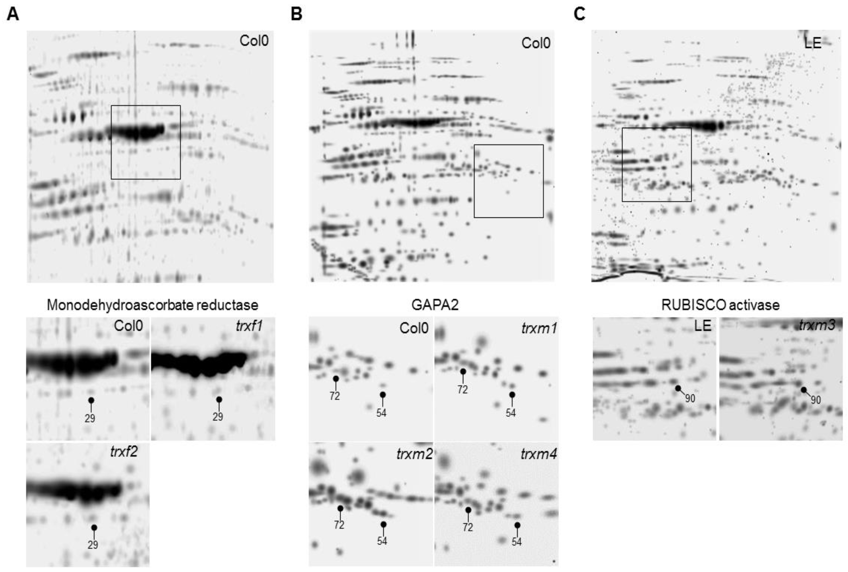
The trxm1 and trxm4 mutants were described previously [23,24] as well as trxm3 [25], trxf1 [26,27] and trxf2 [26]. Figure 1 shows that there is no detected TRX expression in trxf1, trxm1, trxm3 and trxm4, with these lines being considered as knockout ones, whilst a slight level of transcripts can be observed for trxf2 and trxm2 as it was described. The level of expression of all the mutants was clearly sufficiently low to validate the results obtained. The proteomic approach was performed to study protein profiles in these plastidial TRX f and m mutants. For this, the total protein extracts from rosettes of 25 dpg plants of the mutant lines trxf1, trxf2, trxm1, trxm2, trxm3 and trxm4 and of the wild type lines Col0 and Ler were analyzed in the “Unidad de Proteómica of the Universidad of Córdoba” (Córdoba, Spain). An optimal concentration and purity degree of the protein extracts were reached for a high level of separation of the peptides using two dimensional (2-DE) electrophoresis. Figure 2 shows the spots corresponding to the peptides of up- and down-regulated proteins in rosettes from trxf1 and trxf2 (Figure 2A), trxm1, trxm2 and trxm4 (Figure 2B), and trxm3 (Figure 2C), mutants in comparison with Columbia (Col0) and Landsberg erecta LE (Table 1, Table 2 and Table 3. The master gels from each three replicates gel mutant lines were obtained and normalized by using the software PDQuest® (Bio-rad). Three replicates gel of Col0 control was developed for each TRX type (f and m), as Ler for trxm3 (Figure 2). Also, PDQuest® (Bio-rad) was used to select, in each gel of the mutant lines, those spots where the expression was down regulated or up regulated when compared to the same spot located in the gels of the control plants. The proteins contained in the spots were picked, digested and the peptides identified by MS (Table S2). The comparison with databases allowed us to identify the proteins that could contain the different peptides. Out of the 200 analyzed and resolved spots, a total of 86 differentially expressed proteins were identified (Table S2). The trxf1 and trxf2 mutants had a proportionally larger number of down regulated proteins (15 out of 20 and 13 out of 21, respectively), whilst the trxm mutants had a larger number of up regulated proteins (16 out of 26 in trxm1, 18 out of 23 in trxm2, eight out of 10 in trxm3, and 21 out of 25 in trxm4) (Figure 3A). Despite the sequence similarities between the TRX f1 and f2, in the trxf1 and trxf2 lines nine proteins showed a different regulation (Table 1): glutamate-glyoxylate aminotransferase 1 (GGAT1), aminomethyltransferase, 5-methyl tetrahydropteroyl triglutamate-homocysteine methyltransferase 1, ribulose-bisphosphate carboxylase oxygenase (RUBISCO) activase, β-D-glucopyranosyl abscisate β-glucosidase, glyceraldehyde-3-phosphate dehydrogenase (GAPC2), monodehydroascorbate reductase, V-type ATP synthase, and Chaperonin 60 subunit β1. Nevertheless, apart from these differences, 10 out of 24 (41.7%) of the analyzed proteins were down regulated in both mutant lines while four out of 24 (16.7%) were up regulated. Data analysis revealed that the largest number of down regulated biological processes corresponded to the trxf mutants (Figure 3B). The other m type TRXs mutants mostly showed up regulated processes, especially trxm2, trxm3 and trxm4 (Figure 3B).
The four trxm mutant lines only shared one differentially expressed protein (β carbonic anhydrase 2), while the trxm1, trxm2, and trxm4 lines shared up to 13 differentially expressed proteins (39.4%, Table 2 and Table 3). However, some of the differentially expressed proteins identified were up regulated in one mutant line but down regulated in another one, as in the case of the ferredoxin-NADP reductase 1 or the chlorophyll a–b binding protein 2. Interestingly, nine proteins underwent similar changes in these three trxm lines, eight up-regulated and only one down-regulated (ferredoxin-NADP reductase 2). Some interesting proteins up-regulated, affecting key pathways, were the two chloroplast isoforms of glyceraldehyde-3-phosphate dehydrogenase GAPB and GAPA2 and the mitochondrial enzymes serine hydroxymethyltransferase 1 and glycine dehydrogenase 1 (amino acid metabolism). Regarding carbon fixation, the RUBISCO large subunit was down regulated in trxm1 and up regulated in trxm4, whereas its regulator RUBISCO activase was up-regulated in trxm2, trxm3, and trxm4. Interestingly, no photosynthetic genes were differentially expressed in the trxm3 mutant; nevertheless, the up-regulation of photosystem II stability/assembly factor HCF136 in the trxm1, trxm2 and trxm4 mutant lines is significant.
No coincident protein was observed in the trxf and trxm mutants. Nevertheless, the trxf1 and trxf2 lines shared one differentially expressed protein with the trxm1, trxm2 and trxm4 mutants: the ferredoxin-NADP reductase 1, though this protein was up-regulated in trxf1, trxf2, and trxm4 and down-regulated in trxm1 and trxm2. In addition, in the trxf and trxm3 mutants only myrosinase 2, a ABA signalling protein, was differentially expressed (down-regulated).
Regarding hormonal processes, in the trxm mutants only trxm3 displayed changes in ABA and JA signalling. Adjustments in the trxf mutants were limited to ABA signalling with a down-regulation of myrosinase 2 in both mutant lines and of β-D-glucopyranosyl abscisate β-glucosidase (activating the glucose-conjugated inactive ABA), but only in the trxf1 mutant. In Table 1, Table 2 and Table 3, we can observe the metabolic alterations mostly corresponded to changes in the level of glycine, alanine, glutamine, and methionine synthesizing/hydrolyzing enzymes in chloroplasts, mitochondria, peroxisomes, and cytosol. However, we could not observe any change in the amino acid metabolism in the trxm3 mutant.
All the identified spots corresponded to proteins with putative general functions related to the Calvin–Benson cycle, photosynthesis, stress response, hormone signalling, protein turnover, and to unknown processes (Figure 4, Table 1, Table 2, Table 3 and Table S2). The most represented functions fell into the metabolism category, containing 30–58% of the spots analysed, especially in the trxf mutants. The Calvin–Benson cycle was also well represented in all the mutants, ranging from 10.5% to 20%. Surprisingly, photosynthesis was affected in all the mutants except for trxm3, especially in the trxm1, trxm2, and trxm4 lines (Figure 4).
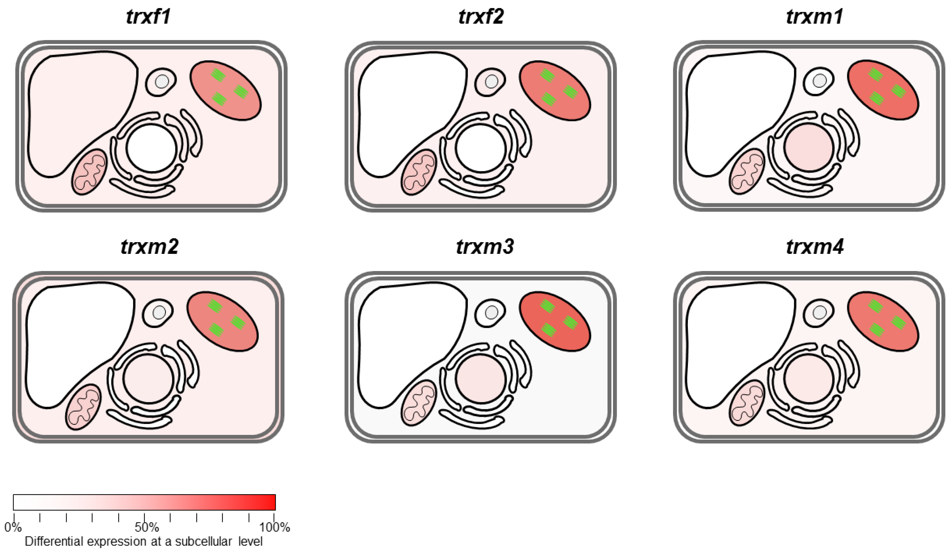
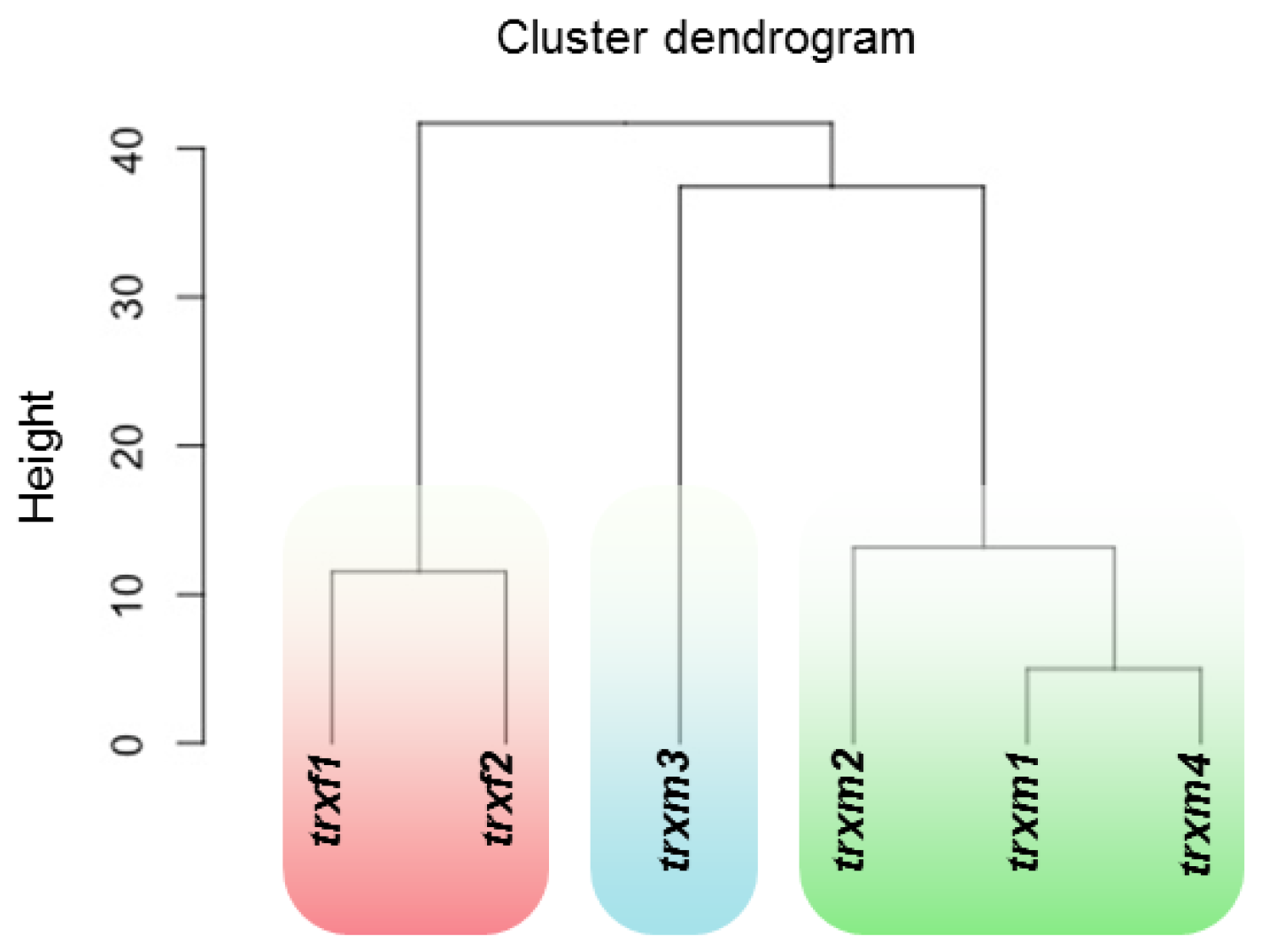
At a sub-cellular level, most of the differential expressed proteins were predicted to be chloroplastic, ranging from 47% to 59% of the total spots identified in each mutant (Figure 5). Differential expression in TRX mutants also affected non-chloroplast proteins as they were also predicted to be located in other sub-cellular compartments such as mitochondria, peroxisomes, cytosol, nucleus, Golgi/endoplasmic reticulum, and vacuole (enumerated according to the frequency of appearance in the proteomic analyses). Taking into account the affected processes, we applied a hierarchical clustering to our proteomic data (Figure 6). Two major clusters contained the trxf and the trxm mutants. Within the trxm cluster, two sub-clusters separated the trxm3 line from the trxm1, trxm2, and trxm4 mutants. Interestingly, according to our clustering analysis, trxm1 might be functionally closer to trxm4 than to trxm2. These data are according to recent results obtained in our laboratory (unpublished data).

4. Discussion

The existence of an elevated number of plastidial thioredoxins, amongst which are two f type TRXs and four m type TRXs in the chloroplast makes it difficult to identify and separate their different functions. A great deal of effort has been made to reach important conclusions and different authors have reported previously protein targets and suggested specific roles for several of the plastidial TRXs. Due to similarities with the f and m type TRXs, these isoforms might both carry out overlapping as specific functions in the redox regulation of the plant physiological processes. However, it has been difficult to find a clear border to delimit the role of each plastidial TRX described up to now. Beyond the study of the different reduction degree of the target proteins, our aim has been to know whether the pattern response of the Arabidopsis proteome is specific for each plastidial TRX mutant characterized. In this study we have carried out a proteomic profile of Arabidopsis mutant lines lacking f1, f2, m1, m2, m3 and m4 type TRXs.
The analysis of the 2-DE pattern revealed a total of 86 different protein-spot intensities out of the 200 that were resolved in all images (Figure 2 and Table S2). The differentially expressed proteins in the mutants showed particular patterns, supporting the hypothesis of a specific functionality for each TRX f or m isoform. A higher number of down regulated proteins were observed for trxf mutants, mainly trxf1; whilst more up regulated proteins occurred in the trxm lines, principally the trxm4 line (Figure 3A), suggesting that their roles are separate and affected.
Almost 59% of the differentially expressed proteins are plastidial, but other affected proteins are localized in mitochondria, cytosol, peroxisomes and nucleus (Figure 5), indicating the connection between the processes occurring either in the chloroplast or in other localizations when one plastidial TRX is missing. Likely, redox imbalance provoked by the lack of TRXs f/m isoforms in chloroplasts is triggering retrograde signalling to readjust the biological functions affected as we noticed differentially expressed proteins in other organelles. Moreover, we cannot discard other factors regulating the protein levels as protein degradation or stabilization by proteases and chaperonins, respectively, as discussed later.
As expected for the chloroplast redox enzymes involved in photosynthesis-related processes, among the proteins found that predominate are those which are functionally related with electronic transport. The presence of several subunits of the chloroplastidic and mithocondrial ATP synthase complexes is noteworthy. The subunit α of the chloroplastidic ATP synthase was down regulated in the trxf1 and trxf2, whilst the subunit β was up regulated in these lines. Interestingly, the subunits β and γ (the latter being up-regulated in trxm3) have been reported to be targets of plastidial TRXs [1,28]. However, it has been described that under low irradiance, NADPH thioredoxin reductase C (NTRC) is required for the redox modulation of the subunit γ of ATP synthase [29]. As the subunit 1 of the mitochondrial ATP synthase is also affected in trxf1, trxf2 and trxm3 mutants, these patterns of up/down regulation would be an indication of the importance and complexity of the regulation of ATP synthesis in plants suggesting the significance of balancing chloroplastic and mitochondrial ATP levels in response to redox changes. Remarkably, and similarly to the above-mentioned chloroplast subunits, the mitochondrial subunit 1 also seems to be redox regulated [1,30].
The link between chloroplast and mitochondrion is also evidenced with proteins responding to redox changes in the chloroplast involved in amino acid metabolism, such as serine hydroxyl methyl transferase or glycine dehydrogenase in trxm1, trxm2 and trxm4 and amino methyl transferase in trxf1; and even proteins with unknown functions such as the protein with identity number B3H6G1 in the trxm1, trxm2 and trxm4 mutants (Table S2).
These results do not necessarily imply dual localization chloroplast/mitochondria of the referred isoforms (though this cannot be ruled out), but rather they demonstrate the relationship between the metabolism of the chloroplast and the mitochondria. The observed differences at a proteomic level reveal, once again, a certain degree of functional specificity of each isoform of analyzed plastidial TRX.
Metabolism, the Calvin–Benson cycle, photosynthesis, response to stress, and hormone signaling are the functional categories most affected when one of the f or m type TRXs is absent. However, the lack of TRX f1 or f2 provokes a down-regulation in cell general processes, whilst the influence of the absence of TRXs m is the opposite (Figure 3). Most of the proteins differentially expressed in the trxf mutants relate to its function in metabolism in general, and to the synthesis of ATP specifically. In the same way, it seems that TRXs m1, m2 and m4 are involved in the redox regulation of the photosynthesis. TRXs f and m3 are likely to be involved in the redox regulation during stress situations as monodehydroascorbate reductase (in the stress response category) and myrosinase 2 (in the ABA signaling category) have been reported as TRXs targets (Table 1 and Table 3).
Taking into consideration the data obtained, it seems that the protein differentially expressed in the mutants fell into different functional categories which can be organized in function of the processes affected or their sub-cellular localization. Thus, as expected in a photosynthetic organ like the leaf, among the predominate proteins found are those which are functionally related with electronic transport with the only exception of trxm3.
Likewise, photosynthesis appears highly unstable in these mutants, amongst which a number of the electron transport or Calvin-Benson cycle proteins are up regulated or down regulated, as is described in the Results section. Interestingly, factor HCF136 was up regulated in the trxm1, trxm2 and trxm4 mutant lines, but down regulated in the trxf1 and trxf2 lines. It has been reported that factor HCF136 is involved in the assembly of an intermediary complex of PSII [31], suggesting that it is possible to detect putative alterations or increased instability of PSII in these mutants.
Previous studies have reported that TRXs are able to transfer reducing equivalents to the redox protein HCF164 in the thylakoid lumen [32], and that these proteins are necessary for the biogenesis of PSI. Additionally, evidence showed that HCF164 serves as transducer of reducing equivalents to proteins in the thylakoid lumen. Consequently, decreased levels of m type TRXs might lead to a redox imbalance in the thylakoid lumen and the instability of redox regulated proteins components of the PSII as PsbO subunits [14]. Interestingly, PSI subunit levels do not seem to be influenced in the TRX mutants analyzed, ruling out the possibility of different spot intensities (detection threshold) as both photosystems have a similar stoichiometry in the thylakoid membranes. Curiously, apart from proteins participating in primary metabolism or photosynthesis, other less represented proteins did not appear in our analyses, suggesting that these spots were not abundant enough to be detected with our experimental conditions. A clear example is the absence among the identified spots of the plastid TRXs that we were analyzing. However, this fact does not invalidate our results.
Some authors have attributed the f and m type TRXs to being part of the mechanisms involved in the control of the binding of the light harvesting chlorophyl (LHC) antenna complexes to the PSI and PSII through the activation of a serine/threonine kinase, SNT7 [33]. This is relevant because two components of binding factors to the chlorophyll a/b of the PSII are differentially expressed in trxm1 (Lhcb1B2 is up regulated and Lhcb2C1 is down regulated), trxm2 (Lhcb1B2 is up regulated) and trxm4 (Lhcb2C1 is up regulated). Additionally, trxm1 was the only one to have differentially expressed two subunits of the photolysis water complex (PsbO2 is up regulated and PsbP1 is down regulated), essential for generating the reducing power during the electron transport chain. TRX m1 is probably closely related to the redox regulation of the photosynthesis process.
In relation to the carbon fixation, it was not surprising to detect the large subunit of the RUBISCO protein among the differentially expressed proteins, due to its relevant position in carbon fixation. The RUBISCO large subunit was found to be down regulated in all the trxf and trxm1 lines. However, we cannot rule out the possibility that these changes might correspond to post-translationally modifications of the RUBISCO large subunit as we did not observe differences in the phenotypes with respect to wild-type plants due to a putative defect in carbon fixation. RUBISCO activase, involved in the regulation of the small subunit of RUBISCO appeared up regulated in the trxm2, trxm3 and trxm4 lines and down regulated in the trxf2 mutant, indicating a specific action when a change occurs in CO2 fixation during the loss of the redox homeostasis in plants. It is well known that several Calvin-Benson cycle enzymes are redox regulated, so it is not surprising to find them in the proteome of the mutants. However, the list is certainly lower due to the high plasticity of the plant to adapt under different adverse environments.
Due to their importance in key positions in the Calvin-Benson cycle the following proteins are particularly relevant: fructose-bisphosphate aldolase (up regulated in trxm1, trxm2 and trxm3) and transketolase 1 (down regulated in trxf1, trxf2 and trxm1); and the plastidial isoforms of GADPH, found to be up regulated in trxm1, trxm2 and trxm4 mutants; which were confirmed in the gene expression analysis (data not shown). It seems clear that redox activation/regulation by TRXs has a direct effect on the balance of carbohydrate synthesis and distribution as an optimal cell redox homeostasis environment is mandatory for the proper functioning of several processes, essentially carbon metabolism and the photosynthesis.
An interesting group was that of the proteins involved in redox homeostasis, with the most relevant being the subunits 1 and 2 of ferredoxin NADP+ reductase (FNR) (not described so far as a target of plastidial TRXs) and chloroplastidic and peroxisomal monodehydroascorbate reductase (MDAR), the relationship between these proteins and the redox regulation through TRXs is well known [34]. One of the possible functions of plastidial TRXs could be to maintain homeostasis against abiotic stress conditions, such as salinity during germination [35]. It seems that f type TRXs possibly play a more relevant role in controlling the homeostasis conditions inside the cells than previously thought.
Enzymes were also found to be involved in the response to biotic stress, such as the myrosinase (down regulated in trxf1, trxf2 and trxm3). In addition, the epithiospecifier protein, up regulated in trxm3, is also functionally related to myrosinase [36]. Similarly, GDSL esterase/lipase endothelial cell specific molecule1 (ESM1) was found to be up-regulated in trxm1, trxm2 and trxm4 mutants and it has been related to the response to insects [37].
Moreover, several interesting proteins related with the metabolism of amino acids, proteins and lipids have also been identified. Differentially expressed enzymes, mainly related with the synthesis and transfer of amino and methyl groups were found to be up regulated. Concerning the metabolism of proteins, proteases appeared, as well as factors involved in the translation process and proteins with chaperone activity. Particularly Relevant are the chaperonin 60 subunit β1 (up regulated in trxf1 and down regulated in trxf2) and chaperonin 60 subunit β2 (up-regulated in trxm2 and trxm3), suggesting that the protein folding and assembly are likely to be related to redox regulation. Previous studies have shown that the chaperonin 60 subunit β may be protecting the photosynthtetic components during stress situations [38].
Although the number of proteins with unknown functions was very low, a few examples without characterization were found (four), such as the protein with identity number B3H6G1 up regulated in trxm1, trxm2 and trxm4 (mentioned above). It is reasonable to think that a deep characterization would be necessary to identify new and specific functions of these plastidial thioredoxins.

5. Conclusions

In this study we have attempted to shed some light and to get closer to functionally defining the different isoforms of f and m type TRXs in Arabidopsis and to clarify the blurred frontier that exists, in most cases, between specificity and functional redundancy in multi-gene families in plants. Even though there are numerous studies on plastidial TRXs, the existing information is still scarce, from a functional point of view. Despite this, more studies are necessary, this is the first time a broad view is showing what processes are affected when one of the f or m type TRXs is lacking. We were aware of the complexity of the task. The results we have achieved from the approach followed in this study, which consisted of a comparative analysis of knockout/down mutants for the six isoforms of f and m type TRXs, should help to better understand the functional role of f and m type TRXs in Arabidopsis and to open up new research paths in the study of processes regulated by these enzymes.

Supplementary Materials

The following are available online at https://www.mdpi.com/2076-3921/8/3/54/s1, Table S1: gene-specific oligonucleotides used for quantitative PCR, Table S2: list of identified proteins in trxf1, trxf2 trxm1, trxm2, txm3, and trxm4 mutants.

Author Contributions

Funding acquisition, Mariam Sahrawy (M.S.); investigation, Juan Fernández–Trijueque (J.F.-T.), Antonio–Jesús Serrato (A.-J.S.) and M.S.; methodology, J.F.-T. and A.-J.S.; project administration, M.S.; supervision, A.-J.S. and M.S.; visualization, A.-J.S. and M.S.; writing—original draft, A.-J.S. and M.S.; writing—review and editing, A.-J.S. and M.S.

Funding

This work has been funded by research project BIO2009-07297, BIO2015-65272 from the Spanish Ministry of Science and Innovation and the European Fund for Regional Development, project P07-CVI-2795 and BIO 154 from the Andalusian Regional Government, Spain, and project BIO2012-33292, from the Spanish Ministry of Economy and Competitiveness.

Acknowledgments

The authors thank Trinidad Moreno and Sabrina De Brasi for their technical support, and Angela Tate for helpful editorial feedback on the manuscript. Protein spot digestion and MALDI-TOF analysis were carried out in the Universidad of Cordoba UCO-SCAI proteomics facility, a member of Carlos III Networked Proteomics Platform, ProteoRed-ISCIII.

Conflicts of Interest

The authors declare no conflict of interest.

References

  1. Montrichard, F.; Alkhalfioui, F.; Yano, H.; Vensel, W.H.; Hurkman, W.J.; Buchanan, B.B. Thioredoxin targets in plants: The first 30 years. J. Proteom. 2009, 72, 452–474. [Google Scholar] [CrossRef] [PubMed]
  2. Buchanan, B.B.; Schurmann, P.; Wolosiuk, R.A.; Jacquot, J.P. The ferredoxin/thioredoxin system: From discovery to molecular structures and beyond. Photosynth. Res. 2002, 73, 215–222. [Google Scholar] [CrossRef] [PubMed]
  3. Barajas-López, J.D.; Serrato, A.J.; Cazalis, R.; Meyer, Y.; Chueca, A.; Reichheld, J.P.; Sahrawy, M. Circadian regulation of chloroplastic f and m thioredoxins through control of the CCA1 transcription factor. J. Exp. Bot. 2011, 62, 2039–2051. [Google Scholar] [CrossRef] [PubMed]
  4. Chibani, K.; Wingsle, G.; Jacquot, J.P.; Gelhaye, E.; Rouhier, N. Comparative genomic study of the thioredoxin family in photosynthetic organisms with emphasis on Populus trichocarpa. Mol. Plant 2009, 2, 308–322. [Google Scholar] [CrossRef] [PubMed]
  5. Motohashi, K.; Kondoh, A.; Stumpp, M.T.; Hisabori, T. Comprehensive survey of proteins targeted by chloroplast thioredoxin. Proc. Natl. Acad. Sci. USA 2001, 8, 11224–11229. [Google Scholar] [CrossRef] [PubMed]
  6. Collin, V.; Issakidis-Bourguet, E.; Marchand, C.; Hirasawa, M.; Lancelin, J.M.; Knaff, D.B.; Miginiac-Maslow, M. The Arabidopsis plastidial thioredoxins: New functions and new insights into specificity. J. Biol. Chem. 2003, 278, 23747–23752. [Google Scholar] [CrossRef] [PubMed]
  7. Lemaire, S.D.; Michelet, L.; Zaffagnini, M.; Massot, V.; Issakidis-Bourguet, E. Thioredoxins in chloroplasts. Curr. Genet. 2007, 51, 343–365. [Google Scholar] [CrossRef] [PubMed]
  8. Serrato, A.J.; Fernández-Trijueque, J.; Barajas-López, J.D.; Chueca, A.; Sahrawy, M. Plastid thioredoxins: A “one-for-all” redox-signaling system in plants. Front. Plant Sci. 2013, 4, 1–10. [Google Scholar] [CrossRef] [PubMed]
  9. Chiadmi, M.; Navaza, A.; Miginiac-Maslow, M.; Jacquot, J.P.; Cherfils, J. Redox signalling in the chloroplast: Structure of oxidized pea fructose-1,6-bisphosphate phosphatase. EMBO J. 1999, 18, 6809–6815. [Google Scholar] [CrossRef] [PubMed]
  10. Schürmann, P.; Wolosiuk, R.A.; Breazeale, V.D.; Buchanan, B.B. 2 Proteins function in regulation of photosynthetic CO2 assimilation in chloroplasts. Nature 1976, 263, 257–258. [Google Scholar] [CrossRef]
  11. Buchanan, B.B.; Crawford, N.A.; Wolosiuk, R.A. Ferredoxin-thioredoxin system functions with effectors in activation of NADP glyceraldehyde 3-phosphate dehydrogenase of barley seedlings. Plant Sci. Let. 1978, 12, 257–264. [Google Scholar] [CrossRef]
  12. Jacquot, J.P.; Vidal, J.; Gadal, P.; Schürmann, P. Evidence for existence of several enzyme-specific thioredoxins in plants. FEBS Let. 1978, 96, 243–246. [Google Scholar] [CrossRef]
  13. Buchanan, B.B.; Balmer, Y. Redox regulation: A broadening horizon. Ann. Rev. Plant Biol. 2005, 56, 187–220. [Google Scholar] [CrossRef] [PubMed]
  14. Balmer, Y.; Vensel, W.H.; DuPont, F.M.; Buchanan, B.B.; Hurkman, W.J. Proteome of amyloplasts isolated from developing wheat endosperm presents evidence of broad metabolic capability. J. Exp. Bot. 2006, 57, 1591–1602. [Google Scholar] [CrossRef] [PubMed]
  15. Barajas-López, J.D.; Serrato, A.J.; Olmedilla, A.; Chueca, A.; Sahrawy, M. Localization in roots and flowers of pea chloroplast thioredoxin f and m proteins reveals new roles in non-photosynthetic organs. Plant Physiol. 2007, 145, 1–15. [Google Scholar]
  16. Hisabory, T. Thioredoxin affinity chromatography: A useful method for further understanding the thioredoxin network. J. Exp. Bot. 2005, 56, 1463–1468. [Google Scholar] [CrossRef] [PubMed]
  17. Yoshida, K.; Hisabory, T. Two distinct redox cascades cooperatively regulate chloroplast functions and sustain plant viability. Proc. Natl. Acad. Sci. USA 2016, 113, E3967–E3976. [Google Scholar] [CrossRef] [PubMed]
  18. Valot, B.; Negroni, L.; Zivy, M.; Gianinazzi, S.; Dumas-Gaudot, E. A mass spectrometric approach to identify arbuscular mycorrhiza-related proteins in root plasma membrane fractions. Proteom. J. 2006, S145–S155. [Google Scholar] [CrossRef] [PubMed]
  19. Soto-Suarez, M.; Serrato, A.J.; Rojas-González, J.A.; Bautista, R.; Sahrawy, M. Transcriptomic and proteomic approach to identify differentially expressed genes and proteins in Arabidopsis thaliana mutants lacking chloroplastic 1 and cytosolic FBPases reveals several levels of metabolic regulation. BMC Plant Biol. 2016, 16, 258–274. [Google Scholar] [CrossRef] [PubMed]
  20. Ramagli, L.S.; Rodríguez, L.V. Quantitation of microgram amounts of protein in two-dimensional polyacrylamide-gel sample buffer electrophoresis electrophoresis. Electrophoresis 1985, 6, 559–563. [Google Scholar] [CrossRef]
  21. Yan, J.X.; Wait, R.; Berkelman, T.; Harry, R.A.; Westbrook, J.A.; Wheeler, C.H.; Dunn, M.J. A modified silver staining protocol for visualization of proteins compatiblewith matrix-assisted laser desorption/ionization and electrospray ionization massspectrometry. Electrophoresis Int. J. 2000, 21, 3666–3672. [Google Scholar] [CrossRef]
  22. Castillejo, M.; Fernández-Aparicio, M.; Rubiales, D. Proteomic analysis by twodimensional differential in gel electrophoresis (2D DIGE) of the early response of Pisum sativum to Orobanchecrenata. J. Exp. Bot. 2012, 63, 107–119. [Google Scholar] [CrossRef] [PubMed]
  23. Courteille, A.; Vesa, S.; Sanz-Barrio, R.; Cazale, A.C.; Becuwe-Linka, N.; Farran, I.; Havaux, M.; Rey, P.; Rumeau, D. Thioredoxin m4 controls photosynthetic alternative electron pathways in Arabidopsis. Plant Physiol. 2013, 161, 508–520. [Google Scholar] [CrossRef] [PubMed]
  24. Wang, P.; Liu, J.; Liu, B.; Feng, D.; Da, Q.; Shu, S.; Su, J.; Zhang, Y.; Wang, J.; Wang, H.-B. Evidence for a role of chloroplastic m-type thioredoxins in the biogenesis of photosystem II in Arabidopsis. Plant Physiol. 2013, 163, 1710–1728. [Google Scholar] [CrossRef] [PubMed]
  25. Benitez-Alfonso, Y.; Cilia, M.; San Roman, A.; Thomas, C.; Maule, A.; Hearn, S.; Jackson, D. Control of Arabidopsis meristem development by thioredoxin-dependent regulation of intercelular transport. Proc. Natl. Acad. Sci. USA 2009, 106, 3615–3620. [Google Scholar] [CrossRef] [PubMed]
  26. Yoshida, K.; Hara, S.; Hisabori, T. Thioredoxin Selectivity for Thiol-Based Redox Regulation of Target Proteins in Chloroplasts. J. Biol. Chem. 2015, 115, 14278–14288. [Google Scholar] [CrossRef] [PubMed]
  27. Thormaehlen, I.; Ruber, J.; Von Roepenack-Lahaye, E.; Ehrlich, S.; Massot, V.; Huemmer, C.; Tezycka, J.; Issakidis-Bourguet, E.; Geigenberger, P. Inactivation of thioredoxin f1 leads to decreased light activation of ADP-glucose pyrophosphorylase and altered diurnal starch turnover in leaves of Arabidopsis plants. Plant Cell Environ. 2013, 36, 16–29. [Google Scholar] [CrossRef] [PubMed]
  28. Stumpp, M.T.; Motohashi, K.; Hisabori, T. Chloroplast thioredoxin mutants without active-site cysteines facilitate the reduction of the regulatory disulphide bridge on the gamma-subunit of chloroplast ATP synthase. Biochem. J. 1999, 341 Pt 1, 157–163. [Google Scholar] [CrossRef]
  29. Carrillo, L.R.; Froehlich, J.E.; Cruz, J.A.; Savage, L.J.; Kramer, D.M. Multi-level regulation of the chloroplast ATP synthase: The chloroplast NADPH thioredoxin reductase C (NTRC) is required for redox modulation specifically under low irradiance. Plant J. 2016, 87, 654–663. [Google Scholar] [CrossRef] [PubMed]
  30. Balmer, Y.; Vensel, W.H.; Tanaka, C.K.; Hurkman, W.J.; Gelhay, E.; Rouhier, N.; Jacquot, J.P.; Manieri, W.; Schürmann, P.; Droux, M.; et al. Thioredoxin links redox to the regulation of fundamental processes of plant mitochondria. Proc. Natl. Acad. Sci. USA 2004, 101, 2642–2647. [Google Scholar] [CrossRef] [PubMed] [Green Version]
  31. Plücken, H.; Müller, B.; Grohmann, D.; Westhoff, P.; Eichacker, L. The HCF136 protein is essential for assembly of the photosystem II reaction center in Arabidopsis thaliana. FEBS Lett. 2002, 532, 85–90. [Google Scholar] [CrossRef]
  32. Motohashi, K.; Hisabori, T. HCF164 receives reducing equivalents from stromal thioredoxin across the thylakoid membrane and mediates reduction of target proteins in the thylakoid lumen. J. Biol. Chem. 2006, 281, 35039–35047. [Google Scholar] [CrossRef] [PubMed]
  33. Rintamäki, E.; Martinsuo, P.; Pursisheimo, S.; Aro, E.M. Coorperative regulation of light-harvesting complex II phosphorylation via the plastoquinol and ferredoxin-thioredoxin system in chloroplasts. Proc. Natl. Acad. Sci. USA 2000, 97, 11644–11649. [Google Scholar] [CrossRef] [PubMed]
  34. Balmer, Y.; Vensel, W.H.; Cai, N.; Manieri, W.; Schürmann, P.; Hurkman, W.J.; Buchanan, B.B. A complete ferredoxin/thioredoxin system regulates fundamental processes in amyloplasts. Proc. Natl. Acad. Sci. USA 2006, 103, 2988–2993. [Google Scholar] [CrossRef] [PubMed]
  35. Fernández-Trijueque, J.; Barajas-López, J.D.; Chueca, A.; Cazalis, R.; Sahrawy, M.; Serrato, A.J. Plastid thioredoxins f and m are related to the developing and salinity response of post-germinating seeds of Pisum sativum. Plant Sci. 2012, 188–189, 82–88. [Google Scholar] [CrossRef] [PubMed]
  36. Lambrix, V.; Reichelt, M.; Mitchell-Olds, T.; Kliebenstein, D.J.; Gershenzon, J. The Arabidopsis epithiospecifier protein promotes the hydrolysis of glucosinolates to nitriles and influences Trichoplusiani herbivory. Plant Cell 2001, 13, 2793–2807. [Google Scholar] [CrossRef] [PubMed]
  37. Zhang, Z.-Y.; Ober, J.A.; Kliebenstein, D.J. The gene controlling the quantitative trait locus EPITHIOSPECIFIER MODIFIER1 alters glucosinolate hydrolysis and insect resistance in Arabidopsis. Plant Cell 2006, 18, 1524–1536. [Google Scholar] [CrossRef] [PubMed]
  38. Salvucci, M.E. Association of Rubisco activase with chaperonin-60β: A possible mechanism for protecting photosynthesis during heat stress. J. Exp. Bot. 2008, 59, 1923–1933. [Google Scholar] [CrossRef] [PubMed]
Figure 1. Thioredoxins (TRXs) f and m transcript levels in trxf and trxm mutants.
Antioxidants 08 00054 g001
Figure 2. DE images from rosette of trxf1, f2, m1, m2, m3 and m4 mutants. 2-D images of total proteins from rosettes of trx f1 and f2 (A), m1, m2 and m4 (B), and m3 (C) mutants in comparison with Col0 or Ler (LE). Numbers correspond to the protein spots identified by MALDI-TOF/TOF analysis (Table S2). The figure shows the representative experiments carried out with some examples of proteins identified in each gel.
Antioxidants 08 00054 g002
Figure 3. Processes down- or up-regulated in the trx mutants, number of coincident spots identified by proteomic analyses. (A) number of proteins down- or up-regulated in trxf or trxm mutants; (B) biological processes affected in each mutant line.
Antioxidants 08 00054 g003
Figure 4. Degree of significance of the biological processes affected in the trx mutants in relation to the number peptides predicted to be involved in a process.
Antioxidants 08 00054 g004
Figure 5. Differential expression shown at a sub-cellular level. The red color intensity is indicating if differential expressions are significant in a given organelle.
Antioxidants 08 00054 g005
Figure 6. Hierarchical clustering segregates trx mutants according to the biological processes affected. For calculations, the R statistical environment (The R Foundation, Vienna, Austria) available at http://www.R-project.org was used.
Antioxidants 08 00054 g006
Table 1. Differentially expressed proteins identified by MS in the trxf1 and trxf2 mutants, organized inthe functional category, the gene code and the subcellular localization and whether the protein has been reported as a Thioredoxins (TRX) target. The color code indicates fold change in protein abundance.
Functional CategoryProteinGene IDLocationTRX TargetSpottrxf1trxf2
Amino acids metabolismGlutamate-glyoxylate aminotransferase 1At1g23310Per.-16
AminomethyltransferaseAt1g11860Mit.-8
Glutamate-glyoxylate aminotransferase 2At1g70580Per.-21
5-methyltetrahydropteroyltrigluta-mate-homocysteine methyltransferase 1At5g17920Per.-33
5-methyltetrahydropteroyltrigluta-mate-homocysteine methyltransferase 2At3g03780Cy.-12
Calvin-Benson cycle RUBISCO large subunitAtCg00490Ch.Yes5
Transketolase1At3g60750Ch.Yes20
RUBISCO activaseAt2g39730Ch.Yes27
ATP synthesisATP synthase subunit ßAtCg00480Ch.Yes15
ATP synthase subunit αAtCg00120Ch.Yes4, 24
ATP synthase subunit 1AtMg01190Mit.Yes25
PhotosynthesisPSII stability/assembly factor HCF136At5g23120Ch.-9
Ferredoxin-NADP reductase 1At5g66190Ch.-17
ABA signallingβ-D-glucopyranosyl abscisate β-glucosidaseAt1g52400ER-3
Myrosinase 2At5g25980n.d.Yes22
GlycolisisTriosephosphate isomeraseAt3g55440Mit.-31
Glyceraldehyde-3-phosphate dehydrogenase C2 (GAPC2)At1g13440Cy.-26
Stress responseJacalin-Related lectinAt3g16470n.d.-7
Monodehydroascorbate reductaseAt1g63940Ch., Mit.Yes29
Protein biosynthesisElongation factor TuAt4g20360Ch.Yes6
ATP hydrolysisV-type ATP synthaseAt1g78900V.-2
PSII biogenesisPSII stability/assembly factor HCF136At5g23120Ch.-30
Refolding activityChaperonin 60 subunit ß1At1g55490Ch.-13
Tricarboxylic acid cycleMalate dehydrogenase 1At1g53240Mit.Yes35
Protein abundance change relative to the control (Col0). Antioxidants 08 00054 i001
Ch., chloroplast; Mit., mitochondria; Per., peroxisome; Cy., cytosol; V., vacuole; ER, endoplasmic reticulum; n.d., not determined. Proteins with a Confidence Interval C.I.% ≥ 95% are shown. According to Montrichard et al. (2009) [1], reported thioredoxin targets are shown. NADP: nicotinamide-adenine-dinucleotide phosphate; HCF: high chlorophyll fluorescence; ABA: absicic acid; PSII: photosystem II; RUBISCO: ribulose bisphosphate carboxylase/oxygenase.
Table 2. Differentially expressed proteins identified by MS in the trxm1, trxm2, and trxm4 mutants, organized in the functional category, the gene code and the subcellular localization and whether the protein has been reported as a TRX target. The color code indicates fold change in protein abundance.
Functional CategoryProteinGene IDLocationTRX TargetSpottrxm1trxm2trxm4
Calvin-Benson cycleTransketolaseAt3g60750Ch.Yes38
RUBISCO large subunitAtCg00490Ch.Yes28
Fructose-bisphosphate aldolase 2At4g38970Ch.Yes59, 103
RUBISCO activaseAt2g39730Ch.Yes65
Glyceraldehyde-3-phosphate dehydrogenase B GAPB)At1g42970Ch.Yes68
Glyceraldehyde-3-phosphate dehydrogenase A2 (GAPA2)At1g12900Ch.Yes54, 72
Amino acids metabolismSerine hydroxymethyltransferase 1At4g37930Mit.Yes101
Glycine dehydrogenase (decarboxylating) 1At4g33010Mit.-100
Probable phosphoglycerate mutase 2At3g08590Mit.-47
Glutamate-glyoxylate aminotransferase 1At1g23310Per.Yes97
5-methyltetrahydropteroyltrigluta-mate-homocysteine methyltransferase 1At5g17920Cy.Yes45, 98
PhotosynthesisChlorophyll a-b binding protein 2At1g29920Ch.Yes42
Ferredoxin-NADP reductase 1At5g66190Ch.-43
Chlorophyll a-b binding proteinAt2g34420Ch.-56
Oxygen-evolving enhancer protein 2-1At1g06680Ch.Yes57
Oxygen-evolving enhancer protein 1-2At3g50820Ch.Yes41
Ferredoxin-NADP reductase 2At1g20020Ch.-104
Stress responseUncharacterized proteinAt2g37660Ch.-40
Heat shock 70 kDa protein 3At3g09440N.-46
Monodehydroascorbate reductase 1At3g52880Per.Yes70
PSII stabilization/repairPhotosystem II stability/assembly factor HCF136At5g23120Ch.-67
Protease Do-like 1At3g27925Ch.-39
Protein transportChaperone protein ClpC1At5g50920Ch.-37
ATP synthesisATP synthase subunit betaAtCg00480Ch.Yes93
GlycolysisGlyceraldehyde-3-phosphate dehydrogenase GAPC2At1g13440Cy.Yes53
Carbohydrate metabolismChloroplast stem-loop binding protein of 41 kDa bAt1g09340Ch.-64
Carbon utilizationβ carbonic anhydrase 2At5g14740Ch.-55
Lipid degradationGDSL esterase/lipase ESM1At3g14210N.-105
Protein refoldingChaperonin 60 subunit beta 2At3g13470Ch.Yes96
UnkownPolyketide cyclase/dehydrase and lipid transport superfamily proteinAt4g14500Mit.-61
Uncharacterized proteinAt2g37660Ch.-40
Uncharacterized proteinAt5g05113Mit.-74
Disease resistance protein (NBS-LRR class) familyAt5g40060n.d.-75
Protein abundance change relative to the control (Col0). Antioxidants 08 00054 i002
Ch., chloroplast; Mit., mitochondria; Per., peroxisome; Cy., cytosol; V., vacuole; N., nucleus; n.d., not determined. Proteins with Protein Scores C.I.% ≥ 95% are shown. According to Montrichard et al. (2009) [1], reported thioredoxin targets are shown. ESM1: epithiospecifier modifier 1.
Table 3. Differentially expressed proteins identified by MS in the trxm3 mutant, organized in the functional category, the gene code and the subcellular localization and whether the protein has been reported as a TRX target. The color code indicates fold change in protein abundance.
Functional CategoryProteinGene IDLocationTRX TargetSpottrxm3
Calvin-Benson cycleRUBISCO activaseAt2g39730Ch.Yes90
Fructose-bisphosphate aldolase 2At4g38970Ch.Yes91
ATP synthesisATP synthase subunit 1AtMG01190Mit.Yes82
ATP synthase γ chain 1At4g04640Ch.Yes85
JA signalling /responseEpithiospecifier proteinAt1g54040N.-84
Lipoxygenase 2At3g45140Ch.Yes76
ABA signallingMyrosinase 2At5g25980n.d.Yes77
Carbon utilizationβ carbonic anhydrase 2At5g14740Ch.Yes92
Refolding activityChaperonin 60 subunit β 2At3g13470Ch.-87
Stress responseMonodehydroascorbate reductaseAt1g63940Ch., Mit.Yes83
Protein abundance change relative to the control (Ler). Antioxidants 08 00054 i003
Ch., chloroplast; Mit., mitochondria; N., nucleus; n.d., not determined. Proteins with Protein Scores C.I.% ≥ 95% are shown. According to Montrichard et al. (2009) [1], reported thioredoxin targets are shown. JA: jamonic acid.