Betaine in Metabolic Dysfunction-Associated Steatotic Liver Disease: Mechanisms of Action and Therapeutic Potential

 ,
,  ,
,  , and
, and
Abstract
1. Introduction
Literature Search Strategy
2. Physiological Role of Betaine
2.1. Betaine as an Osmoprotectant and Chemical Chaperone
2.2. Betaine as a Methyl Group Donor

| Experimental Model | Model/Intervention | Betaine Dose/Concentration and Treatment Duration | Main Findings |
|---|---|---|---|
| Animal models | |||
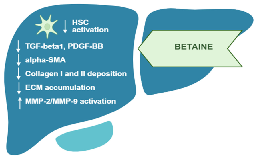
| C57BL/6 mice | TAA-induced liver fibrosis | 2% (w/v) in drinking water; 6 weeks (weeks 2–8) | Reduced TGF-β1 and PDGF-BB signaling, modulated MMP-2/MMP-9/TIMP-1, and decreased collagen I and III deposition [69] |
| C57BL/6 mice | TAA-induced liver fibrosis | 2% (w/v) in drinking water; 6 weeks (weeks 2–8) | Reduced hepatocellular injury, oxidative stress, and inflammation [70] |
| C57BL/6 mice | Methionine–choline-deficient diet-induced NAFLD | 1.5% (w/v) in drinking water; 6 weeks | Improved liver histology and reduced hepatic steatosis and hepatocellular injury [71] |
| C57BL/6 mice | Methionine–choline-deficient diet-induced fatty liver | 1.5% (w/v) in drinking water; 6 weeks | Reduced oxidative stress, inflammation, and apoptosis and modulated autophagy and Akt/mTOR signaling [72] |
| C57BL mice | High-fat diet-induced insulin resistance and fatty liver | 1% (w/v) in drinking water; 14 weeks (preventive) or final 4 weeks after 14 weeks of HFD (therapeutic) | Improved hepatic insulin signaling and insulin sensitivity, with increased IRS-1/Akt/GSK3β activation and hepatic glycogen content and reduced steatosis [84] |
| Obese mice | Diet-induced obesity; betaine supplementation | 2% (w/v) in drinking water; 8 to 25 weeks | Improved glucose metabolism and glucose utilization in liver and skeletal muscle and reduced systemic inflammation [85] |
| ApoE−/− mice | High-fat diet-induced NAFLD | 2% (w/v) in drinking water; 8 weeks | Reduced hepatic lipid accumulation through FGF10/AMPK signaling, suppression of lipogenesis, and enhancement of fatty acid oxidation [86] |
| Rats | Fructose-induced NAFLD | 62.5, 125, or 250 mg/kg; 4 weeks (weeks 5–8) | Reduced hepatic steatosis and ER stress through regulation of LXRα/PPARα signaling [87] |
| Mice | High-fat diet-induced hepatic steatosis | 1% (w/v) in drinking water; 13 weeks | Improved hepatic lipid and iron homeostasis, including regulation of ZIP14, FTL/FTH, ferroportin, HAMP, and BMP2/BMP6–SMAD signaling [88] |
| Mice | High-fat diet-induced MASLD | 0.3% (w/w) in diet; from the start of the intervention | Reduced hepatic iron accumulation and lipid peroxidation and attenuated ferroptosis through activation of the Nrf2/GPX4 pathway [89] |
| ICR mice | CDAHFD-induced NAFLD | 0.2–1% (w/v) in drinking water; 1 week; 0.5% used in mechanistic autophagy experiments | Enhanced autophagy and reduced hepatic lipid accumulation and ER stress [90] |
| Rats | High-fat diet-induced NAFLD | 1% betaine in the diet; 3 weeks | Improved sulfur-amino acid metabolism and antioxidant status and reduced oxidative stress [91] |
| Cell-based studies | |||
| Primary human hepatocytes | Insulin-resistant hepatocytes; betaine treatment | 0.63–20 mM; 24 h | Increased IRS-1 and Akt activation without changes in insulin receptor expression or activation, supporting a post-receptor insulin-sensitizing effect [84] |
| AML12 hepatocytes | Palmitic acid-induced lipotoxicity and ferroptosis; betaine treatment | 200 μM; 24–48 h | Reduced iron accumulation and lipid peroxidation and increased Nrf2/GPX4/SLC7A11 signaling, resulting in attenuation of ferroptosis [89] |
| L8824 fish hepatocyte cell line | Lipid-overload model; betaine treatment | 400 μM | Reduced lipid accumulation and enhanced VLDL secretion through regulation of the HNF4α/MTTP pathway [92] |
| Human adipocytes | Hypoxia-induced inflammatory response; betaine treatment | 250 μM; 8–20 h | Reduced expression of hypoxia-induced inflammatory adipokines, including IL-6 and TNF-α [93] |
3. The Effects of Betaine in MASLD/MASH
3.1. Betaine Modulates Insulin Resistance and Lipid Metabolism in MASLD/MASH
3.1.1. Effects of Betaine on Hepatic Insulin Signaling
3.1.2. Effects of Betaine on Hepatic Lipid Metabolism
3.1.3. Betaine, One-Carbon Metabolism, and Insulin Sensitivity
3.1.4. Effects of Betaine on Oxidative Stress- and Inflammation-Associated Insulin Resistance
3.2. Betaine Restores Redox, Mitochondrial, and Endoplasmic Reticulum Homeostasis Across the MASLD–MASH–HCC Continuum
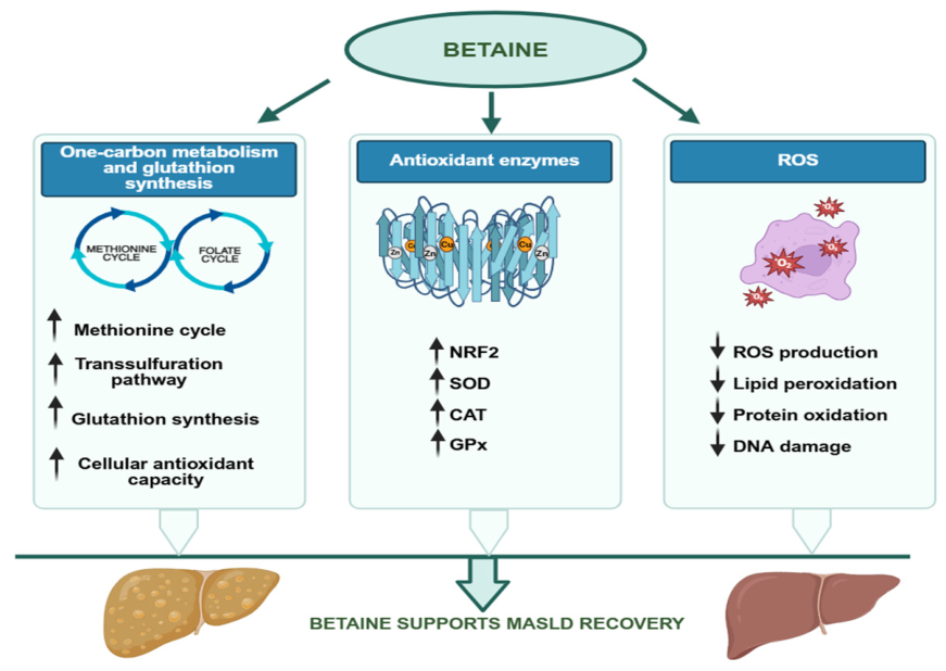
3.2.1. Betaine Mitigates Oxidative Stress
3.2.2. Betaine Preserves Mitochondrial Homeostasis
3.2.3. Betaine Alleviates Endoplasmic Reticulum Stress
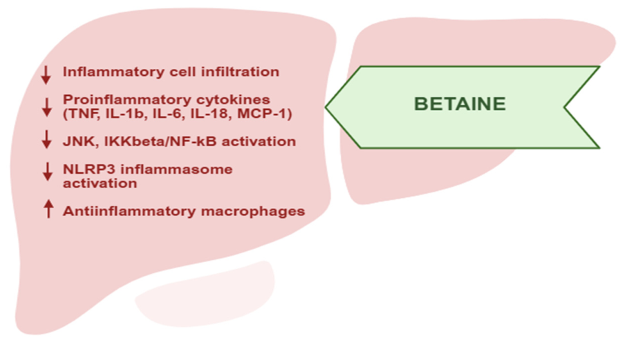
3.3. Betaine Mitigates Inflammation and Fibrosis During MASLD Progression
4. Conclusions
Author Contributions
Funding
Institutional Review Board Statement
Informed Consent Statement
Data Availability Statement
Conflicts of Interest
Abbreviations
| ACC | Acetyl-CoA carboxylase |
| Akt | Protein kinase B |
| AMPK | AMP-activated protein kinase |
| ApoE−/− | Apolipoprotein E-deficient |
| ATF6 | Activating transcription factor 6 |
| BGT1 | Betaine/GABA transporter 1 |
| BHMT | Betaine–homocysteine methyltransferase |
| CD36 | Cluster of differentiation 36 |
| ChREBP | Carbohydrate-responsive element-binding protein |
| CPT1 | Carnitine palmitoyltransferase 1 |
| CPR | Cytochrome P450 oxidoreductase |
| db/db | Leptin receptor-deficient diabetic mouse model |
| ECM | Extracellular matrix |
| eIF2α | Eukaryotic initiation factor 2 alpha |
| ER | Endoplasmic reticulum |
| EU | European Union |
| FASN | Fatty acid synthase |
| FDA | U.S. Food and Drug Administration |
| FGF10 | Fibroblast growth factor 10 |
| FGF21 | Fibroblast growth factor 21 |
| FOXO1 | Forkhead box O1 |
| FTO | Fat mass and obesity-associated protein |
| FXR | Farnesoid X receptor |
| GNMT | Glycine N-methyltransferase |
| GSK3β | Glycogen synthase kinase 3 beta |
| HCC | Hepatocellular carcinoma |
| HMGB1 | High-mobility group box 1 |
| HSCs | Hepatic stellate cells |
| IKKβ | Inhibitor of nuclear factor kappa B kinase beta |
| IL-1β | Interleukin 1 beta |
| IL-6 | Interleukin 6 |
| IL-18 | Interleukin 18 |
| iNOS | Inducible nitric oxide synthase |
| IRE1α | Inositol-requiring enzyme 1 alpha |
| IRS-1 | Insulin receptor substrate 1 |
| JAK/STAT | Janus kinase/signal transducer and activator of transcription |
| JNK | c-Jun N-terminal kinase |
| LPS | Lipopolysaccharide |
| LXRα | Liver X receptor alpha |
| m6A | N6-methyladenosine |
| MASH | Metabolic dysfunction-associated steatohepatitis |
| MASLD | Metabolic dysfunction-associated steatotic liver disease |
| MAT | Methionine adenosyltransferase |
| MCP-1 | Monocyte chemoattractant protein 1 |
| MMP-2 | Matrix metalloproteinase 2 |
| MMP-9 | Matrix metalloproteinase 9 |
| mTORC1 | Mechanistic target of rapamycin complex 1 |
| MTTP | Microsomal triglyceride transfer protein |
| MyD88 | Myeloid differentiation primary response protein 88 |
| NF-κB | Nuclear factor kappa B |
| NLRP3 | NLR family pyrin domain-containing 3 |
| NO | Nitric oxide |
| NRF-1 | Nuclear respiratory factor 1 |
| PDGF-BB | Platelet-derived growth factor BB |
| PEMT | Phosphatidylethanolamine N-methyltransferase |
| PERK | Protein kinase RNA-like endoplasmic reticulum kinase |
| PGC-1α | Peroxisome proliferator-activated receptor gamma coactivator 1 alpha |
| PI3K | Phosphoinositide 3-kinase |
| PKCε | Protein kinase C epsilon |
| PPARα | Peroxisome proliferator-activated receptor alpha |
| PPARγ | Peroxisome proliferator-activated receptor gamma |
| ROS | Reactive oxygen species |
| SAH | S-adenosylhomocysteine |
| SAM | S-adenosylmethionine |
| SCD1 | Stearoyl-CoA desaturase 1 |
| SCFAs | Short-chain fatty acids |
| sIgA | Secretory immunoglobulin A |
| SIRT1 | Sirtuin 1 |
| SLC6A12 | Solute carrier family 6 member 12 |
| SOCS3 | Suppressor of cytokine signaling 3 |
| SREBP-1c | Sterol regulatory element-binding protein 1c |
| T2DM | Type 2 diabetes mellitus |
| TFAM | Mitochondrial transcription factor A |
| TGF-β1 | Transforming growth factor beta 1 |
| TGR5 | Takeda G protein-coupled receptor 5 |
| TIMP-1 | Tissue inhibitor of metalloproteinases 1 |
| TLR4 | Toll-like receptor 4 |
| TNF-α | Tumor necrosis factor alpha |
| TonEBP/NFAT5 | Tonicity-responsive enhancer-binding protein (nuclear factor of activated T cells 5) |
| TXNIP | Thioredoxin-interacting protein |
| UPR | Unfolded protein response |
| VLDL | Very-low-density lipoprotein |
| α-SMA | Alpha-smooth muscle actin |
References
- Younossi, Z.M.; Golabi, P.; Paik, J.M.; Henry, A.; Van Dongen, C.; Henry, L. The global epidemiology of nonalcoholic fatty liver disease (NAFLD) and nonalcoholic steatohepatitis (NASH): A systematic review. Hepatology 2023, 77, 1335–1347. [Google Scholar] [CrossRef] [Scilit] [PubMed]
- Younossi, Z.M.; Kalligeros, M.; Henry, L. Epidemiology of metabolic dysfunction-associated steatotic liver disease. Clin. Mol. Hepatol. 2025, 31, S32–S50. [Google Scholar] [CrossRef] [Scilit] [PubMed]
- Targher, G.; Valenti, L.; Byrne, C.D. Metabolic dysfunction-associated steatotic liver disease. N. Engl. J. Med. 2025, 393, 683–698. [Google Scholar] [CrossRef] [Scilit] [PubMed]
- Tilg, H.; Petta, S.; Stefan, N.; Targher, G. Metabolic dysfunction-associated steatotic liver disease in adults: A review. JAMA 2026, 335, 163–174. [Google Scholar] [CrossRef] [Scilit] [PubMed]
- Huang, H.T.; Hewitt, M.; Li, W.; Alazawi, W. Real-world evidence in metabolic dysfunction-associated steatotic liver disease (MASLD): Insights, challenges, and future directions. Lancet Reg. Health Eur. 2025, 62, 101557. [Google Scholar] [CrossRef] [Scilit] [PubMed]
- Olaez-Ramos, C.E.; Mojica-Zamudio, K.L.; Sandoval-Rodríguez, A.; Armendáriz-Borunda, J. Epidemiology and diagnostic of MASLD in Latin America: A yet to be met challenge. Front. Pharmacol. 2026, 16, 1700072. [Google Scholar] [CrossRef] [Scilit] [PubMed]
- Stefan, N.; Yki-Järvinen, H.; Neuschwander-Tetri, B.A. Metabolic dysfunction-associated steatotic liver disease: Heterogeneous pathomechanisms and effectiveness of metabolism-based treatment. Lancet Diabetes Endocrinol. 2025, 13, 134–148. [Google Scholar] [CrossRef] [Scilit] [PubMed]
- Romanos, M.; Garcia Cordova, J.M.; Villamarin, J.; Pazmino Zurita, J.D.; Acosta, A. Obesity and metabolic dysfunction-associated steatotic liver disease (MASLD): A literature review on pathophysiology and treatment. Diabetes Obes. Metab. 2026, 28, 19–30. [Google Scholar] [CrossRef] [Scilit] [PubMed]
- Kim, B.K. Metabolic dysfunction-associated steatotic liver disease and obesity: Pathogenesis, diagnostics, risk stratification, and therapeutic approach. Kaohsiung J. Med. Sci. 2026, e70227. [Google Scholar] [CrossRef] [Scilit] [PubMed]
- Zhang, Y.; Wei, Y.; Liu, H.; Guo, Y. Protective effect of exercise on metabolic dysfunction-associated fatty liver disease: Potential epigenetic mechanisms (Review). Int. J. Mol. Med. 2025, 56, 146. [Google Scholar] [CrossRef] [Scilit] [PubMed]
- Maldonado-Rojas, C.D.A.; Zuarth-Vazquez, J.M.; Uribe, M.; Barbero-Becerra, V.J. Insulin resistance and metabolic dysfunction-associated steatotic liver disease (MASLD): Pathways of action of hypoglycemic agents. Ann. Hepatol. 2024, 29, 101182. [Google Scholar] [CrossRef] [Scilit] [PubMed]
- Syed-Abdul, M.M. Lipid metabolism in metabolic-associated steatotic liver disease (MASLD). Metabolites 2023, 14, 12. [Google Scholar] [CrossRef] [Scilit] [PubMed]
- Carli, F.; Della Pepa, G.; Sabatini, S.; Vidal-Puig, A.; Gastaldelli, A. Lipid metabolism in MASLD and MASH: From mechanism to the clinic. JHEP Rep. 2024, 6, 101185. [Google Scholar] [CrossRef] [Scilit] [PubMed]
- Jin, J.; Cheng, Y. Mitochondrial dysfunction in metabolic-syndrome-related MASLD/MASH: Metabolic mechanisms and therapeutic perspectives. Metabolites 2026, 16, 489. [Google Scholar] [CrossRef] [Scilit] [PubMed]
- Radosavljevic, T.; Brankovic, M.; Samardzic, J.; Djuretić, J.; Vukicevic, D.; Vucevic, D.; Jakovljevic, V. Altered mitochondrial function in MASLD: Key features and promising therapeutic approaches. Antioxidants 2024, 13, 906. [Google Scholar] [CrossRef] [Scilit] [PubMed]
- Zhou, M.; Lv, J.; Chen, X.; Shi, Y.; Chao, G.; Zhang, S. From gut to liver: Exploring the crosstalk between the gut–liver axis and oxidative stress in metabolic dysfunction-associated steatotic liver disease. Ann. Hepatol. 2025, 30, 101777. [Google Scholar] [CrossRef] [Scilit] [PubMed]
- Zhao, G.; He, F.; Wu, C.; Li, P.; Li, N.; Deng, J.; Zhu, G.; Ren, W.; Peng, Y. Betaine in inflammation: Mechanistic aspects and applications. Front. Immunol. 2018, 9, 1070. [Google Scholar] [CrossRef] [Scilit] [PubMed]
- Dong, T.; Li, J.; Liu, Y.; Zhou, S.; Wei, X.; Hua, H.; Tang, K.; Zhang, X.; Wang, Y.; Wu, Z.; et al. Roles of immune dysregulation in MASLD. Biomed. Pharmacother. 2024, 170, 116069. [Google Scholar] [CrossRef] [Scilit] [PubMed]
- Capuccio, S.; Cocuzza, C.; Di Marco, G.L.; Scamporrino, A.; Piro, S.; Russello, M. Metabolic dysfunction-associated steatotic liver disease (MASLD): From steatosis to systemic metabolic failure: Classical pathophysiology, emerging systemic mechanisms and modifiable lifestyle determinants. Nutrients 2026, 18, 2316. [Google Scholar] [CrossRef] [Scilit] [PubMed]
- Steinberg, G.R.; Carpentier, A.C.; Wang, D. Integrative metabolism in MASLD and MASH: Pathophysiology and emerging mechanisms. J. Hepatol. 2025, 83, 584–595. [Google Scholar] [CrossRef] [Scilit] [PubMed]
- Huang, D.Q.; Wong, V.W.S.; Rinella, M.E.; Boursier, J.; Lazarus, J.V.; Yki-Järvinen, H.; Loomba, R. Metabolic dysfunction-associated steatotic liver disease in adults. Nat. Rev. Dis. Primers 2025, 11, 14. [Google Scholar] [CrossRef] [Scilit] [PubMed]
- Gallego-Durán, R.; Carrillo-Fernández, P.P.; Gracia-Sancho, J. Pathophysiology of metabolic dysfunction associated steatotic liver disease. Clín. Gastroenterol. Méx. 2025, 1, 313–322. [Google Scholar] [CrossRef] [Scilit]
- Miller, D.M.; McCauley, K.F.; Dunham-Snary, K.J. Metabolic Dysfunction-Associated Steatotic Liver Disease (MASLD): Mechanisms, Clinical Implications and Therapeutic Advances. Endocrinol. Diabetes Metab. 2025, 8, e70132. [Google Scholar] [CrossRef] [Scilit] [PubMed]
- Tantu, M.T.; Farhana, F.Z.; Haque, F.; Mkoo, K.M.; Qiao, L.; Ross, A.G.; Shiddiky, M.J.A. Pathophysiology, noninvasive diagnostics and emerging personalized treatments for metabolic associated liver diseases. npj Gut Liver 2025, 2, 18. [Google Scholar] [CrossRef] [Scilit]
- Younossi, Z.M.; Kalligeros, M.; Wong, V.W.; Al-Naamani, K.M.; Tacke, F.; Tsochatzis, E.; Crespo, J.; El-Kassas, M.; Bril, F.; Arrese, M.; et al. Updated global consensus recommendations for risk stratification, treatment initiation, and response monitoring in metabolic dysfunction-associated steatotic liver disease. Clin. Gastroenterol. Hepatol. 2026; in press. [CrossRef] [Scilit] [PubMed]
- Younossi, Z.M.; Zelber-Sagi, S.; Lazarus, J.V. Global consensus recommendations for metabolic dysfunction-associated steatotic liver disease and steatohepatitis. Gastroenterology 2025, 169, 1017–1032. [Google Scholar] [CrossRef] [Scilit] [PubMed]
- Kumar, A. Global MASLD consensus: Inclusive in scope, exclusive in therapy? Gastroenterology 2026, 170, 644–645. [Google Scholar] [CrossRef] [Scilit] [PubMed]
- Bhadoria, A.S.; Sinha, S. Re-evaluating lifestyle interventions in normal weight metabolic dysfunction-associated steatotic liver disease. Gastroenterology 2026, 170, 645–646. [Google Scholar] [CrossRef] [Scilit] [PubMed]
- Pekarska, K.; Brennan, P.N.; Ivancovsky Wajcman, D.; Towey, J.; Cox, L.; Weatherby, K.; McPherson, S.; Stine, J.; Willemse, J.; Li, W. Nutrition in MASLD: A patient-focused, evidence-based clinician’s guide. Frontline Gastroenterol. 2026, 17, 262–281. [Google Scholar] [CrossRef] [Scilit]
- Arumugam, M.K.; Paal, M.C.; Donohue, T.M.; Ganesan, M.; Osna, N.A.; Kharbanda, K.K. Beneficial effects of betaine: A comprehensive review. Biology 2021, 10, 456. [Google Scholar] [CrossRef] [Scilit] [PubMed]
- Buonaiuto, G.; Federiconi, A.; Vecchiato, C.G.; Benini, E.; Mordenti, A.L. Betaine dietary supplementation: Healthy aspects in human and animal nutrition. Antioxidants 2025, 14, 771. [Google Scholar] [CrossRef] [Scilit] [PubMed]
- Yang, Z.-J.; Huang, S.-Y.; Cheng, J.; Zeng, J.-W.; Wusiman, M.; Li, H.-B.; Zhu, H.-L. Betaine: A comprehensive review on dietary sources, health benefits, mechanisms of action, and application. Trends Food Sci. Technol. 2025, 159, 104993. [Google Scholar] [CrossRef] [Scilit]
- Singh, S.K.; Yadav, P.; Bishnolia, M.; Manhar, N.; Kumar, P.; Navik, U.; Weiskirchen, R. Betaine as a regulator of metabolism, epigenetics, and cellular osmoprotection: Therapeutic implications in cardiometabolic and renal diseases. Front. Pharmacol. 2026, 17, 1883688. [Google Scholar] [CrossRef] [Scilit] [PubMed]
- Craig, S.A.S. Betaine in human nutrition. Am. J. Clin. Nutr. 2004, 80, 539–549. [Google Scholar] [CrossRef] [Scilit] [PubMed]
- Eklund, M.; Bauer, E.; Wamatu, J.; Mosenthin, R. Potential nutritional and physiological functions of betaine in livestock. Nutr. Res. Rev. 2005, 18, 31–48. [Google Scholar] [CrossRef] [Scilit] [PubMed]
- Luo, D.; Wang, X.; Cao, S.; Ba, L.; Chen, J. Recent Advances in Betaine: Extraction, Identification, and Bioactive Functions in Human Health. Food Sci. Nutr. 2025, 13, e70173. [Google Scholar] [CrossRef] [Scilit] [PubMed]
- Day, C.R.; Kempson, S.A. Betaine chemistry, roles, and potential use in liver disease. Biochim. Biophys. Acta 2016, 1860, 1098–1106. [Google Scholar] [CrossRef] [Scilit] [PubMed]
- Dobrijević, D.; Pastor, K.; Nastić, N.; Ozogul, F.; Krulj, J.; Kokić, B.; Bartkiene, E.; Rocha, J.M.; Kojić, J. Betaine as a functional ingredient: Metabolism, health-promoting attributes, food sources, applications and analysis methods. Molecules 2023, 28, 4824. [Google Scholar] [CrossRef] [Scilit] [PubMed]
- Ueland, P.M. Choline and betaine in health and disease. J. Inherit. Metab. Dis. 2011, 34, 3–15. [Google Scholar] [CrossRef] [Scilit] [PubMed]
- Hoffman, J.R.; Ratamess, N.A.; Kang, J.; Rashti, S.L.; Faigenbaum, A.D. Effect of betaine supplementation on power performance and fatigue. J. Int. Soc. Sports Nutr. 2009, 6, 7. [Google Scholar] [CrossRef] [Scilit] [PubMed]
- Lever, M.; Slow, S. The clinical significance of betaine, an osmolyte with a key role in methyl group metabolism. Clin. Biochem. 2010, 43, 732–744. [Google Scholar] [CrossRef] [Scilit] [PubMed]
- Liu, Y.; Li, Q.; Liu, R.; Wang, Z.; Zhao, S. Exploring the multifunctional roles of betaine: Traditional applications, emerging technologies, and green chemistry innovations. Foods 2026, 15, 737. [Google Scholar] [CrossRef] [Scilit] [PubMed]
- Geng, L.; Ping, J.; Wu, R.; Yan, H.; Zhang, H.; Zhuang, Y.; Ning, T.; Wang, J.; Liang, C.; Zhang, J.; et al. Systematic profiling reveals betaine as an exercise mimetic for geroprotection. Cell 2025, 188, 5403–5425.e33. [Google Scholar] [CrossRef] [Scilit] [PubMed]
- Xu, Y.; Gao, J.; Wang, M.; Zhang, H. Exercise-mimicking effects of betaine in chronic disease prevention and management. Front. Nutr. 2026, 12, 1762908. [Google Scholar] [CrossRef] [Scilit] [PubMed]
- Liu, Y.; Lin, J. Betaine: A promising novel anti-aging substance as an exercise mimetic. Front. Pharmacol. 2025, 16, 1672934. [Google Scholar] [CrossRef] [Scilit] [PubMed]
- DiGiacomo, K.; Simpson, S.; Leury, B.J.; Dunshea, F.R. Dietary Betaine Impacts Metabolic Responses to Moderate Heat Exposure in Sheep. Animals 2023, 13, 1691. [Google Scholar] [CrossRef] [Scilit] [PubMed]
- Alharazi, S.I.; Kutby, A.M.; Howladar, S.M. Glycine betaine as an osmolyte for plant protection against abiotic stress: A comprehensive review. J. Chem. Nutr. Biochem. 2025, 6, 30–56. [Google Scholar] [CrossRef] [Scilit]
- Ratriyanto, A.; Mosenthin, R. Osmoregulatory function of betaine in alleviating heat stress in poultry. J. Anim. Physiol. Anim. Nutr. 2018, 102, 1634–1650. [Google Scholar] [CrossRef] [Scilit] [PubMed]
- Ratriyanto, A.; Mosenthin, R.; Bauer, E.; Eklund, M. Metabolic, osmoregulatory and nutritional functions of betaine in monogastric animals. Asian-Australas. J. Anim. Sci. 2009, 22, 1461–1476. [Google Scholar] [CrossRef] [Scilit]
- Willingham, B.D.; Ragland, T.J.; Ormsbee, M.J. Betaine supplementation may improve heat tolerance: Potential mechanisms in humans. Nutrients 2020, 12, 2939. [Google Scholar] [CrossRef] [Scilit] [PubMed]
- Wang, H.; Li, S.; Fang, S.; Yang, X.; Feng, J. Betaine improves intestinal functions by enhancing digestive enzymes, ameliorating intestinal morphology, and enriching intestinal microbiota in high-salt stressed rats. Nutrients 2018, 10, 907. [Google Scholar] [CrossRef] [Scilit] [PubMed]
- Teixidó, N.; Cañamás, T.P.; Usall, J.; Torres, R.; Magan, N.; Viñas, I. Accumulation of the compatible solutes glycine betaine and ectoine in osmotic stress adaptation and heat shock cross-protection in the biocontrol agent Pantoea agglomerans CPA-2. Lett. Appl. Microbiol. 2005, 41, 248–252. [Google Scholar] [CrossRef] [Scilit] [PubMed]
- De Angelis, E.; Borghetti, P.; Passeri, B.; Cavalli, V.; Ferrari, L.; Andrani, M.; Martelli, P.; Saleri, R. Hyperosmotic stress induces the expression of organic osmolyte transporters in porcine intestinal cells and betaine exerts a protective effect on the barrier function. Biomedicines 2024, 12, 2391. [Google Scholar] [CrossRef] [Scilit] [PubMed]
- Halterman, J.A.; Kwon, H.M.; Wamhoff, B.R. Tonicity-independent regulation of the osmosensitive transcription factor TonEBP (NFAT5). Am. J. Physiol. Cell Physiol. 2012, 302, C1–C8. [Google Scholar] [CrossRef] [Scilit] [PubMed]
- Lee, H.H.; An, S.M.; Ye, B.J.; Lee, J.H.; Yoo, E.J.; Jeong, G.W.; Kang, H.J.; Alfadda, A.A.; Lim, S.W.; Park, J.; et al. TonEBP/NFAT5 promotes obesity and insulin resistance by epigenetic suppression of white adipose tissue beiging. Nat. Commun. 2019, 10, 3536. [Google Scholar] [CrossRef] [Scilit] [PubMed]
- Choi, S.Y.; Lee-Kwon, W.; Kwon, H.M. The evolving role of TonEBP as an immunometabolic stress protein. Nat. Rev. Nephrol. 2020, 16, 352–364. [Google Scholar] [CrossRef] [Scilit] [PubMed]
- Eronina, T.B.; Mikhaylova, V.V.; Chebotareva, N.A.; Tugaeva, K.V.; Kurganov, B.I. Effect of betaine and arginine on interaction of αB-crystallin with glycogen phosphorylase b. Int. J. Mol. Sci. 2022, 23, 3816. [Google Scholar] [CrossRef] [Scilit] [PubMed]
- Eronina, T.B.; Mikhaylova, V.V.; Chebotareva, N.A.; Kleymenov, S.Y.; Pivovarova, A.V.; Kurganov, B.I. Combined action of chemical chaperones on stability, aggregation and oligomeric state of muscle glycogen phosphorylase b. Int. J. Biol. Macromol. 2022, 203, 406–416. [Google Scholar] [CrossRef] [Scilit] [PubMed]
- Bortz, J.; Obeid, R. The shuttling of methyl groups between folate and choline pathways. Nutrients 2025, 17, 2495. [Google Scholar] [CrossRef] [Scilit] [PubMed]
- Zhao, Y.; Liang, J.; Ma, W.; Xiao, J.; Cao, H. The SAM-m6A axis as an unexplored therapeutic hub for plant-derived regulation of disease metabolism. Pharmacol. Res. 2026, 225, 108114. [Google Scholar] [CrossRef] [Scilit] [PubMed]
- Liu, Z.; Li, X.; Wang, T.; Zhang, H.; Li, X.; Xu, J.; Zhang, Y.; Zhao, Z.; Yang, P.; Zhou, C.; et al. SAH and SAM/SAH ratio associate with acute kidney injury in critically ill patients: A case-control study. Clin. Chim. Acta 2024, 553, 117726. [Google Scholar] [CrossRef] [Scilit] [PubMed]
- Caudill, M.A.; Wang, J.C.; Melnyk, S.; Pogribny, I.P.; Jernigan, S.; Collins, M.D.; Santos-Guzman, J.; Swendseid, M.E.; Cogger, E.A.; James, S.J. Intracellular S-Adenosylhomocysteine Concentrations Predict Global DNA Hypomethylation in Tissues of Methyl-Deficient Cystathionine Beta-Synthase Heterozygous Mice. J. Nutr. 2001, 131, 2811–2818. [Google Scholar] [CrossRef] [Scilit] [PubMed]
- Wu, Y.; Yu, J.; Song, H.; Lu, D.; Li, Z.; Li, X.; Ding, Z.; Zhou, L.; Ma, J.; Zhang, Y.; et al. Profiling of RNA N6-methyladenosine methylation reveals the critical role of m6A in betaine alleviating hepatic steatosis. Sci. Rep. 2025, 15, 7298. [Google Scholar] [CrossRef] [Scilit] [PubMed]
- Zhu, Q.; Hu, Y.; Li, M.; Yang, H.; Zhao, L.; Zhao, Y. N6-Methyladenosine (m6A)-Mediated Regulation of Lipid Metabolism: Molecular Mechanisms, Pathological Implications, and Therapeutic Perspectives. Biomolecules 2026, 16, 101. [Google Scholar] [CrossRef] [Scilit] [PubMed]
- Yang, Z.J.; Huang, S.Y.; Zhong, K.Y.; Huang, W.G.; Huang, Z.H.; He, T.T.; Yang, M.T.; Wusiman, M.; Zhou, D.D.; Chen, S.; et al. Betaine alleviates cognitive impairment induced by homocysteine through attenuating NLRP3-mediated microglial pyroptosis in an m6A-YTHDF2-dependent manner. Redox Biol. 2024, 69, 103026. [Google Scholar] [CrossRef] [Scilit] [PubMed]
- Djuretić, J.; Filipovic, J.; Brankovic, M.; Stankovic, S.; Samardzic, J.; Vucevic, D.; Radosavljevic, T. Macrophage inhibitory factor in myocardial oxidative stress and inflammation during thioacetamide-induced liver fibrosis: Modulation by betaine. Curr. Issues Mol. Biol. 2025, 47, 728. [Google Scholar] [CrossRef] [Scilit] [PubMed]
- Liu, S.; Wang, D.; Li, B.; Li, K.; Dai, X.; Cheng, L.; Wang, J.; Huang, T.; Tang, Y.; Xiao, Y. Dietary betaine intake and risk of mortality in patients with coronary artery disease: The prospective Guangdong coronary artery disease cohort. Br. J. Nutr. 2023, 130, 10–19. [Google Scholar] [CrossRef] [Scilit] [PubMed]
- Ashtary-Larky, D.; Bagheri, R.; Ghanavati, M.; Asbaghi, O.; Tinsley, G.M.; Mombaini, D.; Kooti, W.; Kashkooli, S.; Wong, A. Effects of betaine supplementation on cardiovascular markers: A systematic review and meta-analysis. Crit. Rev. Food Sci. Nutr. 2022, 62, 6516–6533. [Google Scholar] [CrossRef] [Scilit] [PubMed]
- Radosavljevic, T.; Vukicevic, D.; Djuretić, J.; Gopcevic, K.; Labudovic Borovic, M.; Stankovic, S.; Samardzic, J.; Radosavljevic, M.; Vucevic, D.; Jakovljevic, V. The role of macrophage inhibitory factor in TAA-induced liver fibrosis in mice: Modulatory effects of betaine. Biomedicines 2024, 12, 1337. [Google Scholar] [CrossRef] [Scilit] [PubMed]
- Vukićević, D.; Rovčanin, B.; Gopčević, K.; Stanković, S.; Vučević, D.; Jorgačević, B.; Mladenović, D.; Vesković, M.; Samardžić, J.; Ješić, R.; et al. The role of MIF in hepatic function, oxidative stress, and inflammation in thioacetamide-induced liver injury in mice: Protective effects of betaine. Curr. Med. Chem. 2021, 28, 3249–3268. [Google Scholar] [CrossRef] [Scilit] [PubMed]
- Vesković, M.; Labudović-Borović, M.; Mladenović, D.; Jadžić, J.; Jorgačević, B.; Vukićević, D.; Vučević, D.; Radosavljević, T. Effect of betaine supplementation on liver tissue and ultrastructural changes in methionine-choline-deficient diet-induced NAFLD. Microsc. Microanal. 2020, 26, 997–1006. [Google Scholar] [CrossRef] [Scilit] [PubMed]
- Vesković, M.; Mladenović, D.; Milenković, M.; Tošić, J.; Borozan, S.; Gopčević, K.; Labudović-Borović, M.; Dragutinović, V.; Vučević, D.; Jorgačević, B.; et al. Betaine modulates oxidative stress, inflammation, apoptosis, autophagy, and Akt/mTOR signaling in methionine-choline deficiency-induced fatty liver disease. Eur. J. Pharmacol. 2019, 848, 39–48. [Google Scholar] [CrossRef] [Scilit] [PubMed]
- Vesković, M.; Labudović-Borović, M.; Zaletel, I.; Rakočević, J.; Mladenović, D.; Jorgačević, B.; Vučević, D.; Radosavljević, T. The effects of betaine on the nuclear fractal dimension, chromatin texture, and proliferative activity in hepatocytes in a mouse model of nonalcoholic fatty liver disease. Microsc. Microanal. 2018, 24, 132–138. [Google Scholar] [CrossRef] [Scilit] [PubMed]
- Jorgačević, B.; Stanković, S.; Filipović, J.; Samardžić, J.; Vučević, D.; Radosavljević, T. Betaine modulating MIF-mediated oxidative stress, inflammation and fibrogenesis in thioacetamide-induced nephrotoxicity. Curr. Med. Chem. 2022, 29, 5254–5267. [Google Scholar] [CrossRef] [Scilit] [PubMed]
- Yang, Y.; Jin, X.; Lu, D.; Wu, Y.; Meng, F.; Zhang, M.; Wang, J.; Cui, X.; Li, T.; Wang, S.; et al. Betaine decreases hepatic lipid deposition through DNA 5mC and RNA m6A methylation-mediated regulation of fatty acid metabolic gene expression in laying hens. Poult. Sci. 2025, 104, 105496. [Google Scholar] [CrossRef] [Scilit] [PubMed]
- Liu, J.; Liu, Y.; Chen, Y.; Liu, Y.; Huang, C.; Luo, Y.; Wang, X. Betaine alleviates nonalcoholic fatty liver disease (NAFLD) via a mechanism involving BHMT/FTO/m6A/PGC1α signaling. J. Nutr. Biochem. 2024, 134, 109738. [Google Scholar] [CrossRef] [Scilit] [PubMed]
- Obeid, R.; Awwad, H.; Heine, G.H.; Emrich, I.E.; Fliser, D.; Zawada, A.M.; Geisel, J. Plasma concentrations of trimethylamine-N-oxide, choline, and betaine in patients with moderate to advanced chronic kidney disease and their relation to cardiovascular and renal outcomes. J. Ren. Nutr. 2024, 34, 530–538. [Google Scholar] [CrossRef] [Scilit] [PubMed]
- Norouzzadeh, M.; Kalantar, H.; Khorsandi, L.; Mohtadi, S.; Khodayar, M.J. Betaine ameliorates arsenic-induced kidney injury in mice by mitigating oxidative stress-mediated inflammation. Arch. Biochem. Biophys. 2024, 758, 110076. [Google Scholar] [CrossRef] [Scilit] [PubMed]
- Hosseini-Esfahani, F.; Koochakpoor, G.; Golzarand, M.; Mirmiran, P.; Azizi, F. Dietary intakes of choline and betaine and incidence of type 2 diabetes: Tehran Lipid and Glucose Study. Metab. Syndr. Relat. Disord. 2023, 21, 573–580. [Google Scholar] [CrossRef] [Scilit] [PubMed]
- Salahi, P.; Alirezaei, M.; Kheradmand, A.; Rocky, A.J. Betaine: A promising micronutrient in diet intervention for ameliorating maternal blood biochemical alterations in gestational diabetes mellitus. Int. J. Pept. Res. Ther. 2020, 26, 1177–1184. [Google Scholar] [CrossRef] [Scilit]
- Chen, W.; Xu, M.; Xu, M.; Wang, Y.; Zou, Q.; Xie, S.; Wang, L. Effects of betaine on non-alcoholic liver disease. Nutr. Res. Rev. 2022, 35, 28–38. [Google Scholar] [CrossRef] [Scilit] [PubMed]
- Wang, C.; Ma, C.; Gong, L.; Dai, S.; Li, Y. Preventive and therapeutic role of betaine in liver disease: A review on molecular mechanisms. Eur. J. Pharmacol. 2021, 912, 174604. [Google Scholar] [CrossRef] [Scilit] [PubMed]
- Zhu, G.; Zhao, Q.; Ran, J.; Zhang, H.; Yin, S.; Issa, R.; Shi, J.; Wu, C.; Li, J. The role of hepatocyte epigenetics in the pathogenesis of metabolic dysfunction-associated steatotic liver disease. Commun. Med. 2026, 6, 276. [Google Scholar] [CrossRef] [Scilit] [PubMed]
- Kathirvel, E.; Morgan, K.; Malysheva, O.V.; Caudill, M.A.; Morgan, T.R. Betaine for the prevention and treatment of insulin resistance and fatty liver in a high-fat dietary model of insulin resistance in C57BL mice. Front. Nutr. 2024, 11, 1409972. [Google Scholar] [CrossRef] [Scilit] [PubMed]
- Xu, G.; Pan, H.; Fan, L.; Zhang, L.; Li, J.; Cheng, S.; Meng, L.; Shen, N.; Liu, Y.; Li, Y.; et al. Dietary betaine improves glucose metabolism in obese mice. J. Nutr. 2024, 154, 1309–1320.177. [Google Scholar] [CrossRef] [Scilit] [PubMed]
- Chen, W.; Zhang, X.; Xu, M.; Jiang, L.; Zhou, M.; Liu, W.; Chen, Z.; Wang, Y.; Zou, Q.; Wang, L. Betaine prevented high-fat diet-induced NAFLD by regulating the FGF10/AMPK signaling pathway in ApoE−/− mice. Eur. J. Nutr. 2021, 60, 1655–1668. [Google Scholar] [CrossRef] [Scilit] [PubMed]
- Ge, C.X.; Yu, R.; Xu, M.X.; Li, P.Q.; Fan, C.Y.; Li, J.M.; Kong, L.D. Betaine prevented fructose-induced NAFLD by regulating the LXRα/PPARα pathway and alleviating endoplasmic reticulum stress in rats. Eur. J. Pharmacol. 2016, 770, 154–164. [Google Scholar] [CrossRef] [Scilit] [PubMed]
- Li, Y.; Jiang, W.; Feng, Y.; Wu, L.; Jia, Y.; Zhao, R. Betaine alleviates high-fat diet-induced disruption of hepatic lipid and iron homeostasis in mice. Int. J. Mol. Sci. 2022, 23, 6263. [Google Scholar] [CrossRef] [Scilit] [PubMed]
- Zhai, X.J.; Xiao, C.; Yang, C.R.; Ding, Z.R.; Wang, D.M.; Liu, J.Y.; Miao, Y. Betaine attenuates high-fat-diet-induced metabolism-associated steatotic liver disease via the inhibition of ferroptosis through the Nrf2/GPX4 axis. FASEB J. 2025, 39, e71315. [Google Scholar] [CrossRef] [Scilit] [PubMed]
- Chen, J.; Liu, B.; Yao, X.; Yang, X.; Sun, J.; Yi, J.; Xue, F.; Zhang, J.; Shen, Y.; Chen, B.; et al. AMPK/SIRT1/PGC-1α signaling pathway: Molecular mechanisms and targeted strategies from energy homeostasis regulation to disease therapy. CNS Neurosci. Ther. 2025, 31, e70657. [Google Scholar] [CrossRef] [Scilit] [PubMed]
- Ahn, C.W.; Jun, D.S.; Na, J.D.; Choi, Y.J.; Kim, Y.C. Alleviation of hepatic fat accumulation by betaine involves reduction of homocysteine via up-regulation of betaine-homocysteine methyltransferase (BHMT). Biochem. Biophys. Res. Commun. 2016, 477, 440–447. [Google Scholar] [CrossRef] [Scilit] [PubMed]
- Dong, Y.; Li, C.; Jiang, M.; Duan, L.; Zhang, W.; Chen, X. Phosphatidylcholine ameliorates palmitic acid-induced lipotoxicity by facilitating endoplasmic reticulum and mitochondria contacts in intervertebral disc degeneration. JOR Spine 2025, 8, e70062. [Google Scholar] [CrossRef] [Scilit] [PubMed]
- Olli, K.; Lahtinen, S.; Rautonen, N.; Tiihonen, K. Betaine reduces the expression of inflammatory adipokines caused by hypoxia in human adipocytes. J. Nutr. 2013, 143, 43–49. [Google Scholar] [CrossRef] [Scilit] [PubMed]
- Xie, Y.; Ding, N.; Sun, J.; Jin, X.; Zhang, X.; Sun, J.; Xu, H.; Bing, H.; Guan, L.; Li, Y.; et al. Oral betaine ameliorates high-fat diet-induced hepatic steatosis in mice by inducing miR-96-5p targeting IGF1R. J. Funct. Foods 2025, 134, 107060. [Google Scholar] [CrossRef] [Scilit]
- Rui, L. Hepatic selective insulin resistance at the intersection of insulin signaling and MASLD. Cell Metab. 2024, 36, 947–968. [Google Scholar] [CrossRef] [Scilit] [PubMed]
- Burchfield, J.G.; Diaz-Vegas, A.; James, D.E. The insulin signalling network. Nat. Metab. 2025, 7, 1745–1764. [Google Scholar] [CrossRef] [Scilit] [PubMed]
- Baptista, C.; Esteves, F.; Fallowfield, J.A.; Kendall, T.J.; Nelson, L.J.; Kranendonk, M. Deciphering cytochrome P450 reductase role in MASLD: Molecular mechanisms and pathophysiological implications. Nat. Rev. Gastroenterol. Hepatol. 2026, 23, 655–668. [Google Scholar] [CrossRef] [Scilit] [PubMed]
- Joselit, Y.; Nanobashvili, K.; Jack-Roberts, C.; Greenwald, E.; Malysheva, O.V.; Caudill, M.A.; Saxena, A.; Jiang, X. Maternal betaine supplementation affects fetal growth and lipid metabolism of high-fat-fed mice in a temporal-specific manner. Nutr. Diabetes 2018, 8, 41. [Google Scholar] [CrossRef] [Scilit] [PubMed]
- Młodzik-Czyżewska, M.A.; Szwengiel, A.; Chmurzyńska, A. Betaine and B12 intake, glutathione concentration, and MTHFR, PEMT, and MTHFD1 genotypes are associated with diabetes-related parameters in Polish adults. J. Nutr. 2025, 155, 111–121. [Google Scholar] [CrossRef] [Scilit] [PubMed]
- Smiles, W.J.; Ovens, A.J.; Kemp, B.E.; Galic, S.; Petersen, J.; Oakhill, J.S. New developments in AMPK and mTORC1 cross-talk. Essays Biochem. 2024, 68, 321–336. [Google Scholar] [CrossRef] [Scilit] [PubMed]
- Hwang, S.; Choi, Y.J.; Park, M.H.; Kim, D.H. Betaine regulates hepatic inflammation in obese mice: FOXO6 activation. Yakhak Hoeji 2026, 70, 122–133. [Google Scholar] [CrossRef] [Scilit]
- Park, M.H.; Kim, D.H. Betaine suppresses lipid accumulation in liver: Inhibition of FoxO6 and PPARγ interaction. J. Med. Food 2024, 27, 1231–1242. [Google Scholar] [CrossRef] [Scilit] [PubMed]
- Wang, L.; Chen, L.; Tan, Y.; Wei, J.; Chang, Y.; Jin, T.; Zhu, H. Betaine supplement alleviates hepatic triglyceride accumulation of apolipoprotein E-deficient mice via reducing methylation of peroxisome proliferator-activated receptor alpha promoter. Lipids Health Dis. 2013, 12, 34. [Google Scholar] [CrossRef] [Scilit] [PubMed]
- Zhou, J.; Zhu, B.; Bing, Z.; Wang, T.; Zhao, Y. The gut–liver axis in MASLD: From host–microbiome crosstalk to precision therapeutics. Microorganisms 2026, 14, 471. [Google Scholar] [CrossRef] [Scilit] [PubMed]
- Perumal, S.K.; Arumugam, M.K.; Osna, N.A.; Rasineni, K.; Kharbanda, K.K. Betaine regulates the gut–liver axis: A therapeutic approach for chronic liver diseases. Front. Nutr. 2025, 12, 1478542. [Google Scholar] [CrossRef] [Scilit] [PubMed]
- Wu, W.; Feng, J.; Jiang, D.; Zhou, X.; Jiang, Q.; Cai, M.; Wang, X.; Shan, T.; Wang, Y. AMPK regulates lipid accumulation in skeletal muscle cells through FTO-dependent demethylation of N6-methyladenosine. Sci. Rep. 2017, 7, 41606. [Google Scholar] [CrossRef] [Scilit] [PubMed]
- Barbhuiya, P.A.; Patowary, P.; Pathak, M.P. Epitranscriptomic remodeling: M6A methylation as a master regulator of de novo lipogenesis in MASLD. Curr. Hepatol. Rep. 2026, 25, 27. [Google Scholar] [CrossRef] [Scilit]
- Deng, Y.; Dong, Y.; Zhang, S.; Feng, Y. Targeting mitochondrial homeostasis in the treatment of non-alcoholic fatty liver disease: A review. Front. Pharmacol. 2024, 15, 1463187. [Google Scholar] [CrossRef] [Scilit] [PubMed]
- Mejía-Guzmán, J.E.; Belmont-Hernández, R.A.; Chávez-Tapia, N.C.; Uribe, M.; Nuño-Lámbarri, N. Metabolic-dysfunction-associated steatotic liver disease: Molecular mechanisms, clinical implications, and emerging therapeutic strategies. Int. J. Mol. Sci. 2025, 26, 2959. [Google Scholar] [CrossRef] [Scilit] [PubMed]
- Chen, X.; Luo, J.; Yang, L.; Guo, Y.; Fan, Y.; Liu, J.; Sun, J.; Zhang, Y.; Jiang, Q.; Chen, T.; et al. miR-143-mediated responses to betaine supplement repress lipogenesis and hepatic gluconeogenesis by targeting MAT1A and MAPK11. J. Agric. Food Chem. 2022, 70, 7981–7992. [Google Scholar] [CrossRef] [Scilit] [PubMed]
- Deminice, R.; da Silva, R.P.; Lamarre, S.G.; Kelly, K.B.; Jacobs, R.L.; Brosnan, M.E.; Brosnan, J.T. Betaine supplementation prevents fatty liver induced by a high-fat diet: Effects on one-carbon metabolism. Amino Acids 2015, 47, 839–846. [Google Scholar] [CrossRef] [Scilit]
- Zhu, H.; Wang, L. Betaine, DNA methylation and nonalcoholic fatty liver disease. In Betaine: Chemistry, Analysis, Function and Effects; Preedy, V.R., Ed.; Royal Society of Chemistry: Cambridge, UK, 2015; pp. 311–328. [Google Scholar]
- Dong, X.; Wang, J.; Zhao, M.; Du, X.; Fan, H.; Fu, Y.; Gong, Z.; Miao, S. Betaine alleviates high-fat diet-induced excessive lipid deposition in gibel carp hepatopancreas and L8824 cells by enhancing VLDL secretion through the HNF4α/MTTP pathway. Aquac. Nutr. 2024, 2024, 8886237. [Google Scholar] [CrossRef] [Scilit] [PubMed]
- Wen, C.; Leng, Z.; Chen, Y.; Ding, L.; Wang, T.; Zhou, Y. Betaine alleviates heat stress-induced hepatic and mitochondrial oxidative damage in broilers. J. Poult. Sci. 2021, 58, 103–109. [Google Scholar] [CrossRef] [Scilit] [PubMed]
- Yousefi, M.; Molayemraftar, T.; Hoseini, S.M.; Ghafarifarsani, H. Dietary betaine improves immune function, antioxidant status, immune-related signaling molecules, and disease resistance in fish species. Aquac. Nutr. 2025, 2025, 4596572. [Google Scholar] [CrossRef] [Scilit] [PubMed]
- Kathirvel, E.; Morgan, K.; Nandgiri, G.; Sandoval, B.C.; Caudill, M.A.; Bottiglieri, T.; French, S.W.; Morgan, T.R. Betaine improves nonalcoholic fatty liver and associated hepatic insulin resistance: A potential mechanism for hepatoprotection by betaine. Am. J. Physiol. Gastrointest. Liver Physiol. 2010, 299, G1068–G1077. [Google Scholar] [CrossRef] [Scilit] [PubMed]
- Szkudelska, K.; Szkudelski, T. The anti-diabetic potential of betaine: Mechanisms of action in rodent models of type 2 diabetes. Biomed. Pharmacother. 2022, 150, 112946. [Google Scholar] [CrossRef] [Scilit] [PubMed]
- Kim, D.H.; Kim, S.M.; Lee, B.; Lee, E.K.; Chung, K.W.; Moon, K.M.; An, H.J.; Kim, K.M.; Yu, B.P.; Chung, H.Y. Effect of betaine on hepatic insulin resistance through FOXO1-induced NLRP3 inflammasome. J. Nutr. Biochem. 2017, 45, 104–114. [Google Scholar] [CrossRef] [Scilit] [PubMed]
- de Deus, I.J.; Martins-Silva, A.F.; Fagundes, M.M.A.; Paula-Gomes, S.; Silva, F.G.D.E.; da Cruz, L.L.; de Abreu, A.R.R.; de Queiroz, K.B. Role of NLRP3 inflammasome and oxidative stress in hepatic insulin resistance and the ameliorative effect of phytochemical intervention. Front. Pharmacol. 2023, 14, 1188829. [Google Scholar] [CrossRef] [Scilit] [PubMed]
- Svobodová, G.; Horní, M.; Velecká, E.; Boušová, I. Metabolic dysfunction-associated steatotic liver disease-induced changes in the antioxidant system: A review. Arch. Toxicol. 2025, 99, 1–22. [Google Scholar] [CrossRef] [Scilit] [PubMed]
- Liu, Y.; Wang, Y.; Zhou, J.; Li, H.; Liu, C.; Zhong, B.; Liu, J.; Liu, L.; Zhang, L.; Sun, L. Restoring metabolic flexibility: Targeting organelle interaction networks in the pathogenesis and therapy of MASLD. Front. Cell Dev. Biol. 2025, 13, 1718799. [Google Scholar] [CrossRef] [Scilit] [PubMed]
- Zawieja, E.; Chmurzynska, A. Betaine and aging: A narrative review of findings, possible mechanisms, research perspectives, and practical recommendations. Ageing Res. Rev. 2025, 104, 102634. [Google Scholar] [CrossRef] [Scilit] [PubMed]
- Ko, J.W.; Lee, Y.; Jang, Y.; Kwon, Y.H. Protective effects of taurine and betaine against neurotoxicity via inhibition of endoplasmic reticulum stress and inflammation signaling in the brain of mice fed a Western diet. J. Funct. Foods 2024, 113, 106022. [Google Scholar] [CrossRef] [Scilit]
- Saadati, S.; Godini, R.; Reddy, A.; Teede, H.; Mousa, A. Metabolic crossroads in insulin resistance: Exploring lipid dysregulation and inflammation. Front. Immunol. 2025, 16, 1692742. [Google Scholar] [CrossRef] [Scilit] [PubMed]
- Dua, A.; Kumari, R.; Singh, M.; Kumar, R.; Pradeep, S.; Ojesina, A.I.; Kumar, R. Metabolic dysfunction-associated steatotic liver disease (MASLD): The interplay of gut microbiome, insulin resistance, and diabetes. Front. Med. 2025, 12, 1618275. [Google Scholar] [CrossRef] [Scilit] [PubMed]
- Noblet, B.; Benhamed, F.; O’Sullivan, I.; Zhang, W.; Filhoulaud, G.; Montagner, A.; Polizzi, A.; Marmier, S.; Burnol, A.-F.; Guilmeau, S.; et al. Dual regulation of TXNIP by ChREBP and FoxO1 in liver. iScience 2021, 24, 102218. [Google Scholar] [CrossRef] [Scilit] [PubMed]
- Li, L.; Sun, L.; Liang, X.; Ou, Q.; Tan, X.; Li, F.; Lai, Z.; Ding, C.; Chen, H.; Yu, X.; et al. Maternal betaine supplementation ameliorates fatty liver disease in offspring mice by inhibiting hepatic NLRP3 inflammasome activation. Nutr. Res. Pract. 2023, 17, 1084–1098. [Google Scholar] [CrossRef] [Scilit] [PubMed]
- Zhang, M.; Wang, X.L.; Shi, H.; Meng, L.Q.; Quan, H.F.; Yan, L.; Yang, H.F.; Peng, X.D. Betaine inhibits NLRP3 inflammasome hyperactivation and regulates microglial M1/M2 phenotypic differentiation, thereby attenuating lipopolysaccharide-induced depression-like behavior. J. Immunol. Res. 2022, 2022, 9313436. [Google Scholar] [CrossRef] [Scilit] [PubMed]
- Shi, H.; Wang, X.L.; Quan, H.F.; Yan, L.; Pei, X.Y.; Wang, R.; Peng, X.D. Effects of betaine on LPS-stimulated activation of microglial M1/M2 phenotypes by suppressing TLR4/NF-κB pathways in N9 cells. Molecules 2019, 24, 367. [Google Scholar] [CrossRef] [Scilit] [PubMed]
- Tian, J.; Zhao, Y.; Wang, L.; Li, L. Role of TLR4/MyD88/NF-κB signaling in heart and liver-related complications in a rat model of type 2 diabetes mellitus. J. Int. Med. Res. 2021, 49, 300060521997590. [Google Scholar] [CrossRef] [Scilit] [PubMed]
- Zhang, W.; Wang, L.W.; Wang, L.K.; Li, X.; Zhang, H.; Luo, L.P.; Song, J.C.; Gong, Z.J. Betaine protects against high-fat-diet-induced liver injury by inhibition of high-mobility group box 1 and Toll-like receptor 4 expression in rats. Dig. Dis. Sci. 2013, 58, 3198–3206. [Google Scholar] [CrossRef] [Scilit] [PubMed]
- Ren, G.; Bai, C.; Yi, S.; Cong, Q.; Zhu, Y. Mechanisms and therapeutic strategies for MAFLD targeting TLR4 signaling pathways. J. Innate Immun. 2024, 16, 45–55. [Google Scholar] [CrossRef] [Scilit] [PubMed]
- Wang, L.; Dong, Z.; Zhang, Y.; Peng, L. Emerging roles of high-mobility group box-1 in liver disease. J. Clin. Transl. Hepatol. 2024, 12, 1043–1056. [Google Scholar] [CrossRef] [Scilit]
- Contreras-Zentella, M.L.; Hernández-Espinosa, L.C.; Hernández-Muñoz, R. Oxidative stress in liver metabolic dysfunction and diseases, with a focus on hepatogenic diabetes: Effect of alcohol consumption. Antioxidants 2025, 14, 1494. [Google Scholar] [CrossRef] [Scilit] [PubMed]
- Kuchay, M.S.; Choudhary, N.S.; Ramos-Molina, B. Pathophysiological underpinnings of metabolic dysfunction-associated steatotic liver disease. Am. J. Physiol. Cell Physiol. 2025, 328, C1637–C1666. [Google Scholar] [CrossRef] [Scilit] [PubMed]
- Accacha, S.; Barillas-Cerritos, J.; Srivastava, A.; Ross, F.; Drewes, W.; Gulkarov, S.; De Leon, J.; Reiss, A.B. From childhood obesity to metabolic dysfunction-associated steatotic liver disease (MASLD) and hyperlipidemia through oxidative stress during childhood. Metabolites 2025, 15, 287. [Google Scholar] [CrossRef] [Scilit] [PubMed]
- Zhao, K.; Zhang, H.; Ding, W.; Yu, X.; Hou, Y.; Liu, X.; Li, X.; Wang, X. Adipokines regulate the development and progression of MASLD through organellar oxidative stress. Hepatol. Commun. 2025, 9, e0639. [Google Scholar] [CrossRef] [Scilit] [PubMed]
- Jaara, H.S.; Torres, S. Mitochondrial ROS, a trigger for mitochondrial dysfunction and inflammasome activation and a therapeutic target in liver diseases. Explor. Dig. Dis. 2024, 3, 474–503. [Google Scholar] [CrossRef] [Scilit]
- LeFort, K.R.; Rungratanawanich, W.; Song, B.J. Contributing roles of mitochondrial dysfunction and hepatocyte apoptosis in liver diseases through oxidative stress, post-translational modifications, inflammation, and intestinal barrier dysfunction. Cell. Mol. Life Sci. 2024, 81, 34. [Google Scholar] [CrossRef] [Scilit] [PubMed]
- McCaffrey, J.M.; Ibdah, J.A. Effects of diet and exercise on mitochondrial health in metabolic dysfunction-associated steatotic liver disease (MASLD): Role of ceramides. Nutrients 2025, 17, 2972. [Google Scholar] [CrossRef] [Scilit] [PubMed]
- Jun, J.H.; Du, K.; Dutta, R.K.; Maeso-Diaz, R.; Oh, S.H.; Wang, L.; Gao, G.; Ferreira, A.; Hill, J.; Pullen, S.S.; et al. The senescence-associated secretome of Hedgehog-deficient hepatocytes drives MASLD progression. J. Clin. Investig. 2024, 134, e180310. [Google Scholar] [CrossRef] [Scilit] [PubMed]
- Jones, A.K.; Bajrami, B.; Campbell, M.K.; Erzurumluoglu, A.M.; Guo, Q.; Chen, H.; Zhang, X.; Zeveleva, S.; Kvaskoff, D.; Brunner, A.D.; et al. mARC1 in MASLD: Modulation of lipid accumulation in human hepatocytes and adipocytes. Hepatol. Commun. 2024, 8, e0365. [Google Scholar] [CrossRef] [Scilit] [PubMed]
- Zhang, M.; Zhang, H.; Li, H.; Lai, F.; Li, X.; Tang, Y.; Min, T.; Wu, H. Antioxidant mechanism of betaine without free radical scavenging ability. J. Agric. Food Chem. 2016, 64, 7921–7930. [Google Scholar] [CrossRef] [Scilit] [PubMed]
- Mu, C.; Wang, S.; Wang, Z.; Tan, J.; Yin, H.; Wang, Y.; Dai, Z.; Ding, D.; Yang, F. Mechanisms and therapeutic targets of mitochondria in the progression of metabolic dysfunction-associated steatotic liver disease. Ann. Hepatol. 2025, 30, 101774. [Google Scholar] [CrossRef] [Scilit] [PubMed]
- Li, X.; Chen, W.; Jia, Z.; Xiao, Y.; Shi, A.; Ma, X. Mitochondrial dysfunction as a pathogenesis and therapeutic strategy for metabolic dysfunction-associated steatotic liver disease. Int. J. Mol. Sci. 2025, 26, 4256. [Google Scholar] [CrossRef] [Scilit] [PubMed]
- Caputo, V.; Tarantino, G.; Santini, S.J.; Fracassi, G.; Balsano, C. The role of epigenetic control of mitochondrial (dys)function in MASLD onset and progression. Nutrients 2023, 15, 4757. [Google Scholar] [CrossRef] [Scilit] [PubMed]
- Herzig, S.; Shaw, R.J. AMPK: Guardian of metabolism and mitochondrial homeostasis. Nat. Rev. Mol. Cell Biol. 2018, 19, 121–135. [Google Scholar] [CrossRef] [Scilit] [PubMed]
- Wang, H.; Liu, X.; Wang, C.; Yu, S.; Yang, X.; Cao, X.; Luo, M.; Liu, S.; Zhang, C. Natural active botanical metabolites: Targeting AMPK signaling pathway to treat metabolic dysfunction-associated fatty liver disease. Front. Pharmacol. 2025, 16, 1611400. [Google Scholar] [CrossRef] [Scilit] [PubMed]
- Seo, J.; Kwon, D.; Kim, S.H.; Byun, M.R.; Lee, Y.H.; Jung, Y.S. Role of autophagy in betaine-promoted hepatoprotection against non-alcoholic fatty liver disease in mice. Curr. Res. Food Sci. 2024, 8, 100663. [Google Scholar] [CrossRef] [Scilit] [PubMed]
- Ruan, P.; Zheng, Y.; Dong, Z.; Wang, Y.; Xu, Y.; Chen, H. Research progress in the regulation of autophagy and mitochondrial homeostasis by AMPK signaling channels. Zhonghua Wei Zhong Bing Ji Jiu Yi Xue 2024, 36, 425–429. [Google Scholar] [CrossRef] [PubMed]
- Kwon, D.Y.; Jung, Y.S.; Kim, S.J.; Park, H.K.; Park, J.H.; Kim, Y.C. Impaired sulfur-amino acid metabolism and oxidative stress in nonalcoholic fatty liver are alleviated by betaine supplementation in rats. J. Nutr. 2009, 139, 63–68. [Google Scholar] [CrossRef] [Scilit] [PubMed]
- Sánchez-Tapia, M.; Tobón-Cornejo, S.; Noriega, L.G.; Vázquez-Manjarrez, N.; Coutiño-Hernández, D.; Granados-Portillo, O.; Román-Calleja, B.M.; Ruíz-Margáin, A.; Macías-Rodríguez, R.U.; Tovar, A.R.; et al. Hepatic steatosis can be partly generated by the gut microbiota-mitochondria axis via 2-oleoyl glycerol and reversed by a combination of soy protein, chia oil, curcumin and nopal. Nutrients 2024, 16, 2594. [Google Scholar] [CrossRef] [Scilit] [PubMed]
- Schnabl, B.; Damman, C.J.; Carr, R.M. Metabolic dysfunction-associated steatotic liver disease and the gut microbiome: Pathogenic insights and therapeutic innovations. J. Clin. Investig. 2025, 135, e186423. [Google Scholar] [CrossRef] [Scilit] [PubMed]
- Zhang, R.; Yan, Z.; Zhong, H.; Luo, R.; Liu, W.; Xiong, S.; Liu, Q.; Liu, M. Gut microbial metabolites in MASLD: Implications of mitochondrial dysfunction in the pathogenesis and treatment. Hepatol. Commun. 2024, 8, e0484. [Google Scholar] [CrossRef] [Scilit] [PubMed]
- Karin, M.; Kim, J.Y. Endoplasmic reticulum stress at the forefront of fatty liver diseases and cancer. Pharmacol. Rev. 2025, 77, 100096. [Google Scholar] [CrossRef] [Scilit] [PubMed]
- Zhang, K.; Kaufman, R.J. From endoplasmic-reticulum stress to the inflammatory response. Nature 2008, 454, 455–462. [Google Scholar] [CrossRef] [Scilit] [PubMed]
- Ajoolabady, A.; Kaplowitz, N.; Lebeaupin, C.; Kroemer, G.; Kaufman, R.J.; Malhi, H.; Ren, J. Endoplasmic reticulum stress in liver diseases. Hepatology 2023, 77, 619–639. [Google Scholar] [CrossRef] [Scilit] [PubMed]
- Wang, X.; Zhang, L.; Dong, B. Molecular mechanisms in MASLD/MASH-related HCC. Hepatology 2025, 82, 1303–1324. [Google Scholar] [CrossRef] [Scilit] [PubMed]
- Todoric, J.; Di Caro, G.; Reibe, S.; Henstridge, D.C.; Green, C.R.; Vrbanac, A.; Ceteci, F.; Conche, C.; McNulty, R.; Shalapour, S.; et al. Fructose-stimulated de novo lipogenesis is promoted by inflammation. Nat. Metab. 2020, 2, 1034–1045. [Google Scholar] [CrossRef] [Scilit] [PubMed]
- Li, Y.; Yang, P.; Ye, J.; Xu, Q.; Wu, J.; Wang, Y. Updated mechanisms of MASLD pathogenesis. Lipids Health Dis. 2024, 23, 117. [Google Scholar] [CrossRef] [Scilit] [PubMed]
- Kunja, C.; Kumar, V.; Kodam, P.; Gopu, C.D.; Maity, S. ER stress sensors at the ER-mitochondrial interface, controlling mitochondrial health in neurodegenerative diseases. Front. Neurosci. 2025, 19, 1665272. [Google Scholar] [CrossRef] [Scilit] [PubMed]
- Degechisa, S.T.; Dabi, Y.T.; Gizaw, S.T. The mitochondrial-associated endoplasmic reticulum membranes: A platform for the pathogenesis of inflammation-mediated metabolic diseases. Immun. Inflamm. Dis. 2022, 10, e647. [Google Scholar] [CrossRef] [Scilit] [PubMed]
- Wang, J.; He, W.; Tsai, P.J.; Chen, P.H.; Ye, M.; Guo, J.; Su, Z. Mutual interaction between endoplasmic reticulum and mitochondria in nonalcoholic fatty liver disease. Lipids Health Dis. 2020, 19, 72. [Google Scholar] [CrossRef] [Scilit] [PubMed]
- Cinaroglu, A.; Gao, C.; Imrie, D.; Sadler, K.C. Activating transcription factor 6 plays protective and pathological roles in steatosis due to endoplasmic reticulum stress in zebrafish. Hepatology 2011, 54, 495–508. [Google Scholar] [CrossRef] [Scilit] [PubMed]
- Steinberg, G.R.; Carpentier, A.C.; Wang, D. MASH: The nexus of metabolism, inflammation, and fibrosis. J. Clin. Investig. 2025, 135, e186420. [Google Scholar] [CrossRef] [Scilit] [PubMed]
- Heikenwälder, M.; Tacke, F. Immune mechanisms linking metabolic injury to inflammation and fibrosis in fatty liver disease: Novel insights into cellular communication circuits. J. Hepatol. 2022, 77, 1136–1160. [Google Scholar] [CrossRef] [Scilit] [PubMed]
- Fomin, M.; Zapf, K.; Schieffer, E.; Freitag, J.; Bange, G.; Schieffer, B. From lipid overload to autophagy collapse: How lipid dysregulation drives chronic inflammation and metabolic disease. Inflamm. Res. 2026, 75, 138. [Google Scholar] [CrossRef] [Scilit] [PubMed]
- Lau, H.C.H.; Zhang, X.; Yu, J. Gut microbiome in metabolic dysfunction-associated steatotic liver disease and associated hepatocellular carcinoma. Nat. Rev. Gastroenterol. Hepatol. 2025, 22, 619–638. [Google Scholar] [CrossRef] [Scilit] [PubMed]
- Cui, C.; Gao, S.; Shi, J.; Wang, K. Gut–liver axis: The role of intestinal microbiota and their metabolites in the progression of metabolic dysfunction-associated steatotic liver disease. Gut Liver 2025, 19, 479–507. [Google Scholar] [CrossRef] [Scilit] [PubMed]
- Albillos, A.; de Gottardi, A.; Rescigno, M. The gut-liver axis in liver disease: Pathophysiological basis for therapy. J. Hepatol. 2020, 72, 558–577. [Google Scholar] [CrossRef] [Scilit] [PubMed]
- Hsu, C.L.; Schnabl, B. The gut–liver axis and gut microbiota in health and liver disease. Nat. Rev. Microbiol. 2023, 21, 719–733. [Google Scholar] [CrossRef] [Scilit] [PubMed]
- Bahitham, W.; Banoun, Y.; Aljahdali, M.; Almuaiqly, G.; Bahshwan, S.M.; Aljahdali, L.; Sanai, F.M.; Rosado, A.S.; Sergi, C.M. “Trust your gut”: Exploring the connection between gut microbiome dysbiosis and the advancement of metabolic associated steatotic liver disease (MASLD)/metabolic associated steatohepatitis (MASH): A systematic review of animal and human studies. Front. Nutr. 2025, 12, 1637071. [Google Scholar] [CrossRef] [Scilit] [PubMed]
- Vallianou, N.G.; Kounatidis, D.; Psallida, S.; Vythoulkas-Biotis, N.; Adamou, A.; Zachariadou, T.; Kargioti, S.; Karampela, I.; Dalamaga, M. NAFLD/MASLD and the gut-liver axis: From pathogenesis to treatment options. Metabolites 2024, 14, 366. [Google Scholar] [CrossRef] [Scilit] [PubMed]
- Jin, M.; Shen, Y.; Pan, T.; Zhu, T.; Li, X.; Xu, F.; Betancor, M.B.; Jiao, L.; Tocher, D.R.; Zhou, Q. Dietary betaine mitigates hepatic steatosis and inflammation induced by a high-fat diet by modulating the Sirt1/SREBP-1/PPARα pathway in juvenile black seabream (Acanthopagrus schlegelii). Front. Immunol. 2021, 12, 694720. [Google Scholar] [CrossRef] [Scilit] [PubMed]
- Wu, J.; He, C.; Bu, J.; Luo, Y.; Yang, S.; Ye, C.; Yu, S.; He, B.; Yin, Y.; Yang, X. Betaine attenuates LPS-induced downregulation of occludin and claudin-1 and restores intestinal barrier function. BMC Vet. Res. 2020, 16, 75. [Google Scholar] [CrossRef] [Scilit] [PubMed]
- Chen, Q.; Wang, Y.; Jiao, F.; Shi, C.; Pei, M.; Wang, L.; Gong, Z. Betaine inhibits Toll-like receptor 4 responses and restores intestinal microbiota in acute liver failure mice. Sci. Rep. 2020, 10, 21850. [Google Scholar] [CrossRef] [Scilit] [PubMed]
- Ferenc, K.; Sokal-Dembowska, A.; Helma, K.; Motyka, E.; Jarmakiewicz-Czaja, S.; Filip, R. Modulation of the gut microbiota by nutrition and its relationship to epigenetics. Int. J. Mol. Sci. 2024, 25, 1228. [Google Scholar] [CrossRef] [Scilit] [PubMed]
- Li, S.; Xu, S.; Zhao, Y.; Wang, H.; Feng, J. Dietary betaine addition promotes hepatic cholesterol synthesis, bile acid conversion, and export in rats. Nutrients 2020, 12, 1399. [Google Scholar] [CrossRef] [Scilit] [PubMed]
- Milosevic, I.; Vujovic, A.; Barac, A.; Djelic, M.; Korac, M.; Radovanovic Spurnic, A.; Gmizic, I.; Stevanovic, O.; Djordjevic, V.; Russo, E.; et al. Gut-liver axis, gut microbiota, and its modulation in the management of liver diseases: A review of the literature. Int. J. Mol. Sci. 2019, 20, 395. [Google Scholar] [CrossRef] [Scilit] [PubMed]
- Wang, L.; Cao, Z.M.; Zhang, L.L.; Li, J.M.; Lv, W.L. The role of gut microbiota in some liver diseases: From an immunological perspective. Front. Immunol. 2022, 13, 923599. [Google Scholar] [CrossRef] [Scilit] [PubMed]
- Zhao, N.; Yang, Y.; Chen, C.; Jing, T.; Hu, Y.; Xu, H.; Wang, S.; He, Y.; Liu, E.; Cui, J. Betaine supplementation alleviates dextran sulfate sodium-induced colitis via regulating the inflammatory response, enhancing the intestinal barrier, and altering gut microbiota. Food Funct. 2022, 13, 12814–12826. [Google Scholar] [CrossRef] [Scilit] [PubMed]
- Li, Z.; Pu, J.; Chen, X.; Chen, Y.; Peng, X.; Cai, J.; Jia, G.; Zhao, H.; Tian, G. Betaine addition to the diet alleviates intestinal injury in growing rabbits during the summer heat through the AAT/mTOR pathway. J. Anim. Sci. Biotechnol. 2024, 15, 41. [Google Scholar] [CrossRef] [Scilit] [PubMed]
- Bajaj, J.S.; Heuman, D.M.; Hylemon, P.B.; Sanyal, A.J.; White, M.B.; Monteith, P.; Noble, N.A.; Unser, A.B.; Daita, K.; Fisher, A.R.; et al. Altered profile of human gut microbiome is associated with cirrhosis and its complications. J. Hepatol. 2014, 60, 940–947. [Google Scholar] [CrossRef] [Scilit] [PubMed]
- Chakraborti, C.K. New-found link between microbiota and obesity. World J. Gastrointest. Pathophysiol. 2015, 6, 110–119. [Google Scholar] [CrossRef] [Scilit] [PubMed]
- Du, J.; Zhang, P.; Luo, J.; Shen, L.; Zhang, S.; Gu, H.; He, J.; Wang, L.; Zhao, X.; Gan, M.; et al. Dietary betaine prevents obesity through gut microbiota-driven microRNA-378a family. Gut Microbes 2021, 13, 1862612. [Google Scholar] [CrossRef] [Scilit] [PubMed]
- Bingül, İ.; Aydın, A.F.; Başaran-Küçükgergin, C.; Doğan-Ekici, I.; Çoban, J.; Doğru-Abbasoğlu, S.; Uysal, M. High-fat diet plus carbon tetrachloride-induced liver fibrosis is alleviated by betaine treatment in rats. Int. Immunopharmacol. 2016, 39, 199–207. [Google Scholar] [CrossRef] [Scilit] [PubMed]
- Yu, K.; Li, Q.; Shi, G.; Li, N. Involvement of epithelial-mesenchymal transition in liver fibrosis. Saudi J. Gastroenterol. 2018, 24, 5–11. [Google Scholar] [CrossRef] [Scilit] [PubMed]
- Di Gregorio, J.; Robuffo, I.; Spalletta, S.; Giambuzzi, G.; De Iuliis, V.; Toniato, E.; Martinotti, S.; Conti, P.; Flati, V. The epithelial-to-mesenchymal transition as a possible therapeutic target in fibrotic disorders. Front. Cell Dev. Biol. 2020, 8, 607483. [Google Scholar] [CrossRef] [Scilit] [PubMed]
- Abdelmalek, M.F.; Sanderson, S.O.; Angulo, P.; Soldevila-Pico, C.; Liu, C.; Peter, J.; Keach, J.; Cave, M.; Chen, T.; McClain, C.J.; et al. Betaine for nonalcoholic fatty liver disease: Results of a randomized placebo-controlled trial. Hepatology 2009, 50, 1818–1826. [Google Scholar] [CrossRef] [Scilit] [PubMed]
- Lim, A.S.; Cruz, S.N.; Uddin, A.A.; Malysheva, O.; Caudill, M.A.; Alonso, C.; Montilla, A.; Karsdal, M.A.; Leeming, D.J.; Mayorca-Guiliani, A.E.; et al. Pilot trials of oral betaine in participants with metabolic dysfunction-associated steatotic liver disease and elevated alanine aminotransferase. Hepatology 2026, 83, 1515–1528. [Google Scholar] [CrossRef] [Scilit] [PubMed]



Disclaimer/Publisher’s Note: The statements, opinions and data contained in all publications are solely those of the individual author(s) and contributor(s) and not of MDPI and/or the editor(s). MDPI and/or the editor(s) disclaim responsibility for any injury to people or property resulting from any ideas, methods, instructions or products referred to in the content. |
© 2026 by the authors. Licensee MDPI, Basel, Switzerland. This article is an open access article distributed under the terms and conditions of the Creative Commons Attribution (CC BY) license.
Share and Cite
Radosavljevic, T.; Djuretic, J.; Brankovic, M.; Samardzic, J.; Curuvija, I.; Vucevic, D. Betaine in Metabolic Dysfunction-Associated Steatotic Liver Disease: Mechanisms of Action and Therapeutic Potential. Antioxidants 2026, 15, 1209. https://doi.org/10.3390/antiox15091209
Radosavljevic T, Djuretic J, Brankovic M, Samardzic J, Curuvija I, Vucevic D. Betaine in Metabolic Dysfunction-Associated Steatotic Liver Disease: Mechanisms of Action and Therapeutic Potential. Antioxidants. 2026; 15(9):1209. https://doi.org/10.3390/antiox15091209
Chicago/Turabian StyleRadosavljevic, Tatjana, Jasmina Djuretic, Milica Brankovic, Janko Samardzic, Ivana Curuvija, and Danijela Vucevic. 2026. "Betaine in Metabolic Dysfunction-Associated Steatotic Liver Disease: Mechanisms of Action and Therapeutic Potential" Antioxidants 15, no. 9: 1209. https://doi.org/10.3390/antiox15091209
APA StyleRadosavljevic, T., Djuretic, J., Brankovic, M., Samardzic, J., Curuvija, I., & Vucevic, D. (2026). Betaine in Metabolic Dysfunction-Associated Steatotic Liver Disease: Mechanisms of Action and Therapeutic Potential. Antioxidants, 15(9), 1209. https://doi.org/10.3390/antiox15091209