Probiotics and Extracellular Vesicles as Redox Modulators in Wound Healing: From Microbial Therapeutics to Engineered Nanotherapeutic Strategies
 , ,
, ,
Abstract
1. Introduction
2. Redox Imbalance and Oxidative Stress in Wound Healing
2.1. Phases of Wound Healing and Redox Dynamics
2.2. Dual Roles of ROS in Wound Healing
2.3. Oxidative Stress and Healing Failure in Chronic Wounds
2.4. ROS-Induced Cellular Dysfunction and MMP Activation
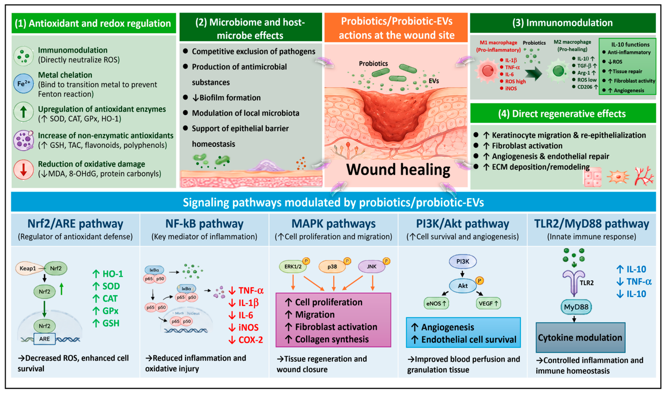
3. Antioxidant and Redox-Regulatory Roles of Probiotics in Wound Healing
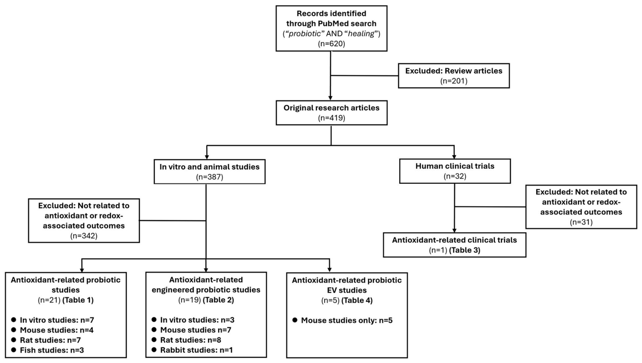
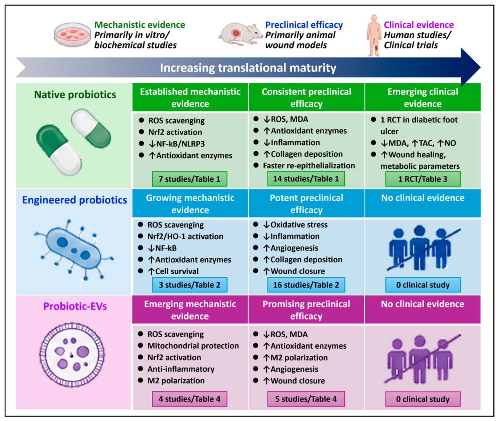
3.1. Literature Screening and Study Classification
3.2. Intrinsic Antioxidant Properties of Probiotics in Wound Repair

| Probiotic | Model | Redox Modulation | Signaling Pathway | Immune Modulation | Wound Healing | Reference |
|---|---|---|---|---|---|---|
| L. plantarum | In vitro: epithelial cell scratch and H2O2-induced oxidative stress; In vivo: mouse skin wound model | Oxidative stress protection ↑; cell viability ↑ (60–100% protection) | NR | IL-6 ↑ (early phase); IL-10 ↑ (late phase); inflammation modulation (early pro-inflammatory → later anti-inflammatory transition) | ↑ | Dubey et al., 2021 [99] |
| L. plantarum | Rat burn wound model | ROS scavenging ↑ (DPPH activity ↑); metal chelation ↑; reducing power ↑ (antioxidant capacity ↑) | NR | Inflammation/infiltration ↓ | ↑ | Elmansy et al., 2022 [104] |
| L. plantarum | In vitro keratinocyte model with M. furfur infection | Nrf2 ↑ (restored from downregulation induced by M. furfur); ROS-related signaling indirectly modulated via AhR pathway | AhR ↓; CYP1A1 ↓; Nrf2 ↑; NLRP3 inflammasome ↓ (Caspase-1 ↓; IL-1β ↓; IL-18 ↓) | IL-1β ↓; IL-18 ↓; Inflammation ↓; Inflammasome activation ↓ | ↑ | Fusco et al., 2023 [107] |
| L. plantarum | Mouse skin wound model | NR | CARD9-mediated signaling ↑; NF-κB–related pathway (via C-type lectin receptors) involved; MyD88-independent | M2 polarization ↑; IL-10 ↑; chemokines ↑ (CCL5, CXCL1, CXCL2); leukocyte infiltration ↑ | ↑ | Ishi et al., 2023 [112] |
| L. plantarum | Mouse infected skin wound model | Free radical scavenging ↑ (DPPH assay, up to ~90% inhibition); ROS ↓ | NR | IL-6 ↓; IL-10 ↑; inflammation ↓ | Dubey et al., 2023 [100] | |
| L. plantarum | In vitro: HUVEC scratch wound model; In vivo: rat diabetic skin wound model | NR | NLRP3 inflammasome ↓; Caspase-1 ↓; GSDMD ↓ (pyroptosis ↓) | IL-1β ↓; IL-18 ↓; inflammation ↓; pyroptosis ↓ | ↑ | Wang et al., 2024 [108] |
| L. plantarum | Rat diabetic infected skin wound model | Antioxidant compounds present (GC-MS) | NR | IL-10 ↑; IL-6 ↓; inflammation ↓ | ↑ | Narang et al., 2026 [101] |
| L. plantarum | In vitro: fibroblast scratch wound model; in ovo CAM angiogenesis model | NR | TGF-β1/Smad2/3/4 ↑ | IL-1β ↓; IL-6 ↓; iNOS ↓ (anti-inflammatory) | Demir et al., 2025 [109] | |
| L. rhamnosus | Zebrafish caudal fin amputation model under oxytetracycline exposure | ROS ↑ (restored amputation-induced ROS production) | Wnt signaling ↑ (wnt3a ↑; lef1 ↑; axin2 ↑) | Neutrophil recruitment ↓; apoptosis ↓; inflammation modulation ↑ | ↑ | Wang et al., 2023 [106] |
| L. rhamnosus | In vitro HaCaT scratch wound under TNF-α-induced inflammatory condition | SOD-like activity ↑; DPPH radical scavenging ↑; oxidative stress protection ↑ (H2O2-induced cy totoxicity ↓) | A2A receptor-mediated FAK ↑/AKT ↑; ERK ↑; p38 ↑ | Bidirectional NO regulation: NO ↑ (basal condition); NO ↓ (under LPS stimulation); anti-inflammatory effect ↑ | ↑ | Chae et al., 2025 [113] |
| L. reuteri | Rat skin wound model | MPO ↓ (oxidative/inflammatory enzyme ↓); antioxidant effect↑ | NR | Inflammation ↓; neutrophil activity ↓ (via MPO ↓) | ↑ | Khodaii et al., 2019 [118] |
| Lactobacillus sp. Ca6 | Rat skin wound model | DPPH radical scavenging ↑; Reducing power ↑; β-carotene bleaching inhibition ↑; Metal chelating activity ↑ | NR | Inflammation ↓ | ↑ | Trabelsi et al., 2017 [114] |
| B. bifidum | Rat peptic ulcer wound model | SOD ↑; GPx ↑; antioxidant capacity ↑ | NR | IL-8 ↑; neutrophil recruitment ↑; macrophage infiltration ↑ | ↑ | Almasyan et al., 2023 [119] |
| B. bifidum | Rat oral ulcer model | NR | TLR2/TGF-β1/Smad3 (suggested, not directly validated); NF-κB ↓ (indirect evidence via TNF-α, IL-6 reduction) | TNF-α ↓; IL-6 ↓; IL-10 ↑; M2 polarization ↑; inflammation ↓ | ↑ | Li et al., 2026 [111] |
| E. mundtii | In vitro fibroblast scratch wound model | Antioxidant activity ↑ (DPPH scavenging ~40%); ROS ↓ | NR | NR | ↑ | Fidan et al., 2023 [115] |
| L. mesenteroides | In vitro keratinocyte infected scratch wound model | Oxidative stress index ↓; TAC ↑; ROS ↓ | NR | IL-6 ↓; inflammation ↓ | ↑ | Altves et al., 2024 [116] |
| S. putrefaciens | Fish skin wound model | SOD ↑; CAT ↑; GSR ↔; peroxidase activity ↑; antioxidant system ↑ | NR | IL-10 ↑; TGF-β1 ↑; IL-1β ↓; IL-8 ↓; inflammation ↓ | ↑ | Chen et al., 2020 [102] |
| L. casei, L. plantarum | In vitro: H2O2-induced oxidative stress; In vivo: mouse skin wound model | ROS scavenging ↑ (DPPH assay); MSC survival under oxidative stress (~4–5×) ↑; antioxidant genes ↑ (SOD-1 ↑, HO-1 ↑, MT-1 ↑) | HO-1 ↑; antioxidant gene upregulation (SOD-1, MT-1 | Inflammation ↓ | ↑ | Kazemi et al., 2022 [105] |
| L. casei, L. plantarum, multiple Lactobacilli spp. | In vitro fibroblast scratch wound model | ROS ↓ (DPPH scavenging ↑up to ~60%) | MMP-1/2/3/9/10 ↓; TIMP-1/2 ↑ | NR | ↑ | Shirzad et al., 2018 [117] |
| L. casei, L. gasseri, L. paracasei, B. lactis, S. thermophilus | In vitro: fibroblast scratch and H2O2-induced oxidative stress | ROS ↓; oxidative damage ↓; cell viability ↑ under oxidative stress | NR | IL-6 ↓; IL-1β ↓; inflammation ↓ | ↑ | Michels et al., 2023 [120] |
| S. thermophilus, L. plantarum, L. acidophilus, B. longum, B. infantis, B. breve, L. bulgaricus | In vitro keratinocyte scratch wound model | NO production ↑ (nitrite levels ↑ via NOS2 activity) | NOS2/NO pathway ↑; NOS2 expression ↑; (effect abolished by NOS2 inhibitor, aminoguanidine) | IL-6 ↑; IL-8 ↑ (associated with NOS2 activation | ↑ | Lombardi et al., 2019 [110] |
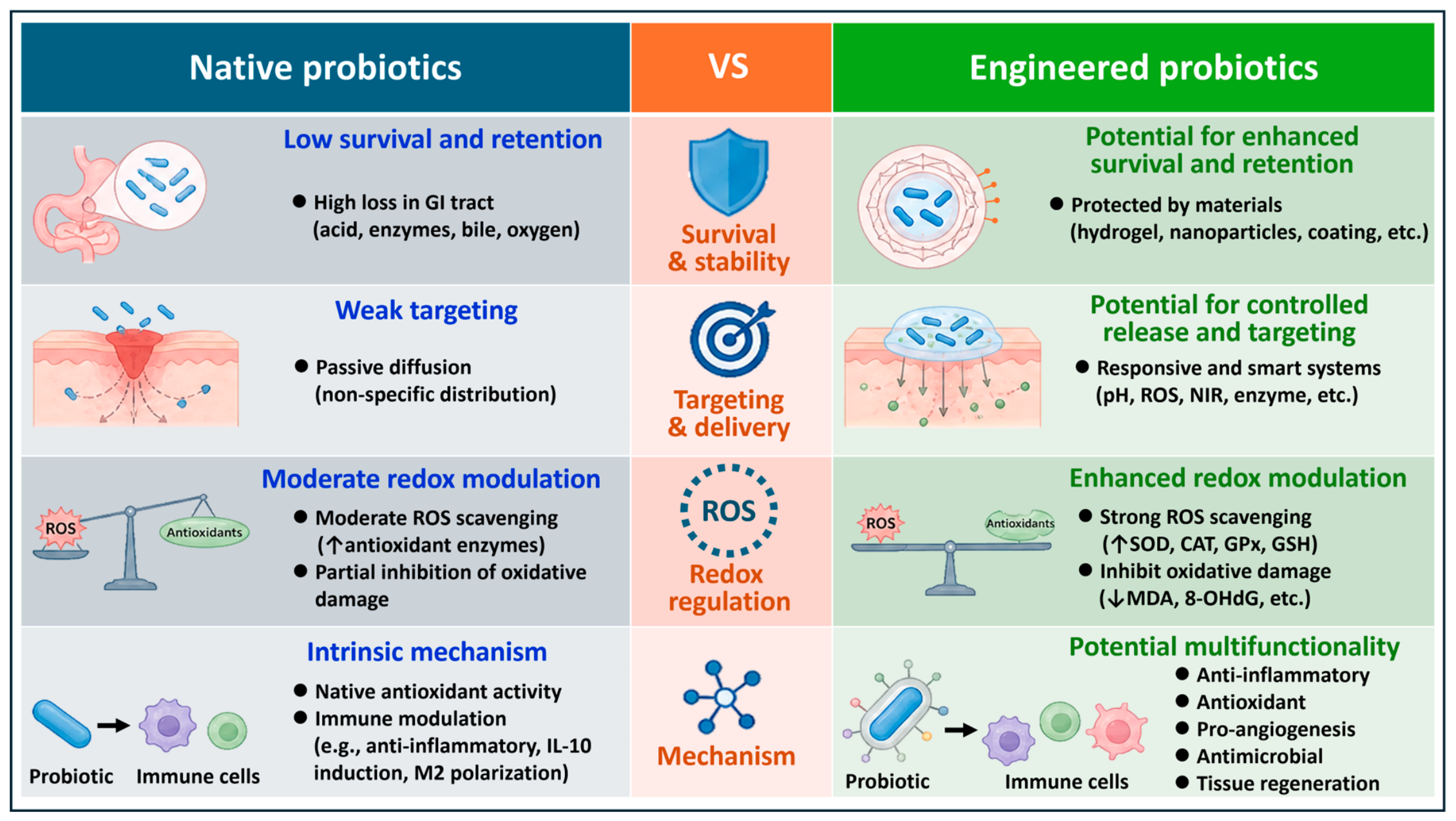
3.3. Engineering Strategies for Probiotic Redox Modulation in Wound Repair
| Category | Probiotic (Model) | Engineering Strategy | Redox Modulation | Signaling Pathway | Immune Modulation | Wound Healing | Reference |
|---|---|---|---|---|---|---|---|
| Hydrogel | L. rhamnosus (Mouse wound) | Live probiotic-loaded pH/ROS dual-responsive sodium alginate hydrogel (SA-SPBA@L.rha) | ROS ↓ (·OH, H2O2, ·O2− scavenging ↑ ~80–100%) | NR | Inflammation ↓; inflammatory cell infiltration ↓ | ↑↑ | Miao et al., 2024 [125] |
| L. rhamnosus (Mouse wound) | Live probiotic encapsulated in multifunctional living hydrogel (PMBV-PVA hydrogel; nanoSe biosynthesized intracellularly + ceramide-coated bacteria) | ROS ↓; H2O2 ↓; NO ↓; nanoSe-mediated ROS scavenging | NF-κB ↓ (p-NF-κB, p65 ↓); PI3K/Akt/mTOR ↑ | M2 polarization ↑; M1 markers ↓; IL-6 ↓; TNF-α ↓; IL-1β↓; inflammation ↓ | ↑↑ | Liu et al., 2025 [126] | |
| L. rhamnosus (Rat wound) | Live probiotic encapsulated in injectable photo-crosslinkable PEG hydrogel with calcium phosphate nanoparticles (CP-LGG@Gel; in situ light-activated) | NR | TNF signaling pathway involvement (transcriptomics); cytokine–cytokine receptor interaction ↑ | M2 polarization ↑; IL-10 ↑; TNF-α ↓; inflammation↓ | ↑↑ | Guo et al., 2026 [132] | |
| L. reuteri (Mouse wound) | Metal-phenolic self-assembly shielded live probiotic (L. reuteri@FeTA) in hydrogel (Gel/L@FeTA) | NR | Angiogenesis-related proteins ↑ (VEGF ↑; CD31 ↑) | TNF-α ↓; IL-1β ↓; IL-10 ↑; TGF-β ↑ (anti-inflammatory ↑) | ↑↑ | Zhou et al., 2023 [129] | |
| L. reuteri (Mouse wound) | Viable probiotic encapsulated in calcium alginate hydrogel (LR@CAH; with acid-responsive hydrogen-producing material) | ROS ↓; oxidative stress ↓ (via glucose consumption + hydrogen production); antioxidant enzyme activity ↑ | NR | Inflammation ↓ | ↑↑ | Wang et al., 2025 [127] | |
| L. reuteri (Rat wound) | Reuterin incorporated into 3D-printed double-layer hydrogel (ACG-GC/Reu/SPS-PDMS) | NR | NF-κB ↓; AMPK ↓; IL-17 ↓; NLRP3 inflammasome activation; MAPK, TGF-β, PI3K-Akt, TNF, VEGF pathways ↑ | IL-10 ↑; TGF-β ↑; Arg-1↑; M2 polarization ↑; TNF-α ↓; IL-1β ↓; iNOS ↓; inflammation ↓ | ↑↑ | Feng et al., 2025 [133] | |
| L. reuteri (Rat wound) | Reuterin-crosslinked chitosan hydrogel (injectable, pH-responsive release system) | ROS ↓; DPPH scavenging ↑ (>60%); oxidative stress–induced cell damage ↓; cell viability under H2O2 ↑ | NR | Inflammation/infiltration ↓; TGF-β1 ↓ | ↑↑ | Gao et al., 2025 [144] | |
| L. casei (Rat wound) | Probiotic extracts + NO donor = biomimetic phage-like microparticles, further encapsulated in GO-doped hydrogel | NO release ↑; nitrosative stress ↑; intracellular ROS ↑ (in bacteria); oxidative stress ↑ | NR | Inflammation/infiltration ↓; bacterial toxicity ↓ | ↑↑ | Gong et al., 2022 [128] | |
| B. subtilis (Rat wound) | Living probiotic encapsulated in metal–phenolic networks (MPNs) and loaded into KGM/XG hydrogel (KGXM-PCB@Bsubtilis) | ROS ↓ (intracellular ROS scavenging ↑); antioxidant activity ↑ (DPPH/ABTS scavenging ↑) | NR | IL-10 ↑; TGF-β ↑; IL-6 ↓; iNOS ↓; inflammation ↓ | ↑↑ | Wu et al., 2025 [103] | |
| P. pentosaceus (Rat wound) | Live probiotic incorporated in hydrogel (C-phycocyanin-loaded whey protein nanofibril hydrogel) | MDA ↓ (54.76–98.58%); GSH ↑; TAC ↑ (antioxidant capacity ↑) | COX-2 inhibition ↑ (phycocyanin-related) | Inflammation/infiltration ↓; microbiome balance ↑; immune response modulation ↑ | ↑↑ | El-Dein et al., 2026 [134] | |
| Nanofiber | L. rhamnosus (Antioxidant model) | Probiotic exopolysaccharide (EPS-P14)-based electrospun nanofibrous membrane (EPS/PEO ± Cu/Ag-doped bioactive glass); no live bacteria | ROS scavenging ↑ (DPPH ~65–70%; FRAP reducing power ↑) | NR | NR | ↑↑ | Akhtach et al., 2025 [145] |
| L. casei (Mouse wound) | Probiotic loaded silk fibroin/alginate scaffold | NR | Endoplasmic reticulum stress ↓ | M2 polarization ↑; inflammation ↓ | ↑↑ | Dou et al., 2023 [135] | |
| Microparticle | L. plantarum (scratch wound) | Cell-free supernatant-mediated biosynthesis of silver nanoparticles (AgNPs) | ROS ↑ (AgNP-induced oxidative stress for antibacterial effect); antioxidant activity ↑ (DPPH scavenging 9.9–52.4%) | NR | NR | ↑↑ | Vijayakumar et al., 2023 [131] |
| L. plantarum (Mouse wound) | Viable probiotic incorporated in sponge dressing + nanocurcumin (CSLNs; solid lipid nanoparticles) | LPO ↓; Catalase ↑; GSH ↑ (antioxidant enzymes ↑) | NR | TNF-α ↓; MMP-9 ↓ (inflammation ↓); VEGF ↑; TGF-β ↑ | ↑↑ | Sandhu et al., 2023 [137] | |
| L. plantarum (Rat wound) | Live probiotic encapsulated in lyophilized polymeric microparticles (chitosan–alginate) + prebiotic (FOS) | ROS-related oxidative stress ↓; FRAP (TAC) ↑; MDA (lipid peroxidation) ↓ | NR | Inflammation/infiltration ↓ | ↑↑ | Farahani et al., 2023 [146] | |
| L. rhamnosus (Rat wound) | Live probiotic loaded in Dextran-Gelatin-Gellan Gum composite microspheres (DGGcm) biomaterial (sustained release system) | NR | NR | IL-6 ↓; IL-10 ↑; inflammation ↓ | ↑↑ | Diao et al., 2025 [138] | |
| L. rhamnosus (Antioxidant model) | Live probiotic coated with γ-Fe2O3 nanoparticles (nanobiohybrid; γ-Fe2O3 NPs–LR) via electrostatic self-assembly | ROS ↑ (from γ-Fe2O3 NPs antibacterial mechanism); antioxidant activity ↑ (DPPH scavenging ↑); SOD/CAT ↑ (implied from L. rhamnosus secretion); oxidative stress scavenging ↑ | NR | NR | NR | Shingade et al., 2026 [147] | |
| Dressing | L. fermentum (Rat wound) | Live probiotic embedded in patch (lyophilized bacteria in alginate microbeads within gas-permeable dressing producing NO) | NR | NO signaling ↑ (nitric oxide-mediated wound healing process) | Inflammation/infiltration ↓ | ↑↑ | Jones et al., 2012 [136] |
| Other | L. rhamnosus (Mouse wound) | Live probiotic engineered bio-heterojunction (P-bioHJ): LG conjugated with MXene (Ti3C2) quantum dots + FeS + LOx; NIR-triggered system | ROS scavenging ↑; intracellular ROS ↓; SOD ↑; CAT ↑; MDA ↓; hydroxyl radical clearance ↑ | NF-κB ↓; IκBα ↓; TNF-α ↓; TNF signaling modulation (RNA-seq); anti-inflammatory pathway inhibition | Inflammation ↓; TNF-α ↓; IL-6 ↓; anti-inflammatory effect ↑ | ↑↑ | Qin et al., 2024 [130] |
3.4. Clinical Evidence of Probiotic Antioxidant Effects in Wound Repair
| Probiotic | Intervention | Design | Population | Biomarker (Antioxidant/Oxidative Stress) | Clinical Outcome | Reference |
|---|---|---|---|---|---|---|
| L. acidophilus, L. casei, L. fermentum, B. bifidum (multi-strain) | Oral live probiotics (2 × 10⁹ CFU/g each, oral capsule) | Randomized, double-blind, placebo-controlled trial | Patients with diabetic foot ulcer, n = 60 (30 vs. 30), age 40–85, duration: 12 weeks | MDA ↓; TAC ↑; NO ↑; GSH ↔; hs-CRP ↓ | Ulcer length, width, depth ↓; wound healing rate ↑; improved glycemic control (FPG, HbA1c ↓); cholesterol ↓; inflammation ↓ | Mohseni, 2018 (IRCT201603085623N68) [148] |
| Strain/EV Source | EV Preparation & Characterization | EV Properties/Cargo | Treatment | Experimental Model | Redox/Immune Findings | Proposed Pathway | Mechanistic Validation | Wound/Tissue Outcome | Reference |
|---|---|---|---|---|---|---|---|---|---|
| L. reuteri DSM 20016; MVs | Cell-free culture supernatant; centrifugation (5000× g, 20 min) + 0.45-μm filtration + ultracentrifugation (129,000× g, 1.5 h; repeated at 129,000× g, 16 h); TEM, SEM, DLS | Mean size ≈60 nm (DLS); spherical membrane vesicles; 3-HPA identified by LC–MS; vesicle concentration and purity assessment NR; endotoxin/bacterial component assessment NR | Natural MVs; oral mucosal wound: topical MVs (10 μL, 200 μg/mL, twice daily); cutaneous wound: MV-loaded hydrogel, 200 μg MVs on days 0 and 7 | In vitro: RAW 264.7 macrophages (LPS-induced inflammation); In vivo: mouse oral mucosal ulcer model + mouse skin wound model | Redox: ROS ↓; mitochondrial ROS ↓; MDA ↓; NO ↓; mitochondrial membrane potential stabilization ↑; CI activity ↑/CII normalization; oxidative stress ↓ Immune: IL-10 ↑; TNF-α ↓; IL-1β ↓; IL-6 ↓; M2 macrophage polarization ↑ (CD206↑, Arg-1↑; iNOS↓); inflammation ↓ | 3-HPA-associated inhibition of mitochondrial permeability/mPTP opening → reduced oxidative stress → anti-inflammatory macrophage phenotype | Yes—cargo identification and pharmacological pathway interrogation: LC–MS identified 3-HPA; isolated 3-HPA reproduced redox/immunomodulatory effects; cyclosporin A/CypD-associated mPTP inhibition used to interrogate mitochondrial permeability mechanism; direct molecular target of 3-HPA not established | Wound closure ↑; re-epithelialization ↑; inflammatory infiltration ↓; collagen organization ↑; hair follicle formation ↑; tissue regeneration ↑; mucosal healing ↑ | Chen et al., 2024 [92] |
| L. reuteri; MVs | Gradient differential ultracentrifugation; morphology characterization; NTA; transcriptomic analysis of MV-treated cells | Mean size ~120 nm (NTA); vesicle concentration NR; molecular cargo profiling NR; purity assessment NR; endotoxin/bacterial component assessment NR | Natural MVs, biomaterial-engineered delivery; MVs covalently anchored to carboxylated chitosan and incorporated into oxidized hyaluronic acid hydrogel (Gel-MVs) for controlled local release; dose NR | In vitro: cell prolifera-tion/migration assays; In vivo: mouse skin wound model | Redox: Direct redox outcomes NR Immune: Inflammation ↓ | Transcriptome-associated regulation of angiogenesis/metabolic pathways; VEGF/CD31-associated vascularization↑ | Pathway-associated/transcriptomic evidence only; no inhibitor, knockdown, cargo-depletion, or rescue validation reported | Angiogenesis ↑; vasculariza-tion ↑; wound closure ↑; cell proliferation ↑; cell migration ↑; collagen deposition ↑ | Zhou et al., 2025 [95] * |
| L. reuteri; MVs † | Differential centrifugation + ultracentrifugation (100,000× g); Cryo-TEM; NTA; UPLC-ESI-MS/MS metabolomic profiling | Mean size ~145 nm; concentration NR; metabolomic profiling identified major bioactive constituents, including abundant phospholipids; purity assessment NR; endotoxin/bacterial component assessment NR | Natural MVs; 20 μg/mL in vitro; ROS-responsive DNA hydrogel (MVs-Gel) for controlled local release ± NIR photothermal therapy; in vivo MV dose NR | Mouse diabetic infected skin wound model | Redox: ROS ↓; antioxidant activity ↑; apoptosis ↓ (Bax ↓/Bcl-2 ↑); Nrf2/HO-1 ↑ Immune: IL-10 ↑; TGF-β ↑; TNF-α ↓; IL-6 ↓; M2 macrophage polarization ↑(CD206 ↑; CD86 ↓); inflammation ↓ | Nrf2/HO-1 -mediated redox regulation ↑; NF-κB -mediated inflammation↓; PI3K/Akt ↑; AMPK ↑ | Pathway-associated validation by Western blot and transcriptomics/GSEA; no inhibitor, knockdown, cargo-depletion, or rescue validation reported | Wound clo-sure ↑; re-epithelialization ↑; keratinocyte migration ↑; angiogenesis ↑(CD31 ↑); neovasculari-zation ↑; col-lagen deposi-tion ↑; α-SMA ↑; antibacterial activity ↑ | Tai et al., 2026 [96] |
| L. rhamnosus GG (ATCC 53103); LGG-MVs | Differential centrifugation + 0.22-μm filtration + ultracentrifugation (150,000× g, 2 h, twice) with ultrafiltration; TEM; NTA; zeta-potential analysis; miRNA sequencing | Size 30–200 nm, main peak ~180 nm; zeta potential ~−19.4 mV; concentration estimated by BCA protein assay; miRNA profiling identified 494 miRNAs, with miR-21-5p highly enriched; purity assessment not formally reported; endotoxin/bacterial component assessment NR | Natural LGG-EVs; in vitro 50 ng/μL; in vivo 50 μg EVs in 100 μL PBS by daily subcutaneous injection around wounds | In vitro: scratch wound (HaCaT keratinocytes; HUVEC en-dothelial cells); in vivo: mouse skin wound model | Redox: Direct redox outcomes NR Immune: Direct imunemodulation endpoints NR | miR-21-5p-mediated PI3K/AKT–HIF1α signaling ↑; VEGF-associated angiogenic signaling↑ | Yes—miRNA cargo profiling + miR-21-5p inhibitor functional validation; inhibition attenuated LGG-EV-induced proliferation/migration and abolished p-AKT/HIF1α activation; no rescue experiment reported | Wound clo-sure ↑; re-epithelialization ↑; keratinocyte proliferation ↑; endothelial cell prolifera-tion/migration ↑; angiogene-sis ↑; granula-tion tissue ↑; collagen dep-osition ↑ | Wang et al., 2024 [94] |
| L. casei DSM 20011; L. plantarum NCIMB 8826; MVs | Centrifugation + 0.45-μm filtration + ultracentrifugation (100,000× g) + SEC; cryo-TEM; NTA; LC–MS/MS proteomics; SEM of BPs | ~50–150 nm; NTA-based particle concentration reported; culture-dependent protein cargo; thioredoxin identified in L. casei pH 6.5 MVs; SEC-purified; endotoxin/bacterial component assessment NR | Natural MVs; direct MV-loaded or bacteriomimetic microparticle-loaded HEC hydrogel; topical application; in vivo dose NR | In vitro: Ha-CaT keratino-cyte scratch wound; In vivo: mouse tail wound model | Redox: Direct redox outcomes NR Immune: TNF ↓; IL-10 modulation; IL-10/TNF ratio ↑; an-ti-inflammatory effect ↑; neutrophil infiltration ↓; M2-like ef-fect↑ | No specific pathway established; proteomic composition associated with culture-dependent immunomodulation | Proteomic association only; no inhibitor, knockdown, cargo-depletion, or rescue validation | BP hydrogel: early wound-width reduction/re-epithelialization ↑; dermal thickness ↓; revascularization ↑; overall wound-closure time ↔; collagen deposition ↔ | Kuhn et al., 2024 [93] |
4. Extracellular Vesicles: A Redox-Modulating Delivery System
4.1. Biology of Extracellular Vesicles
4.2. EV-Mediated Redox Regulation Through Antioxidant Enzymes and microRNA Cargo
4.3. Therapeutic Advantages of EVs over Conventional Antioxidant Delivery Systems
4.4. Emerging Interest in Microbial and Probiotic-Derived Extracellular Vesicles
5. Probiotic-Derived EVs: Characteristics and Biological Potential
5.1. Sources and Biological Characteristics of Probiotic-Derived EVs
5.2. Immunomodulatory and Antioxidant Functions of Probiotic-Derived EVs
5.3. Comparative Positioning of Probiotic- and MSC-Derived EVs
| Feature | Probiotic-Derived EVs | MSC-Derived EVs |
|---|---|---|
| Source | Beneficial gut or skin-associated probiotics (e.g., Lactobacillus, Lactiplantibacillus, Akkermansia muciniphila, Bifidobacterium) | MSCs (e.g., bone marrow, adipose tissue, umbilical cord) |
| Manufacturing Cost | Potentially low; may be more amenable to bacterial culture-based production, although GMP-grade manufacturing cost has not been established | Generally associated with more complex mammalian cell culture and GMP-grade production requirements |
| Production Stability | Theoretically scalable through bacterial culture-based production; clinical-grade scalability and batch consistency remain to be demonstrated | May be affected by donor variability, cell state/senescence, culture conditions, and batch-to-batch variation |
| PAMP/MAMP-associated Immunogenicity | Potential innate immune activation by MAMPs/PAMPs, including peptidoglycan, lipoteichoic acid, lipoproteins, and, where applicable, LPS-associated components; effects are strain-, cargo-, purity-, and dose-dependent | Generally lower PAMP/MAMP-related immunogenicity, although donor-, culture-, cargo-, and product-related immune effects still require evaluation |
| Major Antioxidant Cargo | Bacterial enzymes, stress proteins, SCFA-associated metabolites, thioredoxin-related proteins, regulatory sRNAs | miR-21, miR-146a, antioxidant enzymes (SOD, catalase), HO-1-related signaling molecules |
| Immunomodulatory Functions | Regulation of TLR/NF-κB/Nrf2 pathways; promotion of M2 macrophage polarization | Suppression of excessive inflammation; enhancement of M2 macrophage polarization and fibroblast activation |
| Barrier Penetration/Wound Delivery | Small nanoscale vesicles facilitate tissue penetration and cellular uptake; frequently incorporated into hydrogels or other biomaterial platforms for enhanced wound retention | Effective delivery through local injection, topical application, and biomaterial-assisted delivery systems |
| Engineering Flexibility | Potentially amenable to parental-strain genetic engineering and programmable cargo modification; direct validation in therapeutic probiotic-EV manufacturing remains limited | Established preclinical strategies include genetic modification, cargo loading, and surface functionalization Can be engineered for siRNA/protein loading and surface targeting modifications |
| Oral/Noninvasive Administration Potential | Theoretical potential for oral or other noninvasive delivery and microbiota-mediated systemic effects; biodistribution and therapeutic efficacy remain insufficiently validated | Primarily administered through local injection or topical application |
| Gut–Skin Axis Potential | Potential to participate in microbiota–gut–skin communication pathways, but direct wound-healing evidence remains limited | Limited evidence supporting gut–skin axis modulation |
| Clinical Maturity | Early preclinical stage; wound-healing evidence remains limited and no direct human therapeutic validation is currently available | More advanced preclinical and early clinical development across regenerative indications, although clinical validation for wound healing remains limited |
| Regulatory Classification | No dedicated classification established; may intersect with biologic, microbial/postbiotic, nanomedicine, or engineered EV product frameworks depending on composition and intended use | Generally considered within biologic/EV therapeutic frameworks, although product classification and regulatory requirements remain jurisdiction- and product-dependent |
| Major Advantages | Potentially lower-cost bacterial culture-based production, theoretical scalability, engineering potential, and microbiome-associated biological properties | More extensive regenerative and wound-healing evidence and greater translational maturity |
| Major Limitations | Limited direct human evidence, insufficient clinical-grade manufacturing data, EV and cargo heterogeneity, PAMP/MAMP-associated immunogenicity, and unresolved regulatory classification | High production cost, donor variability, limited scalability |
| Potency Assays | No standardized wound-specific potency assay established; mechanism-relevant assays may include redox, immunomodulatory, epithelial, or angiogenic endpoints | More extensively developed functional assays are available, but standardized indication-specific potency assays remain an important translational requirement |
| Quality Control Requirements | Requires standardized strain/culture conditions, EV isolation and purification, physicochemical characterization, cargo assessment, sterility/contaminant testing, and functional potency testing | Requires standardized donor/cell-source qualification, culture conditions, EV isolation and characterization, sterility, identity/purity assessment, and potency testing |
| Batch-to-Batch Reproducibility | Potentially affected by bacterial strain, growth phase, culture conditions, isolation method, and cargo heterogeneity; clinical-grade reproducibility remains unestablished | Affected by donor variability, cell state, culture conditions, passage, isolation method, and manufacturing process; substantial standardization efforts are ongoing |
| References | [122,211,212,218,219,222,232,257,258,260,261,262,263] | [251,252,253,254,255,256,259,264,265] |
5.4. Translational Opportunities and Current Limitations of Probiotic-Derived EVs
| Feature | Live Probiotics | Probiotic-EVs |
|---|---|---|
| Basic Structure | Viable microorganisms containing intact cell walls, cytoplasmic components, and genomic DNA/RNA | Cell-free nanosized lipid bilayer vesicles containing bioactive cargo |
| Mechanism of Action | Act through colonization, microbial competition, metabolite secretion, and host immune modulation | Deliver functional cargo directly to host cells, including proteins, lipids, RNAs, and metabolites |
| Redox-Regulatory Capacity | Certain strains produce antioxidant metabolites such as SCFAs and GSH | EVs can directly transport antioxidant enzymes and redox-regulatory molecules (e.g., SOD-like proteins, miRNAs) |
| Immunomodulatory Effects | Activate TLR/NOD-associated signaling and influence macrophage polarization | Modulate immune responses through vesicle-associated surface molecules and regulatory RNA cargo |
| Routes of Administration | Oral, topical, spray-based, or local administration | Topical delivery, spray application, or incorporation into hydrogels and biomaterials |
| Penetration across Wound Barriers | Limited by bacterial size and cell wall structure | Small vesicle size (~50–200 nm) may facilitate tissue penetration and cellular uptake |
| Requirement for Survival/Colonization | Require viability and interaction with local microbiota for sustained activity | Non-replicative; therapeutic effects mediated through direct cargo delivery |
| Engineering Potential | Genetically engineerable strains capable of expressing therapeutic factors (e.g., FGF2, CXCL12) | EV engineering remains early-stage but allows programmable cargo loading and surface modification |
| Safety Considerations | Potential risks of bacterial translocation, infection, or horizontal gene transfer | Reduced infection risk due to acellular nature; lower likelihood of uncontrolled colonization |
| Regulatory Challenges | Often regulated within probiotic, food, or live biotherapeutic frameworks | Greater standardization and characterization challenges as microbial nanovesicle therapeutics |
| Research Maturity | Supported by multiple clinical studies, including dermatologic and gastrointestinal applications | Predominantly preclinical evidence from cell culture and animal studies |
| References | [27,34,270] | [93,273,274] |
6. Engineering and Therapeutic Optimization of Probiotic-Derived EVs
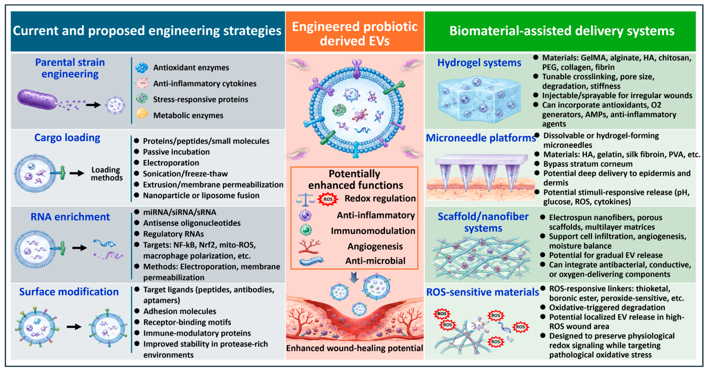
6.1. Bioengineering Strategies for Probiotic-Derived EVs
- Parental engineering: Currently, one potential approach to change the composition of EVs in probiotics is the so-called parental engineering. As the physiological state and genetic information of the EV cargo largely derive from the parental microorganism, it is possible to selectively increase the level of therapeutic molecules in the vesicles by engineering the bacteria [279,280,281]. However, in practice, parental bacterial strains may potentially be genetically engineered to produce an abundance of antioxidant enzymes, anti-inflammatory mediators, proteins involved in stress reactions, or peptides with a regeneration capacity that, during the biogenesis of vesicles, may be incorporated into the EV cargo [35,282,283]. Various studies in engineered microbial or related experimental systems have addressed the use of catalase, SOD, thioredoxin-related proteins, or heme oxygenase-related pathways to enhance ROS-scavenging activity in inflammatory tissues [284,285,286]. In a similar fashion, engineered probiotics can be used to produce proteins that could modulate macrophage polarization, epithelial repair, or mitochondrial oxidative homeostasis [123,287,288,289,290,291]. Parentally engineered EVs may offer a stable and scalable approach to generating EVs with a more consistent therapeutic cargo profile in comparison with EVs generated by post-isolation modification. This approach has yet to be tested in the context of EVs derived from probiotics in chronic wound models, however.
- Cargo loading: Many strategies of EV engineering have been explored primarily in mammalian-EV and broader EV systems, and these may also be applicable to probiotic-EVs. After EVs have been isolated, therapeutic molecules can be loaded into EVs, along with the endogenous bacterial load to give EVs an extra boost in biological activity. Incorporation of antioxidant proteins or peptides or small-molecule therapeutics into the interior of vesicles can be achieved by protein loading techniques such as passive incubation, membrane permeabilization, sonication, freeze-thaw cycling, extrusion or electroporation [292,293,294,295]. In addition, integration of EVs with biomimetic nanoparticles or liposomal fusion systems has been explored in broader experimental systems for more effective encapsulation and delivery stability [296,297,298]. However, the therapeutic advantages of these cargo-loading approaches remain to be validated specifically in probiotic-EV-based chronic wound models. These methods may be especially useful in scenarios where the concentration of endogenous bacterial cargo is too low to be used for therapy. Importantly, the cargo loading strategies also provide opportunities for integrating more than one therapeutic function in a single vesicle platform, such as antioxidative, anti-inflammatory, antimicrobial, and regenerative functions.
- RNA enrichment: Another emerging strategy with potential application to probiotic-EVs is RNA engineering. It is now becoming abundantly clear that sRNA, microRNA, and other nucleic acid cargoes are naturally present in EVs produced by bacteria and are able to modulate inflammatory and oxidative signaling pathways in the host [299,300]. Based on this idea, a number of engineering strategies have been developed to selectively enrich EVs or bacterial vesicles with therapeutic RNA, although their application to probiotic-derived EVs remains less established. These include loading with miRNAs, siRNAs, antisense oligonucleotides, or other regulatory RNAs that could be used to regulate NF-κB activation pathways, Nrf2 signaling pathways, mitochondrial ROS production pathways, or macrophage polarization pathways [301,302]. Among the most popular approaches to transferring nucleic acid cargo to isolated vesicles are electroporation and permeabilization of membrane vesicles through the addition of membrane permeabilizing agents [303,304,305]. While the efficiency of loading EVs with RNA and the stability of EVs are still technical issues to be resolved, this suggests the potential for probiotic-EVs to serve as a biologically compatible nanoscale RNA delivery system for targeting redox modulation. However, there is still limited direct validation of RNA-engineered probiotic-EVs for redox modulation in chronic wounds.
- Surface modification: Surface engineering and the incorporation of vesicles by display modification have also been explored as methods for the improvement of EV targeting and interaction with cells. A number of studies have investigated the possibility of engineering the surface of bacterial EVs using targeting ligands, adhesion peptides, receptor-binding motifs and/or immune-modulatory surface proteins, as these surface components strongly impact the vesicle’s tropism, immune recognition and uptake by the cells [159,306]. For instance, changes to the surfaces of vesicles might be made to optimize reactions with inflamed endothelial tissue, activated macrophages, damaged epithelium, or the regions of wounds with high ROS levels [307,308,309]. Surface display engineering may additionally be used to enhance EV stability in wound settings, where protease concentrations are high, or to help with the incorporation of EVs into biomaterial-assisted delivery systems, like hydrogels and scaffolds [310,311,312]. Many of the strategies still exist at the very early stages of preclinical research, but overall, they illustrate the potential for rational engineering of EVs derived from probiotics to optimize the composition of EVs, enhance targeting to cells and tissues, and maximize therapeutic potential through manipulation of redox properties within the wound microenvironment. Most of these approaches have been investigated in bacterial-EV or nanovesicle systems and have yet to be tested specifically for probiotic-EVs in chronic wounds.
6.2. Biomaterial-Assisted Delivery Systems
- Hydrogel: Of these biomaterial approaches, hydrogel-assisted delivery has been directly examined using EVs derived from probiotics or probiotic membrane vesicles in preclinical wound/tissue-repair models [92,93,95,96]. In contrast, most of the advanced designs of hydrogels and multifunctional or responsive hydrogels below are adapted from more general studies of EVs and biomaterial delivery and have yet to be directly tested with EVs derived from probiotic bacteria. Hydrogel-based delivery systems are among the most widely studied approaches for localized EV delivery in broader EV and regenerative medicine research. Due to their high water content, structural flexibility, and tunable physicochemical properties, hydrogels can mimic aspects of the native ECM and could also serve as reservoirs for sustained EV release [311,315,316]. Probiotic-EVs could be encapsulated in natural or synthetic hydrogel matrices containing compounds like gelatin methacrylate (GelMA), alginate, hyaluronic acid, chitosan, polyethylene glycol (PEG), collagen or fibrin-based polymers in practice [317,318,319]. Then, release kinetics can be further tuned by tuning the crosslinking density, degradation rate, pore size, and stiffness of the hydrogel [320,321,322]. Injectable and sprayable hydrogel formulations have been investigated as well, which might be helpful for delivering the formulation into irregularly shaped sites and reducing tissue damage during delivery [323,324,325]. Multifunctional hydrogels can be loaded with antioxidant reagents, antimicrobial peptides, oxygen-producing compounds, conductive polymers, or anti-inflammatory molecules that could potentially act synergistically with the actions of the probiotic EVs to favor tissue regeneration and/or local redox balance [326,327,328]. However, these multifunctional combinations are yet to be confirmed in wound models using probiotic EVs.
- Microneedle: Microneedle-assisted delivery systems are another promising way in which to increase EV penetration into damaged skin tissues. Conventional topical delivery will present some problems, such as insufficient penetration of the stratum corneum and insufficient diffusion into fibrotic and/or biofilm wound surfaces [329,330,331]. Some of these limitations can be addressed by microneedle arrays, which can transport EVs directly into deeper layers of the epidermis and dermis with minimal pain and tissue damage at the microscale size [332,333,334]. Probiotic-EVs could potentially be embedded in the microneedles that are made of polymers like hyaluronic acid, gelatin, silk fibroin, or polyvinyl alcohol (PVA), which are dissolvable, biodegradable, or hydrogel-forming [335,336,337]. After delivery into tissue, these systems could allow gradual release of EV cargo into the local wound microenvironment. The use of microneedle-based delivery could be beneficial, especially for diabetic wounds and very inflamed tissues, to enhance EV penetration and retention and decrease dosing frequencies. Microneedle-assisted delivery has not been directly shown for probiotic EVs in chronic wound models so far, and is considered a potential method of delivery based on extended EV and nanomedicine platforms.
- Scaffold: Bioactive scaffolds and nanofiber-based wound dressings are other methods that are able to retain EVs for a longer period of time and provide a scaffold for spatially organized tissue regeneration. Nanofiber structures, such as electrospun nanofibers, porous scaffolds, multilayer matrices, and ECM-mimetic biomaterials, can be designed to encourage cells to infiltrate, maintain moisture levels, promote angiogenesis and/or allow for a step-wise release of EVs over time [338,339]. The composition of the scaffold, fiber diameter, surface charge, and porosity of the scaffold can all affect the efficiency of loading EVs, release from the scaffold, and uptake by cells in wound tissue [308,340,341]. Another property of some of the scaffolds is their use of antibacterial materials or oxygen-releasing or conductive biomaterials to further enhance the ability to regenerate in chronic wound environments [342,343,344]. Scaffold-assisted systems might allow for a longer period of therapeutic contact at the wound surface and increased structural support, as compared to direct EV injection. For the moment, there are no direct studies on the scaffold-assisted delivery of EVs derived from probiotics in chronic wound models, and the proposed use is thus extrapolated from general EV and biomaterial studies.
- ROS-sensitive material: Recently, ROS-sensitive materials or linkers have been identified as potential platforms for the controlled delivery of EVs in the oxidative wound environment. Since chronic non-healing wounds are associated with an excessive amount of ROS, several biomaterial systems have been created where the cargo of EVs is released preferably under oxidative conditions [8,10,345]. These systems frequently involve ROS-sensitive chemical groups like thioketal bonds, boronic ester-containing polymers, peroxide-sensitive nanoparticles, or oxidation-degradable crosslinkers, which have the ability to break down their structures in the presence of higher ROS levels [346,347,348]. In general, in ROS-responsive delivery systems, sequential escalations of oxidative stress may cause degradation of the biomaterial and subsequent local EV release [349,350,351]. This approach could potentially be used to selectively improve therapeutic delivery in areas of high oxidative damage and maintain redox signaling. Notably, ROS-responsive systems also open up opportunities for more spatially controlled and sustained redox modulation than with conventional passive delivery systems. However, the application of probiotic-derived EVs in chronic wounds via ROS-responsive delivery is still an emerging strategy and needs to be validated directly.
6.3. Targeted Redox Modulation in Wound Microenvironments
- Fibroblasts: Fibroblasts are the main cells involved in wound repair, deposition of extracellular matrix (ECM), synthesis of collagen, wound contraction, and wound remodeling. Continued oxidative stress can induce senescence of the fibroblasts and reduce their ability to proliferate, migrate, and produce matrix, which impairs the development of granulation tissue and delays wound closure [353]. Emerging preclinical evidence indicates that probiotic-derived EVs can improve the oxidative and inflammatory wound microenvironment and promote tissue remodeling. In cutaneous wound models, MVs produced from L. reuteri improved wound repair and collagen deposition, with a reduction in pro-inflammatory macrophage activation and oxidative stress [92], and probiotic MV-based hydrogel application also improved dermal remodeling and collagen organization and deposition [93,95]. More recently, MVs derived from L. reuteri were found to directly decrease intracellular ROS and apoptosis under diabetic wound-like conditions and, when added to an inflammation-responsive hydrogel, to increase organized collagen deposition and myofibroblast-associated α-SMA expression in vivo [96]. These observations imply that probiotic-EVs can create a favorable microenvironment for fibroblast-mediated repair, but the direct mechanistic evidence of redox restoration caused by probiotic-EVs in dermal fibroblasts remains scarce.
- 2.
- Keratinocytes: Keratinocytes are key effector cells in re-epithelialization and epidermal barrier restoration during wound healing. Oxidative stress can affect keratinocyte proliferation, migration, cytoskeletal organization, mitochondrial function, and stress-responsive signaling, which would result in delayed epidermal regeneration and wound closure [357,358,359,360]. Recent original studies provide direct evidence that probiotic-derived EVs can protect keratinocytes and enhance their reparative functions. EVs from L. rhamnosus GG stimulated the proliferation and migration of keratinocytes in vitro and accelerated re-epithelialization in a full-thickness skin wound model, with the EV-derived miR-21-5p playing a key role in these effects [94]. More recently, MVs derived from L. reuteri were found to decrease intracellular ROS and apoptosis in HaCaT keratinocytes under diabetic wound-like conditions, maintain mitochondrial morphology and membrane potential, restore impaired keratinocyte migration, and activate Nrf2/HO-1-mediated antioxidant signaling [96].
- 3.
- Macrophages: One of the key connections between oxidative stress and chronic inflammation and poor tissue repair is the dysregulation of macrophages. In chronic wounds, macrophages are continually activated in a pro-inflammatory state, leading to high levels of ROS, reactive nitrogen species, proteases, and inflammatory cytokines, while failure to transition to reparative macrophages is associated with failure of inflammatory resolution and delayed wound healing [363,364]. Importantly, direct evidence from wound models supports macrophages as a major cellular target of probiotic-derived vesicles. Membrane vesicles from L. reuteri decreased pro-inflammatory macrophages and facilitated an anti-inflammatory phenotypic switch in mucosal and cutaneous wounds [92]. Mechanistically, MVs decreased oxidative stress in activated macrophages, and the metabolite 3-hydroxypropionaldehyde, associated with vesicles, decreased mitochondrial permeability and stabilized mitochondrial membrane potential and was thus directly connected to macrophage phenotypic reprogramming and wound repair [92].
- 4.
- Angiogenesis and mitochondrial oxidative homeostasis: Impaired angiogenesis and reduced tissue oxygenation are major pathological features of chronic wounds, particularly diabetic and ischemic wounds [56,367]. In these scenarios, endothelial dysfunction is tightly associated with oxidative stress and disrupted mitochondrial homeostasis. While transient or low levels of mitochondrial ROS can be important in physiological angiogenic signaling, excess or chronic mtROS can cause endothelial dysfunction, decrease nitric oxide bioavailability, and cause problems with endothelial migration and vascular homeostasis [368,369]. Thus, restoration of endothelial redox balance is likely to be important for effective neovascularization.
7. Challenges, Translational Barriers, and Future Directions
7.1. Technical and Translational Challenges
- Standardization, isolation variability, and cargo heterogeneity: The main methodological and biological sources of variability that impact the reproducibility, standardization, and translational development of EVs from probiotics are listed in Table 7. The lack of standardized methods in the isolation, purification, and characterization of EVs is at present one of the major challenges. Various methods of ultracentrifugation, filtration, precipitation kits, density gradients, or chromatography-based methods are used in different studies, leading to significant differences in vesicle purity, yield, size distribution, and content of vesicles [370,371]. Significantly, there are differences in the efficiency of recovery among these various methods, as well as differences in selectivity for different subpopulations of EVs and differences in the extent to which non-vesicular proteins, nucleic acids, or other bacterial components are co-isolated. Therefore, discrepancies among these studies might be attributed to methodological inconsistencies instead of intrinsic biological variability between EVs from different probiotics.
- 2.
- Immunogenicity: Biosafety and immunogenicity are other significant issues. Probiotic-derived EVs are derived from bacteria, which contain structurally immunogenic components like peptidoglycan fragments, lipoteichoic acid, lipoproteins, or endotoxin-associated molecules that have the ability to stimulate innate immune signaling pathways, unlike mammalian EVs [276,277,278]. While some inflammation may be desirable for antimicrobial defense or wound healing, there is potential for chronic wounds with ongoing inflammation and an immune response to become chronic and problematic if the immune system is stimulated excessively or in an uncontrolled manner. Interestingly, the immunological effect of probiotic-EVs seems to be quite dependent on bacterial species, vesicle composition, purification, dose, and route of administration [363,374]. However, even for probiotics generally regarded as safe, the cargo profile of vesicles might be greatly different based on environmental conditions [375]. Moreover, biodistribution and systemic accumulation, activation of off-target immune cells, and effects on the microbiome are not fully understood. These uncertainties underscore the importance of more thorough safety assessment, particularly if engineered probiotic-EVs, carrying exogenous proteins or synthetic regulatory RNAs or targeted surface ligands, were ever to be developed for human clinical applications.
- 3.
- Regulatory Issues: In addition to biological safety, regulatory and manufacturing issues are other important obstacles relating to translation. Currently, no special regulatory guidelines for EV therapeutics of probiotic origins exist. Whether these should be considered as belonging to the class of ‘biologics’, ‘postbiotics’, ‘nanomedicines’, ‘microbial products’ or ‘engineered extracellular vesicle therapeutics’ remains to be determined, and different regulatory pathways may have significantly different manufacturing/quality control requirements [149,376]. There are also practical challenges in large-scale production, as it would be technically challenging to ensure EV yield consistency, cargo composition, and functional potency between different batches. The challenge is even more complicated for engineered probiotic EVs that contain synthetic biology circuits, RNA therapeutics, and/or responsiveness to external cues, where additional factors such as genetic stability, biosafety containment, and reproducibility are involved. Furthermore, there are challenges with the storage of the vesicles and the preservation of their biological activity, and the integrity of the vesicles might be compromised over long storage times, lyophilization and/or freeze-thaw cycles [377,378]. Overall, these restrictions suggest that there is still a lot more to do in order to transform the current experimental nanovesicles into real-world applicable redox therapeutics for chronic wound management that can be produced at a large scale and approved for regulatory use, all derived from probiotics.
7.2. Limitations of Current Experimental Models
- Acute Wound Bias: One problem is that, in most cases, acute wound models are used and fail to simulate the pathological complexity of chronic non-healing wounds that are found in clinical scenarios; hence, this is acute wound bias. Most of the available studies have tested probiotic-EVs in short-duration wound healing models in otherwise healthy rodents, where wound healing, even in the absence of probiotic-EVs, progresses relatively efficiently [92,93,94,95,96]. While it may seem very promising to lower ROS levels, inflammatory cytokines, and/or wound size, these decreases may not accurately reflect the chronic oxidative stress, ischemia, biofilm formation, metabolic dysfunction, vascular insufficiency, or immune dysfunction associated with chronic wounds in diabetic, obese, aging, or peripheral vascular disease patients. Consequently, many experimental systems are probably overoptimistic in their therapeutic claims and underestimate the biological hurdles encountered in the real-world wound environment.
- Lack of Chronic Wound Models: A further challenge is that there is a relative lack of models that are mechanistically well-developed enough to include both chronic redox imbalance and host–microbiome interactions in a single model. Chronic wounds are not just a static pathological defect in the tissue, but a dynamic pathological site characterized by the constant interaction of various elements, including inflammatory immune cells, senescent fibroblasts, dysfunctional keratinocytes, endothelial dysfunction, microbial colonization of the wound, degradation of the ECM, and mitochondrial oxidative stress [379,380]. Most existing probiotic-EV studies, however, have been in simplified in vitro systems, or in animal experiments of relatively short duration that are not reflective of these multifaceted processes [92,93,94,95,96]. In particular, there is very limited research focused on how probiotic EVs act in a polymicrobial biofilm-associated wound, ischemic tissues, a diabetic microenvironment, or in a recurrent inflammatory condition during long-term administration. Likewise, although the concept of the microbiota–gut–skin axis is gaining traction, the evidence that orally administered probiotic EVs can lead to modulation of distal wound repair through modulation of the redox state in the host is still scanty. As a result, many of these proposed mechanisms have not been definitively validated in physiologically relevant models of disease but are only inferential.
- Insufficient Human Validation: Perhaps the most important gap is the near-total lack of human validation studies. The clinical data on probiotic-EV therapeutics remain extremely scarce, especially in wound healing applications, when compared to conventional probiotics and MSC-derived EVs [148]. The majority of current information is based on cell culture studies or on early-stage animal studies; controlled human trials of safety, biodistribution, dosing, pharmacodynamics, or long-term efficacy are virtually absent.
- Limited Biodistribution Studies: More importantly, the biodistribution aspects of EVs derived from probiotics after topical, oral, or injectable administration, their persistence in wound tissues, the preferred cells that internalize them, and the composition of the EVs at the physiological level are still unknown [36].
- Unclear Pharmacokinetics: Pharmacokinetic and biodistribution studies are still particularly underdeveloped but are important for optimization and regulatory approval. In vivo characterization of variables like EV half-life, in vivo accumulation, clearance routes, EV cargo stability, and dose–response relationships are still too poorly understood [381,382]. In addition, the dynamics of bacterial EVs with resident microbiota, host immunity, and host metabolic pathways may vary significantly, and the biological behavior in humans may be quite different from what is seen in a controlled laboratory system [216,383].
7.3. Future Directions in Probiotic-EV Therapeutics
- Precision medicine: Precision redox therapeutics have the potential to become a large area of research for probiotic-EVs in the future. There is significant variation in the level of oxidative stress, immune activation, ischemia, microbial dysbiosis, and tissue degeneration that can occur in chronic wounds and between patients and different types of wounds [363,384]. Thus, it is very likely that next-generation probiotic-EVs will not be just antioxidants but more personalized therapeutic systems, which selectively modulate certain pathways of a pathological process. Next-generation EV therapeutics might not try to indiscriminately inhibit ROS, but rather they might target inappropriately amplified ROS signals linked to chronic inflammatory and mitochondrial disease.
- Gut–skin axis: There is also a new focus area that is rapidly emerging: the microbiota–gut–skin axis [385,386]. There is now growing evidence that microbial-derived vesicles could be involved in long-range host–microbiome interactions via immune, metabolic, and epithelial signaling pathways that connect the gut with other tissues, for instance, the skin [235,387]. Probiotic EVs are nanoscale particles that interact with intestinal epithelial cells and immune cells to possibly exert an effect on systemic inflammatory or oxidative pathways in addition to wound sites [249]. This suggests that administration of probiotic-EVs via the oral route may play indirect roles in wound healing in the future via regulation of systemic redox homeostasis, macrophage activation, metabolic control and microbiota-related immune activation [212,257]. However, there is still a lot of work to be performed to understand the effects in vivo, such as biodistribution, intestinal uptake, host–microbiome interactions, and long-range signaling mechanisms.
- Hybrid platform: Another interesting pathway toward going would be the use of EVs from probiotics in a hybrid therapeutic platform with advanced biomaterials or nanomedicine techniques [388,389]. Newly designed probiotic-EVs could be useful components of future multifunctional therapeutic systems that could control oxidative stress, inflammation, infection, angiogenesis, and tissue regeneration at the same time. Hybrid platforms can enhance the stability, tissue penetration, controlled release, and therapeutic duration of EVs in hostile chronic wound environments through the involvement of nanoparticles, ROS-scavenging materials, conductive biomaterials, oxygen-generating systems, or smart wound dressings [140,141,142,143,390,391]. In the future, such integrated systems might enable more coordinated modulation of the complicated pathological network that is characteristic of the failure to heal chronic wounds.
- MSC-EV combination: MSC-derived EVs and probiotic-derived EVs can be used together to create a combination that could offer unique therapeutic benefits. While the strong regenerative and angiogenic properties of MSC-EVs are noteworthy, probiotic-EVs may provide complementary functions that are related to the microbiome, such as immunomodulation and redox-regulation [392,393]. Combining different abilities of vesicles of stem cell origin and microbial EVs derived from bacteria or yeast may thus be a future approach to combine the tissue-repairing properties of the vesicles with the antioxidative and immune-modulating characteristics of the microbial ones. These strategies may help in the treatment of chronic wounds that are simultaneously associated with other factors such as excess ROS, vascular impairment, microbial dysbalance and chronic inflammation. Importantly, combinational EV systems may provide wider therapeutic coverage than each individual platform.
- Microbiome engineering: Progress in personalized microbiome engineering is also likely to radically change the future of probiotic-EV therapeutics. With the ever-developing multi-omics technologies, synthetic biology and artificial intelligence (AI) microbial design, future probiotic strains could potentially be engineered to enable the production of vesicles with finely tuned cargo designs that are specific to certain pathological conditions or patient populations [287,394]. In the future, possibly, engineered microbial systems could be used to produce “designer EVs” loaded with an optimized mix of antioxidant enzymes, immunoregulatory RNAs, targeting ligands, or regenerative signaling molecules which are optimized for the specific characteristics of the wound [395]. While at present many of these concepts are still speculative, they are a clear indication of the increasing overlap of microbiome sciences and precision regenerative medicine.
- Clinical translation: Finally, the clinical translation of probiotic-derived EVs will need to be successful to bring the EVs to the clinical setting. In the future, successful development will require an improvement not only in biological activity, but also in the production of reproducible manufacturing protocols, standardized isolation protocols, scalable production systems, biosafety validation, and regulatory frameworks available for the development of nanovesicle therapeutics derived from microorganisms [375,376]. Together, it will be necessary to fill the existing translational gap by creating more clinically relevant chronic wound models, conducting pharmacokinetic studies, performing biodistribution analysis, and developing well-designed human trials. All these emerging directions show the potential applications of probiotic-derived EVs as an essential tool for next-generation precision therapeutic approaches for chronic wound management, especially for wounds associated with high oxidative stress, immune imbalance, and a poor wound repair process.
| Source of Variability/Limitation | Key Factor | Potential Impact on Probiotic-EV Preparation | Implications for Wound-healing Studies and Translation | Recommended Consideration | Reference |
|---|---|---|---|---|---|
| Isolation and Purification Variability | Ultracentrifugation, filtration, precipitation, density-gradient separation, chromatography | Differences in EV yield, purity, size distribution, recovery of EV subpopulations, and co-isolated non-vesicular components | Reduced inter-study comparability; apparent differences in biological activity may partly arise from isolation methodology | Standardized protocols; detailed reporting of isolation/purification procedures; assessment of purity and recovery | [370,371] |
| Culture-dependent Heterogeneity | Bacterial strain, growth phase, culture medium, oxygen availability, nutrient status, environmental stress | Altered EV production, physicochemical properties, and molecular composition | EVs from the same probiotic species may exhibit different biological activities between laboratories or batches | Standardize and report strain identity and culture/harvest conditions | [372,373] |
| Cargo Heterogeneity | Variation in proteins, lipids, metabolites, and nucleic acids | Differences in antioxidant, immunomodulatory, antimicrobial, and regenerative potency | Complicates identification of active components, mechanistic interpretation, and comparison of therapeutic efficacy | Comprehensive cargo profiling and identification of cargo–function relationships | [372,373,375] |
| Characterization Variability | NTA, electron microscopy, proteomics, lipidomics, RNA sequencing and other analytical platforms | Differences in reported EV concentration, size, morphology, and cargo composition | Difficulties in comparing EV preparations and establishing reproducible quality attributes | Use complementary characterization methods and standardized reporting criteria | [370,371,372,373] |
| Dose Normalization | Particle number, protein concentration, bacterial culture volume, or other normalization approaches | Equivalent reported doses may not represent equivalent amounts of biologically active EV material | Confounds dose–response comparisons and therapeutic efficacy across wound models | Establish biologically relevant and consistently reported dosing metrics | [212,371,372,373] |
| Batch-to-batch Variability | Variation in culture, isolation, purification, and cargo composition | Inconsistent EV composition and functional potency | Challenges reproducibility, quality control, scale-up, and therapeutic manufacturing | Define critical quality attributes and functional potency assays | [372,373] |
| Biological and Mechanistic Heterogeneity | Different recipient cells, wound models, EV uptake, cargo-dependent signaling | Variable responses in macrophages, keratinocytes, fibroblasts, and endothelial cells | Limits generalization of mechanisms across wound types and experimental models | Validate mechanisms across multiple relevant cellular and in vivo wound models | [92,93,94,95,96,365,366,375] |
| Scale-up and Clinical Translation | Manufacturing scale, purification efficiency, storage stability, quality control | Changes in EV yield, integrity, cargo, or potency during large-scale production | Difficulty achieving reproducible therapeutic products suitable for clinical use | Develop scalable GMP-compatible production, storage, release criteria, and potency testing | [149,371,372,373,376,377,378] |
| Preclinical Domain | Minimum Requirement | Suggested Minimum Evidence Before Clinical Translation |
|---|---|---|
| Production standardization | Standardized culture conditions | Defined probiotic strain and reproducible culture medium, growth phase, oxygen conditions, culture duration, and harvest conditions across batches |
| EV preparation | Standardized isolation and purification | Reproducible isolation/purification workflow with documented EV recovery, purity, and removal/assessment of non-vesicular contaminants |
| EV identity and quality | Size, concentration, and morphology characterization | Reproducible particle-size distribution and concentration together with morphological confirmation using complementary characterization methods |
| Molecular composition | Protein, RNA, lipid, and metabolite cargo profiling | Representative molecular profiling with assessment of cargo consistency across independent production batches |
| Microbiological safety | Sterility and contaminant testing | Absence of viable parental bacteria and unintended microbial contamination, with assessment of relevant immunostimulatory bacterial contaminants/components |
| Biological potency | Mechanism-relevant potency assay | Reproducible quantitative assay linked to the proposed wound-healing mechanism, such as redox modulation, immune regulation, keratinocyte migration, or angiogenic activity |
| Dose optimization | Dose-response analysis | Demonstration of dose-dependent biological activity and definition of an effective and tolerable dose range using a clearly reported dose-normalization method |
| Pharmacology | Biodistribution and persistence | Wound retention, target-cell uptake, systemic exposure, off-target organ distribution, persistence, and clearance characterized for the intended administration route |
| Safety | Local and systemic toxicity | Assessment of local inflammation/tissue toxicity, systemic immune responses, major-organ toxicity, and clinically relevant laboratory safety parameters |
| Disease relevance | Chronic wound model validation | Therapeutic efficacy reproduced in at least one clinically relevant impaired-healing model incorporating features such as diabetes, ischemia, aging, or persistent inflammation/oxidative stress |
| Infection relevance | Biofilm or infection-model testing | Efficacy and safety evaluated in clinically relevant infected wounds and, where feasible, polymicrobial biofilm-associated wound models |
| Comparative efficacy | Comparison with standard wound care | Direct comparison with an appropriate standard-of-care or clinically relevant wound-treatment comparator, rather than vehicle control alone |
| Durability | Long-term safety and recurrence assessment | Follow-up beyond initial wound closure to assess tissue quality, delayed toxicity/immunogenicity, durability of repair, and wound recurrence |
8. Conclusions
Supplementary Materials
Author Contributions
Funding
Institutional Review Board Statement
Informed Consent Statement
Data Availability Statement
Acknowledgments
Conflicts of Interest
Abbreviations
References
- Choudhary, V.; Choudhary, M.; Bollag, W.B. Exploring Skin Wound Healing Models and the Impact of Natural Lipids on the Healing Process. Int. J. Mol. Sci. 2024, 25, 3790. [Google Scholar] [CrossRef] [Scilit] [PubMed]
- Al-Taweel, R.; Hammad, A.S.; Tajammul, A.; Crovella, S.; Al-Asmakh, M. Wounds and the Microbiota: The Healing Interplay Between Host and Microbial Communities. Int. J. Mol. Sci. 2025, 26, 11365. [Google Scholar] [CrossRef] [Scilit] [PubMed]
- Guo, S.; DiPietro, L.A. Factors Affecting Wound Healing. J. Dent. Res. 2010, 89, 219–229. [Google Scholar] [CrossRef] [Scilit] [PubMed]
- Boleti, A.P.A.; Jacobowski, A.C.; Frihling, B.E.F.; Cruz, M.V.; Santos, K.; Migliolo, L.; de Andrade, L.R.M.; Macedo, M.L.R. Wound Healing: Molecular Mechanisms, Antimicrobial Peptides, and Emerging Technologies in Regenerative Medicine. Pharmaceuticals 2025, 18, 1525. [Google Scholar] [CrossRef] [Scilit] [PubMed]
- Redmond, M.C.; Gethin, G.; Finn, D.P. A Review of Chronic Wounds and Their Impact on Negative Affect, Cognition, and Quality of Life. Int. Wound J. 2025, 22, e70748. [Google Scholar] [CrossRef] [Scilit] [PubMed]
- Luong, A.D.; Maruthapandi, M.; Luong, J.H.T. Advanced Grafting Biomaterials and Technologies in Chronic Wound Care: Mechanisms, Clinical Outcomes, and Therapeutic Integration. J. Funct. Biomater. 2026, 17, 239. [Google Scholar] [CrossRef] [Scilit] [PubMed]
- Ukaegbu, K.; Allen, E.; Svoboda, K.K.H. Reactive Oxygen Species and Antioxidants in Wound Healing: Mechanisms and Therapeutic Potential. Int. Wound J. 2025, 22, e70330. [Google Scholar] [CrossRef] [Scilit] [PubMed]
- Xiong, Y.; Wu, Q.P.; Wang, H.; Shahbazi, M.A.; Mi, B.B. ROS-responsive drug delivery systems for chronic wound healing: Advances, challenges, and translational perspectives. Mil. Med. Res. 2026, 13, 100019. [Google Scholar] [CrossRef] [Scilit] [PubMed]
- Mittal, M.; Siddiqui, M.R.; Tran, K.; Reddy, S.P.; Malik, A.B. Reactive oxygen species in inflammation and tissue injury. Antioxid. Redox Signal 2014, 20, 1126–1167. [Google Scholar] [CrossRef] [Scilit] [PubMed]
- Dong, Y.; Wang, Z. ROS-scavenging materials for skin wound healing: Advancements and applications. Front. Bioeng. Biotechnol. 2023, 11, 1304835. [Google Scholar] [CrossRef] [Scilit] [PubMed]
- Khorsandi, K.; Hosseinzadeh, R.; Esfahani, H.; Zandsalimi, K.; Shahidi, F.K.; Abrahamse, H. Accelerating skin regeneration and wound healing by controlled ROS from photodynamic treatment. Inflamm. Regen. 2022, 42, 40. [Google Scholar] [CrossRef] [Scilit] [PubMed]
- Dilshad, M.; Shah, A.; Muhammad, H.; Haleem, A. The significance of antioxidants and their cellular mechanisms that defend against oxidative stress: A review. Next Res. 2026, 10, 101908. [Google Scholar] [CrossRef] [Scilit]
- McCarty, T.Y.; Kearney, C.J. Human dermal fibroblast senescence in response to single and recurring oxidative stress. Front. Aging 2025, 6, 1504977. [Google Scholar] [CrossRef] [Scilit] [PubMed]
- Daré, R.G.; Lopes, L.B.; Petri-Fink, A.; Rothen-Rutishauser, B. Advancing preclinical research with reconstructed in vitro skin models mimicking non-healing wounds. Int. J. Pharm. X 2026, 11, 100518. [Google Scholar] [CrossRef] [Scilit] [PubMed]
- Cabral-Pacheco, G.A.; Garza-Veloz, I.; Castruita-De la Rosa, C.; Ramirez-Acuna, J.M.; Perez-Romero, B.A.; Guerrero-Rodriguez, J.F.; Martinez-Avila, N.; Martinez-Fierro, M.L. The Roles of Matrix Metalloproteinases and Their Inhibitors in Human Diseases. Int. J. Mol. Sci. 2020, 21, 9739. [Google Scholar] [CrossRef] [Scilit] [PubMed]
- Cheng, M.; Liu, Y.; Suo, M.; Wang, K.; Chen, X.; Li, Z. Immunometabolism: A Novel Therapeutic Target and Its Pharmacological Modulation for Intervertebral Disc Degeneration. Int. J. Mol. Sci. 2026, 27, 1133. [Google Scholar] [CrossRef] [Scilit] [PubMed]
- Comino-Sanz, I.M.; Lopez-Franco, M.D.; Castro, B.; Pancorbo-Hidalgo, P.L. The Role of Antioxidants on Wound Healing: A Review of the Current Evidence. J. Clin. Med. 2021, 10, 3558. [Google Scholar] [CrossRef] [Scilit] [PubMed]
- Huang, X.; He, D.; Pan, Z.; Luo, G.; Deng, J. Reactive-oxygen-species-scavenging nanomaterials for resolving inflammation. Mater. Today Bio 2021, 11, 100124. [Google Scholar] [CrossRef] [Scilit] [PubMed]
- Zhang, W.; Chen, L.; Xiong, Y.; Panayi, A.C.; Abududilibaier, A.; Hu, Y.; Yu, C.; Zhou, W.; Sun, Y.; Liu, M.; et al. Antioxidant Therapy and Antioxidant-Related Bionanomaterials in Diabetic Wound Healing. Front. Bioeng. Biotechnol. 2021, 9, 707479. [Google Scholar] [CrossRef] [Scilit] [PubMed]
- Fitzmaurice, S.D.; Sivamani, R.K.; Isseroff, R.R. Antioxidant therapies for wound healing: A clinical guide to currently commercially available products. Ski. Pharmacol. Physiol. 2011, 24, 113–126. [Google Scholar] [CrossRef] [Scilit] [PubMed]
- Yamakawa, S.; Hayashida, K. Advances in surgical applications of growth factors for wound healing. Burns Trauma 2019, 7, 10. [Google Scholar] [CrossRef] [Scilit] [PubMed]
- Chandra, P.; Faizan, M.; Porwal, M.; Sharma, H.; Sachan, N. An Overview and Review of Growth Factors in Wound Healing: Emerging Trends and Innovations. Curr. Diabetes Rev. 2026, 22, 1–27. [Google Scholar] [CrossRef] [Scilit] [PubMed]
- Viana-Mendieta, P.; Sanchez, M.L.; Benavides, J. Rational selection of bioactive principles for wound healing applications: Growth factors and antioxidants. Int. Wound J. 2022, 19, 100–113. [Google Scholar] [CrossRef] [Scilit] [PubMed]
- Ling Wang, A.Y.; Aviña, A.E.; Liu, Y.-Y.; Kao, H.-K. Skin bioprinting for burn reconstruction: From stem cell integration to smart in situ regenerative systems. Int. J. Bioprinting 2025, 11, 130–196. [Google Scholar] [CrossRef] [Scilit]
- Wang, A.Y.L.; Avina, A.E.; Liu, Y.Y.; Kao, H.K. Stromal vascular fraction in canine osteoarthritis: Advantages, applications, and insights for veterinary practitioners. Front. Vet. Sci. 2025, 12, 1586629. [Google Scholar] [CrossRef] [Scilit] [PubMed]
- Wang, A.Y.L.; Kao, H.K.; Liu, Y.Y.; Loh, C.Y.Y. Engineered extracellular vesicles derived from pluripotent stem cells: A cell-free approach to regenerative medicine. Burns Trauma 2025, 13, tkaf013. [Google Scholar] [CrossRef] [Scilit] [PubMed]
- Wang, A.Y.L.; Avina, A.E.; Liu, Y.Y.; Kao, H.K. Microbial allies in skin trauma recovery: From immune modulation to engineered probiotic therapeutics. Burns Trauma 2026, 14, tkaf068. [Google Scholar] [CrossRef] [Scilit] [PubMed]
- Wang, A.Y.L. Human Induced Pluripotent Stem Cell-Derived Exosomes as a New Therapeutic Strategy for Various Diseases. Int. J. Mol. Sci. 2021, 22, 1769. [Google Scholar] [CrossRef] [Scilit] [PubMed]
- Hsu, H.H.; Wang, A.Y.L.; Loh, C.Y.Y.; Pai, A.A.; Kao, H.K. Therapeutic Potential of Exosomes Derived from Diabetic Adipose Stem Cells in Cutaneous Wound Healing of db/db Mice. Pharmaceutics 2022, 14, 1206. [Google Scholar] [CrossRef] [Scilit] [PubMed]
- Wang, A.Y.L.; Loh, C.Y.Y.; Shen, H.H.; Hsieh, S.Y.; Wang, I.K.; Chuang, S.H.; Wei, F.C. Topical Application of Human Wharton’s Jelly Mesenchymal Stem Cells Accelerates Mouse Sciatic Nerve Recovery and is Associated with Upregulated Neurotrophic Factor Expression. Cell Transpl. 2019, 28, 1560–1572. [Google Scholar] [CrossRef] [Scilit] [PubMed]
- Zhang, J.; Shi, W.; Qu, D.; Yu, T.; Qi, C.; Fu, H. Extracellular vesicle therapy for traumatic central nervous system disorders. Stem Cell Res. Ther. 2022, 13, 442. [Google Scholar] [CrossRef] [Scilit] [PubMed]
- Visan, A.I.; Birnaz, A.; Negut, I. Stimuli-Responsive Nanomaterial-Based Biosensor Structures for Wound Care: pH, ROS, and Temperature Sensing Strategies. Micromachines 2026, 17, 306. [Google Scholar] [CrossRef] [Scilit] [PubMed]
- Lee, J.; Kwon, S.; Yang, Y.; Choi, J. Cell-Specific Extracellular Vesicles Targeting Strategies for Immune Modulation in Inflammatory Diseases. Pharmaceutics 2026, 18, 697. [Google Scholar] [CrossRef] [Scilit] [PubMed]
- Dominguez Rubio, A.P.; D’Antoni, C.L.; Piuri, M.; Perez, O.E. Probiotics, Their Extracellular Vesicles and Infectious Diseases. Front. Microbiol. 2022, 13, 864720. [Google Scholar] [CrossRef] [Scilit] [PubMed]
- Zheng, Z.; Liu, X.; Guo, K.; Wu, S.; Cai, W.; Li, Y.; Wang, Y.; Zhang, C.; Hu, H.; Shi, Y. Bioengineered probiotics derived bacterial extracellular vesicle as bioactive nanocarrier for the local VEGF expression to accelerate wound healing. J. Nanobiotechnology 2026, 24, 287. [Google Scholar] [CrossRef] [Scilit] [PubMed]
- Dell’Atti, F.; Abreu, H.; Malfa, P.; Raineri, D.; Cappellano, G.; Chiocchetti, A. Probiotic-derived extracellular vesicles: The next breakthrough in postbiotics for rheumatoid arthritis. Front. Immunol. 2025, 16, 1620185. [Google Scholar] [CrossRef] [Scilit] [PubMed]
- Wang, Y.; Li, Q.; Chen, X.; Nie, X.; Li, Z.; Xie, J. Probiotic-derived extracellular vesicles: A novel approach for managing metabolic disorders. J. Future Foods 2026, in press. [Google Scholar] [CrossRef] [Scilit]
- Shi, Y.; Li, S.; Yang, S. Probiotic-derived extracellular vesicles: A novel weapon against viral infections. Biosci. Microbiota Food Health 2025, 44, 245–250, Correction in Biosci. Microbiota Food Health 2026, 45, 84. https://doi.org/10.12938/bmfh.2025-027c.. [Google Scholar] [CrossRef] [Scilit] [PubMed]
- Deng, J.; Li, Q.; Wang, F. Novel administration strategies for tissue-specific delivery of extracellular vesicles. Extracell. Vesicle 2024, 4, 100057. [Google Scholar] [CrossRef] [Scilit]
- Ozgok Kangal, M.K.; Kopitnik, N.L. Physiology, Wound Healing. In StatPearls; StatPearls Publishing: Treasure Island, FL, USA, 2026. [Google Scholar]
- Jin, C.; Jin, Y.; Ding, Z.; Nuch, K.S.; Han, M.; Shim, J.; Chien, P.N.; Heo, C.Y. Cellular and Molecular Mechanisms of Wound Repair: From Biology to Therapeutic Innovation. Cells 2025, 14, 1850. [Google Scholar] [CrossRef] [Scilit] [PubMed]
- Liu, S.; Liu, J.; Wang, Y.; Deng, F.; Deng, Z. Oxidative Stress: Signaling Pathways, Biological Functions, and Disease. MedComm 2025, 6, e70268. [Google Scholar] [CrossRef] [Scilit] [PubMed]
- Yang, D.; Elner, S.G.; Bian, Z.M.; Till, G.O.; Petty, H.R.; Elner, V.M. Pro-inflammatory cytokines increase reactive oxygen species through mitochondria and NADPH oxidase in cultured RPE cells. Exp. Eye Res. 2007, 85, 462–472. [Google Scholar] [CrossRef] [Scilit] [PubMed]
- Bryan, N.; Ahswin, H.; Smart, N.; Bayon, Y.; Wohlert, S.; Hunt, J.A. Reactive oxygen species (ROS)—A family of fate deciding molecules pivotal in constructive inflammation and wound healing. Eur. Cells Mater. 2012, 24, 249–265. [Google Scholar] [CrossRef] [Scilit] [PubMed]
- Fang, W.C.; Lan, C.E. The Epidermal Keratinocyte as a Therapeutic Target for Management of Diabetic Wounds. Int. J. Mol. Sci. 2023, 24, 4290. [Google Scholar] [CrossRef] [Scilit] [PubMed]
- Kunkemoeller, B.; Kyriakides, T.R. Redox Signaling in Diabetic Wound Healing Regulates Extracellular Matrix Deposition. Antioxid. Redox Signal 2017, 27, 823–838. [Google Scholar] [CrossRef] [Scilit] [PubMed]
- Mathew-Steiner, S.S.; Roy, S.; Sen, C.K. Collagen in Wound Healing. Bioengineering 2021, 8, 63. [Google Scholar] [CrossRef] [Scilit] [PubMed]
- Hunt, M.; Torres, M.; Bachar-Wikstrom, E.; Wikstrom, J.D. Cellular and molecular roles of reactive oxygen species in wound healing. Commun. Biol. 2024, 7, 1534. [Google Scholar] [CrossRef] [Scilit] [PubMed]
- Sultana, S.; Foti, A.; Dahl, J.U. Bacterial Defense Systems against the Neutrophilic Oxidant Hypochlorous Acid. Infect. Immun. 2020, 88, e00964-19. [Google Scholar] [CrossRef] [Scilit] [PubMed]
- Tran, N.; Mills, E.L. Redox regulation of macrophages. Redox Biol. 2024, 72, 103123. [Google Scholar] [CrossRef] [Scilit] [PubMed]
- Miricescu, D.; Badoiu, S.C.; Stanescu, S., II; Totan, A.R.; Stefani, C.; Greabu, M. Growth Factors, Reactive Oxygen Species, and Metformin-Promoters of the Wound Healing Process in Burns? Int. J. Mol. Sci. 2021, 22, 9512. [Google Scholar] [CrossRef] [Scilit] [PubMed]
- Kim, Y.W.; Byzova, T.V. Oxidative stress in angiogenesis and vascular disease. Blood 2014, 123, 625–631. [Google Scholar] [CrossRef] [Scilit] [PubMed]
- Chua, C.C.; Hamdy, R.C.; Chua, B.H. Upregulation of vascular endothelial growth factor by H2O2 in rat heart endothelial cells. Free Radic. Biol. Med. 1998, 25, 891–897. [Google Scholar] [CrossRef] [Scilit] [PubMed]
- Antunes, M.A.; Lopes-Pacheco, M.; Rocco, P.R.M. Oxidative Stress-Derived Mitochondrial Dysfunction in Chronic Obstructive Pulmonary Disease: A Concise Review. Oxid. Med. Cell Longev. 2021, 2021, 6644002. [Google Scholar] [CrossRef] [Scilit] [PubMed]
- Zhong, Z.; Wu, K.; Wang, J.; Chen, G.; Luo, H.; Chen, G.; Sun, C.; Guo, D.; Li, L.; Li, L.; et al. Inflammation-driven mitochondrial dysfunction and ROS accumulation orchestrate pulmonary fibrotic remodeling in sepsis. Redox Biol. 2026, 94, 104225. [Google Scholar] [CrossRef] [Scilit] [PubMed]
- Wang, R.; Gu, S.; Kim, Y.H.; Lee, A.; Lin, H.; Jiang, D. Diabetic Wound Repair: From Mechanism to Therapeutic Opportunities. MedComm 2025, 6, e70406. [Google Scholar] [CrossRef] [Scilit] [PubMed]
- Duarte, J.; Mascarenhas-Melo, F.; Pires, P.C.; Veiga, F.; Paiva-Santos, A.C. Multifunctional hydrogels-based therapies for chronic diabetic wound healing. Eur. Polym. J. 2024, 211, 113026. [Google Scholar] [CrossRef] [Scilit]
- Nouvong, A.; Ambrus, A.M.; Zhang, E.R.; Hultman, L.; Coller, H.A. Reactive oxygen species and bacterial biofilms in diabetic wound healing. Physiol. Genom. 2016, 48, 889–896. [Google Scholar] [CrossRef] [Scilit] [PubMed]
- Liu, Z.J.; Roy, A.; Zheng, Y.; Annabi, N. Harnessing carbon nanomaterials for reactive oxygen species regulation: Insights into generation, scavenging, and sensing. Adv. Drug Deliv. Rev. 2025, 224, 115651. [Google Scholar] [CrossRef] [Scilit] [PubMed]
- Chen, R.; Xu, H.; Li, X.; Dong, J.; Wang, S.; Hao, J.; Liang, G. Role of oxidative stress in post-burn wound healing. Burns Trauma 2025, 13, tkaf040. [Google Scholar] [CrossRef] [Scilit] [PubMed]
- Pan, Y.; Chen, L.; Chen, Y.; Thomas, E.R.; Zhou, S.; Yang, Y.; Liu, K.; Wu, J.; Li, X. Mitochondrial dysfunction in diabetic ulcers: Pathophysiological mechanisms and targeted therapeutic strategies. Front. Cell Dev. Biol. 2025, 13, 1625474. [Google Scholar] [CrossRef] [Scilit] [PubMed]
- Chen, Y.; Li, H.; He, W. Mitochondria-mediated inflammation and diabetic wound healing: Mechanisms and therapeutic strategies. Front. Pharmacol. 2026, 17, 1786693. [Google Scholar] [CrossRef] [Scilit] [PubMed]
- Buranasin, P.; Kominato, H.; Mizutani, K.; Mikami, R.; Saito, N.; Takeda, K.; Iwata, T. Influence of Reactive Oxygen Species on Wound Healing and Tissue Regeneration in Periodontal and Peri-Implant Tissues in Diabetic Patients. Antioxidants 2023, 12, 787. [Google Scholar] [CrossRef] [Scilit] [PubMed]
- Manful, C.F.; Fordjour, E.; Subramaniam, D.; Sey, A.A.; Abbey, L.; Thomas, R. Antioxidants and Reactive Oxygen Species: Shaping Human Health and Disease Outcomes. Int. J. Mol. Sci. 2025, 26, 7520. [Google Scholar] [CrossRef] [Scilit] [PubMed]
- Bellanti, F.; Coda, A.R.D.; Trecca, M.I.; Lo Buglio, A.; Serviddio, G.; Vendemiale, G. Redox Imbalance in Inflammation: The Interplay of Oxidative and Reductive Stress. Antioxidants 2025, 14, 656. [Google Scholar] [CrossRef] [Scilit] [PubMed]
- Manful, C.F.; Fordjour, E.; Ikumoinein, E.; Abbey, L.; Thomas, R. Therapeutic Strategies Targeting Oxidative Stress and Inflammation: A Narrative Review. BioChem. 2025, 5, 35. [Google Scholar] [CrossRef] [Scilit]
- Tan, H.Y.; Wang, N.; Li, S.; Hong, M.; Wang, X.; Feng, Y. The Reactive Oxygen Species in Macrophage Polarization: Reflecting Its Dual Role in Progression and Treatment of Human Diseases. Oxid. Med. Cell Longev. 2016, 2016, 2795090. [Google Scholar] [CrossRef] [Scilit] [PubMed]
- Xu, X.; Pang, Y.; Fan, X. Mitochondria in oxidative stress, inflammation and aging: From mechanisms to therapeutic advances. Signal Transduct. Target. Ther. 2025, 10, 190. [Google Scholar] [CrossRef] [Scilit] [PubMed]
- Lopes, F.B.; Sarandy, M.M.; Novaes, R.D.; Valacchi, G.; Goncalves, R.V. OxInflammatory Responses in the Wound Healing Process: A Systematic Review. Antioxidants 2024, 13, 823. [Google Scholar] [CrossRef] [Scilit] [PubMed]
- Lohr, J.M.; Raffetto, J.D.; Dexter, D.J.; Regulski, M.J.; Edmonds, M.E.; Ozsvath, K.J.; Blakely, M.M. A synergistic multimodality treatment approach to address the key drivers of wound chronicity. J. Vasc. Surg. Venous Lymphat. Disord. 2026, 14, 102348, Correction in J. Vasc. Surg. Venous Lymphat. Disord. 2026, 14, 102458. https://doi.org/10.1016/j.jvsv.2026.102458.. [Google Scholar] [CrossRef] [Scilit] [PubMed]
- Deepak, K.; Shee, M.; Roy, P.K.; Mandal, M. Reactive oxygen species in cell signaling and cell death: A redox perspective on metabolic and inflammatory disorders. Cell Death 2026, 2, 100009. [Google Scholar] [CrossRef] [Scilit]
- Landen, N.X.; Li, D.; Stahle, M. Transition from inflammation to proliferation: A critical step during wound healing. Cell Mol. Life Sci. 2016, 73, 3861–3885. [Google Scholar] [CrossRef] [Scilit] [PubMed]
- Cialdai, F.; Risaliti, C.; Monici, M. Role of fibroblasts in wound healing and tissue remodeling on Earth and in space. Front. Bioeng. Biotechnol. 2022, 10, 958381. [Google Scholar] [CrossRef] [Scilit] [PubMed]
- Chen, Y.S.; Tian, H.X.; Rong, D.C.; Wang, L.; Chen, S.; Zeng, J.; Xu, H.; Mei, J.; Wang, L.Y.; Liou, Y.L.; et al. ROS homeostasis in cell fate, pathophysiology, and therapeutic interventions. Mol. Biomed. 2025, 6, 89. [Google Scholar] [CrossRef] [Scilit] [PubMed]
- Cano Sanchez, M.; Lancel, S.; Boulanger, E.; Neviere, R. Targeting Oxidative Stress and Mitochondrial Dysfunction in the Treatment of Impaired Wound Healing: A Systematic Review. Antioxidants 2018, 7, 98. [Google Scholar] [CrossRef] [Scilit] [PubMed]
- Silveira, L.L.; Sarandy, M.M.; Novaes, R.D.; Morais-Santos, M.; Goncalves, R.V. OxInflammation Affects Transdifferentiation to Myofibroblasts, Prolonging Wound Healing in Diabetes: A Systematic Review. Int. J. Mol. Sci. 2024, 25, 8992. [Google Scholar] [CrossRef] [Scilit] [PubMed]
- Sampson, N.; Berger, P.; Zenzmaier, C. Redox signaling as a therapeutic target to inhibit myofibroblast activation in degenerative fibrotic disease. Biomed. Res. Int. 2014, 2014, 131737. [Google Scholar] [CrossRef] [Scilit] [PubMed]
- Liu, C.; Niu, X.; Xu, Y.; Liu, W. Multidimensional Regulatory Mechanisms and Targeted Therapeutic Strategies for Inhibited Keratinocyte Proliferation in Diabetic Wounds. Drug Des. Devel Ther. 2026, 20, 598433. [Google Scholar] [CrossRef] [Scilit] [PubMed]
- Siegenthaler, B.; Defila, C.; Muzumdar, S.; Beer, H.D.; Meyer, M.; Tanner, S.; Bloch, W.; Blank, V.; Schafer, M.; Werner, S. Nrf3 promotes UV-induced keratinocyte apoptosis through suppression of cell adhesion. Cell Death Differ. 2018, 25, 1749–1765, Correction in Cell Death Differ. 2020, 27, 402. https://doi.org/10.1038/s41418-019-0437-z.. [Google Scholar] [CrossRef] [Scilit] [PubMed]
- Kolluru, G.K.; Bir, S.C.; Kevil, C.G. Endothelial dysfunction and diabetes: Effects on angiogenesis, vascular remodeling, and wound healing. Int. J. Vasc. Med. 2012, 2012, 918267. [Google Scholar] [CrossRef] [Scilit] [PubMed]
- Walczak, I.; Tarnawska, M.; Stawarska, K.; Braczko, A.; Paterek, A.; Rolski, F.; Leszek, P.; Smolarek, D.; Kapusta, M.; Narajczyk, M.; et al. Endothelial cell senescence and mitochondrial dysfunction in vascular ageing. Ageing Res. Rev. 2026, 118, 103119. [Google Scholar] [CrossRef] [Scilit] [PubMed]
- Caley, M.P.; Martins, V.L.; O’Toole, E.A. Metalloproteinases and Wound Healing. Adv. Wound Care 2015, 4, 225–234. [Google Scholar] [CrossRef] [Scilit] [PubMed]
- Kazemi, A.; Fathy, M.; Jahanian, A.; Khanali, J.; Ostadi, Y.; Babajani, A.; Tayebi, T.; Niknejad, H. The role of MMPs and TIMPs in regenerative medicine: From pathological ECM remodeling to therapeutic applications. Biomed. Pharmacother. 2025, 191, 118457. [Google Scholar] [CrossRef] [Scilit] [PubMed]
- Park, J.Y.; Choi, Y.; Kim, H.D.; Kuo, H.H.; Chang, Y.C.; Kim, C.H. Matrix Metalloproteinases and Their Inhibitors in the Pathogenesis of Epithelial Differentiation, Vascular Disease, Endometriosis, and Ocular Fibrotic Pterygium. Int. J. Mol. Sci. 2025, 26, 5553. [Google Scholar] [CrossRef] [Scilit] [PubMed]
- Gilchrist, D.E.; Ju, J.A.; Martin, S.S.; Vitolo, M.I. The Pro-Metastatic Roles of ROS. Antioxidants 2026, 15, 529. [Google Scholar] [CrossRef] [Scilit] [PubMed]
- Zhang, W.; Xiang, Y.; Lu, C.; Wang, F.; Ren, H.; Wu, H.; Yan, M. The extracellular matrix in inflammation and cancer. Mol. Biomed. 2026, 7, 21. [Google Scholar] [CrossRef] [Scilit] [PubMed]
- Wenta, T.; Nastaly, P.; Lipinska, B.; Manninen, A. Remodeling of the extracellular matrix by serine proteases as a prerequisite for cancer initiation and progression. Matrix Biol. 2024, 134, 197–219. [Google Scholar] [CrossRef] [Scilit] [PubMed]
- Quintero-Fabian, S.; Arreola, R.; Becerril-Villanueva, E.; Torres-Romero, J.C.; Arana-Argaez, V.; Lara-Riegos, J.; Ramirez-Camacho, M.A.; Alvarez-Sanchez, M.E. Role of Matrix Metalloproteinases in Angiogenesis and Cancer. Front. Oncol. 2019, 9, 1370. [Google Scholar] [CrossRef] [Scilit] [PubMed]
- Ioannou, P.; Katsoulieris, E.; Afratis, N.A. Matrix Dynamics and Microbiome Crosstalk: Matrix Metalloproteinases as Key Players in Disease and Therapy. Int. J. Mol. Sci. 2025, 26, 3621. [Google Scholar] [CrossRef] [Scilit] [PubMed]
- Zhang, L.; Chen, N.; Chen, H.; Tang, C.; Wang, J.; Wang, Y.; Zhang, Y.; Guo, H.; Yuan, J. Recent advances of engineered probiotics for therapeutic applications. Biodes Res. 2025, 7, 100039. [Google Scholar] [CrossRef] [Scilit] [PubMed]
- Allahverdi, M.; Dadmehr, M. Probiotic innovation: From next-generation strains and advanced encapsulation strategies to probiotic engineering. J. Food Compos. Anal. 2026, 153, 108917. [Google Scholar] [CrossRef] [Scilit]
- Chen, Y.; Huang, X.; Liu, A.; Fan, S.; Liu, S.; Li, Z.; Yang, X.; Guo, H.; Wu, M.; Liu, M.; et al. Lactobacillus Reuteri Vesicles Regulate Mitochondrial Function of Macrophages to Promote Mucosal and Cutaneous Wound Healing. Adv. Sci. 2024, 11, e2309725. [Google Scholar] [CrossRef] [Scilit] [PubMed]
- Kuhn, T.; Aljohmani, A.; Frank, N.; Zielke, L.; Mehanny, M.; Laschke, M.W.; Koch, M.; Hoppstadter, J.; Kiemer, A.K.; Yildiz, D.; et al. A cell-free, biomimetic hydrogel based on probiotic membrane vesicles ameliorates wound healing. J. Control Release 2024, 365, 969–980. [Google Scholar] [CrossRef] [Scilit] [PubMed]
- Wang, J.; Li, X.; Zhao, X.; Yuan, S.; Dou, H.; Cheng, T.; Huang, T.; Lv, Z.; Tu, Y.; Shi, Y.; et al. Lactobacillus rhamnosus GG-derived extracellular vesicles promote wound healing via miR-21-5p-mediated re-epithelization and angiogenesis. J. Nanobiotechnology 2024, 22, 644. [Google Scholar] [CrossRef] [Scilit] [PubMed]
- Zhou, C.; Cao, H.; Wang, Y.; Yao, C.; Zou, Y.; Liu, J.; Li, N.; Yuan, T.; Liang, J.; Wang, Q.; et al. Anchoring of Probiotic-Membrane Vesicles in Hydrogels Facilitates Wound Vascularization. ACS Nano 2025, 19, 3325–3338. [Google Scholar] [CrossRef] [Scilit] [PubMed]
- Tai, Q.; Feng, H.; Tang, Y.; Ye, Y.; Xie, S.; Tang, X.; Peng, F.; Hu, X.; Fan, Z.; Liao, Y. Inflammation-responsive DNA hydrogel integrating probiotic outer membrane vesicles for infection monitoring and precision therapy of diabetic wounds. J. Control Release 2026, 391, 114594. [Google Scholar] [CrossRef] [Scilit] [PubMed]
- Wang, A.Y.L.; Avina, A.E.; Liu, Y.Y.; Chang, Y.C.; Kao, H.K. Transcription Factor Blimp-1: A Central Regulator of Oxidative Stress and Metabolic Reprogramming in Chronic Inflammatory Diseases. Antioxidants 2025, 14, 183. [Google Scholar] [CrossRef] [Scilit] [PubMed]
- Avina, A.E.; De Paz, D.; Huang, S.C.; Chen, K.H.; Chang, Y.C.; Lee, C.M.; Lin, C.H.; Wei, F.C.; Wang, A.Y.L. IL-10 modified mRNA monotherapy prolongs survival after composite facial allografting through the induction of mixed chimerism. Mol. Ther. Nucleic Acids 2023, 31, 610–627. [Google Scholar] [CrossRef] [Scilit] [PubMed]
- Dubey, A.K.; Podia, M.; Priyanka; Raut, S.; Singh, S.; Pinnaka, A.K.; Khatri, N. Insight Into the Beneficial Role of Lactiplantibacillus plantarum Supernatant Against Bacterial Infections, Oxidative Stress, and Wound Healing in A549 Cells and BALB/c Mice. Front. Pharmacol. 2021, 12, 728614. [Google Scholar] [CrossRef] [Scilit] [PubMed]
- Dubey, A.K.; Sharma, M.; Parul; Raut, S.; Gupta, P.; Khatri, N. Healing wounds, defeating biofilms: Lactiplantibacillus plantarum in tackling MRSA infections. Front. Microbiol. 2023, 14, 1284195. [Google Scholar] [CrossRef] [Scilit] [PubMed]
- Narang, A.; Kaur, K.; Bedi, N.; Rajput, N.; Kaur, G.; Kaur, S. Targeting Microbial Biofilms and Promoting Wound Healing in MRSA-Infected Diabetic Rats using Postbiotics of Lactiplantibacillus plantarum. Probiotics Antimicrob. Proteins 2026, 18, 756–774. [Google Scholar] [CrossRef] [Scilit] [PubMed]
- Chen, Z.; Ceballos-Francisco, D.; Guardiola, F.A.; Esteban, M.Á. Dietary administration of the probiotic Shewanella putrefaciens to experimentally wounded gilthead seabream (Sparus aurata L.) facilitates the skin wound healing. Sci. Rep. 2020, 10, 11029. [Google Scholar] [CrossRef] [Scilit] [PubMed]
- Wu, Q.; Lu, Z.; Wang, L.; Peng, S.; Wang, Z.; Qiu, Y.; Liao, Z.; Wang, Y.; Qin, X. Konjac glucomannan/xanthan gum hydrogels loaded with metal-phenolic networks encapsulated probiotic to promote infected wound healing. Carbohydr. Polym. 2025, 353, 123243. [Google Scholar] [CrossRef] [Scilit] [PubMed]
- Elmansy, E.A.; Elkady, E.M.; Asker, M.S.; Abdou, A.M.; Abdallah, N.A.; Amer, S.K. Exopolysaccharide produced by Lactiplantibacillus plantarum RO30 isolated from Romi cheese: Characterization, antioxidant and burn healing activity. World J. Microbiol. Biotechnol. 2022, 38, 245. [Google Scholar] [CrossRef] [Scilit] [PubMed]
- Kazemi, A.; Ataellahi Eshkoor, P.; Saeedi, P.; Halabian, R. Evaluation of antioxidant and antibacterial effects of lactobacilli metabolites- preconditioned bone marrow mesenchymal stem cells in skin lesions amelioration. Bioorg Chem. 2022, 124, 105797. [Google Scholar] [CrossRef] [Scilit] [PubMed]
- Wang, J.; Wang, L.; Wang, Q.; Liu, C.; Zheng, L. Lacticaseibacillus rhamnosus GG enhances fin regeneration under oxytetracycline exposure via activating Wnt signaling and modulating gut microbiota. Fish. Shellfish. Immunol. 2023, 142, 109155. [Google Scholar] [CrossRef] [Scilit] [PubMed]
- Fusco, A.; Perfetto, B.; Savio, V.; Chiaromonte, A.; Torelli, G.; Donnarumma, G.; Baroni, A. Regulatory Ability of Lactiplantibacillus plantarum on Human Skin Health by Counteracting In Vitro Malassezia furfur Effects. J. Fungi 2023, 9, 1153. [Google Scholar] [CrossRef] [Scilit] [PubMed]
- Wang, X.; Li, X.; Liu, J.; Tao, Y.; Wang, T.; Li, L. Lactobacillus Plantarum Promotes Wound Healing by Inhibiting the NLRP3 Inflammasome and Pyroptosis Activation in Diabetic Foot Wounds. J. Inflamm. Res. 2024, 17, 1707–1720. [Google Scholar] [CrossRef] [Scilit] [PubMed]
- Demir, A.; Aslim, B. Investigation of multifaceted wound healing effect of exopolysaccharide (EPS) produced from probiotic strain Lactiplantibacillus plantarum GD2 as in vitro and in ovo. Sci. Rep. 2025, 15, 36512. [Google Scholar] [CrossRef] [Scilit] [PubMed]
- Lombardi, F.; Palumbo, P.; Mattei, A.; Augello, F.R.; Cifone, M.G.; Giuliani, M.; Cinque, B. Soluble Fraction from Lysates of Selected Probiotic Strains Differently Influences Re-Epithelialization of HaCaT Scratched Monolayer through a Mechanism Involving Nitric Oxide Synthase 2. Biomolecules 2019, 9, 756. [Google Scholar] [CrossRef] [Scilit] [PubMed]
- Li, M.; Yan, Y.; Gong, T.; Liu, X.; Liu, Y.; Zheng, B. Sprayable dynamic Schiff-base hydrogel engineers an oxygen-limited microenvironment for viable Bifidobacterium bifidum delivery and accelerated oral ulcer healing. Biomater. Adv. 2026, 183, 214766. [Google Scholar] [CrossRef] [Scilit] [PubMed]
- Ishi, S.; Kanno, E.; Tanno, H.; Kurosaka, S.; Shoji, M.; Imai, T.; Yamaguchi, K.; Kotsugai, K.; Niiyama, M.; Kurachi, H.; et al. Cutaneous wound healing promoted by topical administration of heat-killed Lactobacillus plantarum KB131 and possible contribution of CARD9-mediated signaling. Sci. Rep. 2023, 13, 15917. [Google Scholar] [CrossRef] [Scilit] [PubMed]
- Chae, D.; Oh, S.W.; Choi, Y.S.; Kang, D.J.; Park, C.W.; Lee, J.; Seo, W.S. First Report on Microbial-Derived Polydeoxyribonucleotide: A Sustainable and Enhanced Alternative to Salmon-Based Polydeoxyribonucleotide. Curr. Issues Mol. Biol. 2025, 47, 41. [Google Scholar] [CrossRef] [Scilit] [PubMed]
- Trabelsi, I.; Ktari, N.; Ben Slima, S.; Triki, M.; Bardaa, S.; Mnif, H.; Ben Salah, R. Evaluation of dermal wound healing activity and in vitro antibacterial and antioxidant activities of a new exopolysaccharide produced by Lactobacillus sp.Ca(6). Int. J. Biol. Macromol. 2017, 103, 194–201. [Google Scholar] [CrossRef] [Scilit] [PubMed]
- Fidan, A.; Ugras, S. First isolation of a probiotic candidate Enterococcus mundtii from Herniaria glabra L. and evaluation of its wound healing activity. FEMS Microbiol. Lett. 2023, 370, fnad083. [Google Scholar] [CrossRef] [Scilit] [PubMed]
- Altves, S.; Celik, F.S.; Gunes, C.E.; Bilecen, K. Exploring the therapeutic potential of Leuconostoc mesenteroides lysates in wound healing and immune modulation on keratinocyte cells. Arch. Dermatol. Res. 2024, 316, 548. [Google Scholar] [CrossRef] [Scilit] [PubMed]
- Shirzad, M.; Hamedi, J.; Motevaseli, E.; Modarressi, M.H. Anti-elastase and anti-collagenase potential of Lactobacilli exopolysaccharides on human fibroblast. Artif. Cells Nanomed. Biotechnol. 2018, 46, 1051–1061. [Google Scholar] [CrossRef] [Scilit] [PubMed]
- Khodaii, Z.; Afrasiabi, S.; Hashemi, S.A.; Ardeshirylajimi, A.; Natanzi, M.M. Accelerated wound healing process in rat by probiotic Lactobacillus reuteri derived ointment. J. Basic Clin. Physiol. Pharmacol. 2019, 30, 20180150. [Google Scholar] [CrossRef] [Scilit] [PubMed]
- Almasyan, R.; Jafari, P.; Farjanikish, G.; Noorbazargan, H. Metabiotic extracted from Bifidobacterium bifidum modulates antioxidant capacity and inflammatory responses during peptic ulcer healing in male wistar rats: A preliminary study. Iran. J. Microbiol. 2023, 15, 102–110. [Google Scholar] [CrossRef] [Scilit] [PubMed]
- Michels, M.; Corneo, E.; Rocha, L.B.G.; Dias, R.; Voytena, A.P.L.; Rossetto, M.; Ramlov, F.; Dal-Pizzol, F.; Jesus, G.F.A. Paraprobiotics strains accelerate wound repair by stimulating re-epithelialization of NIH-3T3 cells, decreasing inflammatory response and oxidative stress. Arch. Microbiol. 2023, 205, 134. [Google Scholar] [CrossRef] [Scilit] [PubMed]
- Sahoo, D.; Rodriguez, E.; Nguyen, K.; Chintapula, U. Probiotic Bacteria as Therapeutics and Biohybrid Drug Carriers: Advances, Design Strategies, and Future Outlook. ACS Appl. Bio Mater. 2025, 8, 7513–7534. [Google Scholar] [CrossRef] [Scilit] [PubMed]
- Xiang, X.; Zhou, Z.; Rao, X.; Jiang, X.R. Recent advances of engineered bacteria for therapeutic applications. Mol. Ther. 2026, 34, 714–733. [Google Scholar] [CrossRef] [Scilit] [PubMed]
- Duan, S.; Wang, Y.; Zhan, S.; Ye, Z.; Luo, T.; Zhou, Y.; Xu, H.; Huang, H. Engineered probiotics: A new era in treating inflammatory bowel disease. J. Transl. Med. 2025, 23, 1223. [Google Scholar] [CrossRef] [Scilit] [PubMed]
- Babysulatha Sasidharan, N.; Hely, S.; John, S.; Arun, K.; Raveendran, N.J.; Rishitha, G.; Kumar, S.S.; Nair, K.A.; Pal, S.; Sunilkumar, D.; et al. Postbiotics as Multifunctional Bioactives: Mechanistic Insights and Translational Applications in Host Physiology and Microbial Ecosystem Modulation. Microorganisms 2026, 14, 1230. [Google Scholar] [CrossRef] [Scilit] [PubMed]
- Miao, Y.; Wei, J.; Chen, X.; Shi, J.; Zhang, L.; Wang, L.; Yang, J.; Ma, L.; Duan, J. Evaluation of living bacterial therapy assisted by pH/reactive oxygen species dual-responsive sodium alginate-based hydrogel for wound infections. Int. J. Biol. Macromol. 2024, 271, 132536. [Google Scholar] [CrossRef] [Scilit] [PubMed]
- Liu, Y.; Yang, M.; Li, Y.; Liu, Y.; Su, H.; Zhang, W.; Liu, Y.; Zhang, Q.; Wang, C. A multifunctional living hydrogel for the synergistic management of infected diabetic wounds. Mater. Today Bio 2025, 32, 101787, Correction in Mater. Today Bio. 2026, 37, 102844. https://doi.org/10.1016/j.mtbio.2026.102844.. [Google Scholar] [CrossRef] [Scilit] [PubMed]
- Wang, Y.; Shi, L.; Lu, J.; Wang, F.; Zhou, Z.; Wang, Y.; Du, X.; Qin, D.; Chen, F.; Shao, D.; et al. Probiotic active gel promotes diabetic wound healing through continuous local glucose consumption and antioxidant. J. Nanobiotechnology 2025, 23, 62. [Google Scholar] [CrossRef] [Scilit] [PubMed]
- Gong, C.; Guan, W.; Liu, X.; Zheng, Y.; Li, Z.; Zhang, Y.; Zhu, S.; Jiang, H.; Cui, Z.; Wu, S. Biomimetic Bacteriophage-Like Particles Formed from Probiotic Extracts and NO Donors for Eradicating Multidrug-Resistant Staphylococcus aureus. Adv. Mater. 2022, 34, e2206134. [Google Scholar] [CrossRef] [Scilit] [PubMed]
- Zhou, C.; Zou, Y.; Xu, R.; Han, X.; Xiang, Z.; Guo, H.; Li, X.; Liang, J.; Zhang, X.; Fan, Y.; et al. Metal-phenolic self-assembly shielded probiotics in hydrogel reinforced wound healing with antibiotic treatment. Mater. Horiz. 2023, 10, 3114–3123. [Google Scholar] [CrossRef] [Scilit] [PubMed]
- Qin, M.; Zhang, X.; Ding, H.; Chen, Y.; He, W.; Wei, Y.; Chen, W.; Chan, Y.K.; Shi, Y.; Huang, D.; et al. Engineered Probiotic Bio-Heterojunction with Robust Antibiofilm Modality via “Eating” Extracellular Polymeric Substances for Wound Regeneration. Adv. Mater. 2024, 36, e2402530. [Google Scholar] [CrossRef] [Scilit] [PubMed]
- Vijayakumar, G.; Kim, H.J.; Rangarajulu, S.K. In Vitro Antibacterial and Wound Healing Activities Evoked by Silver Nanoparticles Synthesized through Probiotic Bacteria. Antibiotics 2023, 12, 141. [Google Scholar] [CrossRef] [Scilit] [PubMed]
- Guo, J.; Lei, W.; Li, B.; Li, K.; Li, J.; Wang, Z.; Liu, H.; Huang, C.; Shen, Y. Injectable living hydrogel as engineered biotherapeutic to promote tooth-extraction wound healing and alveolar bone regeneration. Bioact. Mater. 2026, 59, 355–369. [Google Scholar] [CrossRef] [Scilit] [PubMed]
- Feng, L.; Peng, Q.; Miao, L.; Cai, C.; Tay, F.R.; Zhou, S.; Zhang, Y.; Liu, Z.; Wang, X.; Jiao, Y.; et al. “Monitor-and-treat” that integrates bacterio-therapeutics and bio-optics for infected wound management. Bioact. Mater. 2025, 48, 118–134. [Google Scholar] [CrossRef] [Scilit] [PubMed]
- El-Dein, A.N.; Soliman, T.N.; Ezzat, A.; Abd El-Fattah, M.A.; Aly, H.F.; Younis, E.A.; Flefil, N.S. Innovative Hydrogel Formulation Combining Phycocyanin and Probiotic for Enhancing Skin Regeneration and Accelerated Wound Healing: A Preclinical Investigation in Wistar Rats. Probiotics Antimicrob. Proteins 2026, 18, 2037–2055. [Google Scholar] [CrossRef] [Scilit] [PubMed]
- Dou, Z.; Li, B.; Wu, L.; Qiu, T.; Wang, X.; Zhang, X.; Shen, Y.; Lu, M.; Yang, Y. Probiotic-Functionalized Silk Fibroin/Sodium Alginate Scaffolds with Endoplasmic Reticulum Stress-Relieving Properties for Promoted Scarless Wound Healing. ACS Appl. Mater. Interfaces 2023, 15, 6297–6311. [Google Scholar] [CrossRef] [Scilit] [PubMed]
- Jones, M.; Ganopolsky, J.G.; Labbe, A.; Gilardino, M.; Wahl, C.; Martoni, C.; Prakash, S. Novel nitric oxide producing probiotic wound healing patch: Preparation and in vivo analysis in a New Zealand white rabbit model of ischaemic and infected wounds. Int. Wound J. 2012, 9, 330–343. [Google Scholar] [CrossRef] [Scilit] [PubMed]
- Sandhu, S.K.; Raut, J.; Kumar, S.; Singh, M.; Ahmed, B.; Singh, J.; Rana, V.; Rishi, P.; Ganesh, N.; Dua, K.; et al. Nanocurcumin and viable Lactobacillus plantarum based sponge dressing for skin wound healing. Int. J. Pharm. 2023, 643, 123187. [Google Scholar] [CrossRef] [Scilit] [PubMed]
- Diao, W.; Li, W.; Li, P.; Cai, X.; Xiao, Z.; Zhou, J. DGGcm Loaded with LGG for Promoting Diabetic Infected Wound Healing. Macromol. Biosci. 2025, 25, e00237. [Google Scholar] [CrossRef] [Scilit] [PubMed]
- Cai, Y.; Xu, D.; Chong, Z.E.; Chen, E.S.A.; Gan, S.W.; Bok, A.S.K.; Lu, W.F.; Zhai, W.; Yen, C.C. Multifunctional composite microgels: From structural design to biomedical applications. Mater. Today Bio 2026, 38, 103127. [Google Scholar] [CrossRef] [Scilit] [PubMed]
- Liu, X.; Zhang, H.; Chen, L.; Zheng, Z.; Li, W.; Huang, C.; Zhou, H.; Chen, Y.; Jiang, Z.; Liang, J.; et al. Advanced wound healing with Stimuli-Responsive nanozymes: Mechanisms, design and applications. J. Nanobiotechnology 2025, 23, 479. [Google Scholar] [CrossRef] [Scilit] [PubMed]
- Yun, M.; Langford, L.; Russell, L.; Ndiforamang, N.; Zhang, A.; Bai, W. Emerging stimuli-responsive hydrogels for enhancing chronic wound healing. RSC Appl. Polym. 2026, 4, 53–82. [Google Scholar] [CrossRef] [Scilit] [PubMed]
- Liu, X.; Huang, X.; Liu, N.; Zhu, L.; Zhao, S.; Tang, S.; Wang, P. Advances in smart stimuli-responsive materials for oral wound healing. Front. Chem. 2025, 13, 1725373. [Google Scholar] [CrossRef] [Scilit] [PubMed]
- Barik, S.; Keswani, K.; Ray, P.; Chakraborty, R.; Mohini, S.; Banoth, E.; Kumar, P. Emerging Smart Biomaterials/Devices to Accelerate Chronic Wound Healing by Modulating Wound Microenvironments. Small 2025, 21, e06791. [Google Scholar] [CrossRef] [Scilit] [PubMed]
- Gao, Y.; Wang, P.; Huang, S.; Zheng, Q.; Wu, S.; Huang, J.; Chen, Z.; Gao, B. Antibacterial reuterin as a multifunctional crosslinker for constructing chitosan-based hydrogels to promote infected wound healing. Int. J. Biol. Macromol. 2025, 316, 144494. [Google Scholar] [CrossRef] [Scilit] [PubMed]
- Akhtach, S.; Tabia, Z.; Belkhou, R.; Mabrouk, K.E. Bacterial exopolysaccharide and metal-doped bioactive glass nanoparticles-based electrospun membranes for chronic wound healing. Int. J. Pharm. 2025, 676, 125594. [Google Scholar] [CrossRef] [Scilit] [PubMed]
- Farahani, F.H.; Moraffah, F.; Samadi, N.; Sharifzadeh, M.; Motasadizadeh, H.; Vatanara, A. Improved infectious burn wound healing by applying lyophilized particles containing probiotics and prebiotics. Int. J. Pharm. 2023, 636, 122800. [Google Scholar] [CrossRef] [Scilit] [PubMed]
- Shingade, J.A.; Padalkar, N.S.; Park, J.P.; Patil, A.R. Biocompatible nanobiohybrid of maghemite nanoparticle-coated probiotics: A synergistic platform for enhanced antibacterial, antibiofilm, and antioxidant activities. N. Biotechnol. 2026, 93, 130–142. [Google Scholar] [CrossRef] [Scilit] [PubMed]
- Mohseni, S.; Bayani, M.; Bahmani, F.; Tajabadi-Ebrahimi, M.; Bayani, M.A.; Jafari, P.; Asemi, Z. The beneficial effects of probiotic administration on wound healing and metabolic status in patients with diabetic foot ulcer: A randomized, double-blind, placebo-controlled trial. Diabetes Metab. Res. Rev. 2018, 34, e2970. [Google Scholar] [CrossRef] [Scilit] [PubMed]
- Primorac, D.; Brlek, P.; Bulic, L.; Hrvatin, N.; Skaro, V.; Projic, P.; Glavan, M.; Oleru, I.; Rocheteau, P.; Tremolada, C.; et al. Therapeutic Potential of Extracellular Vesicles: From Biogenesis, Isolation and Molecular Characterization to Addressing Translational Gaps and Regulatory Barriers. Int. J. Mol. Sci. 2026, 27, 1676. [Google Scholar] [CrossRef] [Scilit] [PubMed]
- Vaiciuleviciute, R.; Schubert, M.; Guan, K.; Bernotiene, E. Extracellular vesicles in the heart: Mediators of intercellular communication in health and disease in vitro. Cell Commun. Signal 2026, 24, 329. [Google Scholar] [CrossRef] [Scilit] [PubMed]
- Sheta, M.; Taha, E.A.; Lu, Y.; Eguchi, T. Extracellular Vesicles: New Classification and Tumor Immunosuppression. Biology 2023, 12, 110. [Google Scholar] [CrossRef] [Scilit] [PubMed]
- Jin, Y.; Ma, L.; Zhang, W.; Yang, W.; Feng, Q.; Wang, H. Extracellular signals regulate the biogenesis of extracellular vesicles. Biol. Res. 2022, 55, 35. [Google Scholar] [CrossRef] [Scilit] [PubMed]
- Tsai, Y.J.; Liao, Y.H. Development of extracellular vesicles in diagnostics and therapeutics: From extracellular vesicles to precision medicine. J. Chin. Med. Assoc. 2026, 89, 18–24. [Google Scholar] [CrossRef] [Scilit] [PubMed]
- Kumar, M.A.; Baba, S.K.; Sadida, H.Q.; Marzooqi, S.A.; Jerobin, J.; Altemani, F.H.; Algehainy, N.; Alanazi, M.A.; Abou-Samra, A.B.; Kumar, R.; et al. Extracellular vesicles as tools and targets in therapy for diseases. Signal Transduct. Target. Ther. 2024, 9, 27. [Google Scholar] [CrossRef] [Scilit] [PubMed]
- Lorite, P.; Dominguez, J.N.; Palomeque, T.; Torres, M.I. Extracellular Vesicles: Advanced Tools for Disease Diagnosis, Monitoring, and Therapies. Int. J. Mol. Sci. 2024, 26, 189. [Google Scholar] [CrossRef] [Scilit] [PubMed]
- Chiaradia, E.; Tancini, B.; Emiliani, C.; Delo, F.; Pellegrino, R.M.; Tognoloni, A.; Urbanelli, L.; Buratta, S. Extracellular Vesicles under Oxidative Stress Conditions: Biological Properties and Physiological Roles. Cells 2021, 10, 1763. [Google Scholar] [CrossRef] [Scilit] [PubMed]
- Zhang, L.; Tang, R.; Liang, Z.; Wu, Y.; Liu, J.; Pi, J.; Zhang, H. Impact of varying oxidative stress levels on the secretion characteristics and protein cargo profiling of extracellular vesicles derived from duck intestinal epithelial cells. Poult. Sci. 2025, 104, 106070. [Google Scholar] [CrossRef] [Scilit] [PubMed]
- Benedikter, B.J.; Weseler, A.R.; Wouters, E.F.M.; Savelkoul, P.H.M.; Rohde, G.G.U.; Stassen, F.R.M. Redox-dependent thiol modifications: Implications for the release of extracellular vesicles. Cell Mol. Life Sci. 2018, 75, 2321–2337. [Google Scholar] [CrossRef] [Scilit] [PubMed]
- Meng, F.; Deng, Y.; Li, W.; Zheng, A.; Lu, M.; Xing, H. Engineered extracellular vesicles: From design strategies to therapeutic applications. Extracell. Vesicle 2026, 7, 100104. [Google Scholar] [CrossRef] [Scilit]
- Sun, Y.; Sun, F.; Xu, W.; Qian, H. Engineered Extracellular Vesicles as a Targeted Delivery Platform for Precision Therapy. Tissue Eng. Regen. Med. 2023, 20, 157–175. [Google Scholar] [CrossRef] [Scilit] [PubMed]
- Chen, T.; Chen, D.; Su, W.; Liang, J.; Liu, X.; Cai, M. Extracellular vesicles as vital players in drug delivery: A focus on clinical disease treatment. Front. Bioeng. Biotechnol. 2025, 13, 1600227. [Google Scholar] [CrossRef] [Scilit] [PubMed]
- Wang, K.; He, S.; Wang, Y.; Guo, S.; Zhang, F.; Wang, Y.; Dong, W.; Zhang, L.; Wang, X.; Li, Y. Extracellular vesicles: A new frontier in deciphering the mechanisms of traditional Chinese medicine. Pharmacol. Res. 2025, 219, 107890. [Google Scholar] [CrossRef] [Scilit] [PubMed]
- Wang, J.; Li, Y.; Dullaart, R.P.F.; Olinga, P.; Moshage, H. Extracellular Vesicle-Mediated Delivery of Antioxidant Enzymes: Emerging Insights and Translational Opportunities. Antioxidants 2025, 14, 1504. [Google Scholar] [CrossRef] [Scilit] [PubMed]
- Stavros, S.; Gerede, A.; Moustakli, E.; Zikopoulos, A.; Tsakiridis, I.; Messini, C.; Potiris, A.; Anagnostaki, I.; Arkoulis, I.; Topis, S.; et al. Extracellular Vesicles in Human Reproduction: Integrating Redox-Mitochondrial Signaling with Multi-Omics and AI-Driven Biomarker Discovery. Cells 2026, 15, 955. [Google Scholar] [CrossRef] [Scilit] [PubMed]
- Mas-Bargues, C.; Huete-Acevedo, J.; Arnal-Forne, M.; Sireno, L.; Perez, V.; Borras, C. Extracellular Vesicles as Epigenetic Regulators of Redox Homeostasis: A Systematic Review and Meta-Analysis. Antioxidants 2025, 14, 532. [Google Scholar] [CrossRef] [Scilit] [PubMed]
- Manzoor, T.; Saleem, A.; Farooq, N.; Dar, L.A.; Nazir, J.; Saleem, S.; Ismail, S.; Gugjoo, M.B.; Shiekh, P.A.; Ahmad, S.M. Extracellular vesicles derived from mesenchymal stem cells—A novel therapeutic tool in infectious diseases. Inflamm. Regen. 2023, 43, 17. [Google Scholar] [CrossRef] [Scilit] [PubMed]
- Wang, L.; Wu, Y.; Yao, R.; Li, Y.; Wei, Y.; Cao, Y.; Zhang, Z.; Wu, M.; Zhu, H.; Yao, Y.; et al. The role of mesenchymal stem cell-derived extracellular vesicles in inflammation-associated programmed cell death. Nano Today 2023, 50, 101865. [Google Scholar] [CrossRef] [Scilit]
- Jena, A.B.; Samal, R.R.; Bhol, N.K.; Duttaroy, A.K. Cellular Red-Ox system in health and disease: The latest update. Biomed. Pharmacother. 2023, 162, 114606. [Google Scholar] [CrossRef] [Scilit] [PubMed]
- Mas-Bargues, C.; Sanz-Ros, J.; Romero-Garcia, N.; Huete-Acevedo, J.; Dromant, M.; Borras, C. Small extracellular vesicles from senescent stem cells trigger adaptive mechanisms in young stem cells by increasing antioxidant enzyme expression. Redox Biol. 2023, 62, 102668. [Google Scholar] [CrossRef] [Scilit] [PubMed]
- Borras, C.; Mas-Bargues, C.; Sanz-Ros, J.; Roman-Dominguez, A.; Gimeno-Mallench, L.; Ingles, M.; Gambini, J.; Vina, J. Extracellular vesicles and redox modulation in aging. Free Radic. Biol. Med. 2020, 149, 44–50. [Google Scholar] [CrossRef] [Scilit] [PubMed]
- Aparicio-Trejo, O.E.; Aranda-Rivera, A.K.; Osorio-Alonso, H.; Martinez-Klimova, E.; Sanchez-Lozada, L.G.; Pedraza-Chaverri, J.; Tapia, E. Extracellular Vesicles in Redox Signaling and Metabolic Regulation in Chronic Kidney Disease. Antioxidants 2022, 11, 356. [Google Scholar] [CrossRef] [Scilit] [PubMed]
- Huete-Acevedo, J.; Mas-Bargues, C.; Arnal-Forne, M.; Atencia-Rabadan, S.; Sanz-Ros, J.; Borras, C. Role of Redox Homeostasis in the Communication Between Brain and Liver Through Extracellular Vesicles. Antioxidants 2024, 13, 1493. [Google Scholar] [CrossRef] [Scilit] [PubMed]
- Nahar, N.; Sohag, M.S.U. Advancements in mitochondrial-targeted antioxidants: Organelle-specific drug delivery for disease management. Adv. Redox Res. 2025, 17, 100142. [Google Scholar] [CrossRef] [Scilit]
- Park, M.N.; Kim, M.; Lee, S.; Kang, S.; Ahn, C.H.; Tallei, T.E.; Kim, W.; Kim, B. Targeting Redox Signaling Through Exosomal MicroRNA: Insights into Tumor Microenvironment and Precision Oncology. Antioxidants 2025, 14, 501. [Google Scholar] [CrossRef] [Scilit] [PubMed]
- Li, Y.; Tan, J.; Miao, Y.; Zhang, Q. MicroRNA in extracellular vesicles regulates inflammation through macrophages under hypoxia. Cell Death Discov. 2021, 7, 285. [Google Scholar] [CrossRef] [Scilit] [PubMed]
- Caserta, S.; Ghezzi, P. Release of redox enzymes and micro-RNAs in extracellular vesicles, during infection and inflammation. Free Radic. Biol. Med. 2021, 169, 248–257. [Google Scholar] [CrossRef] [Scilit] [PubMed]
- Ruiz, G.P.; Camara, H.; Fazolini, N.P.B.; Mori, M.A. Extracellular miRNAs in redox signaling: Health, disease and potential therapies. Free Radic. Biol. Med. 2021, 173, 170–187. [Google Scholar] [CrossRef] [Scilit] [PubMed]
- Ghosh-Mitra, A.; Patel, M.; Das, S. The Role of MicroRNAs Carried by Extracellular Vesicles in Tumorigenesis Through Reprogramming the Mitochondrial Information Processing System. Int. J. Mol. Sci. 2026, 27, 5112. [Google Scholar] [CrossRef] [Scilit] [PubMed]
- Perrier, Q.; Lisi, V.; Fisherwellman, K.; Lablanche, S.; Asthana, A.; Orlando, G.; Maiocchi, S. Therapeutic transplantation of mitochondria and Extracellular Vesicles: Mechanistic insights into mitochondria bioenergetics, redox signaling, and organelle dynamics in preclinical models. Free Radic. Biol. Med. 2025, 238, 473–495. [Google Scholar] [CrossRef] [Scilit] [PubMed]
- Olivieri, F.; Prattichizzo, F.; Giuliani, A.; Matacchione, G.; Rippo, M.R.; Sabbatinelli, J.; Bonafe, M. miR-21 and miR-146a: The microRNAs of inflammaging and age-related diseases. Ageing Res. Rev. 2021, 70, 101374. [Google Scholar] [CrossRef] [Scilit] [PubMed]
- Wlodarski, A.; Szymczak-Pajor, I.; Kasznicki, J.; Antanaviciute, E.M.; Szymanska, B.; Sliwinska, A. Dysregulated miR-21/SOD3, but Not miR-30b/CAT, Profile in Elderly Patients with Carbohydrate Metabolism Disorders: A Link to Oxidative Stress and Metabolic Dysfunction. Int. J. Mol. Sci. 2025, 26, 4127. [Google Scholar] [CrossRef] [Scilit] [PubMed]
- Temur, A.A.; Razooqi, N.f.; Sabri, I.A. Circulating miR-21, Oxidative Stress Biomarkers, and Inflammatory Indices in Ulcerative Colitis. Int. J. Med. All Body Health Res. 2026, 7, 59–66. [Google Scholar] [CrossRef] [Scilit]
- Yuan, M.; Yang, X.; Duscher, D.; Xiong, H.; Ren, S.; Xu, X.; Wang, C.; Chen, J.; Liu, Y.; Machens, H.G.; et al. Overexpression of microRNA-21-5p prevents the oxidative stress-induced apoptosis of RSC96 cells by suppressing autophagy. Life Sci. 2020, 256, 118022. [Google Scholar] [CrossRef] [Scilit] [PubMed]
- Zhang, L.; Wang, I.C.; Meng, S.; Xu, J. miR-146a Decreases Inflammation and ROS Production in Aged Dermal Fibroblasts. Int. J. Mol. Sci. 2024, 25, 6821. [Google Scholar] [CrossRef] [Scilit] [PubMed]
- Fumimoto, C.; Yamauchi, N.; Minagawa, E.; Umeda, M. MiR-146a Is Mutually Regulated by High Glucose-Induced Oxidative Stress in Human Periodontal Ligament Cells. Int. J. Mol. Sci. 2024, 25, 10702. [Google Scholar] [CrossRef] [Scilit] [PubMed]
- Xie, Y.; Chu, A.; Feng, Y.; Chen, L.; Shao, Y.; Luo, Q.; Deng, X.; Wu, M.; Shi, X.; Chen, Y. MicroRNA-146a: A Comprehensive Indicator of Inflammation and Oxidative Stress Status Induced in the Brain of Chronic T2DM Rats. Front. Pharmacol. 2018, 9, 478. [Google Scholar] [CrossRef] [Scilit] [PubMed]
- Chawra, H.S.; Agarwal, M.; Mishra, A.; Chandel, S.S.; Singh, R.P.; Dubey, G.; Kukreti, N.; Singh, M. MicroRNA-21’s role in PTEN suppression and PI3K/AKT activation: Implications for cancer biology. Pathol.-Res. Pract. 2024, 254, 155091. [Google Scholar] [CrossRef] [Scilit] [PubMed]
- Huang, J.; Qi, Z. MiR-21 mediates the protection of kaempferol against hypoxia/reoxygenation-induced cardiomyocyte injury via promoting Notch1/PTEN/AKT signaling pathway. PLoS ONE 2020, 15, e0241007. [Google Scholar] [CrossRef] [Scilit] [PubMed]
- Virga, F.; Cappellesso, F.; Stijlemans, B.; Henze, A.T.; Trotta, R.; Van Audenaerde, J.; Mirchandani, A.S.; Sanchez-Garcia, M.A.; Vandewalle, J.; Orso, F.; et al. Macrophage miR-210 induction and metabolic reprogramming in response to pathogen interaction boost life-threatening inflammation. Sci. Adv. 2021, 7, eabf0466. [Google Scholar] [CrossRef] [Scilit] [PubMed]
- Essandoh, K.; Li, Y.; Huo, J.; Fan, G.C. MiRNA-Mediated Macrophage Polarization and its Potential Role in the Regulation of Inflammatory Response. Shock 2016, 46, 122–131. [Google Scholar] [CrossRef] [Scilit] [PubMed]
- Peng, Y.; Liu, B.; Xu, S.; Qu, R.; Zhang, R.; Wang, C.; Liu, Y.; Wei, H.; Tian, Q.; Zhang, M.; et al. miR-223-3p improves macrophage polarization balance and mitochondrial metabolism to ameliorate uveitis by orchestrating P65(Lys310) acetylation through RBPJ/HDAC1 axis. Cell Mol. Biol. Lett. 2026, 31, 55. [Google Scholar] [CrossRef] [Scilit] [PubMed]
- Sprenkle, N.T.; Serezani, C.H.; Pua, H.H. MicroRNAs in Macrophages: Regulators of Activation and Function. J. Immunol. 2023, 210, 359–368. [Google Scholar] [CrossRef] [Scilit] [PubMed]
- Li, X.; Han, Y.; Meng, Y.; Yin, L. Small RNA-big impact: Exosomal miRNAs in mitochondrial dysfunction in various diseases. RNA Biol. 2024, 21, 1–20. [Google Scholar] [CrossRef] [Scilit] [PubMed]
- Babuta, M.; Szabo, G. Extracellular vesicles in inflammation: Focus on the microRNA cargo of EVs in modulation of liver diseases. J. Leukoc. Biol. 2022, 111, 75–92. [Google Scholar] [CrossRef] [Scilit] [PubMed]
- Yang, Z.-R.; Yang, X.; Du, K.; Xu, Y.; Suo, H.; Jiang, H.; Zhu, J.; Yang, L. Advancements in hemostatic and bioactive materials for full-course wound management: From hemostasis to scar prevention. Mater. Today Adv. 2026, 30, 100812. [Google Scholar] [CrossRef] [Scilit]
- Altanam, S.Y.; Darwish, N.; Bakillah, A. Exploring the Interplay of Antioxidants, Inflammation, and Oxidative Stress: Mechanisms, Therapeutic Potential, and Clinical Implications. Diseases 2025, 13, 309. [Google Scholar] [CrossRef] [Scilit] [PubMed]
- Su, X.; Wang, H.; Li, Q.; Chen, Z. Extracellular Vesicles: A Review of Their Therapeutic Potentials, Sources, Biodistribution, and Administration Routes. Int. J. Nanomed. 2025, 20, 3175–3199. [Google Scholar] [CrossRef] [Scilit] [PubMed]
- Wang, Z.; Xing, H.; Huang, Y.; Lu, M. Extracellular vesicle-based targeted RNA therapies against cancer. Extracell. Vesicle 2025, 6, 100083. [Google Scholar] [CrossRef] [Scilit]
- Du, S.; Guan, Y.; Xie, A.; Yan, Z.; Gao, S.; Li, W.; Rao, L.; Chen, X.; Chen, T. Extracellular vesicles: A rising star for therapeutics and drug delivery. J. Nanobiotechnology 2023, 21, 231. [Google Scholar] [CrossRef] [Scilit] [PubMed]
- Baby, H.M.; Zhang, H.; Selvadoss, A.; Pathrikar, T.V.; Bajpayee, A.G. Rational design of extracellular vesicles for targeted drug delivery across physiological barriers. Nano Today 2026, 66, 102920. [Google Scholar] [CrossRef] [Scilit]
- Song, H.; Chen, X.; Hao, Y.; Wang, J.; Xie, Q.; Wang, X. Nanoengineering facilitating the target mission: Targeted extracellular vesicles delivery systems design. J. Nanobiotechnology 2022, 20, 431. [Google Scholar] [CrossRef] [Scilit] [PubMed]
- Li, Q.; Ding, Y.; Shi, Y.; Qiu, C.; Lei, L.; Li, S.; Zhu, Z.; Zheng, J.; Qin, C.; Wang, K.; et al. 80 years of extracellular vesicles: From discovery to clinical translation. Extracell. Vesicles Circ. Nucl. Acids 2026, 7, 165–233. [Google Scholar] [CrossRef] [Scilit] [PubMed]
- Stein, J.B.; Arif, M.; Cheung, H.; Lee, K.-B. Advanced engineering strategies of extracellular vesicles for enhanced central nervous system regeneration and disease modulation. Cell Biomater. 2026, in press. [Google Scholar] [CrossRef] [Scilit]
- Cai, Y.; Zhang, H.; Fan, X.; Fu, H.; Li, H.; Fu, X.; Wang, X.; Yuan, C.; Lin, K.; Zhuang, Y. Hit the bull’s eye: Engineered extracellular vesicles for targeted therapy. Bioact. Mater. 2026, 64, 324–358. [Google Scholar] [CrossRef] [Scilit] [PubMed]
- Pintilei, P.S.; Binaymotlagh, R.; Chronopoulou, L.; Palocci, C. The Role of Natural Hydrogels in Enhancing Wound Healing: From Biomaterials to Bioactive Therapies. Pharmaceutics 2025, 17, 1243. [Google Scholar] [CrossRef] [Scilit] [PubMed]
- Lin, X.; Zhang, X.; Wang, Y.; Chen, W.; Zhu, Z.; Wang, S. Hydrogels and hydrogel-based drug delivery systems for promoting refractory wound healing: Applications and prospects. Int. J. Biol. Macromol. 2025, 285, 138098. [Google Scholar] [CrossRef] [Scilit] [PubMed]
- Joorabloo, A.; Liu, T. Recent advances in reactive oxygen species scavenging nanomaterials for wound healing. Exploration 2024, 4, 20230066. [Google Scholar] [CrossRef] [Scilit] [PubMed]
- Mohite, P.; Puri, A.; Munde, S.; Ade, N.; Budar, A.; Singh, A.K.; Datta, D.; Mangmool, S.; Singh, S.; Chittasupho, C. Bioactive Compound-Fortified Nanomedicine in the Modulation of Reactive Oxygen Species and Enhancement of the Wound Healing Process: A Review. Pharmaceutics 2025, 17, 855. [Google Scholar] [CrossRef] [Scilit] [PubMed]
- Wang, A.Y.L.; Chen, K.H.; Lin, H.C.; Loh, C.Y.Y.; Chang, Y.C.; Avina, A.E.; Lee, C.M.; Chu, I.M.; Wei, F.C. Sustained Release of Tacrolimus Embedded in a Mixed Thermosensitive Hydrogel for Improving Functional Recovery of Injured Peripheral Nerves in Extremities. Pharmaceutics 2023, 15, 508. [Google Scholar] [CrossRef] [Scilit] [PubMed]
- Suri, K.; D’Souza, A.; Huang, D.; Bhavsar, A.; Amiji, M. Bacterial extracellular vesicle applications in cancer immunotherapy. Bioact. Mater. 2023, 22, 551–566. [Google Scholar] [CrossRef] [Scilit] [PubMed]
- Lee, S.; Jo, E.; Lee, E.Y.; Choi, D. Proteomics of extracellular vesicles derived from Lactobacillus: Implications in gut immune homeostasis and therapeutic potential. Biomed. Pharmacother. 2026, 194, 118955. [Google Scholar] [CrossRef] [Scilit] [PubMed]
- Li, Y.; Liu, Y.; Wei, X.; Wang, C.; Zahoor Khan, M.; Ma, Q. Probiotic extracellular vesicles reprogram macrophage immunometabolism: From gut crosstalk to host health. Gut Microbes 2026, 18, 2614115. [Google Scholar] [CrossRef] [Scilit] [PubMed]
- Hu, W.; Wang, W.; Chen, Z.; Chen, Y.; Wang, Z. Engineered exosomes and composite biomaterials for tissue regeneration. Theranostics 2024, 14, 2099–2126. [Google Scholar] [CrossRef] [Scilit] [PubMed]
- Zahid, A.; Ismail, H.; Wilson, J.C.; Grice, I.D. Bioengineering Outer-Membrane Vesicles for Vaccine Development: Strategies, Advances, and Perspectives. Vaccines 2025, 13, 767. [Google Scholar] [CrossRef] [Scilit] [PubMed]
- Nahui Palomino, R.A.; Vanpouille, C.; Costantini, P.E.; Margolis, L. Microbiota-host communications: Bacterial extracellular vesicles as a common language. PLoS Pathog. 2021, 17, e1009508. [Google Scholar] [CrossRef] [Scilit] [PubMed]
- Macia, L.; Nanan, R.; Hosseini-Beheshti, E.; Grau, G.E. Host- and Microbiota-Derived Extracellular Vesicles, Immune Function, and Disease Development. Int. J. Mol. Sci. 2019, 21, 107. [Google Scholar] [CrossRef] [Scilit] [PubMed]
- Jahromi, L.P.; Fuhrmann, G. Bacterial extracellular vesicles: Understanding biology promotes applications as nanopharmaceuticals. Adv. Drug Deliv. Rev. 2021, 173, 125–140. [Google Scholar] [CrossRef] [Scilit] [PubMed]
- Zhang, X.; Wang, Y.; E, Q.; Naveed, M.; Wang, X.; Liu, Y.; Li, M. The biological activity and potential of probiotics-derived extracellular vesicles as postbiotics in modulating microbiota-host communication. J. Nanobiotechnology 2025, 23, 349. [Google Scholar] [CrossRef] [Scilit] [PubMed]
- Zhang, K.; Dong, Y.; Ding, Y.; Wang, X.; Liu, T.; Zhong, W.; Cao, H. Illuminating prospects of probiotic Akkermansia muciniphila in intestinal inflammation and carcinogenesis. Microbiol. Res. 2025, 299, 128240. [Google Scholar] [CrossRef] [Scilit] [PubMed]
- Huang, J.; Yu, Y.; Feng, Z.; Yin, Y.; Liu, Y.; Liu, X.; Yu, R. Cross-Kingdom Dialogue of Microbial Messengers: Multi-Target Regulatory Mechanisms and Therapeutic Strategies of Gut Microbiota-Derived Extracellular Vesicles in Metabolic Diseases. Int. J. Nanomed. 2025, 20, 12573–12591. [Google Scholar] [CrossRef] [Scilit] [PubMed]
- Li, M.; Mao, B.; Tang, X.; Zhang, Q.; Zhao, J.; Chen, W.; Cui, S. Lactic acid bacteria derived extracellular vesicles: Emerging bioactive nanoparticles in modulating host health. Gut Microbes 2024, 16, 2427311. [Google Scholar] [CrossRef] [Scilit] [PubMed]
- Jian, H.; Liu, Y.; Wang, X.; Dong, X.; Zou, X. Akkermansia muciniphila as a Next-Generation Probiotic in Modulating Human Metabolic Homeostasis and Disease Progression: A Role Mediated by Gut–Liver–Brain Axes? Int. J. Mol. Sci. 2023, 24, 3900. [Google Scholar] [CrossRef] [Scilit] [PubMed]
- Haas-Neill, S.; Forsythe, P. A Budding Relationship: Bacterial Extracellular Vesicles in the Microbiota-Gut-Brain Axis. Int. J. Mol. Sci. 2020, 21, 8899. [Google Scholar] [CrossRef] [Scilit] [PubMed]
- Zhao, Q.; Lai, J.; Jiang, Y.; Cui, E.; Chang, H.; Pan, R.; Li, P.; Shao, J.Z.; Zheng, J.; Chen, Y. Lactiplantibacillus plantarum -derived extracellular vesicles alleviate acute lung injury by inhibiting ferroptosis of macrophages. J. Nanobiotechnology 2025, 23, 307. [Google Scholar] [CrossRef] [Scilit] [PubMed]
- Yadav, P.; Debnath, N.; Pradhan, D.; Mehta, P.K.; Kumar, A.; Yadav, M.L.; Yadav, A.K. Probiotic Lactobacillus-Derived Extracellular Vesicles: Insights Into Disease Prevention and Management. Mol. Nutr. Food Res. 2025, 69, e70013. [Google Scholar] [CrossRef] [Scilit] [PubMed]
- Gonzalez-Lozano, E.; Garcia-Garcia, J.; Galvez, J.; Hidalgo-Garcia, L.; Rodriguez-Nogales, A.; Rodriguez-Cabezas, M.E.; Sanchez, M. Novel Horizons in Postbiotics: Lactobacillaceae Extracellular Vesicles and Their Applications in Health and Disease. Nutrients 2022, 14, 5296. [Google Scholar] [CrossRef] [Scilit] [PubMed]
- Ye, W.Y.; Cai, Y. Akkermansia muciniphila: A microbial guardian against oxidative stress-gut microbiota crosstalk and clinical prospects. J. Transl. Med. 2025, 23, 1169. [Google Scholar] [CrossRef] [Scilit] [PubMed]
- Wang, Y.; Duan, C.; Yu, X. Extracellular vesicles of Gram-negative and Gram-positive probiotics. J. Microbiol. 2025, 63, e2506005. [Google Scholar] [CrossRef] [Scilit] [PubMed]
- Zhang, Y.; Song, M.; Fan, J.; Guo, X.; Tao, S. Impact of probiotics-derived extracellular vesicles on livestock gut barrier function. J. Anim. Sci. Biotechnol. 2024, 15, 149. [Google Scholar] [CrossRef] [Scilit] [PubMed]
- Jeong, D.; Kim, M.J.; Park, Y.; Chung, J.; Kweon, H.S.; Kang, N.G.; Hwang, S.J.; Youn, S.H.; Hwang, B.K.; Kim, D. Visualizing extracellular vesicle biogenesis in gram-positive bacteria using super-resolution microscopy. BMC Biol. 2022, 20, 270. [Google Scholar] [CrossRef] [Scilit] [PubMed]
- Briaud, P.; Carroll, R.K. Extracellular Vesicle Biogenesis and Functions in Gram-Positive Bacteria. Infect. Immun. 2020, 88, e00433-20. [Google Scholar] [CrossRef] [Scilit] [PubMed]
- Kozakiewicz, D.; Razim, A.; Schabussova, I.; Gorska, S. Immunomodulatory properties of probiotic extracellular vesicles- do they have the potential to fill the gap in therapeutic strategies for allergies? Cell Commun. Signal 2025, 23, 549. [Google Scholar] [CrossRef] [Scilit] [PubMed]
- Chen, Y.; Yang, Q.; Yang, J.; Zhang, J. Modular design for drug delivery across hierarchical gastrointestinal barriers: From materials to systems. Mater. Today Bio 2026, 38, 103125. [Google Scholar] [CrossRef] [Scilit] [PubMed]
- Dagnelie, M.A.; Corvec, S.; Khammari, A.; Dreno, B. Bacterial extracellular vesicles: A new way to decipher host-microbiota communications in inflammatory dermatoses. Exp. Dermatol. 2020, 29, 22–28. [Google Scholar] [CrossRef] [Scilit] [PubMed]
- Qian, D.; Xu, P.; Wang, X.; Du, C.; Zhao, X.; Xu, J. Bacterial extracellular vesicles for gut microbiome–host communication and drug development. Acta Pharm. Sin. B 2025, 15, 1816–1840. [Google Scholar] [CrossRef] [Scilit] [PubMed]
- Maione, A.; Matuozzo, M.; Imparato, M.; D’Ambrosio, C.; de Alteriis, E.; Guida, M.; Scaloni, A.; Galdiero, E. Extracellular Vesicles from the Probiotic Yeast Pichia kudriavzevii: Proteomic Characterization and Modulation of Immune and Defense Responses in an Induced Inflammation Model of Intestinal Epithelial Cells. Nutrients 2026, 18, 912. [Google Scholar] [CrossRef] [Scilit] [PubMed]
- Vidal-Veuthey, B.; Gonzalez, D.; Cardenas, J.P. Role of microbial secreted proteins in gut microbiota-host interactions. Front. Cell Infect. Microbiol. 2022, 12, 964710. [Google Scholar] [CrossRef] [Scilit] [PubMed]
- Yu, Y.; Bu, Y.; Shen, C.; Lin, B.; Lu, C.; Zhu, L.; Shen, Z.; Li, C.; Li, X. Bacteriocin-transport-inspired oral peptide-probiotic delivery ameliorates IBD complications via autophagy and gut homeostasis. Sci. Adv. 2026, 12, eadz9069. [Google Scholar] [CrossRef] [Scilit] [PubMed]
- Bourebaba, Y.; Marycz, K.; Mularczyk, M.; Bourebaba, L. Postbiotics as potential new therapeutic agents for metabolic disorders management. Biomed. Pharmacother. 2022, 153, 113138. [Google Scholar] [CrossRef] [Scilit] [PubMed]
- Yu, S.; Zhao, Z.; Hao, P.; Qiu, Y.; Zhao, M.; Zhou, G.; Zhang, C.; Kang, J.; Li, P. Biological Functions and Cross-Kingdom Host Gene Regulation of Small RNAs in Lactobacillus plantarum-Derived Extracellular Vesicles. Front. Microbiol. 2022, 13, 944361. [Google Scholar] [CrossRef] [Scilit] [PubMed]
- Ahmadi Badi, S.; Bruno, S.P.; Moshiri, A.; Tarashi, S.; Siadat, S.D.; Masotti, A. Small RNAs in Outer Membrane Vesicles and Their Function in Host-Microbe Interactions. Front. Microbiol. 2020, 11, 1209. [Google Scholar] [CrossRef] [Scilit] [PubMed]
- Ji, Y.; Li, X.; Yao, X.; Sun, J.; Yi, J.; Shen, Y.; Chen, B.; Sun, H. Macrophage polarization: Molecular mechanisms, disease implications, and targeted therapeutic strategies. Front. Immunol. 2025, 16, 1732718. [Google Scholar] [CrossRef] [Scilit] [PubMed]
- Kong, Y.; Zhang, Q.; Wang, S.; Li, R.; Fu, C.; Wei, Q. Mitochondrial metabolism regulated macrophage phenotype in myocardial infarction. Biomed. Pharmacother. 2024, 180, 117494. [Google Scholar] [CrossRef] [Scilit] [PubMed]
- Liu, Q.; Yu, Z.; Tian, F.; Zhao, J.; Zhang, H.; Zhai, Q.; Chen, W. Surface components and metabolites of probiotics for regulation of intestinal epithelial barrier. Microb. Cell Fact. 2020, 19, 23. [Google Scholar] [CrossRef] [Scilit] [PubMed]
- Pirolli, N.H.; Raufman, J.P.; Jay, S.M. Therapeutic Potential and Translational Challenges for Bacterial Extracellular Vesicles in Inflammatory Bowel Disease. Inflamm. Bowel Dis. 2025, 31, 1723–1739. [Google Scholar] [CrossRef] [Scilit] [PubMed]
- Wang, Y.; Zhang, Y.; Song, S.; Chen, J.; Guo, H.; Lin, Y. Food-derived extracellular vesicles: An emerging intervention strategy for inflammatory bowel disease. Crit. Rev. Food Sci. Nutr. 2025, 66, 4669–4689. [Google Scholar] [CrossRef] [Scilit] [PubMed]
- Chen, X.; Li, Q.; Xie, J.; Nie, S. Immunomodulatory Effects of Probiotic-Derived Extracellular Vesicles: Opportunities and Challenges. J. Agric. Food Chem. 2024, 72, 19259–19273. [Google Scholar] [CrossRef] [Scilit] [PubMed]
- Morishita, M. Functional Modification of Probiotic-Derived Extracellular Vesicles Based on Bioorthogonal Reaction for Immunotherapy: Hydrogelation and Immunoactivities. Yakugaku Zasshi 2025, 145, 883–888. [Google Scholar] [CrossRef] [Scilit] [PubMed]
- Kurata, A.; Uegaki, K. Recent advances in understanding the role of extracellular vesicles from probiotics in intestinal immunity signaling. Biochem. Soc. Trans. 2025, 53, 419–429. [Google Scholar] [CrossRef] [Scilit] [PubMed]
- El-Saadony, M.T.; Saad, A.M.; Sitohy, M.; Alkafaas, S.S.; Dladla, M.; Ghosh, S.; Mohammed, D.M.; Ibrahim, E.H.; Fahmy, M.A.; Elkelish, A.; et al. Probiotics and human health: Biological activities, nutritional aspects, immunomodulatory properties, applications, and future perspectives-a comprehensive review. Front. Immunol. 2026, 16, 1713426. [Google Scholar] [CrossRef] [Scilit] [PubMed]
- Shimizu, Y.; Inoue, Y.; Matsuura, N.; Ishii, T.; Sowa, Y.; Sunami, H.; Ntege, E.H. Mesenchymal stromal cell-derived extracellular vesicles in regenerative medicine: Standardisation, bioengineering and clinical translation. Regen. Ther. 2026, 31, 101058. [Google Scholar] [CrossRef] [Scilit] [PubMed]
- Kou, M.; Huang, L.; Yang, J.; Chiang, Z.; Chen, S.; Liu, J.; Guo, L.; Zhang, X.; Zhou, X.; Xu, X.; et al. Mesenchymal stem cell-derived extracellular vesicles for immunomodulation and regeneration: A next generation therapeutic tool? Cell Death Dis. 2022, 13, 580. [Google Scholar] [CrossRef] [Scilit] [PubMed]
- McLaughlin, C.; Datta, P.; Singh, Y.P.; Lo, A.; Horchler, S.; Elcheva, I.A.; Ozbolat, I.T.; Ravnic, D.J.; Koduru, S.V. Mesenchymal Stem Cell-Derived Extracellular Vesicles for Therapeutic Use and in Bioengineering Applications. Cells 2022, 11, 3366. [Google Scholar] [CrossRef] [Scilit] [PubMed]
- Murali, V.P.; Holmes, C.A. Mesenchymal stromal cell-derived extracellular vesicles for bone regeneration therapy. Bone Rep. 2021, 14, 101093. [Google Scholar] [CrossRef] [Scilit] [PubMed]
- Aziziyan, F.; Asl, S.S.; Mahdipour, M.; Fard, R.N.; Sheykhhasan, M. Mesenchymal stem cell-derived extracellular vesicles in musculoskeletal regeneration: Mechanisms, applications, and future prospects. Stem Cell Res. Ther. 2026, 17, 66. [Google Scholar] [CrossRef] [Scilit] [PubMed]
- Jung, H.; Jung, Y.; Seo, J.; Bae, Y.; Kim, H.S.; Jeong, W. Roles of extracellular vesicles from mesenchymal stem cells in regeneration. Mol. Cells 2024, 47, 100151. [Google Scholar] [CrossRef] [Scilit] [PubMed]
- Li, X.; Li, X.; Tang, M.; Yuan, X.; Wang, Y.; Liu, L.; Zhao, J.; Qiao, W.; Zhang, Y.; Chen, L. Gut-to-Body: Regulatory Roles of Probiotic Extracellular Vesicles in Healthy Homeostasis. J. Future Foods 2025, in press. [Google Scholar] [CrossRef] [Scilit]
- Wu, Y.; Chen, K.; Hu, T.; Luo, Y.; Zhang, Y.; Yang, G.; Wang, J.; Zhu, Y. Lactobacillus johnsonii-derived extracellular vesicles modulate gut microbiota metabolites and macrophage-related immune responses against Salmonella Typhimurium infection. Vet. Res. 2026, 57, 95. [Google Scholar] [CrossRef] [Scilit] [PubMed]
- Shimizu, Y.; Ntege, E.H.; Inoue, Y.; Matsuura, N.; Sunami, H.; Sowa, Y. Optimizing mesenchymal stem cell extracellular vesicles for chronic wound healing: Bioengineering, standardization, and safety. Regen. Ther. 2024, 26, 260–274. [Google Scholar] [CrossRef] [Scilit] [PubMed]
- Jing, Q.; Liu, H.; Wu, D. Engineered bacteria-enabled biosensing: Integration with artificial intelligence for enhanced diagnostic precision. Sens. Diagn. 2026, 5, 625–648. [Google Scholar] [CrossRef] [Scilit]
- Li, N.; Yin, L.; Wang, J.; Zhang, J.; Tong, Y. Programmable probiotics as next-generation living therapeutics: Bridging synthetic biology and precision medicine. Curr. Opin. Biotechnol. 2025, 96, 103375. [Google Scholar] [CrossRef] [Scilit] [PubMed]
- Patra, D. Synthetic Biology-Enabled Engineering of Probiotics for Precision and Targeted Therapeutic Delivery Applications. Exon 2024, 1, 54–66. [Google Scholar] [CrossRef] [Scilit]
- Sanwlani, R.; Bramich, K.; Mathivanan, S. Role of probiotic extracellular vesicles in inter-kingdom communication and current technical limitations in advancing their therapeutic utility. Extracell. Vesicles Circ. Nucl. Acids 2024, 5, 509–526. [Google Scholar] [CrossRef] [Scilit] [PubMed]
- Aguiar Koga, B.A.; Fernandes, L.A.; Fratini, P.; Sogayar, M.C.; Carreira, A.C.O. Role of MSC-derived small extracellular vesicles in tissue repair and regeneration. Front. Cell Dev. Biol. 2022, 10, 1047094. [Google Scholar] [CrossRef] [Scilit] [PubMed]
- Wu, T.; Liu, Y.; Wang, S.; Shi, C. MSC-Derived Extracellular Vesicles: Roles and Molecular Mechanisms for Tissue Repair. Int. J. Nanomed. 2025, 20, 7953–7974. [Google Scholar] [CrossRef] [Scilit] [PubMed]
- Kurt, S.; Kimiz-Gebologlu, I.; Cinar, O.; Oncel, S.S. Natural nanocarriers in oncology: The targeting and delivery capabilities of extracellular vesicles. OpenNano 2026, 28, 100287. [Google Scholar] [CrossRef] [Scilit]
- Jimenez-Gastelum, G.R.; Villegas-Mercado, C.E.; Cota-Quintero, J.L.; Arzola-Rodriguez, S.I.; Ramos-Payan, R.; Bermudez, M. Hydrogels Modulating the Microbiome: Therapies for Tissue Regeneration with Infection Control. Gels 2025, 11, 584. [Google Scholar] [CrossRef] [Scilit] [PubMed]
- Fan, M.H.; Pi, J.K.; Zou, C.Y.; Jiang, Y.L.; Li, Q.J.; Zhang, X.Z.; Xing, F.; Nie, R.; Han, C.; Xie, H.Q. Hydrogel-exosome system in tissue engineering: A promising therapeutic strategy. Bioact. Mater. 2024, 38, 1–30. [Google Scholar] [CrossRef] [Scilit] [PubMed]
- Ding, S.; Liu, S.; Zhou, C.; Wang, S.; Gao, R.; Kang, S.; Zhang, C.; Huang, P.; Liu, W.; Feng, Z.; et al. Reactive Oxygen Species-Responsive Microgels Loaded with Vascular Endothelial Growth Factor─Engineered Endothelial Extracellular Vesicles for Diabetic Wound Healing via Microenvironment Reprogramming. ACS Appl. Mater. Interfaces 2026, 18, 16184–16204. [Google Scholar] [CrossRef] [Scilit] [PubMed]
- Umekar, M.; Chaudhary, A.A.; Manghani, M.; Shidhaye, S.; Khajone, P.; Mahore, J.; Rudayni, H.A.; Trivedi, R. Probiotics in Nanotechnology-Driven Wound Healing: From Mechanistic Insight to Clinical Promise. Pharmaceutics 2025, 17, 805. [Google Scholar] [CrossRef] [Scilit] [PubMed]
- Rhim, W.K.; Kim, J.Y.; Lee, S.Y.; Cha, S.G.; Park, J.M.; Park, H.J.; Park, C.G.; Han, D.K. Recent advances in extracellular vesicle engineering and its applications to regenerative medicine. Biomater. Res. 2023, 27, 130. [Google Scholar] [CrossRef] [Scilit] [PubMed]
- Wang, J.; Zhao, W.; Hu, X.; Wang, Y.; Yang, P.; Zheng, D.; Wang, Y.; Ma, G.; Shi, M.; Wei, W.; et al. Endogenous Engineering Reprograms Extracellular Vesicles for Enhanced Therapeutic Function. Adv. Sci. 2026, 13, e24231. [Google Scholar] [CrossRef] [Scilit] [PubMed]
- Choi, D.; Lee, E.Y. Standardizing Bacterial Extracellular Vesicle Purification: A Call for Consensus. J. Microbiol. Biotechnol. 2025, 35, e2506011. [Google Scholar] [CrossRef] [Scilit] [PubMed]
- Sawant, H.; Bihl, J.; Borthakur, A. A Simplified Method for the Isolation of Extracellular Vesicles from Probiotic Bacteria and Their Characterization. Int. J. Mol. Sci. 2025, 26, 1058. [Google Scholar] [CrossRef] [Scilit] [PubMed]
- Li, Y.; Li, J.; Dullaart, R.P.F.; Moshage, H. Bacterial Extracellular Vesicles in the Pathogenesis and Treatment of Liver Diseases. J. Gastroenterol. Hepatol. 2026, 41, 880–894. [Google Scholar] [CrossRef] [Scilit] [PubMed]
- Park, S.H.; Son, Y.M. Bacterial Membrane Vesicles as Versatile Platforms for Systemic and Mucosal Vaccines. Vaccines 2026, 14, 440. [Google Scholar] [CrossRef] [Scilit] [PubMed]
- Yin, R.; Wang, T.; Dai, H.; Han, J.; Sun, J.; Liu, N.; Dong, W.; Zhong, J.; Liu, H. Immunogenic molecules associated with gut bacterial cell walls: Chemical structures, immune-modulating functions, and mechanisms. Protein Cell 2023, 14, 776–785. [Google Scholar] [CrossRef] [Scilit] [PubMed]
- Kimbrell, M.R.; Warshakoon, H.; Cromer, J.R.; Malladi, S.; Hood, J.D.; Balakrishna, R.; Scholdberg, T.A.; David, S.A. Comparison of the immunostimulatory and proinflammatory activities of candidate Gram-positive endotoxins, lipoteichoic acid, peptidoglycan, and lipopeptides, in murine and human cells. Immunol. Lett. 2008, 118, 132–141. [Google Scholar] [CrossRef] [Scilit] [PubMed]
- Jiang, X.; You, L.; Zhang, Z.; Cui, X.; Zhong, H.; Sun, X.; Ji, C.; Chi, X. Biological Properties of Milk-Derived Extracellular Vesicles and Their Physiological Functions in Infant. Front. Cell Dev. Biol. 2021, 9, 693534. [Google Scholar] [CrossRef] [Scilit] [PubMed]
- Ju, J.; Neuen, S.M.L.; van Zandvoort, M.; Keulers, T.G.H.; Rouschop, K.M.A. Extracellular vesicles: Cargo loading, degradation and secretory pathways, and their intersection with autophagy. Extracell. Vesicles Circ. Nucl. Acids 2025, 6, 360–385. [Google Scholar] [CrossRef] [Scilit] [PubMed]
- Huang, W.; Zhao, M.; Li, L.; Chen, Q.; Yang, M.; Zhou, C.; Qi, M.; Wang, Y.; Luo, L.; Liang, W.; et al. Extracellular vesicle biomarkers: Current status and future perspectives as novel tools in liquid biopsy. Front. Immunol. 2026, 17, 1853671. [Google Scholar] [CrossRef] [Scilit] [PubMed]
- Asai, T.; Yoshikawa, S.; Ikeda, Y.; Taniguchi, K.; Sawamura, H.; Tsuji, A.; Matsuda, S. Encouraging Tactics with Genetically Modified Probiotics to Improve Immunity for the Prevention of Immune-Related Diseases including Cardio-Metabolic Disorders. Biomolecules 2022, 13, 10. [Google Scholar] [CrossRef] [Scilit] [PubMed]
- Bai, M.; Li, Z.; Shi, T.; Li, X.; Li, J.; Ma, J.; Huang, L.; Fan, Z. Exogenous Extracellular Vesicles as Emerging Platforms in Translational Medicine. BIO Integr. 2025, 6, 27. [Google Scholar] [CrossRef] [Scilit]
- Liu, J.; Han, X.; Zhang, T.; Tian, K.; Li, Z.; Luo, F. Reactive oxygen species (ROS) scavenging biomaterials for anti-inflammatory diseases: From mechanism to therapy. J. Hematol. Oncol. 2023, 16, 116. [Google Scholar] [CrossRef] [Scilit] [PubMed]
- Akter, S.; Madhuvilakku, R.; Kar, A.K.; Nila, I.S.; Liu, P.; Inuzuka, H.; Wei, W.; Hong, Y. Reactive oxygen species (ROS) in cancer: From mechanism to therapeutic implications. Signal Transduct. Target. Ther. 2026, 11, 111. [Google Scholar] [CrossRef] [Scilit] [PubMed]
- Alhaj Sulaiman, A.; Katanaev, V.L. Beyond Antioxidants: How Redox Pathways Shape Cellular Signaling and Disease Outcomes. Antioxidants 2025, 14, 1142. [Google Scholar] [CrossRef] [Scilit] [PubMed]
- Muhammad, S.; Li, M.; Jia, Q.; Ijaz, M.; Liang, S.; Zeng, W.; Chen, D.; Zhang, Y.; Du, X.; Song, W.; et al. Advances in the engineering of living probiotics for cancer immunotherapy. Theranostics 2026, 16, 1164–1226. [Google Scholar] [CrossRef] [Scilit] [PubMed]
- Ma, J.; Lyu, Y.; Liu, X.; Jia, X.; Cui, F.; Wu, X.; Deng, S.; Yue, C. Engineered probiotics. Microb. Cell Fact. 2022, 21, 72. [Google Scholar] [CrossRef] [Scilit] [PubMed]
- Lv, C.; Hu, X.; Li, X.; Shi, W.; Li, W.; He, Y.; Li, H.; Bai, J.; Li, Z.; Wen, Z.; et al. Engineered probiotic alleviates ulcerative colitis by inhibiting M1 macrophage polarization via glycolytic reprogramming. Bioeng. Transl. Med. 2025, 10, e70067. [Google Scholar] [CrossRef] [Scilit] [PubMed]
- Altin-Yavuzarslan, G.; Brooks, S.M.; Park, J.O.; Reed, K.B.; Flynn, M.; Lanier, O.L.; Lu, H.; Alper, H.S.; Nelson, A. Theranostic Probiotic Engineered Living Materials That Dynamically Respond to Inflammation Markers. Adv. Healthc. Mater. 2026, 15, e03809. [Google Scholar] [CrossRef] [Scilit] [PubMed]
- Chen, X.; Guo, H.; Li, Y.; Lin, Q.; Liu, H.; Hong, L.; Wang, L.; Zhong, J.; Ni, D.; Wang, Z. Carbon-Coated Probiotics Restore Intestinal Homeostasis to Promote Damage Repair in Inflammatory Bowel Disease. Adv. Sci. 2025, 12, e09812. [Google Scholar] [CrossRef] [Scilit] [PubMed]
- Iqbal, Z.; Rehman, K.; Mahmood, A.; Shabbir, M.; Liang, Y.; Duan, L.; Zeng, H. Exosome for mRNA delivery: Strategies and therapeutic applications. J. Nanobiotechnology 2024, 22, 395. [Google Scholar] [CrossRef] [Scilit] [PubMed]
- Zhang, L.; Shi, C.; Yan, L.; Zhang, X.; Ji, X.; Li, L.; He, X.; Tan, Y.; Zhao, N.; Lu, C. The Application of Exosomes from Different Sources Loaded with Natural Small-Molecule Compounds in Disease. Int. J. Nanomed. 2025, 20, 12363–12392. [Google Scholar] [CrossRef] [Scilit] [PubMed]
- Brezgin, S.; Danilik, O.; Yudaeva, A.; Kachanov, A.; Kostyusheva, A.; Karandashov, I.; Ponomareva, N.; Zamyatnin, A.A., Jr.; Parodi, A.; Chulanov, V.; et al. Basic Guide for Approaching Drug Delivery with Extracellular Vesicles. Int. J. Mol. Sci. 2024, 25, 10401. [Google Scholar] [CrossRef] [Scilit] [PubMed]
- Zhu, H.; Zhang, C.; Ma, S.; Xu, Z.; Liang, C.; Cheng, F.; Chen, H. Engineering exosomal cargo loading via endogenous molecular pathways: Strategies to enhance therapeutic potential. Interdiscip. Med. 2026, 4, e70119. [Google Scholar] [CrossRef] [Scilit]
- Pourtalebi Jahromi, L.; Kronast, B.; Munkert, J.; Sana, L.; Koch, M.; Danzer, H.; Dormeyer, S.; Jiang, S.; Herrmann, F.; Weiler, M.; et al. Bioengineered Bacterial Vesicles and Biomimetic Hybrids Eliminate Biofilms and Balance the Gut Microbiome. Small 2026, 22, e2502551. [Google Scholar] [CrossRef] [Scilit] [PubMed]
- Li, L.; He, D.; Guo, Q.; Zhang, Z.; Ru, D.; Wang, L.; Gong, K.; Liu, F.; Duan, Y.; Li, H. Exosome-liposome hybrid nanoparticle codelivery of TP and miR497 conspicuously overcomes chemoresistant ovarian cancer. J. Nanobiotechnology 2022, 20, 50. [Google Scholar] [CrossRef] [Scilit] [PubMed]
- Wu, S.; Yun, J.; Tang, W.; Familiari, G.; Relucenti, M.; Wu, J.; Li, X.; Chen, H.; Chen, R. Therapeutic m(6)A Eraser ALKBH5 mRNA-Loaded Exosome-Liposome Hybrid Nanoparticles Inhibit Progression of Colorectal Cancer in Preclinical Tumor Models. ACS Nano 2023, 17, 11838–11854. [Google Scholar] [CrossRef] [Scilit] [PubMed]
- Charpentier, L.A.; Stanton, B.A. Bacterial EVs contain small RNAs and transfer RNAs that regulate inflammation in lung infections. Front. Immunol. 2026, 17, 1677190. [Google Scholar] [CrossRef] [Scilit] [PubMed]
- Liu, B.D.; Akbar, R.; Oliverio, A.; Thapa, K.; Wang, X.; Fan, G.C. Bacterial Extracellular Vesicles in the Regulation of Inflammatory Response and Host-Microbe Interactions. Shock 2024, 61, 175–188. [Google Scholar] [CrossRef] [Scilit] [PubMed]
- Munir, J.; Yoon, J.K.; Ryu, S. Therapeutic miRNA-Enriched Extracellular Vesicles: Current Approaches and Future Prospects. Cells 2020, 9, 2271. [Google Scholar] [CrossRef] [Scilit] [PubMed]
- Ye, Y.; Jia, Z.; Feng, J.; Sun, C.; Chen, L.; Lei, L.; Zhu, Z.; Liu, L. Plant-derived exosome-like nanovesicles: From biological characteristics to precision medicine applications as multifunctional delivery platforms. Mater. Today Adv. 2026, 30, 100848. [Google Scholar] [CrossRef] [Scilit]
- Mediratta, K.; Diab, M.D.; Han, P.; Hu, H.; Wang, L. Emerging Strategies for Cargo Loading and Engineering of Extracellular Vesicles for Breast Cancer Treatment. Nanomaterials 2025, 15, 1418. [Google Scholar] [CrossRef] [Scilit] [PubMed]
- Xu, H.; Liu, R.; Zhou, H.; Kong, B.; Shen, K.; Zhao, T.; Du, X.; Zhang, H.; Song, H.; Guo, D.; et al. Engineered small extracellular vesicles as bioactive materials: Integrating engineering strategies for cargo loading and targeted delivery systems. Bioact. Mater. 2026, 59, 96–134. [Google Scholar] [CrossRef] [Scilit] [PubMed]
- Han, Y.; Jones, T.W.; Dutta, S.; Zhu, Y.; Wang, X.; Narayanan, S.P.; Fagan, S.C.; Zhang, D. Overview and Update on Methods for Cargo Loading into Extracellular Vesicles. Processes 2021, 9, 356. [Google Scholar] [CrossRef] [Scilit] [PubMed]
- Johnson, V.; Vasu, S.; Kumar, U.S.; Kumar, M. Surface-Engineered Extracellular Vesicles in Cancer Immunotherapy. Cancers 2023, 15, 2838. [Google Scholar] [CrossRef] [Scilit] [PubMed]
- Lei, X.; Ring, S.; Jin, S.; Singh, S.; Mahnke, K. Extracellular Vesicles and Their Role in Skin Inflammatory Diseases: From Pathogenesis to Therapy. Int. J. Mol. Sci. 2025, 26, 3827. [Google Scholar] [CrossRef] [Scilit] [PubMed]
- Ding, L.; Zhang, T.; Pan, Y.; Liu, J.; Ma, T.; Zhou, H.; Zhu, Q.; Tai, Z.; Chen, Z. Extracellular vesicles in chronic wound therapy: Engineering strategies and advanced delivery systems for enhanced regeneration. Mater. Today Bio 2025, 35, 102298. [Google Scholar] [CrossRef] [Scilit] [PubMed]
- Li, L.; Wang, Z.; Wang, K.; Fu, S.; Li, D.; Wang, M.; Cao, Y.; Zhu, H.; Li, Z.; Weng, L.; et al. Paintable Bioactive Extracellular Vesicle Ink for Wound Healing. ACS Appl. Mater. Interfaces 2023, 15, 25427–25436. [Google Scholar] [CrossRef] [Scilit] [PubMed]
- Zheng, D.; Ruan, H.; Chen, W.; Zhang, Y.; Cui, W.; Chen, H.; Shen, H. Advances in extracellular vesicle functionalization strategies for tissue regeneration. Bioact. Mater. 2023, 25, 500–526. [Google Scholar] [CrossRef] [Scilit] [PubMed]
- Norahan, M.H.; Pedroza-Gonzalez, S.C.; Sanchez-Salazar, M.G.; Alvarez, M.M.; Trujillo de Santiago, G. Structural and biological engineering of 3D hydrogels for wound healing. Bioact. Mater. 2023, 24, 197–235. [Google Scholar] [CrossRef] [Scilit] [PubMed]
- Hashemi, A.; Ezati, M.; Nasr, M.P.; Zumberg, I.; Provaznik, V. Extracellular Vesicles and Hydrogels: An Innovative Approach to Tissue Regeneration. ACS Omega 2024, 9, 6184–6218. [Google Scholar] [CrossRef] [Scilit] [PubMed]
- Shen, A.Z.; Taha, M.; Ghannoum, M.; Tyring, S.K. Biofilms and Chronic Wounds: Pathogenesis and Treatment Options. J. Clin. Med. 2025, 14, 7784. [Google Scholar] [CrossRef] [Scilit] [PubMed]
- Ghosh, R.; Singh, P.; Pandit, A.H.; Tariq, U.; Bhunia, B.K.; Kumar, A. Emerging Technological Advancement for Chronic Wound Treatment and Their Role in Accelerating Wound Healing. ACS Appl. Bio Mater. 2024, 7, 7101–7132. [Google Scholar] [CrossRef] [Scilit] [PubMed]
- Revete, A.; Aparicio, A.; Cisterna, B.A.; Revete, J.; Luis, L.; Ibarra, E.; Segura Gonzalez, E.A.; Molino, J.; Reginensi, D. Advancements in the Use of Hydrogels for Regenerative Medicine: Properties and Biomedical Applications. Int. J. Biomater. 2022, 2022, 3606765. [Google Scholar] [CrossRef] [Scilit] [PubMed]
- Khan, M.U.A.; Stojanovic, G.M.; Abdullah, M.F.B.; Dolatshahi-Pirouz, A.; Marei, H.E.; Ashammakhi, N.; Hasan, A. Fundamental properties of smart hydrogels for tissue engineering applications: A review. Int. J. Biol. Macromol. 2024, 254, 127882. [Google Scholar] [CrossRef] [Scilit] [PubMed]
- Munteanu, C.; Prifti, E.; Achim, L.; Silaghi, C.N.; Marza, S.M. Next-Generation Hydrogels Integrating Natural Antioxidants and Microbiome Modulators for Improved Cancer Management. Gels 2026, 12, 249. [Google Scholar] [CrossRef] [Scilit] [PubMed]
- Wang, Y.; Li, J.; Feng, L.; Zhang, X.; Yang, H.; Huang, J. Recent advances in gelatin methacrylate (GelMA)−based hydrogels for oral tissue engineering: Applications, dilemmas, and future prospects. Mater. Des. 2026, 265, 115954. [Google Scholar] [CrossRef] [Scilit]
- Huang, Y.; Zhu, X.; Zhou, J.; Li, H.; Zhang, W.; Shen, D.; Huang, Z.; Zhang, T.; Zhuang, L.; Qin, L.; et al. Hydrogel Microspheres for Biomedical Applications. Small Sci. 2026, 6, e202500453. [Google Scholar] [CrossRef] [Scilit] [PubMed]
- Rahman, M.; Teimouri, S.; Roy, P.R.; Raposo, A.; Alturki, H.A.; Kasapis, S. Strategies for Regulating the Release Kinetics of Bioactive Compounds from Biopolymeric Hydrogels. Gels 2025, 11, 986. [Google Scholar] [CrossRef] [Scilit] [PubMed]
- Kong, H.J.; Alsberg, E.; Kaigler, D.; Lee, K.Y.; Mooney, D.J. Controlling Degradation of Hydrogels via the Size of Cross-Linked Junctions. Adv. Mater. 2004, 16, 1917–1921. [Google Scholar] [CrossRef] [Scilit] [PubMed]
- Yang, P.; Boer, G.; Snow, F.; Williamson, A.; Cheeseman, S.; Samarasinghe, R.M.; Rifai, A.; Priyam, A.; Elnathan, R.; Guijt, R.; et al. Test and tune: Evaluating, adjusting and optimising the stiffness of hydrogels to influence cell fate. Chem. Eng. J. 2025, 505, 159295. [Google Scholar] [CrossRef] [Scilit]
- Hoang, T.T.T.; Luu, C.H.; Kim, J.H.; Leach, J.K.; Park, K.D. Advancing injectable hydrogels for wound treatment: Targeted control of oxidative stress toward personalized regeneration. Burns Trauma 2025, 13, tkaf051. [Google Scholar] [CrossRef] [Scilit] [PubMed]
- Xiao, J.; Dai, X.; An, X.; Tang, F.; Zhang, S.; Lu, S.; Wang, M.; Zhang, Q.; Gan, D. Precision strategies for enhanced chronic skin wound healing using multifunctional hydrogels. Precis. Med. Eng. 2026, 3, 100054. [Google Scholar] [CrossRef] [Scilit]
- Nie, L.; Lu, Y.; Guo, W. Sprayable Hydrogel Dressings in Wound-Healing Applications. Bioengineering 2026, 13, 618. [Google Scholar] [CrossRef] [Scilit] [PubMed]
- Liu, M.; Wang, T.; Wang, X.; Ren, W.; Zhang, Y.; Li, L.; Diao, H. Multifunctional hydrogels: Therapeutic strategies and advances in inflammation. Eur. Polym. J. 2025, 235, 114064. [Google Scholar] [CrossRef] [Scilit]
- He, J.; Chen, J.; Liu, T.; Qin, F.; Wei, W. Research Progress of Multifunctional Hydrogels in Promoting Wound Healing of Diabetes. Int. J. Nanomed. 2025, 20, 7549–7578. [Google Scholar] [CrossRef] [Scilit] [PubMed]
- Jeon, M.J.; Seol, Y.; Jeong, Y.; Dutta, S.D.; Lim, K.-T. Multifunctional Hydrogel-Based Scaffolds: Integrating Conductive Nanomaterials for Smart Wound Healing Applications. Gels 2026, 12, 501. [Google Scholar] [CrossRef] [Scilit] [PubMed]
- Saghazadeh, S.; Rinoldi, C.; Schot, M.; Kashaf, S.S.; Sharifi, F.; Jalilian, E.; Nuutila, K.; Giatsidis, G.; Mostafalu, P.; Derakhshandeh, H.; et al. Drug delivery systems and materials for wound healing applications. Adv. Drug Deliv. Rev. 2018, 127, 138–166. [Google Scholar] [CrossRef] [Scilit] [PubMed]
- Cui, M.; Wiraja, C.; Chew, S.W.T.; Xu, C. Nanodelivery Systems for Topical Management of Skin Disorders. Mol. Pharm. 2021, 18, 491–505. [Google Scholar] [CrossRef] [Scilit] [PubMed]
- Brito, S.; Baek, M.; Bin, B.H. Skin Structure, Physiology, and Pathology in Topical and Transdermal Drug Delivery. Pharmaceutics 2024, 16, 1403. [Google Scholar] [CrossRef] [Scilit] [PubMed]
- Rajput, A.; Patil, A.; Kandhare, P.; Pawar, A. Application of microneedle arrays in cosmetics: Promises, advances, and challenges. Med. Nov. Technol. Devices 2024, 24, 100325. [Google Scholar] [CrossRef] [Scilit]
- Khatik, R.; Sahu, J.K.; Bhowmik, S.; Rai, I.; Kumari, M.; Dwivedi, M. Biodegradable Microneedle for Enhanced Transdermal Drug Delivery: Trends and Techniques. Methods Protoc. 2025, 8, 134. [Google Scholar] [CrossRef] [Scilit] [PubMed]
- Barnum, L.; Samandari, M.; Schmidt, T.A.; Tamayol, A. Microneedle arrays for the treatment of chronic wounds. Expert. Opin. Drug Deliv. 2020, 17, 1767–1780. [Google Scholar] [CrossRef] [Scilit] [PubMed]
- Li, J.; Yan, Z.; Mao, D.; Liu, X.; Lee, P.; Lyu, Y.; Feng, N. Microneedle technology in the treatment of ocular diseases: Advances and applications. Asian J. Pharm. Sci. 2026, 21, 101129. [Google Scholar] [CrossRef] [Scilit] [PubMed]
- Xu, C.; Wu, F.; Duan, Z.; Rajbanshi, B.; Qi, Y.; Qin, J.; Dai, L.; Liu, C.; Jin, T.; Zhang, B.; et al. Microneedle-aided nanotherapeutics delivery and nanosensor intervention in advanced tissue regeneration. J. Nanobiotechnology 2025, 23, 330. [Google Scholar] [CrossRef] [Scilit] [PubMed]
- Hulimane Shivaswamy, R.; Binulal, P.; Benoy, A.; Lakshmiramanan, K.; Bhaskar, N.; Pandya, H.J. Microneedles as a Promising Technology for Disease Monitoring and Drug Delivery: A Review. ACS Mater. Au 2025, 5, 115–140. [Google Scholar] [CrossRef] [Scilit] [PubMed]
- Nazarnezhad, S.; Baino, F.; Kim, H.W.; Webster, T.J.; Kargozar, S. Electrospun Nanofibers for Improved Angiogenesis: Promises for Tissue Engineering Applications. Nanomaterials 2020, 10, 1609. [Google Scholar] [CrossRef] [Scilit] [PubMed]
- Muniyandi, P.; Palaninathan, V.; Veeranarayanan, S.; Ukai, T.; Maekawa, T.; Hanajiri, T.; Mohamed, M.S. ECM Mimetic Electrospun Porous Poly (L-lactic acid) (PLLA) Scaffolds as Potential Substrates for Cardiac Tissue Engineering. Polymers 2020, 12, 451. [Google Scholar] [CrossRef] [Scilit] [PubMed]
- Ju, Y.; Hu, Y.; Yang, P.; Xie, X.; Fang, B. Extracellular vesicle-loaded hydrogels for tissue repair and regeneration. Mater. Today Bio 2023, 18, 100522. [Google Scholar] [CrossRef] [Scilit] [PubMed]
- Lee, J.R. Recent Advances in Polymer-Based Immunomodulatory Nanomaterials for Wound Healing. Polymers 2026, 18, 1391. [Google Scholar] [CrossRef] [Scilit] [PubMed]
- Liu, Y.; Li, W.; Ge, L. Conductive biomaterials for chronic wound healing. Adv. Interv. Mater. 2026, 1, 100001. [Google Scholar] [CrossRef] [Scilit]
- Altalbawy, F.M.A.; Mukhlif, B.A.M.; Hussen, A.; Mohammed, J.S.; S, R.J.; Singh, A.; Mishra, S.B.; Chauhan, A.S.; Astaneh, M.E.; Fereydouni, N. Regenerative potential of PRP-based scaffolds in chronic wound healing: Mechanisms, advances, and therapeutic insights. Regen. Ther. 2025, 30, 278–298. [Google Scholar] [CrossRef] [Scilit] [PubMed]
- Ferraz, M.P. Biomaterial-Based Strategies for Infection Control in Chronic Wounds. Appl. Sci. 2026, 16, 3390. [Google Scholar] [CrossRef] [Scilit]
- Kharaziha, M.; Salehi, S.; Shokri, M.; Ahmadi Tafti, S.M.; Scheibel, T. ROS-Scavenging Multifunctional Microneedle Patch Facilitating Wound Healing. Adv. Healthc. Mater. 2025, 14, e01886. [Google Scholar] [CrossRef] [Scilit] [PubMed]
- Gao, F.; Xiong, Z. Reactive Oxygen Species Responsive Polymers for Drug Delivery Systems. Front. Chem. 2021, 9, 649048. [Google Scholar] [CrossRef] [Scilit] [PubMed]
- Hegde, B.M.; Rashmi, N.G. Redox-responsive oral drug delivery systems for inflammatory bowel disease: Analytical challenges, design strategies, and translational outlook. Eur. J. Pharm. Sci. 2026, 224, 107577. [Google Scholar] [CrossRef] [Scilit] [PubMed]
- Chi, T.; Sang, T.; Wang, Y.; Ye, Z. Cleavage and Noncleavage Chemistry in Reactive Oxygen Species (ROS)-Responsive Materials for Smart Drug Delivery. Bioconjug Chem. 2024, 35, 1–21. [Google Scholar] [CrossRef] [Scilit] [PubMed]
- Zhang, Y.; Wu, D.; Zhou, C.; Bai, M.; Wan, Y.; Zheng, Q.; Fan, Z.; Wang, X.; Yang, C. Engineered extracellular vesicles for tissue repair and regeneration. Burns Trauma 2024, 12, tkae062. [Google Scholar] [CrossRef] [Scilit] [PubMed]
- Song, W.; Xue, J.; Jia, P.; Xu, Y.; Jiang, H.; Yuan, Z.; Zhang, T.; Zhang, W.; Zhao, J.; Qiu, X.; et al. Cell-derived vesicle-modified biomaterials for tissue repair and regeneration. Mater. Today Bio 2026, 38, 103263. [Google Scholar] [CrossRef] [Scilit] [PubMed]
- Dayem, A.A.; Yan, E.; Do, M.; Kim, Y.; Lee, Y.; Cho, S.G.; Kim, D.H. Engineering extracellular vesicles for ROS scavenging and tissue regeneration. Nano Converg. 2024, 11, 24, Correction in Nano Converg. 2025, 12, 5. https://doi.org/10.1186/s40580-024-00470-1.. [Google Scholar] [CrossRef] [Scilit] [PubMed]
- Hunt, M.; Torres, M.; Bachar-Wikstrom, E.; Wikstrom, J.D. Multifaceted roles of mitochondria in wound healing and chronic wound pathogenesis. Front. Cell Dev. Biol. 2023, 11, 1252318. [Google Scholar] [CrossRef] [Scilit] [PubMed]
- Boraldi, F.; Lofaro, F.D.; Bonacorsi, S.; Mazzilli, A.; Garcia-Fernandez, M.; Quaglino, D. The Role of Fibroblasts in Skin Homeostasis and Repair. Biomedicines 2024, 12, 1586. [Google Scholar] [CrossRef] [Scilit] [PubMed]
- Aboulgheit, A.; Karbasiafshar, C.; Zhang, Z.; Sabra, M.; Shi, G.; Tucker, A.; Sodha, N.; Abid, M.R.; Sellke, F.W. Lactobacillus plantarum probiotic induces Nrf2-mediated antioxidant signaling and eNOS expression resulting in improvement of myocardial diastolic function. Am. J. Physiol. Heart Circ. Physiol. 2021, 321, H839–H849. [Google Scholar] [CrossRef] [Scilit] [PubMed]
- Shahannaz, D.C.; Sugiura, T. EV-Encapsulated Mitochondrial miRNAs: Enhancing Cardiomyocyte Bioenergetics. Int. J. Mol. Sci. 2026, 27, 2224. [Google Scholar] [CrossRef] [Scilit] [PubMed]
- Iorio, R.; Petricca, S.; Di Emidio, G.; Falone, S.; Tatone, C. Mitochondrial Extracellular Vesicles (mitoEVs): Emerging mediators of cell-to-cell communication in health, aging and age-related diseases. Ageing Res. Rev. 2024, 101, 102522. [Google Scholar] [CrossRef] [Scilit] [PubMed]
- Pastar, I.; Stojadinovic, O.; Yin, N.C.; Ramirez, H.; Nusbaum, A.G.; Sawaya, A.; Patel, S.B.; Khalid, L.; Isseroff, R.R.; Tomic-Canic, M. Epithelialization in Wound Healing: A Comprehensive Review. Adv. Wound Care 2014, 3, 445–464. [Google Scholar] [CrossRef] [Scilit] [PubMed]
- Zhuang, Z.M.; Guo, K.; Wu, Z.R.; Wang, Y.; Zhong, X.C.; Chen, C.Y.; Feng, Z.X.; Yu, Y.Z.; Zhang, H.Q.; Tan, M.H.; et al. Skin regeneration axis under metal-based nanozymes: From chronic wound healing to structural and functional restoration. J. Adv. Res. 2026, in press. [Google Scholar] [CrossRef] [Scilit] [PubMed]
- Sabnam, S.; Rizwan, H.; Pal, S.; Pal, A. CEES-induced ROS accumulation regulates mitochondrial complications and inflammatory response in keratinocytes. Chem. Biol. Interact. 2020, 321, 109031. [Google Scholar] [CrossRef] [Scilit] [PubMed]
- Sharma, A.; Srivastava, R.; Gnyawali, S.C.; Bhasme, P.; Anthony, A.J.; Xuan, Y.; Trinidad, J.C.; Sen, C.K.; Clemmer, D.E.; Roy, S.; et al. Mitochondrial Bioenergetics of Functional Wound Closure is Dependent on Macrophage-Keratinocyte Exosomal Crosstalk. ACS Nano 2024, 18, 30405–30420. [Google Scholar] [CrossRef] [Scilit] [PubMed]
- Wang, J.; Zhang, Y.; Yuan, S.; Zhang, M.; Zhang, X.; Chen, J.; Dou, H.; Cheng, H.; Ding, X. Gut probiotic Akkermansia muciniphila-derived extracellular vesicles deliver miR-21-5p to alleviate atopic dermatitis by targeting TLR4 signaling. Nano Res. 2026, 19, 94908421. [Google Scholar] [CrossRef] [Scilit]
- Xie, Y.; Lv, G.; Su, D.; Li, M.; Xu, Q.; Chen, H.; Cheng, F.; Dai, D. Lactobacillus plantarum-derived cytoplasmic membrane vesicles as novel anti-inflammatory nanotherapeutics for psoriasis management. Front. Immunol. 2025, 16, 1647466. [Google Scholar] [CrossRef] [Scilit] [PubMed]
- Dai, Y.; Chen, Y. Targeting persistently activated inflammatory microenvironment to promote chronic wound healing. Front. Immunol. 2025, 16, 1708358. [Google Scholar] [CrossRef] [Scilit] [PubMed]
- Schilrreff, P.; Alexiev, U. Chronic Inflammation in Non-Healing Skin Wounds and Promising Natural Bioactive Compounds Treatment. Int. J. Mol. Sci. 2022, 23, 4928. [Google Scholar] [CrossRef] [Scilit] [PubMed]
- Kim, W.; Lee, E.J.; Bae, I.H.; Myoung, K.; Kim, S.T.; Park, P.J.; Lee, K.H.; Pham, A.V.Q.; Ko, J.; Oh, S.H.; et al. Lactobacillus plantarum-derived extracellular vesicles induce anti-inflammatory M2 macrophage polarization in vitro. J. Extracell. Vesicles 2020, 9, 1793514. [Google Scholar] [CrossRef] [Scilit] [PubMed]
- Jiang, B.; Huang, J. Influences of bacterial extracellular vesicles on macrophage immune functions. Front. Cell Infect. Microbiol. 2024, 14, 1411196. [Google Scholar] [CrossRef] [Scilit] [PubMed]
- Patel, S.; Srivastava, S.; Singh, M.R.; Singh, D. Mechanistic insight into diabetic wounds: Pathogenesis, molecular targets and treatment strategies to pace wound healing. Biomed. Pharmacother. 2019, 112, 108615. [Google Scholar] [CrossRef] [Scilit] [PubMed]
- Luo, Z.; Yao, J.; Wang, Z.; Xu, J. Mitochondria in endothelial cells angiogenesis and function: Current understanding and future perspectives. J. Transl. Med. 2023, 21, 441. [Google Scholar] [CrossRef] [Scilit] [PubMed]
- Incalza, M.A.; D’Oria, R.; Natalicchio, A.; Perrini, S.; Laviola, L.; Giorgino, F. Oxidative stress and reactive oxygen species in endothelial dysfunction associated with cardiovascular and metabolic diseases. Vasc. Pharmacol. 2018, 100, 1–19. [Google Scholar] [CrossRef] [Scilit] [PubMed]
- Suresh, P.S.; Zhang, Q. Comprehensive Comparison of Methods for Isolation of Extracellular Vesicles from Human Plasma. J. Proteome Res. 2025, 24, 2956–2967. [Google Scholar] [CrossRef] [Scilit] [PubMed]
- Liangsupree, T.; Multia, E.; Riekkola, M.-L. Recent advances in modern extracellular vesicle isolation and separation techniques. J. Chromatogr. A 2026, 1767, 466602. [Google Scholar] [CrossRef] [Scilit] [PubMed]
- Dai, K.; Liao, B.; Huang, X.; Liu, Q. Consistency in bacterial extracellular vesicle production: Key to their application in human health. Extracell. Vesicles Circ. Nucl. Acids 2025, 6, 1–20. [Google Scholar] [CrossRef] [Scilit] [PubMed]
- Cannizzaro, F.; Gallo, A.; La Scala, S.; Gallo, G.; Faddetta, T. Bacterial Extracellular Vesicles in Biotechnology: Current Challenges and Strategies for Production Enhancement. Fermentation 2026, 12, 86. [Google Scholar] [CrossRef] [Scilit]
- Riaz, M.; Iqbal, M.Z.; Klar, A.S.; Biedermann, T. Immunomodulatory Mechanisms of Chronic Wound Healing: Translational and Clinical Relevance. MedComm 2025, 6, e70378. [Google Scholar] [CrossRef] [Scilit] [PubMed]
- Chernov, V.M.; Chernova, O.A.; Markelova, M.I.; Trushin, M.V. Probiotics under Selective Pressure: Novel Insights and Biosafety Challenge. Arch. Razi Inst. 2024, 79, 1165–1182. [Google Scholar] [CrossRef] [Scilit] [PubMed]
- Wang, C.K.; Tsai, T.H.; Lee, C.H. Regulation of exosomes as biologic medicines: Regulatory challenges faced in exosome development and manufacturing processes. Clin. Transl. Sci. 2024, 17, e13904. [Google Scholar] [CrossRef] [Scilit] [PubMed]
- Sivanantham, A.; Jin, Y. Impact of Storage Conditions on EV Integrity/Surface Markers and Cargos. Life 2022, 12, 697. [Google Scholar] [CrossRef] [Scilit] [PubMed]
- Ahmadian, S.; Jafari, N.; Tamadon, A.; Ghaffarzadeh, A.; Rahbarghazi, R.; Mahdipour, M. Different storage and freezing protocols for extracellular vesicles: A systematic review. Stem Cell Res. Ther. 2024, 15, 453. [Google Scholar] [CrossRef] [Scilit] [PubMed]
- Pastar, I.; Balukoff, N.C.; Marjanovic, J.; Chen, V.Y.; Stone, R.C.; Tomic-Canic, M. Molecular Pathophysiology of Chronic Wounds: Current State and Future Directions. Cold Spring Harb. Perspect. Biol. 2023, 15, a041243. [Google Scholar] [CrossRef] [Scilit] [PubMed]
- Yang, Q.; Liu, C.; Wang, Q.; An, J.; Cai, Y. Infection-driven proliferative phase impairment in chronic wounds: A mechanistic framework for precision regenerative therapy. Front. Immunol. 2026, 17, 1803023. [Google Scholar] [CrossRef] [Scilit] [PubMed]
- Buschmann, D.; Mussack, V.; Byrd, J.B. Separation, characterization, and standardization of extracellular vesicles for drug delivery applications. Adv. Drug Deliv. Rev. 2021, 174, 348–368. [Google Scholar] [CrossRef] [Scilit] [PubMed]
- Kooijmans, S.A.A.; de Jong, O.G.; Schiffelers, R.M. Exploring interactions between extracellular vesicles and cells for innovative drug delivery system design. Adv. Drug Deliv. Rev. 2021, 173, 252–278. [Google Scholar] [CrossRef] [Scilit] [PubMed]
- White, J.R.; Dauros-Singorenko, P.; Hong, J.; Vanholsbeeck, F.; Phillips, A.; Swift, S. The role of host molecules in communication with the resident and pathogenic microbiota: A review. Med. Microecol. 2020, 4, 100005. [Google Scholar] [CrossRef] [Scilit]
- Maan, M.; Joshi, S.; Saini, A. Deciphering skin architecture, wound pathophysiology and molecular mechanisms of healing: Insights into immune response and therapeutic strategies. Front. Bioeng. Biotechnol. 2026, 14, 1812108. [Google Scholar] [CrossRef] [Scilit] [PubMed]
- De Pessemier, B.; Grine, L.; Debaere, M.; Maes, A.; Paetzold, B.; Callewaert, C. Gut-Skin Axis: Current Knowledge of the Interrelationship between Microbial Dysbiosis and Skin Conditions. Microorganisms 2021, 9, 353. [Google Scholar] [CrossRef] [Scilit] [PubMed]
- Munteanu, C.; Turti, S.; Marza, S.M. Unraveling the Gut–Skin Axis: The Role of Microbiota in Skin Health and Disease. Cosmetics 2025, 12, 167. [Google Scholar] [CrossRef] [Scilit]
- Diaz-Garrido, N.; Badia, J.; Baldoma, L. Microbiota-derived extracellular vesicles in interkingdom communication in the gut. J. Extracell. Vesicles 2021, 10, e12161. [Google Scholar] [CrossRef] [Scilit] [PubMed]
- Singh, H.; Balusamy, S.R.; Sukweenadhi, J.; Saravanan, M.; Aruchamy, M.; Mijakovic, I.; Singh, P. Smart hybrid nanomaterials for chronic infections: Microbiome-responsive and sustainable therapeutic platforms. J. Nanobiotechnology 2025, 23, 698. [Google Scholar] [CrossRef] [Scilit] [PubMed]
- Chennamsetty, S.; Keerikkadu, M.; Shetty, A.; Bangera, P.D.; Tippavajhala, V.K.; Rathnanand, M. Biohybrid nanocarriers for targeted drug delivery by integrating biological interfaces with synthetic platforms for site-specific therapeutics. Int. J. Pharm. 2026, 687, 126400. [Google Scholar] [CrossRef] [Scilit] [PubMed]
- Mo, Y.; Zhou, T.; Li, W.; Niu, Y.; Sheu, C. Advances in Nanohybrid Hydrogels for Wound Healing: From Functional Mechanisms to Translational Prospects. Gels 2025, 11, 483. [Google Scholar] [CrossRef] [Scilit] [PubMed]
- Huang, C.; Yuan, W.; Chen, J.; Wu, L.P.; You, T. Construction of Smart Biomaterials for Promoting Diabetic Wound Healing. Molecules 2023, 28, 1110. [Google Scholar] [CrossRef] [Scilit] [PubMed]
- Dabrowska, S.; Andrzejewska, A.; Janowski, M.; Lukomska, B. Immunomodulatory and Regenerative Effects of Mesenchymal Stem Cells and Extracellular Vesicles: Therapeutic Outlook for Inflammatory and Degenerative Diseases. Front. Immunol. 2020, 11, 591065. [Google Scholar] [CrossRef] [Scilit] [PubMed]
- Shen, M.; Zhu, D.; Li, T.; Wei, S.; Wang, X.; Zheng, M. Outer membrane vesicles as novel therapeutics for heart repair. Extracell. Vesicle 2025, 6, 100092. [Google Scholar] [CrossRef] [Scilit]
- Chen, A.; Gong, Y.; Wu, S.; Du, Y.; Liu, Z.; Jiang, Y.; Li, J.; Miao, Y.B. Navigating a challenging path: Precision disease treatment with tailored oral nano-armor-probiotics. J. Nanobiotechnology 2025, 23, 72. [Google Scholar] [CrossRef] [Scilit] [PubMed]
- Tamborini, M.; Ribecco, V.; Stanzani, E.; Sironi, A.; Tambalo, M.; Franzone, D.; Florio, E.; Fraviga, E.; Saulle, C.; Gagliani, M.C.; et al. Extracellular vesicles released by glioblastoma cancer cells drive tumor invasiveness via Connexin-43 gap junctions. Neuro-Oncol. 2025, 27, 2843–2860. [Google Scholar] [CrossRef] [Scilit] [PubMed]


Disclaimer/Publisher’s Note: The statements, opinions and data contained in all publications are solely those of the individual author(s) and contributor(s) and not of MDPI and/or the editor(s). MDPI and/or the editor(s) disclaim responsibility for any injury to people or property resulting from any ideas, methods, instructions or products referred to in the content. |
© 2026 by the authors. Licensee MDPI, Basel, Switzerland. This article is an open access article distributed under the terms and conditions of the Creative Commons Attribution (CC BY) license.
Share and Cite
Wang, A.Y.L.; Aviña, A.E.; Lin, J.T.-K.; Liu, Y.-Y.; Lin, M.H.; Kao, H.-K. Probiotics and Extracellular Vesicles as Redox Modulators in Wound Healing: From Microbial Therapeutics to Engineered Nanotherapeutic Strategies. Antioxidants 2026, 15, 1172. https://doi.org/10.3390/antiox15091172
Wang AYL, Aviña AE, Lin JT-K, Liu Y-Y, Lin MH, Kao H-K. Probiotics and Extracellular Vesicles as Redox Modulators in Wound Healing: From Microbial Therapeutics to Engineered Nanotherapeutic Strategies. Antioxidants. 2026; 15(9):1172. https://doi.org/10.3390/antiox15091172
Chicago/Turabian StyleWang, Aline Yen Ling, Ana Elena Aviña, Jerry Tsing-Kai Lin, Yen-Yu Liu, Min Hsuan Lin, and Huang-Kai Kao. 2026. "Probiotics and Extracellular Vesicles as Redox Modulators in Wound Healing: From Microbial Therapeutics to Engineered Nanotherapeutic Strategies" Antioxidants 15, no. 9: 1172. https://doi.org/10.3390/antiox15091172
APA StyleWang, A. Y. L., Aviña, A. E., Lin, J. T.-K., Liu, Y.-Y., Lin, M. H., & Kao, H.-K. (2026). Probiotics and Extracellular Vesicles as Redox Modulators in Wound Healing: From Microbial Therapeutics to Engineered Nanotherapeutic Strategies. Antioxidants, 15(9), 1172. https://doi.org/10.3390/antiox15091172