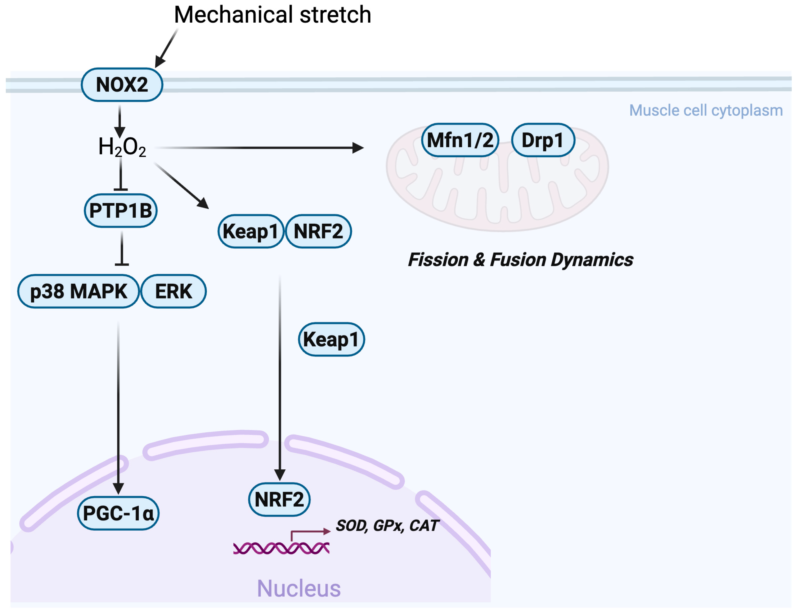
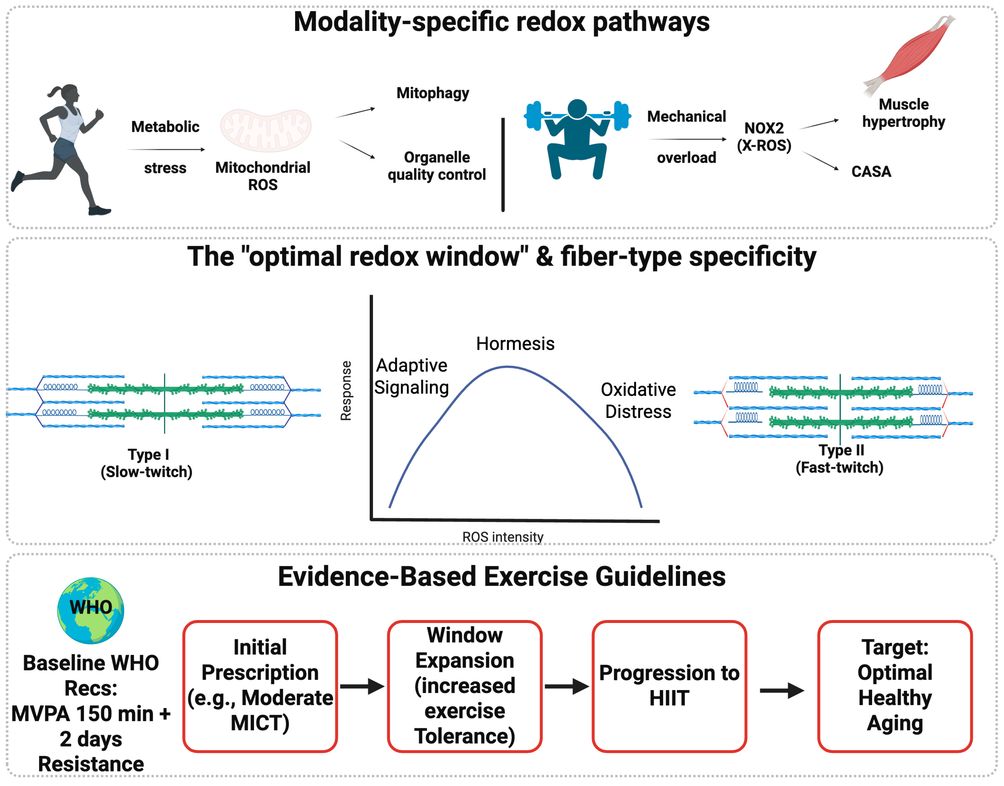
Skeletal Muscle Redox Signaling in Health and Disease: From Molecular Mechanisms to Therapeutic Exercise Strategies

 , ,
, ,  and
and
Abstract
1. Introduction
2. Molecular Mechanisms of Redox Adaptation
2.1. Redox Hormesis and the Optimal Redox Window
2.2. Sources and Compartmentalization of RONS in Skeletal Muscle
2.3. Endogenous Antioxidant and Redox-Buffering Systems
2.4. Redox Signaling in Physiological Adaptation
3. Exercise-Induced Redox Responses and Training Paradigms
3.1. Exercise Modality-Specific Redox Landscapes
3.2. Muscle Damage, Inflammation, Recovery and Overload
4. Translational Aspects
4.1. Antioxidant Supplementation and Redox Adaptation
4.2. Aging and Chronic Disease
4.3. Exercise Protocols for Health: Practical Translational Principles
5. Methodological Considerations and Future Directions
6. Conclusions
Author Contributions
Funding
Institutional Review Board Statement
Informed Consent Statement
Data Availability Statement
Conflicts of Interest
Abbreviations
| AMPK | AMP-activated protein kinase |
| CAT | Catalase |
| DAMPs | Damage-associated molecular patterns |
| DOMS | Delayed onset muscle soreness |
| Drp1 | Dynamin-related protein 1 |
| E-C | Excitation–contraction |
| ER | Endoplasmic reticulum |
| ERK | Extracellular signal-regulated kinase |
| ETC | Electron transport chain |
| EVs | Extracellular vesicles |
| GPx | Glutathione peroxidases |
| Grp75 | Glucose-regulated protein 75 |
| H2O2 | Hydrogen peroxide |
| HIIT | High-intensity interval training |
| IL-18 | Interleukin-18 |
| IL-1β | Interleukin-1 beta |
| IP3R | Inositol 1,4,5-trisphosphate receptor |
| Keap1 | Kelch-like ECH-associated protein 1 |
| M1 | Classically activated macrophages |
| M2 | Alternatively activated macrophages |
| MAMs | Mitochondria-associated membranes |
| Mfn1/2 | Mitofusin 1 and 2 |
| mtROS | Mitochondrial ROS |
| MVPA | Moderate-to-vigorous physical activity |
| NAD+ | Nicotinamide adenine dinucleotide |
| NADPH | Nicotinamide adenine dinucleotide phosphate |
| NLRP3 | NOD-like receptor family pyrin domain containing 3 |
| NMJ | Neuromuscular junction |
| NOS | Nitric oxide synthase |
| NOX | NADPH oxidase |
| NRF2 | Nuclear factor erythroid 2-related factor 2 |
| p38 MAPK | p38 mitogen-activated protein kinase |
| PGC-1α | Peroxisome proliferator-activated receptor-γ coactivator-1α |
| PTMs | Post-translational modifications |
| PTP1B | Protein tyrosine phosphatase 1B |
| RBE | Repeated bout effect |
| RNS | Reactive nitrogen species |
| RONS | Reactive oxygen and nitrogen species |
| ROS | Reactive oxygen species |
| RyR1 | Ryanodine receptor 1 |
| SERCA | Sarcoplasmic reticulum Ca2+-ATPase |
| SIT | Sprint interval training |
| SODs | Superoxide dismutases |
| SR | Sarcoplasmic reticulum |
| TBARS | Thiobarbituric acid reactive substances |
| UPRmt | Mitochondrial unfolded protein response |
| VDAC | Voltage-dependent anion channel |
References
- Pataky, M.W.; Heppelmann, C.J.; Sevits, K.J.; Asokan, A.K.; Kumar, A.P.; Klaus, K.A.; Dasari, S.; Kunz, H.E.; Strub, M.D.; Robinson, M.M.; et al. Aerobic and resistance exercise-regulated phosphoproteome and acetylproteome modifications in human skeletal muscle. Nat. Commun. 2025, 16, 5700. [Google Scholar] [CrossRef]
- Feng, M.; Li, M.; Lou, J.; Wu, G.; Gao, T.; Wu, F.; Tan, Y.; Zhang, N.; Zhao, Y.; Zhao, L.; et al. Early-life exercise extends healthspan but not lifespan in mice. Nat. Commun. 2025, 16, 6328. [Google Scholar] [CrossRef] [PubMed]
- Xu, J.; Liao, C.; Yin, C.C.; Li, G.; Zhu, Y.; Sun, F. In situ structural insights into the excitation-contraction coupling mechanism of skeletal muscle. Sci. Adv. 2024, 10, eadl1126. [Google Scholar] [CrossRef]
- MoTr, P.A.C.S.G.; Lead, A.; MoTr, P.A.C.S.G. Temporal dynamics of the multi-omic response to endurance exercise training. Nature 2024, 629, 174–183. [Google Scholar] [CrossRef]
- Edman, S.; Jones, R.G., III; Jannig, P.R.; Fernandez-Gonzalo, R.; Norrbom, J.; Thomas, N.T.; Khadgi, S.; Koopmans, P.J.; Morena, F.; Chambers, T.L.; et al. The 24-hour molecular landscape after exercise in humans reveals MYC is sufficient for muscle growth. EMBO Rep. 2024, 25, 5810–5837. [Google Scholar] [CrossRef]
- Haller, N.; Widauer, H.L.; Strepp, T.; Nunes, N.; Blumkaitis, J.C.; Wenger, M.; Stoggl, T.; Aglas, L. How intense is high-intensity interval training? Biomarker responses and associations with training load and fitness. iScience 2025, 28, 113738. [Google Scholar] [CrossRef]
- Schwiete, C.; Roth, C.; Mester, J.; Broich, H.; Behringer, M. Overlaps of Skeletal Muscle Fatigue and Skeletal Muscle Damage: The Muscle Injury Continuum. Sports Med. Open 2025, 11, 73. [Google Scholar] [CrossRef]
- Henriquez-Olguin, C.; Knudsen, J.R.; Raun, S.H.; Li, Z.; Dalbram, E.; Treebak, J.T.; Sylow, L.; Holmdahl, R.; Richter, E.A.; Jaimovich, E.; et al. Cytosolic ROS production by NADPH oxidase 2 regulates muscle glucose uptake during exercise. Nat. Commun. 2019, 10, 4623. [Google Scholar] [CrossRef] [PubMed]
- Henriquez-Olguin, C.; Meneses-Valdes, R.; Raun, S.H.; Gallero, S.; Knudsen, J.R.; Li, Z.; Li, J.; Sylow, L.; Jaimovich, E.; Jensen, T.E. NOX2 deficiency exacerbates diet-induced obesity and impairs molecular training adaptations in skeletal muscle. Redox Biol. 2023, 65, 102842. [Google Scholar] [CrossRef] [PubMed]
- Prosser, B.L.; Khairallah, R.J.; Ziman, A.P.; Ward, C.W.; Lederer, W.J. X-ROS signaling in the heart and skeletal muscle: Stretch-dependent local ROS regulates [Ca(2+)]i. J. Mol. Cell. Cardiol. 2013, 58, 172–181. [Google Scholar] [CrossRef]
- Slavin, M.B.; Kumari, R.; Hood, D.A. ATF5 is a regulator of exercise-induced mitochondrial quality control in skeletal muscle. Mol. Metab. 2022, 66, 101623. [Google Scholar] [CrossRef]
- Craige, S.M.; Mammel, R.K.; Amiri, N.; Willoughby, O.S.; Drake, J.C. Interplay of ROS, mitochondrial quality, and exercise in aging: Potential role of spatially discrete signaling. Redox Biol. 2024, 77, 103371. [Google Scholar] [CrossRef] [PubMed]
- Powers, S.K.; Radak, Z.; Ji, L.L.; Jackson, M. Reactive oxygen species promote endurance exercise-induced adaptations in skeletal muscles. J. Sport Health Sci. 2024, 13, 780–792. [Google Scholar] [CrossRef] [PubMed]
- Meizoso-Huesca, A.; Lamboley, C.R.; Krycer, J.R.; Hodson, M.P.; Hudson, J.E.; Launikonis, B.S. Muscle-specific Ryanodine receptor 1 properties underlie limb-girdle muscular dystrophy 2B/R2 progression. Nat. Commun. 2025, 16, 3056. [Google Scholar] [CrossRef]
- Steinz, M.M.; Beard, N.; Shorter, E.; Lanner, J.T. Stable oxidative posttranslational modifications alter the gating properties of RyR1. J. Gen. Physiol. 2024, 156, e202313515. [Google Scholar] [CrossRef] [PubMed]
- Braun, J.L.; Fajardo, V.A. Spaceflight increases sarcoplasmic reticulum Ca(2+) leak and this cannot be counteracted with BuOE treatment. npj Microgravity 2024, 10, 78. [Google Scholar] [CrossRef]
- Khodabukus, A.; Prabhu, N.K.; Roberts, T.; Buldo, M.; Detwiler, A.; Fralish, Z.D.; Kondash, M.E.; Truskey, G.A.; Koves, T.R.; Bursac, N. Bioengineered Model of Human LGMD2B Skeletal Muscle Reveals Roles of Intracellular Calcium Overload in Contractile and Metabolic Dysfunction in Dysferlinopathy. Adv. Sci. 2024, 11, e2400188. [Google Scholar] [CrossRef]
- McAdam, J.S.; Craig, M.P.; Graham, Z.A.; Peoples, B.; Tuggle, S.C.; Seay, R.S.; Lavin, K.M.; Gargus, A.B.; O’Bryan, S.M.; Yang, S.; et al. Multidimensional biocircuitry of exercise adaptation: Integrating in vivo and ex vivo phenomics with miRNA mapping. Physiol. Genom. 2025, 57, 526–550. [Google Scholar] [CrossRef]
- Bishop, D.J.; Lee, M.J.; Picard, M. Exercise as Mitochondrial Medicine: How Does the Exercise Prescription Affect Mitochondrial Adaptations to Training? Annu. Rev. Physiol. 2025, 87, 107–129. [Google Scholar] [CrossRef]
- Wyckelsma, V.L.; Murgia, M.; Kamandulis, S.; Gastaldello, S.; Brazaitis, M.; Snieckus, A.; Eimantas, N.; Paasuke, M.; Edman, S.; Apro, W.; et al. Antioxidant supplementation blunts the proteome response to 3 weeks of sprint interval training preferentially in human type 2 muscle fibres. J. Physiol. 2025. Epub ahead of printing. [Google Scholar] [CrossRef]
- Larsen, S. Acute antioxidant supplementation and performance—Should this be considered. Free Radic. Biol. Med. 2024, 224, 301–309. [Google Scholar] [CrossRef]
- Xirouchaki, C.E.; Jia, Y.; McGrath, M.J.; Greatorex, S.; Tran, M.; Merry, T.L.; Hong, D.; Eramo, M.J.; Broome, S.C.; Woodhead, J.S.T.; et al. Skeletal muscle NOX4 is required for adaptive responses that prevent insulin resistance. Sci. Adv. 2021, 7, eabl4988. [Google Scholar] [CrossRef]
- Sies, H.; Jones, D.P. Reactive oxygen species (ROS) as pleiotropic physiological signalling agents. Nat. Rev. Mol. Cell Biol. 2020, 21, 363–383. [Google Scholar] [CrossRef]
- Caporossi, D.; Jackson, M.J.; Henriquez-Olguin, C. Intracellular and extracellular redox signals during exercise and aging. Free Radic. Biol. Med. 2026, 242, 521–534. [Google Scholar] [CrossRef] [PubMed]
- Sies, H.; Mailloux, R.J.; Jakob, U. Author Correction: Fundamentals of redox regulation in biology. Nat. Rev. Mol. Cell Biol. 2024, 25, 758. [Google Scholar] [CrossRef] [PubMed]
- James, C.; Lloyd, E.M.; Arthur, P.G. Temporal changes in thiol-oxidized plasma albumin are associated with recovery from exercise-induced muscle damage after a marathon. Physiol. Rep. 2024, 12, e70155. [Google Scholar] [CrossRef] [PubMed]
- Tabata, F.; Wada, Y.; Kawakami, S.; Miyaji, K. Serum Albumin Redox States: More Than Oxidative Stress Biomarker. Antioxidants 2021, 10, 503. [Google Scholar] [CrossRef]
- Haller, N.; Reichel, T.; Zimmer, P.; Behringer, M.; Wahl, P.; Stoggl, T.; Kruger, K.; Simon, P. Blood-Based Biomarkers for Managing Workload in Athletes: Perspectives for Research on Emerging Biomarkers. Sports Med. 2023, 53, 2039–2053. [Google Scholar] [CrossRef]
- Hacker, S.; Keck, J.; Reichel, T.; Eder, K.; Ringseis, R.; Kruger, K.; Kruger, B. Biomarkers in Endurance Exercise: Individualized Regulation and Predictive Value. Transl. Sports Med. 2023, 2023, 6614990. [Google Scholar] [CrossRef]
- Vorhauser, J.; Roumeliotis, T.I.; Coupe, D.; Leung, J.K.; Yu, L.; Bohlig, K.; Zerjatke, T.; Glauche, I.; Nadler, A.; Choudhary, J.S.; et al. A redox switch in p21-CDK feedback during G2 phase controls the proliferation-cell cycle exit decision. Mol. Cell 2025, 85, 3241–3255 e3211. [Google Scholar] [CrossRef]
- Lamontagne, F.; Paz-Trejo, C.; Zamorano Cuervo, N.; Grandvaux, N. Redox signaling in cell fate: Beyond damage. Biochim. Biophys. Acta Mol. Cell Res. 2024, 1871, 119722. [Google Scholar] [CrossRef]
- Xia, Q.; Casas-Martinez, J.C.; Zarzuela, E.; Munoz, J.; Miranda-Vizuete, A.; Goljanek-Whysall, K.; McDonagh, B. Peroxiredoxin 2 is required for the redox mediated adaptation to exercise. Redox Biol. 2023, 60, 102631. [Google Scholar] [CrossRef]
- Noone, J.; Mucinski, J.M.; DeLany, J.P.; Sparks, L.M.; Goodpaster, B.H. Understanding the variation in exercise responses to guide personalized physical activity prescriptions. Cell Metab. 2024, 36, 702–724. [Google Scholar] [CrossRef]
- Weber, D.; Thimm, J.; Seiz, T.; Kochlik, B.; Raupbach, J.; Burkle, A.; Grune, T.; Gruber, M.; Moreno-Villanueva, M. Association between redox biomarkers, DNA damage and aerobic capacity before and after physical stress in young men. Redox Biol. 2025, 85, 103764. [Google Scholar] [CrossRef]
- Xu, B.; Guo, S. Daytime eccentric exercise and its impact on inflammatory markers and antioxidant defenses in physically active young men. Front. Immunol. 2025, 16, 1655034. [Google Scholar] [CrossRef]
- Feng, W.; Wang, Y.; Gu, X.; Yu, D.; Liu, Z. Exercise as a modulator of systemic inflammation and oxidative stress biomarkers across clinical and healthy populations: An umbrella meta-analysis. BMC Sports Sci. Med. Rehabil. 2025, 17, 360. [Google Scholar] [CrossRef]
- Kano, R.; Kusano, T.; Takeda, R.; Shirakawa, H.; Poole, D.C.; Kano, Y.; Hoshino, D. Eccentric contraction increases hydrogen peroxide levels and alters gene expression through Nox2 in skeletal muscle of male mice. J. Appl. Physiol. 2024, 137, 778–788. [Google Scholar] [CrossRef] [PubMed]
- Broome, S.C.; Whitfield, J.; Janssens, K.; Hawley, J.A. MitoQ supplementation does not impact redox responses to acute exercise in skeletal muscle of older individuals. Redox Biol. 2025, 88, 103927. [Google Scholar] [CrossRef] [PubMed]
- Cipriano, A.; Viviano, M.; Feoli, A.; Milite, C.; Sarno, G.; Castellano, S.; Sbardella, G. NADPH Oxidases: From Molecular Mechanisms to Current Inhibitors. J. Med. Chem. 2023, 66, 11632–11655. [Google Scholar] [CrossRef] [PubMed]
- Kumar, R.A.; Mariani, V.; Yang, C.; Ferreira, L.F. Skeletal muscle knockout of NAD(P)H oxidase 2 delays the development of isotonic diaphragm fatigue in mice. Free Radic. Biol. Med. 2025, 240, 284–295. [Google Scholar] [CrossRef]
- Meneses-Valdes, R.; Gallero, S.; Henriquez-Olguin, C.; Jensen, T.E. Exploring NADPH oxidases 2 and 4 in cardiac and skeletal muscle adaptations—A cross-tissue comparison. Free Radic. Biol. Med. 2024, 223, 296–305. [Google Scholar] [CrossRef]
- Chavoshan, B.; Sander, M.; Sybert, T.E.; Hansen, J.; Victor, R.G.; Thomas, G.D. Nitric oxide-dependent modulation of sympathetic neural control of oxygenation in exercising human skeletal muscle. J. Physiol. 2002, 540, 377–386. [Google Scholar] [CrossRef]
- Cheng, A.J.; Yamada, T.; Rassier, D.E.; Andersson, D.C.; Westerblad, H.; Lanner, J.T. Reactive oxygen/nitrogen species and contractile function in skeletal muscle during fatigue and recovery. J. Physiol. 2016, 594, 5149–5160. [Google Scholar] [CrossRef]
- Mueller, B.J.; Roberts, M.D.; Mobley, C.B.; Judd, R.L.; Kavazis, A.N. Nitric oxide in exercise physiology: Past and present perspectives. Front. Physiol. 2024, 15, 1504978. [Google Scholar] [CrossRef]
- Sies, H. Dynamics of intracellular and intercellular redox communication. Free Radic. Biol. Med. 2024, 225, 933–939. [Google Scholar] [CrossRef] [PubMed]
- Fuentes-Lemus, E.; Davies, M.J. Effect of crowding, compartmentalization and nanodomains on protein modification and redox signaling—Current state and future challenges. Free Radic. Biol. Med. 2023, 196, 81–92. [Google Scholar] [CrossRef] [PubMed]
- Booth, D.M.; Enyedi, B.; Geiszt, M.; Varnai, P.; Hajnoczky, G. Redox Nanodomains Are Induced by and Control Calcium Signaling at the ER-Mitochondrial Interface. Mol. Cell 2016, 63, 240–248. [Google Scholar] [CrossRef]
- Chen, J.; Yap, M.C.; Bassot, A.; Pascual, D.M.; Makio, T.; Zimmermann, J.; Mast, H.; Bhat, R.; Fleury, S.G.; Fan, Y.; et al. The ER thioredoxin-related transmembrane protein TMX2 controls redox-mediated tethering of ER-mitochondria contacts. Cell Rep. 2025, 44, 116486. [Google Scholar] [CrossRef] [PubMed]
- Kaihara, K.; Kai, H.; Chiba, Y.; Naruse, K.; Iribe, G. Stretch-induced reactive oxygen species contribute to the Frank-Starling mechanism. J. Physiol. 2024, 602, 4347–4362. [Google Scholar] [CrossRef]
- Henriquez-Olguin, C.; Meneses-Valdes, R.; Kritsiligkou, P.; Fuentes-Lemus, E. From workout to molecular switches: How does skeletal muscle produce, sense, and transduce subcellular redox signals? Free Radic. Biol. Med. 2023, 209, 355–365. [Google Scholar] [CrossRef]
- Luin, E.; Giniatullin, R.; Sciancalepore, M. Effects of H(2)O(2) on electrical membrane properties of skeletal myotubes. Free Radic. Biol. Med. 2011, 50, 337–344. [Google Scholar] [CrossRef] [PubMed]
- Schiffer, T.A.; Carvalho, L.; Guimaraes, D.; Boeder, A.; Wikstrom, P.; Carlstrom, M. Specific NOX4 Inhibition Preserves Mitochondrial Function and Dampens Kidney Dysfunction Following Ischemia-Reperfusion-Induced Kidney Injury. Antioxidants 2024, 13, 489. [Google Scholar] [CrossRef]
- Jackson, M.J. Exercise-induced adaptations to homeostasis of reactive oxygen species in skeletal muscle. Free Radic. Biol. Med. 2024, 225, 494–500. [Google Scholar] [CrossRef]
- Zhou, B.; Chen, X.; Ding, R.; Bi, Z.; Zhao, T.; Zhou, R.; Xu, M.; Li, J.; Jiang, X.; Li, H. Biosilica nanoparticulate scavengers for the therapy of hepatic ischemia-reperfusion injury in preclinical models. Nat. Commun. 2025, 16, 7650. [Google Scholar] [CrossRef]
- Zhao, L.; Zhang, T.; Zhou, X. Ameliorating Acute Kidney Injury Induced by Ischemia-Reperfusion by Targeting Purine Metabolism. Int. J. Mol. Sci. 2025, 26, 11886. [Google Scholar] [CrossRef]
- Chuaiphichai, S.; Chu, S.M.; Carnicer, R.; Kelly, M.; Bendall, J.K.; Simon, J.N.; Douglas, G.; Crabtree, M.J.; Casadei, B.; Channon, K.M. Endothelial cell-specific roles for tetrahydrobiopterin in myocardial function, cardiac hypertrophy, and response to myocardial ischemia-reperfusion injury. Am. J. Physiol. Heart Circ. Physiol. 2023, 324, H430–H442. [Google Scholar] [CrossRef]
- Liu, C.C.; Zhang, Y.; Kim, Y.J.; Hamilton, E.J.; Xu, B.; Limas, J.; McCracken, S.A.; Morris, J.M.; Makris, A.; Hennessy, A.; et al. beta3-adrenergic agonist counters oxidative stress and Na(+)-K(+) pump inhibitory S-glutathionylation of placental cells: Implications for preeclampsia. Am. J. Physiol. Cell Physiol. 2025, 328, C27–C39. [Google Scholar] [CrossRef] [PubMed]
- Chen, F.F.; Zhang, Y.H.; Wu, Z.C.; Du, K.; Chen, X.; Lu, Y.; Hu, Q.; Du, A.; Du, S.; Wang, J.; et al. Piezo1 activation in endothelial cells aggravates microvascular ischemia-reperfusion injury in limbs by enhancing ferroptosis. Exp. Mol. Med. 2026, 58, 143–160. [Google Scholar] [CrossRef]
- Zhang, J.; Wu, D.; Lei, R.; Zhang, F.; Wu, W.; Wu, Y.; Zhang, Z.; Han, D.; Chen, L.; Yu, Y.; et al. Loss of hepatic ME1 ameliorates MASLD by Suppressing peroxisomal beta-Oxidation and Activating Lipophagy/Lipolysis. J. Adv. Res. 2025. Epub ahead of printing. [Google Scholar] [CrossRef]
- Loopmans, S.; Rohlenova, K.; van Brussel, T.; Stockmans, I.; Moermans, K.; Peredo, N.; Carmeliet, P.; Lambrechts, D.; Stegen, S.; Carmeliet, G. The pentose phosphate pathway controls oxidative protein folding and prevents ferroptosis in chondrocytes. Nat. Metab. 2025, 7, 182–195. [Google Scholar] [CrossRef] [PubMed]
- Wang, L.; Wang, C.C. Oxidative protein folding fidelity and redoxtasis in the endoplasmic reticulum. Trends Biochem. Sci. 2023, 48, 40–52. [Google Scholar] [CrossRef]
- Beretta, M.; Santos, C.X.; Molenaar, C.; Hafstad, A.D.; Miller, C.C.; Revazian, A.; Betteridge, K.; Schroder, K.; Streckfuss-Bomeke, K.; Doroshow, J.H.; et al. Nox4 regulates InsP(3) receptor-dependent Ca(2+) release into mitochondria to promote cell survival. EMBO J. 2020, 39, e103530. [Google Scholar] [CrossRef]
- Loncke, J.; Kaasik, A.; Bezprozvanny, I.; Parys, J.B.; Kerkhofs, M.; Bultynck, G. Balancing ER-Mitochondrial Ca(2+) Fluxes in Health and Disease. Trends Cell Biol. 2021, 31, 598–612. [Google Scholar] [CrossRef]
- Yuan, M.; Gong, M.; He, J.; Xie, B.; Zhang, Z.; Meng, L.; Tse, G.; Zhao, Y.; Bao, Q.; Zhang, Y.; et al. IP3R1/GRP75/VDAC1 complex mediates endoplasmic reticulum stress-mitochondrial oxidative stress in diabetic atrial remodeling. Redox Biol. 2022, 52, 102289. [Google Scholar] [CrossRef]
- Boardman, N.T.; Trani, G.; Scalabrin, M.; Romanello, V.; Wust, R.C.I. Intracellular to Interorgan Mitochondrial Communication in Striated Muscle in Health and Disease. Endocr. Rev. 2023, 44, 668–692. [Google Scholar] [CrossRef]
- Heaton, R.A.; Ball, S.T.; Staunton, C.A.; Mouly, V.; Jones, S.W.; McArdle, A.; Jackson, M.J. Peroxiredoxin 2 mediates redox-stimulated adaptations to oxidative phosphorylation induced by contractile activity in human skeletal muscle myotubes. Free Radic. Biol. Med. 2025, 227, 395–406. [Google Scholar] [CrossRef]
- Flensted-Jensen, M.; Weinreich, C.M.; Kleis-Olsen, A.S.; Hansen, F.; Skyggelund, N.S.; Pii, J.R.; Whitlock, R.; Karlsen, A.; Ingersen, A.; Reihmane, D.; et al. Resistance-based training improves mitochondrial capacity and redox balance in aging adults, independent of polyphenol supplementation. Redox Biol. 2025, 89, 103972. [Google Scholar] [CrossRef]
- Powers, S.K.; Lategan-Potgieter, R.; Goldstein, E. Exercise-induced Nrf2 activation increases antioxidant defenses in skeletal muscles. Free Radic. Biol. Med. 2024, 224, 470–478. [Google Scholar] [CrossRef]
- Griffith, M.; Araujo, A.; Travasso, R.; Salvador, A. The architecture of redox microdomains: Cascading gradients and peroxiredoxins’ redox-oligomeric coupling integrate redox signaling and antioxidant protection. Redox Biol. 2024, 69, 103000. [Google Scholar] [CrossRef]
- Edman, S.; Flockhart, M.; Larsen, F.J.; Apro, W. Need for speed: Human fast-twitch mitochondria favor power over efficiency. Mol. Metab. 2024, 79, 101854. [Google Scholar] [CrossRef]
- Vasileiadou, O.; Nastos, G.G.; Chatzinikolaou, P.N.; Papoutsis, D.; Vrampa, D.I.; Methenitis, S.; Margaritelis, N.V. Redox Profile of Skeletal Muscles: Implications for Research Design and Interpretation. Antioxidants 2023, 12, 1738. [Google Scholar] [CrossRef]
- Horwath, O.; Moberg, M.; Edman, S.; Philp, A.; Apro, W. Ageing leads to selective type II myofibre deterioration and denervation independent of reinnervative capacity in human skeletal muscle. Exp. Physiol. 2025, 110, 277–292. [Google Scholar] [CrossRef]
- Thomas, A.C.Q.; Stead, C.A.; Burniston, J.G.; Phillips, S.M. Exercise-specific adaptations in human skeletal muscle: Molecular mechanisms of making muscles fit and mighty. Free Radic. Biol. Med. 2024, 223, 341–356. [Google Scholar] [CrossRef]
- Hoffman, N.J.; Whitfield, J.; Xiao, D.; Radford, B.E.; Suni, V.; Blazev, R.; Yang, P.; Parker, B.L.; Hawley, J.A. Phosphoproteomics Uncovers Exercise Intensity-Specific Skeletal Muscle Signaling Networks Underlying High-Intensity Interval Training in Healthy Male Participants. Sports Med. 2025, 55, 1983–2004. [Google Scholar] [CrossRef]
- Li, B.; Ming, H.; Qin, S.; Nice, E.C.; Dong, J.; Du, Z.; Huang, C. Redox regulation: Mechanisms, biology and therapeutic targets in diseases. Signal Transduct. Target. Ther. 2025, 10, 72. [Google Scholar] [CrossRef]
- Martinez-Ferran, M.; Berlanga, L.A.; Barcelo-Guido, O.; Matos-Duarte, M.; Vicente-Campos, D.; Sanchez-Jorge, S.; Romero-Morales, C.; Munguia-Izquierdo, D.; Pareja-Galeano, H. Antioxidant vitamin supplementation on muscle adaptations to resistance training: A double-blind, randomized controlled trial. Nutrition 2023, 105, 111848. [Google Scholar] [CrossRef]
- Raun, S.H.; Henriquez-Olguin, C.; Frank, E.; Schlabs, F.; Hahn, N.J.; Knudsen, J.R.; Ali, M.S.; Andersen, N.R.; Moller, L.L.V.; Davey, J.; et al. Skeletal muscle Rac1 mediates exercise training adaptations towards muscle glycogen resynthesis and protein synthesis. Redox Biol. 2025, 86, 103844. [Google Scholar] [CrossRef]
- Seisenbacher, G.; Nakic, Z.R.; Borras, E.; Sabido, E.; Sauer, U.; de Nadal, E.; Posas, F. Redox proteomics reveal a role for peroxiredoxinylation in stress protection. Cell Rep. 2025, 44, 115224. [Google Scholar] [CrossRef]
- Veal, E.A.; Kritsiligkou, P. How are hydrogen peroxide messages relayed to affect cell signalling? Curr. Opin. Chem. Biol. 2024, 81, 102496. [Google Scholar] [CrossRef]
- Xu, H.; Ahn, B.; Van Remmen, H. Impact of aging and oxidative stress on specific components of excitation contraction coupling in regulating force generation. Sci. Adv. 2022, 8, eadd7377. [Google Scholar] [CrossRef]
- Qin, F.; Siwik, D.A.; Lancel, S.; Zhang, J.; Kuster, G.M.; Luptak, I.; Wang, L.; Tong, X.; Kang, Y.J.; Cohen, R.A.; et al. Hydrogen peroxide-mediated SERCA cysteine 674 oxidation contributes to impaired cardiac myocyte relaxation in senescent mouse heart. J. Am. Heart Assoc. 2013, 2, e000184. [Google Scholar] [CrossRef]
- Sun, J.; Xin, C.; Eu, J.P.; Stamler, J.S.; Meissner, G. Cysteine-3635 is responsible for skeletal muscle ryanodine receptor modulation by NO. Proc. Natl. Acad. Sci. USA 2001, 98, 11158–11162. [Google Scholar] [CrossRef]
- Nikolaienko, R.; Bovo, E.; Kahn, D.; Gracia, R.; Jamrozik, T.; Zima, A.V. Cysteines 1078 and 2991 cross-linking plays a critical role in redox regulation of cardiac ryanodine receptor (RyR). Nat. Commun. 2023, 14, 4498. [Google Scholar] [CrossRef]
- Londhe, A.D.; Bergeron, A.; Curley, S.M.; Zhang, F.; Rivera, K.D.; Kannan, A.; Coulis, G.; Rizvi, S.H.M.; Kim, S.J.; Pappin, D.J.; et al. Regulation of PTP1B activation through disruption of redox-complex formation. Nat. Chem. Biol. 2020, 16, 122–125. [Google Scholar] [CrossRef]
- Buttari, B.; Tramutola, A.; Rojo, A.I.; Chondrogianni, N.; Saha, S.; Berry, A.; Giona, L.; Miranda, J.P.; Profumo, E.; Davinelli, S.; et al. Proteostasis Decline and Redox Imbalance in Age-Related Diseases: The Therapeutic Potential of NRF2. Biomolecules 2025, 15, 113. [Google Scholar] [CrossRef]
- Kuzu, O.F.; Granerud, L.J.T.; Saatcioglu, F. Navigating the landscape of protein folding and proteostasis: From molecular chaperones to therapeutic innovations. Signal Transduct. Target. Ther. 2025, 10, 358. [Google Scholar] [CrossRef]
- Diaz-Castro, F.; Tunon-Suarez, M.; Rivera, P.; Botella, J.; Cancino, J.; Figueroa, A.M.; Gutierrez, J.; Cantin, C.; Deldicque, L.; Zbinden-Foncea, H.; et al. A single bout of resistance exercise triggers mitophagy, potentially involving the ejection of mitochondria in human skeletal muscle. Acta Physiol. 2024, 240, e14203. [Google Scholar] [CrossRef]
- Turkel, I.; Kubat, G.B.; Fatsa, T.; Acet, O.; Ozerklig, B.; Yazgan, B.; Simsek, G.; Singh, K.K.; Kosar, S.N. Acute treadmill exercise induces mitochondrial unfolded protein response in skeletal muscle of male rats. Biochim. Biophys. Acta Bioenerg. 2025, 1866, 149532. [Google Scholar] [CrossRef]
- Gaspar, R.S.; Katashima, C.K.; Crisol, B.M.; Carneiro, F.S.; Sampaio, I.; Silveira, L.D.R.; Silva, A.; Cintra, D.E.; Pauli, J.R.; Ropelle, E.R. Physical exercise elicits UPR(mt) in the skeletal muscle: The role of c-Jun N-terminal kinase. Mol. Metab. 2023, 78, 101816. [Google Scholar] [CrossRef]
- Apablaza, P.; Borquez, J.C.; Mendoza, R.; Silva, M.; Tapia, G.; Espinosa, A.; Troncoso, R.; Videla, L.A.; Juretic, N.; Del Campo, A. Exercise Induces an Augmented Skeletal Muscle Mitochondrial Unfolded Protein Response in a Mouse Model of Obesity Produced by a High-Fat Diet. Int. J. Mol. Sci. 2023, 24, 5654. [Google Scholar] [CrossRef]
- Kim, Y.M.; Youn, S.W.; Sudhahar, V.; Das, A.; Chandhri, R.; Cuervo Grajal, H.; Kweon, J.; Leanhart, S.; He, L.; Toth, P.T.; et al. Redox Regulation of Mitochondrial Fission Protein Drp1 by Protein Disulfide Isomerase Limits Endothelial Senescence. Cell Rep. 2018, 23, 3565–3578. [Google Scholar] [CrossRef]
- Thaher, O.; Wolf, C.; Dey, P.N.; Pouya, A.; Wullner, V.; Tenzer, S.; Methner, A. The thiol switch C684 in Mitofusin-2 mediates redox-induced alterations of mitochondrial shape and respiration. Neurochem. Int. 2018, 117, 167–173. [Google Scholar] [CrossRef]
- Yapa, N.M.B.; Lisnyak, V.; Reljic, B.; Ryan, M.T. Mitochondrial dynamics in health and disease. FEBS Lett. 2021, 595, 1184–1204. [Google Scholar] [CrossRef]
- Kramer, P.A.; Duan, J.; Gaffrey, M.J.; Shukla, A.K.; Wang, L.; Bammler, T.K.; Qian, W.J.; Marcinek, D.J. Fatiguing contractions increase protein S-glutathionylation occupancy in mouse skeletal muscle. Redox Biol. 2018, 17, 367–376. [Google Scholar] [CrossRef]
- Qaisar, R.; Bhaskaran, S.; Ranjit, R.; Sataranatarajan, K.; Premkumar, P.; Huseman, K.; Van Remmen, H. Restoration of SERCA ATPase prevents oxidative stress-related muscle atrophy and weakness. Redox Biol. 2019, 20, 68–74. [Google Scholar] [CrossRef]
- Kano, R.; Tabuchi, A.; Tanaka, Y.; Shirakawa, H.; Hoshino, D.; Poole, D.C.; Kano, Y. In vivo cytosolic H(2)O(2) changes and Ca(2+) homeostasis in mouse skeletal muscle. Am. J. Physiol. Regul. Integr. Comp. Physiol. 2024, 326, R43–R52. [Google Scholar] [CrossRef]
- Place, N.; Ivarsson, N.; Venckunas, T.; Neyroud, D.; Brazaitis, M.; Cheng, A.J.; Ochala, J.; Kamandulis, S.; Girard, S.; Volungevicius, G.; et al. Ryanodine receptor fragmentation and sarcoplasmic reticulum Ca2+ leak after one session of high-intensity interval exercise. Proc. Natl. Acad. Sci. USA 2015, 112, 15492–15497. [Google Scholar] [CrossRef]
- Watanabe, D.; Wada, M. Cellular mechanisms underlying overreaching in skeletal muscle following excessive high-intensity interval training. Am. J. Physiol. Cell Physiol. 2025, 328, C921–C938. [Google Scholar] [CrossRef]
- Hargreaves, M.; Spriet, L.L. Author Correction: Skeletal muscle energy metabolism during exercise. Nat. Metab. 2020, 2, 990. [Google Scholar] [CrossRef]
- Granata, C.; Caruana, N.J.; Botella, J.; Jamnick, N.A.; Huynh, K.; Kuang, J.; Janssen, H.A.; Reljic, B.; Mellett, N.A.; Laskowski, A.; et al. High-intensity training induces non-stoichiometric changes in the mitochondrial proteome of human skeletal muscle without reorganisation of respiratory chain content. Nat. Commun. 2021, 12, 7056. [Google Scholar] [CrossRef]
- Flensted-Jensen, M.; Gram, M.; Dela, F.; Helge, J.W.; Larsen, S. Six weeks of high intensity cycle training reduces H(2)O(2) emission and increases antioxidant protein levels in obese adults with risk factors for type 2 diabetes. Free Radic. Biol. Med. 2021, 173, 1–6. [Google Scholar] [CrossRef]
- Margaritelis, N.V.; Paschalis, V.; Theodorou, A.A.; Kyparos, A.; Nikolaidis, M.G. Redox basis of exercise physiology. Redox Biol. 2020, 35, 101499. [Google Scholar] [CrossRef] [PubMed]
- Southern, W.M.; Johnson, E.E.; Fasbender, E.K.; Fallon, K.S.; Cavazos, C.L.; Lowe, D.A.; Rodney, G.G.; Ervasti, J.M. Impaired hydrogen sulfide biosynthesis underlies eccentric contraction-induced force loss in dystrophin-deficient skeletal muscle. J. Clin. Investig. 2025, 135, e176942. [Google Scholar] [CrossRef] [PubMed]
- Coleman, A.K.; Joca, H.C.; Shi, G.; Lederer, W.J.; Ward, C.W. Tubulin acetylation increases cytoskeletal stiffness to regulate mechanotransduction in striated muscle. J. Gen. Physiol. 2021, 153, e202012743. [Google Scholar] [CrossRef]
- Lavigne, C.; Mons, V.; Lemineur, C.; Meste, O.; Leftheriotis, G.; Blain, G.M. Physiological mechanisms underlying enhanced performance with blood flow restriction training: Neuromuscular, vascular and metabolic adaptations. J. Physiol. 2025. Epub ahead of printing. [Google Scholar] [CrossRef] [PubMed]
- Petrick, H.L.; Pignanelli, C.; Barbeau, P.A.; Churchward-Venne, T.A.; Dennis, K.; van Loon, L.J.C.; Burr, J.F.; Goossens, G.H.; Holloway, G.P. Blood flow restricted resistance exercise and reductions in oxygen tension attenuate mitochondrial H(2)O(2) emission rates in human skeletal muscle. J. Physiol. 2019, 597, 3985–3997. [Google Scholar] [CrossRef]
- Wu, J.; Ren, B.; Wang, D.; Lin, H. Regulatory T cells in skeletal muscle repair and regeneration: Recent insights. Cell Death Dis. 2022, 13, 680. [Google Scholar] [CrossRef]
- Song, D.; Chen, Y.; Wang, P.; Cheng, Y.; Shyh-Chang, N. Lin28a forms an RNA-binding complex with Igf2bp3 to regulate m(6)A-modified stress response genes in stress granules of muscle stem cells. Cell Prolif. 2024, 57, e13707. [Google Scholar] [CrossRef]
- Chung, J.D.; Porrello, E.R.; Lynch, G.S. Muscle regeneration and muscle stem cells in metabolic disease. Free Radic. Biol. Med. 2025, 227, 52–63. [Google Scholar] [CrossRef]
- Zhang, H.; Xiao, X.; Pan, Z.; Dokudovskaya, S. mTOR signaling networks: Mechanistic insights and translational frontiers in disease therapeutics. Signal Transduct. Target. Ther. 2025, 10, 428. [Google Scholar] [CrossRef]
- Bell, L.; Ruddock, A.; Boriel, J.; Maden-Wilkinson, T.; Thompson, S.W.; Wright, K.J.; Burke, K.; Rogerson, D. Effects of a 5-Day Back Squat Overreaching Protocol on Strength Performance, Perceived Recovery and Wellness Responses: A Pilot Trial. J. Funct. Morphol. Kinesiol. 2025, 10, 227. [Google Scholar] [CrossRef]
- Fiala, O.; Hanzlova, M.; Borska, L.; Fiala, Z.; Holmannova, D. Beyond physical exhaustion: Understanding overtraining syndrome through the lens of molecular mechanisms and clinical manifestation. Sports Med. Health Sci. 2025, 7, 237–248. [Google Scholar] [CrossRef]
- Granata, C.; Oliveira, R.S.; Little, J.P.; Renner, K.; Bishop, D.J. Sprint-interval but not continuous exercise increases PGC-1alpha protein content and p53 phosphorylation in nuclear fractions of human skeletal muscle. Sci. Rep. 2017, 7, 44227. [Google Scholar] [CrossRef]
- Hostrup, M.; Lemminger, A.K.; Stocks, B.; Gonzalez-Franquesa, A.; Larsen, J.K.; Quesada, J.P.; Thomassen, M.; Weinert, B.T.; Bangsbo, J.; Deshmukh, A.S. High-intensity interval training remodels the proteome and acetylome of human skeletal muscle. eLife 2022, 11, e69802. [Google Scholar] [CrossRef] [PubMed]
- Hov, H.; Wang, E.; Lim, Y.R.; Trane, G.; Hemmingsen, M.; Hoff, J.; Helgerud, J. Aerobic high-intensity intervals are superior to improve V̇O(2max) compared with sprint intervals in well-trained men. Scand. J. Med. Sci. Sports 2023, 33, 146–159. [Google Scholar] [CrossRef] [PubMed]
- Powers, S.K.; Deminice, R.; Ozdemir, M.; Yoshihara, T.; Bomkamp, M.P.; Hyatt, H. Exercise-induced oxidative stress: Friend or foe? J. Sport Health Sci. 2020, 9, 415–425. [Google Scholar] [CrossRef] [PubMed]
- Flockhart, M.; Nilsson, L.C.; Tais, S.; Ekblom, B.; Apro, W.; Larsen, F.J. Excessive exercise training causes mitochondrial functional impairment and decreases glucose tolerance in healthy volunteers. Cell Metab. 2021, 33, 957–970 e956. [Google Scholar] [CrossRef]
- Crisol, B.M.; Rocha, M.B.; Franco, B.; Morelli, A.P.; Katashima, C.K.; Junior, S.J.A.; Carneiro, F.S.; Braga, R.R.; Bricola, R.S.; de Azambuja, G.; et al. Excessive exercise elicits poly (ADP-ribose) Polymerase-1 activation and global protein PARylation driving muscle dysfunction and performance impairment. Mol. Metab. 2025, 96, 102135. [Google Scholar] [CrossRef]
- Zhang, H.; Qi, G.; Wang, K.; Yang, J.; Shen, Y.; Yang, X.; Chen, X.; Yao, X.; Gu, X.; Qi, L.; et al. Oxidative stress: Roles in skeletal muscle atrophy. Biochem. Pharmacol. 2023, 214, 115664. [Google Scholar] [CrossRef]
- Stozer, A.; Vodopivc, P.; Krizancic Bombek, L. Pathophysiology of exercise-induced muscle damage and its structural, functional, metabolic, and clinical consequences. Physiol. Res. 2020, 69, 565–598. [Google Scholar] [CrossRef]
- Shu, L.Z.; Zhang, X.L.; Ding, Y.D.; Lin, H. From inflammation to bone formation: The intricate role of neutrophils in skeletal muscle injury and traumatic heterotopic ossification. Exp. Mol. Med. 2024, 56, 1523–1530. [Google Scholar] [CrossRef] [PubMed]
- Kitajima, Y.; Suzuki, N.; Nunomiya, A.; Osana, S.; Yoshioka, K.; Tashiro, Y.; Takahashi, R.; Ono, Y.; Aoki, M.; Nagatomi, R. The Ubiquitin-Proteasome System Is Indispensable for the Maintenance of Muscle Stem Cells. Stem Cell Rep. 2018, 11, 1523–1538. [Google Scholar] [CrossRef]
- Blackburn, D.M.; Sahinyan, K.; Hernandez-Corchado, A.; Lazure, F.; Richard, V.; Raco, L.; Perron, G.; Zahedi, R.P.; Borchers, C.H.; Lepper, C.; et al. The E3 ubiquitin ligase Nedd4L preserves skeletal muscle stem cell quiescence by inhibiting their activation. iScience 2024, 27, 110241. [Google Scholar] [CrossRef] [PubMed]
- Han, L.; Qiu, Y.; He, L.; Gao, C.; Zhou, N.; Fu, X.; Xu, J.; Lin, L.; Wang, H.; Biswal, S.; et al. Non-canonical roles of Keap1/Nrf2 in regulating quiescence and early activation in adult muscle stem cells. Nat. Commun. 2025, 16, 10505. [Google Scholar] [CrossRef] [PubMed]
- Calvo-Rubio, M.; Garcia-Domiguez, E.; Tamayo-Torres, E.; Soto-Rodriguez, S.; Olaso-Gonzalez, G.; Ferrucci, L.; de Cabo, R.; Gomez-Cabrera, M.C. The repeated bout effect evokes the training-induced skeletal muscle cellular memory. Free Radic. Biol. Med. 2024, 225, 247–254. [Google Scholar] [CrossRef]
- Boyd, L.; Deakin, G.B.; Devantier-Thomas, B.; Singh, U.; Doma, K. The Effects of Pre-conditioning on Exercise-Induced Muscle Damage: A Systematic Review and Meta-analysis. Sports Med. 2023, 53, 1537–1557. [Google Scholar] [CrossRef]
- Zhong, Z.; Liang, S.; Sanchez-Lopez, E.; He, F.; Shalapour, S.; Lin, X.J.; Wong, J.; Ding, S.; Seki, E.; Schnabl, B.; et al. New mitochondrial DNA synthesis enables NLRP3 inflammasome activation. Nature 2018, 560, 198–203. [Google Scholar] [CrossRef]
- Xian, H.; Watari, K.; Sanchez-Lopez, E.; Offenberger, J.; Onyuru, J.; Sampath, H.; Ying, W.; Hoffman, H.M.; Shadel, G.S.; Karin, M. Oxidized DNA fragments exit mitochondria via mPTP- and VDAC-dependent channels to activate NLRP3 inflammasome and interferon signaling. Immunity 2022, 55, 1370–1385 e1378. [Google Scholar] [CrossRef]
- Billingham, L.K.; Stoolman, J.S.; Vasan, K.; Rodriguez, A.E.; Poor, T.A.; Szibor, M.; Jacobs, H.T.; Reczek, C.R.; Rashidi, A.; Zhang, P.; et al. Mitochondrial electron transport chain is necessary for NLRP3 inflammasome activation. Nat. Immunol. 2022, 23, 692–704. [Google Scholar] [CrossRef]
- Chaweewannakorn, C.; Tsuchiya, M.; Koide, M.; Hatakeyama, H.; Tanaka, Y.; Yoshida, S.; Sugawara, S.; Hagiwara, Y.; Sasaki, K.; Kanzaki, M. Roles of IL-1alpha/beta in regeneration of cardiotoxin-injured muscle and satellite cell function. Am. J. Physiol. Regul. Integr. Comp. Physiol. 2018, 315, R90–R103. [Google Scholar] [CrossRef]
- Swanson, K.V.; Deng, M.; Ting, J.P. The NLRP3 inflammasome: Molecular activation and regulation to therapeutics. Nat. Rev. Immunol. 2019, 19, 477–489. [Google Scholar] [CrossRef]
- Chen, Y.F.; Lee, C.W.; Li, Y.J.; Lin, W.T.; Chen, H.Y.; Chen, Y.C.; Lin, C.H.; Ho, J.H.; Lu, L.F.; Chien, S.; et al. Temporal single-cell sequencing analysis reveals that GPNMB-expressing macrophages potentiate muscle regeneration. Exp. Mol. Med. 2025, 57, 1232–1245. [Google Scholar] [CrossRef]
- Krasniewski, L.K.; Chakraborty, P.; Cui, C.Y.; Mazan-Mamczarz, K.; Dunn, C.; Piao, Y.; Fan, J.; Shi, C.; Wallace, T.; Nguyen, C.; et al. Single-cell analysis of skeletal muscle macrophages reveals age-associated functional subpopulations. eLife 2022, 11, e77974. [Google Scholar] [CrossRef]
- Merlin, J.; Ivanov, S.; Dumont, A.; Sergushichev, A.; Gall, J.; Stunault, M.; Ayrault, M.; Vaillant, N.; Castiglione, A.; Swain, A.; et al. Non-canonical glutamine transamination sustains efferocytosis by coupling redox buffering to oxidative phosphorylation. Nat. Metab. 2021, 3, 1313–1326. [Google Scholar] [CrossRef]
- Wculek, S.K.; Heras-Murillo, I.; Mastrangelo, A.; Mananes, D.; Galan, M.; Miguel, V.; Curtabbi, A.; Barbas, C.; Chandel, N.S.; Enriquez, J.A.; et al. Oxidative phosphorylation selectively orchestrates tissue macrophage homeostasis. Immunity 2023, 56, 516–530 e519. [Google Scholar] [CrossRef]
- Ryan, E.M.; Sadiku, P.; Coelho, P.; Watts, E.R.; Zhang, A.; Howden, A.J.M.; Sanchez-Garcia, M.A.; Bewley, M.; Cole, J.; McHugh, B.J.; et al. NRF2 Activation Reprograms Defects in Oxidative Metabolism to Restore Macrophage Function in Chronic Obstructive Pulmonary Disease. Am. J. Respir. Crit. Care Med. 2023, 207, 998–1011. [Google Scholar] [CrossRef]
- Eggelbusch, M.; Shi, A.; Broeksma, B.C.; Vazquez-Cruz, M.; Soares, M.N.; de Wit, G.M.J.; Everts, B.; Jaspers, R.T.; Wust, R.C.I. The NLRP3 inflammasome contributes to inflammation-induced morphological and metabolic alterations in skeletal muscle. J. Cachexia Sarcopenia Muscle 2022, 13, 3048–3061. [Google Scholar] [CrossRef] [PubMed]
- Irazoki, A.; Gordaliza-Alaguero, I.; Frank, E.; Giakoumakis, N.N.; Seco, J.; Palacin, M.; Guma, A.; Sylow, L.; Sebastian, D.; Zorzano, A. Disruption of mitochondrial dynamics triggers muscle inflammation through interorganellar contacts and mitochondrial DNA mislocation. Nat. Commun. 2023, 14, 108. [Google Scholar] [CrossRef] [PubMed]
- Collado-Mateo, D.; Lavin-Perez, A.M.; Penacoba, C.; Del Coso, J.; Leyton-Roman, M.; Luque-Casado, A.; Gasque, P.; Fernandez-Del-Olmo, M.A.; Amado-Alonso, D. Key Factors Associated with Adherence to Physical Exercise in Patients with Chronic Diseases and Older Adults: An Umbrella Review. Int. J. Environ. Res. Public Health 2021, 18, 2023. [Google Scholar] [CrossRef]
- Feigel, E.D.; Koltun, K.J.; Lovalekar, M.; Kargl, C.K.; Bird, M.B.; Forse, J.N.; Patel, V.J.; Martin, B.J.; Nagle, E.F.; Friedl, K.E.; et al. Association of allostatic load measured by allostatic load index on physical performance and psychological responses during arduous military training. Physiol. Rep. 2025, 13, e70273. [Google Scholar] [CrossRef] [PubMed]
- Ottensmeyer, J.; Esch, A.; Baeta, H.; Sieger, S.; Gupta, Y.; Rathmann, M.F.; Jeschke, A.; Jacko, D.; Schaaf, K.; Schiffer, T.; et al. Force-induced dephosphorylation activates the cochaperone BAG3 to coordinate protein homeostasis and membrane traffic. Curr. Biol. 2024, 34, 4170–4183 e4179. [Google Scholar] [CrossRef]
- Aiken, C.T.; Kaake, R.M.; Wang, X.; Huang, L. Oxidative stress-mediated regulation of proteasome complexes. Mol. Cell. Proteom. 2011, 10, R110 006924. [Google Scholar] [CrossRef]
- Walsh, N.P.; Halson, S.L.; Sargent, C.; Roach, G.D.; Nedelec, M.; Gupta, L.; Leeder, J.; Fullagar, H.H.; Coutts, A.J.; Edwards, B.J.; et al. Sleep and the athlete: Narrative review and 2021 expert consensus recommendations. Br. J. Sports Med. 2020, 55, 356–368. [Google Scholar] [CrossRef] [PubMed]
- Maughan, R.J.; Burke, L.M.; Dvorak, J.; Larson-Meyer, D.E.; Peeling, P.; Phillips, S.M.; Rawson, E.S.; Walsh, N.P.; Garthe, I.; Geyer, H.; et al. IOC consensus statement: Dietary supplements and the high-performance athlete. Br. J. Sports Med. 2018, 52, 439–455. [Google Scholar] [CrossRef] [PubMed]
- Felix-Soriano, E.; Stanford, K.I. Exerkines and redox homeostasis. Redox Biol. 2023, 63, 102748. [Google Scholar] [CrossRef] [PubMed]
- Castano, C.; Mirasierra, M.; Vallejo, M.; Novials, A.; Parrizas, M. Delivery of muscle-derived exosomal miRNAs induced by HIIT improves insulin sensitivity through down-regulation of hepatic FoxO1 in mice. Proc. Natl. Acad. Sci. USA 2020, 117, 30335–30343. [Google Scholar] [CrossRef]
- Ristow, M.; Zarse, K.; Oberbach, A.; Kloting, N.; Birringer, M.; Kiehntopf, M.; Stumvoll, M.; Kahn, C.R.; Bluher, M. Antioxidants prevent health-promoting effects of physical exercise in humans. Proc. Natl. Acad. Sci. USA 2009, 106, 8665–8670. [Google Scholar] [CrossRef]
- Paulsen, G.; Cumming, K.T.; Holden, G.; Hallen, J.; Ronnestad, B.R.; Sveen, O.; Skaug, A.; Paur, I.; Bastani, N.E.; Ostgaard, H.N.; et al. Vitamin C and E supplementation hampers cellular adaptation to endurance training in humans: A double-blind, randomised, controlled trial. J. Physiol. 2014, 592, 1887–1901. [Google Scholar] [CrossRef]
- Paulsen, G.; Hamarsland, H.; Cumming, K.T.; Johansen, R.E.; Hulmi, J.J.; Borsheim, E.; Wiig, H.; Garthe, I.; Raastad, T. Vitamin C and E supplementation alters protein signalling after a strength training session, but not muscle growth during 10 weeks of training. J. Physiol. 2014, 592, 5391–5408. [Google Scholar] [CrossRef]
- Powers, S.K.; Schrager, M. Redox signaling regulates skeletal muscle remodeling in response to exercise and prolonged inactivity. Redox Biol. 2022, 54, 102374. [Google Scholar] [CrossRef]
- Nitti, M.; Marengo, B.; Furfaro, A.L.; Pronzato, M.A.; Marinari, U.M.; Domenicotti, C.; Traverso, N. Hormesis and Oxidative Distress: Pathophysiology of Reactive Oxygen Species and the Open Question of Antioxidant Modulation and Supplementation. Antioxidants 2022, 11, 1613. [Google Scholar] [CrossRef]
- Bredefeld, C.; Hussain, M.M.; Averna, M.; Black, D.D.; Brin, M.F.; Burnett, J.R.; Charriere, S.; Cuerq, C.; Davidson, N.O.; Deckelbaum, R.J.; et al. Guidance for the diagnosis and treatment of hypolipidemia disorders. J. Clin. Lipidol. 2022, 16, 797–812. [Google Scholar] [CrossRef]
- Margaritelis, N.V.; Paschalis, V.; Theodorou, A.A.; Kyparos, A.; Nikolaidis, M.G. Antioxidant supplementation, redox deficiencies and exercise performance: A falsification design. Free Radic. Biol. Med. 2020, 158, 44–52. [Google Scholar] [CrossRef]
- McKeegan, K.; Mason, S.A.; Trewin, A.J.; Keske, M.A.; Wadley, G.D.; Della Gatta, P.A.; Nikolaidis, M.G.; Parker, L. Reactive oxygen species in exercise and insulin resistance: Working towards personalized antioxidant treatment. Redox Biol. 2021, 44, 102005. [Google Scholar] [CrossRef]
- Mason, S.A.; Trewin, A.J.; Parker, L.; Wadley, G.D. Antioxidant supplements and endurance exercise: Current evidence and mechanistic insights. Redox Biol. 2020, 35, 101471. [Google Scholar] [CrossRef]
- Lopez-Otin, C.; Blasco, M.A.; Partridge, L.; Serrano, M.; Kroemer, G. Hallmarks of aging: An expanding universe. Cell 2023, 186, 243–278. [Google Scholar] [CrossRef]
- Gulen, M.F.; Samson, N.; Keller, A.; Schwabenland, M.; Liu, C.; Gluck, S.; Thacker, V.V.; Favre, L.; Mangeat, B.; Kroese, L.J.; et al. cGAS-STING drives ageing-related inflammation and neurodegeneration. Nature 2023, 620, 374–380. [Google Scholar] [CrossRef]
- Forman, H.J.; Zhang, H. Author Correction: Targeting oxidative stress in disease: Promise and limitations of antioxidant therapy. Nat. Rev. Drug Discov. 2021, 20, 652. [Google Scholar] [CrossRef]
- Ashcroft, S.P.; Stocks, B.; Egan, B.; Zierath, J.R. Exercise induces tissue-specific adaptations to enhance cardiometabolic health. Cell Metab. 2024, 36, 278–300. [Google Scholar] [CrossRef]
- Xu, X.; Pang, Y.; Fan, X. Mitochondria in oxidative stress, inflammation and aging: From mechanisms to therapeutic advances. Signal Transduct. Target. Ther. 2025, 10, 190. [Google Scholar] [CrossRef]
- Healy, R.D.; Smith, C.; Woessner, M.N.; Levinger, I. Relationship between VO2peak, VO2 Recovery Kinetics, and Muscle Function in Older Adults. Gerontology 2023, 69, 1278–1283. [Google Scholar] [CrossRef]
- Henriquez-Olguin, C.; Renani, L.B.; Arab-Ceschia, L.; Raun, S.H.; Bhatia, A.; Li, Z.; Knudsen, J.R.; Holmdahl, R.; Jensen, T.E. Adaptations to high-intensity interval training in skeletal muscle require NADPH oxidase 2. Redox Biol. 2019, 24, 101188. [Google Scholar] [CrossRef]
- Santiago-Fernandez, O.; Coletto, L.; Tasset, I.; Kaushik, S.; Concepcion, A.R.; Qaisar, R.; Macho-Gonzalez, A.; Lindenau, K.; Diaz, A.; Khawaja, R.R.; et al. Age-related decline of chaperone-mediated autophagy in skeletal muscle leads to progressive myopathy. Nat. Metab. 2025, 7, 2589–2611. [Google Scholar] [CrossRef]
- Ham, D.J.; Borsch, A.; Lin, S.; Thurkauf, M.; Weihrauch, M.; Reinhard, J.R.; Delezie, J.; Battilana, F.; Wang, X.; Kaiser, M.S.; et al. The neuromuscular junction is a focal point of mTORC1 signaling in sarcopenia. Nat. Commun. 2020, 11, 4510. [Google Scholar] [CrossRef]
- Liu, L.; Kim, S.; Buckley, M.T.; Reyes, J.M.; Kang, J.; Tian, L.; Wang, M.; Lieu, A.; Mao, M.; Rodriguez-Mateo, C.; et al. Exercise reprograms the inflammatory landscape of multiple stem cell compartments during mammalian aging. Cell Stem Cell 2023, 30, 689–705 e684. [Google Scholar] [CrossRef]
- Motanova, E.; Pirazzini, M.; Negro, S.; Rossetto, O.; Narici, M. Impact of ageing and disuse on neuromuscular junction and mitochondrial function and morphology: Current evidence and controversies. Ageing Res. Rev. 2024, 102, 102586. [Google Scholar] [CrossRef]
- Wang, Q.; Sun, Y.; Li, T.Y.; Auwerx, J. Mitophagy in the pathogenesis and management of disease. Cell Res. 2026, 36, 11–37. [Google Scholar] [CrossRef]
- Hope, H.C.; de Sostoa, J.; Ginefra, P.; Andreatta, M.; Chiang, Y.H.; Ronet, C.; Pich-Bavastro, C.; Corria Osorio, J.; Kuonen, F.; Auwerx, J.; et al. Age-associated nicotinamide adenine dinucleotide decline drives CAR-T cell failure. Nat. Cancer 2025, 6, 1524–1536. [Google Scholar] [CrossRef]
- Picca, A.; Faitg, J.; Auwerx, J.; Ferrucci, L.; D’Amico, D. Mitophagy in human health, ageing and disease. Nat. Metab. 2023, 5, 2047–2061. [Google Scholar] [CrossRef]
- Hughes, R.P.; Carlini, N.A.; Fleenor, B.S.; Harber, M.P. Mitochondrial-targeted antioxidant ingestion acutely blunts VO(2max) in physically inactive females. Physiol. Rep. 2023, 11, e15871. [Google Scholar] [CrossRef]
- Kelly, G.; Kataura, T.; Panek, J.; Ma, G.; Salmonowicz, H.; Davis, A.; Kendall, H.; Brookes, C.; Ayine-Tora, D.M.; Banks, P.; et al. Suppressed basal mitophagy drives cellular aging phenotypes that can be reversed by a p62-targeting small molecule. Dev. Cell 2024, 59, 1924–1939 e1927. [Google Scholar] [CrossRef]
- Zheng, J.J.; Pena Calderin, E.; Hill, B.G.; Bhatnagar, A.; Hellmann, J. Exercise Promotes Resolution of Acute Inflammation by Catecholamine-Mediated Stimulation of Resolvin D1 Biosynthesis. J. Immunol. 2019, 203, 3013–3022. [Google Scholar] [CrossRef]
- Bull, F.C.; Al-Ansari, S.S.; Biddle, S.; Borodulin, K.; Buman, M.P.; Cardon, G.; Carty, C.; Chaput, J.P.; Chastin, S.; Chou, R.; et al. World Health Organization 2020 guidelines on physical activity and sedentary behaviour. Br. J. Sports Med. 2020, 54, 1451–1462. [Google Scholar] [CrossRef]
- Milani, J.; Milani, M.; Verboven, K.; Cipriano, G., Jr.; Hansen, D. Exercise intensity prescription in cardiovascular rehabilitation: Bridging the gap between best evidence and clinical practice. Front. Cardiovasc. Med. 2024, 11, 1380639. [Google Scholar] [CrossRef]
- Meng, Q.; Su, C.H. The Impact of Physical Exercise on Oxidative and Nitrosative Stress: Balancing the Benefits and Risks. Antioxidants 2024, 13, 573. [Google Scholar] [CrossRef]
- Mueller, S.; Winzer, E.B.; Duvinage, A.; Gevaert, A.B.; Edelmann, F.; Haller, B.; Pieske-Kraigher, E.; Beckers, P.; Bobenko, A.; Hommel, J.; et al. Effect of High-Intensity Interval Training, Moderate Continuous Training, or Guideline-Based Physical Activity Advice on Peak Oxygen Consumption in Patients With Heart Failure With Preserved Ejection Fraction: A Randomized Clinical Trial. JAMA 2021, 325, 542–551. [Google Scholar] [CrossRef]
- Brown, T.M.; Pack, Q.R.; Aberegg, E.; Brewer, L.C.; Ford, Y.R.; Forman, D.E.; Gathright, E.C.; Khadanga, S.; Ozemek, C.; Thomas, R.J.; et al. Correction to: Core Components of Cardiac Rehabilitation Programs: 2024 Update: A Scientific Statement From the American Heart Association and the American Association of Cardiovascular and Pulmonary Rehabilitation. Circulation 2025, 151, e965–e966. [Google Scholar] [CrossRef]
- Taylor, J.L.; Myers, J.; Bonikowske, A.R. Practical guidelines for exercise prescription in patients with chronic heart failure. Heart Fail. Rev. 2023, 28, 1285–1296. [Google Scholar] [CrossRef]
- Gassner, M.; Balaskovits, P.; Melchior, P.; Wagner, M.; Wagner, K.H.; Konig, D. Reactive oxygen species and IL-6 responses to resistance-based HIIT: A time-dependent analysis. Free Radic. Biol. Med. 2026, 242, 250–257. [Google Scholar] [CrossRef]
- Lomiwes, D.; Barnes, M.; Shaw, O.; Ngametua, N.; Sawyer, G.; Burr, N.; Hedderley, D.; Kanon, A.; Bear, T.; Carroll, A.; et al. The Efficacy of New Zealand Greenshell Mussel Powder Supplementation in Supporting Muscle Recovery Following Eccentric Exercise-Induced Muscle Damage in Healthy, Untrained Adult Males. Nutrients 2023, 15, 2316. [Google Scholar] [CrossRef]
- Franklin, B.A.; Eijsvogels, T.M.H.; Pandey, A.; Quindry, J.; Toth, P.P. Physical activity, cardiorespiratory fitness, and cardiovascular health: A clinical practice statement of the American Society for Preventive Cardiology Part II: Physical activity, cardiorespiratory fitness, minimum and goal intensities for exercise training, prescriptive methods, and special patient populations. Am. J. Prev. Cardiol. 2022, 12, 100425. [Google Scholar] [CrossRef]
- Kitzman, D.W.; Whellan, D.J.; Duncan, P.; Pastva, A.M.; Mentz, R.J.; Reeves, G.R.; Nelson, M.B.; Chen, H.; Upadhya, B.; Reed, S.D.; et al. Physical Rehabilitation for Older Patients Hospitalized for Heart Failure. N. Engl. J. Med. 2021, 385, 203–216. [Google Scholar] [CrossRef]
- Etayo-Urtasun, P.; Izquierdo, M.; Saez de Asteasu, M.L. Effectiveness of post-discharge exercise interventions in older adults following acute hospitalisation: A systematic review and meta-analysis. Lancet Healthy Longev. 2025, 6, 100730. [Google Scholar] [CrossRef]
- Nayor, M.; Shah, R.V.; Miller, P.E.; Blodgett, J.B.; Tanguay, M.; Pico, A.R.; Murthy, V.L.; Malhotra, R.; Houstis, N.E.; Deik, A.; et al. Metabolic Architecture of Acute Exercise Response in Middle-Aged Adults in the Community. Circulation 2020, 142, 1905–1924. [Google Scholar] [CrossRef]
- Wouters, P.; Schoots, T.; Niemeijer, V.; Spee, R.F.; Kemps, H. Does recovery from submaximal exercise predict response to cardiac resynchronisation therapy? Open Heart 2022, 9, e002047. [Google Scholar] [CrossRef]
- Pocovi, N.C.; Lin, C.C.; French, S.D.; Graham, P.L.; van Dongen, J.M.; Latimer, J.; Merom, D.; Tiedemann, A.; Maher, C.G.; Clavisi, O.; et al. Effectiveness and cost-effectiveness of an individualised, progressive walking and education intervention for the prevention of low back pain recurrence in Australia (WalkBack): A randomised controlled trial. Lancet 2024, 404, 134–144. [Google Scholar] [CrossRef]
- Gabbett, T.; Sancho, I.; Dingenen, B.; Willy, R.W. When progressing training loads, what are the considerations for healthy and injured athletes? Br. J. Sports Med. 2021, 55, 947–948. [Google Scholar] [CrossRef]
- Bell, L.; Strafford, B.W.; Coleman, M.; Androulakis Korakakis, P.; Nolan, D. Integrating Deloading into Strength and Physique Sports Training Programmes: An International Delphi Consensus Approach. Sports Med. Open 2023, 9, 87. [Google Scholar] [CrossRef]
- Kobayashi, Y.; Long, J.; Dan, S.; Johannsen, N.M.; Talamoa, R.; Raghuram, S.; Chung, S.; Kent, K.; Basina, M.; Lamendola, C.; et al. Correction to: Strength training is more effective than aerobic exercise for improving glycaemic control and body composition in people with normal-weight type 2 diabetes: A randomised controlled trial. Diabetologia 2024, 67, 1725–1726. [Google Scholar] [CrossRef]
- Dun, Y.; Hu, P.; Ripley-Gonzalez, J.W.; Zhou, N.; Li, H.; Zhang, W.; Chen, M.; Zheng, Q.; Cui, N.; Wu, S.; et al. Effectiveness of a multicomponent exercise program to reverse pre-frailty in community-dwelling Chinese older adults: A randomised controlled trial. Age Ageing 2022, 51, afac026. [Google Scholar] [CrossRef]
- Specht, K.S.; Kant, S.; Addington, A.K.; McMillan, R.P.; Hulver, M.W.; Learnard, H.; Campbell, M.; Donnelly, S.R.; Caliz, A.D.; Pei, Y.; et al. Nox4 mediates skeletal muscle metabolic responses to exercise. Mol. Metab. 2021, 45, 101160. [Google Scholar] [CrossRef]
- Henriquez-Olguin, C.; Meneses-Valdes, R.; Jensen, T.E. Compartmentalized muscle redox signals controlling exercise metabolism—Current state, future challenges. Redox Biol. 2020, 35, 101473. [Google Scholar] [CrossRef]
- Murphy, M.P.; Bayir, H.; Belousov, V.; Chang, C.J.; Davies, K.J.A.; Davies, M.J.; Dick, T.P.; Finkel, T.; Forman, H.J.; Janssen-Heininger, Y.; et al. Guidelines for measuring reactive oxygen species and oxidative damage in cells and in vivo. Nat. Metab. 2022, 4, 651–662. [Google Scholar] [CrossRef]
- Pak, V.V.; Ezerina, D.; Lyublinskaya, O.G.; Pedre, B.; Tyurin-Kuzmin, P.A.; Mishina, N.M.; Thauvin, M.; Young, D.; Wahni, K.; Martinez Gache, S.A.; et al. Ultrasensitive Genetically Encoded Indicator for Hydrogen Peroxide Identifies Roles for the Oxidant in Cell Migration and Mitochondrial Function. Cell Metab. 2020, 31, 642–653 e646. [Google Scholar] [CrossRef] [PubMed]
- Hoehne, M.N.; Jacobs, L.; Lapacz, K.J.; Calabrese, G.; Murschall, L.M.; Marker, T.; Kaul, H.; Trifunovic, A.; Morgan, B.; Fricker, M.; et al. Spatial and temporal control of mitochondrial H(2)O(2) release in intact human cells. EMBO J. 2022, 41, e109169. [Google Scholar] [CrossRef] [PubMed]
- Geissel, F.; Lang, L.; Husemann, B.; Morgan, B.; Deponte, M. Deciphering the mechanism of glutaredoxin-catalyzed roGFP2 redox sensing reveals a ternary complex with glutathione for protein disulfide reduction. Nat. Commun. 2024, 15, 1733. [Google Scholar] [CrossRef]
- Smolyarova, D.D.; Podgorny, O.V.; Bilan, D.S.; Belousov, V.V. A guide to genetically encoded tools for the study of H(2)O(2). FEBS J. 2022, 289, 5382–5395. [Google Scholar] [CrossRef] [PubMed]
- Gest, A.M.M.; Sahan, A.Z.; Zhong, Y.; Lin, W.; Mehta, S.; Zhang, J. Molecular Spies in Action: Genetically Encoded Fluorescent Biosensors Light up Cellular Signals. Chem. Rev. 2024, 124, 12573–12660. [Google Scholar] [CrossRef] [PubMed]
- Meng, J.; Fu, L.; Liu, K.; Tian, C.; Wu, Z.; Jung, Y.; Ferreira, R.B.; Carroll, K.S.; Blackwell, T.K.; Yang, J. Global profiling of distinct cysteine redox forms reveals wide-ranging redox regulation in C. elegans. Nat. Commun. 2021, 12, 1415. [Google Scholar] [CrossRef]
- Shi, Y.; Fu, L.; Yang, J.; Carroll, K.S. Wittig reagents for chemoselective sulfenic acid ligation enables global site stoichiometry analysis and redox-controlled mitochondrial targeting. Nat. Chem. 2021, 13, 1140–1150. [Google Scholar] [CrossRef]
- Barnett, D.; Zimmer, T.S.; Booraem, C.; Palaguachi, F.; Meadows, S.M.; Xiao, H.; Wong, M.Y.; Luo, W.; Gan, L.; Chouchani, E.T.; et al. Mitochondrial complex III-derived ROS amplify immunometabolic changes in astrocytes and promote dementia pathology. Nat. Metab. 2025, 7, 2300–2323. [Google Scholar] [CrossRef]



| Feature | Mechanistic Models (Cell/Animal) | Human Exercise Studies |
|---|---|---|
| Source Identification | Direct: Uses Genetic KO/Inhibitor for NOX2, NOX4 and Mito. | Indirect: inferred from systemic markers (TBARS, PC) or biopsy analysis |
| Spatial Resolution | High: Subcellular nanodomains (Mito vs. SR) via real-time sensors. | Low: Global muscle tissue or systemic redox status; limited subcellular data. |
| Causal Evidence | Definitive: Proves ROS as a necessity for adaptation via loss-of-function. | Correlative: Observational associations between redox flux and performance. |
| Temporal Dynamics | Real-time: Monitoring ROS transients during active contraction. | Snapshot: Biopsies collected at discrete time points post-exercise. |
| Exercise Modality | Predominant Redox Source | Candidate Adaptive Pathways | Strength of Evidence (Humans) |
|---|---|---|---|
| Endurance (MICT) | Mitochondrial (Complex I/III), NOX4 | PGC-1α, AMPK, SIRT1, NRF2 | High: Extensive longitudinal training data. |
| Resistance | NOX2 (X-ROS), Mechanical-stretch. | mTORC1, Satellite cells, Myogenin | Moderate: Mechanistic links largely inferred. |
| HIIT/SIT | Mixed (Mito-ROS & NOX flux) | PGC-1α, NRF2, Mitochondrial biogenesis | Moderate to High: Emerging clinical evidence. |
| Eccentric-heavy | NOX2, Inflammatory cells | NF-κB, Satellite cell proliferation | Moderate: Clear post-exercise oxidative markers. |
Disclaimer/Publisher’s Note: The statements, opinions and data contained in all publications are solely those of the individual author(s) and contributor(s) and not of MDPI and/or the editor(s). MDPI and/or the editor(s) disclaim responsibility for any injury to people or property resulting from any ideas, methods, instructions or products referred to in the content. |
© 2026 by the authors. Licensee MDPI, Basel, Switzerland. This article is an open access article distributed under the terms and conditions of the Creative Commons Attribution (CC BY) license.
Share and Cite
Yun, H.R.; Singh, M.K.; Han, S.; Ranbhise, J.S.; Seo, H.; Kim, S.S.; Kang, I. Skeletal Muscle Redox Signaling in Health and Disease: From Molecular Mechanisms to Therapeutic Exercise Strategies. Antioxidants 2026, 15, 678. https://doi.org/10.3390/antiox15060678
Yun HR, Singh MK, Han S, Ranbhise JS, Seo H, Kim SS, Kang I. Skeletal Muscle Redox Signaling in Health and Disease: From Molecular Mechanisms to Therapeutic Exercise Strategies. Antioxidants. 2026; 15(6):678. https://doi.org/10.3390/antiox15060678
Chicago/Turabian StyleYun, Hyeong Rok, Manish Kumar Singh, Sunhee Han, Jyotsna S. Ranbhise, Hanjoon Seo, Sung Soo Kim, and Insug Kang. 2026. "Skeletal Muscle Redox Signaling in Health and Disease: From Molecular Mechanisms to Therapeutic Exercise Strategies" Antioxidants 15, no. 6: 678. https://doi.org/10.3390/antiox15060678
APA StyleYun, H. R., Singh, M. K., Han, S., Ranbhise, J. S., Seo, H., Kim, S. S., & Kang, I. (2026). Skeletal Muscle Redox Signaling in Health and Disease: From Molecular Mechanisms to Therapeutic Exercise Strategies. Antioxidants, 15(6), 678. https://doi.org/10.3390/antiox15060678