Article

Oxidative Status as an Indicator of Gonadal Maturation in Three Species of Mediterranean Sea Urchin


by
Pedro A. Álvarez
1,
Alberto Coll
2,
María Elena Díaz-Casado
3,
Félix Hidalgo
4,
Eva E. Rufino-Palomares
5,
Amalia Pérez-Jiménez
4 and
Cristina E. Trenzado
2,*
1
Servicio de Desarrollo Pesquero, Delegación Territorial de Agricultura, Pesca, Agua y Desarrollo Rural en Granada, Junta de Andalucía, 18013 Granada, Spain
2
Departamento de Biología Celular, Universidad de Granada, 18071 Granada, Spain
3
Departamento de Fisiología, Universidad de Granada, 18071 Granada, Spain
4
Departamento de Zoología, Universidad de Granada, 18071 Granada, Spain
5
Departamento de Bioquímica y Biología Molecular I, Universidad de Granada, 18071 Granada, Spain
*
Author to whom correspondence should be addressed.
Antioxidants 2026, 15(4), 516; https://doi.org/10.3390/antiox15040516
Submission received: 25 February 2026 / Revised: 13 April 2026 / Accepted: 15 April 2026 / Published: 21 April 2026
(This article belongs to the Section Health Outcomes of Antioxidants and Oxidative Stress)

Abstract

Sea urchins are invertebrates that play a crucial role in marine ecosystems by controlling benthic algal communities and whose natural populations are being affected by different biotic and abiotic factors. Triggering physiological processes promotes the activation of certain metabolic pathways, so oxidative status markers could be a suitable tool to asses maturation stage in which natural populations are. Antioxidant status of three species of Mediterranean Sea urchins, A. lixula, P. lividus and S. granularis, was evaluated in gonadal and digestive tissue. Superoxide dismutase (SOD), catalase (CAT), glutathione peroxidase (GPx), glutathione reductase (GR), glutathione s-transferase (GST), glucose 6-phosphate dehydrogenase (G6PDH), NAD(P)H: quinone oxidoreductase (NQO1) and lipid peroxidation were assayed. Significant differences were found among species, displaying in general higher antioxidant activity in A. lixula and S. granularis compared to P. lividus. A significant effect of sex was observed with females exhibiting a higher gonadosomatic index and higher levels of lipid peroxidation mainly in A. lixula. These results seem to be related to metabolic fluctuations associated with the gonadal maturation stage. Changes in digestive tissue were less evident, but some differences among species could be related to triggered digestive processes for replenishment of energy reserves in gonads. Oxidative status can be a useful complementary tool to evaluate gonadal condition in species of sea urchin from the same habitat. Integrative physiological and biochemical studies will contribute to the knowledge of invertebrate physiology.

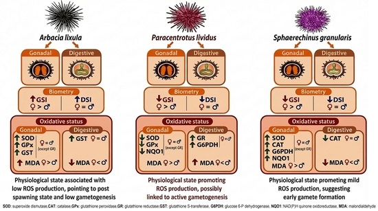
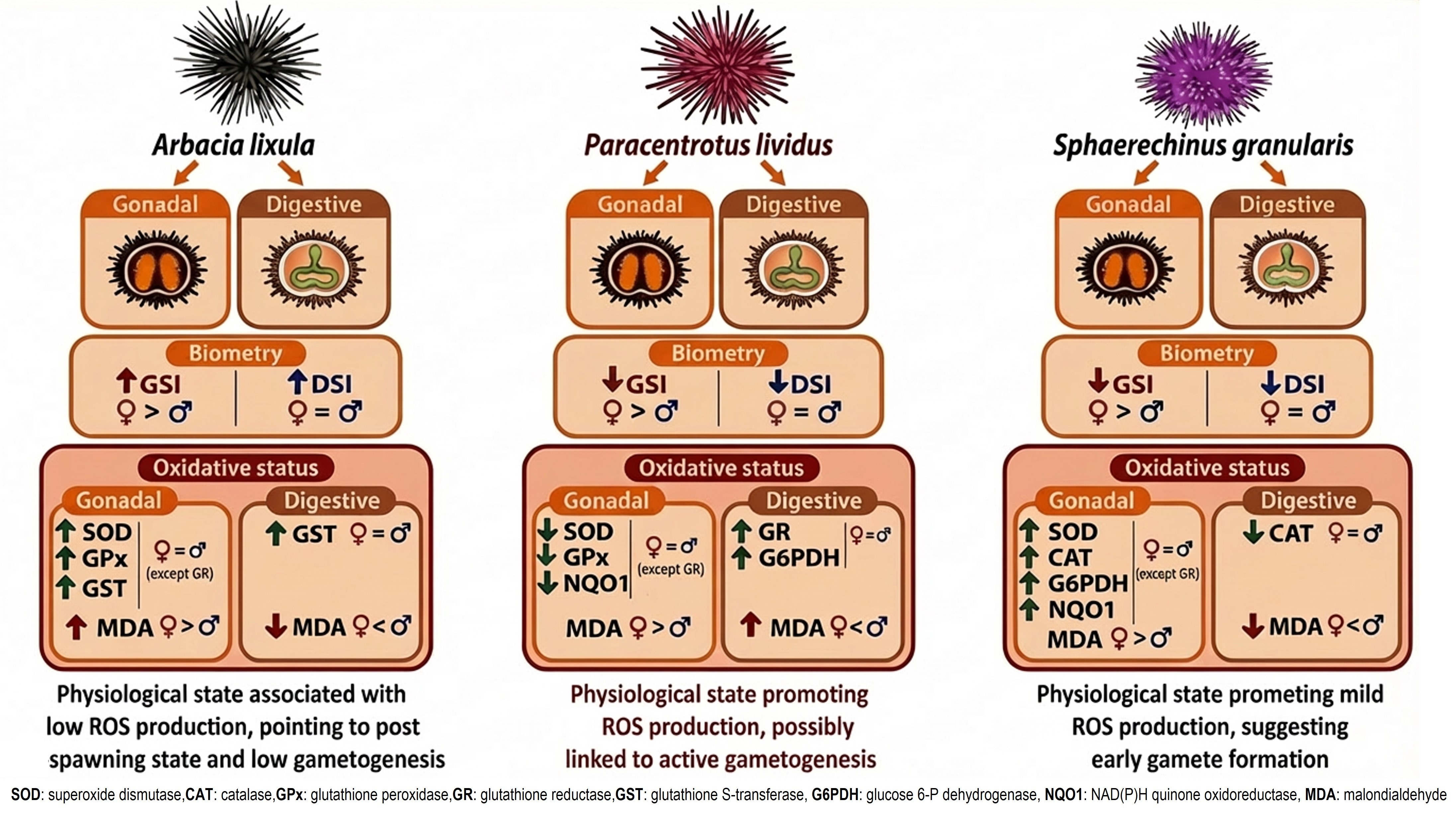
Graphical Abstract

1. Introduction

In marine ecosystems, commercial fishing represents major anthropogenic pressure leading to depleted shellfish populations. Additionally, unusual increases in sea temperature leading to a consequent water tropicalization have facilitated the expansion of invasive species, such as brown algae Rugulopteryx okamurae [1,2] reducing biodiversity by affecting Natura 2000 areas [3]. In parallel, thermal anomalies represent a major environmental stressor capable of triggering acute physiological disturbances in marine organisms, particularly in ectothermic invertebrates whose biological processes are tightly regulated by ambient temperature [4].
Sea urchins (Echinodermata) are key organisms of Mediterranean subtidal ecosystems, playing a central role in controlling benthic algal communities [5]. Paracentrotus lividus (Lamarck 1816) is one of the most exploited species of echinoderms in the Mediterranean Sea due to the traditional gastronomic demand of its gonads and whose populations are being affected up to 65% in southern Spain [5,6]. In Mediterranean seabeds P. lividus coexists with two other species, Arbacia lixula (Linnaeus 1758) and Sphaerechinus granularis (Lamarck 1816), the latter less commonly harvested but with the decline of P. lividus populations, it has become an alternative for local consumption. Due to the critical situation of sea urchin populations in the south of Spain, the authorities decreed the closure of the fishery for the common sea urchin P. lividus within this area [7]. To this sense, the General Fisheries Commission for the Mediterranean (GFCMs) of the Food and Agriculture Organization of the United Nations (FAO) highlights the need to deal with the decline in natural stocks and the relevance of evaluating and preserving reproductive potential of sea urchins [8].
Sea urchins have been widely used as biological markers to assess the effects of both classic and emerging environmental stressors and seawater alterations, such as pH, chemical pollutants or microplastics [9,10]. Due to the thermophilic nature of their larvae, biological processes can be activated by abiotic signals that may depend primarily on photoperiod and temperature [11].
In marine invertebrates, seasonal variations in metabolic rate are assumed to entail corresponding alterations in reactive oxygen species (ROS) formation [12]. Gonadal maturation is a seasonal physiological process that involves the mobilization of energy reserves for gametogenesis, entailing a metabolic activation that can trigger the production of reactive oxygen species (ROS) [13]. In sea urchins, a high antioxidant response has been demonstrated during the stages associated with the peak of gamete formation [10] which could also occur as a defense strategy during the growing phase of gametogenesis, to ensure good offspring quality [9]. Thus, the molecular mechanism of ROS in sea urchin gonads involves a delicate balance between essential signaling for maturation, environmental stress-induced damage, and robust antioxidant defense mechanisms [4]. On the other hand, sea urchin gonads act also as energy reserve tissue [14], establishing a clear dependence on digestive function and depending on external factors like food availability, interspecific competition or anthropogenic pollutants that can affect metabolic pathways and promote oxidative processes [15].
Unchecked ROS accumulation can impair metabolic enzyme activity, reduce ATP production, disrupt protein structure, and compromise cellular membrane integrity, ultimately promoting apoptotic events [16]. To protect tissues from the harmful effects of ROS and mitigate oxidative damage, organisms have a complex antioxidant defense system. Enzymatic defenses include superoxide dismutase (SOD) and catalase (CAT), with the ability to remove superoxide anion and hydrogen peroxide, respectively. Glutathione peroxidase (GPX) provides protection by detoxifying hydrogen peroxide and organic peroxides. Glucose-6-phosphate dehydrogenase (G6PDH) is a crucial enzyme providing NADPH, essential for regeneration of glutathione, by glutathione reductase (GR) activity, which converts oxidized glutathione (GSSG) into its reduced form (GSH), neutralizing the damaging effects of free radicals. The enzymes NQO1 (previously defined as DT-diaphorase) and GST have both been reported to increase their activity in aquatic organisms under the effect of pollutants [17,18]. In this context, the assessment of oxidative status using biomarkers of damage and defense (e.g., MDA, total antioxidant capacity, and antioxidant enzymes) has been proposed as a key indicator of physiological status in marine invertebrates, as it integrates the response to multiple environmental stressors and links it to reproductive performance parameters [19].
Since physiological processes of reproduction promote higher energy costs associated with a metabolic activation, the aim of this study is to conduct a comparative study that correlates antioxidant status markers and gonadal maturation among three species of sea urchin from the Mediterranean Sea coast under the same environmental conditions. Integrative physiological and biochemical studies will contribute to the knowledge of invertebrate physiology and could be useful to predict future trends during the reproductive period and develop strategies aimed to promote conservation strategies of natural populations and sustainable fishery management plans.

2. Materials and Methods

2.1. Specimens and Sample Collection

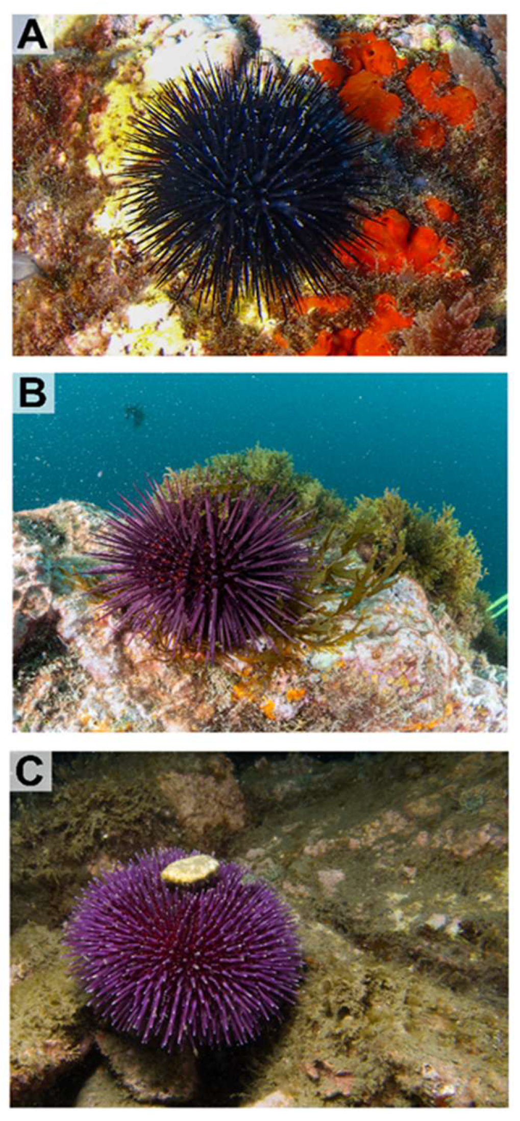
The following three Mediterranean species of sea urchin present in the rocky infralittoral zone of the Calahonda (Granada, Spain; 36°42′09.5″ N, 3°24′43.1″ W) were sampled: Arbacia lixula (1–3 m depth), Sphaerechinus granularis (1–7 m depth), and Paracentrotus lividus (1–3 m depth) (Figure 1). The sampling point is significant due to its biological diversity and hosts one of the richest and most diverse underwater fauna and flora heritages in Europe.
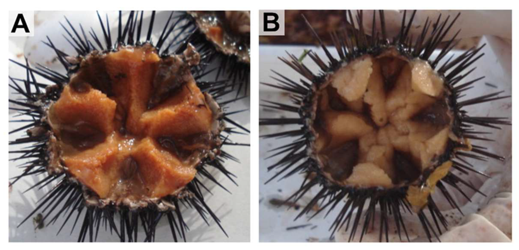
The samples were collected manually from the natural environment in May by scuba diving. Water condition was 17 °C temperature, 37 ‰ salinity, and dissolved oxygen levels above saturation. A total of six individuals of each species (3 males and 3 females) were selected, adjusting the minimum number of collected specimens allowed by responsible authorities for carrying out this experimental assay in nature populations. Animals were measured and weighed, and ex was determined in wet samples of gonads (Figure 2) after they had been crushed and processed by a smear on a slide, checking for the presence of sperm or eggs under optical microscopy (Leica DM 750). Gonadal and digestive tissues were dissected and weighed to obtain the gonadosomatic index (GSI) calculated as (gonad wet weight (g) × 100)/total wet weight (g) and the digestive somatic index (DSI) as (digestive wet weight (g) × 100)/total wet weight (g) according to Martínez-Pita et al. [20]. Thereafter gonadal and digestive tissues were immediately frozen in liquid nitrogen and stored at −80 °C until processing.

2.2. Determination of Oxidative Status

Samples of gonads and extragonadal tissue (digestive) were homogenized (Heidolph Instruments, Schwabach, Germany) in 100 mM Tris, 0.1 m EDTA and 0.1% Triton buffer (pH 7.8), at a 1:4 (w/v) ratio. Homogenates were centrifuged at 30,000 g for 30 min at 4 °C (Sigma 3K30, St. Louis, MO, USA), and the supernatant was collected and distributed in aliquots kept at −80 °C.
Superoxide dismutase (SOD; EC 1.15.1.1) activity was analyzed using the McCord and Fridovich [21] method, which is based on an indirect measurement of activity in relation to the degree of inhibition of a control reaction based on cytochrome c reduction. Catalase (CAT; EC 1.11.1.6) activity was determined using the method originally described by Aebi [22], based on the decrease in absorbance resulting from the degradation of H2O2. Glutathione peroxidase (GPx; EC 1.11.1.9) activity was measured using the Flohé and Günzler [23] method, which is based on an indirect measurement of NADPH oxidation, obtained by coupling with a standard glutathione reductase (GR; EC 1.8.1.7) reaction. GR activity was determined using the Carlberg and Mannervik [24] method, which is based on the decrease in absorbance caused by NADPH oxidation. Glucose 6-phosphate dehydrogenase (G6PDH; EC 1.1.1.49) activity was assayed measuring the change in absorbance due to NADPH production following a modified method of Lohr and Waller [25]. The glutathione S-transferase (GST; EC 2.5.1.18) activity of the samples was determined following the method of Frasco and Guilhermino [26] measuring the increase in absorbance due to the formation of a conjugate between glutathione and 2,4-dinitrochlorobenzene. NAD(P)H: quinone oxidoreductase 1 (NQO1; EC 1.6.5.2.) activity was determined using a modified method of Lemaire et al. [27], based on measurement of the decrease in absorbance caused by reduction of 2,6-dichlorophenol indophenol.
For all the enzymes, except SOD, one unit of activity was defined as the amount of enzyme required to transform one μmol of substrate per minute under the measurement conditions. For SOD, one unit of activity was defined as the amount of enzyme required to generate a 50% inhibition in the reduction in cytochrome c.
The enzymatic activities were expressed as specific activity, for which the soluble protein content in the samples was quantified according to Bradford [28]. Finally, lipid peroxidation was measured based on the content of thiobarbituric acid reactive substances (TBARSs) according to a modified method of Buege and Aust [29]. For the determination of TBARS, malondialdehyde (MDA) was used as a standard.
All measurements were performed with a PowerWave microplate spectrophotometer (Bio-Tek Instrument, Inc., Winooski, VT, USA).

2.3. Statistical Analysis

A two-way ANOVA was used to detect differences in means associated with sea urchin species and sex. ANOVA assumptions were checked via Shapiro–Wilk test and Levene test. When homoscedasticity was not met, a logarithmic transformation was applied to the response variable. Significant main effects were followed up by Tukey’s test as post hoc test when appropriate. Benjamini–Hochberg correction was used to adjust p-values and prevent error inflation due to multiple testing. Statistical significance level was set at α = 0.05
Principal Component Analysis (PCA) was performed on gonadal variables as a descriptive technique to examine correlation between measured variables and tendencies at the multivariate level. Digestive tissue variables were excluded due to low adequacy for PCA according to Kaiser–Meyer–Olkin test. Barlett’s test of sphericity was used to test the assumption that the correlation matrix was different from an identity matrix.

3. Results

3.1. Gonadosomatic and Digestive Somatic Index

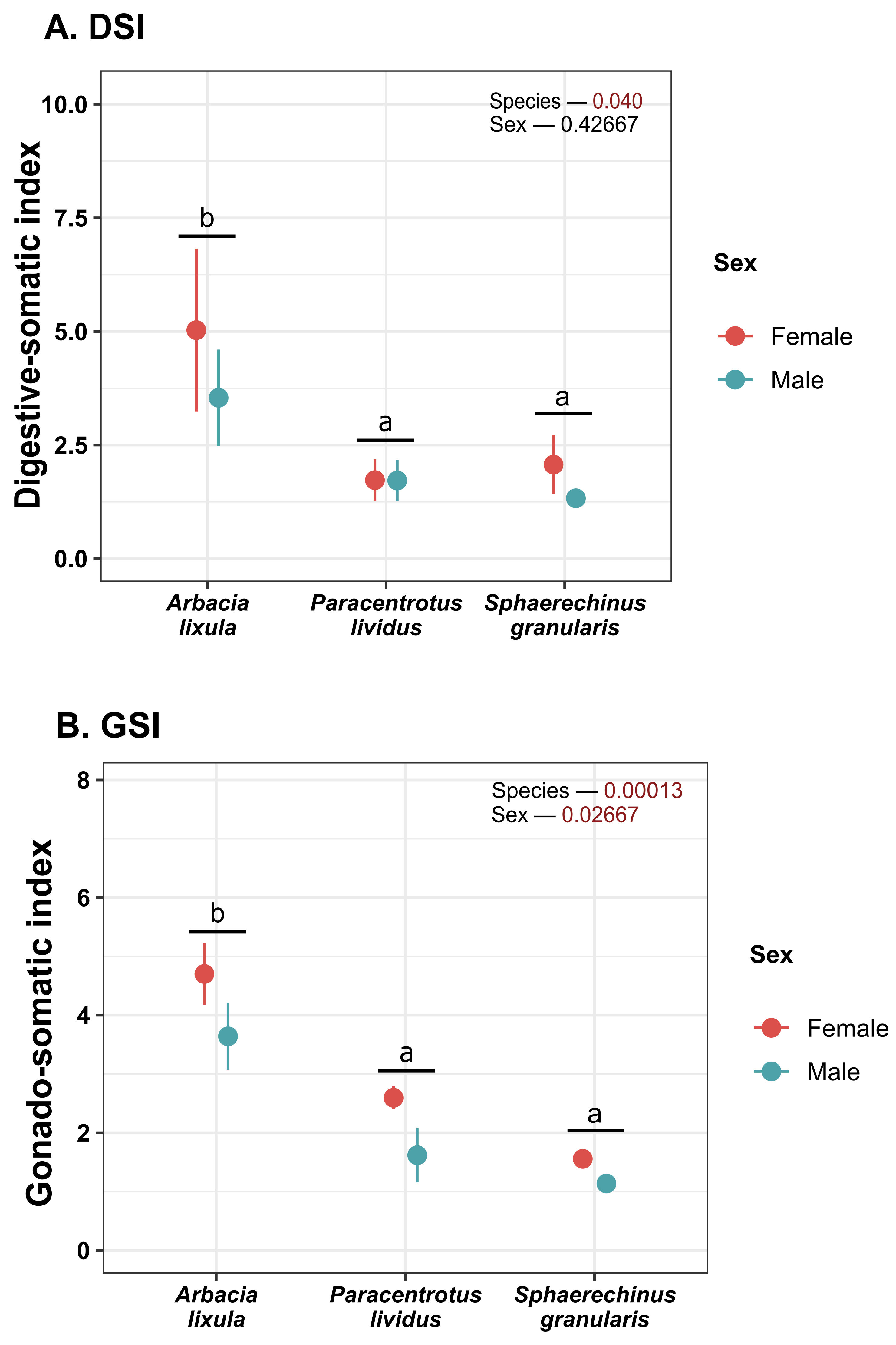
Both gonadosomatic index (GSI) and digestive-somatic index (DSI) were higher in Arbacia lixula than in other species (Figure 3). Female sea urchins also displayed higher GSI than males.

3.2. Gonadal Tissue Oxidative Status

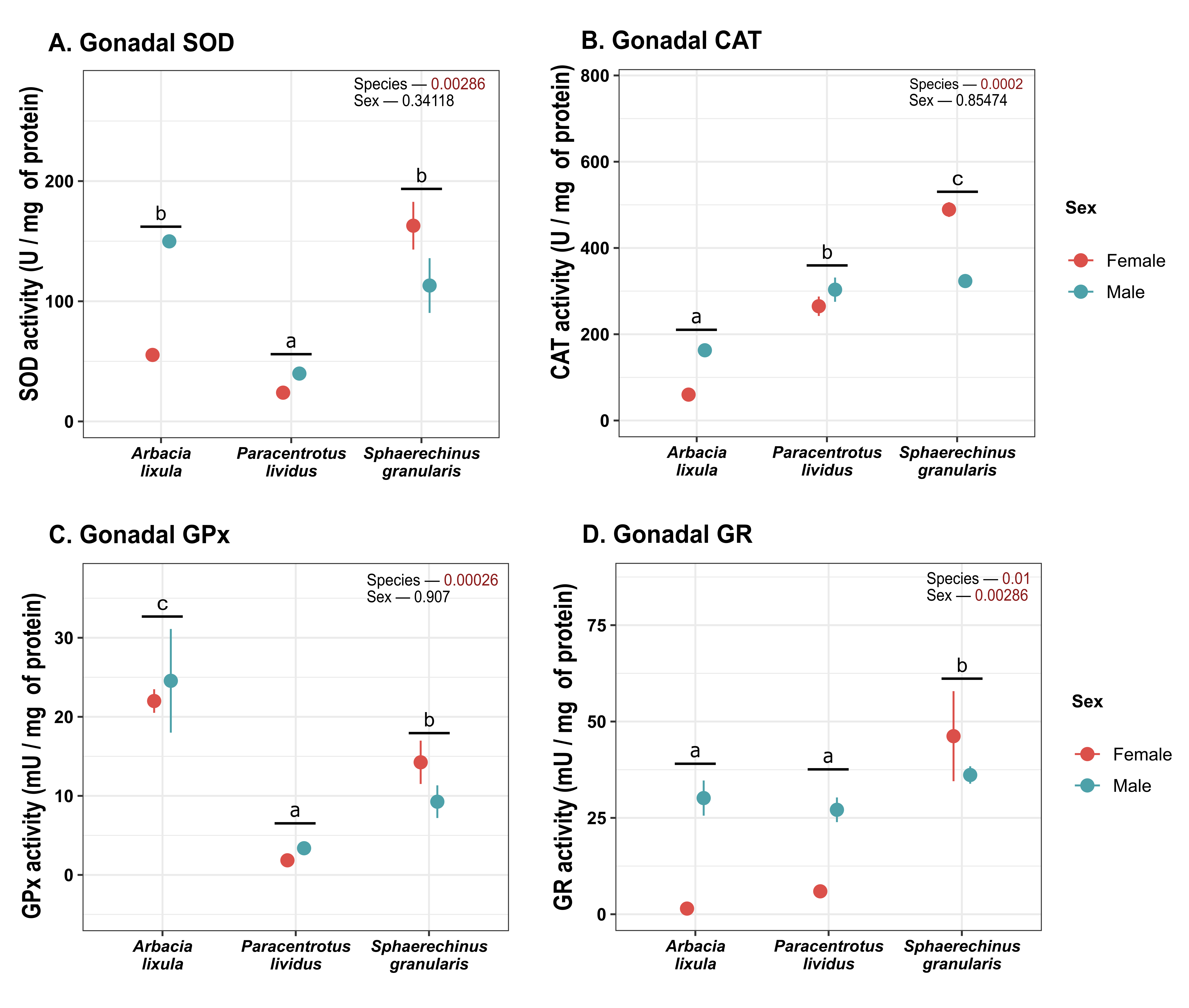
S. granularis and A. lixula had higher SOD activity (Figure 4A) than P. lividus. There were no overall differences between sexes. CAT levels in gonadal tissue varied significantly across species (Figure 4B). A. lixula displayed the lowest activity of the three species, and Sphaerechinus granularis featured the highest. GPx activity was significantly different across species (Figure 4C). A. lixula displayed the highest activity, while P. lividus featured the lowest. Regarding GR (Figure 4D), male sea urchins had higher activity than females and S. granularis featured higher activity than other species.
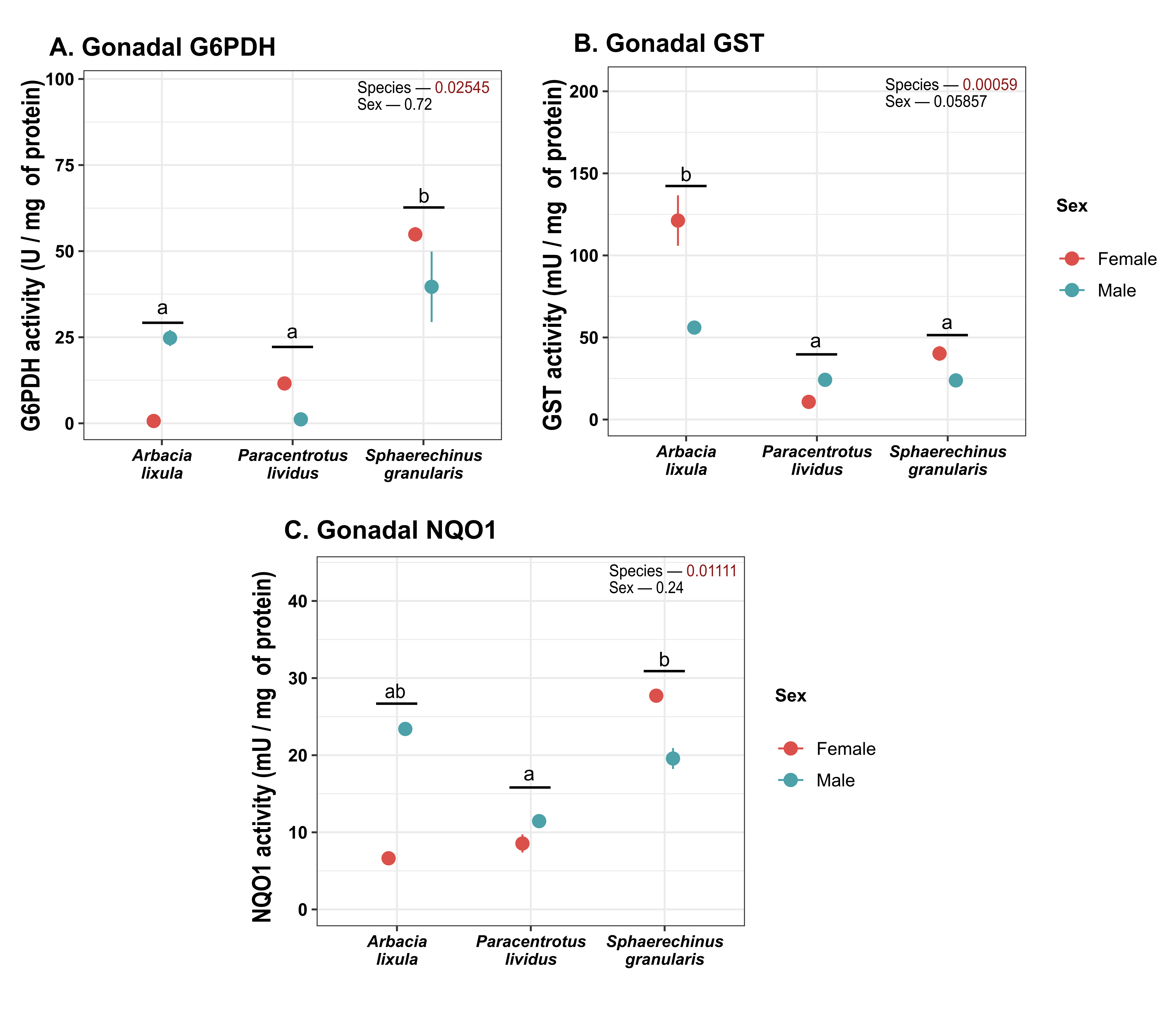
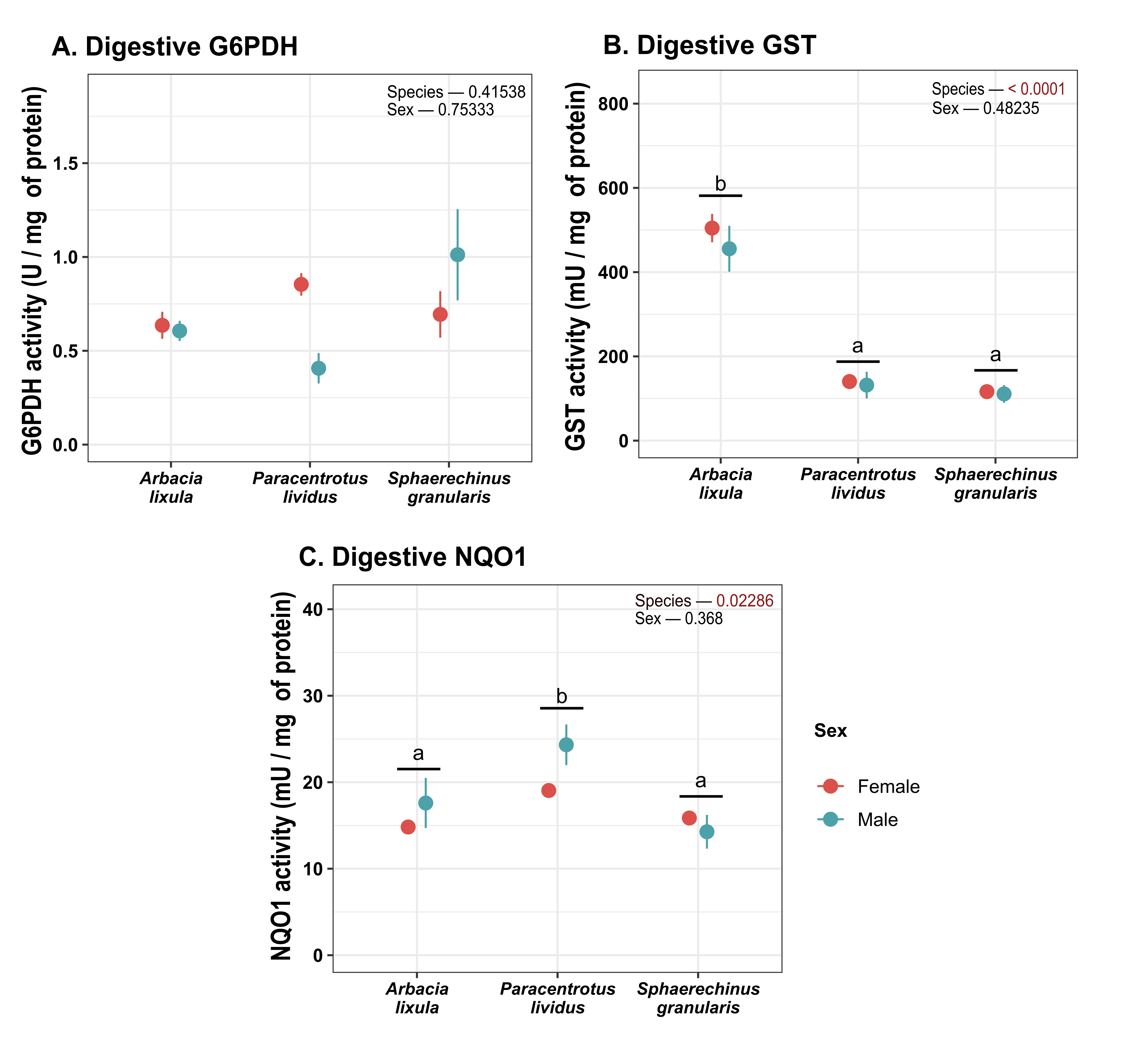
G6PDH activity was higher in S. granularis sea urchins (Figure 5A), while GST activity was found to be higher in A. lixula (Figure 5B). NQO1 activity was higher in S. granularis than in P. lividus (Figure 5C).
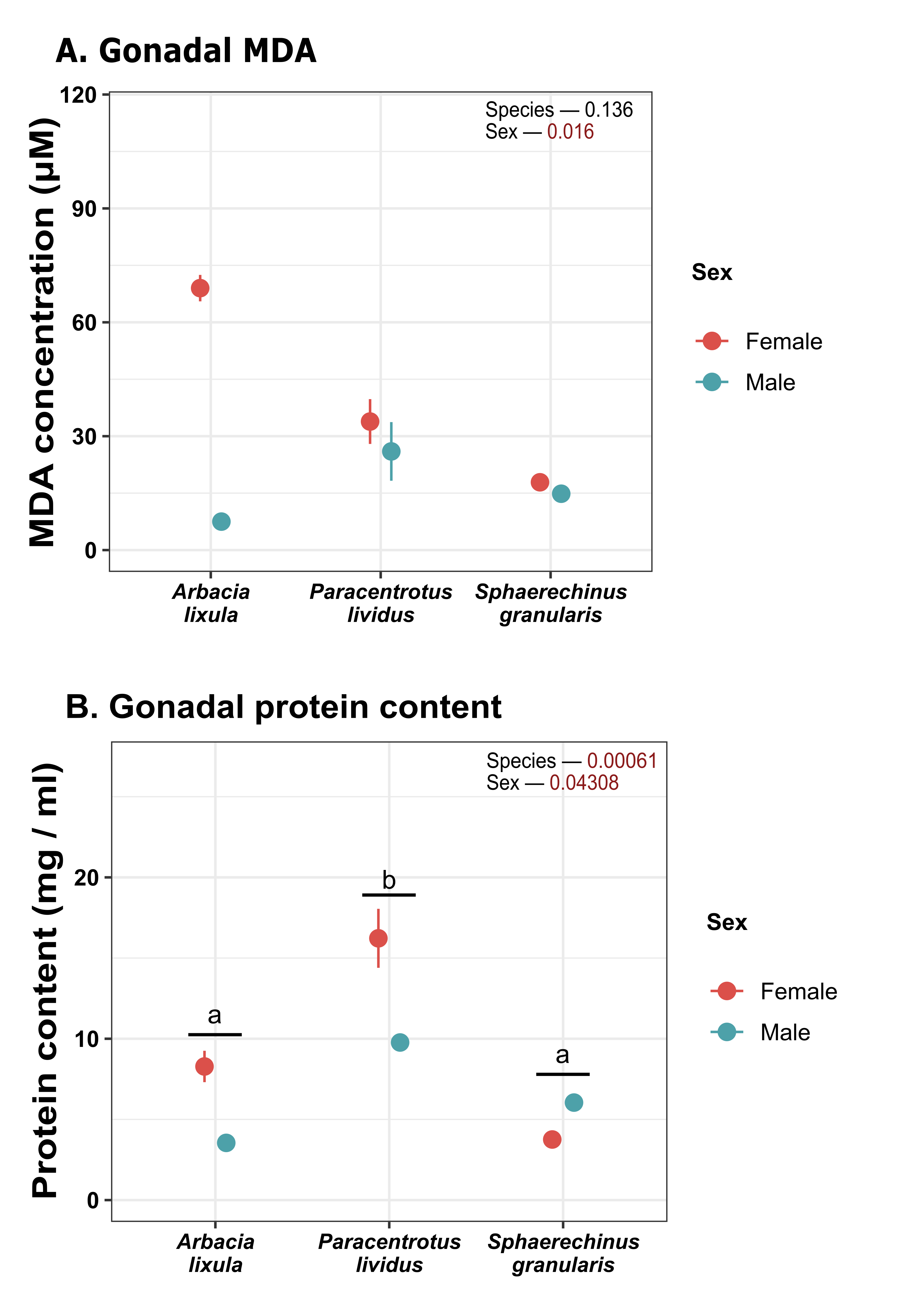
Regarding lipid peroxidation (Figure 6A), females had overall higher MDA content than males in gonadal tissue (Figure 6A). This difference was particularly pronounced in A. lixula, but there were no overall statistical differences between species. Soluble protein levels in gonadal tissue (Figure 6B) were generally higher in females, and P. lividus had the highest protein content for both sexes (16.22 mg/mL in females and 9.77 mg/mL in males).

3.3. Digestive Tissue

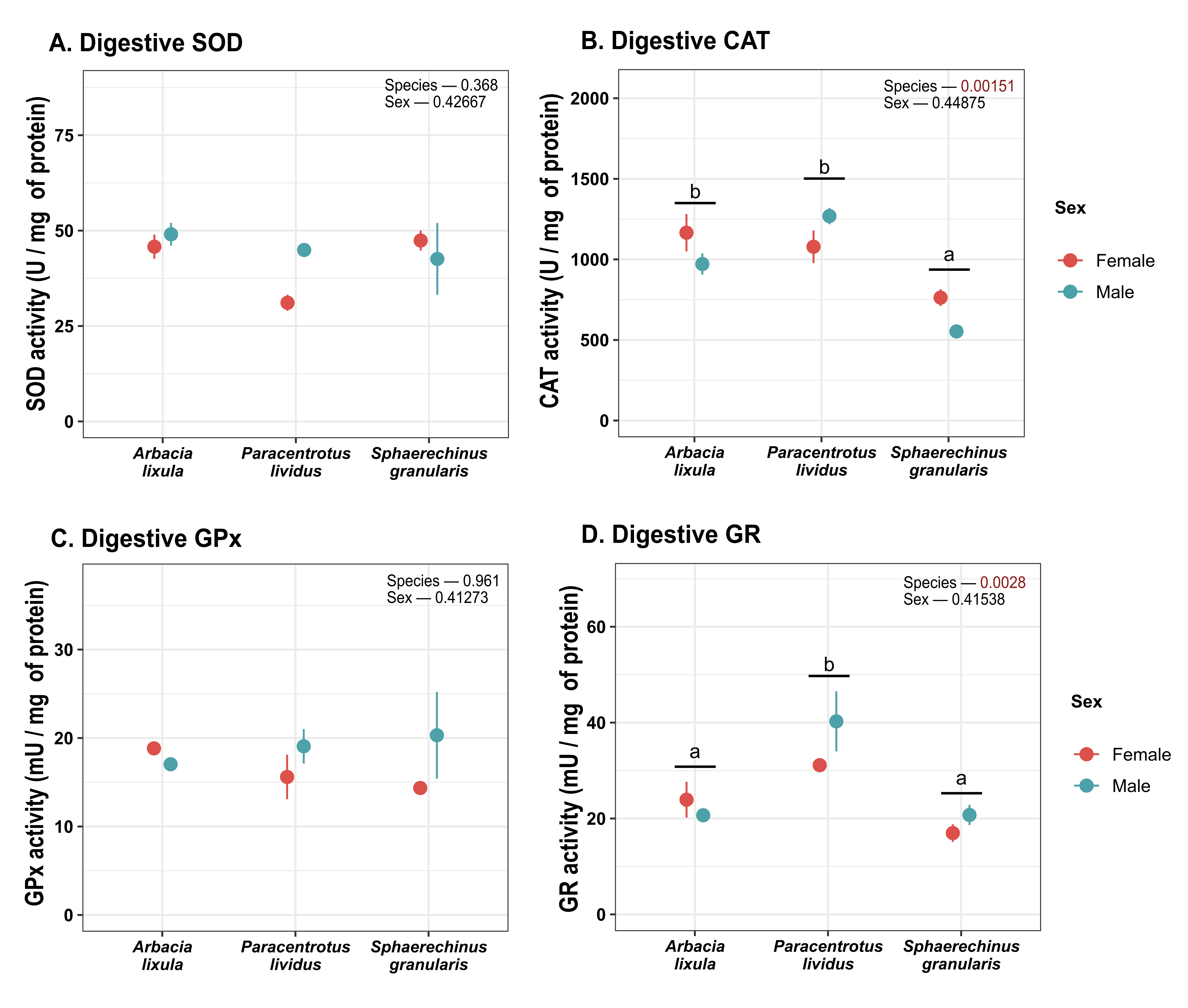
Catalase activity in digestive tissues (Figure 7) was found to be lower for S. granularis sea urchins. In contrast, neither SOD, GPx nor G6PDH activity differed between sea urchin species or sexes in digestive tissue. GR activity was significantly higher for P. lividus (Figure 7 and Figure 8). GST activity (Figure 8) was higher in A. lixula than other sea urchin species. P. lividus sea urchins were also found to display higher NQO1 activity (Figure 8) than other species.
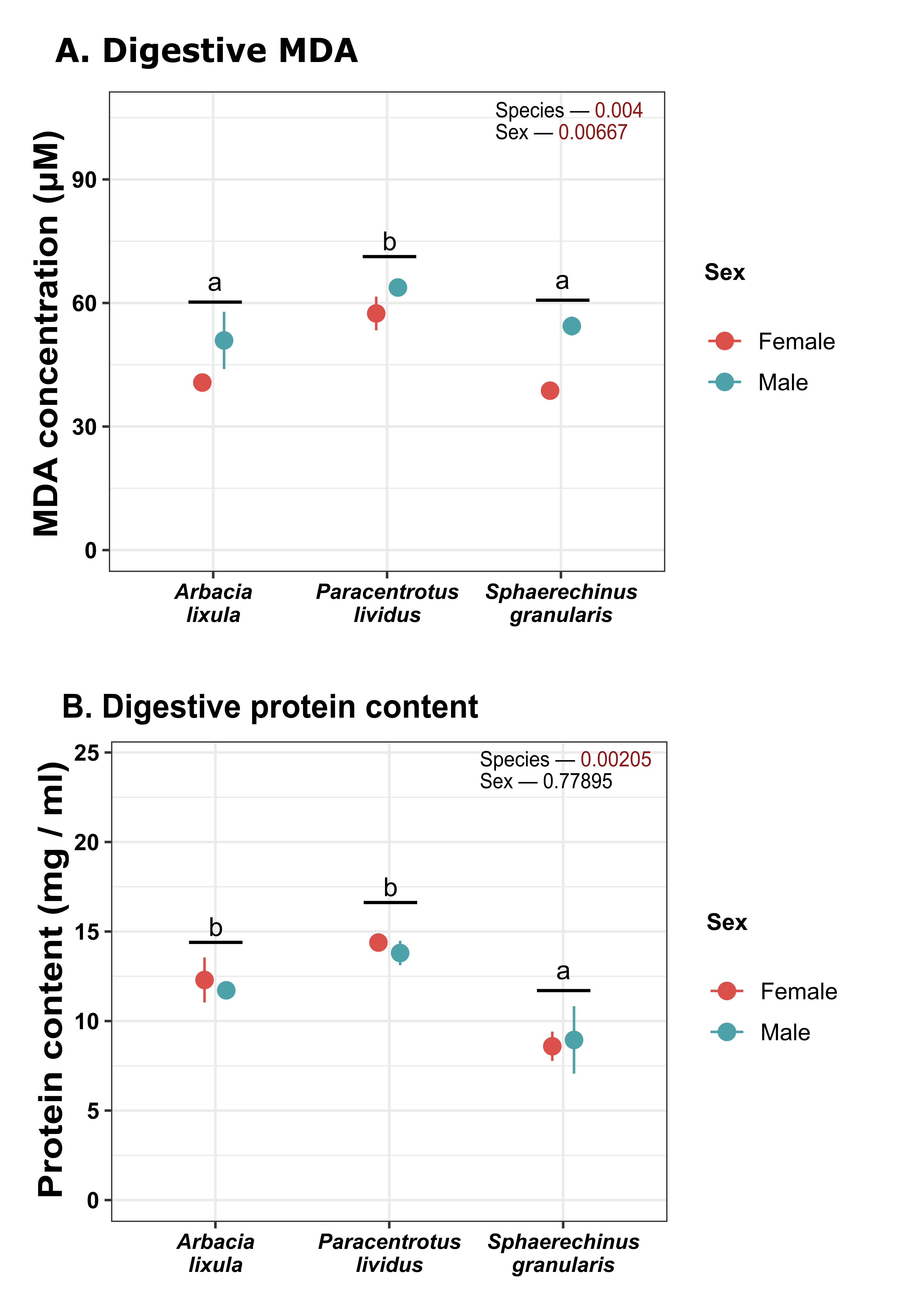
Regarding lipid peroxidation (Figure 9), male sea urchins overall featured higher MDA concentration and there were also differences across species, as P. lividus displayed higher peroxidation levels. Soluble protein (Figure 9) content was found to be lower in S. granularis.

3.4. Principal Component Analysis

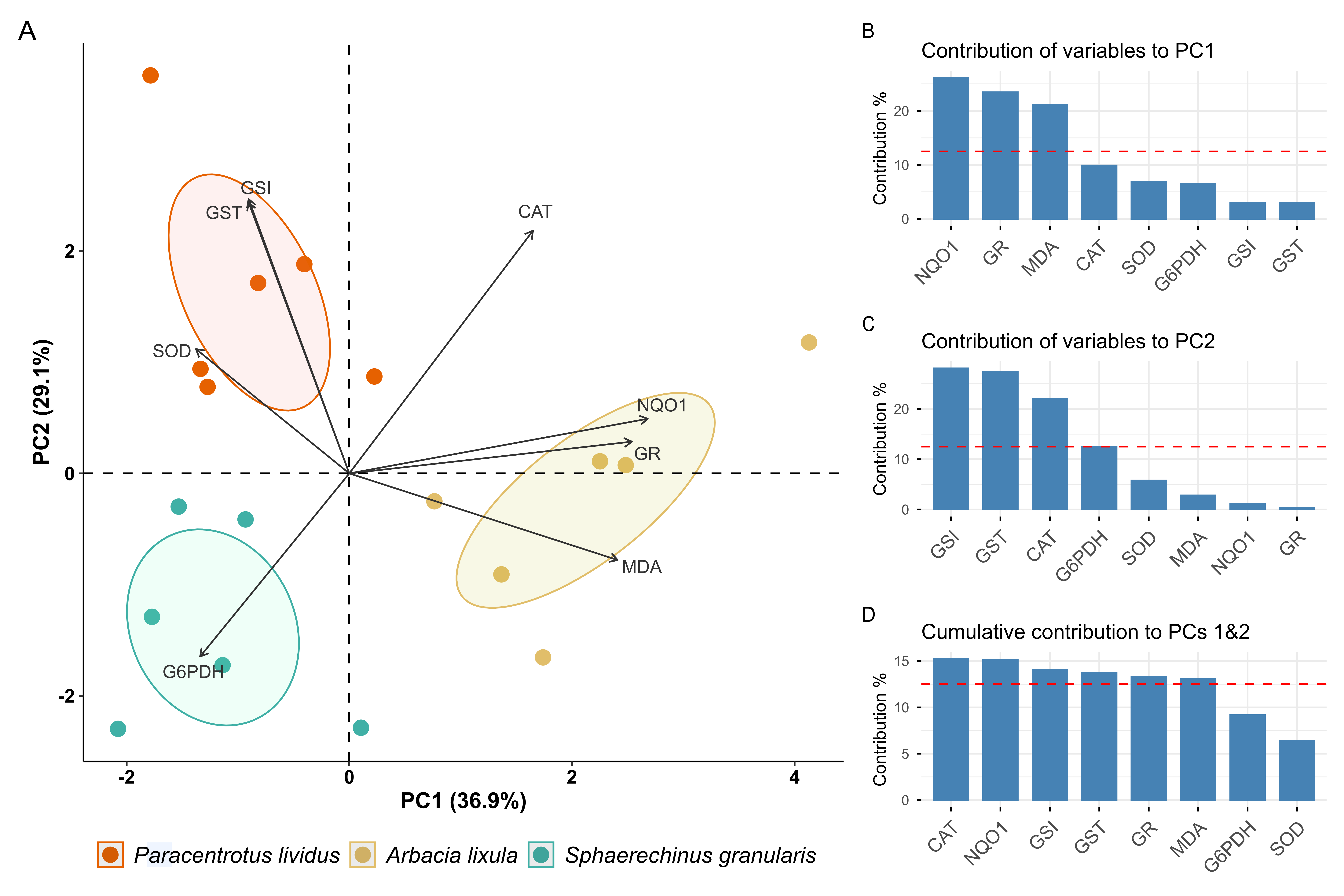
Principal Component Analysis (PCA) was performed on measured gonadal parameters to explore correlation between variables, excluding GPx based on low KMO score that would impact the analysis. Figure 10A illustrates the biplot based on the first two principal components (PC1 and PC2). Observations were grouped based on species, and variable vectors were overlaid on top of the biplot (Figure 10A). PC1 explained 36.9% of initial variance, while PC2 accounted for 29.1% of it. Together, they cumulatively explained 66% of data variance. The histogram in Figure 10B shows variables contribution to PC1, which was mainly spread among NQO1, GR and MDA. All of these variables showed a higher contribution than expected by chance (dashed line) and contributed positively to PC1, indicating positive correlation among them. On the other hand, GSI, GST, CAT and G6PDH were the major contributors to PC2 (Figure 10C), and only G6PDH contributed negatively, indicating negative correlation with GST, CAT and GSI. Overall, CAT, NQO1, GST, GSI, GR and MDA showed high cumulative influence on both PC1 and PC2 (Figure 10D).

4. Discussion

In recent years, physical and chemical changes in the environment, invasive species, and anthropogenic activity have had a negative impact on marine ecosystems and, consequently, on invertebrate species such as sea urchins, which play a crucial role in regulating algal populations due to their position as primary consumers in the food web [30,31].
In the development of restorative actions for aquatic organisms, the assessment of physiological response under particular environmental conditions could be a suitable tool for establishing optimal culture conditions, as was previously evaluated in other invertebrates such as sea anemone [19].
Most echinoderm species show remarkable natural fluctuations which may be related to the regulation of their reproductive processes by external factors [32]. Environmental conditions play a critical role in the maturation, reproduction, and development of sea urchins, with water temperature, food availability, and pH (acidification) being the most influential factors [33]. These factors determine the timing of spawning, the rate of gonadal maturation, and larval quality. On the other hand, several studies have stated that sea urchin response to environmental changes can manifest through changes in oxidative status parameters associated with an adaptive response [9,10].
During the reproductive cycle of sea urchins, the gonads undergo macroscopic changes in size and histological organization and experience a series of established stages after spawning as follows: (i) vitellogenesis and accumulation of energy reserves in specialized cells called nutritive phagocytes; (ii) gamete formation and descent of nutritive phagocytes; (iii) gamete accumulation (pre-spawning) and release (spawning); and finally (iv) gonadal reconstitution based on the formation of nutritive covers. These four stages, which are easily recognizable in females, are reduced to the following three stages in males: (I) accumulation of reserves, (II) formation of gametes, and (III) accumulation of gametes and release of gametes [34,35]. The gonadosomatic index (GSI) of mature sea urchins is a critical indicator of reproductive development [36].
In this study, the differences displayed in GSI could initially be attributed to the different degrees of maturation between species at the time of collection. However, similar behavior displayed in the digestive somatic index (DSI) among species leads us to believe that these differences could instead be attributable to the different relative body diameter (thus weight) rather than significant changes in gonadal or digestive tissue weight. Therefore, although the GSI measurement may be a criterion of interest to assess the state of gonadal maturation [30], when it comes to establishing a comparison between species at the same point in time, it does not appear to be as consistent. In this regard, the analysis of biochemical markers may be a complementary strategy for understanding the state of maturation of different species from a single environment at a single time point.
Sea urchin gonads, besides having a reproductive function, are also capable of acting as reserve organs, with both processes being closely related [14]. Thus, the analysis of digestive tissue can provide information of interest regarding adaptations aimed at meeting greater or lesser energy demands during maturation stages. Reserve cells in gonads, known as nutritive phagocytes, accumulate proteins, lipids and carbohydrates necessary for gamete formation processes and for their metabolic mobilization as an energy source in case of need due to low food availability. These cells can also reabsorb gametes not expelled after spawning as a source of energy [30].
During the stages of gonadal maturation, the activation of gametogenesis involves energy expenditure from gonadal reserves, which promotes the activation of certain metabolic pathways, increasing oxidative processes [37]. This can promote the formation of reactive oxygen species (ROS) and generate a protective response in organisms based on molecular and enzymatic antioxidant defenses [38,39].
Perez et al. [40] demonstrated a positive correlation between gonadal maturation and lipid soluble antioxidant concentration in marine invertebrates, such as the starfish Anasterias antarctica, as a strategy for oxidative damage prevention and gamete protection through allocation of antioxidants to mature gonads.
This evaluation of antioxidant enzymes in three species of sea urchin present on the Mediterranean coast—A. lixula, P. lividus, and S. granularis—displayed differences that could be attributed to the stage of gonadal maturation each of them was in. It is known that during the peak of gametogenesis, sea urchins maintain higher antioxidant levels [10]. One notable thing was that the black sea urchin, A. lixula, showed significantly higher values of the enzymatic antioxidant activity, glutathione peroxidase (GPx) and glutathione transferase (GST). This could be attributed to the fact that this species was in a gonadal stage in which gamete formation was relevant. Interestingly, for both enzymes activity is necessary reduced glutathione (GSH), which is involved in phase II metabolism that plays a crucial role in intracellular protection against reactive oxygen species [41]. According to Lubos et al. [42] the intracellular content of glutathione depends on environmental factors and is maintained in equilibrium between its utilization and its synthesis, an aspect that could be in agreement with environmental effect on maturation processes. On the other hand, GPx shares with catalase (CAT) the function of removing inorganic peroxides. The low activity of the latter could indicate that GPx would have primarily assumed this role triggered by a physiological stage. This would be consistent with the fact, suggesting that the glutathione redox cycle is an important protective mechanism against mild oxidative stress, being catalase more crucial protecting cells against chronic oxidative stress [43].
On the other hand, it is important to highlight the role of GPx removing organic peroxides, like lipoperoxides. Lipids, such as fatty acids (FAs) and polyunsaturated fatty acids (PUFAs), are present in sea urchin gonads and are essential for their physiological processes [34,44]. Several studies have shown an increase in lipid during the stages of gonadal maturation, which then undergoes a clear decline in the final stages and after spawning [45,46]. In this study, females showed higher MDA values in gonadal tissue. The accumulation of lipids in the female gonads of sea urchins is a sex-specific process directly linked to the reproductive cycle, in which females store significantly higher levels of total lipids and PUFAs compared to males. These lipids, which constitute essential energy sources for oogenesis (egg formation) and larval development, reach their peak level before spawning and declining and decreasing and becoming equal in both sexes after spawning [47]. To this respect, Alonso-Alvarez et al. [48] suggested that reproduction might generate oxidative stress, increasing lipid radical content in mature gonads compared to immature gonads and that, in the long term, could affect the reproductive potential of the gonad. Pérez et al. [37] reported an increase in the concentration of TBARS (thiobarbituric acid reactive substances) and lipid radicals during gametogenesis, associated with a decrease in the levels of fat-soluble antioxidants (tocopherol, carotene and equinenone). The trend to higher MDA values in females and particularly in A. lixula (Figure 6) could be consistent with a greater accumulation of PUFAs, fatty acids with a high propensity for oxidation, justifying in this species an increased antioxidant requirement in agreement with higher values of GPx.
The results suggest that A. lixula, at the time of capture in May, was in a period of gonadal maturation and gamete formation linked to high metabolic activity promoting ROS generation. This agrees with the fact that the gonad growth period of A. lixula on Mediterranean coasts has been reported to occur from winter to spring, with most individuals maturing during May–June and spawning from spring to late summer/early autumn, depending on seasonal temperatures [49]. Also, GSI values in A. lixula in this study are within the same range (around 4–5%) reported by these authors at the same season time [49,50].
The evaluation of antioxidant activity in the gonads of the common sea urchin, P. lividus, curiously revealed a trend opposite to that observed in A. lixula for the same enzymes. This response had already been observed in marine invertebrates such as the mussel Mytilus galloprovincialis, where a progressive decrease in antioxidant activity was observed after egg laying despite the availability of food and higher temperatures [51]. This could be consistent with the fact that P. lividus could be in a post-spawning stage at the time of sampling, supported by Uz et al. [52], reporting that in P. lividus from the northeast coast of Spain had already spawned during the months of May–June. At this stage, the gametogenic processes linked to the activation of ROS-producing metabolic pathways would not be a priority, thus justifying a decrease in endogenous antioxidant defenses. In addition, after gonadal spawning, there is a depletion in lipid energy reserves [47], which would justify the low MDA concentration observed in P. lividus and the relative higher protein concentration in gonads of these animals.
The gonadosomatic index values obtained in P. lividus were consistent with those obtained by Raposo et al. [53] (1.5–3%) in individuals of similar size (diameter of 4–5 cm), under the same seasonality (May–June) and temperature (17 °C) in a post-spawning period, being low when compared to other indices that range from 8 to 12% in periods of maturity prior to spawning. This could be indicative that in the animals, at the time they were collected, spawning had probably already taken place, with the gonads presenting a low number of residual gametes and being in an initial stage of energy reserve accumulation.
The violet or purple sea urchin, Sphaerechinus granularis, is an edible species of increasing commercial importance that coexists in Mediterranean waters with P. lividus and A. lixula, although it is not as abundant as the latter two. The general increased activity in most of the enzymes evaluated (SOD, CAT, GPx GR, G6PDH, and NQO1), which tended to be more pronounced in females, could be consistent with the spawning period for this species, which begins in June and can extend until November, with a peak on the southern coast of the Iberian Peninsula in the months of June–August [20]. This could explain why the animals were still in the middle of gametogenesis, with the consequent tendency to generate ROS [37], supporting the high antioxidant activity in most of the enzymes evaluated. This is consistent with reports by Perez et al. [39] in sea urchin, showing an increased lipid radical/a-TH index during preceding spawning stage that could be associated with a higher metabolism in the lipid phase of the gonads. In this study, MDA levels were low, which could be indicative that the antioxidant response was effective in preventing lipid oxidation.
According to GSI variations associated with gonadal maturation, Martinez-Pita et al. [20] reported values ranging from 3 to 5%. for S. granularis in southern Spain during a similar seasonal period of this study. The lower GI values displayed for animals (less than 2%), given the period and water temperature when animals were sampled, it does not appear to be attributable to gamete release stage. However, these GI values agree with Vafidis et al. [54] in specimens from the Aegean coast under the same seasonality and which were in previous stages with gamete formation and maturation. This seems more likely and would justify the increase in gonadal metabolic activity and, therefore, the antioxidant enzyme response. According to the same authors, the trend to more pronounced antioxidant response in females could be due to the advance in maturation processes compared to males. In addition, female sea urchins generally have a higher gonadosomatic index (GSI), often with a higher lipid and carotenoid content, as they invest more energy in egg production [55]. This would support the fact that in this study, females, regardless of species, showed significantly higher GSI than males.
Another factor that could contribute to the low gonadal growth of S. granularis is the reduced availability of food for this species [20,56,57]. Itshould be noted that in Mediterranean waters, the predominant species are A. lixula and P. lividus, whose diet is based on encrusting coralline algae and marine phanerogams and algae from rocky areas, respectively [6,58]. S. granularis inhabits deeper areas and prefers feeding on decaying plant material and epiphytic algae. Low food availability may influence gonad size in S. granularis. These results suggest investment in gonadal growth under high hydrodynamics and probably less favorable food supply [54]. In this sense, food supply would be key in promoting the physiological activity of the digestive tissue, to ensure the replenishment of energy reserves, in which the gonad, as mentioned above, would play a prominent role as reserve tissue.
The results of the assessment of oxidative status in intestinal tissue were not as relevant as those observed in the gonads, nor did they show any sex-related effect. This was to be expected, as metabolic changes in digestive tissue are less pronounced than in gonads. However, certain activation of oxidative processes (CAT, GR and NQO1) linked to a trend to higher lipid hydroperoxides in P. lividus could be consistent with an activation of digestive activity. The fact that this species, as previously hypothesized, could be in a post-spawning period would have justified a metabolic activation for gonadal regeneration and energy reserve replenishment, triggering oxidative processes and antioxidant requirements [59]. On the other hand, during stages prior to egg laying and gonadal maturation, the intestinal function for the regeneration of energy reserves is not yet a priority [34,35,60]. This would be consistent with the lowest levels observed in the digestive tract of the other two species (A. lixula and S. granularis) in terms of lower antioxidant activity and lipid peroxide concentration.
GST activity is an enzyme mainly involved in detoxification processes through the formation of soluble conjugates [61] and previously it has been reported to be a good indicator of environmental stress in sea urchin [62]. A. lixula usually colonizes rocky areas at shallower depths and in this study the sampling point was a dock area where there are hydrocarbons from fuels and agricultural pesticides, favoring the accumulation of xenobiotic agents associated with anthropogenic activity. The marked increase displayed in the intestinal tissue of A. lixula, regardless of sex, is consistent with previously observed for the gonads. This response could be consistent with the reported high capacity of this species to bioaccumulate contaminants [62]. The fact that both tissues (gonadal and digestive) had a similar behavior could reflect the greater sensitivity of this species, explaining the oxidation of gonadal lipids manifested by higher levels of MDA [10].
Principal Component Analysis (PCA) performed on measured gonadal parameters showed that major contributors to PC1 (NQO1 activity, GR activity, and malondialdehyde concentration) are important drivers of the differences between the sampled sea urchins. Similarly, variables such as GST activity, CAT activity and gonadosomatic index (GSI), which were the major contributors to PC2, also are highlighted. Some antioxidant enzyme activities showed positive correlation, such as GR and NQO1, but these were also correlated with malondialdehyde (MDA), indicating that samples that had higher MDA levels also had higher activities of these two enzymes. SOD and GST activity, however, were negatively correlated with lipid peroxidation marker MDA, as well as GSI. There also appears to be some degree of negative correlation between CAT and G6PDH activity levels. P. lividus observations were defined by their higher GST activity and GSI and low MDA content. S. granularis sea urchins featured high G6PDH but low antioxidant activity and lipid peroxidation. Finally, A. lixula was the species with the higher spread in PC1, which suggests a highly variable oxidative state between males and females. They were characterized by higher levels of MDA, NQO1 activity, and GR activity compared to other species.
It is important to note that this study has put forward hypotheses that will need to be further supported in subsequent studies, given certain limitations such as the small number of specimens used in the evaluated parameters. Current regulation regarding sea urchin capture has been a limiting factor, and this, combined with the lack of phenotypic differences between sexes, has meant that specimens were collected only until a statistically minimum number was reached, ensuring a balanced sample size for each sex within each species.
On the other hand, despite the aim of this study being providing a comparative overview of the physiology of gonadal maturation in three species coexisting in the same location and at the same point in time, this objective must be further explored in more extensive studies including a seasonal analysis for each of the species studied, and supplemented with a histological evaluation of the gonads to support gonadal maturation stages described.

5. Conclusions

In short, taking a general look at the results obtained of the three species studied, A. lixula and S. granularis appear to show at the time of collection an antioxidant behavior consistent with a stage of gonadal maturation prior to spawning, in which metabolic processes would be activated by mobilization of energy reserves for gametogenesis. In contrast, P. lividus could be in a period of replenishment of energy reserves after spawning. The analysis of oxidative status markers could be a complementary tool for the evaluation of gonadal maturation status in sea urchins. The different behavior in the three species collected from a single environment at a single time point could be related to an endogenous component associated with their biology modulating gonadal processes.

Author Contributions

Conceptualization, C.E.T. and F.H.; methodology, P.A.Á., M.E.D.-C. and C.E.T.; software, A.C.; formal analysis, P.A.Á., M.E.D.-C., A.C. and C.E.T.; investigation, P.A.Á. and F.H.; resources, F.H; writing—original draft preparation, P.A.Á., A.C. and C.E.T.; writing—review and editing, P.A.Á., A.P.-J., E.E.R.-P. and C.E.T.; supervision, C.E.T. All authors have read and agreed to the published version of the manuscript.

Funding

This research received no external funding.

Institutional Review Board Statement

Ethical approval was not required for this study because sea urchins are included in invertebrate group and therefore falls outside the scope of the Directive 2010/63/EU on the protection of animals used for scientific purposes and its Spanish transposition through R.D. 53/2013, which applies only to vertebrates and cephalopods.

Informed Consent Statement

Not applicable.

Data Availability Statement

The raw data supporting the conclusions of this article will be made available by the authors on request.

Acknowledgments

The authors would like to thank Alejandro José Ibáñez Yuste for his contribution in taking the photographs of the sea urchin species in their natural environment used in this study.

Conflicts of Interest

The authors declare no conflicts of interest.

Abbreviations

The following abbreviations are used in this manuscript:
ROSReactive oxygen species
SODSuperoxide dismutase 
CATCatalase 
GPxGlutathione peroxidase 
GRGlutathione reductase 
GSTGlutathione s-transferase 
G6PDHGlucose 6-phosphate dehydrogenase
NQO1NAD(P)H: quinone oxidoreductase 
MDAMalondialdehyde
TBARSThiobarbituric acid reactive substances
GSIGonadosomatic index
DSIDigestive somatic index
PCAPrincipal Component Analysis 

References

  1. Borriglione, M.; Ruitton, S.; Boyer, F.; Thibault, D.; Blanfuné, A.; Guillemain, D.; Verlaque, M.; Boudouresque, C.F.; Thibaut, T. Impact of the invasive brown alga Rugulopteryx okamurae on the benthic communities in the Northwestern Mediterranean Sea. Estuar. Coast. Shelf Sci. 2024, 310, 109010. [Google Scholar] [CrossRef]
  2. Faria, J.; Prestes, A.C.; Moreu, I.; Martins, G.M.; Neto, A.I.; Cacabelos, E. Arrival and proliferation of the invasive seaweed Rugulopteryx okamurae in NE Atlantic islands. Bot. Mar. 2022, 65, 45–50. [Google Scholar] [CrossRef]
  3. García-Gómez, J.C.; Sempere-Valverde, J.; González, A.R.; Martínez-Chacón, M.; Olaya-Ponzone, L.; Sánchez-Moyano, E.; Ostalé-Valriberas, E.; Megina, C. From exotic to invasive in record time: The extreme impact of Rugulopteryx okamurae (Dictyotales, Ochrophyta) in the strait of Gibraltar. Sci. Total Environ. 2020, 704, 135408. [Google Scholar] [CrossRef] [PubMed]
  4. Johnstone, J.; Nash, S.; Hernandez, E.; Rahman, M.S. Effects of elevated temperature on gonadal functions, cellular apoptosis, and oxidative stress in Atlantic sea urchin Arbacia punculata. Mar. Environ. Res. 2019, 149, 40–49. [Google Scholar] [CrossRef] [PubMed]
  5. Bernal-Ibáñez, A.; Castilla-Gavilán, M.; Sumariva, E.G.; Hachero-Cruzado, I. Invasion context matters: Vulnerability of the sea urchin Paracentrotus lividus to ingestion of Rugulopteryx okamurae increases with invasion time. Mar. Pollut. Bull. 2025, 214, 117826. [Google Scholar] [CrossRef]
  6. Toso, A.; Necci, F.; Martines, A.; Lacorte, R.; Toso, Y.; Gianguzza, P.; Deidun, A.; Ungaro, N.; Costantino, G.; Caforio, M.; et al. Overfishing and sea warming drive the collapse of Paracentrotus lividus. Sci. Rep. 2025, 15, 18733. [Google Scholar] [CrossRef]
  7. BOJA. Orden de 17 de Octubre de 2023, por la Que se Cierra la Pesquería de Erizos y Anémonas en el Litoral de Andalucía, Debido a la Situación Crítica de Sus Poblaciones y el Impacto Generado por la Invasión del Alga Rugulopterix okamurae. Junta de Andalucía. 2023. Available online: https://www.juntadeandalucia.es/boja/2023/204/33 (accessed on 14 December 2025).
  8. FAO. Guidelines on Aquaculture Restocking and Stock Enhancement; FAO: Rome, Italy, 2023. [Google Scholar] [CrossRef]
  9. Asnicar, D.; Stranci, F.; Monti, S.; Badocco, D.; Marčeta, T.; Munari, M.; Marin, M.G. Investigating intraspecific variability in the biological responses of sea urchins (Paracentrotus lividus) to seawater acidification. Environ. Sci. Pollut. Res. 2024, 31, 51687–51701. [Google Scholar] [CrossRef]
  10. Amri, S.; Samar, M.F.; Sellem, F.; Ouali, K. Seasonal antioxidant responses in the sea urchin Paracentrotus lividus (Lamarck 1816) used as a bioindicator of the environmental contamination in the South-East Mediterranean. Mar. Pollut. Bull. 2017, 122, 392–402. [Google Scholar] [CrossRef]
  11. Leach, T.S.; Hofmann, G.E. Marine heatwave temperatures enhance larval performance but are meditated by paternal thermal history and inter-individual differences in the purple sea urchin, Strongylocentrotus purpuratus. Front. Physiol. 2023, 14, 1230590. [Google Scholar] [CrossRef]
  12. Abele, D.; Puntarulo, S. Formation of reactive species and induction of antioxidant defence systems in polar and temperate marine invertebrates and fish. Comp. Biochem. Physiol.-A Mol. Integr. Physiol. 2004, 138, 405–415. [Google Scholar] [CrossRef]
  13. Lesser, M.P. Oxidative stress in marine environments: Biochemistry and physiological ecology. Annu. Rev. Physiol. 2006, 68, 253–278. [Google Scholar] [CrossRef] [PubMed]
  14. Hughes, A.D.; Kelly, M.S.; Barnes, D.K.; Catarino, A.I.; Black, K.D. The dual functions of sea urchin gonads are reflected in the temporal variations of their biochemistry. Mar. Biol. 2006, 148, 789–798. [Google Scholar] [CrossRef]
  15. Beaulieu, M.; Costantini, D. Biomarkers of oxidative status: Missing tools in conservation physiology. Conserv. Physiol. 2014, 2, cou014. [Google Scholar] [CrossRef] [PubMed]
  16. Redza-Dutordoir, M.; Averill-Bates, D.A. Activation of apoptosis signalling pathways by reactive oxygen species. Biochim. Biophys. Acta-Mol. Cell Res. 2016, 1863, 2977–2992. [Google Scholar] [CrossRef]
  17. Do, T.; Vaculciakova, S.; Kluska, K.; Peris-Díaz, M.D.; Priborsky, J.; Guran, R.; Krężel, A.; Adam, V.; Zitka, O. Antioxidant-related enzymes and peptides as biomarkers of metallic nanoparticles (eco)toxicity in the aquatic environment. Chemosphere 2024, 364, 142988. [Google Scholar] [CrossRef]
  18. Ross, D.; Siegel, D. The diverse functionality of NQO1 and its roles in redox control. Redox Biol. 2021, 41, 101950. [Google Scholar] [CrossRef]
  19. Coll, A.; Rufino-Palomares, E.E.; Ramos-Barbero, M.; Ortiz-Maldonado, A.E.; Pantoja-Echevarría, L.M.; González-Ordóñez, I.; Pérez-Jiménez, A.; Trenzado, C.E. Effects of environmental factors on the oxidative status of Anemonia viridis in aquaculture systems. Comp. Biochem. Physiol. Part B Biochem. Mol. Biol. 2025, 275, 111042. [Google Scholar] [CrossRef]
  20. Martínez-Pita, I.; Sánchez-España, A.I.; García, F.J. Gonadal growth and reproduction in the sea urchin Sphaerechinus granularis (Lamarck 1816) (Echinodermata: Echinoidea) in southern Spain. Sci. Mar. 2008, 72, 603–611. [Google Scholar] [CrossRef]
  21. McCord, J.M.; Fridovich, I. Superoxide Dismutase: An enzymic function for erythrocuprein (hemocuprein). J. Biol. Chem. 1969, 244, 6049–6055. [Google Scholar] [CrossRef]
  22. Aebi, H. Catalase in vitro. Methods Enzymol. 1984, 105, 121–126. [Google Scholar] [CrossRef]
  23. Flohé, L.; Günzler, W.A. Assays of glutathione peroxidase. Methods Enzymol. 1984, 105, 114–120. [Google Scholar] [CrossRef]
  24. Carlberg, I.; Mannervik, B. Purification and characterization of the flavoenzyme glutathione reductase from rat liver. J. Biol. Chem. 1975, 250, 5475–5480. [Google Scholar] [CrossRef]
  25. Löhr, G.; Waller, H. Glucose-6-phosphate Dehydrogenase: (Zwischenferment). In Methods of Enzymatic Analysis; Academic Press: New York, NY, USA, 1965; pp. 744–751. [Google Scholar] [CrossRef]
  26. Frasco, M.F.; Guilhermino, L. Effects of dimethoate and beta-naphthoflavone on selected biomarkers of Poecilia reticulata. Fish Physiol. Biochem. 2002, 26, 149–156. [Google Scholar] [CrossRef]
  27. Lemaire, P.; Sturve, J.; Förlin, L.; Livingstone, D.R. Studies on aromatic hydrocarbon quinone metabolism and DT-Diaphorase function in liver of fish species. Mar. Environ. Res. 1996, 42, 317–321. [Google Scholar] [CrossRef]
  28. Bradford, M. A Rapid and Sensitive Method for the Quantitation of Microgram Quantities of Protein Utilizing the Principle of Protein-Dye Binding. Anal. Biochem. 1976, 72, 248–254. [Google Scholar] [CrossRef] [PubMed]
  29. Buege, J.A.; Aust, S.D. Microsomal Lipid Peroxidation. Methods Enzymol. 1978, 52, 302–310. [Google Scholar] [CrossRef]
  30. James, P.; Siikavuopio, S. A Guide to the Sea Urchin Reproductive Cycle and Staging Sea Urchin Gonad Samples; Nofima: Tromsø, Norway, 2018. [Google Scholar]
  31. Ling, S.D.; Scheibling, R.E.; Rassweiler, A.; Johnson, C.R.; Shears, N.; Connell, S.D.; Salomon, A.K.; Norderhaug, K.M.; Pérez-Matus, A.; Hernández, J.C.; et al. Global regime shift dynamics of catastrophic sea urchin overgrazing. Philos. Trans. R. Soc. B Biol. Sci. 2015, 370, 20130269. [Google Scholar] [CrossRef]
  32. Uthicke, S.; Schaffelke, B.; Byrne, M. A boom-bust phylum? Ecological and evolutionary consequences of density variations in echinoderms. Ecol. Monogr. 2009, 79, 3–24. [Google Scholar] [CrossRef]
  33. Zhadan, P.M.; Vaschenko, M.A.; Almyashova, T.N. Effects of Environmental Factors on Reproduction of the Sea Urchin Strongylocentrotus intermedius. In Sea Urchin—From Environment to Aquaculture and Biomedicine; IntechOpen: London, UK, 2017. [Google Scholar] [CrossRef]
  34. Tenuzzo, B.A.; Carata, E.; Mariano, S.; Dini, L. Morphological and Biochemical Profiles of the Gonadal Cycle in the Sea Urchin Paracentrotus lividus: Wild Type vs. Bred. In Sea Urchin—From Environment to Aquaculture and Biomedicine; IntechOpen: London, UK, 2017. [Google Scholar] [CrossRef][Green Version]
  35. Wangensteen, O.S.; Turon, X.; Casso, M.; Palacín, C. The reproductive cycle of the sea urchin Arbacia lixula in northwest Mediterranean: Potential influence of temperature and photoperiod. Mar. Biol. 2013, 160, 3157–3168. [Google Scholar] [CrossRef]
  36. Núñez-González, R.; Sarmiento-Lezcano, A.N.; Caballero, M.J.; Tilic, E.; Castro-Hernández, J.J. Locality matters: Variation in the reproductive cycle and population structure of subtropical sea urchins. PLoS ONE 2025, 20, e0328273. [Google Scholar] [CrossRef]
  37. Perez, A.; Malanga, G.; Puntarulo, S. Reproductive condition associated to changes in the liposoluble antioxidant capacity and the damage to lipids in the sea urchins Loxechinus albus (Echinodermata: Echinoidea). Rev. Cienc. Mar. Y Costeras 2011, 3, 183–194. [Google Scholar]
  38. Chainy, G.B.N.; Paital, B.; Dandapat, J. An Overview of Seasonal Changes in Oxidative Stress and Antioxidant Defence Parameters in Some Invertebrate and Vertebrate Species. Scientifica 2016, 20126, 6126570. [Google Scholar] [CrossRef] [PubMed]
  39. Malanga, G.; Perez, A.; Calvo, J.; Puntarulo, S. The effect of seasonality on oxidative metabolism in the sea urchin Loxechinus albus. Mar. Biol. 2009, 156, 763–770. [Google Scholar] [CrossRef]
  40. Pérez, A.F.; Boy, C.C.; Calcagno, J.; Malanga, G. Reproduction and oxidative metabolism in the brooding sea star Anasterias antarctica (Lütken, 1957). J. Exp. Mar. Biol. Ecol. 2015, 463, 150–157. [Google Scholar] [CrossRef]
  41. Anderson, M.E. Glutathione and Glutathione Delivery Compounds. Adv. Pharmacol. 1996, 38, 65–78. [Google Scholar] [CrossRef]
  42. Lubos, E.; Loscalzo, J.; Handy, D.E. Glutathione peroxidase-1 in health and disease: From molecular mechanisms to therapeutic opportunities. Antioxid. Redox Signal. 2011, 15, 1957–1997. [Google Scholar] [CrossRef]
  43. Yan, H.; Harding, J.J. Glycation-induced inactivation and loss of antigenicity of catalase and superoxide dismutase. Biochem. J. 1997, 328, 599–605. [Google Scholar] [CrossRef]
  44. Marsh, A.G.; Watts, S.A. Chapter 3 Biochemical and energy requirements of gonad development. In Developments in Aquaculture and Fisheries Science; Elsevier: Amsterdam, The Netherlands, 2007; Volume 37. [Google Scholar] [CrossRef]
  45. Küçükdermenci, A.; Lök, A.; Kırtık, A.; Kurtay, E.; Serdar, S.; Yiğitkurt, S. Reproductive pattern and biochemical composition of the sea urchin, Paracentrotus lividus (Lamarck, 1816) in west Aegean Sea-Türkiye. BMC Zool. 2026, 11, 6. [Google Scholar] [CrossRef]
  46. Shim, K.B. Seasonal variations in the biochemical composition of sea urchin (Hemicentrotus pulcherrimus) roe. Iran. J. Fish. Sci. 2024, 23, 457–471. [Google Scholar] [CrossRef]
  47. Rocha, F.; Baião, L.F.; Moutinho, S.; Reis, B.; Oliveira, A.; Arenas, F.; Maia, M.R.; Fonseca, A.J.; Pintado, M.; Valente, L.M. The effect of sex, season and gametogenic cycle on gonad yield, biochemical composition and quality traits of Paracentrotus lividus along the North Atlantic coast of Portugal. Sci. Rep. 2019, 9, 2994. [Google Scholar] [CrossRef]
  48. Alonso-Alvarez, C.; Bertrand, S.; Devevey, G.; Prost, J.; Faivre, B.; Sorci, G. Increased susceptibility to oxidative stress as a proximate cost of reproduction. Ecol. Lett. 2004, 7, 363–368. [Google Scholar] [CrossRef]
  49. Elakkermi, M.; Mezali, K.; Soualili, D.L. Interpopulation variability of the reproductive cycle of Arbacia lixula (Echinodermata: Echinoidea) in the Mostaganem shallow-water area (south-western Mediterranean). Reg. Stud. Mar. Sci. 2021, 45, 101810. [Google Scholar] [CrossRef]
  50. Fenaux, L. Maturation des gonades et cycle saisonnier des larves chez A. lixula, P. lividus et P. microtuberculatus (Echinides) à Villefranche-Sur-Mer. Vie Milieu Ser. A Biol. Mar. 1968, 19, 1–52. [Google Scholar]
  51. Solé, M.; Porte, C.; Albaigés, J. Seasonal variation in the mixed-function oxygenase system and antioxidant enzymes of the mussel Mytilus galloprovincialis. Environ. Toxicol. Chem. 1995, 14, 157–164. [Google Scholar] [CrossRef]
  52. de la Uz, S.; Carrasco, J.F.; Rodríguez, C. Temporal variability of spawning in the sea urchin Paracentrotus lividus from northern Spain. Reg. Stud. Mar. Sci. 2018, 23, 2–7. [Google Scholar] [CrossRef]
  53. Raposo, A.; Ferreira, S.M.; Ramos, R.; Anjos, C.; Gonçalves, S.C.; Santos, P.M.; Baptista, T.; Costa, J.L.; Pombo, A. Reproductive Cycle of the Sea Urchin Paracentrotus lividus (Lamarck, 1816) on the Central West Coast of Portugal: New Perspective on the Gametogenic Cycle. J. Mar. Sci. Eng. 2023, 11, 2366. [Google Scholar] [CrossRef]
  54. Vafidis, D.; Antoniadou, C.; Ioannidi, V. Population density, size structure, and reproductive cycle of the comestible sea urchin Sphaerechinus granularis (Echinodermata: Echinoidea) in the Pagasitikos gulf (Aegean sea). Animals 2020, 10, 1506. [Google Scholar] [CrossRef]
  55. Elmasry, E.; El-Sayed, A.F.M.; Abdelrazek, F.A. Gonadal growth and qualitative color assessment of the sea urchin Paracentrotus lividus Lamarck, 1816 (Echinodermata: Echinoidae) of the Southeastern Mediterranean Sea. Egypt. J. Aquat. Res. 2023, 49, 369–378. [Google Scholar] [CrossRef]
  56. Dolinar, D.; Edwards, M. The metabolic depression and revival of purple urchins (Strongylocentrotus purpuratus) in response to macroalgal availability. J. Exp. Mar. Biol. Ecol. 2021, 545, 151646. [Google Scholar] [CrossRef]
  57. Guillou, M.; Lumingas, L.J.; Michel, C. The effect of feeding or starvation on resource allocation to body components during the reproductive cycle of the sea urchin Sphaerechinus granularis (Lamarck). J. Exp. Mar. Biol. Ecol. 2000, 245, 183–196. [Google Scholar] [CrossRef]
  58. Klaoudatos, D.; Tziantziou, L.; Lolas, A.; Neofitou, N.; Vafidis, D. Population Characteristics of the Upper Infralittoral Sea Urchin Arbacia lixula (Linnaeus, 1758) in Eastern Mediterranean (Central Greece): An Indicator Species for Coastal Water Quality. J. Mar. Sci. Eng. 2022, 10, 395. [Google Scholar] [CrossRef]
  59. Jobling, M. The influences of feeding on the metabolic rate of fishes: A short review. J. Fish Biol. 1981, 18, 385–400. [Google Scholar] [CrossRef]
  60. Arafa, S.; Chouaibi, M.; Sadok, S.; Abed, A.E. The influence of season on the gonad index and biochemical composition of the sea urchin Paracentrotus lividus from the golf of Tunis. Sci. World J. 2012, 2012, 815935. [Google Scholar] [CrossRef]
  61. Carvalho-Neta, R.N.F.; Barbosa, G.L.; Torres, H.S.; Sousa, D.B.P.; da Silva Castro, J.; Santos, D.M.S.; Tchaicka, L.; da Silva de Almeida, Z.; Teixeira, E.G.; Torres, A.R. Changes in Glutathione S-Transferase Activity and Parental Care Patterns in a Catfish (Pisces, Ariidae) as a Biomarker of Anthropogenic Impact in a Brazilian Harbor. Arch. Environ. Contam. Toxicol. 2017, 72, 132–141. [Google Scholar] [CrossRef]
  62. Belmahi, Y.; Khati, W.; Derbal, F.; Belmahi, A.E.; Missawi, O.; Zitouni, N.; Rouane-Hacene, O.; Benabdi, M.; Banni, M. Multi-marker Approach in the Black Sea Urchin Arbacia lixula (Linnaeus, 1758) from Algerian West Coast. Thalassas 2023, 39, 697–713. [Google Scholar] [CrossRef]
Figure 1. Regular sea urchin species of Mediterranean coasts collected in this study: (A) Arbacia lixula; (B) Paracentrotus lividus; (C) Sphaerechinus granularis.
Antioxidants 15 00516 g001
Figure 2. Sampled gonads of Paracentrotus lividus in (A) female specimen and (B) male specimen.
Antioxidants 15 00516 g002
Figure 3. Plot of (A) digestive-somatic index (DSI) and (B) gonadosomatic index (GSI). Values correspond to mean ± SEM. a, b: differences between species regardless of sex.
Antioxidants 15 00516 g003
Figure 4. Plot of (A) CAT; (B) SOD; (C) GPx and (D) GR activity in gonads. Values correspond to mean ± SEM. a, b, c: differences between species regardless of sex. CAT: catalase, SOD: superoxide dismutase, GPx: glutathione peroxidase, GR: glutathione reductase.
Antioxidants 15 00516 g004
Figure 5. Plot of (A) G6PDH; (B) GST and (C) NQO1 activity in gonads. Values correspond to mean ± SEM. a, b: differences between species regardless of sex. G6PDH: glucose 6-phosphate dehydrogenase), GST: glutathione S-transferase), NQO1: NAD(P)H: quinone oxidoreductase 1.
Antioxidants 15 00516 g005
Figure 6. Plot of (A) lipid peroxidation measured as MDA; (B) protein concentration in gonads. Values correspond to mean ± SEM. a, b: differences between species regardless of sex. MDA: malondialdehyde.
Antioxidants 15 00516 g006
Figure 7. Plot of (A) CAT (A); (B) SOD, (C) GPx and (D) GR activity in digestive tissues. Values correspond to mean ± SEM. a, b: differences between species regardless of sex. CAT: catalase, SOD: superoxide dismutase, GPx: glutathione peroxidase, GR: glutathione reductase.
Antioxidants 15 00516 g007
Figure 8. Plot of (A) G6PDH; (B) GST and (C) NQO1 activity in digestive tissues. Values correspond to mean ± SEM. a, b: differences between species regardless of sex. G6PDH: glucose 6-phosphate dehydrogenase), GST: glutathione S-transferase, NQO1: NAD(P)H: quinone oxidoreductase 1.
Antioxidants 15 00516 g008
Figure 9. Plot of (A) lipid peroxidation measured as MDA; (B) protein concentration in digestive tissues. Values correspond to mean ± SEM. a, b: differences between species regardless of sex. MDA: malondialdehyde.
Antioxidants 15 00516 g009
Figure 10. Principal Component Analysis of gonadal variables: (A) PCA biplot with samples grouped based on species, with confident ellipses build on a confidence level of 0.95; (B) percentage contribution of the variables to PC1; (C) percentage contribution of the variables to PC2; (D) cumulative contribution of the variables to both PC1 and PC2. Red dashed lines represent expected average contribution.
Antioxidants 15 00516 g010
Disclaimer/Publisher’s Note: The statements, opinions and data contained in all publications are solely those of the individual author(s) and contributor(s) and not of MDPI and/or the editor(s). MDPI and/or the editor(s) disclaim responsibility for any injury to people or property resulting from any ideas, methods, instructions or products referred to in the content.