Article

Oxidative Stress Disrupts Gill Function in Eriocheir sinensis: Consequences for Ion Transport, Apoptosis, and Autophagy

1
Key Laboratory of Freshwater Fisheries and Germplasm Resources Utilization, Ministry of Agriculture and Rural Affairs, Freshwater Fisheries Research Center, Chinese Academy of Fishery Sciences, Wuxi 214081, China
2
Wuxi Fisheries College, Nanjing Agricultural University, Wuxi 214081, China
3
Fishery Technology Extension Station of Yunnan, Kunming 650034, China
4
Freshwater Fisheries Research Institute of Jiangsu Province, Nanjing 210017, China
*
Author to whom correspondence should be addressed.
These authors contributed equally to this work.
Antioxidants 2025, 14(8), 897; https://doi.org/10.3390/antiox14080897
Submission received: 13 May 2025 / Revised: 8 July 2025 / Accepted: 16 July 2025 / Published: 22 July 2025
(This article belongs to the Special Issue Natural Antioxidants and Aquatic Animal Health—2nd Edition)

Abstract

Oxidative stress is a key mediator of physiological dysfunction in aquatic organisms under environmental challenges, yet its comprehensive impacts on gill physiology require further clarification. This study investigated the molecular and cellular responses of Eriocheir sinensis gills to hydrogen peroxide (H2O2)-induced oxidative stress, integrating antioxidant defense, ion transport regulation, and stress-induced cell apoptosis and autophagy. Morphological alterations in the gill filaments were observed, characterized by septum degeneration, accumulation of haemolymph cells, and pronounced swelling. For antioxidant enzymes like catalase (CAT) and glutathione peroxidase (GPx), activities were enhanced, while superoxide dismutase (SOD) activity was reduced following 48 h of exposure. Overall, the total antioxidant capacity (T-AOC) showed a significant increase. The elevated concentrations of malondialdehyde (MDA) and H2O2 indicated oxidative stress. Ion transport genes displayed distinct transcription patterns: Na+-K+-2Cl co-transporter-1 (NKCC1), Na+/H+ exchanger 3 (NHE3), aquaporin 7 (AQP7), and chloride channel protein 2 (CLC2) were significantly upregulated; the α-subunit of Na+/K+-ATPase (NKAα) and carbonic anhydrase (CA) displayed an initial increase followed by decline; whereas vacuolar-type ATPase (VATP) consistently decreased, suggesting compensatory mechanisms to maintain osmotic balance. Concurrently, H2O2 triggered apoptosis (Bcl2, Caspase-3/8) and autophagy (beclin-1, ATG7), likely mediated by MAPK and AMPK signaling pathways. These findings reveal a coordinated yet adaptive response of crab gills to oxidative stress, providing new insights into the mechanistic basis of environmental stress tolerance in crustaceans.

1. Introduction

In the aquatic environment, stressors such as fluctuations in salinity, alkalinity, temperature, dissolved oxygen levels, and ammonia nitrogen trigger defensive responses in organisms [1,2,3]. These responses are commonly associated with oxidative stress, which arises when reactive oxygen species (ROS) production overwhelms the detoxification capacity of antioxidant defenses [4]. In crustaceans, the gills are multifunctional organs that directly interface with the external environment, playing critical roles not only in gas exchange but also in osmoregulation and immune defense. However, the systemic effects of oxidative stress on gill physiology, including the interplay among antioxidant defense, ion homeostasis, and programmed cell death (such as apoptosis and autophagy), require further discussion.
Excessive levels of ROS can be harmful, leading to lipid peroxidation, protein misfolding, DNA breaks, and damage to cellular structures, which ultimately trigger cell death processes such as apoptosis, autophagy, or necrosis [5,6]. To counteract these harmful effects, organisms activate intricate antioxidant defense mechanisms. Key among these are antioxidant enzymes, including superoxide dismutase (SOD), catalase (CAT), and glutathione peroxidase (GPx), which collaboratively scavenge ROS and restore redox homeostasis [7].
Furthermore, the ion transport systems in gill tissues, which function in maintaining osmoregulation, calcium homeostasis, ammonia excretion, and extracellular pH regulation [8,9], may undergo significant effects under oxidative stress. Many studies on crustacean species have demonstrated gill damage resulting from exposure to salinity and toxic substances. Specifically, the impact of salinity on Na+/K+ ATPase (NKA) and antioxidant enzyme activities in the gills has been well documented [10,11]. Metals have been demonstrated to alter gill structures, hemolymph osmolality, and NKA in a variety of freshwater and marine crustaceans [12,13]. Pesticides can disrupt both respiration and osmoregulation in gill tissues, accompanied by severe oxidative stress, significantly affecting the health and survival of crustaceans [14,15]. Our study focuses on elucidating gene transcription profiles that mediate compensatory regulation of ion transport systems under oxidative stress conditions.
Concurrently, ROS-induced autophagy and apoptosis may serve as either cellular repair mechanisms or pathways to damage [16,17]. Excessive ROS induce apoptosis by activating the caspase cascade system, which is responsible for the induction, transmission, and amplification of apoptosis signals [18]. For example, intracellular stress like oxidative stress activates proapoptotic proteins in the B cell lymphoma 2 (Bcl2) family, including BAX and BAK [19]. This activation triggers mitochondrial outer membrane permeabilization (MOMP), leading to cytochrome c release, ultimately activating the caspase cascade. Additionally, ROS activate the tumor suppressor p53 [20], which subsequently promotes the expression and activity of proapoptotic proteins, such as BAX, thereby advancing the apoptotic process. ROS also serve as important regulators of autophagy. A key mechanism involves ROS-mediated activation of AMPK, which phosphorylates and inhibits mTOR complex 1 (mTORC1) to initiate autophagosome formation [21]. High ROS levels upregulate beclin-1, a critical autophagy regulator, leading to enhanced autophagosome formation [22]. Despite their known individual roles, how oxidative stress coordinates apoptosis and autophagy in crustacean gill tissues—and whether they function synergistically or antagonistically—remains to be elucidated.
Hydrogen peroxide (H2O2) is endogenously produced in cells through multiple pathways, including the mitochondrial electron transport chain at complex I/III, NADPH oxidase (NOX)-mediated superoxide conversion, and peroxisomal β-oxidation reactions. However, environmental stressors promote excessive H2O2 generation via oxidative burst, compromised antioxidant defenses causing electron leakage, and oxidative stress [23,24]. While low H2O2 levels act as signaling molecules in proliferation and immunity, excess H2O2 causes oxidative damage to proteins, lipids, and DNA. H2O2 is commonly used in scientific research as a tool to induce oxidative stress because it can penetrate biological membranes, subsequently causing cell dysfunction or death. Eroicheir sinensis is noted for its economic importance in Chinese aquaculture. In farming practice, the crab is particularly vulnerable to a series of environmental stressors and pathogens that exacerbate oxidative stress, undermining health and productivity. However, to date, there is still a lack of direct evidence regarding how the crab responds to oxidative stress. In this study, H2O2 was employed to elicit oxidative stress in the gill tissues of E. sinensis. We systematically analyzed: (1) antioxidant defense mechanisms, (2) regulatory dynamics of ion transport genes, and (3) cell apoptosis/autophagy, providing a comprehensive understanding of crustacean stress adaptation.

2. Materials and Methods

2.1. Animals and Treatments

Juvenile Chinese mitten crabs (one-year-old) were cultured in laboratory tanks (100 cm × 45 cm × 50 cm). The crabs were acclimated to lab conditions (pH 8.0 ± 0.2, DO > 6.0 mg/L, temperature 25 ± 1 °C) for two weeks. Commercial feed (Jiangsu Haorun Biotech. Co., Ltd., Taizhou, China) was administered twice daily, and one-third of the water volume was replaced every two days. Feeding was ceased one day before the experiment, and only healthy individuals that were in the intermolt stage and had intact appendages were selected for the experiments.
H2O2 solutions of varying concentrations (3, 6, 9, 12, and 15 mmol/L) were prepared by diluting 30% H2O2 in water. Uniformly sized healthy juvenile crabs (mean weight 13.34 ± 2.56 g) were subjected to the specified H2O2 concentrations for 96 h. A control group was placed with no H2O2 in water. Each group consisted of 90 individuals, distributed across three tanks. No food was provided to any of the groups during the experiment. The H2O2 solutions were refreshed daily. Following the treatment, nine juvenile crabs were randomly selected from each group at 24, 48, 72, and 96 h. These crabs were anesthetized via an ice-water bath prior to the collection of gills. Some samples were rapidly frozen in liquid nitrogen and stored at −80 °C for later analysis, while other samples were washed with PBS and soaked in 4% paraformaldehyde (Solarbio, Beijing, China) at room temperature for 24 h to perform paraffin embedding and sectioning.

2.2. Measurement of Antioxidant Parameters

The antioxidant capacity and oxidative stress status of gill tissues were systematically evaluated through a series of standardized assays. Gill tissues were precisely weighed and homogenized in ice-cold physiological saline (0.9% sodium chloride solution, mass-to-volume ratio 1:9) using a high-throughput tissue grinder. The homogenate was then centrifuged at 5000× g for 10 min at 4 °C to obtain the supernatant for analysis. Total protein (TP) concentration was measured using the Coomassie Brilliant Blue method (A045-2 kit), with absorbance readings at 562 nm, and the protein content was calculated. The total antioxidant capacity (T-AOC) was determined by the ABTS radical scavenging assay (A015-2-1 kit), measuring the reduction in absorbance at 593 nm after 5 min incubation at 37 °C. Superoxide dismutase (SOD) activity was evaluated using the hydroxylamine method (A001-1 kit) by monitoring the inhibition of nitrite formation at 450 nm. When the SOD inhibition rate reaches 50%, the corresponding enzyme is one SOD dynamic unit (U). Catalase (CAT) activity was measured via the ammonium molybdate method (A007-1-1 kit) by quantifying residual H2O2 at 405 nm after 60 s of reaction time at 37 °C. The activity of glutathione peroxidase (GPx) was assessed by monitoring the consumption of reduced glutathione at 412 nm (A005-1 kit). Malondialdehyde (MDA) concentration was determined using the thiobarbituric acid (TBA) method (A003-1-1 kit), by reacting MDA with TBA at 95 °C to generate a red product, with absorbance quantified at 532 nm. H2O2 concentration was analyzed using a ferrous oxidation–xylenol orange assay (S0038 kit), measuring the formation of a ferric complex at 520 nm after 30 min incubation at 25 °C. The results were normalized to total protein content. All experimental procedures were strictly performed in accordance with the manufacturers’ protocols for each respective assay kit. The commercial assay kits for TP, T-AOC, SOD, CAT, GPx, and MDA were purchased from Nanjing Jiancheng Bioengineering Institute (Nanjing, China). The H2O2 kit was acquired from Beyotime (Shanghai, China).

2.3. Histological Analysis

The gill tissue, initially fixed in 4% paraformaldehyde, was rinsed under running water for 4 h to remove residual fixative. Subsequently, a gradient ethanol process was employed for dehydration. Once dehydrated, they were embedded in paraffin wax and allowed to solidify. Sections of 5 μm thickness were prepared using a microtome Leica RM2235 (Leica, Nussloch, Germany). The slices were stained with hematoxylin and eosin (H&E) to highlight histological details. Finally, the sections were analyzed under an Leica DM4000 B LED light microscope (Leica, Hüllhorst, Germany) for histological details.

2.4. Quantitative Real-Time PCR (qPCR)

Total RNA was extracted from gills using a commercial kit (RNA-easy Isolation Reagent, R701, Vazyme Biotech, Nanjing, China) according to the manufacturer’s instructions. The concentration and purity of RNA were assessed by measuring the absorbance ratio at 260/280 nm using a NanoPhotometer-N50 (Implen, Munich, Germany). The cDNA was synthesized from 2 μg of total RNA using the PrimeScript™ RT reagent Kit (RR037Q, Takara, Beijing, China). Primers for related genes in this study were designed using the NCBI/Primer-BLAST tool (http://www.ncbi.nlm.nih.gov/tools/primer-blast/, accessed on 14 May 2024). Specific primer sequences and the corresponding GenBank accession numbers are detailed in Table 1. PCR reactions were performed on a CFX Opus 96 Real-Time PCR instrument (Bio-Rad, Hercules, CA, USA) with the following conditions: initial denaturation at 95 °C for 30 s, followed by 40 cycles of 95 °C for 5 s, 60 °C for 30 s, and a melting curve procedure. Each sample was prepared in triplicate. The relative mRNA expression levels of target genes to the internal reference gene were calculated using the 2−ΔΔCT method.

2.5. Data Analysis

The results were presented as mean ± standard deviation (mean ± SD). Data were analyzed using SPSS Statistics 23.0 software (IBM, New York, NY, USA). A one-way ANOVA was employed for variance analysis, and Tukey’s test was used to examine significant differences between groups (p < 0.05). The normality of the distribution and the homogeneity of variances were assessed using the Shapiro–Wilk test and Levene’s test, respectively. GraphPad Prism 8.0 was used for statistical graphing.

3. Results

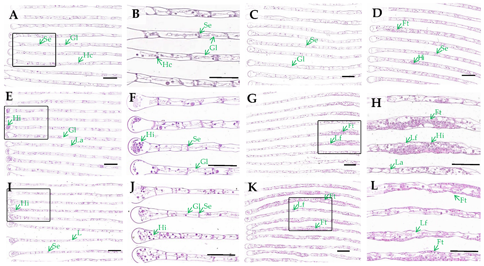
3.1. Gills Histological Observation

Histological examination demonstrated concentration- and time-dependent alterations in the gill morphology of E. sinensis following H2O2 exposure. Control specimens exhibited well-arranged gill filaments with intact septum structure and a normal distribution of haemolymph cells (Figure 1A,B). At 6 mmol/L H2O2, no significant alterations were observed after 24 h (Figure 1C). However, after 72 h, slight thickening of gill filaments was detected (Figure 1D). More severe histopathological manifestations were evident at higher concentrations (12 and 15 mmol/L). At 24 h, a notable increase in haemolymph cells was observed, particularly at the tips of the gill filaments (Figure 1E,F,I,J). By 72 h, the gill filaments exhibited irregular thickening, septum degeneration, and extensive aggregation of haemolymph cells within the gill lumen (Figure 1G,H,K,L). Notably, both the severity of swelling and the affected area of gill filaments exhibited a positive correlation with increasing H2O2 concentrations at the 72 h time point, demonstrating clear dose-dependent pathological progression.

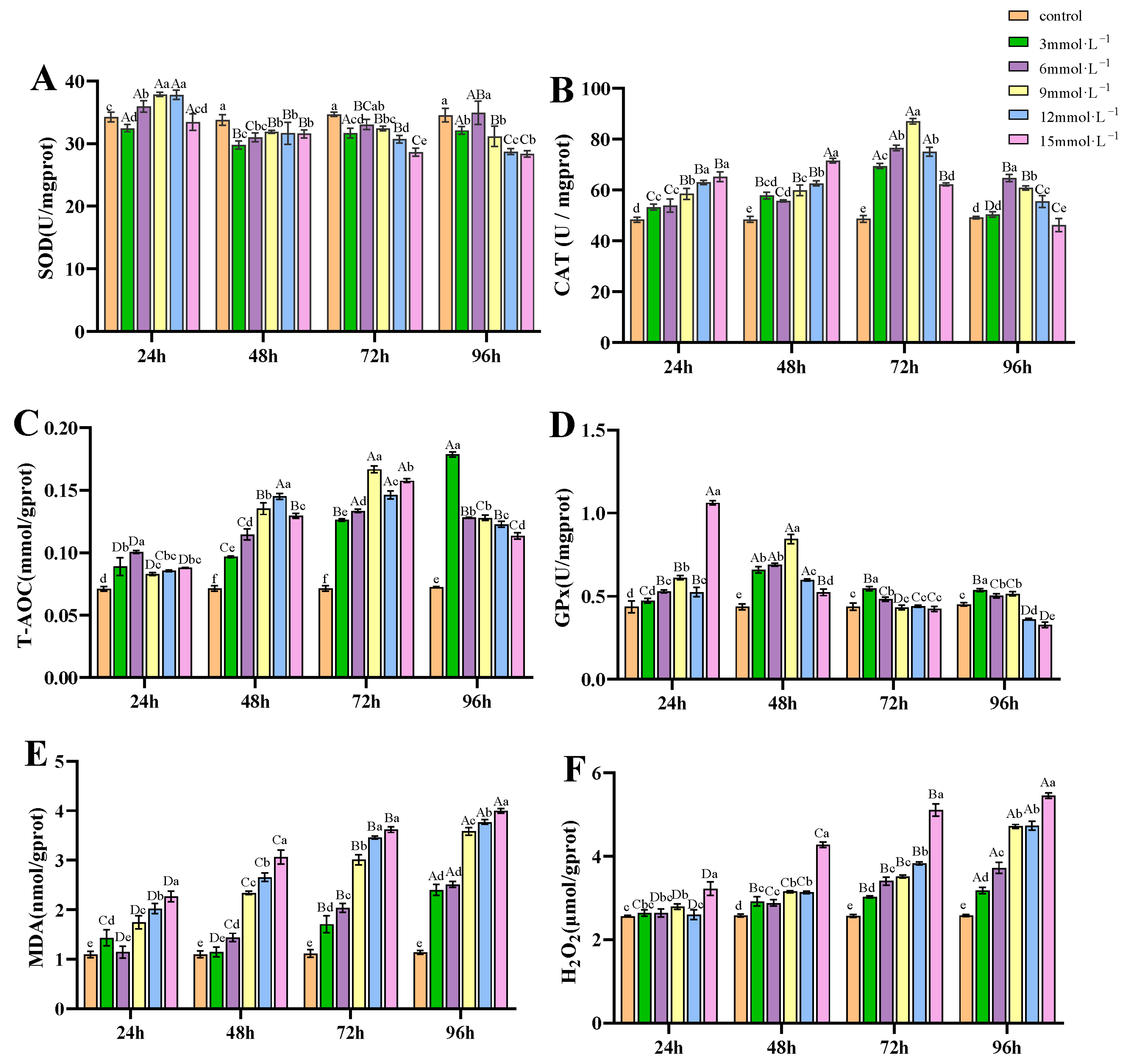
3.2. Impact of H2O2 Exposure on Antioxidant Capacity of Gills

Under H2O2 exposure, the SOD activities in the treatment groups of 6, 9, and 12 mmol/L at 24 h were higher than those in the control; while from 48 h, the SOD activities were significantly reduced compared to the control group (p < 0.05, Figure 2A). CAT activities showed an initial increase and then a decrease under the exposure of H2O2 (p < 0.05), with peak values observed at 72 h for the 3, 6, 9, and 12 mmol/L groups, and at 48 h for the 15 mmol/L group (Figure 2B). T-AOC in the treatment groups was significantly higher than in the control group (p < 0.05, Figure 2C), showing an increased trend from 24 to 96 h of exposure and peaking at 72 h, except for the 3 mmol/L group, which peaked at 96 h. GPx activities increased from 24 to 48 h in the 3, 6, 9, and 12 mmol/L groups, but decreased significantly below control at 96 h in the 12 and 15 mmol/L groups (p < 0.05, Figure 2D). After H2O2 exposure, the concentrations of MDA (Figure 2E) and H2O2 (Figure 2F) in gill tissues exhibited an overall increasing trend as time progressed.

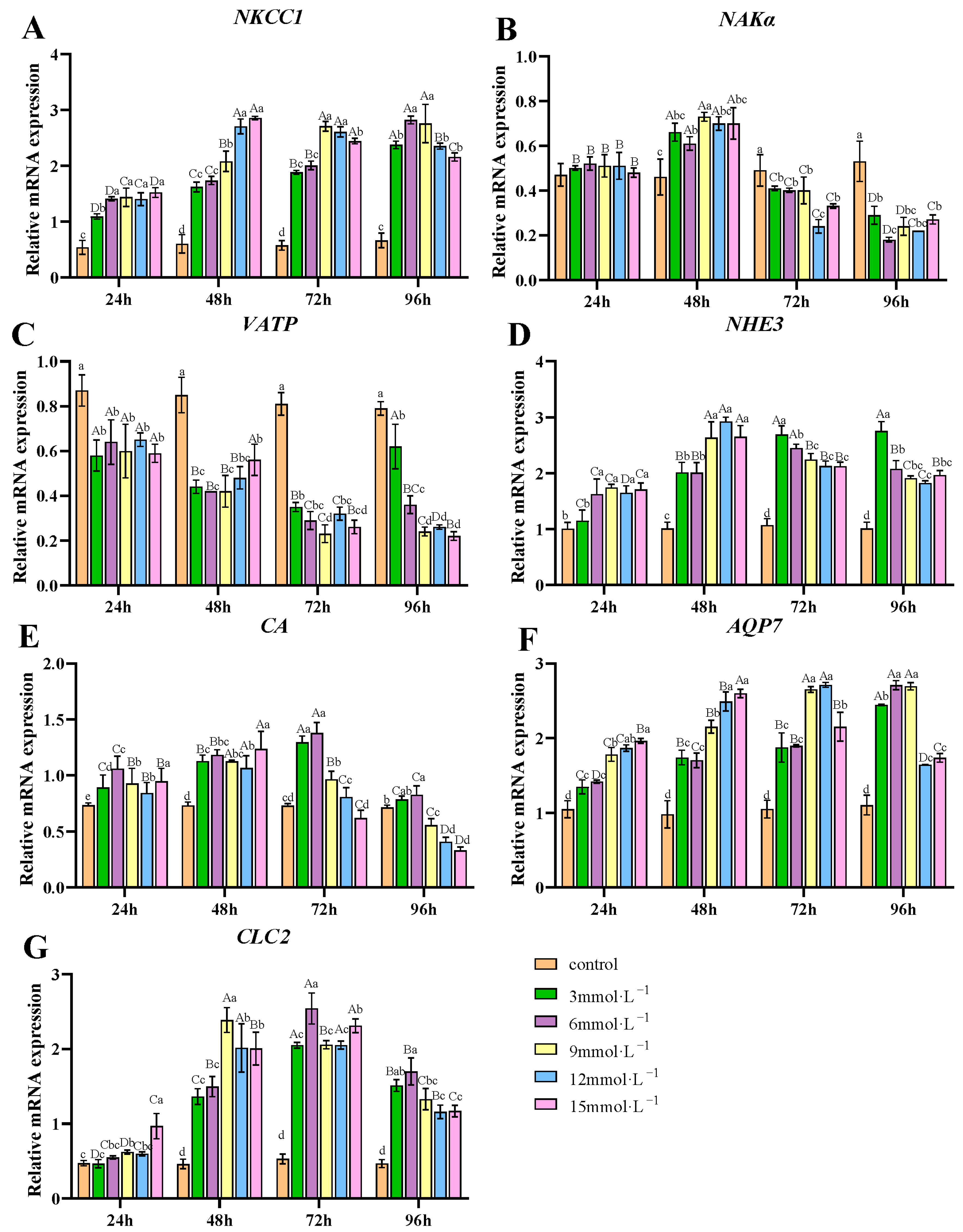
3.3. Effects of H2O2 Exposure on the mRNA Expression of Ion Transport-Related Genes in Gills

H2O2 exposure influenced the transcription of ion transport-related genes in gill tissues (Figure 3). The transcription of NKCC1, NHE3, AQP7, and CLC2 was upregulated after H2O2 exposure (Figure 3A,D,F,G). On the contrary, the transcription level of VATP was suppressed (p < 0.05) after H2O2 exposure (Figure 3C). The mRNA levels of NKAα initially increased across all treatment groups, peaking at 48 h, before subsequently decreasing. By 72 and 96 h, these levels were significantly lower than those observed in the control group (Figure 3B). The mRNA levels for CA initially rose, then declined, with the 3 and 6 mmol/L groups peaking at 72 h, whereas the 9, 12, and 15 mmol/L groups peaked at 48 h (Figure 3E).

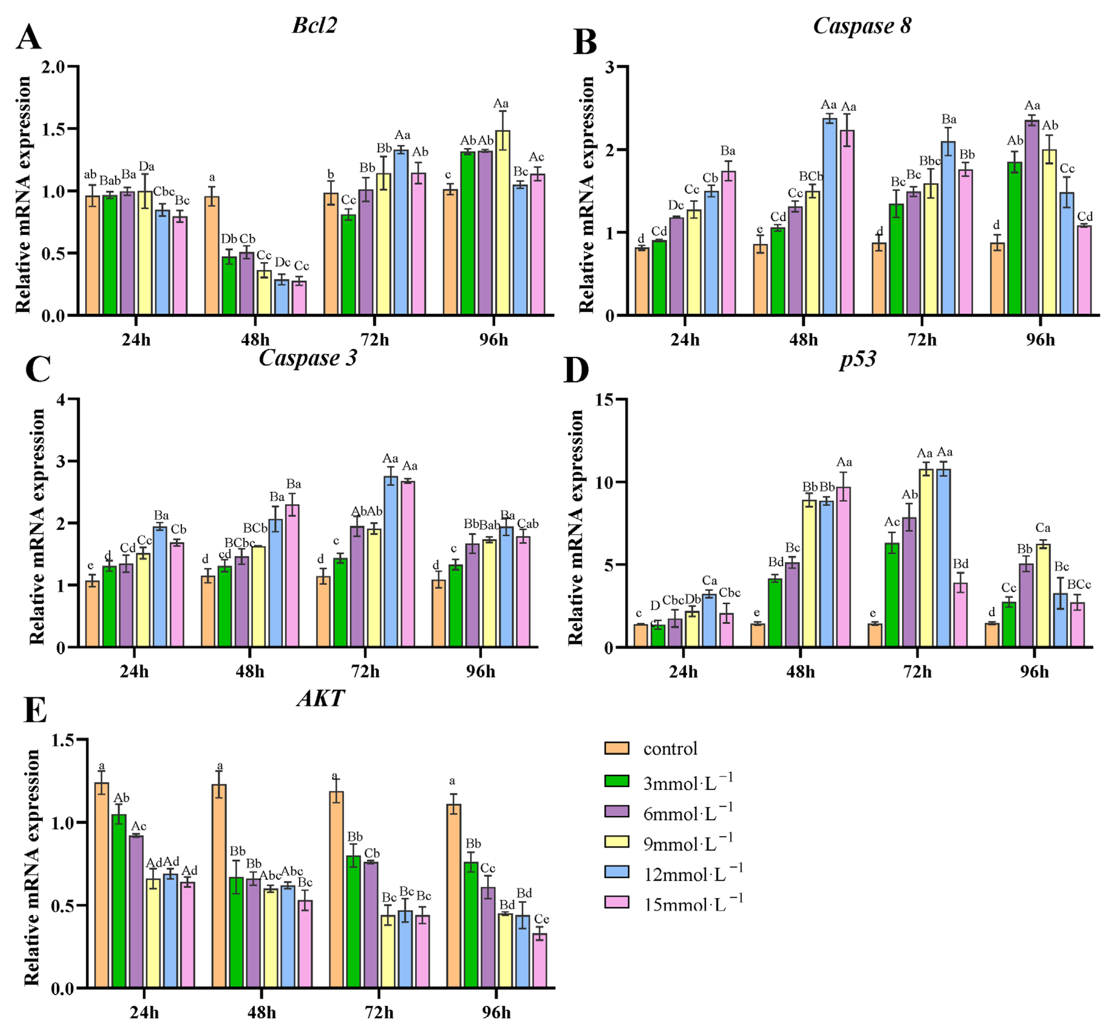
3.4. Effect of H2O2 Exposure on the Transcription of Apoptosis-Related Genes in Gills

Under H2O2 exposure, Bcl2 transcription was significantly inhibited at 48 h, showing values significantly lower than those of the control group (p < 0.05). However, at 72 and 96 h, there was a significant increase compared to 48 h (p < 0.05, Figure 4A). Caspase-8 and Caspase-3 transcription was significantly upregulated at all H2O2 concentrations and time points compared to the control (p < 0.05, Figure 4B,C). For Caspase-8, the 3, 6, and 9 mmol/L treatments showed a progressive increase in transcription over time, while in the 12 and 15 mmol/L groups, it reached its maximum at 48 h. For Caspase-3, its transcription peaked at 72 h. Similarly, the transcription of p53 was significantly higher than the control (p < 0.05), reaching its maximum at 72 h (Figure 4D). The AKT transcription decreased significantly over time (p <0.05), with only a slight increase in the 3 mmol/L treatment group at 72 h (Figure 4E)

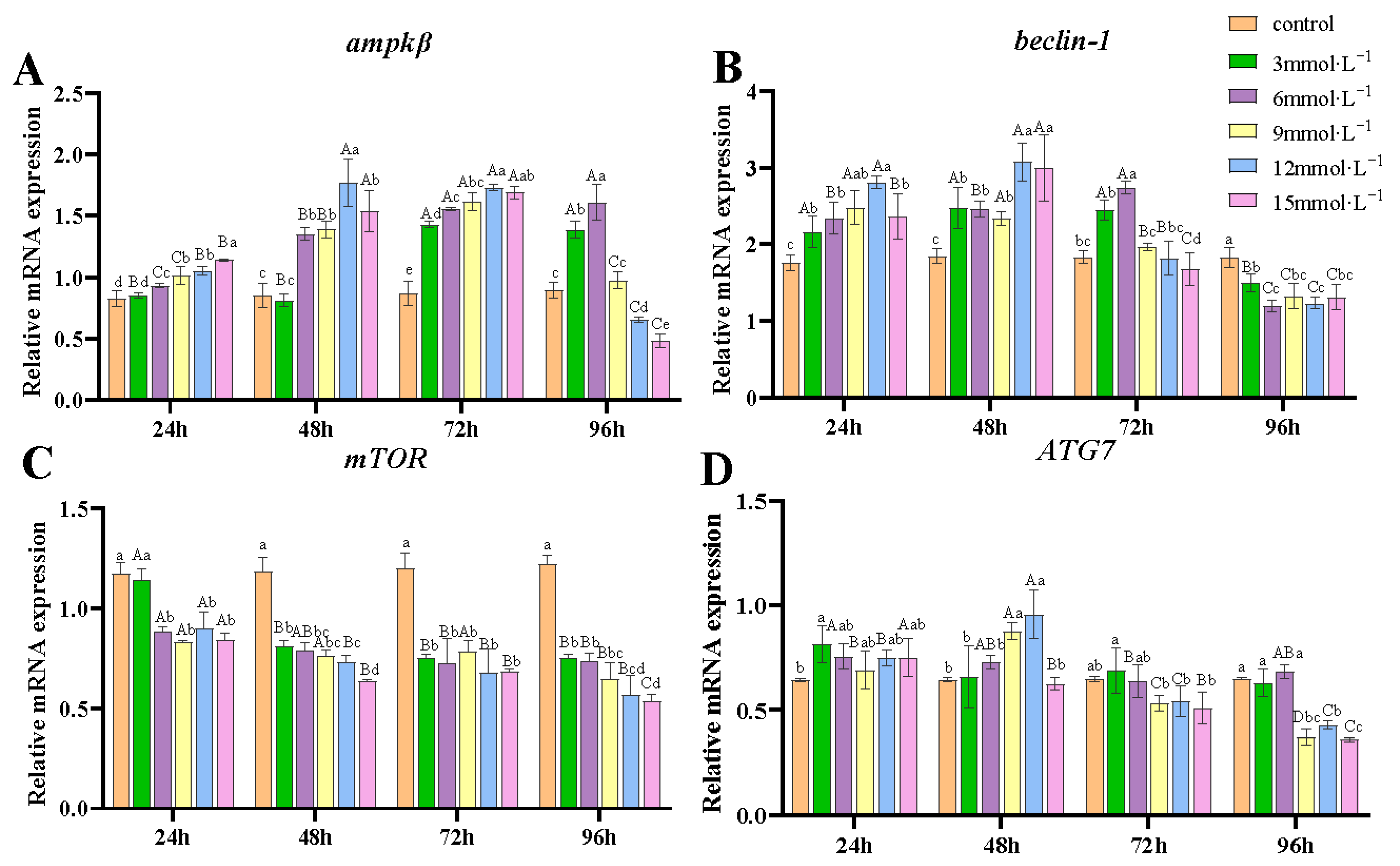
3.5. Effect of H2O2 Exposure on the Transcription of Autophagy-Related Genes in Gills

At the H2O2 concentrations of 6, 9, 12, and 15 mmol/L, the transcription of ampkβ significantly increased from 24 to 72 h compared to the control (p < 0.05), while at the low concentration of 3 mmol/L, the upregulation occurred from 72 to 96 h (Figure 5A). The mRNA levels of beclin-1 were significantly higher than those in the control group at 24 and 48 h (p < 0.05) (Figure 5B). By 96 h, beclin-1 transcription was significantly suppressed compared to the control (p < 0.05). mTOR transcription was significantly downregulated under all conditions (p < 0.05) except at 3 mmol/L concentration after 24 h (Figure 5C). ATG7 transcription showed no significant difference from controls at 3 and 6 mmol/L (p > 0.05), but was significantly upregulated at 9 and 12 mmol/L after 48 h of exposure (Figure 5D). It is noteworthy that transcriptions of ampkβ, beclin-1, and ATG7 were significantly downregulated at 96 h under exposure to 9, 12, and 15 mmol/L concentrations of H2O2.

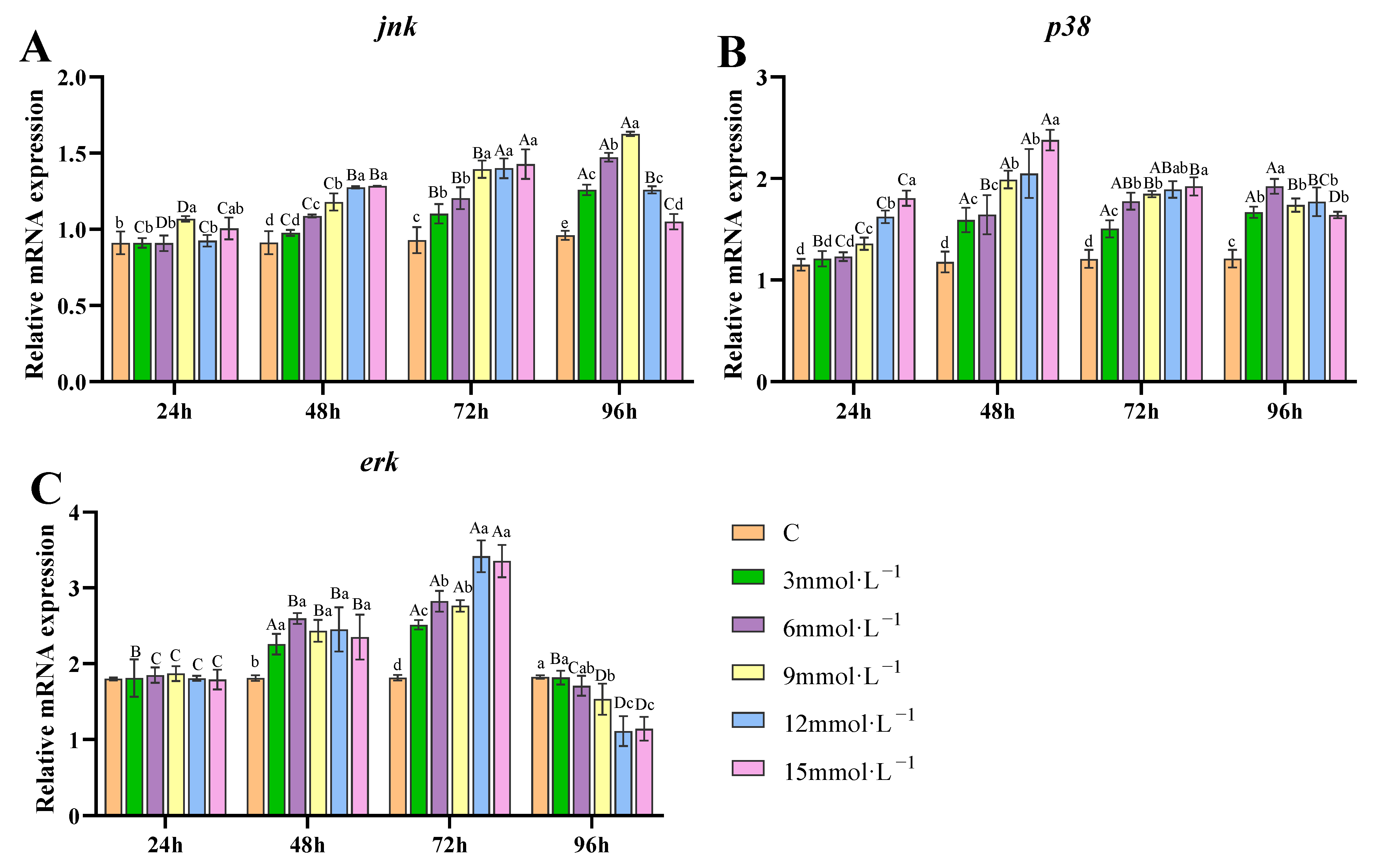
3.6. Effect of H2O2 Exposure on the Transcription of Genes Related to MAPK Pathway in Gills

After H2O2 exposure, mRNA levels of jnk exhibited a gradual increase from 48 to 96 h across all exposure concentrations. Although a slight decrease was observed in the 12 and 15 mmol/L treatment groups, levels remained markedly higher than the control group, with statistical significance (p < 0.05, Figure 6A). During the 96 h exposure, the transcription of p38 exhibited a continuous increasing trend in the 3 and 6 mmol/L groups, with significant upregulation observed from 48 to 96 h. Meanwhile, p38 transcription was markedly upregulated in the 9, 12, and 15 mmol/L groups after 24 h of H2O2 exposure, although it showed a declining trend at 96 h in the high concentration H2O2 groups (p < 0.05, Figure 6B). The transcription of erk was upregulated at 48 and 72 h under H2O2 exposure compared to the control. However, at 96 h, it decreased, showing levels lower than the control after 9, 12, and 15 mmol/L H2O2 exposures (p < 0.05, Figure 6C).

4. Discussion

4.1. Effects of H2O2 on Histological Changes and Antioxidant Capacity of Gills

In crustaceans, gills are a critical organ functioning primarily in respiration and ion regulation. These delicate structures can be particularly vulnerable to various environmental stressors, leading to physiological disorders or damage [29]. Consequently, gills are recognized as critical indicators of stress and disease. Romano and Zeng reported that ammonia-N exposure causes extensive infiltration of haemocytes, epithelial changes, disrupted pillar cells, and lamellae collapse in Portunus pelagicus gills [30]. Acute exposure to cadmium induced an increase in hemocytes in the gill lumen, with edema and even inflammatory foci in Sinopotamon henanense [13]. In this study, H2O2 exposure led to an increase in hemocytes, accompanied by enlarged gill lumen and degeneration of septa, showing hypertrophy of gill filaments at 72 h. Significant hemocyte infiltration within gill filaments serves as a characteristic histological marker of early inflammatory lesions [31]. This physiological state is considered an adaptive response to altered environmental conditions, functioning as a protective mechanism that prevents excessive toxin infiltration from water into gill tissues, thereby averting contamination of the hemolymph. However, this adaptation concurrently reduces the respiratory–excretory surface area of gills, ultimately compromising their functional capacity.
Oxidative stress occurs when the generation of ROS exceeds the scavenging capacity of the antioxidant defense system [32]. The ROS cascade typically begins with superoxide radical (O2•−) formation, which is then converted to hydrogen peroxide (H2O2) through SOD-catalyzed dismutation. H2O2 can subsequently generate hydroxyl radicals (OH) via metal-catalyzed Fenton reactions, where H2O2 reacts with redox-active metals (Fe2+/Cu+) to generate OH [33]. The enzymatic antioxidant defense system plays a critical role in facing excessive free radicals to maintain cellular redox balance [34,35]. Common antioxidant enzymes such as SOD, CAT, and GPx can remove excess ROS and protect the body from oxidative harm [36]. The SOD isoenzymes, including cytoplasmic Cu/Zn-SOD and mitochondrial Mn-SOD, protect cells by neutralizing diffusible O2 into H2O2 and O2, thereby blocking radical propagation. In this study, SOD activity exhibited a transient increase at 24 h of H2O2 exposure, demonstrating an initial adaptive response to stress. However, subsequent activity decline suggested possible H2O2-mediated enzyme inhibition, likely resulting from membrane-permeable H2O2 accumulation exceeding cellular detoxification capacity, as evidenced by elevated gill H2O2 levels. The underlying mechanism may involve excess H2O2 reducing catalytic Cu2+ in Cu/Zn-SOD to Cu+, which then participates in Fenton chemistry, generating damaging hydroxyl radicals and causing enzyme inactivation [37]. In contrast, Mn-SOD maintains relative stability due to the redox-inert nature of its manganese center, thereby avoiding Fenton reactions [38]. Unlike Mn-SOD, Cu/Zn-SOD appears more vulnerable under prolonged H2O2 stress. CAT mediates H2O2 decomposition to water and oxygen, while GPx reduces H2O2 to water using glutathione (GSH) as a substrate. In this study, CAT activity demonstrated a consistent rise from 24 to 72 h of exposure, while GPx activity exhibited a declining trend from 48 to 96 h. These inverse activity patterns reveal functional competition between GPx and CAT in H2O2 clearance beyond their complementary roles [39]. It is known that GPx has a much lower Michaelis–Menten constant value (KM) than CAT, which allows GPx to function efficiently at low concentrations of H2O2 (physiological levels), while CAT is significantly activated only when there is a high accumulation of H2O2 (under oxidative stress). The notable CAT upregulation confirms significant H2O2 accumulation in gill tissues, surpassing CAT’s high KM threshold. Concurrently, GPx’s dual-substrate requirement (H2O2 + GSH) makes it particularly susceptible to ROS overload, which oxidizes its catalytic selenocysteine residue and depletes GSH [40], inactivating enzyme function. T-AOC comprises the integrated function of enzymatic (SOD, CAT, GPx) and non-enzymatic (vitamins E/C, GSH, carotenoids, phenolics) antioxidants in neutralizing diverse free radicals, and is widely used to assess the overall antioxidant capacity in crustaceans under environmental stressors. Results demonstrated a rapid, significant increase in T-AOC levels from 24 to 96 h post-exposure compared to controls. The elevation likely represented a protective compensatory response to oxidative challenge, aimed at neutralizing the increased ROS to maintain cellular redox balance. Irregular changes in antioxidant enzyme activity were also observed in common carp and tilapia treated with H2O2 [24,41]. Moreover, similar results were observed in E. sinensis under oxidative stress induced by various environmental stressors. For example, SOD, CAT, and GPx activities in gills were increased after hypoxia stress, accompanied by increasing MDA content [42]. After cadmium (Cd) exposure, SOD, CAT, and GPx activities in the gills of Charybdis japonica showed an increased trend from 0.5 day to 3 days, but decreased after 6 days of exposure [43].
Both MDA and H2O2 have been utilized to assess the state of cellular oxidative stress. MDA, as a reactive aldehyde, is a byproduct of lipid peroxidation, generated when ROS (especially OH) attack polyunsaturated fatty acids (PUFAs) in cellular membranes [44]. H2O2 functions as both a redox signaling molecule at physiological levels and a toxic oxidant at pathological concentrations. While low levels regulate cell signaling via targeted oxidation, excess H2O2 overwhelms detoxification systems, causing oxidative damage [45]. Both MDA and H2O2 levels in this study revealed rising trends within the gill tissues of the treatment groups. Notably, the observed increase in H2O2 concentrations inversely correlated with SOD activity, suggesting reduced H2O2 generation via SOD, implying potential transmembrane diffusion of exogenous H2O2, and/or generation from alternative pathways such as peroxisomal fatty acid β-oxidation [46] or NOX-mediated production [47]. Excess H2O2 may generate additional OH through Fenton reactions, which continuously attack PUFAs to yield lipid hydroperoxides (LOOH) and ultimately MDA. Although GPx attempts to detoxify LOOH to non-toxic alcohols (LOH), its efficiency becomes progressively compromised due to ROS overload and GSH depletion. The intensified lipid peroxidation cascade ultimately leads to sustained elevation of MDA levels. Other exogenous stressors can also significantly elevate MDA and H2O2 levels. In Scylla serrata, lipid peroxidation and H2O2 were elevated in gills under salinity stress ranging from 10 ppt to 35 ppt [48]. Similarly, after a 14-day exposure to nitrite and sulfide, MDA and H2O2 contents increased significantly in E. sinensis gill tissues [49]. Collectively, evidenced by antioxidant/oxidant parameters and histopathological alterations, our study demonstrates that H2O2 can induce oxidative stress responses in gill tissues.

4.2. Effects of H2O2 Stress on the Ion Transport-Related Genes in Gills

Crustaceans maintain ion stability and fluid pH balance in complex environments through ion transport proteins and enzymes in gill tissues [50], which regulate intracellular and extracellular ion concentrations to support physiological functions. Key ion pumps like NKAα and VATP drive ion uptake and secretion by utilizing the energy released from ATP hydrolysis. VATP is responsible for transporting H+ either out of the cell or into organelles, while NKAα functions in maintaining the balance of Na+ and K+ both inside and outside cells [25]. Ion channels represent another class of membrane transport proteins that mediate selective ion movement, particularly Na+, Ca2+, and Cl. Among these, NKCC1 co-transports ions across the cellular membrane in a ratio of 1Na+:1K+:2Cl [51]. The Na+/H+ exchanger (NHE) facilitates an exchange of Na+ and H+, maintaining the acid-base balance [52]. CA aids in the exchange of Na+ and Cl by catalyzing the hydration of CO2 in gill cells, providing counter ions H+ and HCO3− [53]. CLC2 is responsible for regulating the transmembrane transport of Cl. Our results show that the VATP transcription significantly decreased, while the NAKα transcription was only upregulated at 48 h and then downregulated at 72 and 96 h of exposure. The sustained downregulation of VATP suggests a suppression of proton transport into lysosomes, which may lead to a loss of lysosomal stability [54]. A study indicated that VATP transcript levels in Macrobrachium amazonicum gills were significantly downregulated under salinity stress relative to controls [51]. In contrast, the transient upregulation of NKAα at 48 h implies an immediate compensatory effort to maintain Na+/K+ homeostasis. The suppression of NKAα and VATP translation was possibly an attempt to conserve cellular energy under H2O2 stress by preventing ATP depletion. On the contrary, the mRNA levels of NKCC1, NHE3, CA, and CLC2 showed a significant increase, indicating that H2O2 exposure stimulated the transcription of ion transport enzymes and may further promote ion movements in gills. Some reports showed similar changes in ion transport-related genes under stressors. Zhang et al. reported that the transcription of NKA-α, NKCC1, and NHE first increased and then decreased in the gills of Portunus trituberculatus under long-term ammonia stress [55]. Nan et al. reported that after 14 days of ammonia exposure in Litopenaeus vannamei, NKA-α and NKA-β genes were downregulated in the ammonia-exposed group compared to controls [56]. The transcription of CA increased and then decreased on Macrobrachium nipponense under nanoplastic exposure [57]. A study showed that the differentially expressed genes (DGEs) of antioxidant- and ion transport-related, e.g., NKA and GPx, were upregulated in the gills of E. sinensis under heavy metal cadmium toxicity [58]. In addition to maintaining ionic homeostasis, crustaceans also need to establish water balance within the body through aquaporins (AQPs) [59]. In L. vannamei, it was found that the transcription of AQP7 significantly decreased with reduced salinity levels, hindering cellular water transport [60]. Our study revealed significantly elevated AQP7 mRNA levels following H2O2 exposure, suggesting enhanced water transport activity in gill tissues. This may contribute to the observed gill filament swelling. It is reported that oxidative stress modulates ion transport through both direct and indirect mechanisms. Direct effects involve oxidative modification of critical amino acids, such as cysteine thiol groups, in ion channels, inducing conformational changes that alter their function [61]. Indirect effects occur via (1) ROS-dependent phosphorylation that post-translationally regulates channel activity [62], (2) membrane lipid peroxidation disrupting the structural integrity of channel microenvironments, and (3) activation of NF-κB and Nrf2 signaling cascades that transcriptionally modulate ion transport-related gene expression profiles [63]. These findings demonstrate that oxidative stress dynamically regulates both ion and water transport systems in crustacean gills, possibly leading to impaired osmoregulatory homeostasis.

4.3. Effects of H2O2 Exposure on Apoptosis and Autophagy of Gill Tissues

Oxidative stress can trigger cellular apoptosis, a programmed cell death process critical for maintaining cellular homeostasis [64]. Apoptosis is promoted through two pathways: the extrinsic pathway, which is initiated by direct activation of caspase-8 [65]; and the intrinsic pathway, triggered by mitochondrial dysfunction, leading to cytochrome c release and subsequent caspase activation [66]. Both pathways ultimately converge on the cleavage of executioner caspase-3/7 to induce cell death [67]. In this study, mRNA levels of caspase-8 were significantly upregulated in a dose- and time-dependent manner at 3, 6, and 9 mmol/L concentrations, suggesting the activation of extrinsic apoptosis. Caspase-3 transcripts were significantly upregulated in all H2O2 treatment groups and reached their peak at 72 h, further confirming the activation of apoptosis. Similar results were reported in previous studies, where the mRNA expression patterns of caspase-8/3 were upregulated in the gills of both Sinopotamon henanense after H2O2 exposure [68] and L. vannamei under NH4Cl exposure [69]. Bcl2 is a well-characterized anti-apoptotic protein that suppresses programmed cell death by regulating the mitochondrial pathway of apoptosis [70]. Following 48 h of H2O2 exposure, significant suppression of Bcl2 transcription suggests the initiation of the mitochondrial pathway of apoptosis. By 72 h, an increase in Bcl2 transcription, particularly in treatment groups at concentrations of 12 and 15 mmol/L, may reflect the cells’ adaptation to sustained oxidative stress or an attempt to enhance their survival prospects by elevating Bcl2 transcription. The specific mechanisms require further investigation in future studies. Guan et al. found that abamectin could promote apoptosis in P. clarkii based on the downregulation of Bcl2 and upregulation of caspase-3 [71]. Additionally, Liu et al. demonstrated that the downregulation of Bcl2 and upregulation of caspase-3 induced by LPS stress can be reversed by adding dietary glutathione [72]. Furthermore, oxidative stress may activate specific transcription factors, such as p53, which induces apoptosis by triggering key apoptotic genes, including the mitochondrial proapoptotic factor Bax and the death receptor Fas [73,74]. When exposed to persistent organic pollutants (HBCD or BDE-47), apoptosis could be triggered by oxidative stress through the transcriptional upregulation of p53 in the gills of Macrophthalmus japonicas [20]. Sun et al. reported that hypoxia-induced oxidative stress can cause apoptosis in M. nipponensem, which was proved by the increase in caspase-3 mRNA expression and the upregulation of p53 at transcript and protein levels [75]. In this study, the upregulation of p53 transcripts in gill tissues may induce the transcription of proapoptotic genes to accelerate apoptosis. Furthermore, Akt, a serine-threonine kinase, is crucial for cellular proliferation and survival, as it inhibits apoptosis by phosphorylating various target molecules [76]. Following 48 h of exposure to nitrite or ammonia, transcription of Akt was notably downregulated in the gills of L. vannamei [77]. A similar result was identified by Huang et al., who found that, under prometryn stress, the transcription of Akt was decreased while caspase-3 increased at 48 h [78]. In this study, Akt decreased significantly over time after H2O2 treatment, indicating that apoptosis in the gills of E. sinensis may be associated with the inhibition of the PI3K–Akt signaling pathway [79]. Based on the transcription of these genes, we revealed that H2O2 may promote cell death in the gills of E. sinensis through the apoptosis program.
Autophagy is another cellular strategy for combating stressful environments, particularly under conditions of nutrient or energy deficiency. Increasing evidence suggests that autophagy plays a pivotal role as a sensor in redox signaling during cellular responses [80]. Mild oxidative stress activates the autophagy pathway as a self-protective mechanism. However, severe oxidative stress compromises this process, thereby accelerating cell apoptosis and necrosis [81]. The autophagic process is governed by an intricate network of molecular regulators, including Atg family proteins and lysosomal degradation enzymes. Beclin-1 is a crucial mediator of autophagy, which facilitates the initial formation of phagophore membranes that develop into autophagosomes. The maturation of these autophagic vesicles relies on specialized protein conjugation mechanisms, particularly those involving Atg7 and the LC3 protein. Moreover, cellular energy sensors, specifically the AMPK and mTOR pathways, serve as central regulators that modulate autophagic activity under stress conditions. It has been reported that elevated levels of ROS are associated with increased expression of autophagy regulatory factors, such as beclin-1 and ATG7 [82]. Upregulation of beclin-1 enhances the formation of autophagosomes [83]. However, Bcl2 can bind to beclin-1, inhibiting its activity to prevent excessive autophagy and thus protect cells from damage [84], modulating the balance between the two critical pathways of autophagy and apoptosis. In M. nipponense, hypoxia stress significantly increased the mRNA and protein levels of beclin-1 [85]. Additionally, ROS can activate ampk, which in turn promotes autophagy by inhibiting the transcription of mTOR [28]. Hypoxia induced antioxidant responses, which subsequently trigger autophagy in hepatocytes by upregulating beclin-1, ATG7, and ATG8 at both transcriptional and protein levels in M. nipponense [86]. Spiroplasma eriocheiris infection can induce autophagy accompanied by the increased transcription of ATG7 in E. sinensis [87]. In our study, the beclin-1 transcription was upregulated from 24 to 72 h of exposure but downregulated at 96 h. The transcription of ATG7 increased slightly after 48 h of exposure to H2O2 and was then downregulated. Similarly, ampkβ showed an increased trend after H2O2 exposure from 24 to 72 h, but its levels decreased at 96 h under high concentrations of H2O2 (9, 12, and 15 mmol/L). The upregulation of these genes indicated that mild H2O2 exposure induced autophagy in gill cells, an adaptive response mechanism. However, the downregulation suggested that high concentrations and continuous exposure to H2O2 may inhibit autophagic functions.
The MAPK signaling pathways, including ERK, JNK, and p38, play a pivotal role in cellular apoptosis and autophagy [88]. Under stress conditions, MAPKs integrate various signals through transcription-dependent and independent mechanisms based on the cellular environment and type, ultimately maintaining the balance between cell survival and death [89]. Increased ROS leads to the activation of stress-responsive kinases such as JNK and p38 MAPK, which phosphorylate and stabilize p53. p53 induces apoptosis by transcriptionally activating proapoptotic genes such as Bax and PUMA to disrupt mitochondrial integrity and FAS to initiate extrinsic apoptosis. Additionally, it promotes oxidative stress through PIGs and FDXR, amplifying oxidative stress and apoptotic signaling [90]. Guyton et al. discovered that H2O2 is a key factor in the activation of MAPKs by triggering various receptors [91]. Peng et al. discovered that cadmium could induce antioxidant response in L. vannamei, leading to an increased transcription of JNK, p38, and ERK [92]. Our study observed that exposure to H2O2 upregulated JNK, p38, and ERK in the gill tissues, suggesting a critical role for the MAPK signaling pathway in the response to oxidative stress.
It has been proven that autophagy and apoptosis can be simultaneously regulated and occur within the same tissue. For instance, hypoxia caused severe oxidant stress, activated apoptosis [75] and autophagy [86] in the hepatopancreas of M. nipponense. Similar results were also found in E. sinensis under copper ion stress [93] and sustained hepatopancreatic necrosis disease [94]. Both scenarios are characterized by severe oxidative stress. Research indicates that apoptosis and autophagy exhibit mutual inhibition, though their exact regulatory interplay remains unclear. Both processes share key factors like Beclin-1 and Bcl2, which act as molecular switches [95]. Bcl2 suppresses apoptosis by inhibiting Bax/Bak while also binding and inactivating Beclin-1 to block autophagy. This study further demonstrated an inverse correlation between the expression trends of Bcl2 and Beclin-1. The dual regulatory roles of the AMPK and MAPK pathways in this process are particularly noteworthy. AMPK promotes cell survival by activating autophagy to remove damaged organelles and mitigate oxidative damage, while MAPK subfamilies (such as JNK and p38) drive proapoptotic signaling to eliminate severely compromised cells. This mechanism likely plays a crucial role in the reparative capacity of gill tissues under stress.

5. Conclusions

Our study investigated the multidimensional adaptive response mechanisms of E. sinensis to oxidative stress induced by various concentrations of H2O2 in gill tissues. Initially, the crabs enhanced their antioxidant defense by upregulating key antioxidant enzymes (SOD, CAT, GPx) and increasing T-AOC level, which helped mitigate initial oxidative damage. The stress also challenged the gills’ ion regulation capabilities, requiring adaptive gene transcription changes to maintain cellular homeostasis. However, excessive exposure led to an overwhelmed antioxidant system, peroxide accumulation, gill inflammation, and ion-regulatory dysfunctions. Furthermore, H2O2 triggered the AMPK and MAPK signaling pathways, leading to autophagy and apoptosis. In summary, this study provides insights into the oxidative stress responses of crabs to H2O2 and highlights the critical functions of the gill tissues under such conditions.

Author Contributions

Conceptualization, W.F. and Y.T.; methodology, Q.Y. and J.L.; software, J.Z.; validation, W.F. and Y.X.; formal analysis, G.J. and R.J.; investigation, Q.H. and W.F.; resources, Y.T.; data curation, Q.Y. and J.L.; writing—original draft preparation, W.F. and Q.H.; review and editing, Y.T.; visualization, W.F.; supervision, Y.T.; project administration, Y.T.; funding acquisition, Y.T. All authors have read and agreed to the published version of the manuscript.

Funding

This research was funded by the National Natural Science Foundation of China (No. 32202966), the Key Research and Development Program of Jiangsu Province (No. BE2022360), and the Jiangsu Revitalization of Seed Industry (No. JBGS [2021]031).

Institutional Review Board Statement

The animal study protocol was approved by the Animal Ethics Committee of the Freshwater Fisheries Research Center (FFRC), Chinese Academy of Fishery Sciences (protocol code LAECFFRC-2024-03-18).

Data Availability Statement

The original contributions of this study are detailed within the manuscript. Further inquiries should be directed to the corresponding author.

Acknowledgments

We sincerely appreciate the support provided by China Jiangsu Haorun Biological Industry Group Co., Ltd., especially Guoan Hua and Bo Peng, for their valuable assistance in this study. Their contributions were instrumental in facilitating our research.

Conflicts of Interest

The authors declare no conflicts of interest. The funders had no role in the design of the study; in the collection, analyses, or interpretation of data; in the writing of the manuscript; or in the decision to publish the results.

References

  1. Chang, C.; Mayer, M.; Rivera-Ingraham, G.; Blondeau-Bidet, E.; Wu, W.; Lorin-Nebel, C.; Lee, T. Effects of temperature and salinity on antioxidant responses in livers of temperate (Dicentrarchus labrax) and tropical (Chanos Chanos) marine euryhaline fish. J. Therm. Biol. 2021, 99, 103016. [Google Scholar] [CrossRef] [PubMed]
  2. Fan, Z.; Wu, D.; Li, J.; Li, C.; Zheng, X.; Wang, L. Phosphorus nutrition in Songpu mirror carp (Cyprinus carpio Songpu) during chronic carbonate alkalinity stress: Effects on growth, intestinal immunity, physical barrier function, and intestinal microflora. Front. Immunol. 2022, 13, 900793. [Google Scholar] [CrossRef] [PubMed]
  3. Jiang, X.; Dong, S.; Liu, R.; Huang, M.; Dong, K.; Ge, J.; Gao, Q.; Zhou, Y. Effects of temperature, dissolved oxygen, and their interaction on the growth performance and condition of rainbow trout (Oncorhynchus mykiss). J. Therm. Biol. 2021, 98, 102928. [Google Scholar] [CrossRef]
  4. Singh, S.P.; Ahmad, T.; Sharma, J.; Chakrabarti, R. Effect of temperature on food consumption, immune system, antioxidant enzymes, and heat shock protein 70 of Channa punctata (Bloch, 1793). Fish Physiol. Biochem. 2021, 47, 79–91. [Google Scholar] [CrossRef]
  5. Chen, J.; Zhou, X.; Feng, L.; Liu, Y.; Jiang, J. Effects of glutamine on hydrogen peroxide-induced oxidative damage in intestinal epithelial cells of Jian carp (Cyprinus carpio var. Jian). Aquaculture 2009, 288, 285–289. [Google Scholar] [CrossRef]
  6. Martín, D.; Salinas, M.; Fujita, N.; Tsuruo, T.; Cuadrado, A. Ceramide and reactive oxygen species generated by H2O2 induce caspase-3-independent degradation of Akt/protein kinase B. J. Biol. Chem. 2002, 277, 42943–42952. [Google Scholar] [CrossRef]
  7. Cuypers, A.; Plusquin, M.; Remans, T.; Jozefczak, M.; Keunen, E.; Gielen, H.; Opdenakker, K.; Nair, A.R.; Munters, E.; Artois, T.J. Cadmium stress: An oxidative challenge. Biometals 2010, 23, 927–940. [Google Scholar] [CrossRef]
  8. Depledge, M.H.; Rainbow, P.S. Models of regulation and accumulation of trace metals in marine invertebrates. Comp. Biochem. Physiol. C Pharmacol. Toxicol. Endocrinol. 1990, 97, 1–7. [Google Scholar] [CrossRef]
  9. Rainbow, P.S.; Luoma, S.N. Metal toxicity, uptake and bioaccumulation in aquatic invertebrates—Modelling zinc in crustaceans. Aquat. Toxicol. 2011, 105, 455–465. [Google Scholar] [CrossRef]
  10. Boonsanit, P.; Pairohakul, S. Effects of salinity on haemolymph osmolality, gill Na+/K+ ATPase and antioxidant enzyme activities in the male mud crab Scylla olivacea (Herbst, 1796). Mar. Biol. Res. 2021, 17, 86–97. [Google Scholar] [CrossRef]
  11. Wang, X.; Yao, Q.; Zhang, D.; Lei, X.; Wang, S.; Wan, J.; Liu, H.; Chen, Y.; Zhao, Y.; Wang, G. Effects of acute salinity stress on osmoregulation, antioxidant capacity and physiological metabolism of female Chinese mitten crabs (Eriocheir sinensis). Aquaculture 2022, 552, 737989. [Google Scholar] [CrossRef]
  12. Issartel, J.; Boulo, V.; Wallon, S.; Geffard, O.; Charmantier, G. Cellular and molecular osmoregulatory responses to cadmium exposure in Gammarus fossarum (Crustacea, Amphipoda). Chemosphere 2010, 81, 701–710. [Google Scholar] [CrossRef] [PubMed]
  13. Wang, J.; Zhang, P.; Shen, Q.; Wang, Q.; Liu, D.; Li, J.; Wang, L. The effects of cadmium exposure on the oxidative state and cell death in the gill of freshwater crab Sinopotamon henanense. PLoS ONE 2013, 8, e64020. [Google Scholar] [CrossRef] [PubMed]
  14. Negro, L.; Senkman, E.; Montagna, M.; Collins, P. Freshwater decapods and pesticides: An unavoidable relation in the modern world. In Pesticides in the Modern World-Risks and Benefits; IntechOpen: London, UK, 2011. [Google Scholar]
  15. Silvestre, F.; Trausch, G.; Spano, L.; Devos, P. Effects of atrazine on osmoregulation in the Chinese mitten crab, Eriocheir sinensis. Comp. Biochem. Physiol. C Toxicol. Pharmacol. 2002, 132, 385–390. [Google Scholar] [CrossRef] [PubMed]
  16. Xian, J.; Miao, Y.; Li, B.; Guo, H.; Wang, A. Apoptosis of tiger shrimp (Penaeus monodon) haemocytes induced by Escherichia coli lipopolysaccharide. Comp. Biochem. Physiol. A Mol. Integr. Physiol. 2013, 164, 301–306. [Google Scholar] [CrossRef]
  17. Zheng, N.; Wang, N.; Wang, Z.; Abdallah, G.; Zhang, B.; Wang, S.; Yao, Q.; Chen, Y.; Wang, Q.; Zhang, D. Effect of infection with Aeromonas hydrophila on antioxidant capacity, inflammation response, and apoptosis proteins in Chinese mitten crab (Eriocheir sinensis). Comp. Biochem. Physiol. C Toxicol. Pharmacol. 2022, 252, 109220. [Google Scholar] [CrossRef]
  18. Fan, T.; Han, L.; Cong, R.; Liang, J. Caspase family proteases and apoptosis. Acta Biochim. Et Biophys. Sin. 2005, 37, 719–727. [Google Scholar] [CrossRef]
  19. Cory, S.; Adams, J.M. The Bcl2 family: Regulators of the cellular life-or-death switch. Nat. Rev. Cancer 2002, 2, 647–656. [Google Scholar] [CrossRef]
  20. Park, K.; Kwak, I. Apoptotic p53 Gene Expression in the Regulation of Persistent Organic Pollutant (POP)-Induced Oxidative Stress in the Intertidal Crab Macrophthalmus japonicus. Antioxidants 2022, 11, 771. [Google Scholar] [CrossRef]
  21. Kim, J.; Kundu, M.; Viollet, B.; Guan, K. AMPK and mTOR regulate autophagy through direct phosphorylation of Ulk1. Nat. Cell Biol. 2011, 13, 132–141. [Google Scholar] [CrossRef]
  22. She, C.; Zhu, L.; Zhen, Y.; Wang, X.; Dong, Q. Activation of AMPK protects against hydrogen peroxide-induced osteoblast apoptosis through autophagy induction and NADPH maintenance: New implications for osteonecrosis treatment? Cell. Signal. 2014, 26, 1–8. [Google Scholar] [CrossRef] [PubMed]
  23. He, Q.; Feng, W.; Chen, X.; Xu, Y.; Zhou, J.; Li, J.; Xu, P.; Tang, Y. H2O2-induced oxidative stress responses in Eriocheir sinensis: Antioxidant defense and immune gene expression dynamics. Antioxidants 2024, 13, 524. [Google Scholar] [CrossRef]
  24. Jia, R.; Du, J.; Cao, L.; Li, Y.; Johnson, O.; Gu, Z.; Jeney, G.; Xu, P.; Yin, G. Antioxidative, inflammatory and immune responses in hydrogen peroxide-induced liver injury of tilapia (GIFT, Oreochromis niloticus). Fish Shellfish Immunol. 2019, 84, 894–905. [Google Scholar] [CrossRef] [PubMed]
  25. Qi, T.; Liu, J.; Zhao, P.; Ge, B.; Liu, Q.; Jiang, S.; Wang, Z.; Zhang, H.; Tang, B.; Ding, G. A novel modulation of physiological regulation in cultured Chinese mitten crab (Eriocheir japonica sinensis) in response to consistent salinity changes. Gene 2020, 756, 144914. [Google Scholar] [CrossRef]
  26. Xu, H.; Lyu, S.; Li, Y.; Xu, J.; Lu, B.; Zhao, J. Identification, characterization and expression analysis of ERK2 in Chinese mitten crab Eriocheir sinensis after challenge with LPS and Aeromonas hydrophila. Fish Shellfish Immunol. 2016, 52, 325–333. [Google Scholar] [CrossRef]
  27. Wang, Y.; Nan, X.; Hao, S.; Zhao, K.; Guo, Y.; Wang, Q.; Li, W. AKT regulates hemocyte proliferation via glucose metabolism in Eriocheir sinensis. Fish Shellfish Immunol. 2022, 127, 247–255. [Google Scholar] [CrossRef]
  28. Wang, J.; Sun, L.; Li, X.; Tao, S.; Wang, F.; Shi, Y.; Guan, H.; Yang, Y.; Zhao, Z. Alkali exposure induces autophagy through activation of the MAPKpathway by ROS and inhibition of mTOR in Eriocheir sinensis. Aquat. Toxicol. 2023, 258, 106481. [Google Scholar] [CrossRef]
  29. Soegianto, A.; Charmantier-Daures, M.; Trilles, J.; Charmantier, G. Impact of cadmium on the structure of gills and epipodites of the shrimp Penaeus japonicas (Crustacea: Decapoda). Aquat. Living Resour. 1999, 12, 57–70. [Google Scholar] [CrossRef]
  30. Romano, N.; Zeng, C. Ontogenetic changes in tolerance to acute ammonia exposure and associated gill histological alterations during early juvenile development of the blue swimmer crab, Portunus pelagicus. Aquaculture 2007, 266, 246–254. [Google Scholar] [CrossRef]
  31. Victor, B. Gill tissue pathogenicity and hemocyte behavior in the crab Paratelphusa hydrodromous exposed to lead chloride. J. Environ. Sci. Health Part A 1994, 29, 1011–1034. [Google Scholar]
  32. Wang, L.; Xu, T.; Lei, W.; Liu, D.; Li, Y.; Xuan, R.; Ma, J. Cadmium-induced oxidative stress and apoptotic changes in the testis of freshwater crab, Sinopotamon henanense. PLoS ONE 2011, 6, e27853. [Google Scholar] [CrossRef]
  33. Birnie Gauvin, K.S.S.; Costantini, D.S.S.; Cooke, S.J.S.S.; Willmore, W.G.S.S. A comparative and evolutionary approach to oxidative stress in fish: A review. Fish Fish. 2017, 18, 928–942. [Google Scholar] [CrossRef]
  34. Jia, X.; Zhang, D.; Wang, F.; Dong, S. Immune responses of L itopenaeus vannamei to non-ionic ammonia stress: A comparative study on shrimps in freshwater and seawater conditions. Aquac. Res. 2017, 48, 177–188. [Google Scholar] [CrossRef]
  35. Liu, T.; Zhang, G.; Feng, Y.; Kong, C.; Ayisi, C.L.; Huang, X.; Hua, X. Dietary soybean antigen impairs growth and health through stress-induced non-specific immune responses in Pacific white shrimp, Litopenaeus vannamei. Fish Shellfish Immunol. 2019, 84, 124–129. [Google Scholar] [CrossRef]
  36. Frías-Espericueta, M.G.; Bautista-Covarrubias, J.C.; Osuna-Martínez, C.C.; Delgado-Alvarez, C.; Bojórquez, C.; Aguilar-Juárez, M.; Roos-Muñoz, S.; Osuna-López, I.; Páez-Osuna, F. Metals and oxidative stress in aquatic decapod crustaceans: A review with special reference to shrimp and crabs. Aquat. Toxicol. 2022, 242, 106024. [Google Scholar] [CrossRef] [PubMed]
  37. Ramirez, D.C.; Mejiba, S.E.G.; Mason, R.P. Mechanism of hydrogen peroxide-induced Cu, Zn-superoxide dismutase-centered radical formation as explored by immuno-spin trapping: The role of copper-and carbonate radical anion-mediated oxidations. Free Radic. Biol. Med. 2005, 38, 201–214. [Google Scholar] [CrossRef] [PubMed]
  38. Iranzo, O. Manganese complexes displaying superoxide dismutase activity: A balance between different factors. Bioorganic Chem. 2011, 39, 73–87. [Google Scholar] [CrossRef] [PubMed]
  39. Ighodaro, O.M.; Akinloye, O.A. First line defence antioxidants-superoxide dismutase (SOD), catalase (CAT) and glutathione peroxidase (GPX): Their fundamental role in the entire antioxidant defence grid. Alex. J. Med. 2018, 54, 287–293. [Google Scholar] [CrossRef]
  40. Miyamoto, Y.; Koh, Y.H.; Park, Y.S.; Fujiwara, N.; Sakiyama, H.; Misonou, Y.; Ookawara, T.; Suzuki, K.; Honke, K.; Taniguchi, N. Oxidative stress caused by inactivation of glutathione peroxidase and adaptive responses. Biol. Chem. 2003, 384, 567–574. [Google Scholar] [CrossRef]
  41. Jia, R.; Du, J.; Cao, L.; Feng, W.; He, Q.; Xu, P.; Yin, G. Chronic exposure of hydrogen peroxide alters redox state, apoptosis and endoplasmic reticulum stress in common carp (Cyprinus carpio). Aquat. Toxicol. 2020, 229, 105657. [Google Scholar] [CrossRef]
  42. Yang, X.; Shi, X.; Wu, M.; Pang, Y.; Song, X.; Shi, A.; Niu, C.; Cheng, Y. Effects of melatonin feed on histology and antioxidant ability of the gills and oxygen consumption of Chinese mitten crab (Eriocheir sinensis), exposed to acute hypoxia stress. Aquaculture 2021, 544, 737015. [Google Scholar] [CrossRef]
  43. Pan, L.; Zhang, H. Metallothionein, antioxidant enzymes and DNA strand breaks as biomarkers of Cd exposure in a marine crab, Charybdis japonica. Comp. Biochem. Physiol. C Toxicol. Pharmacol. 2006, 144, 67–75. [Google Scholar] [CrossRef] [PubMed]
  44. Yao, J.; Wang, J.; Liu, L.; Li, Y.; Xun, A.; Zeng, W.; Jia, C.; Wei, X.; Feng, J.; Zhao, L.I. Anti-oxidant effects of resveratrol on mice with DSS-induced ulcerative colitis. Arch. Med. Res. 2010, 41, 288–294. [Google Scholar] [CrossRef] [PubMed]
  45. Sies, H. Hydrogen peroxide as a central redox signaling molecule in physiological oxidative stress: Oxidative eustress. Redox Biol. 2017, 11, 613–619. [Google Scholar] [CrossRef]
  46. Nordgren, M.; Fransen, M. Peroxisomal metabolism and oxidative stress. Biochimie 2014, 98, 56–62. [Google Scholar] [CrossRef]
  47. Paital, B.; Chainy, G. Effects of temperature on complexes I and II mediated respiration, ROS generation and oxidative stress status in isolated gill mitochondria of the mud crab Scylla serrata. J. Therm. Biol. 2014, 41, 104–111. [Google Scholar] [CrossRef]
  48. Paital, B.; Chainy, G.B.N. Antioxidant defenses and oxidative stress parameters in tissues of mud crab (Scylla serrata) with reference to changing salinity. Comp. Biochem. Physiol. C Toxicol. Pharmacol. 2010, 151, 142–151. [Google Scholar] [CrossRef]
  49. Xing, Y.; Feng, Y.; Tian, J.; Zhang, Y.; Han, C.; Liu, X.; Yang, J. Nitrite and sulfide stress affects physiological and metabolic functions of gills in crab Eriocheir sinensis. Aquaculture 2025, 594, 741437. [Google Scholar] [CrossRef]
  50. McNamara, J.C.; Faria, S.C. Evolution of osmoregulatory patterns and gill ion transport mechanisms in the decapod Crustacea: A review. J. Comp. Physiol. B 2012, 182, 997–1014. [Google Scholar] [CrossRef]
  51. Faleiros, R.O.; Goldman, M.H.S.; Furriel, R.P.; McNamara, J.C. Differential adjustment in gill Na+/K+-and V-ATPase activities and transporter mRNA expression during osmoregulatory acclimation in the cinnamon shrimp Macrobrachium amazonicum (Decapoda, Palaemonidae). J. Exp. Biol. 2010, 213, 3894–3905. [Google Scholar] [CrossRef]
  52. Choe, K.P.; Kato, A.; Hirose, S.; Plata, C.; Sindic, A.; Romero, M.F.; Claiborne, J.B.; Evans, D.H. NHE3 in an ancestral vertebrate: Primary sequence, distribution, localization, and function in gills. Am. J. Physiol. Regul. Integr. Comp. Physiol. 2005, 289, R1520–R1534. [Google Scholar] [CrossRef]
  53. Rahim, S.M.; Mazlan, A.G.; Simon, K.D.; Delaunoy, J.P.; Laurent, P. Immunocytochemical localization of carbonic anhydrase in the pseudobranch tissue of the rainbow trout Oncorhynchus mykiss. J. Zhejiang Univ.Sci. B 2014, 15, 194–200. [Google Scholar] [CrossRef] [PubMed]
  54. Shaw, J.P.; Moore, M.N.; Readman, J.W.; Mou, Z.; Langston, W.J.; Lowe, D.M.; Frickers, P.E.; Al-Moosawi, L.; Pascoe, C.; Beesley, A. Oxidative stress, lysosomal damage and dysfunctional autophagy in molluscan hepatopancreas (digestive gland) induced by chemical contaminants. Mar. Environ. Res. 2019, 152, 104825. [Google Scholar] [CrossRef] [PubMed]
  55. Zhang, J.; Zhang, M.; Jayasundara, N.; Ren, X.; Gao, B.; Liu, P.; Li, J.; Meng, X. Physiological and molecular responses in the gill of the swimming crab Portunus trituberculatus during long-term ammonia Stress. Front. Mar. Sci. 2021, 8, 797241. [Google Scholar] [CrossRef]
  56. Nan, Y.; Xiao, M.; Duan, Y.; Yang, Y. Toxicity of ammonia stress on the physiological homeostasis in the gills of Litopenaeus vannamei under seawater and low-salinity conditions. Biology 2024, 13, 281. [Google Scholar] [CrossRef]
  57. Li, Y.; Liu, Z.; Jiang, Q.; Ye, Y.; Zhao, Y. Effects of nanoplastic on cell apoptosis and ion regulation in the gills of Macrobrachium nipponense. Environ. Pollut. 2022, 300, 118989. [Google Scholar] [CrossRef]
  58. Tang, D.; Guo, H.; Shi, X.; Wang, Z. Comparative transcriptome analysis of the gills from the Chinese mitten crab (Eriocheir japonica sinensis) exposed to the heavy metal Cadmium. Turk. J. Fish. Aquat. Sci. 2019, 20, 467–479. [Google Scholar] [CrossRef]
  59. Giffard-Mena, I.; Boulo, V.; Aujoulat, F.; Fowden, H.; Castille, R.; Charmantier, G.; Cramb, G. Aquaporin molecular characterization in the sea-bass (Dicentrarchus labrax): The effect of salinity on AQP1 and AQP3 expression. Comp. Biochem. Physiol. A Mol. Integr. Physiol. 2007, 148, 430–444. [Google Scholar] [CrossRef]
  60. Zhang, D.; Guo, X.; Wang, F.; Dong, S. Effects of periodical salinity fluctuation on the growth, molting, energy homeostasis and molting-related gene expression of Litopenaeus vannamei. J. Ocean Univ. China 2016, 15, 911–917. [Google Scholar] [CrossRef]
  61. Downs, C.A.; Helms, M.N. Regulation of ion transport by oxidants. Am. J. Physiol.-Lung Cell. Mol. Physiol. 2013, 305, L595–L603. [Google Scholar] [CrossRef]
  62. Orfali, R.; Alwatban, A.Z.; Orfali, R.S.; Lau, L.; Chea, N.; Alotaibi, A.M.; Nam, Y.; Zhang, M. Oxidative stress and ion channels in neurodegenerative diseases. Front. Physiol. 2024, 15, 1320086. [Google Scholar] [CrossRef]
  63. Yin, J.; Duan, J.; Cui, Z.; Ren, W.; Li, T.; Yin, Y. Hydrogen peroxide-induced oxidative stress activates NF-κB and Nrf2/Keap1 signals and triggers autophagy in piglets. RSC Adv. 2015, 5, 15479–15486. [Google Scholar] [CrossRef]
  64. Menze, M.A.; Fortner, G.; Nag, S.; Hand, S.C. Mechanisms of apoptosis in Crustacea: What conditions induce versus suppress cell death? Apoptosis 2010, 15, 293–312. [Google Scholar] [CrossRef]
  65. Circu, M.L.; Aw, T.Y. Reactive oxygen species, cellular redox systems, and apoptosis. Free Radic. Biol. Med. 2010, 48, 749–762. [Google Scholar] [CrossRef]
  66. Green, D.R.; Kroemer, G. The pathophysiology of mitochondrial cell death. Science 2004, 305, 626–629. [Google Scholar] [CrossRef] [PubMed]
  67. Wu, M.; Jin, X.; Yu, A.; Zhu, Y.; Li, D.; Li, W.; Wang, Q. Caspase-mediated apoptosis in crustaceans: Cloning and functional characterization of EsCaspase-3-like protein from Eriocheir sinensis. Fish Shellfish Immunol. 2014, 41, 625–632. [Google Scholar] [CrossRef] [PubMed]
  68. Wang, J.; Wang, Q.; Liu, N.; Jing, W.; Wang, L.; Zhou, F. Hydrogen peroxide leads to cell damage and apoptosis in the gill of the freshwater crab Sinopotamon henanense (Crustacea, Decapoda). Hydrobiologia 2014, 741, 13–21. [Google Scholar] [CrossRef]
  69. Li, Y.; Zhang, X.; Tong, R.; Xu, Q.; Zhang, N.; Liao, Q.; Pan, L. Mechanisms of ammonotelism, epithelium damage, cellular apoptosis, and proliferation in gill of Litopenaeus vannamei under NH4Cl exposure. Environ. Sci. Pollut. Res. 2024, 31, 15153–15171. [Google Scholar] [CrossRef]
  70. Alarifi, S.; Ali, H.; Alkahtani, S.; Alessia, M.S. Regulation of apoptosis through bcl-2/bax proteins expression and DNA damage by nano-sized gadolinium oxide. Int. J. Nanomed. 2017, 12, 4541–4551. [Google Scholar] [CrossRef]
  71. Guan, T.; Feng, J.; Zhu, Q.; Wang, L.; Xie, P.; Wang, H.; Li, J. Effects of abamectin on nonspecific immunity, antioxidation, and apoptosis in red swamp crayfish (Procambarus clarkii). Fish Shellfish Immunol. 2023, 142, 109137. [Google Scholar] [CrossRef]
  72. Liu, J.; Liu, W.; Zhang, C.; Xu, C.; Zheng, X.; Zhang, D.; Chi, C. Dietary glutathione supplementation enhances antioxidant activity and protects against lipopolysaccharide-induced acute hepatopancreatic injury and cell apoptosis in Chinese mitten crab, Eriocheir sinensis. Fish Shellfish Immunol. 2020, 97, 440–454. [Google Scholar] [CrossRef] [PubMed]
  73. Fridman, J.S.; Lowe, S.W. Control of apoptosis by p53. Oncogene 2003, 22, 9030–9040. [Google Scholar] [CrossRef] [PubMed]
  74. Li, T.; Kon, N.; Jiang, L.; Tan, M.; Ludwig, T.; Zhao, Y.; Baer, R.; Gu, W. Tumor suppression in the absence of p53-mediated cell-cycle arrest, apoptosis, and senescence. Cell 2012, 149, 1269–1283. [Google Scholar] [CrossRef]
  75. Sun, S.; Gu, Z.; Fu, H.; Zhu, J.; Ge, X.; Xuan, F. Molecular cloning, characterization, and expression analysis of p53 from the oriental river prawn, Macrobrachium nipponense, in response to hypoxia. Fish Shellfish Immunol. 2016, 54, 68–76. [Google Scholar] [CrossRef]
  76. Zhang, X.; Tang, N.; Hadden, T.J.; Rishi, A.K. Akt, FoxO and regulation of apoptosis. Biochim. Et Biophys. Acta Mol. Cell Res. 2011, 1813, 1978–1986. [Google Scholar] [CrossRef]
  77. Zhou, T.; Liu, J.; Chan, S.; Wang, W. Molecular characterization and expression dynamics of three key genes in the PI3K-AKT pathway reveal its involvement in the immunotoxicological responses of the giant river prawn Macrobrachium rosenbergii to acute ammonia and nitrite stress. Ecotoxicol. Environ. Saf. 2021, 208, 111767. [Google Scholar] [CrossRef]
  78. Huang, P.; Du, J.; Cao, L.; Gao, J.; Li, Q.; Sun, Y.; Shao, N.; Zhang, Y.; Xu, G. Effects of prometryn on oxidative stress, immune response and apoptosis in the hepatopancreas of Eriocheir sinensis (Crustacea: Decapoda). Ecotoxicol. Environ. Saf. 2023, 262, 115159. [Google Scholar] [CrossRef]
  79. Sanaei, M.; Razi, S.; Pourbagheri-Sigaroodi, A.; Bashash, D. The PI3K/Akt/mTOR pathway in lung cancer; oncogenic alterations, therapeutic opportunities, challenges, and a glance at the application of nanoparticles. Transl. Oncol. 2022, 18, 101364. [Google Scholar] [CrossRef]
  80. Li, L.; Tan, J.; Miao, Y.; Lei, P.; Zhang, Q. ROS and autophagy: Interactions and molecular regulatory mechanisms. Cell. Mol. Neurobiol. 2015, 35, 615–621. [Google Scholar] [CrossRef]
  81. Sureshbabu, A.; Ryter, S.W.; Choi, M.E. Oxidative stress and autophagy: Crucial modulators of kidney injury. Redox Biol. 2015, 4, 208–214. [Google Scholar] [CrossRef]
  82. Suwansa-ard, S.; Kankuan, W.; Thongbuakaew, T.; Saetan, J.; Kornthong, N.; Kruangkum, T.; Khornchatri, K.; Cummins, S.F.; Isidoro, C.; Sobhon, P. Transcriptomic analysis of the autophagy machinery in crustaceans. BMC Genom. 2016, 17, 587. [Google Scholar] [CrossRef]
  83. Funderburk, S.F.; Wang, Q.J.; Yue, Z. The Beclin 1-VPS34 complex--at the crossroads of autophagy and beyond. Trends Cell Biol. 2010, 20, 355–362. [Google Scholar] [CrossRef] [PubMed]
  84. Levine, B.; Kroemer, G. Autophagy in the pathogenesis of disease. Cell 2008, 132, 27–42. [Google Scholar] [CrossRef] [PubMed]
  85. Zhao, Q.; Sun, X.; Zheng, C.; Xue, C.; Jin, Y.; Zhou, N.; Sun, S. The evolutionarily conserved hif-1/bnip3 pathway promotes mitophagy and mitochondrial fission in crustacean testes during hypoxia. Am. J. Physiol. Regul. Integr. Comp. Physiol. 2023, 324, R128–R142. [Google Scholar] [CrossRef] [PubMed]
  86. Sun, S.; Wu, Y.; Fu, H.; Yang, M.; Ge, X.; Zhu, J.; Xuan, F.; Wu, X. Evaluating expression of autophagy-related genes in oriental river prawn Macrobrachium nipponense as potential biomarkers for hypoxia exposure. Ecotoxicol. Environ. Saf. 2019, 171, 484–492. [Google Scholar] [CrossRef]
  87. Yao, Y.; Meng, X.; Fu, H.; Lu, S.; Yang, J.; Geng, C.; Li, X.; Gu, W.; Zhou, J.; Meng, Q. Two autophagy-related proteins, ATG5 and ATG7, from Eriocheir sinensis involved into Spiroplasma eriocheiris infection. Aquaculture 2024, 582, 740556. [Google Scholar] [CrossRef]
  88. Davis, R.J. Signal transduction by the JNK group of MAP kinases. Cell 2000, 103, 239–252. [Google Scholar] [CrossRef]
  89. Wagner, E.F.; Nebreda, Á.R. Signal integration by JNK and p38 MAPK pathways in cancer development. Nat. Rev. Cancer 2009, 9, 537–549. [Google Scholar] [CrossRef]
  90. Shi, Y.; Nikulenkov, F.; Zawacka-Pankau, J.; Li, H.; Gabdoulline, R.; Xu, J.; Eriksson, S.; Hedström, E.; Issaeva, N.; Kel, A. ROS-dependent activation of JNK converts p53 into an efficient inhibitor of oncogenes leading to robust apoptosis. Cell Death Differ. 2014, 21, 612–623. [Google Scholar] [CrossRef]
  91. Guyton, K.Z.; Liu, Y.; Gorospe, M.; Xu, Q.; Holbrook, N.J. Activation of mitogen-activated protein kinase by H2O2: Role in cell survival following oxidant injury. J. Biol. Chem. 1996, 271, 4138–4142. [Google Scholar] [CrossRef]
  92. Peng, T.; Wang, W.; Gu, M.; Xie, C.; Xiao, Y.; Liu, Y.; Wang, L. Essential roles of Cdc42 and MAPK in cadmium-induced apoptosis in Litopenaeus vannamei. Aquat. Toxicol. 2015, 163, 89–96. [Google Scholar] [CrossRef]
  93. Feng, W.; Su, S.; Song, C.; Yu, F.; Zhou, J.; Li, J.; Jia, R.; Xu, P.; Tang, Y. Effects of copper exposure on oxidative stress, apoptosis, endoplasmic reticulum stress, autophagy and immune response in different tissues of Chinese mitten crab (Eriocheir sinensis). Antioxidants 2022, 11, 2029. [Google Scholar] [CrossRef]
  94. Yan, B.; Liu, X.; Zhou, Y.; Zhang, M.; Fang, P.; Jiang, M.; Yuan, R.; Hu, X.; Cao, G.; Xue, R. Transcriptomic analysis reveals that hepatopancreatic necrosis disease in Eriocheir sinensis (Chinese mitten crabs) may be the result of autophagy and apoptosis. Aquaculture 2020, 515, 734579. [Google Scholar] [CrossRef]
  95. Das, S.; Shukla, N.; Singh, S.S.; Kushwaha, S.; Shrivastava, R. Mechanism of interaction between autophagy and apoptosis in cancer. Apoptosis 2021, 26, 512–533. [Google Scholar] [CrossRef]
Figure 1. Effects of H2O2 exposure on gill morphology (HE-stained): (A,B) control; (C) 6 mmol/L + 24 h; (D) 6 mmol/L + 72 h; (E,F) 12 mmol/L + 24 h; (G,H) 12 mmol/L + 72 h; (I,J) 15 mmol/L + 24 h; (K,L) 15 mmol/L + 72 h. Gl, gill lumen; Se, septum; Hc, haemolymph cell; Hi, haemolymph cell increase; Ft: gill filament thickening. The boxed areas in (A,E,G,I,K) are magnified in (B,F,H,J,L), respectively. Scale bar = 50 µm.
Antioxidants 14 00897 g001
Figure 2. Effects of H2O2 exposure on antioxidant parameters in gills. (A) SOD; (B) CAT; (C) T-AOC; (D) GPx; (E) MDA; (F) H2O2. Distinct lowercase letters indicate significant differences at the same time point (p < 0.05), while distinct uppercase letters indicate significant differences across different time points within the same treatment group (p < 0.05).
Antioxidants 14 00897 g002
Figure 3. Effects of H2O2 exposure on the transcription of ion transport-related genes in gills. (A) NKCC1; (B) NAKα; (C) VATP; (D) NHE3; (E) CA; (F) AQP7; (G) CLC2. Distinct lowercase letters indicate significant differences at the same time point (p < 0.05), while distinct uppercase letters indicate significant differences across different time points within the same treatment group (p < 0.05).
Antioxidants 14 00897 g003
Figure 4. Effects of H2O2 exposure on the transcription of apoptosis-related genes in gills. (A) Bcl2; (B) Caspase8; (C) Caspase3; (D) p53; (E) AKT. Distinct lowercase letters indicate significant differences at the same time point (p < 0.05), while distinct uppercase letters indicate significant differences across different time points within the same treatment group (p < 0.05).
Antioxidants 14 00897 g004
Figure 5. Effects of H2O2 exposure on the transcription of autophagy-related genes in gills. (A) ampkβ; (B) beclin-1; (C) mTOR; (D) ATG7. Distinct lowercase letters indicate significant differences at the same time point (p < 0.05), while distinct uppercase letters indicate significant differences across different time points within the same treatment group (p < 0.05).
Antioxidants 14 00897 g005
Figure 6. Effects of H2O2 exposure on the transcription of MAPK pathway-related genes in gills. (A) jnk; (B) p38; (C) erk. Distinct lowercase letters indicate significant differences at the same time point (p < 0.05), while distinct uppercase letters indicate significant differences across different time points within the same treatment group (p < 0.05).
Antioxidants 14 00897 g006
Table 1. Sequences of primers used in qPCR.
GeneDescriptionPrimer Sequence (5′-3′)Product Length (bp)GenBank Accession Number
NHE3Na+/H+ exchanger 3CGCAACCCACCGTAGCTCAT
TGCTCCGTCTACCGCATCCT
113SRR769751
NKAαNa+/K+-ATPase subunit αTCTGCTTCATCGCCTACTCCA
AGGACTCCATGATACGCGAAC
153KC691291.1
CAcarbonic anhydraseCTCGCAGTTCCACTTCCACT
CGTGTTTCAATGCCTCGTCC
122XM_050884291.1
NKCC1Na+-K+-2Cl cotransporter-1TGCCTCAGGGTCTTGACTACTCC
GCCTCACTGTCTGTTCCGTCT
154MF062032.1
VATPV-type proton ATPase subunit d1-likeAGTTGATGCCTAAATGCC
TCGTCCAAGTCCTGCTC
154XM_050847128.1 [25]
AQP7aquaporin 7CACTCTCGTTGGTGGATGGG
GTGGAGGTGTCCTGGTGC
201XM_050868012
CLCN2Chloride channel protein 2CAGCCCTCAAGCAAACA
GGAGGCGATGGCTATTT
192XM_050870418.1 [25]
jnkc-Jun N-terminal KinaseTACAGTAGAGGTGGGCGACA
TAGGCTCGCTTGGCATGAG
180KC900087
p38p38 mitogen-activated protein kinaseAAGATCACCAGCGATGAGGC
TGCTAGGTAGGGATGGGCAA
183KF582665.1
erkextracellular signal-regulated kinase 2CGCGAGTTGCAGATCCAGAA
CAAGGGGCGATTGGACAACA
170GU002542.1 [26]
Caspase-8Cysteine-aspartic acid protease 8TGGAGCGTCATGGTTCAGAC
CAGACAAGCCACCACTGCTA
161AKS36884.1
Caspase-3Cysteine-aspartic acid protease 3GCTGCTAAGCCAGTAGGCTG
CATATTGCCCACGCTCTGGAA
130MH183147.1
Bcl2bcl-2-like protein1AAAAGGAACCTGTGGCGTCT
GAGACGGCGAGCCTTGATAA
209XM_050860189.1
P53cellular tumor antigen p53-likeTCGACATGGAAGGGAAGCAC
CTGACTTCAAACGGCACAGC
139JQ613218.1
AKTAKT-threonine/serine protein kinaseCAAGATCCTGCGCAAAGACG
CATGACGAAGCAGAGACGGT
148KY412800.1 [27]
ATG7ubiquitin-like modifier-activating enzyme ATG7GCTCTGGGCTTTGACTCCTT
TCGTGTGTGGAATTCCCTGG
167MT543027.1
ampkβ5′-AMP-activated protein kinase subunit beta-1CAATCGTTGACCTCCCAGAA
ACTTCCCTTTCCTTCCCAGAG
232MK676045.1
mTORserine/threonine-protein kinase mTOR-likeAGAAGCTGCATGACTGGGAC
CGGTCACACGACACACTGTA
148XM_050855996.1 [28]
Beclin-1beclin-1-like proteinGCCCATATACTGTGGCGAGG
CCAGGTCAAAGAGCCCAGTT
176MH173046.1
UBEinternal standard geneTTGCGTTCACAACTCGTATCTACC
GTCCGTGAGGAGGGAACAGA
137HQ436509
Disclaimer/Publisher’s Note: The statements, opinions and data contained in all publications are solely those of the individual author(s) and contributor(s) and not of MDPI and/or the editor(s). MDPI and/or the editor(s) disclaim responsibility for any injury to people or property resulting from any ideas, methods, instructions or products referred to in the content.

