Article

Serum 3-Nitrotyrosine in the Cardiovascular Disease of Patients with Systemic Lupus Erythematosus

by
Juan C. Quevedo-Abeledo
1,
Marta Hernández-Díaz
2,
María García-González
2,
Fuensanta Gómez-Bernal
3,
Cristina Almeida-Santiago
1,
Elena Heras-Recuero
4,
Antonia de Vera-González
3,
Alejandra González-Delgado
3,
Pedro Abreu-González
5,
Beatriz Tejera-Segura
6,
Candelaria Martín-González
7,
Miguel Á. González-Gay
4,8,* and
Iván Ferraz-Amaro
2,7,*
1
Division of Rheumatology, Hospital Doctor Negrín, 35010 Las Palmas de Gran Canaria, Spain
2
Division of Rheumatology, Hospital Universitario de Canarias, 38320 San Cristóbal de La Laguna, Spain
3
Division of Central Laboratory, Hospital Universitario de Canarias, 38320 San Cristóbal de La Laguna, Spain
4
Division of Rheumatology, IIS-Fundación Jiménez Díaz, 28040 Madrid, Spain
5
Unit of Physiology, Department of Basic Medical Sciences, University of La Laguna (ULL), 38200 San Cristóbal de La Laguna, Spain
6
Division of Rheumatology, Hospital Insular, 35016 Las Palmas de Gran Canaria, Spain
7
Department of Internal Medicine, University of La Laguna (ULL), 38200 San Cristóbal de La Laguna, Spain
8
Department of Medicine and Psychiatry, University of Cantabria, 39005 Santander, Spain
*
Authors to whom correspondence should be addressed.
Antioxidants 2025, 14(6), 739; https://doi.org/10.3390/antiox14060739
Submission received: 1 April 2025 / Revised: 7 June 2025 / Accepted: 13 June 2025 / Published: 16 June 2025
(This article belongs to the Special Issue Oxidative Stress in Cardiovascular Diseases (CVDs))

Abstract

:
3-Nitrotyrosine (3-NT) is a product of tyrosine nitration mediated by reactive nitrogen species such as peroxynitrite anion and nitrogen dioxide. It serves as an indicator of inflammation, cell damage, and nitric oxide production. Systemic lupus erythematosus (SLE) is an autoimmune disorder characterized by multisystem involvement and increased oxidative stress. Notably, cardiovascular (CV) disease has emerged as the leading cause of mortality among SLE patients. Our objective was to investigate the association between serum 3-NT levels and a wide range of disease characteristics in patients with SLE, with a particular emphasis on CV comorbidity. A total of 214 patients with SLE were enrolled. The serum levels of 3-NT as well as the activity (SLEDAI) and damage index (SLICC-SDI) scores, full lipid profile, insulin resistance indices, and carotid subclinical atherosclerosis were assessed. Multivariable linear regression analysis was carried out to study the relationship between 3-NT and clinical and laboratory disease characteristics, especially focusing on CV comorbidities. Except for body mass index, which showed a significant positive correlation, the demographic data and traditional CV risk factors did not correlate with 3-NT. After multivariable adjustments, several disease characteristics, including the disease duration, activity and damage indices, and autoantibody profile, showed significant positive associations with 3-NT. Regarding CV characteristics, several lipid profile molecules showed significant relationships with 3-NT. This was not the case for insulin resistance and subclinical atherosclerosis. Remarkably, patients with a high CV risk by SCORE2 showed higher 3-NT values compared to those with a low risk, although after the multivariable adjustment, this relationship was attenuated (but still showed a trend). In conclusion, serum 3-NT levels demonstrated significant positive correlations with multiple disease characteristics, including the disease activity and damage and the autoantibody profile. The lipid pattern in the SLE subjects also significantly and independently correlated with the 3-NT values. Our findings highlight the pathophysiological role of 3-NT specifically, and peroxidation in general, in patients with SLE.

1. Introduction

3-Nitrotyrosine (3-NT) is a post-translational protein modification resulting from the action of nitrating agents, which leads to the addition of a nitro (−NO2) group to tyrosine residues, a process known as protein tyrosine nitration [1]. This modification can potentially alter the protein structure and function, contributing to cellular dysfunction in various disease states, and it is considered a specific marker of oxidative damage mediated by peroxynitrite. Numerous reports have shown the presence of 3-NT in various pathologic conditions [2]. For example, elevated levels of 3-NT have been reported in various human pathologies, such as atherosclerosis and cardiovascular disease (CVD) [3], neurodegenerative diseases like multiple sclerosis, Alzheimer’s disease or Parkinson’s disease [4], and lung diseases [5]. The peroxynitrite-mediated oxidation and nitration of biomolecules are hypothesized to contribute to the development of autoimmune diseases. The release of these modified proteins may trigger an immune response, leading to the production of antibodies against self-proteins. Specifically, tyrosine-nitrated proteins can function as neoantigens, potentially inducing the generation of autoantibodies against native proteins in various autoimmune disorders [1]. This mechanism suggests a link between oxidative stress, protein modification, and the breakdown of immune tolerance in autoimmune pathogenesis.
Systemic lupus erythematosus (SLE) is a chronic autoimmune disorder with a heterogeneous clinical presentation and the potential to involve multiple organ systems. The clinical manifestations range from mild cutaneous and articular symptoms to severe, life-threatening complications affecting the renal, hematological, or central nervous systems. Patients with SLE have a markedly increased risk of premature atherosclerosis, and cardiovascular disease (CVD) remains the leading cause of mortality in this population [6].
Oxidative stress has been observed to be increased in SLE [7,8]. This augmented oxidative stress is believed to be involved in causing inflammatory and cellular defects in the pathogenesis of SLE, leading to immune system dysregulation and various disease manifestations [9]. In this regard, a range of inflammatory and cellular markers, notably oxidative modifications to proteins, lipids, and DNA, play a crucial role in disrupting immune system homeostasis. This disruption can precipitate a vigorous autoimmune response, mediated through molecular pathways such as increased NETosis, mTOR pathway activation, and skewed T-cell differentiation [8,10,11].
In this study, we measured 3-NT levels in a well-characterized cohort of patients with SLE. These patients were characterized not only in terms of their disease features but also in aspects related to CVD, including a comprehensive lipid profile, insulin resistance indices, and the presence of subclinical atherosclerosis. Consequently, our aim was to investigate how disease characteristics, with a particular focus on those related to CVD, are associated with 3-NT levels. By examining these associations, we aim to better understand the potential role of oxidative stress in the CV complications often observed in SLE.

2. Materials and Methods

2.1. Study Participants

This cross-sectional study included 214 patients diagnosed with SLE. All participants were aged 18 years or older, had a clinical diagnosis of SLE, and fulfilled at least four of the American College of Rheumatology (ACR) classification criteria for the disease [12]. They had been diagnosed by rheumatologists, were regularly followed up with in rheumatology outpatient clinics, and had a disease duration exceeding 1 year. Historical data were available in their medical records, and they were willing to undergo the specific study procedures. Participation was allowed for patients taking prednisone, at an equivalent dose of ≤10 mg/day, as glucocorticoids are often used in the treatment of SLE. The exclusion criteria included the presence of other autoimmune diseases, severe renal impairment (estimated glomerular filtration rate < 30 mL/min/1.73 m2), an active infection requiring systemic antibiotic or antiviral treatment, a known history of malignancy within the past 5 years, pregnancy or breastfeeding, and an inability to understand or comply with the study procedures. The research was carried out in accordance with the Declaration of Helsinki. The study protocol was approved by the Institutional Review Committee at Hospital Universitario de Canarias and at Hospital Universitario Doctor Negrín (both in Spain), and all subjects provided informed written consent (Approval Number CHUC−2023−48).

2.2. Data Collection

The participants underwent a comprehensive CV risk evaluation through standardized protocols. The baseline assessments included the completion of validated questionnaires detailing the medical history, medication regimens, and lifestyle factors. The clinicians performed physical examinations with a standardized measurement of the anthropometric parameters (weight, height, body mass index) and abdominal circumference. Blood pressure measurements were obtained in the supine position using calibrated equipment and following established protocols. The participants’ self-reported smoking status and antihypertensive medication use were recorded through structured interviews. The researchers conducted systematic reviews of electronic health records to confirm the clinical diagnoses and validate the prescribed pharmaceutical treatments. All of the data collection procedures followed quality-controlled protocols to ensure consistency across the measurements.
The SLE disease activity and damage were evaluated using the Systemic Lupus Erythematosus Disease Activity Index −2000 (SLEDAI−2K) [13] and the Systemic Lupus International Collaborating Clinics/American College of Rheumatology (SLICC/ACR) Damage Index -SDI [14], respectively. For the study proposal, the SLEDAI−2k index was divided into none (0 points), mild (1–5 points), moderate (6–10 points), high (11–19), and very high activity (>20), as previously described [15]. Definitions of Remission in SLE (DORIS) was based on the absence of clinical disease activity, as measured by the clinical SLEDAI−2K = 0 and physician global assessment (PGA) <0.5. The patient may be on antimalarials, low-dose glucocorticoids (e.g., prednisone ≤5 mg/day), and/or maintenance doses of immunosuppressive therapies [16]. Similarly, the Lupus Low Disease Activity State (LLDAS) accepts a SLEDAI−2K ≤4 with no activity from the major organ systems, no new clinical activity compared with the previous assessment, a PGA of ≤1, a prednisone dose of ≤7.5 mg/day, and maintenance doses of antimalarials and immunosuppressive therapies [17]. The Systematic Coronary Risk Evaluation−2 (SCORE2) CV risk tool was assessed, as previously described, using age, gender, smoking status, systolic blood pressure, and non-HDL-cholesterol [18]. The SCORE2 estimates an individual’s 10-year risk of fatal and non-fatal CV disease events in individuals aged 40 to 69 years. For healthy people aged ≥70 years, the SCORE2-OP (older persons) algorithm estimates the 5-year and 10-year fatal and nonfatal CV disease events.

2.3. Laboratory Assessments

The serum cholesterol, triglycerides, and HDL-cholesterol were determined using enzymatic colorimetric assays (Roche Farma, Madrid, Spain). The serum lipoproteins were measured by a quantitative immunoturbidimetric method (Roche Farma, Madrid, Spain). The range for cholesterol was 0.08 to 20.7 mmol/L (intra-assay coefficient of variation: 0.3%), for triglycerides 4 to 1000 mg/dl (intra-assay coefficient of variation: 1.8%), and for HDL-cholesterol 3 to 120 mg/dl (intra-assay coefficient of variation: 0.9%). The atherogenic index was calculated as the total cholesterol to HDL-cholesterol ratio according to the Castelli formula, and the LDL-cholesterol was estimated using the Friedewald equation. The high-sensitivity C-reactive protein (hs-CRP) levels were measured using a high-sensitivity immunoassay. 3-NT was measured using an enzyme-linked immunosorbent assay (ELISA) kit from Invitrogen (Catalogue number ELL008, Thermo Fisher Scientific Inc., Waltham, MA, USA). Both the intra- and inter-coefficients of variability for this assay were maintained below 10%.
The Homeostatic Model Assessment (HOMA) method was utilized to evaluate the insulin resistance and related metabolic parameters. Specifically, the updated HOMA2 model was employed, which allows for the estimation of insulin sensitivity (%S) and β-cell function (%B) using fasting plasma glucose, insulin, or C-peptide concentrations. This model accommodates a broader range of values, including insulin levels between 1 and 2200 pmol/L and glucose concentrations from 1 to 25 mmol/L. In this study, insulin resistance (IR) and %S were calculated based on fasting serum insulin levels, while %B was derived using serum C-peptide concentrations due to its reliability as a marker of β-cell secretion. The HOMA2 computer model provided results for insulin sensitivity expressed as HOMA2-%S, with 100% representing normal sensitivity. The insulin resistance was quantified using HOMA2-IR, which is calculated as the reciprocal of %S. Insulin (Architect Abbott, 2000I) and C peptide (Immulite 2000, Siemens) were determined by chemiluminescent immunometric assays.

2.4. Carotid Ultrasound Assessment

Carotid ultrasonography was conducted to assess the intima–media thickness (cIMT) of the common carotid artery. The evaluation was performed using an EsaoteMylab 70 ultrasound device (Genova, Italy) fitted with a linear probe operating at a 7–12 MHz frequency. The measurements were obtained through the automated Quality Intima Media Thickness in real-time (QIMT) software system utilizing radiofrequency-based technology developed by Esaote (Maastricht, Holland). The evaluation followed the Mannheim consensus recommendations, which outline how to identify plaques in the extracranial carotid arteries that are accessible for assessment [19]. A plaque was defined as a distinct protrusion into the arterial lumen where the carotid intima–media thickness (cIMT) measured more than 1.5 mm. Furthermore, this protrusion had to be at least 50% greater than the surrounding cIMT or cause a narrowing of the arterial lumen by more than 0.5 mm [19].

2.5. Statistical Analysis

The demographic and clinical characteristics in the patients with SLE and the controls were described as the mean ± standard deviation (SD) or percentages for categorical variables. For the non-normally distributed continuous variables, the data were expressed as the median and interquartile range (IQR). Non-normal distribution was assessed using the Shapiro–Wilk test. The relationship of disease characteristics with circulating 3-NT was evaluated by multivariable linear regression analysis. Confounders were selected from the demographics and disease-related data if they had a relationship with 3-NT with a p value less than 0.20 [20]. Given the non-normal distribution of the 3-NT variable in the regression analyses, its inverse multiplied by 1000 was used. The results, expressed as beta coefficients, should be interpreted according to this transformation (1/(ng/mL) × 1000. All of the analyses used a 5% two-sided significance level and were performed using Stata software, version 17/SE (StataCorp, College Station, TX, USA). The p values <0.05 were considered statistically significant.

3. Results

3.1. Demographics and Disease-Related Data of Systemic Lupus Erythematosus Patients

The median serum concentration of 3-nitrotyrosine (3-NT) among individuals with SLE was 5.9 ng/mL, with an interquartile range of 4.7 to 8.0 ng/mL. Table 1 provides an overview of the main features of the 214 participants enrolled in the study. Most of the participants were female (91%), with an average age of 51 ± 12 years. The mean BMI was 28 kg/m2 (SD ± 6), and the mean waist circumference was 91 ± 15 cm. Traditional CV risk factors were present, including current smoking in 19% of the subjects, hypertension in 38%, and obesity in 27%. Statin use was reported in 29% of the cohort, while 28% were on aspirin therapy. Carotid ultrasounds detected plaques in 44% of the patients, and the mean cIMT was 657 ± 144 microns. Further details on the lipid parameters and insulin resistance measures are available in Table 1.
The mean duration of SLE among the participants was 18 years, with a standard deviation of 11 years. According to the SLEDAI score, 37% of the patients exhibited no disease activity, while 52% had mild activity and 11% showed moderate to high activity. The median SDI was 1 (IQR 0–1), and over half of the patients (53%) had an SDI score of at least 1. Prednisone was being used by 35% of the cohort, with a median daily dose of 5 mg (IQR 2.5–5 mg). At the time of enrollment, 75% of the patients were positive for anti-DNA antibodies and 72% for ENA antibodies, with anti-SSA being the most common (detected in 35%). Hydroxychloroquine had been prescribed to 72% of the patients. Other disease-modifying antirheumatic drugs, such as methotrexate (13%) and mycophenolate mofetil (12%), were used less frequently. Further details regarding the SLE-specific characteristics are presented in Table 1.

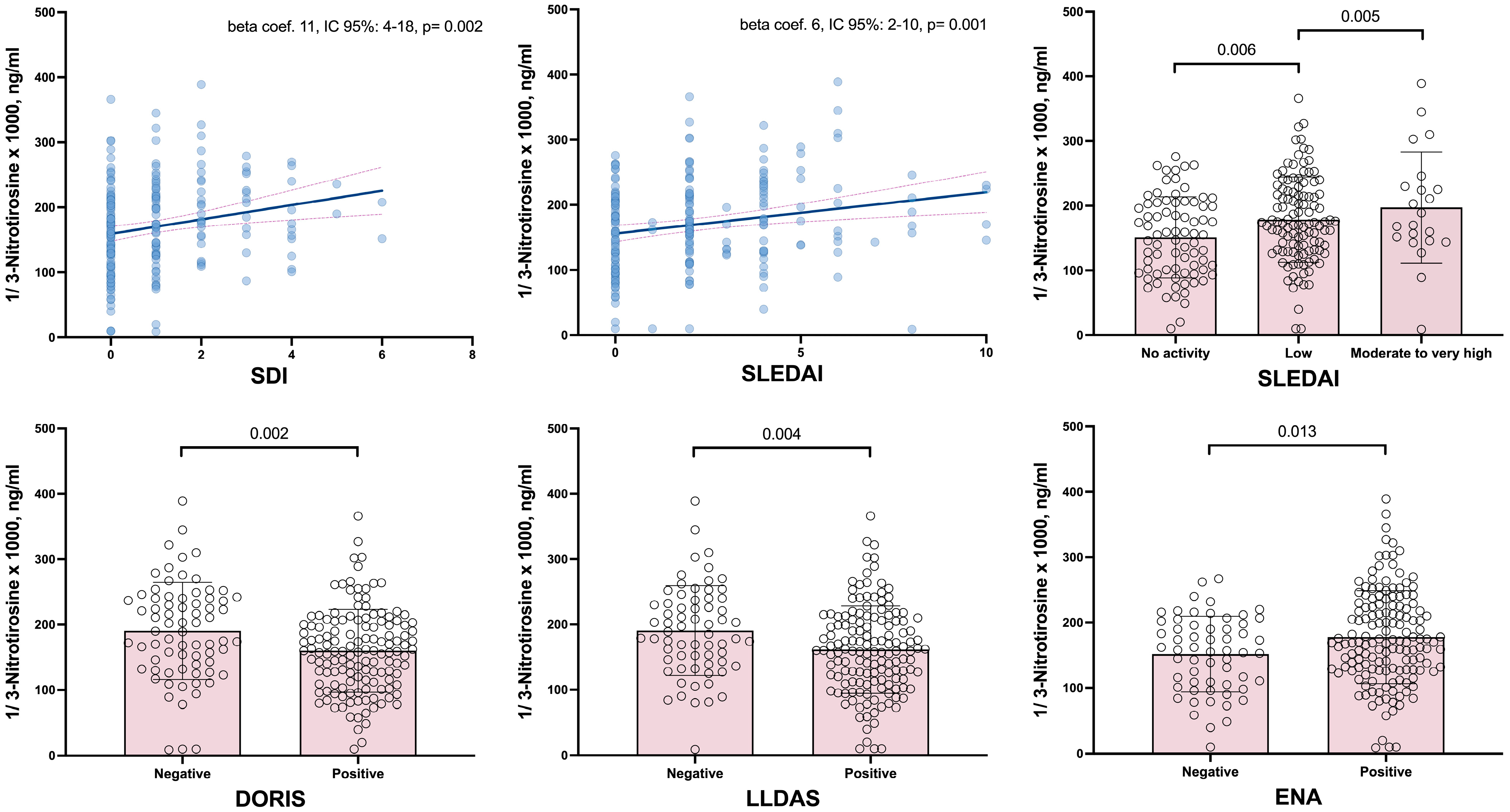
3.2. Relationship Demographics and Disease-Related Data with 3-Nitrotyrosine

Regarding the demographic characteristics, only BMI—whether analyzed as a continuous variable or categorized as ≥30 kg/m2—showed a significant association with 3-NT levels. Notably, classic CV risk factors such as smoking, diabetes, and hypertension, as well as the use of statins or aspirin, were not associated with 3-NT values (Table 2).
On the other hand, numerous associations were observed between the disease characteristics and the serum levels of 3-NT. In this regard, the disease duration, disease-related damage, and disease activity were significantly associated with the serum levels of 3-NT. Specifically, the SDI, both as a continuous variable and when categorized as 0 or ≥1, was associated with higher 3-NT levels. Similarly, the SLEDAI showed that patients with mild or moderate and high disease activity had higher 3-NT values compared to those in remission. Both the SLEDAI−2K and SDI relationships remained significant after multivariate adjustment (Table 2 and Figure 1). Since the SLEDAI−2K and SDI scores are constructed by summing different items corresponding to various domains or organ manifestations, we have broken down, for both calculators, the relationship of their respective items with 3-NT. In the case of SDI, the domains that showed statistical significance with 3-NT values were the CV, articular, and skin domains (Supplementary Table S1). For the SLEDAI−2K, the significant items related to 3-NT were a rash and an elevated anti-DNA (Supplementary Table S2). Furthermore, patients who met the criteria for remission or low disease activity, as defined by the DORIS or LLDAS, exhibited significantly inferior 3-NT levels compared to those who did not achieve these disease states (Table 2 and Figure 1).
Regarding the autoantibody profiles, positive associations with 3-NT were also observed. After adjusting for covariates, the patients who were positive for extractable nuclear antibodies (ENAs), anti-RNP, anti-ribosome, and anti-nucleosome antibodies exhibited significantly higher circulating 3-NT levels. For anti-dsDNA and anti-SSB, although statistical significance was not reached, a trend was noted. Remarkably, positivity for lupus anticoagulant, but not anti-cardiolipin IgG/IgM and anti-β2-glycoprotein I IgG/IgM, showed a significant relationship with 3-NT, although in this case the association was negative. Finally, none of the therapies used by the patients demonstrated a significant association with 3-NT levels (Table 2 and Figure 1).

3.3. Multivariable Analysis of Cardiovascular Disease-Related Factors’ Association with 3-Nitrotyrosine

The lipid profile was prominently associated with 3-NT values (Table 3). In this regard, after the multivariable adjustment, the triglycerides, non-HDL cholesterol, apolipoprotein B, LDL:HDL and ApoB:ApoA1 ratios, and the atherogenic index showed significant positive associations with 3-NT levels. In contrast, the insulin resistance indices did not demonstrate significant relations to 3-NT values. Similarly, the presence of carotid plaque and cIMT was not significantly associated with 3-NT levels. Notably, in the univariable analysis, patients with a high-risk SCORE2 exhibited significantly higher values compared to those at low risk. Although this significance was lost after the multivariable adjustment, a clear trend in this direction was observed (Table 3).

4. Discussion

Our study is the first to evaluate 3-NT in patients with SLE, specifically investigating its relationship with CV disease-related characteristics, including a comprehensive lipid profile, insulin resistance indices, and subclinical atherosclerosis. According to our results, 3-NT, a marker of protein oxidative stress, is associated not only with various disease characteristics but also with the abnormal lipid profile typically observed in these patients. This novel approach offers insights into the complex interplay between oxidative stress, lipid metabolism, and CV risk in SLE patients.
A substantial body of evidence, derived from numerous studies, indicates that patients with SLE exhibit elevated 3-NT levels compared to controls [21,22,23,24,25,26]. Additionally, previous research has explored the role of 3-NT in SLE. For instance, patients with active lupus nephritis have been found to have higher serum 3-NT levels than those without renal disease [21]. Similarly, in a study of 25 patients with SLE, those with high SLEDAI scores or elevated anti-dsDNA antibody levels exhibited increased oxidation compared with patients with low SLEDAI scores or low antibody levels [24]. Another study of 26 SLE patients also found a significant correlation between 3-NT and the SLEDAI, with renal variables within the SLEDAI showing the strongest correlation with serum 3-NT levels [22]. Our study includes the largest and most comprehensive cohort of SLE patients to date in which 3-NT levels have been analyzed. Our findings corroborate the previously mentioned reports by demonstrating a clear association between 3-NT levels and disease activity. This association was further confirmed in the multivariable analysis. Moreover, we describe a positive relationship between 3-NT and disease-related damage, as well as an inverse relationship with states of remission and low disease activity. These findings robustly confirm that 3-NT is positively associated with states of increased disease activity and damage.
We also found a positive association between 3-NT and the disease’s autoantibody profile. Prior descriptions have detailed how the presence of a nitro group on a tyrosine residue can elicit an immunological response [1]. These data indicate a potential role for nitrated epitopes of autologous proteins in the development of autoantibodies in SLE, potentially influencing the disease’s etiopathogenesis. Additionally, and surprisingly, we found a relationship between 3-NT and lupus anticoagulant, but in this case, the relationship was negative. We do not have a precise explanation for this. It is possible that patients with lupus anticoagulant more frequently take aspirin, which could have a negative effect on 3-nitrotyrosine. In this regard, aspirin has antioxidant properties and can reduce oxidative stress, which could potentially lower the levels of 3-NT. Since 3-NT is a marker of oxidative stress and nitrative damage, the aspirin use could dampen the oxidative environment in these patients, resulting in lower levels of 3-NT. In some patients with lupus anticoagulant, thrombotic processes may predominate over inflammatory ones. This could lead to a reduction in systemic inflammation or oxidative stress, which might otherwise decrease 3-NT levels.
A correlation between 3-NT and elevated levels of several lipid profile components was observed. Therefore, 3-NT appeared to be associated with a deleterious lipid profile in terms of the CV risk in patients with SLE. It is known that dyslipidemia can contribute to increased oxidative stress [27]. In this sense, oxidized LDL cholesterol is particularly damaging to blood vessel walls and has been strongly associated with angiographically documented coronary artery disease [28]. In addition, lipoproteins from SLE patients are more oxidized and have greater oxidative potential, which may contribute to accelerated atherosclerosis [29]. Based on our findings, we suggest that 3-NT may contribute to dyslipidemia in SLE and potentially increase the CV risk in these patients.
These findings regarding the association between 3-NT and both disease activity and damage, as well as its relationship with the autoantibody profile, suggest a clear link between the oxidative stress and inflammation marker 3-NT and major clinical outcomes in patients with SLE. We hypothesize that the overproduction of reactive nitrogen species in the pathogenesis of human SLE leads to increased nitration of tyrosine residues on serum proteins and the formation of 3-NT. Importantly, 3-NT has been proven to bind more strongly to SLE immunoglobulins than to other antigens such as dsDNA or native poly L-tyrosine, suggesting the formation of immunogenic nitrotyrosine-bearing epitopes [26]. These modified self-proteins may activate autoreactive T cells and contribute to the breakdown of immunological tolerance, a hallmark of SLE. Furthermore, we believe that the persistent elevation of 3-NT may indicate either ongoing or recurrent bursts of protein nitration, with inefficient repair mechanisms in SLE patients. Our finding of 3-NT serum levels’ association with increased disease activity, nephritis, and CV disease in SLE supports its role as a biomarker linking oxidative stress and inflammation to major clinical outcomes. The formation of immune complexes between SLE IgG and nitrated proteins further implicates 3-NT in the amplification of autoimmune responses and organ damage. Thus, 3-NT not only reflects the burden of oxidative stress and inflammation but may also actively participate in the pathogenesis and progression of critical clinical manifestations in SLE.
In our study, patients classified as high-risk according to the SCORE2 algorithm exhibited significantly elevated 3-NT levels compared to those in the low-risk category. Although this association was attenuated following multivariable adjustment, a notable trend persisted. The observed relationship between 3-NT levels and the SCORE2, a composite measure integrating multiple CV risk factors predictive of CV events, underscores the potential significance of 3-NT as a marker of CV risk in SLE patients. This finding suggests that 3-NT may reflect the cumulative impact of various CV risk factors in SLE, potentially serving as an integrative biomarker of oxidative stress and CV risk. These findings may align with the concept that protein nitration within the arterial wall has been described as reducing endothelial cell adhesion, leading to endothelial denudation, thrombus formation, and phenotypic changes in smooth muscle cells [30]. These processes may collectively contribute to the rupture of atherosclerotic plaques, subsequent thrombus formation, and the occurrence of myocardial infarctions and strokes. Nevertheless, further investigation into the mechanistic links between oxidative stress, as measured by 3-NT, and CV risk in SLE is warranted.
Despite this, we did not observe an association between 3-NT levels and the presence of carotid plaque or cIMT measurements. Atherosclerosis is a chronic process in which CV risk factors play a central role. We believe that the cross-sectional nature of our study, along with the potential confounding effect of traditional CV risk factors, may have limited our ability to detect a relationship between 3-NT and markers of subclinical carotid atherosclerosis.
We acknowledge that we did not include a control group. However, the purpose of our study was not to compare 3-NT levels between controls and SLE patients, but rather to explore the relationship between this molecule and disease characteristics. Additionally, as previously mentioned, the differences in 3-NT serum levels between SLE patients and controls have been thoroughly explored in previous studies. The cross-sectional nature of our work prevents inferring causality, for which prospective studies would be required. In this regard, this cross-sectional design prevents us from drawing conclusions regarding the effect of disease therapies on 3-NT.

5. Conclusions

In conclusion, by analyzing 3-NT levels in relation to CV parameters, our study enhances the understanding of the mechanisms behind the elevated CV risk observed in SLE. Additionally, the relationship between 3-NT and lipid profile abnormalities points to a potential connection between oxidative stress and dyslipidemia in SLE, which may play a role in the accelerated atherosclerosis seen in these patients.

Supplementary Materials

The following supporting information can be downloaded at: https://www.mdpi.com/article/10.3390/antiox14060739/s1; Table S1: Relation of SDI score items to 3-NT serum values; Table S2: SLEDAI−2k items’ relation to 3-NT.

Author Contributions

I.F.-A. and M.Á.G.-G., conception, design, and interpretation and write-up of the data; J.C.Q.-A., M.H.-D., M.G.-G., F.G.-B., C.A.-S., E.H.-R., A.d.V.-G., A.G.-D., P.A.-G., B.T.-S. and C.M.-G., acquisition of the data. All authors have collectively committed to personal accountability for their respective contributions to the work. They have also committed to addressing any inquiries regarding the accuracy or integrity of any aspect of the study, even those outside their personal involvement, with appropriate investigation and resolution, documenting these resolutions in the literature. All authors have read and agreed to the published version of the manuscript.

Funding

This study has been funded by a grant to IF-A by Fundación Canaria Instituto de Investigación Sanitaria de Canarias (FIISC) through the project PIFIISC23/07. Professor Gonzalez-Gay is funded by the Spanish Research Network RICORS—RD24/0007/0031, through Next Generation EU funds, which support the initiatives of the Recovery and Resilience Mechanism (MRR).

Institutional Review Board Statement

The study protocol received approval from the Institutional Review Committee at the Hospital Universitario de Canarias (approval number CHUC_2023_48, 25 May 2023). All participating individuals provided written informed consent. The research was conducted in strict adherence to the applicable guidelines and regulations, as well as in accordance with the principles outlined in the Declaration of Helsinki.

Informed Consent Statement

Informed consent was obtained from all subjects involved in the study.

Data Availability Statement

Data will be made available on request.

Conflicts of Interest

The authors declare that there are no conflicts of interest. Nevertheless, Iván Ferraz-Amaro would like to acknowledge that he has received grants/research support from Abbott, MSD, Janssen, and Roche, as well as consultation fees from company-sponsored speakers’ bureaus associated with Abbott, Pfizer, Roche, Sanofi, Celgene, and MSD. M.A. González-Gay has received research support from GSK as well as consultation/participation fees from company-sponsored speakers’ bureaus associated with GSK, Otsuka, and Sanofi.

Abbreviations

The following abbreviations are used in this manuscript:
3-NT3-Nitrotyrosine
SLESystemic lupus erythematosus
CVCardiovascular
CVDCardiovascular disease
ACRAmerican College of Rheumatology
SLEDAI−2KSystemic Lupus Erythematosus Disease Activity Index
SLICC-SDISystemic Lupus International Collaborating Clinics/American College of Rheumatology Damage Index
DORISDefinitions of Remission in SLE
LLDASLupus Low Disease Activity State
PGAPhysician global assessment
SCORE2Systematic Coronary Risk Evaluation−2
HDLHigh-density cholesterol
SCORE2-OPSystematic Coronary Risk Evaluation−2 Older Persons
LDLLow-density cholesterol
HOMAHomeostatic model assessment
IRInsulin resistance
cIMTCarotid intima–media
SDStandard deviation
IQRInterquartile range
BMIBody mass index
CRPC-reactive protein
ACAsAnticardiolipin antibodies
ANAsAntinuclear antibodies
ENAsExtractible nuclear antibodies
DMARDDisease-modifying antirheumatic drug
ACVAAcute cerebrovascular accident

References

  1. Ahsan, H. 3-Nitrotyrosine: A Biomarker of Nitrogen Free Radical Species Modified Proteins in Systemic Autoimmunogenic Conditions. Hum. Immunol. 2013, 74, 1392–1399. [Google Scholar] [CrossRef] [PubMed]
  2. Souza, J.M.; Peluffo, G.; Radi, R. Protein Tyrosine Nitration--Functional Alteration or Just a Biomarker? Free Radic. Biol. Med. 2008, 45, 357–366. [Google Scholar] [CrossRef]
  3. Daiber, A.; Münzel, T. Increased Circulating Levels of 3-Nitrotyrosine Autoantibodies: Marker for or Maker of Cardiovascular Disease? Circulation 2012, 126, 2371–2373. [Google Scholar] [CrossRef] [PubMed]
  4. Bandookwala, M.; Sengupta, P. 3-Nitrotyrosine: A Versatile Oxidative Stress Biomarker for Major Neurodegenerative Diseases. Int. J. Neurosci. 2020, 130, 1047–1062. [Google Scholar] [CrossRef]
  5. Pennathur, S.; Vivekanandan-Giri, A.; Locy, M.L.; Kulkarni, T.; Zhi, D.; Zeng, L.; Byun, J.; De Andrade, J.A.; Thannickal, V.J. Oxidative Modifications of Protein Tyrosyl Residues Are Increased in Plasma of Human Subjects with Interstitial Lung Disease. Am. J. Respir. Crit. Care Med. 2016, 193, 861–866. [Google Scholar] [CrossRef]
  6. Restivo, V.; Candiloro, S.; Daidone, M.; Norrito, R.; Cataldi, M.; Minutolo, G.; Caracci, F.; Fasano, S.; Ciccia, F.; Casuccio, A.; et al. Systematic Review and Meta-Analysis of Cardiovascular Risk in Rheumatological Disease: Symptomatic and Non-Symptomatic Events in Rheumatoid Arthritis and Systemic Lupus Erythematosus. Autoimmun. Rev. 2022, 21, 102925. [Google Scholar] [CrossRef] [PubMed]
  7. Perl, A. Oxidative Stress in the Pathology and Treatment of Systemic Lupus Erythematosus. Nat. Rev. Rheumatol. 2013, 9, 674–686. [Google Scholar] [CrossRef]
  8. Merino de Paz, N.; García-González, M.; Gómez-Bernal, F.; Quevedo-Abeledo, J.C.; de Vera-González, A.; López-Mejias, R.; Abreu-González, P.; Martín-González, C.; González-Gay, M.; Ferraz-Amaro, I. Relationship between Malondialdehyde Serum Levels and Disease Features in a Full Characterized Series of 284 Patients with Systemic Lupus Erythematosus. Antioxidants 2023, 12, 1535. [Google Scholar] [CrossRef]
  9. Yan, Z.; Chen, Q.; Xia, Y. Oxidative Stress Contributes to Inflammatory and Cellular Damage in Systemic Lupus Erythematosus: Cellular Markers and Molecular Mechanism. J. Inflamm. Res. 2023, 16, 453–465. [Google Scholar] [CrossRef]
  10. Ohl, K.; Tenbrock, K. Oxidative Stress in SLE T Cells, Is NRF2 Really the Target to Treat? Front. Immunol. 2021, 12, 633845. [Google Scholar] [CrossRef]
  11. Shah, D.; Mahajan, N.; Sah, S.; Nath, S.K.; Paudyal, B. Oxidative Stress and Its Biomarkers in Systemic Lupus Erythematosus. J. Biomed. Sci. 2014, 21, 1–13. [Google Scholar] [CrossRef] [PubMed]
  12. Hochberg, M.C. Updating the American College of Rheumatology Revised Criteria for the Classification of Systemic Lupus Erythematosus. Arthritis Rheum. 1997, 40, 1725. [Google Scholar] [CrossRef]
  13. Gladman, D.D.; Ibañez, D.; Urowltz, M.B. Systemic Lupus Erythematosus Disease Activity Index 2000. J. Rheumatol. 2002, 29, 288–291. [Google Scholar]
  14. Gladman, D.; Ginzler, E.; Goldsmith, C.; Fortin, P.; Liang, M.; Urowitz, M.; Bacon, P.; Bombardieri, S.; Hanly, J.; Hay, E.; et al. The Development and Initial Validation of the Systemic Lupus International Collaborating Clinics/American College of Rheumatology Damage Index for Systemic Lupus Erythematosus. Arthritis Rheum. 1996, 39, 363–369. [Google Scholar] [CrossRef]
  15. Mosca, M.; Bombardieri, S. Assessing Remission in Systemic Lupus Erythematosus. Clin. Exp. Rheumatol. 2006, 24, S99–S104. [Google Scholar]
  16. van Vollenhoven, R.F.; Bertsias, G.; Doria, A.; Isenberg, D.; Morand, E.; Petri, M.A.; Pons-Estel, B.A.; Rahman, A.; Ugarte-Gil, M.F.; Voskuyl, A.; et al. 2021 DORIS Definition of Remission in SLE: Final Recommendations from an International Task Force. Lupus Sci. Med. 2021, 8, e000538. [Google Scholar] [CrossRef]
  17. Polachek, A.; Gladman, D.D.; Su, J.; Urowitz, M.B. Defining Low Disease Activity in Systemic Lupus Erythematosus. Arthritis Care Res. 2017, 69, 997–1003. [Google Scholar] [CrossRef] [PubMed]
  18. ESC Cardiovasc Risk Collaboration; SCORE2 working group. SCORE2 Risk Prediction Algorithms: New Models to Estimate 10-Year Risk of Cardiovascular Disease in Europe. Eur. Heart J. 2021, 42, 2439–2454. [Google Scholar] [CrossRef] [PubMed]
  19. Touboul, P.J.; Hennerici, M.G.; Meairs, S.; Adams, H.; Amarenco, P.; Bornstein, N.; Csiba, L.; Desvarieux, M.; Ebrahim, S.; Hernandez Hernandez, R.; et al. Mannheim Carotid Intima-Media Thickness and Plaque Consensus (2004-2006-2011). An Update on Behalf of the Advisory Board of the 3rd, 4th and 5th Watching the Risk Symposia, at the 13th, 15th and 20th European Stroke Conferences, Mannheim, Germany, 2004, Brussels, Belgium, 2006, and Hamburg, Germany, 2011. Cerebrovasc. Dis. 2012, 34, 290–296. [Google Scholar] [CrossRef]
  20. Maldonado, G.; Greenland, S. Simulation Study of Confounder-Selection Strategies. Am. J. Epidemiol. 1993, 138, 923–936. [Google Scholar] [CrossRef]
  21. Oates, J.C.; Christensen, E.F.; Reilly, C.M.; Self, S.E.; Gilkeson, G.S. Prospective Measure of Serum 3-Nitrotyrosine Levels in Systemic Lupus Erythematosus: Correlation with Disease Activity. Proc. Assoc. Am. Physicians 1999, 111, 611–621. [Google Scholar] [CrossRef] [PubMed]
  22. Gilkeson, G.; Cannon, C.; Oates, J.; Reilly, C.; Goldman, D.; Petri, M. Correlation of Serum Measures of Nitric Oxide Production with Lupus Disease Activity. J. Rheumatol. 1999, 26, 318–324. [Google Scholar] [PubMed]
  23. Wang, G.; Pierangeli, S.S.; Papalardo, E.; Ansari, G.A.S.; Khan, M.F. Markers of Oxidative and Nitrosative Stress in Systemic Lupus Erythematosus: Correlation with Disease Activity. Arthritis Rheum. 2010, 62, 2064–2072. [Google Scholar] [CrossRef]
  24. Morgan, P.E.; Sturgess, A.D.; Davies, M.J. Increased Levels of Serum Protein Oxidation and Correlation with Disease Activity in Systemic Lupus Erythematosus. Arthritis Rheum. 2005, 52, 2069–2079. [Google Scholar] [CrossRef] [PubMed]
  25. Khan, F.; Ali, R. Antibodies against Nitric Oxide Damaged Poly L-Tyrosine and 3-Nitrotyrosine Levels in Systemic Lupus Erythematosus. J. Biochem. Mol. Biol. 2006, 39, 189–196. [Google Scholar] [CrossRef]
  26. Khan, F.; Siddiqui, A.A.; Ali, R. Measurement and Significance of 3-Nitrotyrosine in Systemic Lupus Erythematosus. Scand. J. Immunol. 2006, 64, 507–514. [Google Scholar] [CrossRef] [PubMed]
  27. Vekic, J.; Stromsnes, K.; Mazzalai, S.; Zeljkovic, A.; Rizzo, M.; Gambini, J. Oxidative Stress, Atherogenic Dyslipidemia, and Cardiovascular Risk. Biomedicines 2023, 11, 2897. [Google Scholar] [CrossRef]
  28. Tsimikas, S.; Brilakis, E.S.; Miller, E.R.; McConnell, J.P.; Lennon, R.J.; Kornman, K.S.; Witztum, J.L.; Berger, P.B. Oxidized Phospholipids, Lp(a) Lipoprotein, and Coronary Artery Disease. N. Engl. J. Med. 2005, 353, 46–57. [Google Scholar] [CrossRef]
  29. Park, J.K.; Kim, J.Y.; Moon, J.Y.; Ahn, E.Y.; Lee, E.Y.; Lee, E.B.; Cho, K.H.; Song, Y.W. Altered Lipoproteins in Patients with Systemic Lupus Erythematosus Are Associated with Augmented Oxidative Stress: A Potential Role in Atherosclerosis. Arthritis Res. Ther. 2016, 18, 306. [Google Scholar] [CrossRef]
  30. Davies, M.J. Protein Nitration in the Artery Wall: A Contributor to Cardiovascular Disease? Redox Biochem. Chem. 2024, 8, 100032. [Google Scholar] [CrossRef]
Figure 1. Relation of SDI and SLEDAI scores and DORIS, LLDAS, and ENA to 3-NT serum levels. SDI: damage index; SLEDAI: Systemic Lupus Erythematosus Disease Activity Index; LLDAS: Low Lupus Disease Activity State; DORIS: Definitions of Remission In SLE; ENA: extractable autoantibodies.
Antioxidants 14 00739 g001
Table 1. Characteristics of systemic lupus erythematosus patients.
SLE patients
(n = 214)
3-Nitrotyrosine, ng/mL5.9 (4.7–8.0)
Age, years51 ± 12
Female, n (%)194 (91)
Body mass index, kg/m228 ± 6
Abdominal circumference, cm91 ± 15
Waist circumference, cm100 ± 15
Waist to hip ratio0.92 ± 0.33
Cardiovascular co-morbidity
Smoking, n (%)40 (19)
Diabetes, n (%)11 (5)
Hypertension, n (%)82 (38)
Obesity, n (%)58 (27)
Metabolic syndrome, n (%)92 (44)
Statins, n (%)61 (29)
Aspirin, n (%)59 (28)
SCORE2, %2.1 (1.1–3.8)
SCORE2 categories
Low risk171 (80)
Moderate risk28 (13)
High risk15 (7)
Carotid ultrasound
Carotid intima–media thickness, microns657 ± 144
Carotid plaque, n (%)69 (44)
Laboratory data
CRP, mg/dL1.86 (0.8–4.14)
Cholesterol, mg/dL183 ± 40
Triglycerides, mg/dL112 ± 64
HDL-cholesterol, mg/dL55 ± 15
LDL-cholesterol, mg/dL106 ± 33
LDL:HDL-cholesterol ratio2.06 ± 0.82
Non-HDL-cholesterol, mg/dL128 ± 38
Lipoprotein A, mg/dL32 (11–85)
Apolipoprotein A1, mg/dL157 ± 28
Apolipoprotein B, mg/dL87 ± 23
Apo B:Apo A1 ratio0.57 ± 0.17
Atherogenic index3.52 ± 1.09
Insulin resistance indices
Glucose, mg/dL92 ± 17
Insulin, µU/mL14 ± 17
C-peptide, ng/mL2.4 ± 1.69
HOMA2-IR1.10 (0.70–1.80)
HOMA2-S%93 (5–150)
HOMA2-B%-C-peptide122 (95–153)
SLE-related data
Disease duration, years18 ± 11
CRP, mg/dL1.9 (0.8–4.1)
SLICC-DI1 (0–1)
SLICC-DI >=1, n (%)113 (53)
SLEDAI−2K2 (0–4)
SLEDAI categories, n (%)
No activity, n (%)80 (37)
Mild, n (%)111 (52)
Moderate to high, n (%)23 (11)
DORIS, n (%)139 (65)
LLDAS, n (%)146 (68)
Auto-antibody profile
Anti-DNA positive, n (%)160 (75)
Anti-ENA positive, n (%)151 (72)
Anti-SSA, n (%)75 (35)
Anti-SSB, n (%)33 (15)
Anti-Sm, n (%)39 (18)
Anti-RNP, n (%)63 (30)
Anti-ribosome25 (12)
Anti-nucleosome44 (21)
Anti-histone42 (20)
Antiphospholipid syndrome, n (%) 37 (17)
Antiphospholipid autoantibodies, n (%)69 (32)
Lupus anticoagulant, n (%)48 (23)
ACA IgM, n (%)29 (14)
ACA IgG, n (%)29 (14)
Anti beta2 glycoprotein IgM, n (%)20 (10)
Anti beta2 glycoprotein IgG, n (%)21 (10)
Current prednisone, n (%)74 (35)
Prednisone, mg/day5 (2.5–5)
Hydroxychloroquine, n (%)154 (72)
Methotrexate, n (%)28 (13)
Mycophenolate mofetil, n (%)26 (12)
Azathioprine, n (%)20 (9)
Rituximab, n (%)5 (2)
Belimumab, n (%)27 (13)
Data represent means ± SD or median (interquartile range) when data were not normally distributed. BMI: body mass index; C3 C4: complement; CRP: C-reactive protein; LDL: low-density lipoprotein; DMARD: disease-modifying antirheumatic drug; ACA: anticardiolipin; HDL: high-density lipoprotein; ANA: antinuclear antibodies; ENA: extractible nuclear antibodies; LLDAS: Low Lupus Disease Activity State; DORIS: Definitions Of Remission In SLE; SLEDAI: Systemic Lupus Erythematosus Disease Activity Index; SLEDAI categories were defined as follows: 0 no activity, 1–5 mild activity, 6–10 moderate activity, >10 high activity, >20 very high activity; SLICC: Systemic Lupus International Collaborating Clinics/American College of Rheumatology Damage Index.
Table 2. Multivariable analysis of disease-related data association with 3-nitrotyrosine.
1/3-Nitrotyrosine × 1000, ng/mL
Beta Coef. (95% CI), p
Univariable Multivariable
Age, years0.06 (−0.72–0.84)0.88
Female−22 (−53–10)0.18
Body mass index, kg/m21.53 (0.03–3.03)0.046
Abdominal circumference, cm0.42 (−0.21–1.06)0.19
Waist circumference, cm0.51 (−0.12–1.14)0.16
Waist to hip ratio−0.55 (−28.82–27.72)0.97
Cardiovascular co-morbidity
Smoking4 (−19–29)0.69
Diabetes−16 (−58–26)0.45
Hypertension2 (−17–21)0.86
Obesity21 (0.42–42)0.046
Dyslipidemia0.59 (−18.94–20.12)0.95
Statins5 (−16–26)0.65
Aspirin−3 (−24–18)0.81
SLE-related data
Disease duration, years1.04 (0.21–1.87)0.0150.93 (0.09–1.77)0.030
CRP, mg/dl−0.059 (−0.19–0.08)0.40
SLICC-DI11 (4–18)0.00210 (3–17)0.006
SLICC-DI >=120 (11–48)0.00227 (9–45)0.004
SLEDAI−2K6 (2–10)0.0016. (3–10)0.001
SLEDAI categories
No activityref. Ref.
Mild28 (8–47)0.00627 (8–47)0.006
Moderate to very high46 (14–78)0.00546 (15–78)0.004
DORIS−30 (−50–(−11))0.002−28 (−47–(−9))0.004
LLDAS−30 (−49–(−9))0.004−27 (−47–(−7))0.008
Auto-antibody profile
Anti-DNA positive20 (−0.75–42)0.05918 (−3–40)0.089
Anti-ENA positive26 (5–47)0.01327 (6–47)0.011
Anti-SSA7 (−12–27)0.73
Anti-SSB18 (−8–45)0.1721 (−5–47)0.12
Anti-Sm17 (−7–41)0.1614 (−10–38)0.26
Anti-RNP26 (6–46)0.01125 (5–46)0.013
Anti-ribosome36 (7–64)0.01436 (8–64)0.012
Anti-nucleosome31 (8–54)0.00831 (8–53)0.008
Anti-histone13 (−10–36)0.28
Antiphospholipid syndrome−8 (−33–16)0.50
Antiphospholipid autoantibodies−12 (−34–10)0.29
Lupus anticoagulant−29 (−51–(−7))0.010−34 (−56–(−13))0.002
ACA IgM−12 (−39–14)0.36
ACA IgG4 (−23–31)0.79
Anti beta2 glycoprotein IgM−13 (−44–19)0.43
Anti beta2 glycoprotein IgG2 (−29–33)0.91
Current prednisone12 (−8–31)0.24
Prednisone, mg/day2 (−3–6)0.54
Hydroxychloroquine−1 (−21–20)0.96
Methotrexate4 (−23–32)0.76
Mycophenolate mofetil−4 (−33–25)0.77
Azathioprine11 (−21–42)0.51
Rituximab14 (−48–75)0.44
Belimumab22 (−5–50)0.1125 (−3–53)0.077
In this analysis, the serum levels of 3-NT are the dependent variable. The multivariable analysis is adjusted for sex and body mass index. SLEDAI: Systemic Lupus Erythematosus Disease Activity Index; LLDAS: Low Lupus Disease Activity State; DORIS: Definitions Of Remission In SLE; the SLEDAI categories were defined as follows: 0 no activity, 1–5 mild activity, 6–10 moderate activity, >10 high activity, >20 very high activity; SLICC: Systemic Lupus International Collaborating Clinics/American College of Rheumatology Damage Index; CRP: C-reactive protein; ENA: extractable autoantibodies; ACA: anticardiolipin autoantibodies. Significant p values are depicted in bold.
Table 3. Multivariable analysis of cardiovascular disease-related factors’ association with 3-nitrotyrosine.
1/3-Nitrotyrosine × 1000, ng/mL
Beta coef. (95% CI), p
UnivariableMultivariable
Lipid profile
Cholesterol, mg/dL0.2 (−0.058–0.42)0.140.2 (−0.04–0.4)0.097
Triglycerides, mg/dL0.2 (0.06–0.4)0.0090.2 (0.03–0.4)0.024
HDL-cholesterol, mg/dL−0.6 (−1.2–0.09)0.089−0.5 (−1–0.2)0.17
LDL-cholesterol, mg/dL0.2 (−0.05–0.5)0.140.2 (−0.02–0.5)0.070
LDL:HDL-cholesterol ratio16 (5–27)0.00715 (4–27)0.009
Non-HDL-cholesterol, mg/dL0.3 (0.04–0.6)0.0230.3 (−0.05–0.6)0.021
Lipoprotein A, mg/dL0.02 (−0.1–0.2)0.70
Apolipoprotein A1, mg/dL−0.3 (−0.6–0.06)0.11−0.2 (−0.6–0.09)0.15
Apolipoprotein B, mg/dL0.5 (0.06–0.9)0.0250.5 (0.05–0.9)0.029
Apo B:Apo A1 ratio80 (27–134)0.00476 (22–130)0.006
Atherogenic index15 (6–24)0.00114 (5–23)0.002
Insulin resistance indices
Glucose, mg/dL0.1 (−0.4–0.7)0.65
C-peptide, ng/mL2 (−4–7)0.59
Insulin, µU/mL0.2 (−0.4–0.8)0.57
HOMA2-IR2 (−4–7)0.53
HOMA2-S%−0.005 (−0.1–0.1)0.93
HOMA2-B%-C-peptide−0.01 (−0.2–0.2)0.91
Carotid ultrasound
cIMT, microns−0.3 (−0.1–0.05)0.44
Carotid plaque15 (−6–36)0.1714 (−7–36)0.20
SCORE2 calculator0.4 (−2–3)0.77
Low riskref. ref.
Moderate risk−0.5 (−28–27)0.97−6 (−34–22)0.67
High risk44 (6–83)0.02537 (−2–76)0.060
In this analysis, serum levels of 3-NT are dependent variable. HOMA2-IR: insulin resistance index through homeostatic model assessment (calculated with glucose and insulin serum levels). HOMA2-S%: insulin sensitivity index through homeostatic model assessment (calculated with glucose and insulin serum levels). HOMA2-B%-C-peptide: β-cell function index through homeostatic model assessment (calculated with glucose and C-peptide serum levels). SCORE2: Systematic Coronary Risk Assessment; high density lipoprotein; LDL: low density lipoprotein; cIMT: carotid intima–media thickness. Multivariable analysis is adjusted for sex and body mass index. Significant p values are depicted in bold.
Disclaimer/Publisher’s Note: The statements, opinions and data contained in all publications are solely those of the individual author(s) and contributor(s) and not of MDPI and/or the editor(s). MDPI and/or the editor(s) disclaim responsibility for any injury to people or property resulting from any ideas, methods, instructions or products referred to in the content.

