Article

Antimicrobial Evaluation and Fraction-Based Profiling of Basil Essential Oil Against Vaginal Pathogens

1
Department of Food Science and Nutrition, College of Human Ecology, Pusan National University, Busan 46241, Republic of Korea
2
Department of Biomaterials Science, College of Natural Resources and Life Science, Pusan National University, Miryang 50463, Republic of Korea
3
Longevity & Wellbeing Research Center, Pusan National University, Miryang 50463, Republic of Korea
*
Authors to whom correspondence should be addressed.
These authors contributed equally to this work.
Antioxidants 2025, 14(6), 628; https://doi.org/10.3390/antiox14060628
Submission received: 17 April 2025 / Revised: 20 May 2025 / Accepted: 22 May 2025 / Published: 23 May 2025

Abstract

:
Basil (Ocimum basilicum L.) has been traditionally used in various cultures for its medicinal properties. This study evaluated the antioxidant and antimicrobial activities of basil essential oil (BEO) and identified its key bioactive compounds. Antioxidant activity testing, as determined by DPPH and ABTS assays, returned EC50 values of 115.36 and 54.77 µg/mL, respectively. BEO demonstrated significant antimicrobial effects against Gardnerella vaginalis, Fannyhessea vaginae, Chryseobacterium gleum, and Candida albicans, with inhibition zones of up to 25.88 mm and MIC values ranging from 31 to 500 µg/mL. GC-MS (gas chromatography–mass spectrometry) identified monoterpene, phenylpropene, and sesquiterpene derivatives in BEO. In addition, Fraction 3 (Fr. 3) obtained by preparative HPLC had the highest antimicrobial activity, and methyl trans-cinnamate was identified as the primary active compound in this fraction. BEO had no toxic effect on Lactobacillus crispatus or human dermal fibroblasts. These findings support the traditional use of basil and highlight its potential as a safe, natural therapeutic agent with antioxidant and antimicrobial properties.

Graphical Abstract

1. Introduction

Traditionally utilized medicinal herbs consist of various plant species and contribute significantly to the synthesis and development of modern pharmaceuticals [1]. Particularly, in some developing countries, herbal medicines are utilized as the primary treatment for infections, and they are regarded as a valuable resource in ongoing research aimed at discovering natural antibacterial agents [2].
Basil (Ocimum basilicum L.), which is also one of these medicinal plants and a member of the Lamiaceae family, has been traditionally used in the folk medicines of various cultures as a tonic, anthelmintic, diuretic, and antispasmodic, and as a treatment for upper respiratory tract infections [3,4]. Furthermore, modern pharmacological studies have shown that basil has significant antibacterial, anti-inflammatory, and antioxidant activities [5,6,7]. These therapeutic properties are attributed primarily to its rich composition of bioactive compounds, which include monoterpenes, sesquiterpenes, phenylpropanoids, anthocyanins, and phenolic acids [8]. Particularly, the bioactive compounds in basil may promote antimicrobial activity by disrupting the permeability and integrity of bacterial membranes, producing intracellular ATP and potassium ion leakage, ultimately leading to cell death [9]. Thus, basil has been utilized in various ways across different regions and cultures, and its traditional uses are gradually being validated through contemporary scientific research.
Among the extracts derived from basil, basil essential oil (BEO) also has potent antimicrobial effects, particularly against Gram-positive bacteria such as Bacillus sacharolyticus, B. stearothermophilus, B. subtilis, B. thuringiensis, Micrococcus glutamicus, and Sarcina lutea [10]. Given these broad antimicrobial properties, BEO has therapeutic potential for the treatment of a variety of infections, including those affecting the vaginal microbiota. However, few studies have investigated its effects on vaginal infections, such as bacterial vaginosis and candidiasis. Thus, although the therapeutic properties of basil are well documented, the specific mechanisms underlying its antimicrobial activity, particularly in the context of vaginal health, remain insufficiently explored.
A healthy vaginal pH typically ranges from 3.8 to 4.5 and is maintained by beneficial lactobacilli such as Lactobacillus crispatus (L. crispatus), L. jensenii, L. gasseri, and L. coleohominis [11,12,13]. Disruptions to this acidic environment or reductions in lactobacillus populations can lead to vaginal dysbiosis and facilitate the growth of pathogenic microorganisms associated with bacterial vaginosis (BV) and vulvovaginal candidiasis (VVC) [14]. BV is one of the most prevalent vaginal infections in women and arises from disturbances in the vaginal ecosystem due to increases in the relative populations of bacteria such as Gardnerella vaginalis (G. vaginalis), Atopobium vaginae (Fannyhessea vaginae; F. vaginae), and Chryseobacterium gleum (C. gleum) [15,16]. The resulting conditions are characterized by malodorous vaginal discharge, itching, and discomfort [17]. Similarly, VVC is a common condition caused by Candida species, particularly Candida albicans (C. albicans), and has a lifetime prevalence of up to 75% [18].
Various therapeutic approaches, such as antimicrobial agents, probiotics, and lifestyle modifications, have been proposed to manage and prevent BV and VVC. However, the overuse of antibiotics has led to the development of antibiotic-resistant strains and the disruption of the normal vaginal microbiota, particularly to the depletion of beneficial lactic acid bacteria. These challenges highlight the need for phytotherapeutic agents with scientifically validated efficacy and safety profiles to restore vaginal health and prevent recurrent vaginitis.
This study aimed to evaluate the antioxidant and antimicrobial activities of BEO against four vaginitis-associated pathogens, viz., G. vaginalis, F. vaginae, C. gleum, and C. albicans. In addition, we identified the key active antimicrobial compounds within BEO and assessed the cytotoxicity of BEO on beneficial vaginal microbiota and human cells. By integrating traditional medicinal knowledge and modern pharmacological analysis, we undertook to support the potential use of BEO as a safe and effective prophylactic and therapeutic agent for vaginitis.

2. Materials and Methods

2.1. Plant Material Sourcing and Identification

Dried and rubbed basil (Ocimum basilicum L.) powder purchased from Shinyoung FS Co. (Gwangju, Republic of Korea) was freeze-dried using a lyophilizer (Rikakikai Co., Tokyo, Japan) under vacuum at −70 °C for 48 h to preserve heat-sensitive bioactive compounds.

2.2. Essential Oil Extraction

BEO was extracted by steam distillation, as previously described, with slight modifications [19]. Briefly, 400 g of dried basil powder was placed in a distillation apparatus (Dongguan City Niangge Machinery Co., Dongguan, China) containing 2 L of deionized water. Distillation was conducted at 100 ± 5 °C for 65 min or until no additional oil was collected. Modifications included adjusting the distillation time to optimize the oil yield and purity. The obtained BEO was dried over anhydrous sodium sulfate (Na2SO4) to remove residual moisture, and the extraction yield was calculated using the following equation:
Extraction yield (%) = [Weight of dried BEO obtained (g)/Weight of dried basil powder (g)] × 100.

2.3. Phytochemical Analysis

2.3.1. Gas Chromatography–Mass Spectrometry (GC-MS)

The chemical composition of the BEO was analyzed by GC-MS (Agilent, Little Falls, DE, USA). GC was performed with a DB-5MS capillary column (30 m × 0.25 mm i.d. × 0.25 μm film thickness; 5%-phenyl-methylpolysiloxane stationary phase) using helium as the carrier gas, at a flow rate of 1 mL/min and an injector temperature of 220 °C.
The injection volume was 1 µL, and a split injection technique (split ratio 10:1) was used. The oven temperature was programmed as follows: 40 °C for 2 min, 40 °C to 240 °C at 15 °C/min, and 240 °C to 280 °C at 30 °C/min. MS was performed using an ion source temperature of 200 °C and a mass range of 40–400 m/z. The total run time was 18 min, and data acquisition was initiated 5 min after starting the runs. Compounds were identified by comparing the relative retention times and mass spectra with those in the Wiley and NIST (National Institute of Standards and Technology) libraries (Gaithersburg, MD, USA).

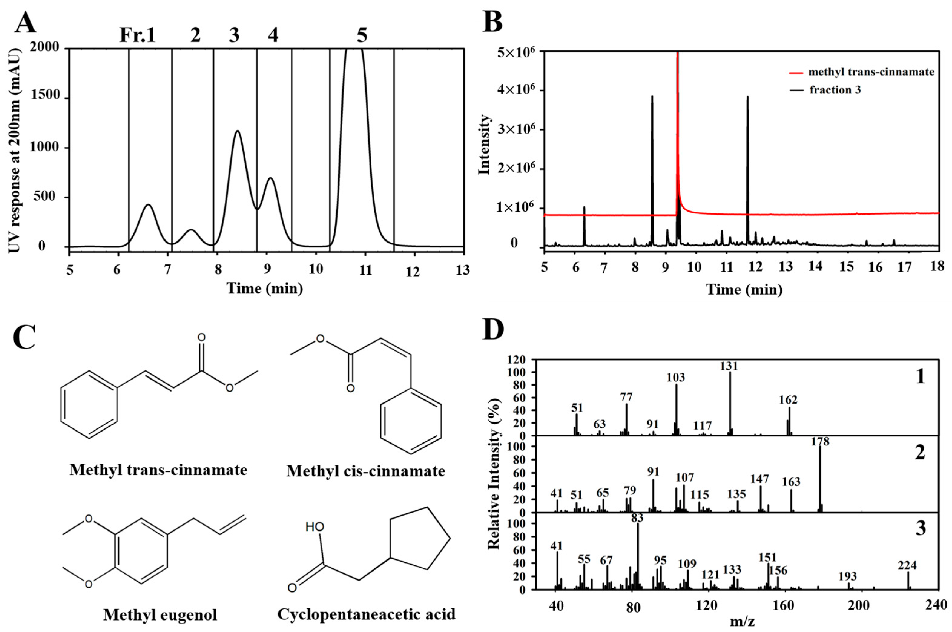
2.3.2. Fractionation of BEO by Preparative HPLC

BEO was fractionated using a preparative HPLC system (LC-forte/R, YMC Co., Kyoto, Japan) equipped with a YMC-Triart Prep C18-S column (250 mm × 10.0 mm, i.d. 10 µm; YMC Co., Ltd., Kyoto, Japan). Prior to the separations, the column was equilibrated for 10 min at room temperature. The BEO was diluted to 0.3% using HPLC-grade methanol and filtered through a 0.45 µm membrane filter. A 100 µL aliquot of this solution was then injected into the system. The mobile phase consisted of solvent A (50% methanol in HPLC-grade water) and solvent B (HPLC-grade acetonitrile). The elution gradient was programmed using a linear gradient (solvent A 70% to 10% over 0–20 min), and the flow rate was maintained at 4.8 mL/min. Peak detection was performed using a photodiode-array detector (PDA) at 200 nm, 260 nm, and 282 nm to target different classes of bioactive compounds. Data acquisition and preprocessing were conducted using Clarity™ chromatography software (version 8.7.1.19, DataApex, Prague, Czech Republic).

2.4. Antioxidant Activity Assays

2.4.1. DPPH Radical Scavenging Activity

DPPH (1,1-diphenyl-2-picrylhydrazyl) radical scavenging activity was assessed using a modified version of the Blois method [20]. Initially, a 60 µM DPPH solution was prepared in 95% ethanol, and the BEO sample was dissolved in dimethyl sulfoxide (DMSO; Duksan Pure Chemicals Co., Ltd., Ansan, Republic of Korea). The assay was conducted by mixing 100 µL of the DPPH solution with 100 µL of a serially diluted BEO solution in a 96-well microplate and incubating the mixture for 30 min in the dark at room temperature. Absorbance was measured at 540 nm using a microplate reader (TECAN Sunrise, Salzburg, Austria).

2.4.2. ABTS Radical Scavenging Activity

The total antioxidant activity of basil essential oil (BEO) was measured using the ABTS (2,2′-azino-bis(3-ethylbenzothiazoline-6-sulfonic acid)) decolorization assay, as described by Re et al. [21], with slight modifications. Briefly, a 7 mM ABTS solution in distilled water was mixed with 2.45 mM potassium persulfate (K2S2O8) and incubated for 12 h in the dark at room temperature to generate ABTS radicals. The resulting ABTS radical cation solution was diluted with 5 mM phosphate-buffered saline (PBS, pH 7.4) to an absorbance of 0.70 ± 0.02 at 734 nm. For the assay, 990 µL of this diluted solution was mixed with 10 µL of the BEO sample solution in a 1.5 mL microcentrifuge tube and incubated for 6 min in the dark. The absorbance was measured at 734 nm using a spectrophotometer (TECAN Sunrise) (Männedorf, Switzerland).

2.5. Antibacterial Testing

2.5.1. Strains and Culture Conditions

G. vaginalis (KCTC 5096), F. vaginae (KCTC 15240), and C. albicans (KCTC 7965) were obtained from the Korean Collection for Type Cultures (KCTC, Jeongeup, Republic of Korea). C. gleum (JCM 2410) was generously provided by Professor Geun Bae Kim of the Department of Microbiology, Chung-Ang University, Seoul, Republic of Korea. The microorganisms were cultured in an incubator (Jisico Co., Ltd., Seoul, Republic of Korea) at 37 °C under the following specific conditions:
G. vaginalis and F. vaginae were grown in tryptic soy broth (TSB; Difco Laboratories, Detroit, MI, USA) supplemented with 5% sheep blood (KisanBio, Seoul, Republic of Korea) and agar powder (Junsei Chemical Co., Tokyo, Japan). Cultures were incubated anaerobically for 24 h in an anaerobic chamber to maintain optimal growth conditions. C. gleum and C. albicans were cultured for 24 h in nutrient broth and YM broth, respectively, both solidified with agar (Difco Laboratories) (Detroit, MI, USA).

2.5.2. Disk Diffusion Test

The antibacterial and antifungal activities of BEO against G. vaginalis, F. vaginae, C. gleum, and C. albicans were assessed using the paper disk–agar plate method, as described by De and Sherwood [22], with slight modifications. The microorganisms were sub-cultured three times and incubated in the selected liquid broth for 12 h. Aliquots (100 µL) of the broth, containing activated microorganisms adjusted to an optical density of 0.1 at 600 nm, were spread on corresponding agar plates. Sterile 8 mm paper disks (ADVANTEC, Tokyo, Japan) were inoculated with 50 µL BEO samples at concentrations of 0, 0.5, 1, 2, or 5 mg/disk. The diameters of the inhibition zones (mm) were measured in triplicate using a digital caliper (BD500-150, Bluetec, Seoul, Republic of Korea) after incubation for 12 to 24 h.

2.5.3. Minimum Inhibitory Concentration (MIC)

MIC assays were conducted using the broth microdilution method, as described by Liu et al. [23]. Initially, microbial suspensions were standardized to an absorbance of 0.1 at 600 nm using a spectrophotometer (TECAN Sunrise). Each microorganism underwent three sequential sub-cultures and a 24 h activation period at 37 °C in the appropriate broth.
Serial twofold dilutions of the samples were prepared; methanol served as the negative control. In sterile 96-well plates (SPL Life Sciences, Pocheon, Republic of Korea), 198 µL bacterial suspensions were combined with 2 µL of serially diluted BEO solution. The MICs were defined as the lowest concentration that inhibited visible microbial growth, as indicated by the absence of turbidity. Microbial growth was also assessed by measuring absorbance at 600 nm to ensure accuracy, and all tests were performed in triplicate.

2.5.4. Relative Microbial Growth Inhibition

Relative microbial growth inhibition was evaluated using a broth microdilution assay, as described by Liu et al. [23]. The microbial suspensions were adjusted to an absorbance of 0.1 at 600 nm and transferred to round-bottomed tubes. BEO samples were added at concentrations ranging from 62 to 500 µg/mL and incubated for 24 h at 37 °C in a shaking incubator (Vision Scientific, Daejeon, Republic of Korea). The microbial suspensions’ turbidities were measured using a spectrophotometer (TECAN Sunrise). All tests were performed in triplicate to ensure reproducibility. Relative microbial growth inhibition percentages were calculated using the following equation:
Relative microbial growth inhibition (%) = [1 − (Abssample/Abscontrol)] × 100.

2.6. Toxicity Assessment

2.6.1. Relative Microbial Viability of Beneficial Bacteria

The microbial viability assay was conducted using L. crispatus KCTC 5054 grown anaerobically for 24 h at 37 °C. Following activation, the liquid culture medium was continuously shaken for 12 h. BEO was added to MRS broth (Difco Laboratories) at concentrations ranging from 0 to 2000 µg/mL.
Microbial viability was assessed using the broth dilution method. Briefly, bacterial suspensions and BEO samples were added to the wells of a 96-well plate (SPL Life Sciences) and incubated for 24 h at 37 °C. The absorbances were measured at 600 nm in triplicate using a spectrophotometer (TECAN Sunrise). Relative microbial viability percentages were calculated using the following equation:
Relative microbial viability (%) = (Abssample/Abscontrol) × 100.

2.6.2. Cytotoxicity of BEO

Human dermal fibroblasts (HDFs) were established from the dermis of young foreskin or adult skin obtained at different anatomical locations. HDFs and human female reproductive tract epithelial cells (HeLa cells) were obtained from the American Type Culture Collection (ATCC, Manassas, VA, USA). The cells were cultured in Dulbecco’s Modified Eagle Medium (DMEM) supplemented with 10% fetal bovine serum (FBS), 2 mM glutamine, 100 U/mL penicillin, and 100 µg/mL streptomycin under a humidified 5% CO2 and 95% air atmosphere at 37 °C. Cell viability was assessed using the MTT assay.

2.7. Statistical Analysis

Statistical analyses were conducted using SPSS Statistics Version 26 (IBM Corp., Armonk, NY, USA). The results are presented as means ± standard deviations (SDs). One-way analysis of variance (ANOVA) followed by Duncan’s multiple range test was used to evaluate the effects of BEO treatment. Statistical significance was accepted for p values < 0.05.

3. Results

3.1. Composition of BEO by GC-MS

The BEO collected by steam distillation was a yellow essential oil with a characteristic aroma. The yield of BEO obtained was 0.33 ± 0.04% (w/w), and the oil was stored in vials at −20 °C in the dark (Figure 1).
The chemical composition of the BEO was determined by GC-MS. Twenty-one compounds were identified (Table 1). The primary constituents were estragole (29.48%), linalool (19.60%), methyl cinnamate (16.72%), trans-α-bergamotene (7.67%), and eucalyptol (6.33%). The BEO also contained various monoterpenes, phenylpropanoids, and sesquiterpenes, which are known to contribute to its antioxidant and antimicrobial properties [24,25,26].

3.2. Isolation and Identification of Antimicrobial Compounds

To isolate the antimicrobial compounds responsible for BEO’s activity, the oil was fractionated by preparative HPLC into five fractions (Fr. 1–5) (Figure 2A). The peaks were collected in separate tubes, and the solvent was removed by evaporation before further analysis.
The antimicrobial activities of the BEO fractions were evaluated using paper disk diffusion tests, MIC assays, and a relative microbial growth inhibition assay. Fr. 3, which was rich in bioactive compounds, exhibited the greatest antimicrobial activity and produced large clear zones of inhibition against vaginitis-associated pathogens (Figure 3). The inhibition zones for Fr. 1–5 ranged from 8.60 ± 0.08 to 17.55 ± 0.05 mm at 0.5 mg/disk. The MIC results confirmed that Fr. 3 exhibited a significant inhibitory effect on all tested microorganisms, consistent with the disk diffusion assay findings (Table 2).
The relative microbial growth inhibition ratios of BEO and its fractions at 500 µg/mL against vaginitis pathogens, using methanol as a control, are shown in Figure 4. The results revealed that BEO and its five fractions exhibited relative microbial growth inhibition ratios ranging from 25.95% to 82.45%.
GC-MS (as described in Section 2.3.1) was used to analyze the primary antimicrobial and antifungal compounds responsible for the high activity of Fr. 3. The major peak in Fr. 3 showed a retention time and mass fragmentation pattern matching those of the methyl trans-cinnamate standard in the GC-MS analysis (Figure 2B). Figure 2C shows the chemical structures of the major compound in Fr. 3, methyl trans-cinnamate, along with other identified constituents including methyl cis-cinnamate, methyleugenol, and cyclopentaneacetic acid. Tandem mass fragmentation identified peaks at m/z 131 and 178, corresponding to methyl cis-cinnamate and methyleugenol, respectively (Figure 2D-1, 2), and cyclopentaneacetic acid was identified based on the presence of a fragment at m/z 83 (Figure 2D-3). The GC-MS profile of Fr. 3 showed a different composition compared to the BEO. This variation was expected, as preparative HPLC fractionation separates compounds based on their polarity and interaction with the stationary phase, resulting in the selective enrichment of specific constituents. In our case, methyl cinnamate, a relatively minor component in the crude oil, was highly concentrated in Fr. 3. Similar outcomes have been reported in studies of essential oil fractionation, where the chemical profiles of fractions differ significantly from that of the parent oil due to targeted isolation processes [27]. In the GC-MS analysis, eugenol was detected in the BEO, whereas methyl eugenol was identified as a major compound in Fr. 3. It is important to note that eugenol and methyl eugenol share a similar chemical structure and may elicit similar fragmentation patterns in GC-MS analysis. Although eugenol was detected in the crude oil, methyl eugenol may have been selectively enriched in Fr. 3 during the fractionation process due to differences in their polarity. Thus, the presence of methyl eugenol in Fr. 3 was likely a result of selective isolation during preparative HPLC. Likewise, cyclopentaneacetic acid, undetected in the crude oil, was found in significant amounts in Fr. 3. It is plausible that this compound, initially present at trace levels, was concentrated through HPLC fractionation. Additionally, its absence in the BEO chromatogram may be attributed to matrix complexity or detection limits of the GC-MS method.

3.3. Antioxidant Activities of BEO

Table 3 presents the EC50 values (half-maximal effective concentration) of BEO for DPPH and ABTS radical scavenging activities. The EC50 of DPPH was 115.36 ± 2.19 µg/mL, while the ABTS assay produced different results.

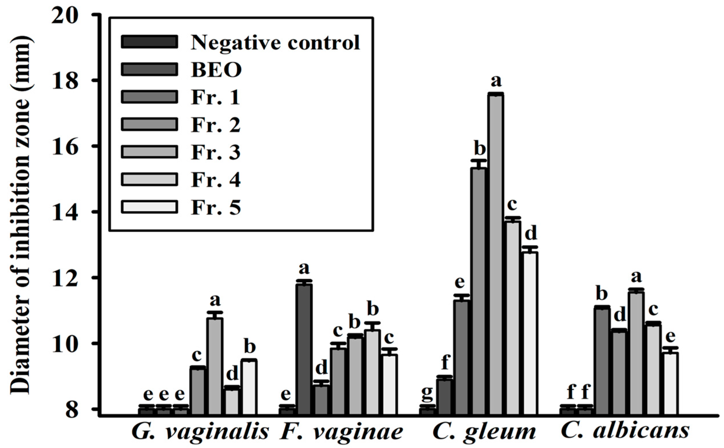
3.4. Disk Diffusion Assays of BEO Against Vaginitis Pathogens

A disk diffusion assay was used to evaluate the antimicrobial activities of BEO against vaginitis-associated pathogens, and the results showed that the BEO exhibited strong dose-dependent antimicrobial effects against all four tested pathogens (Table 4). The inhibition zones ranged from 13.75 ± 0.39 mm to 25.88 ± 0.52 mm at the highest concentration tested (5 mg/disk). In addition, the BEO displayed broad-spectrum effects and inhibited Gram-positive (G. vaginalis and F. vaginae) and Gram-negative bacteria (C. gleum), as well as C. albicans (a dimorphic fungus).

3.5. Evaluation of the MIC Values of BEO

The MIC values of BEO were determined using a broth microdilution assay to assess its antimicrobial activities against the four pathogens. The MIC was defined as the lowest concentration of BEO that inhibited visible bacterial growth after 24 h of incubation at 37 °C. The BEO effectively inhibited all tested pathogens, with MIC values ranging from 31 to 500 µg/mL (Table 4).

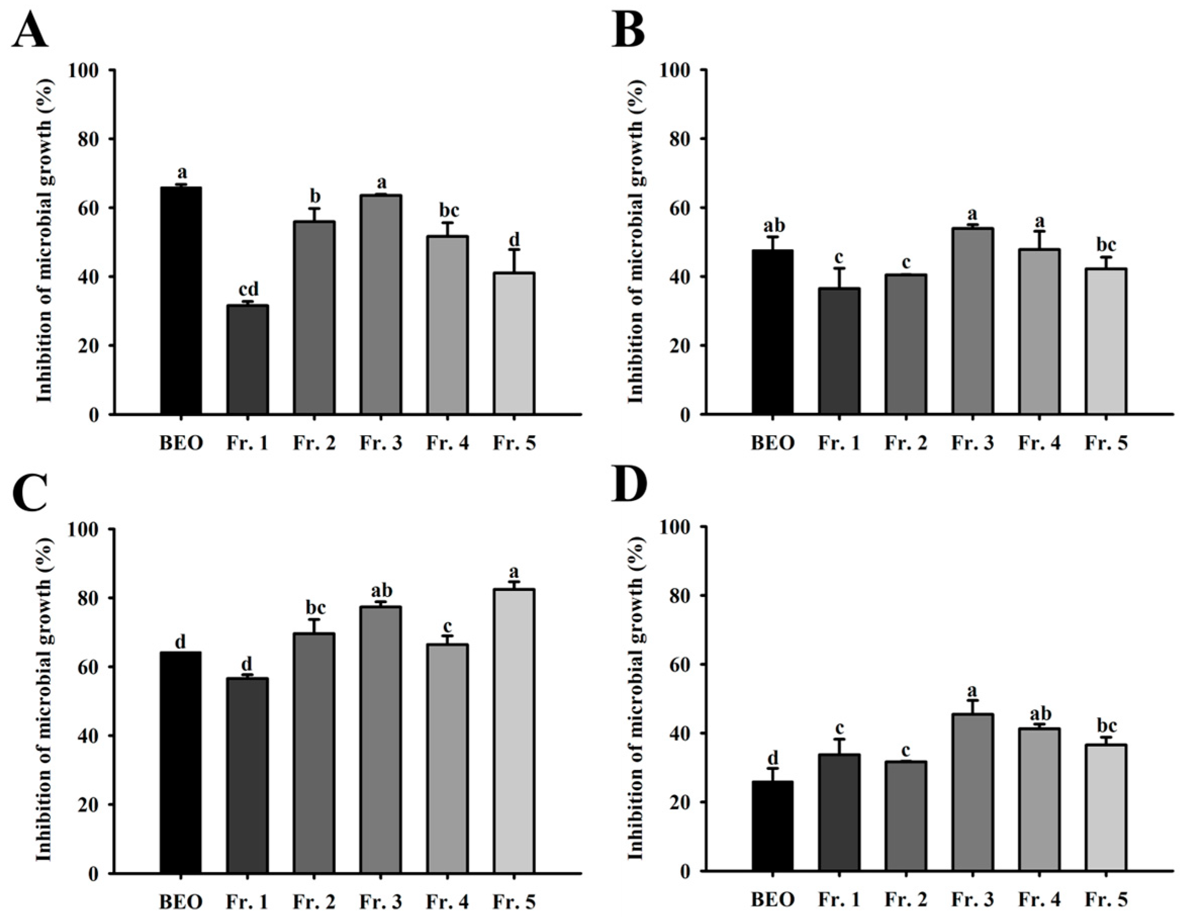
3.6. Relative Microbial Growth Inhibition of Vaginitis Pathogens by BEO

Microbial growth inhibition by BEO was assessed spectrophotometrically, and the results obtained showed it had dose-dependent effects against G. vaginalis, F. vaginae, C. gleum, and C. albicans. At concentrations of 62, 125, 250, and 500 µg/mL, the relative microbial inhibition ratios against G. vaginalis were 66.44 ± 0.23%, 75.55 ± 0.33%, 79.31 ± 1.39%, and 82.21 ± 1.13%, respectively. Against F. vaginae and C. gleum, the inhibition ratios ranged from 28.02 ± 0.55% to 75.86 ± 0.12%. At 500 µg/mL, the BEO inhibited the growth of C. albicans by 84.46 ± 0.32% (Figure 5).

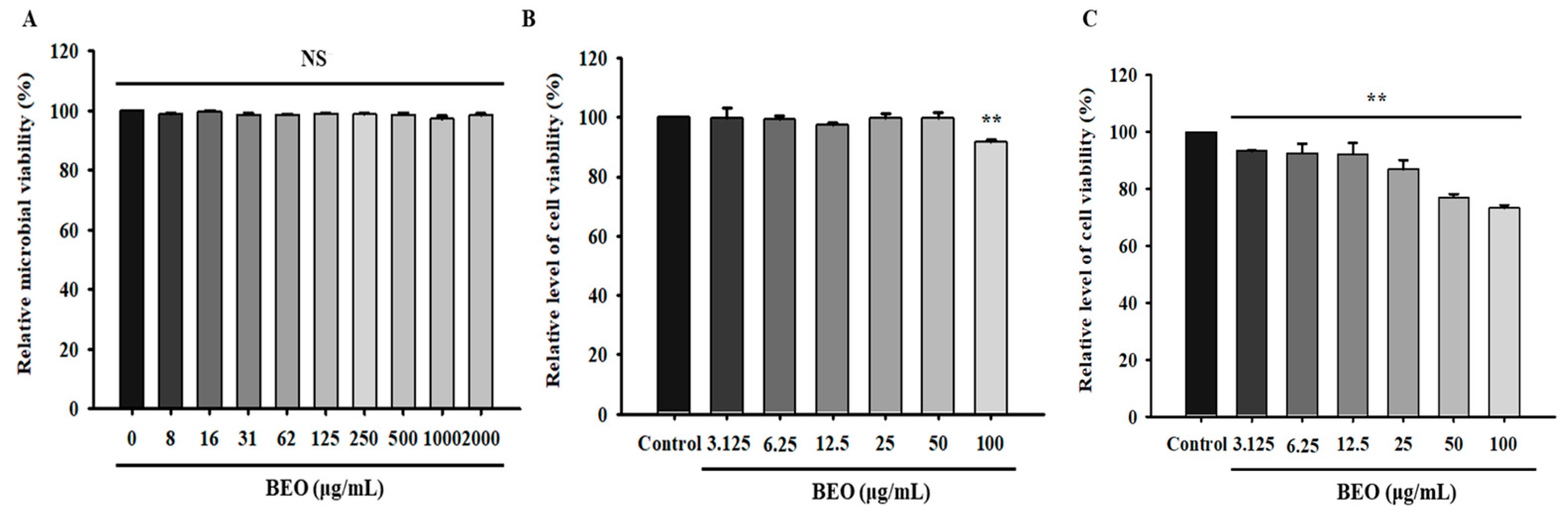
3.7. Safety Assessment of BEO

Ensuring the safety of BEO for topic applications, such as skin cleansers, is essential. In this study, we evaluated its toxic effects on L. crispatus and human dermal cells. To quantitatively assess microbial viability, relative microbial viability was determined spectrophotometrically after 24 h of BEO exposure. The BEO did not significantly affect the viability of L. crispatus, and viability remained above 95% at all tested concentrations (Figure 6A).
To further assess the safety of BEO for skin applications, we examined its effects on human dermal fibroblasts (HDFs) and HeLa cells [28]. HDFs and HeLa cells were incubated with BEO at concentrations ranging from 3.125 to 100 µg/mL for 24 h, and cell viabilities were determined using the MTT assay. No significant reduction in cell viability versus untreated controls was observed at concentrations of 3.125–50 µg/mL. Furthermore, when HDFs were exposed to BEO at 100 µg/mL, cell viability remained high at 91.88 ± 0.67% (Figure 6B), and for HeLa cells, viability ranged from 93.33 ± 0.30% to 73.23 ± 1.11% across the same concentration range, indicating a dose-dependent decrease at higher concentrations (Figure 6C).

4. Discussion

The findings of this study are consistent with those of previous studies, although source-dependent variations in the chemical composition of basil essential oil (BEO) have been reported. Chalchat and Özcan reported high concentrations of estragole (52.60%) and limonene (13.64%) in BEO [29], whereas Kavoosi and Amirghofran identified linalool (31.65%), estragole (17.37%), and methyl cinnamate (15.14%) as the major constituents [30].
The chemical composition of BEO, which includes methyl trans-cinnamate, methyl cis-cinnamate, methyleugenol, and cyclopentaneacetic acid, is probably responsible for its observed antimicrobial and antioxidant activities. Methyl trans-cinnamate, the simplest-structured cinnamate ester, is a significant component in basil and other Ocimum species (Figure 2C) [31,32]. This phytochemical is known for its anti-inflammatory, anti-obesity, anticancer, and antimicrobial properties and is widely utilized in the commodities industry [33,34,35,36]. Furthermore, the identification of these bioactive compounds in BEO suggests that multiple constituents may contribute to the antimicrobial effect of BEO. Synergistic effects, if any, require further investigation through combination testing of individual components. Discrepancies in the chemical composition and activity of BEO may be caused by differences in agroclimatic factors such as climate, season, and geographical conditions, which all influence basil cultivation [37].
Similarly, previous studies have shown that phytocompounds have meaningful antimicrobial activities. Jang et al. reported that the antibacterial activity of the neutral fraction of rhubarb (Rheum rhabarbarum) against C. gleum resulted in an inhibitory zone of 10 mm at a concentration of 10 mg/mL [38]. Similarly, Marcas et al. demonstrated that 20 µL of Bixa orellana L. leaf extract produced inhibition zones of 20.3 mm and 12.0 mm against G. vaginalis and F. vaginae, respectively [39]. Furthermore, Ardestani et al. reported that ajwain (Trachyspermum ammi) essential oil exhibited significant antimicrobial activity against various vaginitis pathogens, with MIC values ranging from 31.5 to 500 µg/mL [40].
While the specific composition and concentrations of active compounds vary across plant species, these findings, along with our own results, support the broader trend that essential oils from diverse botanical sources can exert significant inhibitory effects against vaginal pathogens. The potency of BEO, particularly its low MIC value and broad-spectrum activity, highlights its potential utility as a superior natural antimicrobial agent.
Although Fr. 3 exhibited potent antimicrobial activity in the MIC and microbial growth inhibition assays, the BEO showed stronger inhibition against F. vaginae in the disk diffusion assay. This discrepancy may be attributed to synergistic or additive effects among the multiple constituents in the whole oil, which may be diminished upon fractionation. It is also worth noting that the disk diffusion method has inherent limitations, particularly in evaluating the activity of less volatile or poorly diffusing compounds, and therefore may underestimate the potency of isolated fractions.
Previous reports have emphasized the critical role of phenolic compounds—such as thymol and eugenol—in antimicrobial activity, especially against vaginal pathogens [41,42,43]. Phenolic compounds are also known for their antioxidant effects in cosmetology, largely due to the number and position of hydroxyl groups in their chemical structures [44]. Incorporating such antioxidants in cosmetic formulations can prevent oxidation of lipid components and vitamins [45], suggesting that phytocompounds like eugenol may contribute to both antimicrobial and antioxidant efficacy. In our study, the absence or low abundance of phenolic constituents in Fr. 3 may partially explain its comparatively lower inhibition zone.
In addition, it is essential to ensure the safety of products like BEO intended for human applications. Our cytotoxicity assessments on HDFs and HeLa cells confirmed the biocompatibility of BEO at concentrations that exhibited potent antimicrobial activity. These findings align with those of Syarina et al., who reported that Spirulina platensis extract, when applied to HDFs at 50 µg/mL for 24 h, maintained cell viability greater than 80% [46]. Thus, our findings demonstrate that BEO has potential use for skin application when used at appropriate dosages.

5. Conclusions

BEO demonstrated potent antimicrobial activity against four vaginitis-associated pathogens, viz., G. vaginalis, F. vaginae, C. gleum, and C. albicans, and it had minimal cytotoxic effects on L. crispatus and human dermal fibroblasts. Methyl trans-cinnamate was identified as the primary active compound in BEO, which supports the rationale underlying the traditional medicinal use of Ocimum species. These findings suggest that BEO has significant potential as a natural therapeutic agent for vaginitis and should be subjected to further in vivo and clinical investigations.

Author Contributions

Conceptualization, M.P., J.P. and H.L.; methodology, M.P. and J.P.; validation, M.P. and D.Y.H.; investigation, J.P.; resources, H.L.; data curation, M.P. and J.P.; writing—original draft preparation, M.P.; writing—review and editing, J.P. and D.Y.H.; visualization, J.P.; supervision, S.P. and H.L. All authors have read and agreed to the published version of the manuscript.

Funding

This research received no external funding.

Institutional Review Board Statement

Not applicable.

Informed Consent Statement

Not applicable.

Data Availability Statement

Data are contained within the article.

Conflicts of Interest

The authors declare no conflicts of interest.

Abbreviations

ABTS2,2′-Azino-bis(3-ethylbenzothiazoline-6-sulfonic acid)
ANOVAAnalysis of variance
ATCCAmerican Type Culture Collection
BEOBasil essential oil
BVBacterial vaginosis
DMEMDulbecco’s Modified Eagle Medium
DMSODimethyl sulfoxide
DPPH1,1-Diphenyl-2-picrylhydrazyl
EC50Half-maximal effective concentration
FBSFetal bovine serum
GC-MSGas chromatography–mass spectrometry
HDFHuman dermal fibroblast
NISTNational Institute of Standards and Technology
MICMinimum inhibitory concentration
PBSPhosphate-buffered saline
PDAPhotodiode-array detector
SDStandard deviation
TSBTryptic soy broth
VVCVulvovaginal candidiasis

References

  1. Parham, S.; Kharazi, A.Z.; Bakhsheshi-Rad, H.R.; Nur, H.; Ismail, A.F.; Sharif, S.; RamaKrishna, S.; Berto, F. Antioxidant, Antimicrobial and Antiviral Properties of Herbal Materials. Antioxidants 2020, 9, 1309. [Google Scholar] [CrossRef] [PubMed]
  2. Kennedy, D.A.; Lupattelli, A.; Koren, G.; Nordeng, H. Safety classification of herbal medicines used in pregnancy in a multinational study. BMC Complement. Altern. Med. 2016, 16, 102. [Google Scholar] [CrossRef] [PubMed]
  3. Al Abbasy, D.W.; Pathare, N.; Al-Sabahi, J.N.; Khan, S.A. Chemical composition and antibacterial activity of essential oil isolated from Omani basil (Ocimum basilicum linn.). Asian Pac. J. Trop. Dis. 2015, 5, 645–649. [Google Scholar] [CrossRef]
  4. Hossain, M.A.; Kabir, M.J.; Salehuddin, S.M.; Rahman, S.M.; Das, A.K.; Singha, S.K.; Alam, A.; Rahman, A. Antibacterial properties of essential oils and methanol extracts of sweet basil Ocimum basilicum occurring in Bangladesh. Pharm. Biol. 2010, 48, 504–511. [Google Scholar] [CrossRef]
  5. Akoto, C.O.; Acheampong, A.; Boakye, Y.D.; Naazo, A.A.; Adomah, D.H. Anti-Inflammatory, Antioxidant, and Anthelmintic Activities of Ocimum basilicum (Sweet Basil) Fruits. J. Chem. 2020, 2020, 2153534. [Google Scholar] [CrossRef]
  6. Stanojevic, L.P.; Marjanovic-Balaban, Z.R.; Kalaba, V.D.; Stanojevic, J.S.; Cvetkovic, D.J.; Cakic, M.D. Chemical composition, antioxidant and antimicrobial activity of basil (Ocimum basilicum L.) essential oil. J. Essent. Oil Bear. Plants 2017, 20, 1557–1569. [Google Scholar] [CrossRef]
  7. Teofilović, B.; Grujić-Letić, N.; Gligorić, E.; Rašković, A.; Igić, R.; Vastag, G.; Gadžurić, S. Experimental and computational evaluation of extraction procedure and scavenging capacity of sweet basil extracts (Ocimum basilicum L.). Plant Foods Hum. Nutr. 2021, 76, 240–247. [Google Scholar] [CrossRef]
  8. Makri, O.; Kintzios, S. Ocimum sp.(basil): Botany, cultivation, pharmaceutical properties, and biotechnology. J. Herbs. Spices Med. Plants 2008, 13, 123–150. [Google Scholar] [CrossRef]
  9. Zhakipbekov, K.; Turgumbayeva, A.; Akhelova, S.; Bekmuratova, K.; Blinova, O.; Utegenova, G.; Shertaeva, K.; Sadykov, N.; Tastambek, K.; Saginbazarova, A.; et al. Antimicrobial and other pharmacological properties of Ocimum basilicum, Lamiaceae. Molecules 2024, 29, 388. [Google Scholar] [CrossRef]
  10. Kuorwel, K.K.; Cran, M.J.; Sonneveld, K.; Miltz, J.; Bigger, S.W. Essential oils and their principal constituents as antimicrobial agents for synthetic packaging films. J. Food Sci. 2011, 76, R164–R177. [Google Scholar] [CrossRef]
  11. Antonio, M.A.; Hawes, S.E.; Hillier, S.L. The identification of vaginal lactobacillus species and the demographic and microbiologic characteristics of women colonized by these species. J. Infect. Dis. 1999, 180, 1950–1956. [Google Scholar] [CrossRef] [PubMed]
  12. Boskey, R.; Telsch, K.M.; Whaley, K.J.; Moench, T.R.; Cone, R.A. Acid production by vaginal flora in vitro is consistent with the rate and extent of vaginal acidification. Infect. Immun. 1999, 67, 5170–5175. [Google Scholar] [CrossRef] [PubMed]
  13. Madhivanan, P.; Alleyn, H.N.; Raphael, E.; Krupp, K.; Ravi, K.; Nebhrajani, R.; Anjali, A.R.; Reingold, L.W.; Klausner, J.D. Identification of culturable vaginal lactobacillus species among reproductive age women in Mysore, India. J. Med. Microbiol. 2015, 64, 636–641. [Google Scholar] [CrossRef] [PubMed]
  14. Trinh, H.T.; Lee, I.A.; Hyun, Y.J.; Kim, D.H. Artemisia princeps pamp. essential oil and its constituents eucalyptol and α-terpineol ameliorate bacterial vaginosis and vulvovaginal candidiasis in mice by inhibiting bacterial growth and NF-κB activation. Planta Medica 2011, 77, 1996–2002. [Google Scholar] [CrossRef]
  15. Abou Chacra, L.; Fenollar, F.; Diop, K. Bacterial vaginosis: What do we currently know? Front. Cell. Infect. Microbiol. 2022, 11, 1393. [Google Scholar] [CrossRef]
  16. Jang, J.; Kang, D.H.; Yoon, J.; Kim, H.S. Separation and purification of antimicrobial substance from Syzygium aromaticum Merrill et Perry for treatment of microbial vaginosis. KSBB J. 2018, 33, 285–292. [Google Scholar] [CrossRef]
  17. Pendharkar, S.; Brandsborg, E.; Hammarström, L.; Marcotte, H.; Larsson, P.G. Vaginal colonization by probiotic lactobacilli and clinical outcome in women conventionally treated for bacterial vaginosis and yeast infection. BMC Infect. Dis. 2015, 15, 255. [Google Scholar] [CrossRef]
  18. Hurley, R.; de Louvois, J. Candida vaginitis. Postgrad. Med. J. 1979, 55, 645. [Google Scholar] [CrossRef]
  19. Fachriyah, E.; Wibawa, P.J.; Awaliyah, A. Antibacterial activity of basil oil (Ocimum basilicum L.) and basil oil nanoemulsion. J. Phys. Conf. Ser. 2020, 1524, 12–60. [Google Scholar] [CrossRef]
  20. Blois, M.S. Antioxidant determinations by the use of a stable free radical. Nature 1958, 181, 1199–1200. [Google Scholar] [CrossRef]
  21. Re, R.; Pellegrini, N.; Proteggente, A.; Pannala, A.; Yang, M.; Rice-Evans, C. Antioxidant activity applying an improved ABTS radical cation decolorization assay. Free Radic. Biol. 1999, 26, 1231–1237. [Google Scholar] [CrossRef] [PubMed]
  22. De Beer, E.J.; Sherwood, M.B. The paper-disc agar-plate method for the assay of antibiotic substances. J. Bacteriol. 1945, 50, 459–467. [Google Scholar] [CrossRef]
  23. Liu, M.; Seidel, V.; Katerere, D.R.; Gray, A.I. Colorimetric broth microdilution method for the antifungal screening of plant extracts against yeasts. Methods 2007, 42, 325–329. [Google Scholar] [CrossRef] [PubMed]
  24. Badawy, M.E.; Marei, G.I.; Rabea, E.I.; Taktak, N.E. Antimicrobial and antioxidant activities of hydrocarbon and oxygenated monoterpenes against some foodborne pathogens through in vitro and in silico studies. Pestic. Biochem. Physiol. 2019, 158, 185–200. [Google Scholar] [CrossRef]
  25. Dahham, S.S.; Tabana, Y.M.; Iqbal, M.A.; Ahamed, M.B.; Ezzat, M.O.; Majid, A.S.; Majid, A.M. The anticancer, antioxidant and antimicrobial properties of the sesquiterpene β-caryophyllene from the essential oil of Aquilaria crassna. Molecules 2015, 20, 11808–11829. [Google Scholar] [CrossRef]
  26. Neelam, K.A.; Sharma, K.K. Phenylpropanoids and its derivatives: Biological activities and its role in food, pharmaceutical and cosmetic industries. Crit. Rev. Food Sci. Nutr. 2020, 60, 2655–2675. [Google Scholar] [CrossRef] [PubMed]
  27. Xu, X.; Tang, Z.; Liang, Y. Comparative analysis of plant essential oils by GC-MS coupled with integrated chemometric resolution methods. Anal. Methods 2010, 2, 359–367. [Google Scholar] [CrossRef]
  28. Gawron, G.; Krzyczkowski, W.; Lemke, K.; Ołdak, A.; Kadziński, L.; Banecki, B. Nigella sativa seed extract applicability in preparations against methicillin-resistant Staphylococcus aureus and effects on human dermal fibroblasts viability. J. Ethnopharmacol. 2019, 244, 112–135. [Google Scholar] [CrossRef]
  29. Chalchat, J.C.; Özcan, M.M. Comparative essential oil composition of flowers, leaves and stems of basil (Ocimum basilicum L.) used as herb. Food Chem. 2008, 110, 501–503. [Google Scholar] [CrossRef]
  30. Kavoosi, G.; Amirghofran, Z. Chemical composition, radical scavenging and antioxidant capacity of Ocimum basilicum essential oil. J. Essent. Oil Res. 2017, 29, 189–199. [Google Scholar] [CrossRef]
  31. Krokidi, K.M.; Turner, M.A.; Pearcy, P.A.; Stavros, V.G. A systematic approach to methyl cinnamate photodynamics. Mol. Phys. 2021, 119, e1811910. [Google Scholar] [CrossRef]
  32. Rosas, J.F.; Zoghbi, M.D.G.B.; Andrade, E.H.A.; van den Berg, M.E. Chemical composition of a methyl-(E)-cinnamate Ocimum micranthum Willd. from the Amazon. Flavour Fragr. J. 2005, 20, 161–163. [Google Scholar] [CrossRef]
  33. Chen, Y.Y.; Lee, M.H.; Hsu, C.C.; Wei, C.L.; Tsai, Y.C. Methyl cinnamate inhibits adipocyte differentiation via activation of the CaMKK2–AMPK pathway in 3T3-L1 preadipocytes. J. Agric. Food Chem. 2012, 60, 955–963. [Google Scholar] [CrossRef]
  34. Huang, Q.S.; Zhu, Y.J.; Li, H.L.; Zhuang, J.X.; Zhang, C.L.; Zhou, J.J.; Li, W.G.; Chen, Q.X. Inhibitory effects of methyl trans-cinnamate on mushroom tyrosinase and its antimicrobial activities. J. Agric. Food Chem. 2009, 57, 2565–2569. [Google Scholar] [CrossRef] [PubMed]
  35. Kathirvel, P.; Ravi, S. Chemical composition of the essential oil from basil (Ocimum basilicum Linn.) and its in vitro cytotoxicity against HeLa and HEp-2 human cancer cell lines and NIH 3T3 mouse embryonic fibroblasts. Nat. Prod. Res. 2012, 26, 1112–1118. [Google Scholar] [CrossRef] [PubMed]
  36. Park, K.R.; Lee, H.; Cho, M.; Yun, H.M. A phytochemical constituent, (E)-methyl-cinnamate isolated from Alpinia katsumadai Hayata suppresses cell survival, migration, and differentiation in pre-osteoblasts. Int. J. Mol. Sci. 2020, 21, 3700. [Google Scholar] [CrossRef]
  37. Ahmed, A.F.; Attia, F.A.; Liu, Z.; Li, C.; Wei, J.; Kang, W. Antioxidant activity and total phenolic content of essential oils and extracts of sweet basil (Ocimum basilicum L.) plants. Food Sci. Hum. Wellness 2019, 8, 299–305. [Google Scholar] [CrossRef]
  38. Jang, J.; Kang, D.H.; Yoon, J.; Kim, H.S. Isolation and characterization of an antibacterial substance from Rheum palmatum for treatment of bacterial vaginosis. KSBB J. 2017, 32, 133–139. [Google Scholar] [CrossRef]
  39. Marcas, J.; Romero, L.; Tipiani, O.; Loyola, S.; Tamariz, J. In vitro antimicrobial activity of Bixa orellana L. leaves extract against anaerobic bacteria associated to bacterial vaginosis and Lactobacillus spp. Rev. Peru Med. Exp. Salud Pública 2023, 39, 408–414. [Google Scholar] [CrossRef]
  40. Ardestani, M.M.; Aliahmadi, A.; Toliat, T.; Dalimi, A.; Momeni, Z.; Rahimi, R. Evaluation of antimicrobial activity of Trachyspermum ammi (L.) sprague essential oil and its active constituent, thymol, against vaginal pathogens. Trad. Integr. Med. 2020, 5, 49–58. [Google Scholar] [CrossRef]
  41. Murina, F.; Vicariotto, F.; Di Francesco, S. Thymol, eugenol and lactobacilli in a medical device for the treatment of bacterial vaginosis and vulvovaginal candidiasis. New Microbiol. 2018, 41, 220–224. [Google Scholar] [PubMed]
  42. Saracino, I.M.; Foschi, C.; Pavoni, M.; Spigarelli, R.; Valerii, M.C.; Spisni, E. Antifungal activity of natural compounds vs. Candida spp.: A mixture of cinnamaldehyde and eugenol shows promising in vitro results. Antibiotics 2022, 11, 73. [Google Scholar] [CrossRef]
  43. Didehdar, M.; Chegini, Z.; Shariati, A. Eugenol: A novel therapeutic agent for the inhibition of Candida species infection. Front. Pharmacol. 2022, 13, 872127. [Google Scholar] [CrossRef] [PubMed]
  44. Zagórska-Dziok, M.; Wójciak, M.; Ziemlewska, A.; Nizioł-Łukaszewska, Z.; Hoian, U.; Klimczak, K.; Szczepanek, D.; Sowa, I. Evaluation of the antioxidant, cytoprotective and antityrosinase effects of Schisandra chinensis Extracts and their applicability in skin care product. Molecules 2022, 27, 8877. [Google Scholar] [CrossRef]
  45. Nizioł-Łukaszewska, Z.; Bujak, T. Saponins as natural raw materials for increasing the safety of bodywash cosmetic use. JSD 2018, 21, 67–776. [Google Scholar] [CrossRef]
  46. Syarina, P.N.A.; Karthivashan, G.; Abas, F.; Arulselvan, P.; Fakurazi, S. Wound healing potential of spirulina platensis extracts on human dermal fibroblast cells. EXCLI J. 2015, 14, 385. [Google Scholar] [CrossRef]
Figure 1. Schematic of the steam distillation process for basil essential oil extraction.
Antioxidants 14 00628 g001
Figure 2. (A) Preparative HPLC chromatogram of basil essential oil. (B) GC-MS total ion chromatogram of Fraction 3 from basil essential oil and methyl trans-cinnamate. (C) Chemical structure of main constituents in Fraction 3. (D) Mass spectra of the three peaks of Fraction 3 (1 = methyl cis-cinnamate; 2 = methyleugenol; 3 = cyclopentaneacetic acid).
Antioxidants 14 00628 g002
Figure 3. Disk diffusion test of basil essential oil and fractions: The pathogens investigated were Gardnerella vaginalis KCTC 5096, Fannyhessea vaginae KCTC 15240, Chryseobacterium gleum JCM 2410, and Candida albicans KCTC 7965. BEO: basil essential oil. Values are means ± standard deviations (SDs). a–g Values revealed significant differences using one-way analysis of variance followed by Duncan’s multiple range test (p < 0.05).
Antioxidants 14 00628 g003
Figure 4. Relative microbial growth inhibition of basil essential oil and fractions: (A) Relative microbial growth inhibition against Gardnerella vaginalis. (B) Relative microbial growth inhibition against Fannyhessea vaginae. (C) Relative microbial growth inhibition against Chryseobacterium gleum. (D) Relative microbial growth inhibition against Candida albicans. Values are means ± standard deviations (SDs). a–d Values revealed significant differences using one-way analysis of variance followed by Duncan’s multiple range test (p < 0.05).
Antioxidants 14 00628 g004
Figure 5. Relative microbial growth inhibition of basil essential oil against vaginitis pathogens: (A) Relative microbial growth inhibition toward Gardnerella vaginalis. (B) Relative microbial growth inhibition toward Fannyhessea vaginae. (C) Relative microbial growth inhibition toward Chryseobacterium gleum. (D) Relative microbial growth inhibition toward Candida albicans. Values are means ± standard deviations (SDs). a–d Values revealed significant differences using one-way analysis of variance followed by Duncan’s multiple range test (p < 0.05).
Antioxidants 14 00628 g005
Figure 6. (A) Relative microbial viability of Lactobacillus crispatus after exposure to basil essential oil. (B) The cell viability of HDFs after exposure to basil essential oil. (C) The cell viability of HeLa cells after exposure to basil essential oil. NS: not significant; ** p < 0.05 vs. untreated controls. Values are means ± standard deviations (SDs).
Antioxidants 14 00628 g006
Table 1. Chemical composition of the basil essential oil as determined by GC-MS; n.i: non-identified.
No.CompoundFormulaRt (min)Area (%)
1Bicyclo[3.1.1]heptaneC7H125.0800.67
22-IsopropyltolueneC10H145.5860.48
3l-LimoneneC10H165.6470.45
4EucalyptolC10H18O5.7146.33
5LinaloolC10H18O6.44019.60
6CamphorC10H16O7.0590.37
7EstragoleC10H12O7.59329.48
8(-)-CarvoneC10H14O8.0570.38
9Bornyl acetateC12H20O28.4791.21
10Methyl cinnamateC10H10O28.6422.37
11EugenolC10H12O29.1410.44
12Methyl cinnamateC10H10O29.51916.72
13Bicyclo[5.2.0]nonaneC15H249.5491.72
14CaryophylleneC15H249.9331.07
15trans-alpha-BergamoteneC15H249.9737.67
16alpha-HumuleneC15H2410.2690.53
17n.i-10.4700.48
18Germacrene-DC15H2410.4931.88
19n.i-10.6270.42
20n.i-10.6500.46
21gamma-AmorpheneC15H2410.7592.59
22(E)-alpha-BisaboleneC15H2410.9040.80
23(-)-Caryophyllene oxideC15H24O11.4360.30
24n.i-11.6730.64
25Epi-cadinolC15H26O11.8832.94
100.00
Table 2. The MIC values of basil essential oil fractions against vaginitis pathogens. The pathogens investigated were Gardnerella vaginalis KCTC 5096, Fannyhessea vaginae KCTC 15240, Chryseobacterium gleum JCM 2410, and Candida albicans KCTC 7965. MIC: minimum inhibitory concentration. BEO: basil essential oil.
MICBEOFr. 1Fr. 2Fr. 3Fr. 4Fr. 5
G. vaginalis(μg/mL)125125623162125
F. vaginae(μg/mL)3125025031125125
C. gleum(μg/mL)3112562166216
C. albicans(μg/mL)50025025062125125
Table 3. Antioxidant activities of basil essential oil. DPPH: 1,1-diphenyl-2-picrylhydrazyl. ABTS: 2,2-azono-bis-3-ethylbenzthiazoline-6-sulphonate. EC50: The half-maximal effective concentration. Values are means ± standard deviations (SDs).
DPPHABTS
EC50 (μg/mL)
115.36 ± 2.1954.77 ± 0.29
Table 4. Disk diffusion test for antimicrobial activity against vaginitis pathogens: The pathogens investigated were Gardnerella vaginalis KCTC 5096, Fannyhessea vaginae KCTC 15240, Chryseobacterium gleum JCM 2410, and Candida albicans KCTC 7965. Values are means ± standard deviations (SDs). a–d Values significantly different according to one-way analysis of variance followed by Duncan’s multiple range test (p < 0.05). MIC: minimum inhibitory concentration.
Diameter of Inhibition Zone (mm)MIC
Test Strainsmg/diskμg/mL
00.5125
G. vaginalis8.02 ± 0.01 a8.03 ± 0.02 c9.91 ± 0.35 c11.79 ± 0.20 b13.75 ± 0.39 d125
F. vaginae8.03 ± 0.01 a12.97 ± 0.11 a19.34 ± 0.46 a22.71 ± 0.33 a25.88 ± 0.52 a31
C. gleum8.03 ± 0.01 a8.09 ± 0.01 b11.48 ± 0.13 b13.06 ± 0.02 b17.74 ± 0.25 b31
C. albicans8.02 ± 0.01 a8.02 ± 0.02 c9.03 ± 0.10 d11.07 ± 0.30 d13.95 ± 0.11 c500
Disclaimer/Publisher’s Note: The statements, opinions and data contained in all publications are solely those of the individual author(s) and contributor(s) and not of MDPI and/or the editor(s). MDPI and/or the editor(s) disclaim responsibility for any injury to people or property resulting from any ideas, methods, instructions or products referred to in the content.

