Article

Acute Exposure to Aerosolized Nanoplastics Modulates Redox-Linked Immune Responses in Human Airway Epithelium

by
Joshua D. Breidenbach
1,2,†,
Benjamin W. French
1,†,
Upasana Shrestha
1,3,†,
Zaneh K. Adya
1,
R. Mark Wooten
3,
Andrew M. Fribley
1,
Deepak Malhotra
1,
Steven T. Haller
1,* and
David J. Kennedy
1,*
1
Department of Medicine, College of Medicine and Life Sciences, University of Toledo, Toledo, OH 43614, USA
2
Biochemistry and Biotechnology Group, Bioscience Division, Los Alamos National Laboratory, Los Alamos, NM 87545, USA
3
Department of Medical Microbiology and Immunology, College of Medicine and Life Sciences, University of Toledo, Toledo, OH 43614, USA
*
Authors to whom correspondence should be addressed.
These authors contributed equally to this work.
Antioxidants 2025, 14(4), 424; https://doi.org/10.3390/antiox14040424
Submission received: 11 February 2025 / Revised: 17 March 2025 / Accepted: 26 March 2025 / Published: 31 March 2025

Abstract

:
Micro- and nanoplastics (MPs and NPs) are pervasive environmental pollutants detected in aquatic ecosystems, with emerging evidence suggesting their presence in airborne particles generated by water body motion. Inhalation exposure to airborne MPs and NPs remains understudied despite documented links between occupational exposure to these particles and adverse respiratory outcomes, including airway inflammation, oxidative stress, and chronic respiratory diseases. This study explored the effects of acute NP exposure on a fully differentiated 3D human airway epithelial model derived from 14 healthy donors. Airway epithelium was exposed to aerosolized 50 nm polystyrene NPs at concentrations ranging from 2.5 to 2500 µg/mL for three minutes per day over three days. Functional assays revealed no significant alterations in tissue integrity, cell survival, mucociliary clearance, or cilia beat frequency, suggesting intact epithelial function post-exposure. However, cytokine and chemokine profiling identified a significant five-fold increase in CCL3 (MIP-1α), a neutrophilic chemoattractant, in NP-exposed samples compared to controls. This was corroborated by increased neutrophil chemotaxis in response to conditioned media from NP-exposed tissues, indicating a pro-inflammatory neutrophilic response. Conversely, levels of interleukins (IL-21, IL-2, IL-15), CXCL10, and TGF-β were significantly reduced, suggesting immunomodulatory effects that may impair adaptive immune responses and tissue repair mechanisms. These findings demonstrate that short-term exposure to NP-containing aerosols induces a distinct pro-inflammatory response in airway epithelium, characterized by enhanced neutrophil recruitment and reduced secretion of key immune modulators. These findings underscore the potential for aerosolized NPs to induce oxidative and inflammatory stress, raising concerns about their long-term impact on respiratory health and redox regulation.

1. Introduction

With ever-increasing global industrialization, the use of plastics has skyrocketed [1]. In 2016, an astounding 335 million tons of plastic was produced globally, with an estimated 12.7 million tons entering the ocean from land-based sources. This figure is expected to triple by the year 2050 [2,3]. Due to various physical, chemical, and biological processes, plastics eventually break down into smaller fragments, resulting in the formation of microplastics (MPs; 1 μm–5 mm) and nanoplastics (NPs; <1 μm) [1,4,5]. According to the Guidance on Monitoring of Marine Litter, microplastics can be categorized based on size, falling into the categories of 1–5 mm or 20 μm–1 mm. These microplastics inevitably break down into smaller particles, measuring less than 100 nm [6]. MPs and NPs with a wide range of polymer compositions (e.g., polypropylene (PP), polystyrene (PS), polyethylene (PE)) have been detected in both ocean and freshwater ecosystems, polluting these waters [7,8]. In addition to this, these plastic particles are transported over a wide distance via wind and atmospheric fallout [1,9].
Much research has been conducted to investigate the implications of MP and NP pollution on both local aquatic and terrestrial ecosystems, as well as on human health. Given the ubiquity and persistent nature of MPs and NPs in the environment, human exposure to these pollutants can occur via either ingestion, inhalation, or dermal contact [10]. Following exposure, MPs and NPs exhibit surface reactivity that may drive redox imbalances and exert deleterious toxicity to humans, including inflammation, cytotoxicity, and apoptosis in lungs [11,12,13]; intestinal barrier disruption [14]; increase in pro-inflammatory cytokines like interleukins (ILs) 6 (IL-6), and 1β (IL-1β) and tumor necrosis factor-α (TNFα) [15,16]; and cause oxidative stress, inflammation, and cytotoxicity on cerebral and epithelial human cell lines [17]. PS is among the “Big Five” plastics that dominate global plastic production and consumption, and several studies have linked PS-MPs and -NPs to the induction of inflammatory responses and cellular stress [18,19,20]. In addition to in vitro studies, there are multiple in vivo studies using mice models that present toxicities of MPs and NPs [10,21,22]. One study examined aerosolized PS particles at multiple concentrations for two weeks; the mice showed significant increases in inflammatory cytokines, histopathological changes, and the number of immune cells collected in the bronchoalveolar lavage fluid [16]. In a study involving MP-exposed mice, there was intestinal inflammation and gut microbiota dysbiosis [19]. In another study where mice were exposed to MPs, increased levels of inflammatory cytokines and chemokines and NLR family pyrin domain-containing 3 (NLRP3) inflammasome activation indicated significant lung injury [22].
Many of these studies are primarily focused on the oral route of exposure, with some considering the route of dermal contact to MPs and NPs. There has been limited research conducted into the inhalation of these plastic particles, though recent evidence suggests that natural water body motion is more than sufficient to create airborne water droplets that contain these plastic particles [5]. Additionally, other sources of MPs and NPs include synthetic textiles, sludge byproducts, tire erosion, and occupational exposures. Such exposures have been indicative of airway irritation, neutrophilic inflammation, translocation, increased risk of lung carcinoma and chronic respiratory diseases such as asthma, and even respiratory failure [1,23]. Moreover, MPs and NPs persist in the environment for years, potentially leading to accumulative effects on human health [24].
Considering the widespread distribution of airborne plastics, the current study set out to establish the impact of aerosolized NP exposure on airway epithelial cells using a three-dimensional human airway epithelial cell model. This model was established from a pool of 14 health donor cells and generated multiple layers of cells with functional cilia and mucus production. The culture utilized an air–liquid interface for aerosol exposure, allowing for direct contact of aerosol to tissue culture. Here, we tested the hypothesis that acute exposure to aerosolized NPs induces a pro-inflammatory effect in human airway epithelium. The effect of aerosolized NPs on the model was evaluated with cell survival and functional assays that examined tissue integrity, mucociliary clearance, and cilia beat frequency.

2. Materials and Methods

2.1. Microplastics

Polybead® Microspheres 0.05 μm (polystyrene beads, 2.5% w/v in aqueous solution, 0.05 μm mean diameter, 3.64 × 10¹⁴ particles/mL, cat. 08691-10) were purchased from Polysciences (Warrington, PA, USA).

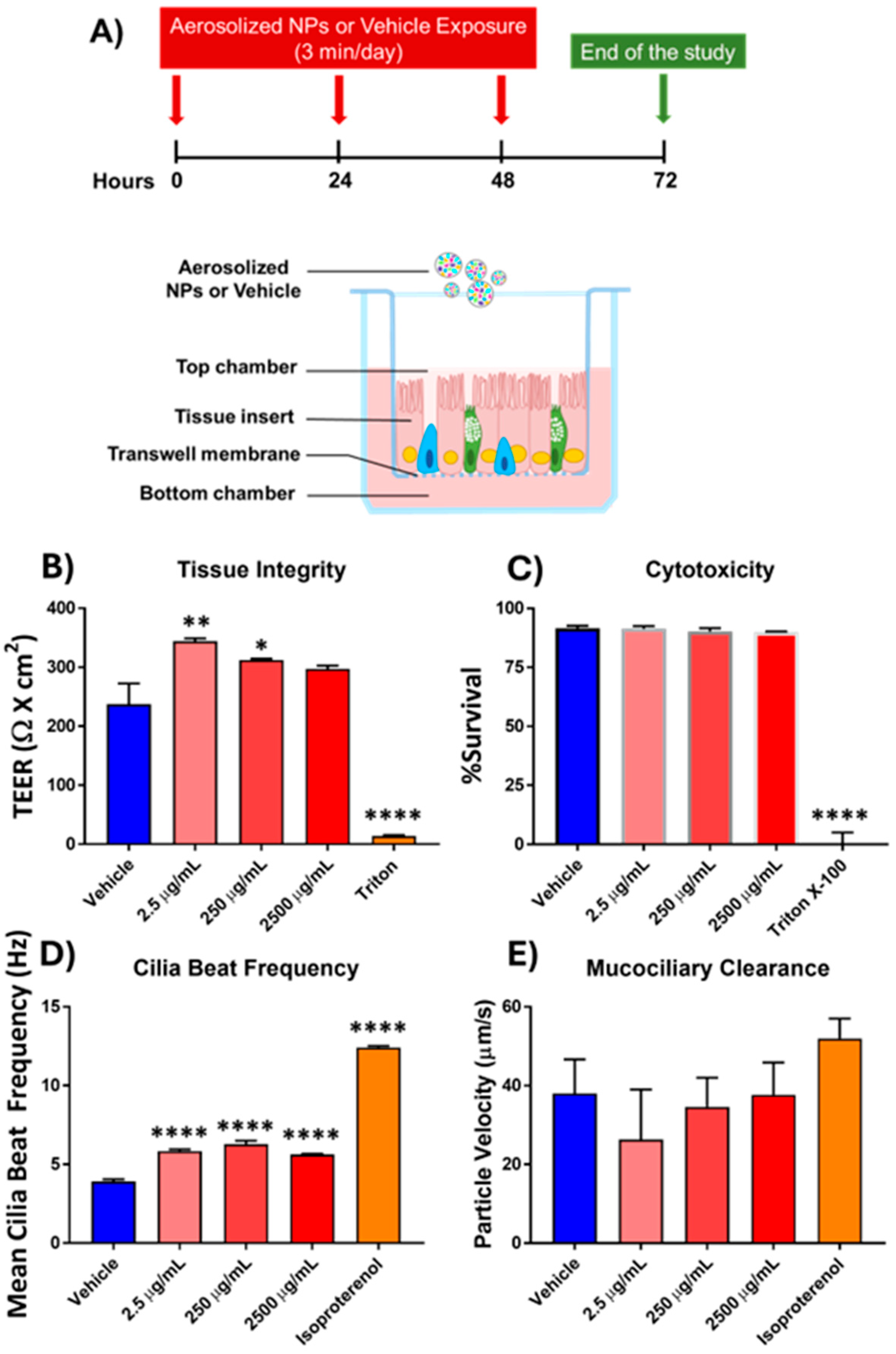
2.2. Primary Cell Culture and Exposure

A 3D model of cultured primary human airway epithelial cells was constructed from primary cells taken from 14 donors, acquired and provided by Epithelix with ethical approval and donor consent (MucilAirTM; Epithelix; Geneva, Switzerland). Donor cells were pooled to reduce variation introduced between donors. These cell cultures were established on 24-well transwell inserts at an air–liquid interface with serum-free MucilAirTM Medium (Epithelix; Geneva, Switzerland). This model presents both functional beating cilia and mucus secretion.
The reconstructed primary 3D airway epithelium was subject to exposure 3x/day for 3 days to either polystyrene plastic beads (0.05 µM mean diameter; Polysciences Inc., Warrington, PA, USA) or vehicle (saline) using the Vitrocell-Cloud nebulization system from Epithelix. This system is designed for dose-controlled and spatially uniform aerosolization of liquid samples on cells cultured with air–liquid interfaces. Each aerosolization exposure lasted approximately 3 min. An estimated 0.43 μL/insert of solution was deposited during each exposure, based on the surface area of the 24-well inserts (0.33 cm2), the surface area of the Vitrocell-Cloud system (136.5 cm2), the 200 μL of solution nebulized per exposure, and an estimated 90% delivery efficacy.

2.3. Tissue Integrity

Tissue integrity was assessed by the transepithelial electrical resistance (TEER) assay. This assay measures electrical resistance via electrodes on both the apical and basal surfaces of the epithelial tissue inserts. As tissues are not as conductive as the electrodes, in-tact tissue presents resistance to the flow of electricity from one side to the other. Therefore, a loss of this resistance indicates a decrease in tissue integrity as there are fewer cells to form a continuous intercellular barrier via intercellular adhesion complexes. Functional epithelium tissues typically present 200–600 Ω × cm2, while deteriorated tissue (broken cellular junctions) measure under 100 Ω × cm2.
To conduct these measurements, 200 μL of normal saline was added to the apical compartment of MucilAirTM cultures and resistance was subsequently measured using an EVOMX volt-ohm-meter (World Precision Instruments UK, Stevenage, UK) for each tested condition. Resistance values (Ω) were converted to TEER (Ω × cm2) using the following formula, TEER (Ω × cm2) = (resistance value (Ω) − 100(Ω)) × 0.33 (cm2), where 100 Ω is the resistance of the membrane and 0.33 cm2 is the total surface of the epithelium.

2.4. Cytotoxicity Assay

Cytotoxicity in the tissue inserts was tested by the lactate dehydrogenase (LDH) assay, measuring levels of LDH in conditioned cell cultured media. As this assay assumes that LDH is a stable enzyme primarily retained within intact cells, increased levels of LDH in conditioned media indicates cell death, and loss of integrity, thereby releasing LDH into the media. On the third day of this study, 24 h after the third and final exposure, 100 μL of basolateral medium was removed from each culture and incubated with the reaction mixture of the LDH Assay Kit-WST (Dojindo; Rockville, MD, USA; ref. CK12-20) following the manufacturer’s instructions. Quantification of LDH in each sample was measured using a microplate reader at 490 nm. To determine the percentage of cytotoxicity, the following equation was used:
C e l l   S u r v i v a l   X = 100 ( % ) A s a m p l e A l o w   c o n t r o l A h i g h   c o n t r o l A l o w   c o n t r o l 100 ( % )
where A = absorbance values, and the high control value was obtained by 10% Triton X-100 apical treatment. The negative controls (vehicle) typically show a low daily basal LDH release, which is due to a physiological cell turnover in the model.

2.5. Cilia Beat Frequency and Mucociliary Clearance

Cilia beat frequency (CBF) and mucociliary clearance (MCC) were assessed by the movement of polystyrene beads along the apical surface of the epithelial tissue insert. Cilia beat frequency was measured via a three-part dedicated setup for this purpose. This system consisted of a camera-equipped microscope, a PCI card, and Epithelix’s Cilia-X CBF analysis software (https://serc.carleton.edu/introgeo/teachingwdata/Fourier.html, accessed on 10 February 2025). CBF is expressed in Hz (cycles or repetitions per second). Videos were generated at 125 frames per second, from which CBF was calculated by Epithelix software. MCC was monitored using a Sony XCD-U100CR camera (Sony; Geneva, Switzerland) connected to an Axiovert 200 M microscope (Zeiss; Oberkochen, Germany) with a 5× objective. Microbead movements were video-tracked at 2 frames per second for 30 images at room temperature. Three videos were taken per insert. Average bead movement velocities (μm/s) were calculated with ImageProPlus 6.0 software.

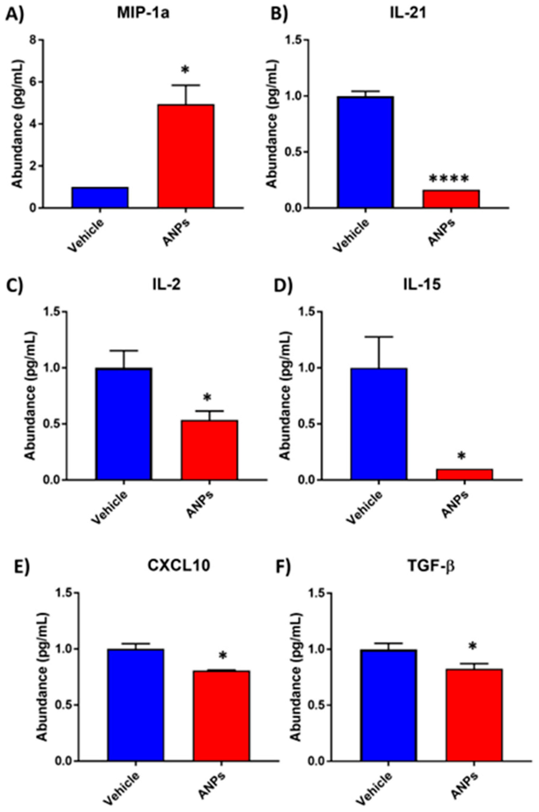
2.6. Protein Secretion
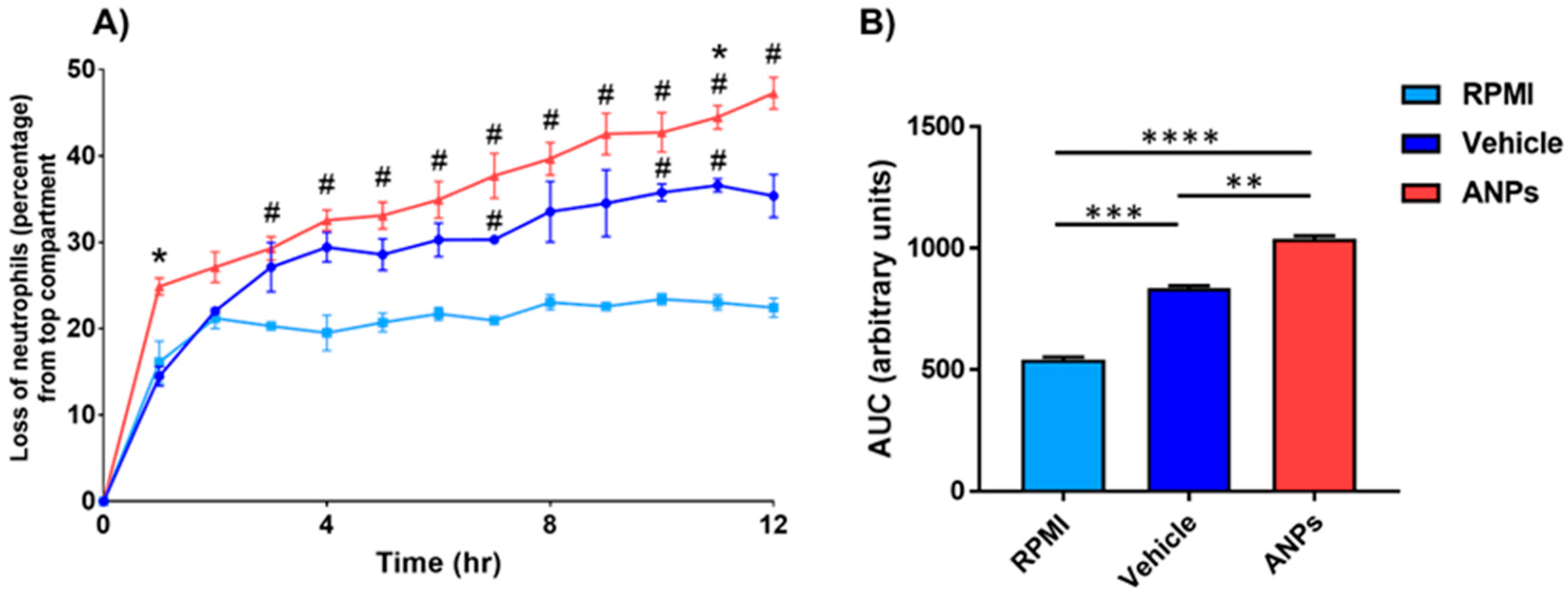
Before each dose, and 24 h after each exposure (plastics or saline), the primary human airway epithelial cell culture media was refreshed. Conditioned media taken from the culture post-exposure were frozen for analysis. To analyze the secreted proteins, the conditioned medium samples from each day were thawed and pooled within groups. Quansys Biosciences (Logan, UT, USA) performed protein measurements via custom multiplex ELISA-based Q-PlexTM technology for quantification of 36 human cytokines and chemokines (Table S1).

2.7. Chemotaxis

To assess the chemotactic potential of the conditioned epithelial medium sample, we performed Boyden chamber assays. Briefly, the Boyden chamber assay is conducted in the 96-well format using HTS Transwell insert plates with 3 µm pores in polycarbonate membranes (Corning, Cat: 3385; Corning, NY, USA). These HTS Transwell insert plates were coated with 50 µg/mL Matrigel (Corning, Cat: 356234; Corning, NY, USA) on each side in RPMI (Caisson Labs, RPL04—phenol red free; Smithfield, UT, USA) + 10% FBS (Gibco, Cat; 26140079; Waltham, MA, USA) at 37 °C for 30 min, followed by 30 min at room temperature. After coating, the plates were placed in a reservoir plates containing 200 uL of dBPS (Caisson Labs, PBL01—without calcium and magnesium; Smithfield, UT) per well while primary human neutrophils (Astarte Biologics/Cellero, Cat: 1025; Lowell, MA, USA) were placed at 1000 cells per well in RPMI + 0.5% FBS above the membrane within the transwell insert. A separate reservoir plate was used for the negative and positive controls (RPMI + 0.5% FBS and 100 nM fMLP (Sigma-Aldrich, F3506; Merck KGaA, Darmstadt, Germany) in RPMI + 0.5% FBS, respectively), as well as potentially attractant conditioned media (conditioned media diluted 1:2 in RPMI + 0.5% FBS). The insert plate was allowed to incubate for 30 min at room temperature before being carefully transferred to the experimental reservoir plate for a 12 h incubation at 37 °C. Chemotaxis was monitored for the entire incubation step on 1 h intervals using an Incucyte S3 (Sartorius AG; Göttingen, Germany) live cell imaging system. Migration from the top chamber to the bottom chamber was noted as a loss of total area of neutrophils on the top portion of the membrane.

2.8. Statistics

All assay-related data were processed using GraphPad Prism version 7.0.5 for Windows (GraphPad Software, San Diego, CA, USA) using Student’s t-test. Multiple groups were tested by ANOVA with Tukey’s multiple comparisons test, followed by multiple t-tests. All the data are expressed as the mean ± standard error of mean (SEM). Significance is marked as a p-value ≤ 0.05. p-value ranges are marked on figures as *, **, ***, or **** for p-values ≤ 0.05, 0.01, 0.001, and 0.0001, respectively.

3. Results

3.1. Effects of Aerosolized Nanoplastics Exposure on Functional Epithelial Measures

Tissue integrity was measured using the TEER assay, and interestingly, treatment with the aerosolized NPs (ANPs) at 2.5 µg/mL showed a significant increase in electrical resistance. This trend appears to continue at 250 and 2500 µg/mL but did not reach significance in either case (n = 3, p-value = 0.0997 and 0.1690, respectively) (Figure 1B). However, this value is within normal TEER values (between 200 and 600 Ω × cm2) of intact epithelium. There was a significant decrease in electrical resistance upon exposure to Triton X-100, indicating severe destruction of the airway epithelial cell barrier integrity.
The LDH assay was used to assess the cytotoxicity imposed by ANPs exposure by measuring levels in the conditioned cell culture media. The acute, repeated exposures to nanoplastic aerosol showed no cytotoxicity, even up to 2500 µg/mL (n = 3, Figure 1C).
CBF is one of several factors that influences the MCC, which aids in maintaining homeostasis and functions as a physical barrier for the immune system. Treatment with ANPs had a significant impact on the cilia beat frequency at every dose but did not appear to be dose-dependent (n = 3, Figure 1D). Isoproterenol was used as a positive control, being known to significantly increase CBF in many mammals, including humans [25,26]. Vehicle-induced CBF had an average of 3.918 Hz (±0.143 SEM); isoproterenol-induced CBF had an average CBF of 12.42 Hz (±0.104 SEM). At each dose of ANP exposure, 2.5 µg/mL, 250 µg/mL, and 2500 µg/mL, the average CBFs were 5.842 Hz (±0.111 SEM), 6.285 Hz (±0.224 SEM), and 5.634 Hz (±0.049 SEM), respectively. Treatment with ANPs resulted in a dose-dependent reduction in MCC, although the change was not significant, as determined by tracking the movement of beads across the cells (n = 3, Figure 1E).

3.2. Differential Secretion of Cytokine and Chemokine Proteins Following Aerosolized Nanoplastics Exposure

Based on the ELISA-based Q-PlexTM quantification of secreted proteins, Figure 2 shows the six cytokines and chemokines observed to be significantly differentially secreted after exposure to aerosolized NPs. Secretion of macrophage inflammatory protein-1 alpha (MIP-1α) was significantly higher following ANP exposure, with an average of 4.94 pg/mL compared to vehicle (average 1 pg/mL). The observed increase in MIP-1α and neutrophilic chemotaxis indicates a pro-inflammatory milieu, potentially driven by oxidative stress induced by ANP exposure. On the other hand, ILs 21, 2, and 15 as well as C-X-C motif chemokine ligand 10 (CXCL10) and transforming growth factor β (TGF-β) were significantly decreased after exposure to aerosolized NPs (n = 3, Figure 2B–F). These significant reductions suggest an impairment of adaptive immune responses and redox-mediated tissue repair mechanisms.

3.3. Enhanced Neutrophilic Chemotaxis Following Aerosolized Nanoplastics Exposure

Interestingly, the vehicle showed higher neutrophilic chemotactic potential than RPMI alone. Beyond the chemotactic potential of the vehicle treatment, conditioned media from NP-exposed cells shows significantly increased chemotactic potential. The conditioned media showed significant increases in neutrophil migration at 10, 14, 16, and 18 h, suggesting an inflammatory response (n = 3, Figure 3A,B). There was a general trend of higher migration than both the vehicle treatment and unconditioned RPMI that did not reach significance outside of those time points.

4. Discussion

Plastic pollution has emerged as a significant global issue, with projections suggesting that by 2050, approximately 12 billion metric tons of plastic wastes will accumulate in landfills or in the natural environment [27]. Large plastic debris undergo fragmentation to form MPs and NPs [28]. In addition to the intentional presence of NPs in various personal care products such as shampoos, facial cleansers, and scrubs, the inevitable breakdown of plastics through physical, chemical, and biological processes also generates NPs [29,30]. NPs have been linked to detrimental health effects on humans [31], including inflammation and damage to the respiratory system [12,32]. In this context, we reported several impacts of aerosolized nanoplastic exposure in primary airway epithelial cells in a 3D cell culture model. Here, we used PS beads as our nanoplastics of choice for different reasons. PS is one of the most widely used plastics found in a diverse array of everyday items, including water bottles, food containers, food packaging, electrical and electronic equipment, and office supplies. Multiple studies have reported PS as among the most abundant plastic particles found in atmospheric deposition, highlighting the potential risks posed by PS particles via airborne exposure [5,33,34]. Moreover, PS-NPs, due to their small yet varying sizes, shapes, and surface charges, can readily traverse the food web, ultimately posing risks to human health [35,36,37].
We utilized a 3D model of cultured primary human airway epithelial cells to investigate the toxicity of ANPs by exposing the epithelial cells to PS-NPs by aerosolization. This study is limited in the variability of NP particle sizes tested and the lack of in vivo exposures conducted. However, the results obtained from human primary 3D cell culture methods are applicable to human health outcomes. This model represents a physiologically relevant model of the human airway, comprising basal cells, mucus-secreting goblet cells, and ciliated cells, showing cilia beating and mucociliary clearance [38,39]. This model has been employed in studies to assess the respiratory toxicity of inhaled drugs targeting respiratory diseases [38] and has been shown to express xenobiotic metabolism enzymes [40]. The single NP size selected does not represent the entire spectrum of nanoplastic particles that become aerosolized, but the 50 nm size is a common model particle size that may have higher uptake in cells [41,42,43].
As shown by the assessments of tissue function, acute exposure to ANPs does not have a significant impact on tissue integrity, cell survival, or mucociliary clearance. Interestingly, the acute ANP exposure did cause an increase in the CBF without a change in MCC, which may suggest nanoplastics negatively impact cilia beat efficiency. Normal CBF is essential for maintaining healthy respiratory functions [44]. CBF is used as an indicator of ciliary activity to evaluate the ciliotoxicity of different compounds upon nasal delivery [45]. Elevated CBF can be indicative of a stress response of the airway epithelium to perceived threats. This could be a protective mechanism, aiming to enhance the clearance of potentially harmful substances [46], such as ANPs in this context. Ciliary beating is a crucial component of the respiratory defense system, as the cilia drive the propulsion of airway surface liquid, which entraps airborne particles, including debris and microorganisms, and directs them toward the oropharynx for clearance via swallowing or coughing them out [47,48]. However, it is crucial to note that the prolonged exposure to ANPs and a persistent increase in CBF could lead to an imbalance in airway surface liquid composition, potentially leading to ciliary fatigue and impairing long-term respiratory epithelial function [49,50]. Such disruptions could have broader implications for airway clearance and host defense mechanisms.
Despite the lack of significant changes in some functional tissue measurements, there are several notable changes in cytokine and chemokine signaling after acute exposure to aerosolized nanoplastics. Amongst the cytokines and chemokines tested, there were six cytokines whose secretions were significantly impacted. The immunomodulatory effects observed in this study may be linked to disruptions in cellular redox signaling, as evidenced by increased pro-inflammatory markers and diminished reparative cytokines. These results align with the hypothesis that nanoplastics contribute to oxidative stress, driving inflammatory and immune dysregulation in the respiratory epithelium. Levels of secreted MIP-1α, a potent neutrophilic chemoattractant, were roughly five-fold higher in the ANP-exposed airway epithelia compared to the control. Elevated levels of MIP-1α are linked not only to inflammation but also to different lung diseases, including asthma, acute respiratory distress syndrome, and pulmonary fibrosis [51,52]. Moreover, overexpression of MIP-1α can amplify neutrophil recruitment, which potentially increases reactive oxygen species (ROS) production and oxidative stress in the airway epithelium [51]. Such ROS production by inflammatory cells and airway epithelial cells disrupt redox homeostasis and activates signaling pathways like NF-κB, which exacerbate inflammation [53,54].
Further evidence for nanoplastics driving a neutrophilic-based inflammatory response comes from the chemotaxis assay. Neutrophils are the most abundant leukocytes in the blood and are rapidly mobilized as a form of initial immune response to combat infections or repair tissue damage [55]. Neutrophils have been implicated in various respiratory disease, such as asthma, chronic obstructive pulmonary disease (COPD), and pulmonary fibrosis [56]. An elevated neutrophil migration rate could be indicative of an active immune response or a pathological condition like COPD [57,58]. Higher neutrophil migration at several time points (10, 14, 16, and 18 h), combined with the increased secretion of MIP-1α provide strong evidence of a neutrophilic-based inflammatory response induced by ANP exposure. These results are in agreement with occupational exposure data that have suggested neutrophilic inflammation as an outcome of aerosolized nanoplastics [59,60]. Neutrophils recruited to sites of inflammation undergo oxidative bursts, producing large amounts of ROS [61]. The excessive ROS production by neutrophils overwhelms epithelial antioxidant defenses, causing redox imbalance, disrupting homeostasis, and contributing to oxidative damage in the airway epithelium, as seen in inflammatory lung conditions [53,62]. While this could be a concern for anyone exposed to ANPs, it is possible that this effect would be amplified in those with chronic neutrophilic asthma and COPD.
The levels of five signaling molecules (IL-21, IL-15, IL-2, CXCL10, and TGFβ) were detected at significantly lower concentrations in the conditioned media. It should be noted that these signaling molecules share several common features in the context of immune regulation and respiratory infections, including intercellular signaling, enhancing Natural Killer (NK) cell functions, and influencing T cell responses [63,64,65]. While they each play a unique role in the immune system, their roles are often heavily interconnected [66]. As such, a reduction in these cytokines following nanoplastic exposure in the airway epithelium has several important implications for immune function and tissue health. Interleukins modulate innate and adaptive immunity, as they coordinate the various T cells (IL-21, -15, -2), B cells (IL-21), and NK cells (IL-21, -15, -2) present in tissues [67,68,69,70,71,72]. Reductions to these interleukins, which are critical for respiratory immunity, is linked to reduced immune cell recruitment to the lungs and could compromise the immune system’s ability to combat pathogens and maintain lung immune surveillance [67,71,72]. Even more, reduction in these key cytokines results in reduced antioxidant defense systems, which leaves lung cells more vulnerable to ROS-induced damage [73]. As an example, a decrease in IL-21 can impact mitochondrial biogenesis and function, which can lead to increased ROS production and further cellular damage [74]. Similarly, CXCL10 is vital for protective immune responses, as it facilitates recruitment and activation of NK cells while also attracting activated T cells, NK cells, and dendritic cells to infection sites [75,76]. Lower CXCL10 levels may potentially result in reduced recruitment of these immune cells to the airway epithelium, leading to an inadequate immune response to inflammation or infections, thereby prolonging the oxidative stress triggered by nano- and microplastics. TGF-β is essential in maintaining lung homeostasis, regulating immune responses, maintaining epithelial integrity, as well as the control of normal tissue repair following lung injury [77]. In addition, it helps regulate immune responses to pulmonary infections, potentially limiting excessive inflammation [78]. Decreased TGF-β could impair epithelial repair and regeneration after damage, hindering tissue healing. Further, TGF-β has a critical role in regulation of oxidative stress [79]. Downregulation of TGF-β may lead to mitochondrial dysfunction, reduced antioxidant defenses, and increased ROS production [80]. However, reduced TGF-β could also reduce fibrosis [81], which may be a protective mechanism in the context of microplastic exposure.
Overall, the reduction in these secreted cytokines and chemokines indicates that nanoplastics play an immunomodulatory effect in the airways, driving a neutrophilic inflammatory response and an oxidative outburst while suppressing key signals that would otherwise trigger robust innate and adaptive immune responses. Micro- and nanoplastics have emerged as significant environmental stressors, and similarly to these tiny plastic particles, other environmental stressors, such as particulate matter and harmful algal bloom (HAB) toxins, contribute to human airway inflammation, redox imbalances, and injury [82,83,84]. Chronic exposure to these airborne pollutants may impair the lung’s natural antioxidant defense systems, such as glutathione (GSH), leading to redox imbalances [85,86]. Therefore, these findings underscore the potential impact of micro- and nanoplastic exposure on respiratory immune function and tissue health.

5. Conclusions

Using ex vivo experiments, we reported that acute exposure to aerosolized nanoplastics induces weakened and dysregulated immune responses in primary airway epithelial cells. This potentially disrupts the balance between different immune cell types and weakens antioxidant defenses, leaving our airway epithelia more vulnerable to ROS-induced damage. Consequently, this affects the long-term ability of the airway epithelium to cope with the environmental stressors like microplastics and HAB aerosol exposure. Due to high (and increasing) levels of pollution and the persistence of plastics in the environment, aerosolized plastic particles will become increasingly relevant in the years to come, and therefore increasingly concerning for global human health. Additional research to investigate the potential risks of ANPs on different human organ systems is important to prompt the development of stringent guidelines and regulations regarding the use of plastics in daily lives.

Supplementary Materials

The following supporting information can be downloaded at: https://www.mdpi.com/article/10.3390/antiox14040424/s1, Table S1: Quantification of human cytokines and chemokines from conditioned cell culture media by custom multiplex ELISA-based Q-PlexTM technology.

Author Contributions

Conceptualization: J.D.B., R.M.W., S.T.H. and D.J.K.; Methodology: J.D.B.; Experimentation: J.D.B.; Formal analysis: J.D.B., Z.K.A. and B.W.F.; Writing—original draft preparation: B.W.F. and U.S.; Writing—reviewing and editing: J.D.B., B.W.F., U.S., Z.K.A., R.M.W., D.M., A.M.F., S.T.H. and D.J.K.; Supervision: D.M., A.M.F., S.T.H. and D.J.K.; Project administration: D.M., A.M.F., S.T.H. and D.J.K.; Funding acquisition: D.M., S.T.H. and D.J.K. All authors have read and agreed to the published version of the manuscript.

Funding

This research was supported by the Ohio Sea Grant College Program, project number NA18OAR4170100, through a grant from the National Oceanic and Atmospheric Administration (NOAA), U.S. Department of Commerce. The authors disclose support for this work from a Harmful Algal Bloom Research Initiative grant from the Ohio Department of Higher Education, the National Institutes of Health (F31HL160178, HL-137004, and 2P01ES028939); the National Science Foundation (OCE-1840715); the David and Helen Boone Foundation Research Fund; and the University of Toledo Women and Philanthropy Genetic Analysis Instrumentation Center. Research reported in this publication was supported by the National Institute of Environmental Health Sciences of the National Institutes of Health under Award Number P01ES028939 (D.M., A.M.F., S.T.H. and D.J.K.) as well as the National Heart, Lung, and Blood Institute under F31 Award Number F31HL160178 (J.D.B.). The content is solely the responsibility of the authors and does not necessarily represent the official views of the National Institutes of Health.

Institutional Review Board Statement

Not applicable.

Informed Consent Statement

Not applicable.

Data Availability Statement

Data are contained within the article or Supplementary Material. Further inquiries may be directed at the corresponding authors.

Acknowledgments

The authors would like to thank the Women and Philanthropy Genetic Analysis Instrumentation Center for providing the facilities for transcriptional analysis.

Conflicts of Interest

The authors declare no conflicts of interest. The funders had no role in the design of the study; in the collection, analyses, or interpretation of data; in the writing of the manuscript; or in the decision to publish the results.

References

  1. Wright, S.L.; Kelly, F.J. Plastic and Human Health: A Micro Issue? Environ. Sci. Technol. 2017, 51, 6634–6647. [Google Scholar] [CrossRef]
  2. Jambeck, J.R.; Geyer, R.; Wilcox, C.; Siegler, T.R.; Perryman, M.; Andrady, A.; Narayan, R.; Law, K.L. Plastic waste inputs from land into the ocean. Science 2015, 347, 768–771. [Google Scholar] [CrossRef] [PubMed]
  3. Noman, M.A.; Miao, L.; Macreadie, P.I.; Adyel, T.M. Time to count plastics in climate action. Science 2025, 387, 1048. [Google Scholar] [CrossRef] [PubMed]
  4. Stapleton, P.A. Micro- and nanoplastic transfer, accumulation, and toxicity in humans. Curr. Opin. Toxicol. 2021, 28, 62–69. [Google Scholar] [CrossRef]
  5. Wright, S.L. Atmospheric microplastic deposition in an urban environment and an evaluation of transport. Environ. Int. 2020, 136, 105411. [Google Scholar] [CrossRef]
  6. Hanke, G.; Galgani, F.; Werner, S.; Oosterbaan, L.; Nilsson, P.; Fleet, D.; Kinsey, S.; Thompson, R.; Van Franeker, J.A.; Vlachogianni, T.; et al. Joint Research Centre: Institute for Environment and Sustainability and MSFD Technical Subgroup on Marine Litter. Guidance on Monitoring of Marine Litter in European Seas, Publications Office. 2013. Available online: https://mcc.jrc.ec.europa.eu/documents/201702074014.pdf (accessed on 10 February 2025).
  7. Li, J.; Liu, H.; Paul Chen, J. Microplastics in freshwater systems: A review on occurrence, environmental effects, and methods for microplastics detection. Water Res. 2018, 137, 362–374. [Google Scholar] [CrossRef] [PubMed]
  8. Hwang, J.; Choi, D.; Han, S.; Jung, S.Y.; Choi, J.; Hong, J. Potential toxicity of polystyrene microplastic particles. Sci. Rep. 2020, 10, 7391. [Google Scholar] [CrossRef]
  9. Trainic, M. Airborne microplastic particles detected in the remote marine atmosphere. Commun. Earth Environ. 2021, 1, 64. [Google Scholar] [CrossRef]
  10. Hirt, N.; Body-Malapel, M. Immunotoxicity and intestinal effects of nano- and microplastics: A review of the literature. Part. Fibre Toxicol. 2020, 17, 57. [Google Scholar] [CrossRef]
  11. Dong, C.-D.; Chen, C.-W.; Chen, Y.-C.; Chen, H.-H.; Lee, J.-S.; Lin, C.-H. Polystyrene microplastic particles: In vitro pulmonary toxicity assessment. J. Hazard. Mater. 2020, 385, 121575. [Google Scholar] [CrossRef]
  12. Xu, M.; Halimu, G.; Zhang, Q.; Song, Y.; Fu, X.; Li, Y.; Li, Y.; Zhang, H. Internalization and toxicity: A preliminary study of effects of nanoplastic particles on human lung epithelial cell. Sci. Total Environ. 2019, 694, 133794. [Google Scholar] [CrossRef] [PubMed]
  13. Hu, J.-q.; Wang, C.-c.; Ma, R.-x.; Qi, S.-q.; Fu, W.; Zhong, J.; Cao, C.; Zhang, X.-l.; Liu, G.-h.; Gao, Y.-d. Co-exposure to polyethylene microplastics and house dust mites aggravates airway epithelial barrier dysfunction and airway inflammation via CXCL1 signaling pathway in a mouse model. Int. Immunopharmacol. 2025, 146, 113921. [Google Scholar] [CrossRef]
  14. Xu, D.; Ma, Y.; Han, X.; Chen, Y. Systematic toxicity evaluation of polystyrene nanoplastics on mice and molecular mechanism investigation about their internalization into Caco-2 cells. J. Hazard. Mater. 2021, 417, 126092. [Google Scholar] [CrossRef]
  15. Hwang, J.; Choi, D.; Han, S.; Choi, J.; Hong, J. An assessment of the toxicity of polypropylene microplastics in human derived cells. Sci. Total Environ. 2019, 684, 657–669. [Google Scholar] [CrossRef] [PubMed]
  16. Jin, Y.J.; Kim, J.E.; Roh, Y.J.; Song, H.J.; Seol, A.; Park, J.; Lim, Y.; Seo, S.; Hwang, D.Y. Characterisation of changes in global genes expression in the lung of ICR mice in response to the inflammation and fibrosis induced by polystyrene nanoplastics inhalation. Toxicol. Res. 2023, 39, 575–599. [Google Scholar] [CrossRef]
  17. Schirinzi, G.F.; Pérez-Pomeda, I.; Sanchís, J.; Rossini, C.; Farré, M.; Barceló, D. Cytotoxic effects of commonly used nanomaterials and microplastics on cerebral and epithelial human cells. Environ. Res. 2017, 159, 579–587. [Google Scholar] [CrossRef] [PubMed]
  18. Jiang, W.; Liu, Y.; Wu, Y.; Zhang, L.; Zhang, B.; Zhou, S.; Zhang, P.; Xu, T.; Wu, M.; Lv, S. Polystyrene nanoplastics of different particle sizes regulate the polarization of pro-inflammatory macrophages. Sci. Rep. 2024, 14, 16329. [Google Scholar] [CrossRef]
  19. Siddiqui, S.A.; Singh, S.; Bahmid, N.A.; Shyu, D.J.H.; Domínguez, R.; Lorenzo, J.M.; Pereira, J.A.M.; Câmara, J.S. Polystyrene microplastic particles in the food chain: Characteristics and toxicity—A review. Sci. Total Environ. 2023, 892, 164531. [Google Scholar] [CrossRef]
  20. Goodman, K.E.; Hua, T.; Sang, Q.-X.A. Effects of Polystyrene Microplastics on Human Kidney and Liver Cell Morphology, Cellular Proliferation, and Metabolism. ACS Omega 2022, 7, 34136–34153. [Google Scholar] [CrossRef]
  21. Li, B.; Ding, Y.; Cheng, X.; Sheng, D.; Xu, Z.; Rong, Q.; Wu, Y.; Zhao, H.; Ji, X.; Zhang, Y. Polyethylene microplastics affect the distribution of gut microbiota and inflammation development in mice. Chemosphere 2020, 244, 125492. [Google Scholar] [CrossRef]
  22. Kwabena Danso, I.; Woo, J.-H.; Hoon Baek, S.; Kim, K.; Lee, K. Pulmonary toxicity assessment of polypropylene, polystyrene, and polyethylene microplastic fragments in mice. Toxicol. Res. 2024, 40, 313–323. [Google Scholar] [CrossRef]
  23. Prata, J.C. Airborne microplastics: Consequences to human health? Environ. Pollut. 2018, 234, 115–126. [Google Scholar] [CrossRef] [PubMed]
  24. Prata, J.C.; da Costa, J.P.; Lopes, I.; Duarte, A.C.; Rocha-Santos, T. Environmental exposure to microplastics: An overview on possible human health effects. Sci. Total Environ. 2020, 702, 134455. [Google Scholar] [CrossRef] [PubMed]
  25. Zhang, L.; Han, D.; Sanderson, M.J. Effect of Isoproterenol on the Regulation of Rabbit Airway Ciliary Beat Frequency Measured with High-Speed Digital and Fluorescence Microscopy. Ann. Otol. Rhinol. Laryngol. 2005, 114, 399–403. [Google Scholar] [CrossRef] [PubMed]
  26. Geary, C.A.; Davis, C.W.; Paradiso, A.M.; Boucher, R.C. Role of CNP in human airways: cGMP-mediated stimulation of ciliary beat frequency. Am. J. Physiol.-Lung Cell. Mol. Physiol. 1995, 268, L1021–L1028. [Google Scholar] [CrossRef]
  27. Geyer, R.; Jambeck, J.R.; Law, K.L. Production, use, and fate of all plastics ever made. Sci. Adv. 2017, 3, e1700782. [Google Scholar] [CrossRef]
  28. Zhang, K.; Hamidian, A.H.; Tubić, A.; Zhang, Y.; Fang, J.K.H.; Wu, C.; Lam, P.K.S. Understanding plastic degradation and microplastic formation in the environment: A review. Environ. Pollut. 2021, 274, 116554. [Google Scholar] [CrossRef]
  29. Alimi, O.S.; Farner Budarz, J.; Hernandez, L.M.; Tufenkji, N. Microplastics and Nanoplastics in Aquatic Environments: Aggregation, Deposition, and Enhanced Contaminant Transport. Environ. Sci. Technol. 2018, 52, 1704–1724. [Google Scholar] [CrossRef]
  30. Hernandez, L.M.; Yousefi, N.; Tufenkji, N. Are There Nanoplastics in Your Personal Care Products? Environ. Sci. Tech. Let. 2017, 4, 280–285. [Google Scholar] [CrossRef]
  31. Kihara, S.; Köper, I.; Mata, J.P.; McGillivray, D.J. Reviewing nanoplastic toxicology: It’s an interface problem. Adv. Colloid. Interface Sci. 2021, 288, 102337. [Google Scholar] [CrossRef]
  32. Lim, D.; Jeong, J.; Song, K.S.; Sung, J.H.; Oh, S.M.; Choi, J. Inhalation toxicity of polystyrene micro(nano)plastics using modified OECD TG 412. Chemosphere 2021, 262, 128330. [Google Scholar] [CrossRef]
  33. Allen, S.; Allen, D.; Phoenix, V.R.; Le Roux, G.; Durántez Jiménez, P.; Simonneau, A.; Binet, S.; Galop, D. Atmospheric transport and deposition of microplastics in a remote mountain catchment. Nat. Geosci. 2019, 12, 339–344. [Google Scholar] [CrossRef]
  34. Cai, L.; Wang, J.; Peng, J.; Tan, Z.; Zhan, Z.; Tan, X.; Chen, Q. Characteristic of microplastics in the atmospheric fallout from Dongguan city, China: Preliminary research and first evidence. Environ. Sci. Pollut. Res. 2017, 24, 24928–24935. [Google Scholar] [CrossRef]
  35. Ekvall, M.T.; Lundqvist, M.; Kelpsiene, E.; Šileikis, E.; Gunnarsson, S.B.; Cedervall, T. Nanoplastics formed during the mechanical breakdown of daily-use polystyrene products. Nanoscale Adv. 2019, 1, 1055–1061. [Google Scholar] [CrossRef] [PubMed]
  36. Paget, V.; Dekali, S.; Kortulewski, T.; Grall, R.; Gamez, C.; Blazy, K.; Aguerre-Chariol, O.; Chevillard, S.; Braun, A.; Rat, P.; et al. Specific Uptake and Genotoxicity induced by Polystyrene Nanobeads with Distinct Surface Chemistry on Human Lung Epithelial Cells and Macrophages. PLoS ONE 2015, 10, e0123297. [Google Scholar] [CrossRef]
  37. Yee, M.S.-L.; Hii, L.-W.; Looi, C.K.; Lim, W.-M.; Wong, S.-F.; Kok, Y.-Y.; Tan, B.-K.; Wong, C.-Y.; Leong, C.-O. Impact of Microplastics and Nanoplastics on Human Health. Nanomaterials 2021, 11, 496. [Google Scholar] [CrossRef]
  38. Balogh Sivars, K.; Sivars, U.; Hornberg, E.; Zhang, H.; Brändén, L.; Bonfante, R.; Huang, S.; Constant, S.; Robinson, I.; Betts, C.J.; et al. A 3D Human Airway Model Enables Prediction of Respiratory Toxicity of Inhaled Drugs In Vitro. Toxicol. Sci. 2017, 162, 301–308. [Google Scholar] [CrossRef]
  39. Huang, S.; Wiszniewski, L.; Constant, S.; Roggen, E. Potential of in vitro reconstituted 3D human airway epithelia (MucilAirTM) to assess respiratory sensitizers. Toxicol. Vitr. 2013, 27, 1151–1156. [Google Scholar] [CrossRef]
  40. Baxter, A.; Thain, S.; Banerjee, A.; Haswell, L.; Parmar, A.; Phillips, G.; Minet, E. Targeted omics analyses, and metabolic enzyme activity assays demonstrate maintenance of key mucociliary characteristics in long term cultures of reconstituted human airway epithelia. Toxicol. Vitr. 2015, 29, 864–875. [Google Scholar] [CrossRef]
  41. Gosselink, I.F.; van Schooten, F.J.; Drittij, M.J.; Höppener, E.M.; Leonhardt, P.; Moschini, E.; Serchi, T.; Gutleb, A.C.; Kooter, I.M.; Remels, A.H. Assessing toxicity of amorphous nanoplastics in airway- and lung epithelial cells using air-liquid interface models. Chemosphere 2024, 368, 143702. [Google Scholar] [CrossRef]
  42. Winiarska, E.; Chaszczewska-Markowska, M.; Ghete, D.; Jutel, M.; Zemelka-Wiacek, M. Nanoplastics Penetrate Human Bronchial Smooth Muscle and Small Airway Epithelial Cells and Affect Mitochondrial Metabolism. Int. J. Mol. Sci. 2024, 25, 4724. [Google Scholar] [CrossRef] [PubMed]
  43. Banerjee, A.; Billey, L.O.; McGarvey, A.M.; Shelver, W.L. Effects of polystyrene micro/nanoplastics on liver cells based on particle size, surface functionalization, concentration and exposure period. Sci. Total Environ. 2022, 836, 155621. [Google Scholar] [CrossRef] [PubMed]
  44. Jing, J.C.; Chen, J.J.; Chou, L.; Wong, B.J.F.; Chen, Z. Visualization and Detection of Ciliary Beating Pattern and Frequency in the Upper Airway using Phase Resolved Doppler Optical Coherence Tomography. Sci. Rep. 2017, 7, 8522. [Google Scholar] [CrossRef]
  45. Inoue, D.; Furubayashi, T.; Ogawara, K.-i.; Kimura, T.; Higaki, K.; Shingaki, T.; Kimura, S.; Tanaka, A.; Katsumi, H.; Sakane, T.; et al. In Vitro Evaluation of the Ciliary Beat Frequency of the Rat Nasal Epithelium Using a High-Speed Digital Imaging System. Biol. Pharm. Bull. 2013, 36, 966–973. [Google Scholar] [CrossRef]
  46. Zhou, H.; Wang, X.; Brighton, L.; Hazucha, M.; Jaspers, I.; Carson, J.L. Increased nasal epithelial ciliary beat frequency associated with lifestyle tobacco smoke exposure. Inhal. Toxicol. 2009, 21, 875–881. [Google Scholar] [CrossRef]
  47. Workman, A.D.; Carey, R.M.; Chen, B.; Saunders, C.J.; Marambaud, P.; Mitchell, C.H.; Tordoff, M.G.; Lee, R.J.; Cohen, N.A. CALHM1-Mediated ATP Release and Ciliary Beat Frequency Modulation in Nasal Epithelial Cells. Sci. Rep. 2017, 7, 6687. [Google Scholar] [CrossRef]
  48. Kuek, L.E.; Lee, R.J. First contact: The role of respiratory cilia in host-pathogen interactions in the airways. Am. J. Physiol. Lung Cell Mol. Physiol. 2020, 319, L603–l619. [Google Scholar] [CrossRef]
  49. Behan, L.; Rubbo, B.; Lucas, J.S.; Dunn Galvin, A. The patient’s experience of primary ciliary dyskinesia: A systematic review. Qual. Life Res. 2017, 26, 2265–2285. [Google Scholar] [CrossRef]
  50. Pifferi, M.; Bush, A.; Di Cicco, M.; Pradal, U.; Ragazzo, V.; Macchia, P.; Boner, A.L. Health-related quality of life and unmet needs in patients with primary ciliary dyskinesia. Eur. Respir. J. 2009, 35, 787–794. [Google Scholar] [CrossRef]
  51. Bhavsar, I.; Miller, C.S.; Al-Sabbagh, M. Macrophage Inflammatory Protein-1 Alpha (MIP-1 alpha)/CCL3: As a Biomarker. General. Methods Biomark. Res. Their Appl. 2015, 1, 223–249. [Google Scholar] [CrossRef]
  52. Alam, R.; York, J.; Boyars, M.; Stafford, S.; Grant, J.A.; Lee, J.; Forsythe, P.; Sim, T.; Ida, N. Increased MCP-1, RANTES, and MIP-1alpha in bronchoalveolar lavage fluid of allergic asthmatic patients. Am. J. Respir. Crit. Care Med. 1996, 153, 1398–1404. [Google Scholar] [CrossRef]
  53. Bezerra, F.S.; Lanzetti, M.; Nesi, R.T.; Nagato, A.C.; Silva, C.P.e.; Kennedy-Feitosa, E.; Melo, A.C.; Cattani-Cavalieri, I.; Porto, L.C.; Valenca, S.S. Oxidative Stress and Inflammation in Acute and Chronic Lung Injuries. Antioxidants 2023, 12, 548. [Google Scholar] [CrossRef]
  54. Rada, B.; Gardina, P.; Myers, T.G.; Leto, T.L. Reactive oxygen species mediate inflammatory cytokine release and EGFR-dependent mucin secretion in airway epithelial cells exposed to Pseudomonas pyocyanin. Mucosal Immunol. 2011, 4, 158–171. [Google Scholar] [CrossRef] [PubMed]
  55. de Oliveira, S.; Rosowski, E.E.; Huttenlocher, A. Neutrophil migration in infection and wound repair: Going forward in reverse. Nat. Rev. Immunol. 2016, 16, 378–391. [Google Scholar] [CrossRef]
  56. Ham, J.; Kim, J.; Ko, Y.G.; Kim, H.Y. The Dynamic Contribution of Neutrophils in the Chronic Respiratory Diseases. Allergy Asthma Immunol. Res. 2022, 14, 361–378. [Google Scholar] [PubMed]
  57. Wu, J.; Hillier, C.; Komenda, P.; Lobato de Faria, R.; Levin, D.; Zhang, M.; Lin, F. A Microfluidic Platform for Evaluating Neutrophil Chemotaxis Induced by Sputum from COPD Patients. PLoS ONE 2015, 10, e0126523. [Google Scholar] [CrossRef]
  58. Stockley, J.A.; Walton, G.M.; Lord, J.M.; Sapey, E. Aberrant neutrophil functions in stable chronic obstructive pulmonary disease: The neutrophil as an immunotherapeutic target. Int. Immunopharmacol. 2013, 17, 1211–1217. [Google Scholar] [CrossRef]
  59. Woo, J.H.; Seo, H.J.; Lee, J.Y.; Lee, I.; Jeon, K.; Kim, B.; Lee, K. Polypropylene nanoplastic exposure leads to lung inflammation through p38-mediated NF-κB pathway due to mitochondrial damage. Part. Fibre Toxicol. 2023, 20, 2. [Google Scholar] [CrossRef]
  60. Zhu, X.; Peng, L.; Song, E.; Song, Y. Polystyrene Nanoplastics Induce Neutrophil Extracellular Traps in Mice Neutrophils. Chem. Res. Toxicol. 2022, 35, 378–382. [Google Scholar] [CrossRef]
  61. Haegens, A.; Heeringa, P.; van Suylen, R.J.; Steele, C.; Aratani, Y.; O’Donoghue, R.J.J.; Mutsaers, S.E.; Mossman, B.T.; Wouters, E.F.M.; Vernooy, J.H.J. Myeloperoxidase Deficiency Attenuates Lipopolysaccharide-Induced Acute Lung Inflammation and Subsequent Cytokine and Chemokine Production1. J. Immunol. 2009, 182, 7990–7996. [Google Scholar] [CrossRef]
  62. Harbort, C.J.; Soeiro-Pereira, P.V.; von Bernuth, H.; Kaindl, A.M.; Costa-Carvalho, B.T.; Condino-Neto, A.; Reichenbach, J.; Roesler, J.; Zychlinsky, A.; Amulic, B. Neutrophil oxidative burst activates ATM to regulate cytokine production and apoptosis. Blood 2015, 126, 2842–2851. [Google Scholar] [CrossRef] [PubMed]
  63. Leonard, W.J.; Wan, C.K. IL-21 Signaling in Immunity. F1000Res 2016, 5, 224. [Google Scholar] [CrossRef]
  64. Toews, G.B. Cytokines and the lung. Eur. Respir. J. 2001, 18, 3s–17s. [Google Scholar] [CrossRef]
  65. Akdis, M.; Aab, A.; Altunbulakli, C.; Azkur, K.; Costa, R.A.; Crameri, R.; Duan, S.; Eiwegger, T.; Eljaszewicz, A.; Ferstl, R.; et al. Interleukins (from IL-1 to IL-38), interferons, transforming growth factor β, and TNF-α: Receptors, functions, and roles in diseases. J. Allergy Clin. Immunol. 2016, 138, 984–1010. [Google Scholar] [CrossRef] [PubMed]
  66. Cytokines in the balance. Nat. Immunol. 2019, 20, 1557. [CrossRef]
  67. Spolski, R.; Wang, L.; Wan, C.K.; Bonville, C.A.; Domachowske, J.B.; Kim, H.P.; Yu, Z.; Leonard, W.J. IL-21 promotes the pathologic immune response to pneumovirus infection. J. Immunol. 2012, 188, 1924–1932. [Google Scholar] [CrossRef]
  68. Spolski, R.; Leonard, W.J. Interleukin-21: Basic Biology and Implications for Cancer and Autoimmunity. Annu. Rev. Immunol. 2008, 26, 57–79. [Google Scholar] [CrossRef]
  69. Verbist, K.C.; Cole, C.J.; Field, M.B.; Klonowski, K.D. A Role for IL-15 in the Migration of Effector CD8 T Cells to the Lung Airways following Influenza Infection. J. Immunol. 2011, 186, 174–182. [Google Scholar] [CrossRef]
  70. Booty, M.G.; Barreira-Silva, P.; Carpenter, S.M.; Nunes-Alves, C.; Jacques, M.K.; Stowell, B.L.; Jayaraman, P.; Beamer, G.; Behar, S.M. IL-21 signaling is essential for optimal host resistance against Mycobacterium tuberculosis infection. Sci. Rep. 2016, 6, 36720. [Google Scholar] [CrossRef]
  71. Verbist, K.C.; Rose, D.L.; Cole, C.J.; Field, M.B.; Klonowski, K.D. IL-15 Participates in the Respiratory Innate Immune Response to Influenza Virus Infection. PLoS ONE 2012, 7, e37539. [Google Scholar] [CrossRef]
  72. Krieg, C.; Létourneau, S.; Pantaleo, G.; Boyman, O. Improved IL-2 immunotherapy by selective stimulation of IL-2 receptors on lymphocytes and endothelial cells. Proc. Natl. Acad. Sci. USA 2010, 107, 11906–11911. [Google Scholar] [CrossRef]
  73. Sharifi-Rad, M.; Anil Kumar, N.V.; Zucca, P.; Varoni, E.M.; Dini, L.; Panzarini, E.; Rajkovic, J.; Tsouh Fokou, P.V.; Azzini, E.; Peluso, I.; et al. Lifestyle, Oxidative Stress, and Antioxidants: Back and Forth in the Pathophysiology of Chronic Diseases. Front. Physiol. 2020, 11, 694. [Google Scholar] [CrossRef] [PubMed]
  74. Isvoranu, G.; Chiritoiu-Butnaru, M. Therapeutic potential of interleukin-21 in cancer. Front. Immunol. 2024, 15, 1369743. [Google Scholar] [CrossRef]
  75. Trifilo, M.J.; Montalto-Morrison, C.; Stiles, L.N.; Hurst, K.R.; Hardison, J.L.; Manning, J.E.; Masters, P.S.; Lane, T.E. CXC chemokine ligand 10 controls viral infection in the central nervous system: Evidence for a role in innate immune response through recruitment and activation of natural killer cells. J. Virol. 2004, 78, 585–594. [Google Scholar] [CrossRef]
  76. Elemam, N.M.; Talaat, I.M.; Maghazachi, A.A. CXCL10 Chemokine: A Critical Player in RNA and DNA Viral Infections. Viruses 2022, 14, 2445. [Google Scholar] [CrossRef]
  77. Bartram, U.; Speer, C.P. The Role of Transforming Growth Factor in Lung Development and Disease. Chest 2004, 125, 754–765. [Google Scholar] [CrossRef] [PubMed]
  78. Branchett, W.J.; Lloyd, C.M. Regulatory cytokine function in the respiratory tract. Mucosal Immunol. 2019, 12, 589–600. [Google Scholar] [CrossRef]
  79. Komai, T.; Inoue, M.; Okamura, T.; Morita, K.; Iwasaki, Y.; Sumitomo, S.; Shoda, H.; Yamamoto, K.; Fujio, K. Transforming Growth Factor-β and Interleukin-10 Synergistically Regulate Humoral Immunity via Modulating Metabolic Signals. Front. Immunol. 2018, 9, 1364. [Google Scholar] [CrossRef]
  80. Ojiaku, C.A.; Yoo, E.J.; Panettieri, R.A., Jr. Transforming Growth Factor β1 Function in Airway Remodeling and Hyperresponsiveness. The Missing Link? Am. J. Respir. Cell Mol. Biol. 2017, 56, 432–442. [Google Scholar] [CrossRef]
  81. Biernacka, A.; Dobaczewski, M.; Frangogiannis, N.G. TGF-β signaling in fibrosis. Growth Factors 2011, 29, 196–202. [Google Scholar] [CrossRef]
  82. Chuang, K.-J.; Chan, C.-C.; Su, T.-C.; Lee, C.-T.; Tang, C.-S. The Effect of Urban Air Pollution on Inflammation, Oxidative Stress, Coagulation, and Autonomic Dysfunction in Young Adults. Am. J. Respir. Crit. Care Med. 2007, 176, 370–376. [Google Scholar] [CrossRef] [PubMed]
  83. Lim, E.Y.; Kim, G.-D. Particulate Matter-Induced Emerging Health Effects Associated with Oxidative Stress and Inflammation. Antioxidants 2024, 13, 1256. [Google Scholar] [CrossRef] [PubMed]
  84. Lad, A.; Hunyadi, J.; Connolly, J.; Breidenbach, J.D.; Khalaf, F.K.; Dube, P.; Zhang, S.; Kleinhenz, A.L.; Baliu-Rodriguez, D.; Isailovic, D.; et al. Antioxidant Therapy Significantly Attenuates Hepatotoxicity following Low Dose Exposure to Microcystin-LR in a Murine Model of Diet-Induced Non-Alcoholic Fatty Liver Disease. Antioxidants 2022, 11, 1625. [Google Scholar] [CrossRef] [PubMed]
  85. Biswas, S.K.; Rahman, I. Environmental toxicity, redox signaling and lung inflammation: The role of glutathione. Mol. Asp. Med. 2009, 30, 60–76. [Google Scholar] [CrossRef]
  86. Lad, A.; Breidenbach, J.D.; Su, R.C.; Murray, J.; Kuang, R.; Mascarenhas, A.; Najjar, J.; Patel, S.; Hegde, P.; Youssef, M.; et al. As We Drink and Breathe: Adverse Health Effects of Microcystins and Other Harmful Algal Bloom Toxins in the Liver, Gut, Lungs and Beyond. Life 2022, 12, 418. [Google Scholar] [CrossRef]
Figure 1. Measurements of functional tissue changes following aerosolized nanoplastic exposure. (A) Schematic of 3D airway epithelial tissue inserts in transwell culture conditions and timeline of exposure. (B) Tissue integrity—TEER of insert wells measured after exposure to aerosolized NPs. Values are displayed as the resistance in ohms multiplied by the area of the tissue insert in cm2. (C) Cytotoxicity—LDH in the epithelium conditioned media measured by LDH assay normalized against vehicle (100% survival). Values are displayed as percent of the average amount recovered from the samples exposed to the positive control (Triton X-100). (D) Cilia beat frequency—the frequency of cilia movement at the apical surface measured by video analysis via custom software. Values are displayed in hertz (Hz). (E) Mucociliary clearance—the net movement caused by cilia at the apical surface measured by high frame-rate videos tracking the movement of beads across the cells within the insert. Values are displayed as mean particle velocity in µm/s. All experiments shown contain n = 3/group. For all the measurements, significance by ANOVA followed by t-tests, where *, **, and **** correspond to p-values ≤ 0.05, 0.01, and 0.0001, respectively.
Antioxidants 14 00424 g001
Figure 2. ELISA-based Q-PlexTM Quantification of secreted proteins in conditioned media post-exposure to aerosolized nanoplastic (ANP) particles. Concentration of cytokine and chemokine proteins (A) MIP-1α, (B) IL-21, (C) IL-2, (D) IL-15, (E) CXCL10, and (F) TGF-β in the conditioned cell culture media recovered from tissue inserts exposed to ANPs or vehicle was determined. Values were normalized by total estimated protein and displayed as pg/mL. All experiments contain n = 3/group. Significance by Student’s t-test where * and **** correspond to p-values ≤ 0.05 and 0.0001, respectively.
Antioxidants 14 00424 g002
Figure 3. Chemotactic potential of neutrophils in conditioned cell culture media of aerosolized nanoplastic (ANP)-exposed cells. (A) Readings were taken at each hour post-exposure and were normalized against the initial surface area covered by neutrophils at 0 h. Loss of neutrophil area indicates higher chemotactic potential. (B) Total area under the curve (AUC) of neutrophil area loss at 12 h. All experiments contain n = 3/time point. Significance was determined by multiple t-tests (A) or a one-way ANOVA followed by t-tests (B) where *, **, ***, and **** correspond to p-values ≤ 0.05, 0.01, 0.001, and 0.0001, respectively. Time points marked with # indicate a difference from RPMI alone.
Antioxidants 14 00424 g003
Disclaimer/Publisher’s Note: The statements, opinions and data contained in all publications are solely those of the individual author(s) and contributor(s) and not of MDPI and/or the editor(s). MDPI and/or the editor(s) disclaim responsibility for any injury to people or property resulting from any ideas, methods, instructions or products referred to in the content.

