Abstract
Bee bread has received attention due to its high nutritional value, especially its phenolic composition, which enhances life quality. The present study aimed to evaluate the chemical and antimicrobial properties of bee bread (BB) samples from Romania. Initially, the bee bread alcoholic extracts (BBEs) were obtained from BB collected and prepared by Apis mellifera carpatica bees. The chemical composition of the BBE was characterized by Fourier Transform Infrared Spectroscopy (FTIR) and the total phenols and flavonoid contents were determined. Also, a UHPLC-DAD-ESI/MS analysis of phenolic compounds (PCs) and antioxidant activity were evaluated. Furthermore, the antimicrobial activity of BBEs was evaluated by qualitative and quantitative assessments. The BBs studied in this paper are provided from 31 families of plant species, with the total phenols content and total flavonoid content varying between 7.10 and 18.30 mg gallic acid equivalents/g BB and between 0.45 and 1.86 mg quercetin equivalents/g BB, respectively. Chromatographic analysis revealed these samples had a significant content of phenolic compounds, with flavonoids in much higher quantities than phenolic acids. All the BBEs presented antimicrobial activity against all clinical and standard pathogenic strains tested. Salmonella typhi, Candida glabrata, Candida albicans, and Candida kefyr strains were the most sensitive, while BBEs’ antifungal activity on C. krusei and C. kefyr was not investigated in any prior research. In addition, this study reports the BBEs’ inhibitory activity on microbial (bacterial and fungi) adhesion capacity to the inert substratum for the first time.
1. Introduction
One of the oldest traces of bee products comes from Spain, Cave Spider, where a rock painting was discovered in 1919. The painting dates from the Neolithic Age and illustrates a human taking honey from wild bees [1]. There are historical beliefs that state the Greeks considered honey and bee products the foods of kings, providing youth and life [2]. Other evidence of the history of beekeeping dates from the 10th century, when the Arab traveler Ibrahim ibn Yaqub wrote about Poland as being a country rich “in food, meat, honey, and arable lands” [3].
From all the statements and oldest records from ancient times, beliefs at present consider that a healthy lifestyle will lead to better health [4]. Therefore, it is believed that ancient people used bee products to treat diverse diseases, which included wounds, ulcers, and bowel problems [5]. The bee products, especially (bee pollen–BP, BB, propolis, honey, bee venom, etc.), were found to have strong effects on physical and mental health. Nevertheless, they have protective and therapeutic effects against several diseases. Moreover, researchers have taken a particular interest in BP and BB as a result of the strong relationship between their nutritional values and their therapeutic properties [6,7,8,9,10,11].
Bee bread is an apitherapeutic product resulting from the lactic fermentation of pollen harvested by honeybees. Collected BP is mixed with digestive enzymes from bees’ salivary glands and stored in a honeycomb with a wax and honey layer. BP and BB have similar metabolic and nutritional values, containing macro- and micronutrients, phenolic acids, and polyphenols [4]. The nectar and pollen from plant flowers provide the BB nutritional value. Recent studies have presented a relevant/direct connection between their composition, physicochemical properties, and therapeutic role in human health [12,13,14,15].
In their work, Bakour et al. [15] studied BB’s chemical composition and bioactive properties and clinched, as expected, that the chemical composition dictates antioxidant properties. Many researchers have shown that BB is a natural source of antioxidants such as PCs, coenzyme Q10, etc. [16]. Additionally, BB is an excellent natural source of bioactive compounds due to its high nutritional value, which is variable according to its botanical origin and geographical region [17,18]. Kieliszek et al. [4] showed that the dietary value of BB is much higher than BP, and it is more digestible due to the higher amount of amino acids and easily assimilated sugars. Similar studies have reported the BB content in water and the presence of significant amounts of proteins, amino acids, carbohydrates, lipids, vitamins, minerals, and polyphenols [16,19,20,21].
The most common PCs in bee products are flavonoids and phenolic acids [22,23]. Studies have discovered the significant role of PCs reflected in the BB’s therapeutic and biological properties [13,24]. As discovered, flavonoids found in BB have important medico-pharmacological applicability in antioxidant, anticancer, anti-inflammatory, neuroprotective, hormone regulation, and antidiabetic activities [4,24,25].
Likewise, BB consists of rich quantities of carotenoids, a group of antioxidant compounds responsible for its colors (red, yellow, orange, brown, etc.) and depends on its botanical origin [6]. Carotenoids sustain cellular growth and regulation and can prevent diseases such as cancer [26]. Depending on the botanical source, BB contains several fat and water-soluble vitamins (A, D, E, K, C, and B complex). Vitamin E is an important antioxidant vitamin found in BB. Additionally, it has diverse biological properties, such as antitumoral, immunostimulatory, anti-inflammatory, and neuroprotective [27].
The BB (as BP) presents anti-inflammatory, antifungal, anticancer, antimicrobial, antioxidant, and gastroprotective properties, as well as neuroprotective and anti-aging activities [24,25,28,29,30,31]. In recent years, there has been a persistent interest regarding the connection between the antimicrobial properties of bee products and antimicrobial resistance to pathogens. Therefore, researchers and scientists have demonstrated that BB possesses microorganisms and bioactive compounds that can provide them with the properties of a probiotic and prebiotic product [32,33,34]. BB is produced through the lactic bacteria fermentation process using microorganisms such as Lactobacillus spp. and Saccharomyces spp. [35]. In a similar context, Toutiaee et al. [36] reported the isolation of Bacillus sp. with probiotic properties, whose bacteria content made BB a considerable source of probiotics. According to Bleha et al. [37], the study’s results obtained from the microbial growth assay show that BB can be used for symbiotic construction.
Other findings reported BB as a powerful biomarker/biomonitor, according to Schaad et al. [38], who quantified pesticides in BB samples collected from honeybees in an agricultural environment in Switzerland. The study results provided a significant basis for monitoring pesticide contamination [39].
From our knowledge, most articles focused on a single BB sample’s physico-chemical and biological characterizations. Only two published studies analyzed BB samples collected from different geographical areas of Greece [28] (18 samples) and Romania [17] (13 samples from other regions compared to those selected in the present study). If in the first study, TF, TP, antioxidant (DPPH and ABTS), and antimicrobial analyses of BB were carried out, in the second, the nutritional properties (fatty acids, proteins, amino acids, and free saccharides) and phenolic compounds were highlighted (flavonol glycoside derivatives, by HPLC), without addressing the antimicrobial analysis. In addition, while both previously mentioned studies analyzed BB samples from local producers, in the case of the studies in this paper, both BB samples from local producers and commercially purchased samples were used. Another comparative study was carried out on 15 samples of Colombian BB [40], and it only aimed to establish protein and lipid levels. Still, other research has been carried out on BB collected from various geographical areas but on a smaller number of samples, such as Ukraine (five samples) [41], Lithuania (nine samples) [42], Portugal (six samples) [43], or Turkey (five samples) [44].
The present study illustrates the palynological analysis, chemical composition, and antioxidant and antimicrobial activities of twelve BB samples. First, botanical origin analysis using scanning electron microscopy (SEM) and a light microscope (LM) was performed. Chemical composition was determined using FTIR spectroscopy, and the PCs were evaluated using spectrophotometric (total phenolic compounds and total flavonoids) and chromatographic methods. Additionally, antioxidant capacity was determined using a spectrophotometric assay. The antimicrobial activity of the BBEs was qualitative and quantitatively evaluated on some pathogenic strains (standard and clinical). In addition, the novelty of this study consists of the inhibitory activity of microbial (bacterial and fungi) adhesion capacity to the inert substratum induced by BBEs, as well as their antifungal activity on Candida krusei and Candida kefyr, which are for the first time reported.
2. Materials and Methods
2.1. Materials
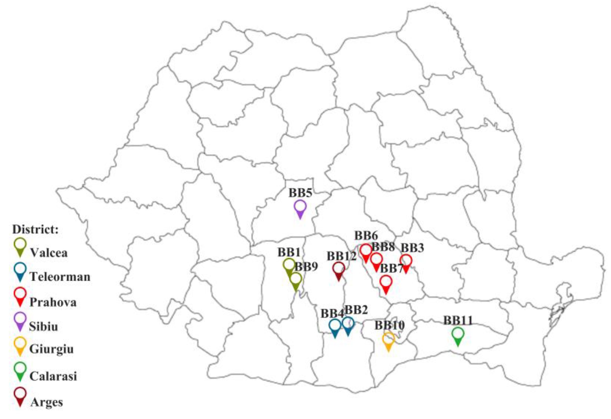
The BB samples were provided by Romanian beekeepers between the spring and summer of 2022 and were deposited at −45 °C. Some samples were extracted from the honeycombs by us and immediately stored in a freezer (BB1-BB3, BB12); others were already removed by the beekeepers with special devices (BB4-BB11). The geographical origin of BB samples is presented in Figure 1. The apiaries were distributed in seven districts of Romania: Arges, Calarasi, Giurgiu, Prahova, Sibiu, Valcea, and Teleorman.
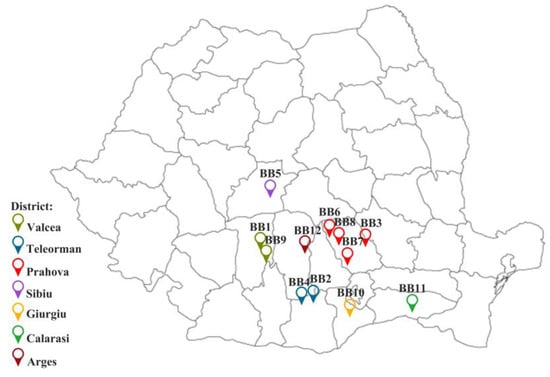
Figure 1.
Distribution of geographical origin of BB samples. Created with ConceptDraw Diagram.
The BBs come from pollen harvested by Apis mellifera carpatica bees from wild flora and house plants, and for these reasons, a palynological analysis was carried out to establish its botanical origin accurately.
2.2. Reagents
Ethanol, glycerol, Folin–Ciocâlteu phenol reagent, sodium carbonate, aluminum chloride, 2,2′-azino-bis (3–ethylbenzothiazoline-6-sulfonate), potassium persulfate, phenolic standards, Nutrient Broth No. 2 (NB), Sabouraud Glucose Agar (Sab) with chloramphenicol, acetic acid (AcA), and crystal violet (CV) were acquired from Sigma-Aldrich (Darmstadt, Germany). Methanol, formic acid, and gallic acid were purchased from Merck (Darmstadt, Germany). All reagents presented high analytical purity, and the strains were part of the Microorganisms Collection of the Department of Microbiology, Faculty of Biology and Research Institute of the University of Bucharest.
2.3. Palynological Analysis
The palynological identification was performed using scanning electron microscopy (SEM) and a light microscope (LM). A total of 2 g of each BB, corresponding to ~500 pollen grains, was considered representative of botanical identification [45]. A quantity of 15 mg of BB was hydrated in 1.5 mL of deionized water. Each suspension was vortexed for 1 min at 20 rpm and immediately spread in 3 equal parts on microscope slides/smooth adhesive surfaces on an aluminum stub.
The SEM images for the determination of the size and morphology of the pollen grains were recorded using a Quanta Inspect F50 (FEI Company, Eindhoven, The Netherlands) scanning electron microscope equipped with a field emission gun electron (FEG) with a 1.2 nm resolution and an energy-dispersive X-ray spectrometer with an MnK resolution of 133 eV. Before the analysis, the BB samples were coated with gold.
The Primostar 3 KMAT Microscope was used for LM analysis. The images were recorded using an Axiocam 208 colour Camera (Carl Zeiss Meditec, Jena, Germany) and Zen Blue 3.4 software. The slides with BB suspensions were dried at room temperature. Glycerol was liquefied at 40 °C, and a drop (100 μL) was put on the slide, which was covered with a thin slide [28].
The pollen grains were identified using online palynological databases (paldat.org and pollenatlas.net (accessed on 1 February 2023)) [46,47] and recent studies [48,49,50,51].
2.4. Bee Bread Extract (BBE) Preparation
The extractions were performed using a method described in a previous study [9]. 1.25 g BB was heated for 10 min at 40 °C with ethanol 70% (v/v) using an ultrasonic bath (Germany, Elmasonic). This process included more steps, such as vortexing (2600 rpm, 3 min), ultrasonication, and centrifugation (4000 rpm at 4 °C, 10 min). The BBEs’ final volume was 25 mL for each sample, and the extracts were stored in closed bottles at −45 °C till analysis.
2.5. Chemical Characterization of BBE
2.5.1. Fourier Transform Infrared Spectroscopy (FTIR)
The FTIR measurements were performed using a Nicolet iS50R spectrometer (Thermo Fisher Scientific, Waltham, MA, USA). The spectra were recorded at room temperature using the attenuated total reflection (ATR) (Thermo Fisher Scientific, Waltham, MA, USA), with 32 scans between 4000 and 400 cm−1 at a resolution of 4 cm−1, with the scanning time being 47 s [52].
2.5.2. Determination of Total Phenols Content (TPC)
The TPC was performed using the Folin–Ciocâlteu method [53]. A total of 0.5 mL of BBE or standard (gallic acid), 5 mL of Folin–Ciocâlteu reagent (diluted 10 times in distilled water), and 4 mL of 1 M of sodium carbonate were homogenized, and the absorbance was measured after 15 min (min) at 746 nm using a Shimadzu UV-1800 spectrophotometer (Kyoto, Japan). A calibration curve was plotted with standard solutions of gallic acid with concentrations varying between 5 and 150 mg/L. TPC was expressed as milligrams (mg) of gallic acid equivalents (GAEs)/gram (g) of the sample [9,53,54,55].
2.5.3. Determination of Total Flavonoids Content (TFC)
The BBEs’ TFC was determined by the aluminum chloride method described in a previous study [9]. The absorbance was measured at 430 nm (spectrophotometer presented in Section 2.5.2), and the TFC was expressed in mg quercetin (QE)/g BB; it was calculated by applying the calibration curve obtained for concentrations of quercetin varying between 0 and 0.12 mg/mL [9,54].
2.5.4. Phenolic Compound Analysis by UHPLC-MS/MS
The measurements were performed using a Q Exactive™ Focus Hybrid Quadrupol OrbiTrap mass spectrometer (ThermoFisher Scientific, Darmstadt, Germany) equipped with heated electrospray ionization (HESI) (ThermoFisher Scientific), coupled with an UltiMate 3000 UHPLC system consisting of a quaternary pump, column oven, and autosampler (ThermoFisher Scientific). Chromatographic separation of phenolic compounds was performed on two columns: Accuacore PFP (50 mm × 2.1 mm, 2.6 μm) and Accuacore PFP (100 mm × 2.1 mm, 2.6 μm) from Thermo Fisher Scientific under the gradient elution of solvent A (water with 0.1% formic acid) and solvent B (methanol with 0.1% formic acid). Chromatographic and mass spectrometric parameters were set according to Ciucure and Geană [56].
Phenolic acids and flavonoids from BBEs were identified according to mass spectra, accurate mass, and characteristic retention time against external standard solutions analyzed under the same conditions. Data-dependent scans with collision-induced dissociations (CIDs) set between 15 and 60 eV were performed for fragmentation studies to confirm each analyzed phenolic compound. Xcalibur software (Version 4.1) was used for instrument control, data acquisition, and data analysis. The ChemSpider internet database of accurate MS data (www.chemspider.com, accessed on 17 November 2023) was used as a reference library to identify compounds of interest [9]. For quantification, a calibration was performed via serial dilution with methanol of the standard mixture of a concentration of 100 mg/L, covering a calibration range between 0 and 10 mg/L [56]. The results are expressed as µg/g of the BB sample.
2.5.5. Determination of Trolox Equivalent Antioxidant Capacity (TEAC)
This method was performed by highlighting the neutralization potential of the cation radical ABTS•+ (2,2′-azino-bis (3–ethylbenzothiazoline-6-sulfonate), expressed as Trolox equivalents. The TEAC method is based on the capacity of the BBE to discolor the blue–green chromophore radical ABTS•+. All steps were described in previous studies [9,57,58], and the spectrophotometric readers were performed at 734 nm. The results are expressed in millimoles of Trolox equivalent (mmol Trolox)/g BB.
2.6. Methods Applied in the Biological Activity of BBE
2.6.1. Qualitative Assessment of the Antimicrobial Activity
The antimicrobial assays were assessed with standard strains (Enterococcus faecalis ATCC 29212, Staphylococcus aureus ATCC 25923, Escherichia coli ATCC 25922, and Pseudomonas aeruginosa ATCC 27853) and clinically isolated strains (Bacillus subtilis, Salmonella typhi 14023, Candida albicans 1688, Candida glabrata, Candida kefyr, and Candida krusei).
The antimicrobial properties of BBE samples (the same extracts characterized previously) were assessed by a spot diffusion assay [9,59,60] and according to the Clinical Laboratory Standards Institute [61]. Bacterial and yeast suspensions (1.5 × 10⁸ CFU/mL and 3 × 10⁸ CFU/mL, respectively) were obtained from 24 h cultures on NB and Sab media. Petri plates with the specific media were seeded with inoculums, and each 20 µL of each BBE was spotted. BBE samples contained ethanol, so an ethanol-based control (CEt) was used. The negative control was considered the sterile medium, and the positive control was the NB/Sab medium inoculated with microbial suspensions. After diffusion, the dishes were incubated at 37 °C for 24 hours (h) for bacteria and 48 h for yeast strains.
2.6.2. Quantitative Assessment of the Antimicrobial Activity
The minimum inhibitory concentration (MIC) assessment was performed using an adapted binary serial microdilution standard assay [9,60,61] in liquid media utilizing 96-well microtiter plates. From every BBE sample, serial two-fold microdilutions were realized in 150 μL of corresponding broth medium seeded with the standard inoculum. Also, a control was performed with ethanol (CEt). The negative and positive controls were performed following the identical steps already detailed. The plates were incubated at 37 °C for 24 h. The MIC values were determined by visual and spectrophotometric analyses measuring the absorbance at 620 nm via the BIOTEK SYNERGY-HTX ELISA multi-mode reader (VT, USA).
2.6.3. Semiquantitative Assessment of the Microbial Adherence to the Inert Substratum
The biofilm development on the inert substratum was assessed utilizing the identical serial two-fold microdilution method [9]. Following 24 h of incubation, the media from dishes (containing binary dilutions of the BBEs) was removed, the wells were washed three times with sterile physiological buffer saline (PBS), and the bacterial cells adhered to the walls were fixed with methanol (5 min) and tinted with 1% CV (15 min). The stained biofilm was resuspended with 33% AcA, and the absorbance was measured at 490 nm [60,61].
2.7. Statistical Analysis
The data results were statistically analyzed with GraphPad Prism 10.2 from GraphPad Software, San Diego, CA (USA). The results are expressed as ±SD (standard deviation) and analyzed using a one-way analysis of variance (one-way ANOVA) and Tukey’s multiple comparisons test according to the method of the experiments performed. The differences between groups were considered statistically significant when the p-value was <0.05.
Principal component analysis (PCA) and hierarchical cluster and heat map analysis (HMA) were performed using Microsoft Excel 2010, Microsoft, Redmond, Washington, DC (USA) and XLSTAT Add-in (15.5.03.3707 software version) by Addinsoft, New York, NY (USA). The chord diagram established by the correlation between botanical families and BB samples was analyzed with OriginPro version 2024 from OriginLab Corporation, Northampton, MA (USA).
3. Results and Discussions
3.1. Palynological Analysis
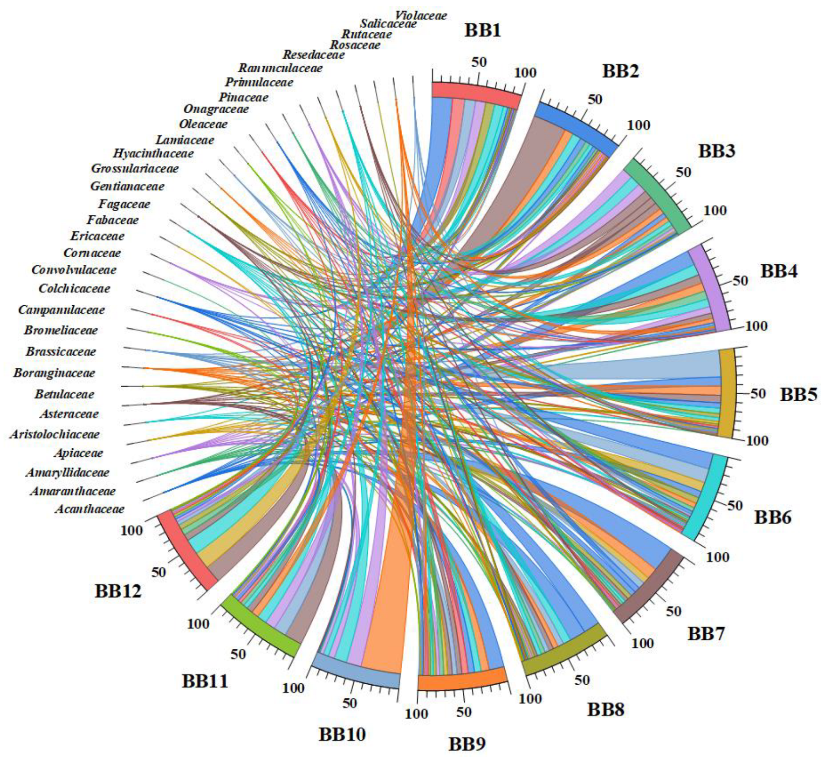
BBs’ taxonomic/botanical assignments were performed using LM and SEM, and Figure 2 and Figure 3 represent the comprehensive results. As a result of the microscopic study, the LM is considered mandatory for determining the relative abundance of species identified. Likewise, the high-resolution SEM images are helpful for the size and morphology determination of the pollen grains, especially the surface pattern, polarity, shape, or dispersal units [62].
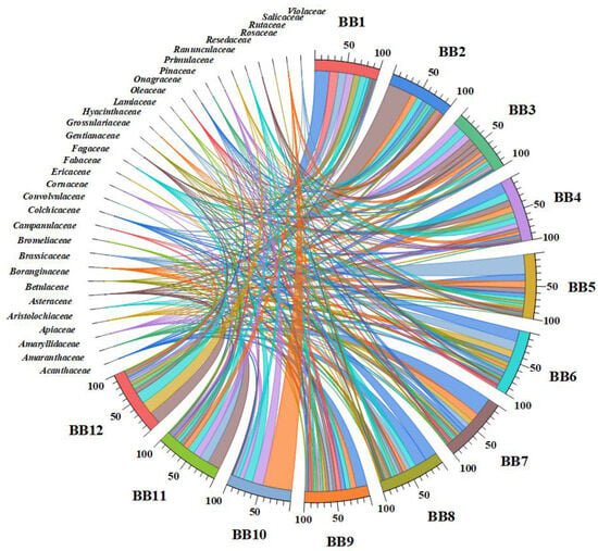
Figure 2.
Chord diagram based on the relationship between botanical families and BB samples.
The data results of the botanical identification presented in Figure 2 indicate 31 families identified and a detailed interconnection between the plant families and BB samples. Also, a high number of plant families and the relative abundance percentages (%) for each BB sample can be observed. For example, BB8 presented pollen grains from an extensive list of plant families (24), and BB6 and BB7 were from 22. Likewise, in BB1, BB3, BB5, and BB9 samples, 21 plant families were identified. BB10 was the least varied sample, containing pollen grains from 9 plant families, with the Salicaceae, Primulaceae, and Fabaceae as the dominant families.
BP from the Fabaceae family is present in all BB samples, which range from 3.00 to 18%, and Acanthaceae, Amaryllidaceae, and Fagaceae plant families are present in 11 BB samples. Likewise, the families predominantly in the 10 BB samples are Amaranthaceae, Asteraceae, Boranginaceae, Onagraceae, and Primulaceae (Figure 2). Otherwise, the high relative abundance is for pollen from the Salicaceae family, with 47% in BB10. BP from the Asteraceae family is predominant in BB2 (39%), followed by the Brassicaceae family in BB5 and the Acanthaceae family in BB7 (30%). The lowest percentages (<1.5%) are presented in BP which comes from the Convolvulaceae (BB12) and Rutaceae (BB8) families.
Multivariate statistical analysis was performed on the plant families identified in BBs to differentiate and group/cluster the BB samples based on the palynological analysis. The PCA plot of the plants’ taxonomic assignments from BP (Figure 3a) showed a clear discrimination of BB12 and some provenience families (see the upper right part of the graph), which are correlated with the relative abundance presented in Figure 2. Likewise, the high frequencies of Amaryllidaceae, Cornaceae, Fabaceae, Fagaceae, Primulaceae, and Salicaceae in pollen from BB2, BB3, BB10, and BB11 are confirmed in the PCA plot. Particularly, BB1 and BB4 are grouped on the left side of Figure 3a, and Resedaceae, Rosaceae, Hyanthaceae, and Grossulariaceae are the representative/specific families from which the pollen of these samples originates. The other families are associated with BB5, BB6, BB7, BB8, and BB9.
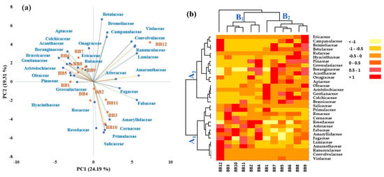
Figure 3.
Discrimination of bee bread samples based on a palynological analysis. (a) Principal component analysis and (b) hierarchical cluster and heat map analysis.
Figure 3.
Discrimination of bee bread samples based on a palynological analysis. (a) Principal component analysis and (b) hierarchical cluster and heat map analysis.
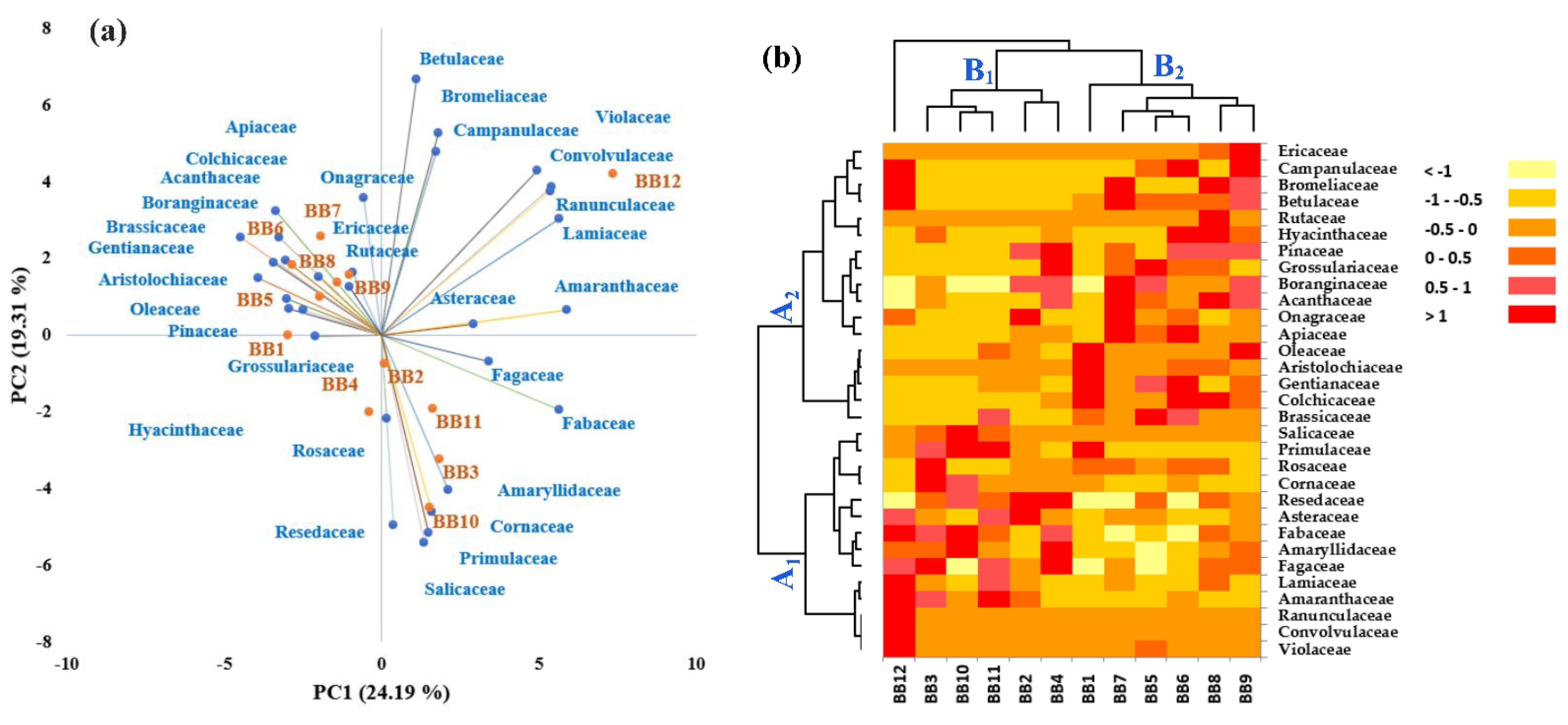
In order to confirm the PCA assay and Chord diagram and differentiate the BB samples as much as possible, hierarchical clustering and heat map assays were performed. According to Figure 3b, the data obtained confirm the previous results. As in Figure 3a, the BB samples were grouped into two main groups (B1 and B2), and this highlighted a discrimination of BB12. The heat map combined with the clusters also expressed a snapshot of the botanical origin of BB samples and was represented by a main group/cluster, which was fused in two subclusters (A1 and A2). Group A1 corresponds to the right side of the PC1-PC2 graph, and A2 to the left side.
Previous studies identified similar botanical origins of pollen grains in BB or BP from Romania [9,17,63,64,65,66]. This study analyzed BB samples from different areas of Romania, with quite varied vegetation, and, for this reason, vast plant botanical families were identified. Furthermore, significant differences in dominant plant families identified depending on the geographical origin of the BB samples were not observed.
3.2. Chemical Composition of BBE
The complex composition of BB can vary widely due to many factors, such as plant and bee species, geographical area, seasonal changes, fermentation strains, beekeeper activities, etc. [67,68]. The alcoholic extracts (BBEs) were analyzed to establish the samples’ chemical compositions.
3.2.1. FTIR Spectroscopy
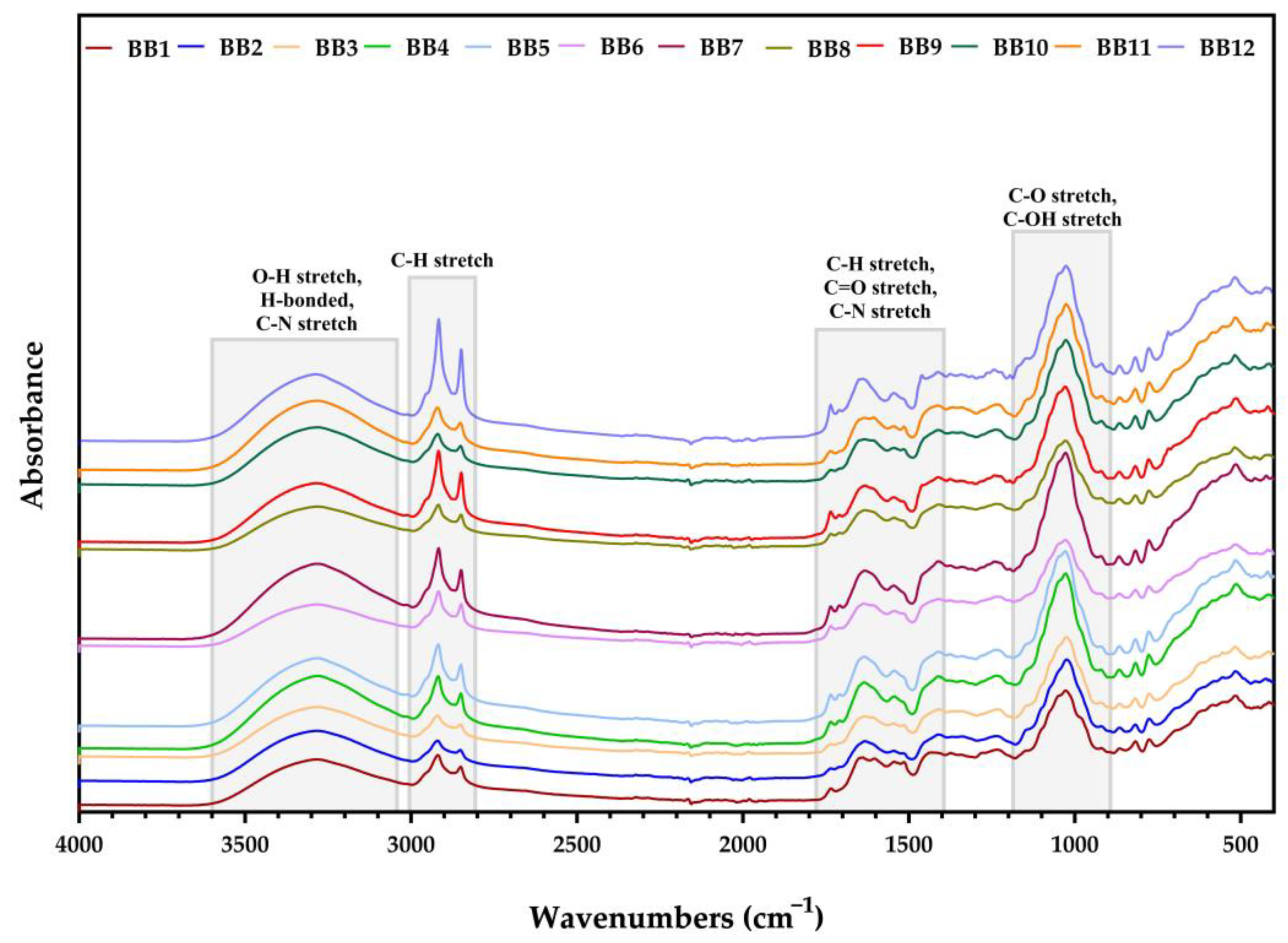
The FTIR spectra for all BB samples were measured between 4000 and 400 cm−1, as illustrated below.
Figure 4 shows that all BB samples presented similar FTIR spectra, which were characterized in previous studies [65,66,69]. Also, all BB samples shared comparable adsorption bands with minor spectral differences.
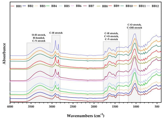
Figure 4.
FTIR spectra for BB.
In the region of 3600–3050 cm−1, the stretching vibrations of O-H and H bonds, which correspond to the presence of water, can be seen [70]. Likewise, in the same intervals, a functional group of amines I and II and amides also exist, which suggests the presence of amino acids and proteins (previously identified in BB) [66]. Between the interval of 3000–2800 cm−1, the peaks were assigned to the symmetric and asymmetric stretching of the C-H groups in carbohydrates and lipids contained in all BB samples [71,72]. Likewise, other peaks related to lipids were observed between the interval of 1790–1400 cm−1, which were attributed to C=O stretching in the ester bond and C-H deformation vibrations of lipids (triglycerides, phospholipids, etc.) [72,73]. Moreover, the C=O and C=C stretching vibrations occurred due to the presence of PCs (sesquiterpenes, phenolic acids, flavonoids, stilbenoids, etc.) [70]. In the same interval, C-N stretching vibrations from amide II were observed [72]. The last gap, 1390–900 cm−1, showed a large peak corresponding to C-O vibrations from carbohydrates and fatty acids, and C-OH groups from polyphenols [66,70,73].
3.2.2. Determinations of TPC, TFC, and TEAC
TPC, TFC, and TEAC results are presented in Table 1 as mean value ± SD.

Table 1.
Total phenols content, total flavonoids content, and antioxidant activity.
The results presented in Table 1 show that BBE8 and BBE10 presented the highest values of TPC (18.30 ± 0.029 and 18.30 ± 0.051 mg GAE/g BB). Correlating the TPC and TFC results with TEAC values is also sometimes difficult. Still, even if the phenolic acids and flavonoids are the primary compounds to determine antioxidant activity, other biomolecules can influence this (stilbenoids, sesquiterpenes, carotenoids, proteins, etc.) [74,75,76,77,78,79]. The antioxidant activity of BBE (corresponding to the TEAC method) had values ranging from 0.02 ± 0.005 to 0.07 ± 0.020 mmol Trolox/g BB.
The TPC, TFC, and antioxidant activity depend upon the plant species from which they are derived. Moreover, Mărgăoan et al. [64] reported the highest antioxidant activity of multifloral BP, with the TEAC method, was for samples that were predominantly from plants of Asteraceae, Brassicaceae, Fabaceae, and Fagaceae plant families. Likewise, the highest levels of TPC were attributed to Rosaceae and Brassicaceae. In contrast, the TFC was highest in BB samples containing BP from Lamiaceae.
Gercek et al. [80] obtained a TFC of 79.21 mg QE/g BP from samples originated from the Asteraceae, Fabaceae, Campanulaceae, Cistaceae, and Rosaceae plant families. In our study, the TFC for BBE11 was 0.95 ± 0.011 mg QE/g BB and BBE12 was 0.85 ± 0.500 mg QE/g BB, and according to the palynological assay, contained pollen from Asteraceae (~21%), Fabaceae (12% and 18%), and Campanulaceae family plant species (2% for BBE12). The BBE3 sample presented the highest TFC (1.86 ± 0.516 mg QE/g BB), probably due to the high polyphenols content of plants that bloom in early spring [9] and belong to families like Acanthaceae, Asteraceae, Colchicaceae, Cornaceae, Fabaceae, Fagaceae, Onagraceae, Primulaceae, Ranunculaceae, Resedaceae, Salicaceae, etc. Furthermore, Jara et al. [81] reported that the Acanthus mollis (Acanthaceae) flower presented a high content of polyphenols and antioxidant activity. Significant levels of PCs and antioxidant activity were also demonstrated in BP from the Oleaceae family [82], which was present the most in BBE1.
3.2.3. Phenolic Compound Profiles by UHPLC-DAD-ESI/MS
A total of 24 compounds, including 9 phenolic acids, 13 flavonoids and derivatives, and stilbene t-resveratrol were unambiguously identified, and sesquiterpene abscisic acid was quantified in the BBE. The quantitative results of the individual phenolic compounds in BB are presented in Table 2.

Table 2.
Phenolic compound content of BB (µg/g).
According to the quantitative data, flavonoids were quantified in higher amounts compared with phenolic acids, being in concordance with the TPC and TFC contents of bee bread measured by UV–Vis spectrophotometric methods (Table 1) and literature data [71]. Moreover, the extracts contained other PCs, like rutin, hesperidin, resveratrol, and abscisic acid (ABA). Overall, the samples had significant quantities of phenolic acids, like p-coumaric acid, gallic acid, caffeic acid, and cinnamic acid. High contents could be observed for quercetin, kaempferol, isorhamnetin, and ABA.
The BP fermentation in the BB formation process changed the bioactive compounds’ amounts and chemical profiles to increase their bioavailability and bioaccessibility [83,84,85]. Also, lactic fermentation by bacteria and/or yeast degrades the pollen wall and releases PCs, which are associated with carbohydrates or proteins [84,86]. Additionally, Zhang et al. [87] reported that the contents of flavonoids in fermented BP increased up to 144.66-fold/units, and of phenolic acids only up to 28.9-fold compared to unfermented BP. The flavonoid and phenolic acid glycosides significantly decreased (in some cases, disappeared), which can be explained by the heteroside hydrolyzation into their aglycone forms due to the microbial fermentation process [88]. Among flavonoid heterosides, the BBEs tested in this study contained only rutin (quercetin-3-rutinoside) and hesperidin (hesperetin-7-rutinoside), while Bayram et al. [44] identified only rutin. In addition, probiotic fermentation can affect the phenolic acids, which, through metabolization in small compounds (like 4-ethyl catechol, dihydrocaffeic acid, etc.), provide more biological properties for bees and humans [84].
According to Table 2, BBE12, BBE2, BBE9, and BBE8 had the highest concentrations of phenolic acids and flavonoids (in this order), but considering the resveratrol and ABA, BBE9 showed the highest content in PCs. Significant ABA contents can be observed in the samples, especially for BBE9, BBE2, BBE3, BBE7, and BBE8. The phytohormone ABA, with multiple regulatory functions in plants, comes from flower nectar. Likewise, ABA enhances the immune system, cold stress tolerance, growth, and prevalence of the nosemosis (Nosema disease) of Apis mellifera bees [68,89,90,91]. Also, for human health, ABA plays an essential role in glucose metabolism, oxidative stress, tumor growth, ischemic retinopathies, and the central nervous system [92,93]. Furthermore, ABA showed an antimicrobial effect on Helicobacter pylori [94] and antiviral properties [95]. Bridi et al. [96] determined in Brassica rapa (Brassicaceae) and Robinia pseudoacacia BP (Fabaceae) 59.00 to 240.00 μg ABA/g BP. In another study [96], the ABA content varied from 25.60 to 355.50 μg ABA/g BP in multi-floral BP from plant species of Asteraceae, Fabaceae, and Rosaceae families.
In a recent study, Bayram et al. [44] reported the composition of their BBE samples, derived from Asteraceae, Fabaceae, Plantaginaceae, and Rosaceae, as gallic acid (0.34–3.47 μg/g BB), kaempferol (3.80–26.81 μg/g BB), quercetin (9.71–39.18 μg/g BB), and rutin (23.30–126.13 μg/g BB), values which were smaller than our results, except for rutin concentration. The results obtained in another study by Gercek et al. [80] on BP samples provided from Asteraceae, Fabaceae, Campanulaceae, Cistacea, and Rosaceae plant species also showed lower PC content values and similar rutin concentrations (115.42 μg/g BP) compared to the results obtained by us for the 12 samples of BB. The palynological profiles of the BB from this study varied, and it was challenging to differentiate the samples according to phenolic profiles and predominant families and correlate them with the preceding BB studies/research. However, the literature data confirm the PCs depicted in Table 2 [15,25,43,71,97,98]. The presence of PCs was also confirmed by FTIR analysis (Figure 4).
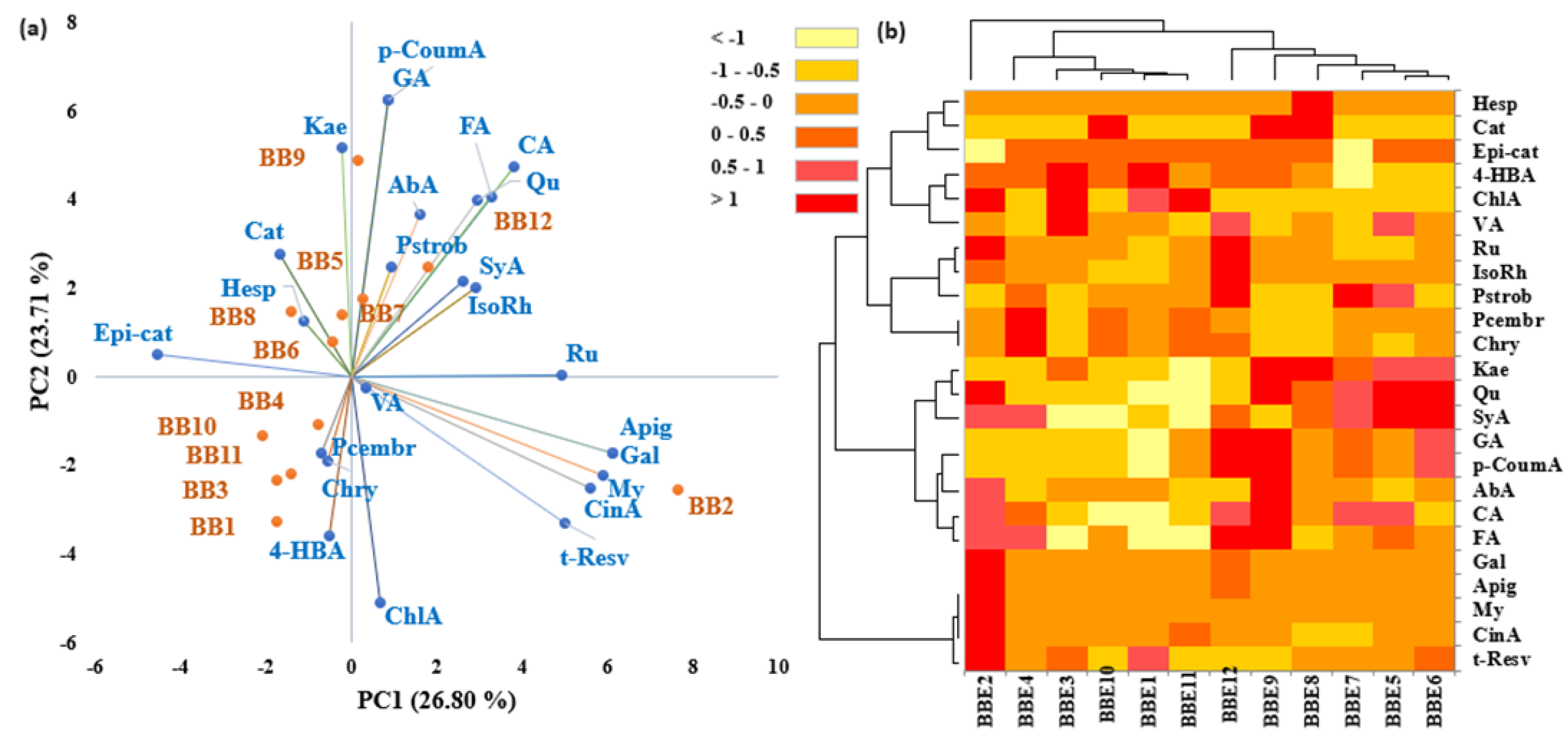
Multivariate statistical analysis, including principal component analysis (PCA) and heat map analysis (HMA), was applied to the phenolic quantitative data in order to differentiate between BB samples with different origins. From the PCA analysis, a clear discrimination of the BB2 sample was observed, which could be correlated with the botanical origin because this sample presented the highest percentage of the Asteraceae family plant species (Figure 2). Furthermore, apigenin (Apig), galangin (Gal), myricetin (My), cinnamic acid (CinA), t-resveratrol (t-Resv), and chlorogenic acid (ChlA) represent specific PCs for BB2 and are distributed on the right-downside area of Figure 5a. Also, the right side of the figure indicates specific PCs for BB7 and BB12, like rutin (Ru), isorhamnetin (IsoRh), syringic acid (SyA), pinostrombin (Pstrob), abscisic acid (AbA), ferulic acid (FA), caffeic acid (CA), quercetin (Qu), gallic acid (GA), and p-coumaric acid (p-CoumA). Corresponding to the palynological analysis, the BB12 sample is specific to BP of plant species of Amaranthaceae, Betulaceae, Bromeliaceae, Concolvulaceae, Lamiaceae, Onagraceae, and Violaceae. The BB7 sample also has plant pollen from the mentioned families, while presents a high content of pollen from the Acanthaceae family.
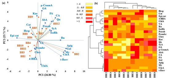
Figure 5.
Discrimination of bee bread samples based on quantitative phenolic compound biomarkers: (a) principal component analysis and (b) hierarchical cluster and heat map analysis.
According to botanical origin (Figure 2 and Figure 3), BB3, BB10, and BB11 present significant contents in pollen from plant species of Amaryllidiceae, Cornaceae, Fabaceae, Fagaceae, Primulaceae, Resedaceae, and Salicaceae families. Linking these results with Figure 5a, the pinocembrin (Pcembr), chrysin (Chry), and 4-hydroxybenzoic acid (4-HBA) are distributed to BB1, BB3, BB4, BB10, and BB11, which are clustered on the left downside of the PCA graph. Kaempferol (kae), catechin (cat), epicatechin (Epi-cat), and hesperidin (Hesp) represent specific PCs for BB5, BB6, BB8, and BB9, which are grouped on the left side of Figure 5a. Likewise, Acanthaceae, Apiaceae, Asteraceae, Boranginaceae, Brassicaceae, Colchicaceae, Oleaceae, Onagraceae, and Pinaceae were the families distinctive for these samples (as seen in Figure 3a). In particular, Hesp is linked to the Ericaceae plant species and is present only in BB8 and BB9, which also have a significant content of phenolic acids and flavonoids.
The hierarchical clusters heat map confirms the PCA results, which highlight a discrimination of BB2. As seen in Figure 5b, the other BB samples are clustered into two main groups, corresponding to the left downside of the PCA graph and the upside.
The heat map of the PC profiles indicates a principal cluster, which corresponds to t-resv, CinA, My, Apig, and Gal, and the BB2 sample, respectively. The main cluster is divided into two sub-clusters, which are distributed into other groups at the same distance.
A recent study [99] highlighted the considerable content in PCs of Vaccinium sp. (CinA, p-CoumA, CA, GA, SyA, Qu, etc.). Only the BB8 sample had pollen from plants of the Rutaceae family, while Hesp was identified in its extract (BBE8). Hesperidin and its derivates are known to be found in the highest concentrations in citrus fruits (Rutaceae family) and in small amounts in Mentha sp. (Lamiaceae) [100]. A fact recently reconfirmed by Bakour et al. [101], who showed that BP with 50% Citrus aurantium had the highest hesperidin content among PCs (488.90 μg/g BP), and also showed more potent antioxidant activity.
3.3. Biological Activity of BBE
3.3.1. Qualitative Assessment of the Antimicrobial Activity
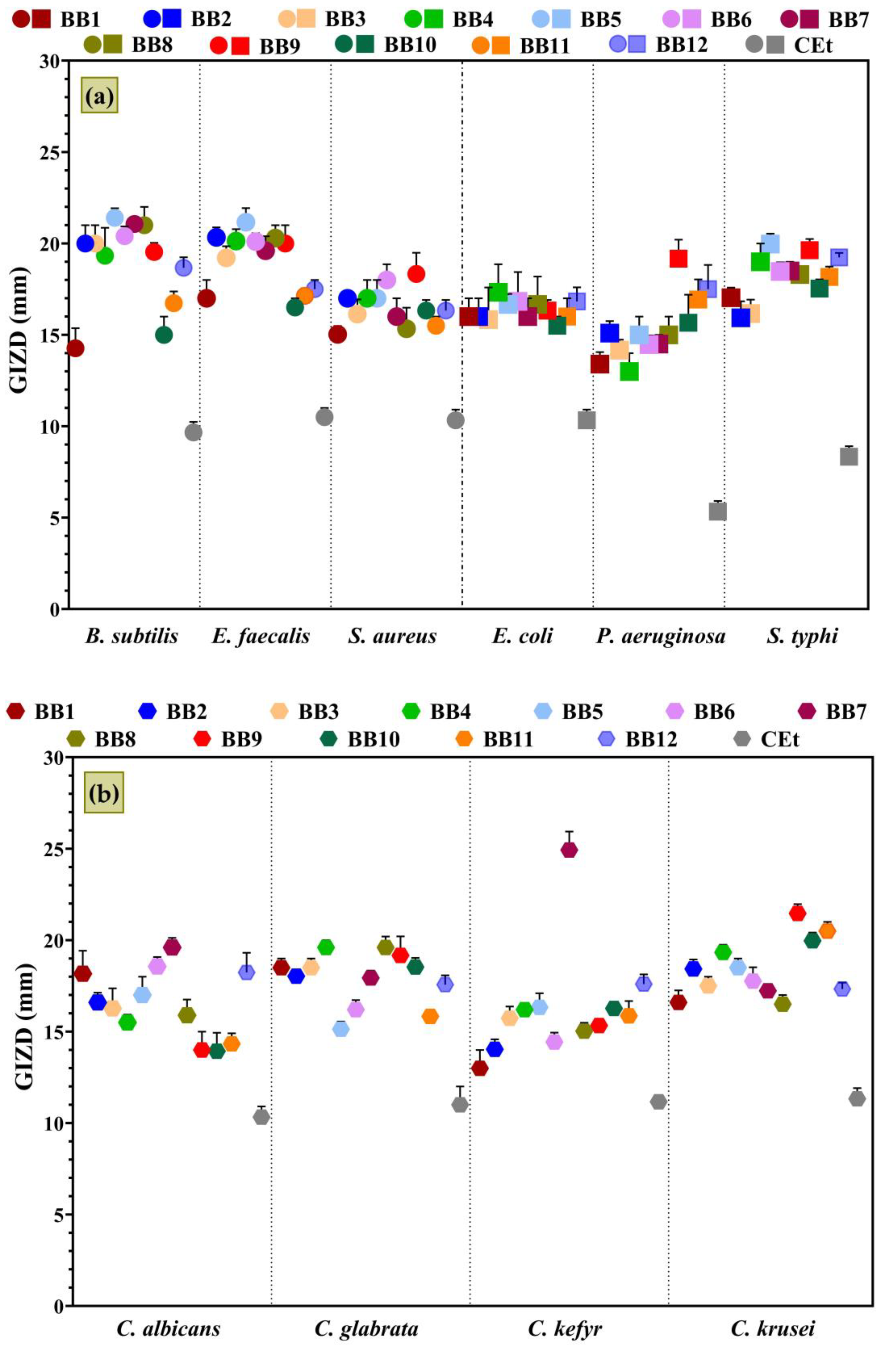
Antimicrobial activity was qualitatively assessed by determining the diameters of the growth inhibition zones that appeared around the spot (of BBE samples) and expressing them as mean values ± SDs (Figure 6).
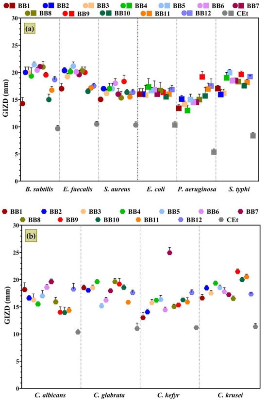
Figure 6.
The growth inhibition zone diameters (GIZDs) of BBEs on selected pathogenic strains: (a) Gram-positive and Gram-negative bacteria; (b) yeasts. The significant influence of the BBEs on each microbial strain was statistically analyzed by one-way ANOVA and Tukey’s multiple comparisons tests. The p-value was <0.01 and the results were statistically significant.
BBE samples presented a significant antimicrobial effect on the growth of all microbial strains tested, and B. subtilis, E. faecalis, S. typhi, C. krusei, and C. glabrata were the most sensitive strains.
The extract with the highest antibacterial activity on Gram-positive bacteria was BBE5, followed by BBE8, BBE1, BBE2, BBE6, and BBE9. The data obtained are linked to the botanical origins because these samples are also clustered in Figure 3. The susceptibility of Gram-positive bacteria to BBEs can be explained by the presence of flavonoids in high amounts, which, with their lipophilicity properties, damage the cell membrane (phospholipid bilayers) and inhibit the respiratory chain and ATP synthesis [102].
In the case of Gram-negative bacteria, BBE5, BBE4, BBE9, and BBE12 showed the highest inhibitory effect. The antimicrobial profiles for yeasts differed, but BBE7 and BBE9 presented the most heightened sensitivity. Besides flavonoids, BBEs contained phenolic acids, which induced damage to the cytoplasmatic membranes of Gram-positive and Gram-negative bacteria, and microscopic fungi [103,104].
Quercetin, kaempferol, and caffeic acid were most likely the compounds responsible for the remarkable antimicrobial activity of the BBE5 sample because these were present in the highest amount in this, have activity on both Gram-positive and Gram-negative bacterial strains, and the first two sometimes acted synergistically [105,106,107]. In addition, Qian et al. [108] revealed that vanillic acid, which had the highest concentration in this sample (BBE5), possessed antimicrobial activity against S. aureus and E. coli. These phenolic compounds were also present (in variable percentages) in other samples with significant antimicrobial activity (BBE8, BBE1, BBE2, BBE6, and BBE9).
Akhir et al. [109] reported that BB ethanol extracts presented more inhibitory effects against B. subtilis and S. aureus than E. coli and S. typhi. Also, Elsayed et al. [110] demonstrated the highest sensitivity for S. aureus (26 mm) and B. subtilis (24 mm) rather than E. coli (18 mm) and C. albicans (15 mm) due to the influence of citrus BB. The GIZDs determined by the Rutaceae plant BB are like the results in Figure 6 and can be correlated with the significant contents in quercetin, rutin, benzoic acid, and hesperidin [110,111]. The antifungal activity of BB has been little addressed until now, the diffusion assay being predominantly applied by Hudz et al. [112] and Elsayed et al. [110]. These two studies obtained growth inhibition diameters between 4 and 8 mm (Candida albicans CCM 8186, Candida glabrata CCM 8270, and Candida tropicalis CCM 8223) and 15 mm (Candida albicans ATCC 10221) for BB samples also extracted with ethanol. Still, according to our knowledge, there are no references to compare our results regarding the antimicrobial activity of BBE against C. kefyr and C. krusei.
3.3.2. Quantitative Evaluation of Antimicrobial Activity
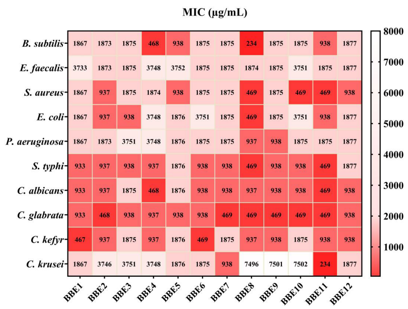
The MIC value was characterized by the smallest concentration of the tested BBEs that inhibited microbial development. The results are shown in Figure 7, expressed as µg/mL.
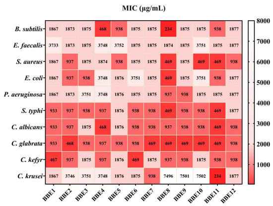
Figure 7.
Quantitative evaluation of the antimicrobial activity of BBEs. Data results marked in intense red indicate the significantly lowest MIC values respectively a significant sensitivity of strains. The scale bar presents the variations in the sensitivity of tested strains from the highest (red) to the lowest (white).
Figure 7 highlights the most significant antimicrobial activity of BBEs on the pathogenic strains. S. typhi and C. glabrata were the most sensitive tested strains and, in contrast, E. faecalis was the most resistant to BBE.
The lowest MIC value was obtained for BBE8 (234 µg/mL), followed by BBE4 (468 µg/mL) on B. subtilis. Also, the extracts showed significant inhibition of S. aureus development, and BBE8, BBE10, BBE11, BBE2, and BBE12 induced the highest sensitivity, a result somehow expected based on the known antimicrobial effects of caffeic acid, quercetin, and kaempferol against this strain. Hesperidin (together with other phenolic compounds) might have contributed to BBE8’s MIC value, given its higher antibacterial effect on Gram-positive than Gram-negative bacteria [113]. The multi-target antibacterial action mechanisms for p-coumaric acid suggested that it could also play an essential role in the fight against pathogenic bacteria, especially against S. aureus and E. coli [114].
In contrast, BBEs determined moderate sensitivity on E. faecalis and P. aeruginosa. C. glabrata, C. albicans, and C. kefyr were the least resistant to the action of the BBEs.
Overall, BBE8, BBE9, BBE2, and BBE4 presented the highest antimicrobial activity, correlated with chemical composition (Figure 4, Table 1 and Table 2). Moreover, according to the statistical assays, the mentioned samples stood out from the others. In agreement with the PCA assays, BBE2 was associated with precise taxonomic assignments (of plants with pollen contribution) and PCs. Also, BBE12 is linked with FA, CA, and Qu, and with specific botanic families (Figure 3).
Abouda et al. [115] reported that BBEs inhibited S. aureus, B. cereus, E. coli, and P. aeruginosa at ½ and ¼ dilutions. Also, Urcan et al. [116] recorded the highest antimicrobial potential of Romanian BB for S. aureus. Another study [15] showed the significant antimicrobial activity of the BB originated from BPs of plants of the Apiaceae (35%), Asteraceae (24%), Fagaceae (16%), Myrtaceae (9%), Punicaceae (6%), and Mimosaceae (5%) botanical families. Bacterial strains B. cereus, S. aureus, and L. monocytogenes were sensitive to BBE, with 0.04–0.17 mg/mL and 0.08–0.35 mg/mL values for MIC and MBC (minimal bactericidal concentration), respectively. Comparable results were obtained for Gram-negative bacteria (E. coli, E. cloacae, and S. typhimurium), which were the least susceptible to BBE.
3.3.3. Semiquantitative Assay of the Microbial Adherence to the Inert Substratum
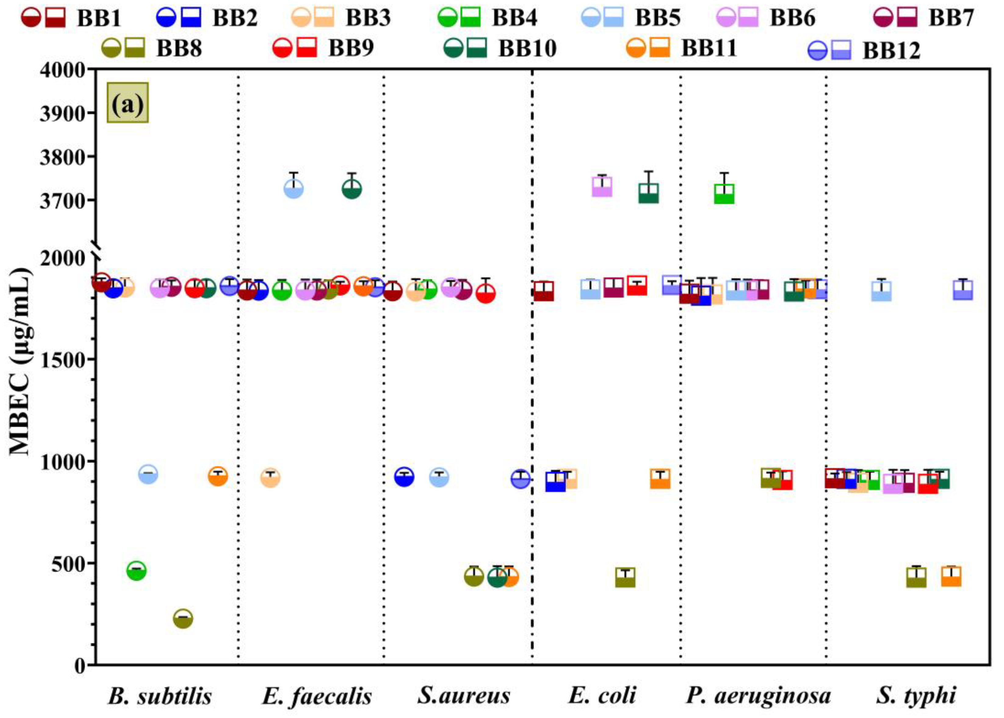
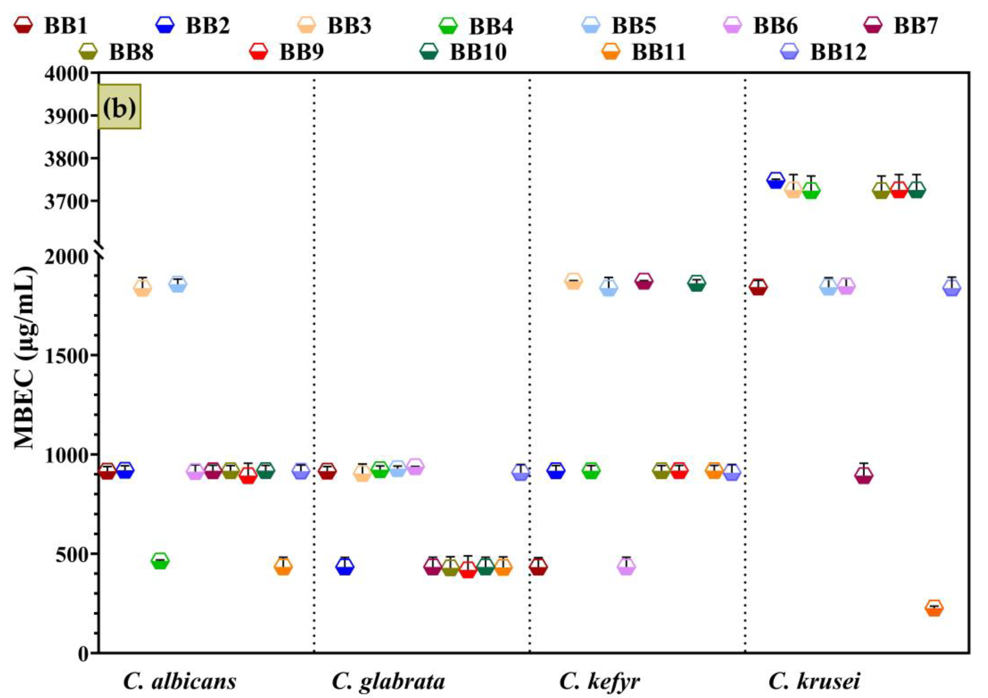
The BBE’s influence on the tested microbial strains’ adherence to the inert substratum is displayed in Figure 8, which represents the minimal biofilm eradication concentration (MBEC) values.
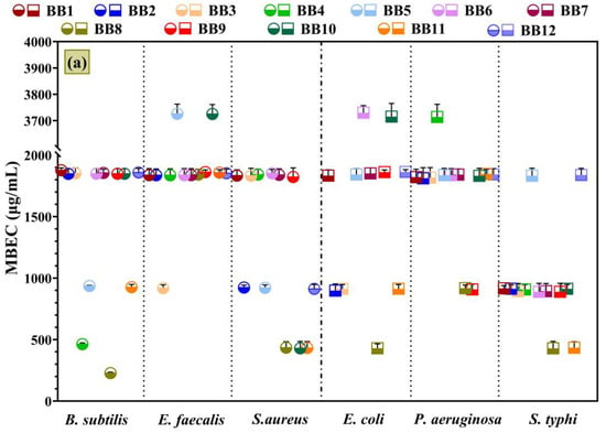
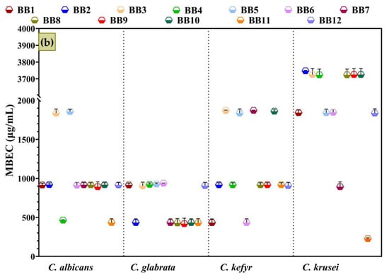
Figure 8.
Graphical chart of the MBEC values: (a) Gram-positive and Gram-negative bacteria; (b) yeasts. The differences between BBEs were statistically analyzed using one-way ANOVA, followed by Tukey’s multiple comparisons tests.
Figure 8’s data confirm the qualitative (Figure 6) and MIC results (Figure 7) and are correlated with the chemical composition (Figure 4 and Table 2) and botanical origin of BBs (Figure 1 and Figure 2). Consequently, S. typhi and C. glabrata depicted the highest sensitivity in the presence of BBE samples. Also, BBEs significantly inhibited the adherence of C. albicans and C. kefyr. For the other strains, BBE samples showed similar antimicrobial profiles, but with some exceptions. For example, BBE8, with the highest TPC content, exhibited the strongest antibiofilm activity against B. subtilis. Also, a great inhibitory effect was displayed against S. aureus, S. typhi, C. albicans, and C. kefyr. Furthermore, BBE2, BBE9, and BBE12, which had a high amount of PCs, displayed significant antibiofilm effects on the tested strains. In addition, in any prior research, the BBs’ influences on adherence to the bacteria or fungi inert substratum were not investigated.
In agreement with the chromatographic results (Table 2), the representative flavonoids for BBEs are isorhamnetin, kaempferol, and quercetin, and their antimicrobial effects are reported in previous studies [117,118,119,120,121]. According to our knowledge, the antimicrobial properties of the isolated PCs fractions from BBEs were not studied due to limited data for BB. Additionally, the inhibitory effect of flavonoids (like quercetin and kaempferol glucosides) isolated from BP against S. aureus, S. epidermidis, P. aeruginosa, E. coli, C. albicans, C. tropicalis, and C. glabrata were reported [122].
BBE8 and BBE9, with significant quercetin and kaempferol amounts, exhibited great antibiofilm effects against P. aeruginosa and C. glabrata. These samples had dominant pollen grains from the Acanthaceae, Colchicaceae, and Ericaceae botanical families. Moreover, in a recent study [28], P. aeruginosa was very sensitive to BB with a significant content of plant pollen from Ericaceae and S. aureus on BB from Brassicaceae; the results were correlated with the high TPC, TFC, and antioxidant activity of BP [123].
BBE5 confirms these findings of antimicrobial activity on B. subtilis, S. aureus, and C. glabrata because it contains 31% Brassicaceae pollen. It is well known that the pollen from Fabaceae species is also associated with an antibacterial effect against P. aeruginosa [28].
In our study, the pollen from the Fabaceae family was present in all samples, but greater contents were found in BBE10, BBE11, and BBE12. In addition, BBE10 had pollen predominantly from the Salicaceae family plant species, which demonstrated its antimicrobial potential [9]. Furthermore, BBE2 had a significant abundance of pollen from Asteraceae family plants and the antimicrobial activity of Achillea setacea, Antennaria dioica, and Helichrysum arenarium, which are found in Romanian flora [124,125] was already proven. Also, BBE2 discrimination is highlighted in the statistical assays of PCs (Figure 5).
Previous research [9] showed that BBEs exhibited greater antimicrobial activity than BPEs with pollen from similar plant species. Also, Pelka et al. [126] reported that BBEs presented a higher antimicrobial effect than BPEs on S. aureus, S. epidermidis, E. coli, and P. aeruginosa, with MIC values that ranged from 2.5 to 10% (v/v) and MBC from 2.5 to 5%. Additionally, Kaškanienè et al. [127] reported that the antibacterial, antifungal, and antioxidant activities increased after the fermentation of bee pollen with Lactococcus lactis and Lactobacillus rhamnosus. It is possible that certain metabolites of these strains may be responsible for antimicrobial activity in some cases. Despite incompletely understanding the inhibition mechanism of bacterial growth, resveratrol contribution can be significant, as well as other compounds that may be present in very low quantities under the detection limit of the applied method [33,128].
4. Conclusions
Bee bread is a promising source of phenolic compounds and antioxidants. The main objective of this study was to investigate the relationship between botanical origin, chemical composition, antioxidant activity, and the effect on selected pathogenic strains.
The palynological analysis revealed a high relative abundance of pollen from plants belonging to Salicaceae, Asteraceae, Brassicaceae, and Acanthaceae families. In total, thirty-one families were identified. In all bee bread samples, pollen “supplied” by plants from the Fabaceae family was always present, while the pollen of the least-represented species is part of the Bromeliaceae, Convovulaceae and Rutaceae families.
The flavonoid concentrations were much higher than the phenolic acids in all bee bread extracts analyzed. The extracts contained mainly rutin, hesperidin, and resveratrol, as well as a high content of quercetin, kaempferol, isorhamnetin, and abscisic acid. There were also significant quantities of p-coumaric acid, gallic acid, caffeic acid, and cinnamic acid. The BBE2, BBE8, BBE9, and BBE12 samples had the highest levels of phenolic acids, flavonoids and heterosides. BBE2 and BBE9 presented the highest concentrations of phenolic compounds, which ranged from 9209.73 and 18,889.74 µg PC/g BB.
The antimicrobial activity of bee bread extracts is strongly linked to chemical composition, antioxidant activity, and pollen botanical origin. The bee bread extracts’ phenolic profiles are complex and different, and it is challenging to attribute the inhibitory growth effect to a single compound or a pollen type from a specific botanic family precisely. Furthermore, a synergistic effect between bioactive compounds is most probably responsible for the biological properties of bee bread.
The bee bread extracts presented a significant antimicrobial effect on the growth of all microbial strains tested. Salmonella typhi and Candida glabrata were the most susceptible tested strains. Also, Candida albicans and Candida kefyr were sensitive to the influence of bee bread extracts.
The present study reported the bee bread extracts’ antibiofilm effects/ inhibitory activity on microbial adhesion capacity to the inert substratum of bacteria or fungi for the first time. Likewise, the samples (BBE8 and BBE9) that had pollen grains dominant from the Acanthaceae, Colchicaceae, and Ericaceae botanical families presented significant quercetin and kaempferol amounts and displayed great antimicrobial effects against Pseudomonas aeruginosa and Candida glabrata. In addition, the sensitivity of Bacillus subtilis, Staphylococcus aureus, and Candida glabrata is linked to pollen from Brassicaceae plant families (BBE5). Significant antimicrobial activity was correlated with pollen from plants belonging to the Salicaceae and Asteraceae families (BBE10 and BBE2, respectively).
Rich in phenolic compounds and with significant antimicrobial properties, bee bread can be a valuable source of natural nutrients and bioactive compounds that enhance human health. Further studies should evaluate the pre- and probiotic potential of bee bread as well as the cytotoxic action to complement the existing data.
Author Contributions
Conceptualization, C.-I.I., E.O. and A.F.; methodology, C.-I.I., E.O., L.-M.D. and A.F.; software, C.-I.I.; validation, E.O., L.-M.D., E.-I.G. and A.F.; formal analysis, C.-I.I., C.C., E.-I.G. and A.S.; investigation, C.-I.I., C.C., E.O. and E.-I.G.; data curation, C.-I.I., E.O., L.-M.D. and E.-I.G.; writing—original draft preparation, C.-I.I., E.O., L.-M.D., E.-I.G. and C.C.; writing—review and editing, C.-I.I., E.O., L.-M.D., A.S. and A.F.; visualization, E.O., L.-M.D. and A.F.; supervision, E.O. and A.F. All authors have read and agreed to the published version of the manuscript.
Funding
The APC was funded by the UNSTPB.
Institutional Review Board Statement
Not applicable.
Informed Consent Statement
Not applicable.
Data Availability Statement
Data are contained within the article.
Acknowledgments
This work was supported by the National Centre for Micro- and Nanomaterials and the Proof of Concept Project of the National University of Science and Technology Politehnica Bucharest.
Conflicts of Interest
The authors declare no conflicts of interest.
References
- Nayik, G.A.; Shah, T.R.; Muzaffar, K.; Wani, S.A.; Gull, A.; Majid, I.; Bhat, F.M. Honey: Its History And Religious Significance: A Review. Univers. J. Pharm. 2014, 3, 5–8. [Google Scholar]
- Khalifa, S.A.M.; Elashal, M.H.; Yosri, N.; Du, M.; Musharraf, S.G.; Nahar, L.; Sarker, S.D.; Guo, Z.; Cao, W.; Zou, X.; et al. Bee Pollen: Current Status and Therapeutic Potential. Nutrients 2021, 13, 1876. [Google Scholar] [CrossRef]
- Madras-Majewska, B.; Majewski, J. Production and Prices of Honey in Researched Apriaries in Poland. Sci. Yearb. Assoc. Agric. Agribus. Econ. 2007, 9, 298–302. [Google Scholar]
- Kieliszek, M.; Piwowarek, K.; Kot, A.M.; Błażejak, S.; Chlebowska-Śmigiel, A.; Wolska, I. Pollen and bee bread as new health-oriented products: A review. Trends Food Sci. Technol. 2018, 71, 170–180. [Google Scholar] [CrossRef]
- Martinotti, S.; Ranzato, E. Honey, Wound Repair and Regenerative Medicine. J. Funct. Biomater. 2018, 9, 34. [Google Scholar] [CrossRef]
- Thakur, M.; Nanda, V. Composition and functionality of bee pollen: A review. Trends Food Sci. Technol. 2020, 98, 82–106. [Google Scholar] [CrossRef]
- Khalifa, S.A.M.; Elashal, M.; Kieliszek, M.; Ghazala, N.E.; Farag, M.A.; Saeed, A.; Xiao, J.; Zou, X.; Khatib, A.; Göransson, U.; et al. Recent insights into chemical and pharmacological studies of bee bread. Trends Food Sci. Technol. 2020, 97, 300–316. [Google Scholar] [CrossRef]
- Margaoan, R.; Strant, M.; Varadi, A.; Topal, E.; Yucel, B.; Cornea-Cipcigan, M.; Campos, M.G.; Vodnar, D.C. Bee Collected Pollen and Bee Bread: Bioactive Constituents and Health Benefits. Antioxidants 2019, 8, 568. [Google Scholar] [CrossRef]
- Ilie, C.I.; Oprea, E.; Geana, E.I.; Spoiala, A.; Buleandra, M.; Gradisteanu Pircalabioru, G.; Badea, I.A.; Ficai, D.; Andronescu, E.; Ficai, A.; et al. Bee Pollen Extracts: Chemical Composition, Antioxidant Properties, and Effect on the Growth of Selected Probiotic and Pathogenic Bacteria. Antioxidants 2022, 11, 959. [Google Scholar] [CrossRef]
- Spoiala, A.; Ilie, C.I.; Ficai, D.; Ficai, A.; Andronescu, E. Synergic Effect of Honey with Other Natural Agents in Developing Efficient Wound Dressings. Antioxidants 2022, 12, 34. [Google Scholar] [CrossRef]
- Ioniță-Mîndrican, C.-B.; Mititelu, M.; Musuc, A.M.; Oprea, E.; Ziani, K.; Neacșu, S.M.; Grigore, N.D.; Negrei, C.; Dumitrescu, D.-E.; Mireșan, H.; et al. Honey and Other Beekeeping Products Intake among the Romanian Population and Their Therapeutic Use. Appl. Sci. 2022, 12, 9649. [Google Scholar] [CrossRef]
- Bakour, M.; Al-Waili, N.S.; El Menyiy, N.; Imtara, H.; Figuira, A.C.; Al-Waili, T.; Lyoussi, B. Antioxidant activity and protective effect of bee bread (honey and pollen) in aluminum-induced anemia, elevation of inflammatory makers and hepato-renal toxicity. J. Food Sci. Technol. 2017, 54, 4205–4212. [Google Scholar] [CrossRef] [PubMed]
- Aylanc, V.; Falcao, S.I.; Vilas-Boas, M. Bee pollen and bee bread nutritional potential: Chemical composition and macronutrient digestibility under in vitro gastrointestinal system. Food Chem. 2023, 413, 135597. [Google Scholar] [CrossRef] [PubMed]
- Ćirić, J.; Haneklaus, N.; Rajić, S.; Baltić, T.; Lazić, I.B.; Đorđević, V. Chemical composition of bee bread (perga), a functional food: A review. J. Trace Elem. Miner. 2022, 2, 100038. [Google Scholar] [CrossRef]
- Bakour, M.; Fernandes, Â.; Barros, L.; Sokovic, M.; Ferreira, I.C.F.R.; Badiaa, l. Bee bread as a functional product: Chemical composition and bioactive properties. LWT 2019, 109, 276–282. [Google Scholar] [CrossRef]
- Urcan, A.C.; Marghitas, L.A.; Dezmirean, D.S.; Bobis, O.; Bonta, V.; Muresan, C.I.; Margaoan, R. Chemical Composition and Biological Activities of Beebread—Review. Bull. Univ. Agric. Sci. Vet. Med. Cluj-Napoca. Anim. Sci. Biotechnol. 2017, 74, 6. [Google Scholar] [CrossRef]
- Urcan, A.C.; Criste, A.D.; Dezmirean, D.S.; Bobiș, O.; Bonta, V.; Dulf, F.V.; Mărgăoan, R.; Cornea-Cipcigan, M.; Campos, M.G. Botanical origin approach for a better understanding of chemical and nutritional composition of beebread as an important value-added food supplement. LWT 2021, 142, 111068. [Google Scholar] [CrossRef]
- Mayda, N.; Özkök, A.; Ecem Bayram, N.; Gerçek, Y.C.; Sorkun, K. Bee bread and bee pollen of different plant sources: Determination of phenolic content, antioxidant activity, fatty acid and element profiles. J. Food Meas. Charact. 2020, 14, 1795–1809. [Google Scholar] [CrossRef]
- Adaskeviciute, V.; Kaskoniene, V.; Kaskonas, P.; Barcauskaite, K.; Maruska, A. Comparison of Physicochemical Properties of Bee Pollen with Other Bee Products. Biomolecules 2019, 9, 819. [Google Scholar] [CrossRef]
- Kutlu, N.; Gerçek, Y.C.; Celik, S.; Bayram, S.; Bayram, N.E. An optimization study for amino acid extraction from bee bread using choline chloride-acetic acid deep eutectic solvent and determination of individual phenolic profile. J. Food Meas. Charact. 2023, 18, 1026–1037. [Google Scholar] [CrossRef]
- Tomás, A.; Falcão, S.I.; Russo-Almeida, P.; Vilas-Boas, M. Potentialities of beebread as a food supplement and source of nutraceuticals: Botanical origin, nutritional composition and antioxidant activity. J. Apic. Res. 2017, 56, 219–230. [Google Scholar] [CrossRef]
- Crozier, A.; Jaganath, I.B.; Clifford, M.N. Dietary phenolics: Chemistry, bioavailability and effects on health. Nat. Prod. Rep. 2009, 26, 1001–1043. [Google Scholar] [CrossRef]
- Tsao, R. Chemistry and biochemistry of dietary polyphenols. Nutrients 2010, 2, 1231–1246. [Google Scholar] [CrossRef]
- Aylanc, V.; Falcão, S.I.; Ertosun, S.; Vilas-Boas, M. From the hive to the table: Nutrition value, digestibility and bioavailability of the dietary phytochemicals present in the bee pollen and bee bread. Trends Food Sci. Technol. 2021, 109, 464–481. [Google Scholar] [CrossRef]
- Othman, Z.A.; Wan Ghazali, W.S.; Noordin, L.; Mohd Yusof, N.A.; Mohamed, M. Phenolic Compounds and the Anti-Atherogenic Effect of Bee Bread in High-Fat Diet-Induced Obese Rats. Antioxidants 2019, 9, 33. [Google Scholar] [CrossRef] [PubMed]
- Metibemu, D.S.; Ogungbe, I.V. Carotenoids in Drug Discovery and Medicine: Pathways and Molecular Targets Implicated in Human Diseases. Molecules 2022, 27, 6005. [Google Scholar] [CrossRef] [PubMed]
- Rizvi, S.; Raza, S.T.; Ahmed, F.; Ahmad, A.; Abbas, S.; Mahdi, F. The Role of Vitamin E in Human Health and Some Diseases. SQU Med. J. 2014, 14, 157–163. [Google Scholar]
- Didaras, N.A.; Kafantaris, I.; Dimitriou, T.G.; Mitsagga, C.; Karatasou, K.; Giavasis, I.; Stagos, D.; Amoutzias, G.D.; Hatjina, F.; Mossialos, D. Biological Properties of Bee Bread Collected from Apiaries Located across Greece. Antibiotics 2021, 10, 555. [Google Scholar] [CrossRef] [PubMed]
- Ben Bacha, A.; Norah, A.O.; Al-Osaimi, M.; Harrath, A.H.; Mansour, L.; El-Ansary, A. The therapeutic and protective effects of bee pollen against prenatal methylmercury induced neurotoxicity in rat pups. Metab. Brain Dis. 2020, 35, 215–224. [Google Scholar] [CrossRef] [PubMed]
- Eleazu, C.; Suleiman, J.B.; Othman, Z.A.; Zakaria, Z.; Nna, V.U.; Hussain, N.H.N.; Mohamed, M. Bee bread attenuates high fat diet induced renal pathology in obese rats via modulation of oxidative stress, downregulation of NF-kB mediated inflammation and Bax signalling. Arch. Physiol. Biochem. 2022, 128, 1088–1104. [Google Scholar] [CrossRef] [PubMed]
- Kosedag, M.; Gulaboglu, M. Pollen and bee bread expressed highest anti-inflammatory activities among bee products in chronic inflammation: An experimental study with cotton pellet granuloma in rats. Inflammopharmacology 2023, 31, 1967–1975. [Google Scholar] [CrossRef]
- Hsu, C.K.; Wang, D.Y.; Wu, M.C. A Potential Fungal Probiotic Aureobasidium melanogenum CK-CsC for the Western Honey Bee, Apis mellifera. J. Fungi 2021, 7, 508. [Google Scholar] [CrossRef]
- Didaras, N.A.; Karatasou, K.; Dimitriou, T.G.; Amoutzias, G.D.; Mossialos, D. Antimicrobial Activity of Bee-Collected Pollen and Beebread: State of the Art and Future Perspectives. Antibiotics 2020, 9, 811. [Google Scholar] [CrossRef]
- Bakour, M.; Laaroussi, H.; Ousaaid, D.; El Ghouizi, A.; Es-Safi, I.; Mechchate, H.; Lyoussi, B. Bee Bread as a Promising Source of Bioactive Molecules and Functional Properties: An Up-To-Date Review. Antibiotics 2022, 11, 203. [Google Scholar] [CrossRef]
- Barta, D.G.; Cornea-Cipcigan, M.; Margaoan, R.; Vodnar, D.C. Biotechnological Processes Simulating the Natural Fermentation Process of Bee Bread and Therapeutic Properties-An Overview. Front. Nutr. 2022, 9, 871896. [Google Scholar] [CrossRef]
- Toutiaee, S.; Mojgani, N.; Harzandi, N.; Moharrami, M.; Mokhberosafa, L. In vitro probiotic and safety attributes of Bacillus spp. isolated from beebread, honey samples and digestive tract of honeybees Apis mellifera. Lett. Appl. Microbiol. 2022, 74, 656–665. [Google Scholar] [CrossRef]
- Bleha, R.; Shevtsova, T.; Kruzík, V.; Skorpilová, T.; Salon, I.; Erban, V.; Brindza, J.; Brovarskyi, V.; Sinica, A. Bee breads from Eastern Ukraine: Composition, physical properties and biological activities. Czech J. Food Sci. 2019, 37, 9–20. [Google Scholar] [CrossRef]
- Schaad, E.; Fracheboud, M.; Droz, B.; Kast, C. Quantitation of pesticides in bee bread collected from honey bee colonies in an agricultural environment in Switzerland. Environ. Sci. Pollut. R. 2023, 30, 56353–56367. [Google Scholar] [CrossRef]
- Murcia-Morales, M.; Heinzen, H.; Parrilla-Vázquez, P.; Gómez-Ramos, M.d.M.; Fernández-Alba, A.R. Presence and distribution of pesticides in apicultural products: A critical appraisal. TrAC Trends Anal. Chem. 2022, 146, 116506. [Google Scholar] [CrossRef]
- Zuluaga, C.M.; Serrato, J.C.; Quicazan, M.C. Chemical, Nutritional and Bioactive Characterization of Colombian Bee-Bread. Chem. Eng. Trans. 2015, 43, 175–180. [Google Scholar] [CrossRef]
- Ivanišová, E.; Kačániová, M.; Frančáková, H.; Petrová, J.; Hutková, J.; Brovarskyi, V.; Velychko, S.; Adamchuk, L.; Schubertová, Z.; Musilová, J. Bee bread—Perspective source of bioactive compounds for future. Potravin. Slovak. J. Food Sci. 2015, 9, 592–598. [Google Scholar] [CrossRef]
- Baltrusaityte, V.; Venskutonis, P.R.; Ceksteryte, V. Antibacterial Activity of Honey and Beebread of Different Origin against S. aureus and S. epidermidis. Food Technol. Biotechnol. 2007, 45, 201–208. [Google Scholar]
- Sobral, F.; Calhelha, R.C.; Barros, L.; Duenas, M.; Tomas, A.; Santos-Buelga, C.; Vilas-Boas, M.; Ferreira, I.C. Flavonoid Composition and Antitumor Activity of Bee Bread Collected in Northeast Portugal. Molecules 2017, 22, 248. [Google Scholar] [CrossRef]
- Bayram, N.E.; Gercek, Y.C.; Çelik, S.; Mayda, N.; Kostić, A.Ž.; Dramićanin, A.M.; Özkök, A. Phenolic and free amino acid profiles of bee bread and bee pollen with the same botanical origin—Similarities and differences. Arab. J. Chem. 2021, 14, 103004–103016. [Google Scholar] [CrossRef]
- Morais, M.; Moreira, L.; Feas, X.; Estevinho, L.M. Honeybee-collected pollen from five Portuguese Natural Parks: Palynological origin, phenolic content, antioxidant properties and antimicrobial activity. Food Chem. Toxicol. 2011, 49, 1096–1101. [Google Scholar] [CrossRef]
- Pollen Atlas. 2023. Available online: https://pollenatlas.net/ (accessed on 1 February 2023).
- PalDat—A Palynological Database. Available online: www.paldat.org (accessed on 1 February 2023).
- Halbritter, H.; Grímsson, S.U.F.; Weber, M.; Hesse, R.Z.M.; Buchner, R.; Frosch-Radivo, M.S.A. Illustrated Pollen Terminology, 2nd ed.; Springer: Cham, Switzerland, 2018. [Google Scholar]
- Klimko, M.; Nowińska, R.; Wilkin, P.; Wiland-Szymańska, J. Pollen Morphology of Some Species of the Genus Sansevieria Petagna (Asparagaceae). Acta Biol. Cracoviensia S. Bot. 2017, 59, 63–75. [Google Scholar] [CrossRef]
- Rahmawati, L.U.; Purwanti, E.; Budiyanto, M.A.K.; Zaenab, S.; Susetyarini, R.E.; Permana, T.I. Identification of Pollen Grains Morphology and Morphometry in Liliaceae. Int. Conf. Life Sci. Technol. Ser. Earth Environ. Sci. 2019, 276, 012031. [Google Scholar] [CrossRef]
- Peng, Y.; Pu, X.; Yu, Q.; Zhou, H.; Huang, T.; Xu, B.; Gao, X. Comparative Pollen Morphology of Selected Species of Blumea DC. and Cyathocline Cass. and Its Taxonomic Significance. Plants 2023, 12, 2909. [Google Scholar] [CrossRef]
- Spoiala, A.; Ilie, C.I.; Dolete, G.; Petrisor, G.; Trusca, R.D.; Motelica, L.; Ficai, D.; Ficai, A.; Oprea, O.C.; Ditu, M.L. The Development of Alginate/Ag NPs/Caffeic Acid Composite Membranes as Adsorbents for Water Purification. Membranes 2023, 13, 591. [Google Scholar] [CrossRef]
- Singleton, V.L.; Rossi, J.A. Colorimetry of Total Phenolics with Phosphomolybdic-Phosphotungstic Acid Reagents. Am. J. Enol. Vitic. 1965, 16, 144–158. [Google Scholar] [CrossRef]
- Marinas, I.; Oprea, E.; Geana, E.-I.; Chifiriuc, C.; Lazar, V. Antimicrobial and antioxidant activity of the vegetative and reproductive organs of Robinia pseudoacacia. J. Serb. Chem. Soc. 2014, 79, 1363–1378. [Google Scholar] [CrossRef]
- Alizadeh, A.; Alizadeh, O.; Amari, G.; Zare, M. Essential Oil Composition, Total Phenolic Content, Antioxidant Activity and Antifungal Properties of Iranian Thymus daenensi ssubsp. daenensis Celak. as in Influenced by Ontogenetical Variation. J. Essent. Oil Bear. Plants 2013, 16, 59–70. [Google Scholar] [CrossRef]
- Ciucure, C.T.; Geana, E.I. Phenolic compounds profile and biochemical properties of honeys in relationship to the honey floral sources. Phytochem. Anal. 2019, 30, 481–492. [Google Scholar] [CrossRef]
- Prior, R.L.; Wu, X.; Schaich, K. Standardized Methods for the Determination of Antioxidant Capacity and Phenolics in Foods and Dietary Supplements. J. Agric. Food Chem. 2005, 53, 4290–4302. [Google Scholar] [CrossRef]
- Pellegrini, N.; Serafini, M.; Colombi, B.; Rio, D.D.; Salvatore, S.; Bianchi, M.; Brighenti, F. Total Antioxidant Capacity of Plant Foods, Beverages and Oils Consumed in Italy Assessed by Three Different In Vitro Assays. J. Nutr. 2003, 133, 2812–2819. [Google Scholar] [CrossRef]
- Motelica, L.; Ficai, D.; Ficai, A.; Trusca, R.D.; Ilie, C.I.; Oprea, O.C.; Andronescu, E. Innovative Antimicrobial Chitosan/ZnO/Ag NPs/Citronella Essential Oil Nanocomposite-Potential Coating for Grapes. Foods 2020, 9, 1801. [Google Scholar] [CrossRef]
- Spoiala, A.; Ilie, C.I.; Trusca, R.D.; Oprea, O.C.; Surdu, V.A.; Vasile, B.S.; Ficai, A.; Ficai, D.; Andronescu, E.; Ditu, L.M. Zinc Oxide Nanoparticles for Water Purification. Materials 2021, 14, 4747–4763. [Google Scholar] [CrossRef]
- Performance Standards for Antimicrobial Susceptibility Testing. In CLSI Supplemenent M100; Clinical and Laboratory Standards Institute: San Antonio, TX, USA, 2021; Volume 41, p. 352.
- Halbritter, H.; Grímsson, S.U.F.; Weber, M.; Hesse, R.Z.M.; Buchner, R.; Frosch-Radivo, M.S.A. Pollen Morphology and Ultrastructure. In Illustrated Pollen Terminology, 2nd ed.; Springer: Cham, Switzerland, 2018; pp. 37–67. [Google Scholar] [CrossRef]
- Oroian, M.; Dranca, F.; Ursachi, F. Characterization of Romanian Bee Pollen-An Important Nutritional Source. Foods 2022, 11, 2633. [Google Scholar] [CrossRef]
- Mărgăoan, R.; Özkök, A.; Keskin, Ş.; Mayda, N.; Urcan, A.C.; Cornea-Cipcigan, M. Bee collected pollen as a value-added product rich in bioactive compounds and unsaturated fatty acids: A comparative study from Turkey and Romania. Food Sci. Technol. 2021, 149, 111925–111936. [Google Scholar] [CrossRef]
- Spulber, R.; Dogaroglu, M.; Babeanu, N.; Popa, O. Physicochemical characteristics of fresh bee pollen from different botanical origins. Rom. Biotechnol. Lett. 2018, 23, 13357–13365. [Google Scholar]
- Isopescu, R.D.; Spulber, R.; Josceanu, A.M.; Mihaiescu, D.E.; Popa, O. Romanian bee pollen classification and property modelling. J. Apic. Res. 2020, 59, 443–451. [Google Scholar] [CrossRef]
- Li, J.-L.; Li, W.-L.; Zhang, J.; Pang, Y.-T.; Xiong, J.; Wu, P.; Wei, B.-R.; Li, X.-J.; Huang, Q.; Tang, Q.-H.; et al. Seasonal Dynamics of the Microbiota and Nutritional Composition in Bee Bread from Apis cerana and Apis mellifera Colonies. Food Res. Int. 2023, 113905. [Google Scholar] [CrossRef]
- Leponiemi, M.; Freitak, D.; Moreno-Torres, M.; Pferschy-Wenzig, E.M.; Becker-Scarpitta, A.; Tiusanen, M.; Vesterinen, E.J.; Wirta, H. Honeybees’ foraging choices for nectar and pollen revealed by DNA metabarcoding. Sci. Rep. 2023, 13, 14753. [Google Scholar] [CrossRef] [PubMed]
- Oroian, M.; Ursachi, F.; Dranca, F. Ultrasound-Assisted Extraction of Polyphenols from Crude Pollen. Antioxidants 2020, 9, 322–337. [Google Scholar] [CrossRef] [PubMed]
- Castiglioni, S.; Astolfi, P.; Conti, C.; Monaci, E.; Stefano, M.; Carloni, P. Morphological, Physicochemical and FTIR Spectroscopic Properties of Bee Pollen Loads from Different Botanical Origin. Molecules 2019, 24, 3974. [Google Scholar] [CrossRef] [PubMed]
- Dranca, F.; Ursachi, F.; Oroian, M. Bee Bread: Physicochemical Characterization and Phenolic Content Extraction Optimization. Foods 2020, 9, 1358. [Google Scholar] [CrossRef]
- Kasprzyk, I.; Depciuch, J.; Grabek-Lejko, D.; Parlinska-Wojtan, M. FTIR-ATR spectroscopy of pollen and honey as a tool for unifloral honey authentication. The case study of rape honey. Food Control 2018, 84, 33–40. [Google Scholar] [CrossRef]
- Pini, R.; Furlanetto, G.; Castellano, L.; Saliu, F.; Rizzi, A.; Tramelli, A. Effects of stepped-combustion on fresh pollen grains: Morphoscopic, thermogravimetric, and chemical proxies for the interpretation of archeological charred assemblages. Rev. Palaeobot. Palynol. 2018, 259, 142–158. [Google Scholar] [CrossRef]
- Treml, J.; Lelakova, V.; Smejkal, K.; Paulickova, T.; Labuda, S.; Granica, S.; Havlik, J.; Jankovska, D.; Padrtova, T.; Hosek, J. Antioxidant Activity of Selected Stilbenoid Derivatives in a Cellular Model System. Biomolecules 2019, 9, 468. [Google Scholar] [CrossRef]
- Julia, M.; Eugenia Marta, K.; María José, N.; Agustín, G.A. Antioxidant Capacity of Anthocyanin Pigments. In Flavonoids; Goncalo, C.J., Ed.; IntechOpen: Rijeka, Croatia, 2017; Chapter 11. [Google Scholar]
- Srivastava, R. Physicochemical, antioxidant properties of carotenoids and its optoelectronic and interaction studies with chlorophyll pigments. Sci. Rep. 2021, 11, 18365. [Google Scholar] [CrossRef]
- Bechir, B.; Imen, R. Potential Antioxidant Activity of Terpenes. In Terpenes and Terpenoids; Shagufta, P., Areej Mohammad, A.-T., Eds.; IntechOpen: Rijeka, Croatia, 2021; Chapter 5. [Google Scholar]
- Rocchetti, G.; Senizza, B.; Zengin, G.; Okur, M.A.; Montesano, D.; Yildiztugay, E.; Lobine, D.; Mahomoodally, M.F.; Lucini, L. Chemical Profiling and Biological Properties of Extracts from Different Parts of Colchicum szovitsii Subsp. Szovitsii. Antioxidants 2019, 8, 632. [Google Scholar] [CrossRef]
- Tawfik, A.I.; Ahmed, Z.H.; Abdel-Rahman, M.F.; Moustafa, A.M. Effect of some bee bread quality on protein content and antioxidant system of honeybee workers. Int. J. Trop. Insect Sci. 2022, 43, 93–105. [Google Scholar] [CrossRef]
- Gercek, Y.C.; Celik, S.; Bayram, S. Screening of Plant Pollen Sources, Polyphenolic Compounds, Fatty Acids and Antioxidant/Antimicrobial Activity from Bee Pollen. Molecules 2021, 27, 117. [Google Scholar] [CrossRef]
- Jara, C.; Leyton, M.; Osorio, M.; Silva, V.; Fleming, F.; Paz, M.; Madrid, A.; Mellado, M. Antioxidant, phenolic and antifungal profiles of Acanthus mollis (Acanthaceae). Nat. Prod. Res. 2017, 31, 2325–2328. [Google Scholar] [CrossRef]
- Castiglioni, S.; Stefano, M.; Astolfi, P.; Pisani, M.; Carloni, P. Characterisation of Bee Pollen from the Marche Region (Italy) According to the Botanical and Geographical Origin with Analysis of Antioxidant Activity and Colour, Using a Chemometric Approach. Molecules 2022, 27, 7996. [Google Scholar] [CrossRef] [PubMed]
- Yang, F.; Chen, C.; Ni, D.; Yang, Y.; Tian, J.; Li, Y.; Chen, S.; Ye, X.; Wang, L. Effects of Fermentation on Bioactivity and the Composition of Polyphenols Contained in Polyphenol-Rich Foods: A Review. Foods 2023, 12, 3315. [Google Scholar] [CrossRef]
- Filannino, P.; Di Cagno, R.; Gambacorta, G.; Tlais, A.Z.A.; Cantatore, V.; Gobbetti, M. Volatilome and Bioaccessible Phenolics Profiles in Lab-Scale Fermented Bee Pollen. Foods 2021, 10, 286. [Google Scholar] [CrossRef]
- Giampieri, F.; Quiles, J.L.; Cianciosi, D.; Forbes-Hernandez, T.Y.; Orantes-Bermejo, F.J.; Alvarez-Suarez, J.M.; Battino, M. Bee Products: An Emblematic Example of Underutilized Sources of Bioactive Compounds. J. Agric. Food Chem. 2022, 70, 6833–6848. [Google Scholar] [CrossRef] [PubMed]
- Kieliszek, M.; Piwowarek, K.; Kot, A.M.; Wojtczuk, M.; Roszko, M.; Bryla, M.; Trajkovska Petkoska, A. Recent advances and opportunities related to the use of bee products in food processing. Food Sci. Nutr. 2023, 11, 4372–4397. [Google Scholar] [CrossRef]
- Zhang, H.; Lu, Q.; Liu, R. Widely targeted metabolomics analysis reveals the effect of fermentation on the chemical composition of bee pollen. Food Chem. 2022, 375, 131908. [Google Scholar] [CrossRef]
- Santos, A.C.D.; Biluca, F.C.; Braghini, F.; Gonzaga, L.V.; Costa, A.C.O.; Fett, R. Phenolic composition and biological activities of stingless bee honey: An overview based on its aglycone and glycoside compounds. Food Res. Int. 2021, 147, 110553. [Google Scholar] [CrossRef]
- Negri, P.; Maggi, M.D.; Ramirez, L.; De Feudis, L.; Szwarski, N.; Quintana, S.; Eguaras, M.J.; Lamattina, L. Abscisic acid enhances the immune response in Apis mellifera and contributes to the colony fitness. Apidologie 2015, 46, 542–557. [Google Scholar] [CrossRef]
- Ramirez, L.; Negri, P.; Sturla, L.; Guida, L.; Vigliarolo, T.; Maggi, M.; Eguaras, M.; Zocchi, E.; Lamattina, L. Abscisic acid enhances cold tolerance in honeybee larvae. Proc. Biol. Sci. 2017, 284, 20162140. [Google Scholar] [CrossRef]
- Szawarski, N.; Saez, A.; Dominguez, E.; Dickson, R.; De Matteis, A.; Eciolaza, C.; Justel, M.; Aliano, A.; Solar, P.; Bergara, I.; et al. Effect of Abscisic Acid (ABA) Combined with Two Different Beekeeping Nutritional Strategies to Confront Overwintering: Studies on Honey Bees’ Population Dynamics and Nosemosis. Insects 2019, 10, 329. [Google Scholar] [CrossRef] [PubMed]
- Balino, P.; Gomez-Cadenas, A.; Lopez-Malo, D.; Romero, F.J.; Muriach, M. Is There A Role for Abscisic Acid, A Proven Anti-Inflammatory Agent, in the Treatment of Ischemic Retinopathies? Antioxidants 2019, 8, 104. [Google Scholar] [CrossRef] [PubMed]
- Pizzio, G.A. Potential Implications of the Phytohormone Abscisic Acid in Human Health Improvement at the Central Nervous System. Ann. Epidemiol. Public Health 2022, 5, 1090. [Google Scholar]
- Kim, S.; Hong, I.; Woo, S.; Jang, H.; Pak, S.; Han, S. Isolation of Abscisic Acid from Korean Acacia Honey with Anti-Helicobacter pylori Activity. Pharmacogn. Mag. 2017, 13, S170–S173. [Google Scholar] [CrossRef]
- Alazem, M.; Lin, N.S. Antiviral Roles of Abscisic Acid in Plants. Front. Plant Sci. 2017, 8, 1760. [Google Scholar] [CrossRef] [PubMed]
- Bridi, R.; Echeverria, J.; Larena, A.; Nunez Pizarro, P.; Atala, E.; De Camargo, A.C.; Oh, W.Y.; Shahidi, F.; Garcia, O.; Ah-Hen, K.S.; et al. Honeybee Pollen From Southern Chile: Phenolic Profile, Antioxidant Capacity, Bioaccessibility, and Inhibition of DNA Damage. Front. Pharmacol. 2022, 13, 775219. [Google Scholar] [CrossRef] [PubMed]
- Habryka, C.; Socha, R.; Juszczak, L. The Influence of Bee Bread on Antioxidant Properties, Sensory and Quality Characteristics of Multifloral Honey. Appl. Sci. 2023, 13, 7913. [Google Scholar] [CrossRef]
- Aylanc, V.; Tomas, A.; Russo-Almeida, P.; Falcao, S.I.; Vilas-Boas, M. Assessment of Bioactive Compounds under Simulated Gastrointestinal Digestion of Bee Pollen and Bee Bread: Bioaccessibility and Antioxidant Activity. Antioxidants 2021, 10, 651. [Google Scholar] [CrossRef] [PubMed]
- Martau, G.A.; Bernadette-Emoke, T.; Odocheanu, R.; Soporan, D.A.; Bochis, M.; Simon, E.; Vodnar, D.C. Vaccinium Species (Ericaceae): Phytochemistry and Biological Properties of Medicinal Plants. Molecules 2023, 28, 1533. [Google Scholar] [CrossRef] [PubMed]
- Pyrzynska, K. Hesperidin: A Review on Extraction Methods, Stability and Biological Activities. Nutrients 2022, 14, 2387. [Google Scholar] [CrossRef] [PubMed]
- Bakour, M.; Laaroussi, H.; Ferreira-Santos, P.; Genisheva, Z.; Ousaaid, D.; Teixeira, J.A.; Lyoussi, B. Exploring the Palynological, Chemical, and Bioactive Properties of Non-Studied Bee Pollen and Honey from Morocco. Molecules 2022, 27, 5777. [Google Scholar] [CrossRef]
- Yuan, G.; Guan, Y.; Yi, H.; Lai, S.; Sun, Y.; Cao, S. Antibacterial activity and mechanism of plant flavonoids to Gram-positive bacteria predicted from their lipophilicities. Sci. Rep. 2021, 11, 10471. [Google Scholar] [CrossRef]
- Lobiuc, A.; Paval, N.E.; Mangalagiu, I.I.; Gheorghita, R.; Teliban, G.C.; Amariucai-Mantu, D.; Stoleru, V. Future Antimicrobials: Natural and Functionalized Phenolics. Molecules 2023, 28, 1114. [Google Scholar] [CrossRef]
- Teodoro, G.R.; Ellepola, K.; Seneviratne, C.J.; Koga-Ito, C.Y. Potential Use of Phenolic Acids as Anti-Candida Agents: A Review. Front. Microbiol. 2015, 6, 1420. [Google Scholar] [CrossRef]
- Nguyen, T.L.A.; Bhattacharya, D. Antimicrobial Activity of Quercetin: An Approach to Its Mechanistic Principle. Molecules 2022, 27, 2494. [Google Scholar] [CrossRef]
- Periferakis, A.; Periferakis, K.; Badarau, I.A.; Petran, E.M.; Popa, D.C.; Caruntu, A.; Costache, R.S.; Scheau, C.; Caruntu, C.; Costache, D.O. Kaempferol: Antimicrobial Properties, Sources, Clinical, and Traditional Applications. Int. J. Mol. Sci. 2022, 23, 15054. [Google Scholar] [CrossRef]
- Khan, F.; Bamunuarachchi, N.I.; Tabassum, N.; Kim, Y.M. Caffeic Acid and Its Derivatives: Antimicrobial Drugs toward Microbial Pathogens. J. Agric. Food Chem. 2021, 69, 2979–3004. [Google Scholar] [CrossRef]
- Qian, W.; Yang, M.; Wang, T.; Sun, Z.; Liu, M.; Zhang, J.; Zeng, Q.; Cai, C.; Li, Y. Antibacterial Mechanism of Vanillic Acid on Physiological, Morphological, and Biofilm Properties of Carbapenem-Resistant Enterobacter hormaechei. J. Food Prot. 2020, 83, 576–583. [Google Scholar] [CrossRef] [PubMed]
- Akhir, R.A.M.; Abu Bakar, M.F.; Sanusi, S.B. Antioxidant and Antimicrobial Activity of Stingless Bee Bread and Propolis Extracts. AIP Conf. Proc. 2017, 1891, 020090. [Google Scholar] [CrossRef]
- Elsayed, N.; El-Din, H.S.; Altemimi, A.B.; Ahmed, H.Y.; Pratap-Singh, A.; Abedelmaksoud, T.G. In Vitro Antimicrobial, Antioxidant and Anticancer Activities of Egyptian Citrus Beebread. Molecules 2021, 26, 2433. [Google Scholar] [CrossRef] [PubMed]
- Kirci, D.; Demirci, F.; Demirci, B. Microbial Transformation of Hesperidin and Biological Evaluation. ACS Omega 2023, 8, 42610–42621. [Google Scholar] [CrossRef] [PubMed]
- Hudz, N.; Yezerska, O.; Grygorieva, O.; Felsöciová, S.; Brindza, J.; Wieczorek, P.P.; Kacániová, M. Analytical Procedure Elaboration of Total Flavonoid Content Determination and Antimicrobial Activity of Bee Bread Extracts. Acta Pol. Pharm. 2019, 76, 439–452. [Google Scholar] [CrossRef]
- Choi, S.S.; Lee, S.H.; Lee, K.A. A Comparative Study of Hesperetin, Hesperidin and Hesperidin Glucoside: Antioxidant, Anti-Inflammatory, and Antibacterial Activities In Vitro. Antioxidants 2022, 11, 1618. [Google Scholar] [CrossRef] [PubMed]
- Lou, Z.; Wang, H.; Rao, S.; Sun, J.; Ma, C.; Li, J. p-Coumaric acid kills bacteria through dual damage mechanisms. Food Control 2012, 25, 550–554. [Google Scholar] [CrossRef]
- Abouda, Z.; Zerdani, I.; Kalalou, I.; Faid, M.; Ahami, M.T. The Antibacterial Activity of Maroccan Bee Bread and Bee-Pollen (Fresh and Dried) against Pathogenic Bacteria. Res. J. Microbiol. 2011, 6, 376–384. [Google Scholar]
- Urcan, A.; Criste, A.; Dezmirean, D.; Bobiș, O.; Mărghitaș, L.; Mărgăoan, R.; Hrinca, A. Antimicrobial Activity of Bee Bread Extracts Against Different Bacterial Strains. Bull. UASVM Anim. Sci. Biotechnol. 2018, 75, 85–91. [Google Scholar] [CrossRef]
- Jiang, L.; Li, H.; Wang, L.; Song, Z.; Shi, L.; Li, W.; Deng, X.; Wang, J. Isorhamnetin Attenuates Staphylococcus aureus-Induced Lung Cell Injury by Inhibiting Alpha-Hemolysin Expression. J. Microbiol. Biotechnol. 2016, 26, 596–602. [Google Scholar] [CrossRef]
- Gutierrez-Venegas, G.; Gomez-Mora, J.A.; Meraz-Rodriguez, M.A.; Flores-Sanchez, M.A.; Ortiz-Miranda, L.F. Effect of flavonoids on antimicrobial activity of microorganisms present in dental plaque. Heliyon 2019, 5, e03013. [Google Scholar] [CrossRef] [PubMed]
- Ming, D.; Wang, D.; Cao, F.; Xiang, H.; Mu, D.; Cao, J.; Li, B.; Zhong, L.; Dong, X.; Zhong, X.; et al. Kaempferol Inhibits the Primary Attachment Phase of Biofilm Formation in Staphylococcus aureus. Front. Microbiol. 2017, 8, 2263. [Google Scholar] [CrossRef] [PubMed]
- Şeker, M.E.; Ay, E.; KaraÇelİk, A.A.; HÜseyİnoĞlu, R.; Efe, D. First determination of some phenolic compounds and antimicrobial activities of Geranium ibericum subsp. jubatum: A plant endemic to Turkey. Turk. J. Chem. 2021, 45, 60–70. [Google Scholar] [CrossRef] [PubMed]
- Li, A.P.; He, Y.H.; Zhang, S.Y.; Shi, Y.P. Antibacterial activity and action mechanism of flavonoids against phytopathogenic bacteria. Pestic. Biochem. Physiol. 2022, 188, 105221. [Google Scholar] [CrossRef]
- Atsalakis, E.; Chinou, I.; Makropoulou, M.; Karabournioti, S.; Graikou, K. Evaluation of Phenolic Compounds in Cistus creticus Bee Pollen from Greece. Antioxidant and Antimicrobial Properties. Nat. Prod. Commun. 2017, 12, 1934578X1701201141. [Google Scholar]
- Rojo, S.; Escuredo, O.; Rodriguez-Flores, M.S.; Seijo, M.C. Botanical Origin of Galician Bee Pollen (Northwest Spain) for the Characterization of Phenolic Content and Antioxidant Activity. Foods 2023, 12, 294. [Google Scholar] [CrossRef] [PubMed]
- Marinas, I.C.; Oprea, E.; Gaboreanu, D.M.; Gradisteanu Pircalabioru, G.; Buleandra, M.; Nagoda, E.; Badea, I.A.; Chifiriuc, M.C. Chemical and Biological Studies of Achillea setacea Herba Essential Oil-First Report on Some Antimicrobial and Antipathogenic Features. Antibiotics 2023, 12, 371. [Google Scholar] [CrossRef]
- Babota, M.; Mocan, A.; Vlase, L.; Crisan, O.; Ielciu, I.; Gheldiu, A.M.; Vodnar, D.C.; Crisan, G.; Paltinean, R. Phytochemical Analysis, Antioxidant and Antimicrobial Activities of Helichrysum arenarium (L.) Moench. and Antennaria dioica (L.) Gaertn. Flowers. Molecules 2018, 23, 409. [Google Scholar] [CrossRef]
- Pelka, K.; Otlowska, O.; Worobo, R.W.; Szweda, P. Bee Bread Exhibits Higher Antimicrobial Potential Compared to Bee Pollen. Antibiotics 2021, 10, 125. [Google Scholar] [CrossRef]
- Kaškonienė, V.; Adaškevičiūtė, V.; Kaškonas, P.; Mickienė, R.; Maruška, A. Antimicrobial and antioxidant activities of natural and fermented bee pollen. Food Biosci. 2020, 34, 100532. [Google Scholar] [CrossRef]
- Vestergaard, M.; Ingmer, H. Antibacterial and antifungal properties of resveratrol. Int. J. Antimicrob. Agents 2019, 53, 716–723. [Google Scholar] [CrossRef] [PubMed]
Disclaimer/Publisher’s Note: The statements, opinions and data contained in all publications are solely those of the individual author(s) and contributor(s) and not of MDPI and/or the editor(s). MDPI and/or the editor(s) disclaim responsibility for any injury to people or property resulting from any ideas, methods, instructions or products referred to in the content. |
© 2024 by the authors. Licensee MDPI, Basel, Switzerland. This article is an open access article distributed under the terms and conditions of the Creative Commons Attribution (CC BY) license (https://creativecommons.org/licenses/by/4.0/).

