
Development and Evaluation of Virola oleifera Formulation for Cutaneous Wound Healing

 ,
, 
 ,
,  ,
, 

Abstract
:1. Introduction
2. Materials and Methods
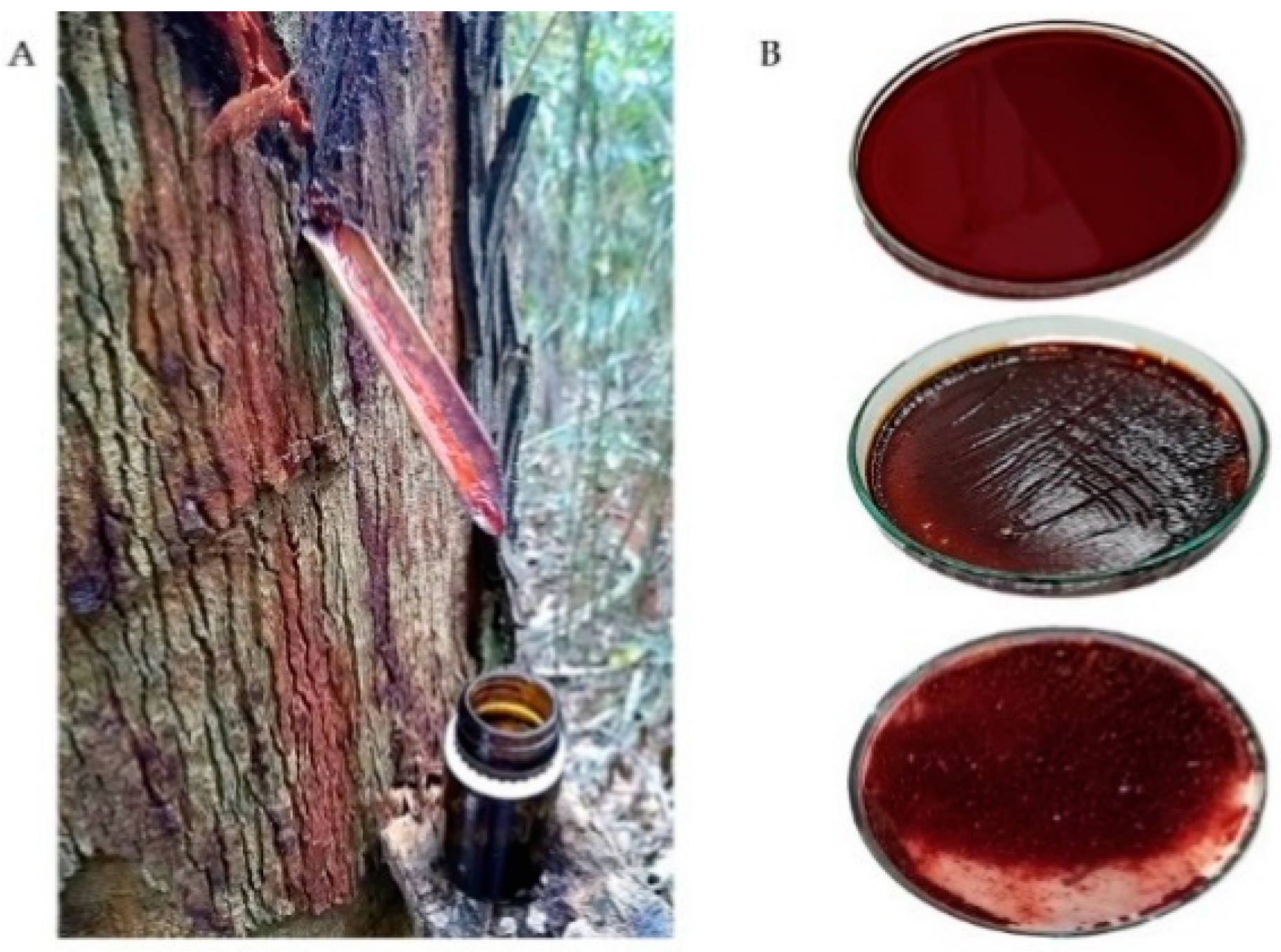
2.1. Resin Material
2.2. Animals
2.3. Cream Composition
2.4. Excisional Wound Model
2.5. Experimental Group
2.6. Wound Size Measurement and Score Wound
2.7. Biochemical Analyses
2.8. Histological Analyses
2.9. Thiobarbituric Acid Reactive Substances (TBARS)
2.10. Advanced Oxidation Protein Products (AOPP)
2.11. In Vitro Assays: ROS Determination
2.11.1. Superoxide Anion Quantification
2.11.2. NO Production
2.12. Statistical Analysis
3. Results
3.1. Biochemical and Body Weight of Experimental Groups
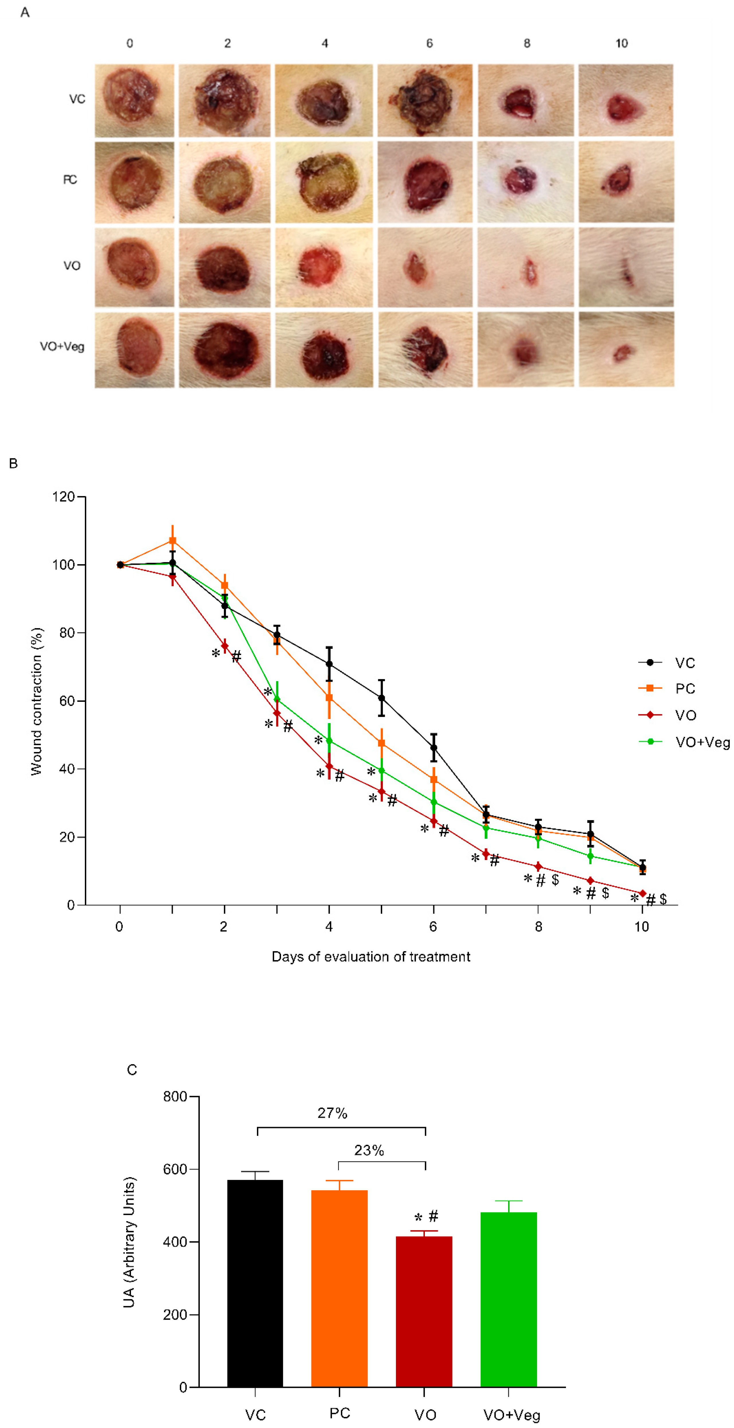
3.2. Healing Effect of Virola Cream
3.3. Dryness Score
3.4. Borders Score
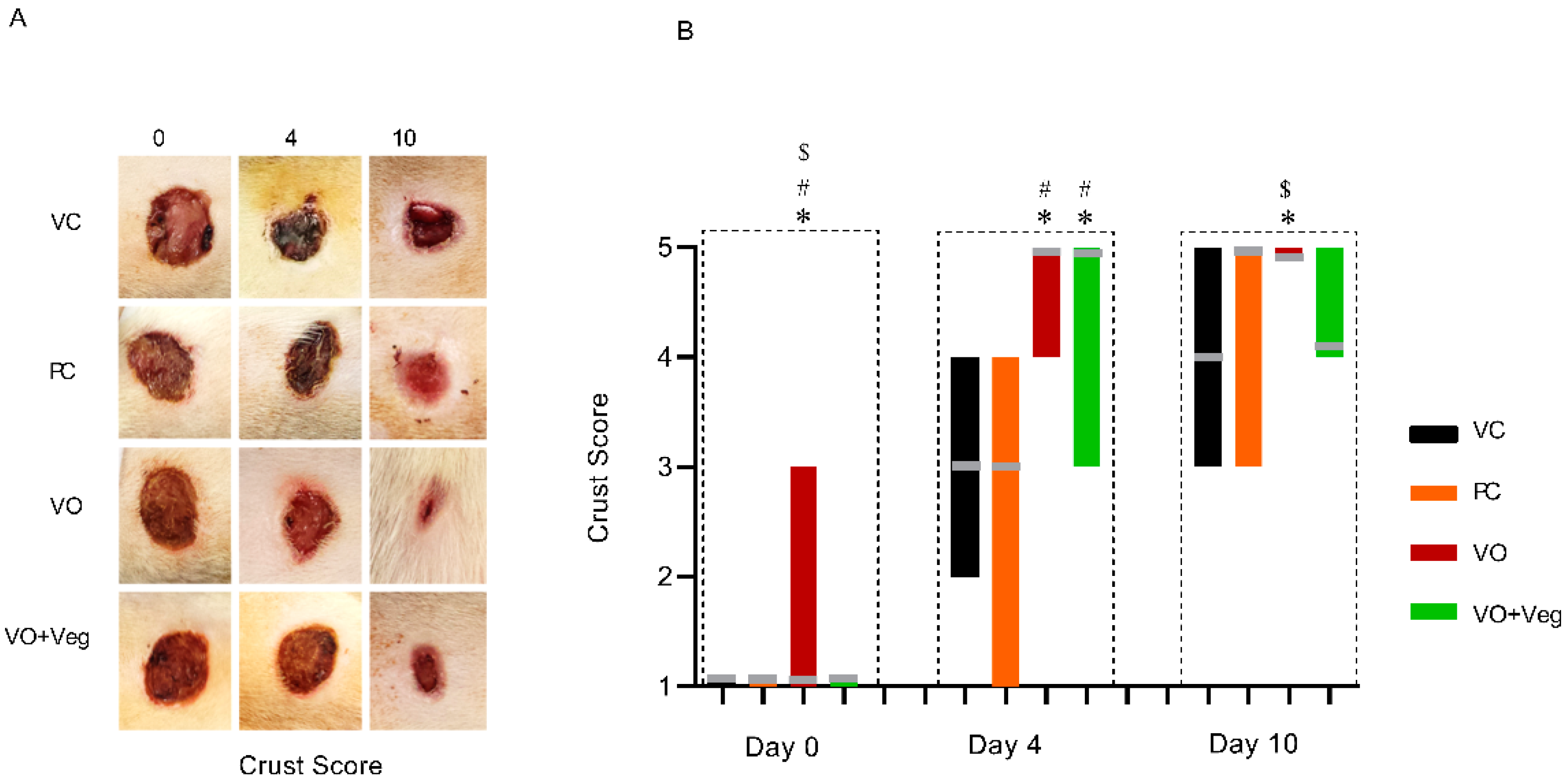
3.5. Crust Score
3.6. Histology
3.7. Lipid Peroxidation (TBARS) in Skin
3.8. Advanced Oxidation Protein Products (AOPP) in Skin
3.9. In Vitro Assays: ROS Determination
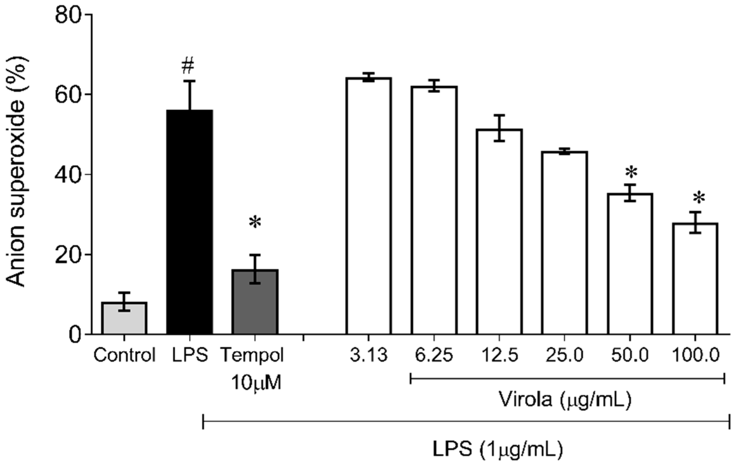
3.9.1. Superoxide Anion Quantification
3.9.2. NO Production
4. Discussion
5. Conclusions
Author Contributions
Funding
Institutional Review Board Statement
Informed Consent Statement
Data Availability Statement
Conflicts of Interest
References
- Pereira, A.C.H.; Lenz, D.; Nogueira, B.V.; Scherer, R.; Andrade, T.U.; da Costa, H.B.; Romão, W.; Pereira, T.M.C.; Endringer, D.C. Gastroprotective activity of the resin from Virola oleifera. Pharm. Biol. 2016, 55, 472–480. [Google Scholar] [CrossRef] [PubMed]
- Santamaría-Aguilar, D.; Aguilar, R.; Lagomarsino, L.P. A taxonomic synopsis of Virola (Myristicaceae) in Mesoamerica, including six new species. PhytoKeys 2019, 134, 1–82. [Google Scholar] [CrossRef] [PubMed]
- Bôa, I.S.F.; Porto, M.L.; Pereira, A.C.H.; Ramos, J.P.L.; Scherer, R.; Oliveira, J.; Nogueira, B.; Meyrelles, S.S.; Vasquez, E.C.; Endringer, D.; et al. Resin from Virola oleifera Protects Against Radiocontrast-Induced Nephropathy in Mice. PLoS ONE 2015, 10, e0144329. [Google Scholar] [CrossRef]
- González-Rodríguez, M.; Ruiz-Fernández, C.; Francisco, V.; Ait Eldjoudi, D.; Farrag AbdElHafez, Y.R.; Cordero-Barreal, A.; Pino, J.; Lago, F.; Campos-Toimil, M.; Rocha Carvalho, G.; et al. Pharmacological Extracts and Molecules from Virola Species: Traditional Uses, Phytochemistry, and Biological Activity. Molecules 2021, 26, 792. [Google Scholar] [CrossRef]
- Coutinho, P.N.; Pereira, B.P.; Pereira, A.C.H.; Porto, M.L.; de Assis, A.L.E.M.; Destefani, A.C.; Meyrelles, S.S.; Vasquez, E.C.; Nogueira, B.V.; de Andrade, T.U.; et al. Chronic administration of antioxidant resin from Virola oleifera attenuates atherogenesis in LDLr−/− mice. J. Ethnopharmacol. 2017, 12, 65–72. [Google Scholar] [CrossRef]
- George, B., II; Janis, J.E.; Attinger, C.E. Wound Healing: An Overview. Plast. Reconstr. Surg. 2006, 117, 1e-S–32e-S. [Google Scholar] [CrossRef]
- Ozgok Kangal, M.K.; Regan, J.P. Wound Healing; StatPearls Publishing: Treasure Island, FL, USA, 2022. [Google Scholar]
- Lindley, L.E.; Stojadinovic, O.; Pastar, I.; Tomic-Canic, M. Biology and Biomarkers for Wound Healing. Plast. Reconstr. Surg. 2016, 138, 18S–28S. [Google Scholar] [CrossRef] [PubMed]
- Vallée, A.; LeCarpentier, Y. TGF-β in fibrosis by acting as a conductor for contractile properties of myofibroblasts. Cell Biosci. 2019, 9, 98. [Google Scholar] [CrossRef] [PubMed]
- Cano Sanchez, M.; Lancel, S.; Boulanger, E.; Neviere, R. Targeting Oxidative Stress and Mitochondrial Dysfunction in the Treatment of Impaired Wound Healing: A Systematic Review. Antioxidants 2018, 7, 98. [Google Scholar] [CrossRef]
- Ellis, P. The impact of smoking on wound healing: The role of the nurse. Br. J. Nurs. 2018, 27, S10–S14. [Google Scholar] [CrossRef]
- Gottrup, F.; Ågren, M.S.; Karlsmark, T. Models for use in wound healing research: A survey focusing on in vitro and in vivo adult soft tissue. Wound Repair Regen. 2000, 8, 83–96. [Google Scholar] [CrossRef] [PubMed]
- Dos Santos Gramma, L.S.; Marques, F.M.; Vittorazzi, C.; de Andrade, T.A.M.; Frade, M.A.C.; de Andrade, T.U.; Endringer, D.C.; Scherer, R.; Fronza, M. Struthanthus vulgaris ointment prevents an over expression of inflammatory response and accelerates the cutaneous wound healing. J. Ethnopharmacol. 2016, 22, 319–327. [Google Scholar] [CrossRef] [PubMed]
- Vittorazzi, C.; Endringer, D.C.; Andrade, T.U.D.; Scherer, R.; Fronza, M. Antioxidant, antimicrobial and wound healing properties of Struthanthus vulgaris. Pharm. Biol. 2016, 54, 331–337. [Google Scholar] [CrossRef] [PubMed]
- Guidoni, M.; Figueira, M.M.; Ribeiro, G.P.; Lenz, D.; Grizotto, P.A.; Pereira, T.D.M.C.; Scherer, R.; Bogusz, S.; Fronza, M. Development and evaluation of a vegetable oil blend formulation for cutaneous wound healing. Arch. Dermatol. Res. 2019, 311, 443–452. [Google Scholar] [CrossRef] [PubMed]
- Levin-Arama, M.; Abraham, L.; Waner, T.; Harmelin, A.; Steinberg, D.M.; Lahav, T.; Harlev, M. Subcutaneous Compared with Intraperitoneal KetamineXylazine for Anesthesia of Mice. J. Am. Assoc. Lab. Anim. Sci. 2016, 55, 794–800. [Google Scholar]
- Ashjazadeh, M.A.; Jahandideh, A.; Abedi, G.; Akbarzadeh, A.; Hesaraki, S. Histopathology and Histomorphological Study of Wound Healing Using Clove Extract Nanofibers (Eugenol) Compared to Zinc Oxide Nanofibers on the Skin of Rats. Arch. Razi Inst. 2019, 74, 267–277. [Google Scholar]
- Caetano, K.S.; Frade, M.A.C.; Minatel, D.G.; Santana, L.; Enwemeka, C.S. Phototherapy Improves Healing of Chronic Venous Ulcers. Photomed. Laser Surg. 2009, 27, 111–118. [Google Scholar] [CrossRef]
- Jamadagni, P.S.; Jamadagni, S.; Mukherjee, K.; Upadhyay, S.; Gaidhani, S.; Hazra, J. Experimental and histopathological observation scoring methods for evaluation of wound healing properties ofJatyadi Ghrita. AYU 2016, 37, 222–229. [Google Scholar] [CrossRef]
- Bradford, M.M. A rapid and sensitive method for the quantitation of microgram quantities of protein utilizing the principle of protein-dye binding. Anal. Biochem. 1976, 7, 248–254. [Google Scholar] [CrossRef]
- Hanasand, M.; Omdal, R.; Norheim, K.B.; Gøransson, L.G.; Brede, C.; Jonsson, G. Improved detection of advanced oxidation protein products in plasma. Clin. Chim. Acta 2012, 413, 901–906. [Google Scholar] [CrossRef]
- Marques, F.M.; Figueira, M.M.; Schmitt, E.F.P.; Kondratyuk, T.P.; Endringer, D.C.; Scherer, R.; Fronza, M. In vitro anti-inflammatory activity of terpenes via suppression of superoxide and nitric oxide generation and the NF-κB signalling pathway. Inflammopharmacology 2019, 27, 281–289. [Google Scholar] [CrossRef] [PubMed]
- Lorençoni, M.F.; Figueira, M.M.; e Silva, M.V.T.; Schmitt, E.F.P.; Endringer, D.C.; Scherer, R.; Barth, T.; Bertolucci, S.K.V.; Fronza, M. Chemical composition and anti-inflammatory activity of essential oil and ethanolic extract of Campomanesia phaea (O. Berg.) Landrum leaves. J. Ethnopharmacol. 2020, 24, 52. [Google Scholar] [CrossRef]
- Green, L.C.; Wagner, D.A.; Glogowski, J.; Skipper, P.L.; Wishnok, J.S.; Tannenbaum, S.R. Analysis of nitrate, nitrite, and [15N] nitrate in biological fluids. Anal. Biochem. 1982, 126, 131–138. [Google Scholar] [CrossRef]
- Dunnill, C.; Patton, T.; Brennan, J.; Barrett, J.; Dryden, M.; Cooke, J.; Leaper, D.; Georgopoulos, N.T. Reactive oxygen species (ROS) and wound healing: The functional role of ROS and emerging ROS-modulating technologies for augmentation of the healing process. Int. Wound J. 2015, 14, 89–96. [Google Scholar] [CrossRef] [PubMed]
- Nouvong, A.; Ambrus, A.M.; Zhang, E.R.; Hultman, L.; Coller, H.A. Reactive oxygen species and bacterial biofilms in diabetic wound healing. Physiol. Genom. 2016, 48, 889–896. [Google Scholar] [CrossRef] [PubMed]
- Hoffmann, M.H.; Griffiths, H.R. The dual role of Reactive Oxygen Species in autoimmune and inflammatory diseases: Evidence from preclinical models. Free Radic. Biol. Med. 2018, 125, 62–71. [Google Scholar] [CrossRef] [PubMed]
- Comino-Sanz, I.M.; López-Franco, M.D.; Castro, B.; Pancorbo-Hidalgo, P.L. The Role of Antioxidants on Wound Healing: A Review of the Current Evidence. J. Clin. Med. 2021, 10, 3558. [Google Scholar] [CrossRef]
- Moseley, R.; Stewart, J.E.; Stephens, P.; Waddington, R.J.; Thomas, D.W. Extracellular matrix metabolites as potential biomarkers of disease activity in wound fluid: Lessons learned from other inflammatory diseases? Br. J. Dermatol. 2004, 150, 401–413. [Google Scholar] [CrossRef]
- Rodriguez, P.G.; Felix, F.N.; Woodley, D.T.; Shim, E.K. The Role of Oxygen in Wound Healing: A Review of the Literature. Dermatol. Surg. 2008, 34, 1159–1169. [Google Scholar] [CrossRef]
- Johnson, J.B.; Broszczak, D.A.; Mani, J.S.; Anesi, J.; Naiker, M. A cut above the rest: Oxidative stress in chronic wounds and the potential role of polyphenols as therapeutics. J. Pharm. Pharmacol. 2022, 74, 485–502. [Google Scholar] [CrossRef]
- Abood, W.N.; Al-Henhena, N.A.; Abood, A.N.; Al-Obaidi, M.M.J.; Ismail, S.; Abdulla, M.A.; Al Batran, R. Wound-healing potential of the fruit extract of Phaleria macrocarpa. Bosn. J. Basic Med. Sci. 2015, 15, 25–30. [Google Scholar] [CrossRef] [PubMed]
- de Aquino, P.E.A.; de Souza, T.D.F.G.; Santos, F.A.; Viana, A.F.S.C.; Louchard, B.O.; Leal, L.K.A.M.; Rocha, T.M.; Evangelista, J.S.A.M.; de Aquino, N.C.; de Alencar, N.M.N.; et al. The Wound Healing Property of N-Methyl-(2S,4R)-trans-4-Hydroxy-L-Proline from Sideroxylon obtusifolium is Related to its Anti-Inflammatory and Antioxidant Actions. J. Evid.-Based Integr. Med. 2019, 24, 25. [Google Scholar] [CrossRef] [Green Version]
- dos Santos, J.S.; Monte-Alto-Costa, A. Caffeic acid phenethyl ester improves burn healing in rats through anti-inflammatory and antioxidant effects. J. Burn Care Res. 2013, 34, 682–688. [Google Scholar] [CrossRef] [PubMed]
- Posadino, A.M.; Cossu, A.; Giordo, R.; Zinellu, A.; Sotgia, S.; Vardeu, A.; Hoa, P.T.; Van Nguyen, L.H.; Carru, C.; Pintus, G. Resveratrol alters human endothelial cells redox state and causes mitochondrial-dependent cell death. Food Chem. Toxicol. 2015, 78, 10–16. [Google Scholar] [CrossRef] [PubMed]
- Pérez-Torres, I.; Guarner-Lans, V.; Rubio-Ruiz, M.E. Reductive Stress in Inflammation-Associated Diseases and the Pro-Oxidant Effect of Antioxidant Agents. Int. J. Mol. Sci. 2017, 18, 2098. [Google Scholar] [CrossRef] [PubMed]
- Espíndola, K.M.M.; Ferreira, R.G.; Narvaez, L.E.M.; Rosario, A.C.R.S.; Da Silva, A.H.M.; Silva, A.G.B.; Vieira, A.P.O.; Monteiro, M.C. Chemical and Pharmacological Aspects of Caffeic Acid and Its Activity in Hepatocarcinoma. Front. Oncol. 2019, 9, 541. [Google Scholar] [CrossRef]
- Morton, L.M.; Phillips, T.J. Wound healing and treating wounds: Differential diagnosis and evaluation of chronic wounds. J. Am. Acad Dermatol. 2016, 74, 589–605. [Google Scholar] [CrossRef]
- Ridiandries, A.; Tan, J.T.M.; Bursill, C.A. The Role of Chemokines in Wound Healing. Int. J. Mol. Sci. 2018, 19, 3217. [Google Scholar] [CrossRef]
- El Ayadi, A.; Jay, J.W.; Prasai, A. Current Approaches Targeting the Wound Healing Phases to Attenuate Fibrosis and Scarring. Int. J. Mol. Sci. 2020, 21, 1105. [Google Scholar] [CrossRef]
- Pereira, L.O.M.; Vilegas, W.; Tangerina, M.M.P.; Arunachalam, K.; Balogun, S.O.; Orlandi-Mattos, P.E.; Colodel, E.M.; Martins, D.T.D.O. Lafoensia pacari A. St.-Hil.: Wound healing activity and mechanism of action of standardized hydroethanolic leaves extract. J. Ethnopharmacol. 2018, 219, 337–350. [Google Scholar] [CrossRef]
- Saleem, A.; Saleem, M.; Akhtar, M.F.; Shahzad, M.; Jahan, S. Correction to: Moringa oleifera leaf extracts attenuate Complete Freund’s adjuvant-induced arthritis in Wistar rats via modulation of inflammatory and oxidative stress biomarkers. Inflammopharmacology 2019, 28, 341–343. [Google Scholar] [CrossRef] [PubMed]
- Ghlissi, Z.; Sayari, N.; Kallel, R.; Bougatef, A.; Sahnoun, Z. Antioxidant, antibacterial, anti-inflammatory and wound healing effects of Artemisia campestris aqueous extract in rat. Biomed. Pharmacother. 2016, 84, 115–122. [Google Scholar] [CrossRef] [PubMed]
- Zbilgin, S.; Acıkara, Ö.B.; Akkol, E.K.; Süntar, I.; Keleş, H.; İşcan, G.S. In vivo wound-healing activity of Euphorbia characias subsp. wulfenii: Isolation and quantification of quercetin glycosides as bioactive compounds. J. Ethnopharmacol. 2018, 5, 400–408. [Google Scholar] [CrossRef] [PubMed]
- Toiu, A.; Vlase, L.; Vodnar, D.C.; Gheldiu, A.-M.; Oniga, I. Solidago graminifolia L. Salisb. (Asteraceae) as a Valuable Source of Bioactive Polyphenols: HPLC Profile, In Vitro Antioxidant and Antimicrobial Potential. Molecules 2019, 24, 2666. [Google Scholar] [CrossRef] [PubMed] [Green Version]


| Body Weight (g) | |||||
|---|---|---|---|---|---|
| Groups | Vehicle (VC) | Positive Control (PC) | Virola 5% (VO) | Virola + Vegederm (VO + Veg) | p |
| Before the injury | 388 ± 37 | 359 ± 48 | 353 ± 7 | 336 ± 29 | 0.925 |
| Day 0 | 360 ± 33 | 334 ± 49 | 317 ± 27 | 337 ± 27 | 0.861 |
| Day 4 | 335 ± 26 | 342 ± 50 | 322 ± 23 | 345 ± 28 | 0.964 |
| Day 10 | 375 ± 31 | 347 ± 40 | 338 ± 19 | 353 ± 24 | 0.878 |
| Parameters | Vehicle (VC) | Positive Control (PC) | Virola 5% (VO) | Virola + Vegederm (VO + Veg) | p |
|---|---|---|---|---|---|
| Day 0 | |||||
| ALT (U/L) | 44 ± 6 | 39 ± 7 | 28 ± 3 | 41 ± 3 | 0.169 |
| AST (U/L) | 134 ± 22 | 97 ± 13 | 72 ± 13 | 97 ± 15 | 0.101 |
| Urea (mg/dL) | 51 ± 3 | 46 ± 1 | 49 ± 2 | 40 ± 1 | 0.436 |
| Urea nitrogen (mg/dL) | 24 ± 1 | 22 ± 1 | 23 ± 1 | 24 ± 1 | 0.552 |
| Serum creatinine (mg/dL) | 0.33 ± 0.01 | 0.32 ± 0.01 | 0.36 ± 0.01 | 0.34 ± 0.03 | 0.500 |
| Day 4 | |||||
| ALT (U/L) | 35 ± 2 | 33 ± 5 | 40 ± 5 | 56 ± 9 | 0.061 |
| AST (U/L) | 120 ± 9 | 95 ± 12 | 133 ± 14 | 161 ± 50 | 0.406 |
| Urea (mg/dL) | 53 ± 3 | 41 ± 4 | 51 ± 9 | 50 ± 2 | 0.446 |
| Urea nitrogen (mg/dL) | 24 ± 2 | 19 ± 2 | 24 ± 4 | 23 ± 1 | 0.541 |
| Serum creatinine (mg/dL) | 0.40 ± 0.01 | 0.25 ± 0.06 | 0.32 ± 0.02 | 0.35 ± 0.01 | 0.424 |
| Day 10 | |||||
| ALT (U/L) | 75 ± 29 | 74 ± 15 | 39 ± 4 | 40 ± 5 | 0.251 |
| AST (U/L) | 217 ± 73 | 263 ± 105 | 120 ± 28 | 119 ± 34 | 0.363 |
| Urea (mg/dL) | 42 ± 4 | 54 ± 4 | 42 ± 6 | 45 ± 3 | 0.207 |
| Urea nitrogen (mg/dL) | 20 ± 2 | 25 ± 2 | 20 ± 3 | 21 ± 1 | 0.204 |
| Serum creatinine (mg/dL) | 0.35 ± 0.03 | 0.37 ± 0.02 | 0.28 ± 0.02 | 0.24 ± 0.06 | 0.422 |
Publisher’s Note: MDPI stays neutral with regard to jurisdictional claims in published maps and institutional affiliations. |
© 2022 by the authors. Licensee MDPI, Basel, Switzerland. This article is an open access article distributed under the terms and conditions of the Creative Commons Attribution (CC BY) license (https://creativecommons.org/licenses/by/4.0/).
Share and Cite
Carvalho, G.R.; Braz, D.S.; Gonçalves, T.C.O.; Aires, R.; Côco, L.Z.; Guidoni, M.; Fronza, M.; Endringer, D.C.; Júnior, A.D.S.; Campos-Toimil, M.; et al. Development and Evaluation of Virola oleifera Formulation for Cutaneous Wound Healing. Antioxidants 2022, 11, 1647. https://doi.org/10.3390/antiox11091647
Carvalho GR, Braz DS, Gonçalves TCO, Aires R, Côco LZ, Guidoni M, Fronza M, Endringer DC, Júnior ADS, Campos-Toimil M, et al. Development and Evaluation of Virola oleifera Formulation for Cutaneous Wound Healing. Antioxidants. 2022; 11(9):1647. https://doi.org/10.3390/antiox11091647
Chicago/Turabian StyleCarvalho, Glaucimeire R., Débora S. Braz, Talita C. O. Gonçalves, Rafaela Aires, Larissa Z. Côco, Marcio Guidoni, Marcio Fronza, Denise C. Endringer, Antonio D. S. Júnior, Manuel Campos-Toimil, and et al. 2022. "Development and Evaluation of Virola oleifera Formulation for Cutaneous Wound Healing" Antioxidants 11, no. 9: 1647. https://doi.org/10.3390/antiox11091647
APA StyleCarvalho, G. R., Braz, D. S., Gonçalves, T. C. O., Aires, R., Côco, L. Z., Guidoni, M., Fronza, M., Endringer, D. C., Júnior, A. D. S., Campos-Toimil, M., Nogueira, B. V., Vasquez, E. C., Campagnaro, B. P., & Pereira, T. M. C. (2022). Development and Evaluation of Virola oleifera Formulation for Cutaneous Wound Healing. Antioxidants, 11(9), 1647. https://doi.org/10.3390/antiox11091647