Review

Anti-Inflammatory and Antioxidant Chinese Herbal Medicines: Links between Traditional Characters and the Skin Lipoperoxidation “Western” Model

by
Jose M. Prieto
1,* and
Guillermo R. Schinella
2,3
1
School of Pharmacy and Biomolecular Sciences, Liverpool John Moores University, Liverpool L3 3AF, UK
2
Facultad de Ciencias Médicas, Universidad Nacional de La Plata, La Plata B1900, Argentina
3
Instituto de Ciencias de la Salud, UNAJ-CICPBA, Florencio Varela BA1888, Argentina
*
Author to whom correspondence should be addressed.
Antioxidants 2022, 11(4), 611; https://doi.org/10.3390/antiox11040611
Submission received: 8 March 2022 / Revised: 17 March 2022 / Accepted: 17 March 2022 / Published: 23 March 2022

Abstract

:
The relationship between lipid peroxidation and inflammation has been accepted as a paradigm in the field of topical inflammation. The underlying biochemical mechanisms may be summarised as unspecific oxidative damage followed by specific oxidative processes as the physio pathological response in skin tissues. In this experimental review we hypothesise that the characteristics attributed by Traditional Chinese Medicine (TCM) to herbal drugs can be linked to their biomolecular activities within the framework of the above paradigm. To this end, we review and collect experimental data from several TCM herbal drugs to create 2D-3D pharmacological and biochemical spaces that are further reduced to a bidimensional combined space. When multivariate analysis is applied to the latter, it unveils a series of links between TCM herbal characters and the skin lipoperoxidation “Western” model. With the help of these patterns and a focused review on their chemical, pharmacological and antioxidant properties we show that cleansing herbs of bitter and cold nature acting through removal of toxins—including P. amurense, Coptis chinensis, S. baicalensis and F. suspensa—are highly correlated with strong inhibition of both lipid peroxidation and eicosanoids production. Sweet drugs—such as A. membranaceus, A. sinensis and P. cocos—act through a specific inhibition of the eicosanoids production. The therapeutic value of the remaining drugs—with low antioxidant or anti-inflammatory activity—seems to be based on their actions on the Qi with the exception of furanocoumarin containing herbs—A. dahurica and A. pubescens—which “expel wind”. A further observation from our results is that the drugs present in the highly active “Cleansing herbs” cluster are commonly used and may be interchangeable. Our work may pave the way to a translation between two medical systems with radically different philosophies and help the prioritisation of active ingredients with specific biomolecular activities of interest for the treatment of skin conditions.

1. Introduction

Topical inflammation underpins almost every skin condition. The search for new, safer therapies to both acute skin conditions such as mechanical injuries and UV exposition and chronic conditions such as eczema, psoriasis, and atopic dermatitis, among many other skin pathologies, is therefore warranted.
The pathophysiological mechanisms of chronic topical inflammatory conditions such as atopic dermatitis and psoriasis are complex. Much emphasis has been put on the extremely complex interplay between the skin and immune system in terms of inflammatory mediators. Therefore, old and current medical approaches favour aggressive anti-inflammatory, immunosuppresive [1] and photoactive [2] drugs (steroids, furanocoumarins, cyclosporine, etc.) with a poor balance between pharmacological and toxicological effects in the long term. Although it is well-recognized that many of these conditions are accompanied by a burst of free radicals and imbalanced antioxidant defences both at both local and systemic levels, antioxidant therapies are yet to be fulfilled [3,4,5]. This is due to a lack of clear target, given the enormous array of chemical species and secondary mediators involved in the cell redox biology [6].

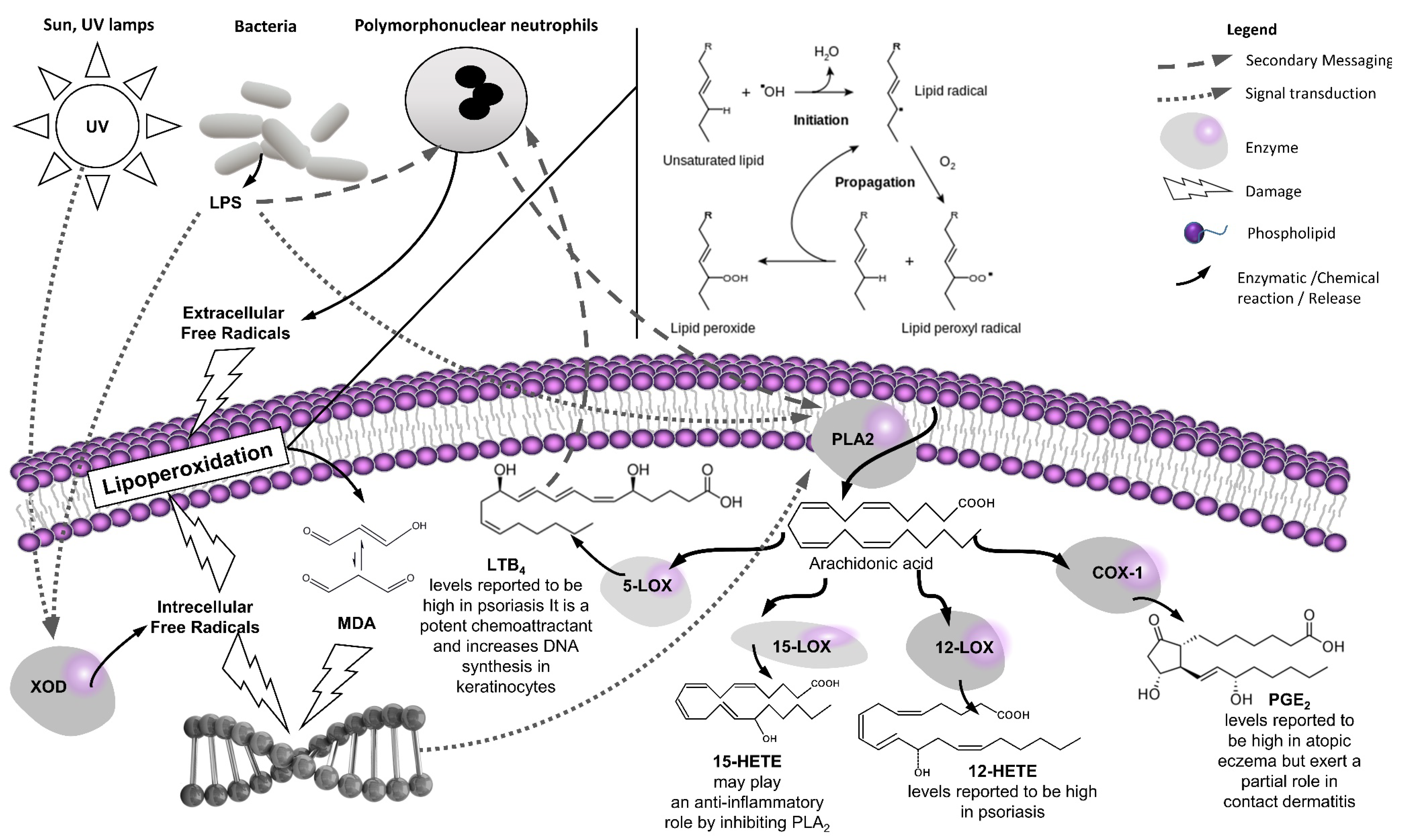
1.1. Introduction to the “Western” Skin Lipoperoxidation Model

The concept of lipid peroxidation as a central pathophysiological process in skin diseases has gained the attention of a part of the research community since the 1980s. The relationship between lipid peroxidation and skin inflammation has been perfectly laid out by Briganti and Picardi [7] and De Luca and Valacchi [8], whilst the underlying biochemical mechanisms were thoroughly reviewed by Guteridge [9] and Niki [10].
Within the theoretical framework laid out by these authors we want to focus on two aspects within the multifaceted and complex interplay between lipoperoxidation and skin inflammation that are pertinent to our paper: the unspecific peroxidation of membrane lipids as a damaging factor [11] and the specific peroxidation of certain membrane lipids (arachidonic acid chiefly) as a response against this damage. The first process physically destabilizes the cell function by altering the structure of its membranes and generates toxic end products such as malonyl dialdehyde (MDA) [12]. The second one generates a family of secondary pro- [13] and anti-inflammatory lipid mediators [14] known as eicosanoids. The most important eicosanoids, prostaglandins and leukotrienes, are biosynthesised from arachidonic acid and are responsible in the early stages of the inflammatory process for the attraction of neutrophils to the affected tissue. On the one hand, high levels of these mediators maintained over time will contribute to a chronic condition [15]. On the other hand, their continuous biosynthesis may also help the resolution of the inflammatory process as they promote the induction of 15-lipoxygenases necessary for the biosynthesis of lipoxin, derived from the ω-6 fatty acid arachidonic acid, and resorbin, protectin, and maresin, derived from the ω-3 fatty acids eicosapentaenoic acid and docosahexaenoic acid, with many of them described as being synthesised by skin cells [16,17]. However, the activity of traditional herbal medicines on these anti-inflammatory mediators is just starting to be scrutinised [18], and therefore we will focus on proinflammatory eicosanoids to study the relationship of curative effects of such herbal drugs with their traditional characters in acute inflammation. Their interplay and actions are summarized in Figure 1.
The inhibition of lipid peroxidation may be a therapeutic target [7]. However, unspecific inhibitors of lipid peroxidation may successfully quench the free radicals and stop the chain reaction leading to lipid peroxides and MDA but could theoretically at the same time impair the synthesis of eicosanoids, which may promote wound healing in the early phases of skin damage. Therefore, a more rational therapeutic approach to this conundrum would be the use of specific inhibitors of the different types of lipoperoxidation in different ratios depending on the stage of the condition. Modern medicine has developed NSAIDs to specifically inhibit eicosanoids production and steroids to reduce the expression of the enzymes involved in their biosynthesis. Yet there is not a defined clinical approach to specifically quench the radicals and stop membrane lipids peroxidation [19].

1.2. Introduction to the “Eastern” Skin Inflammation Model

All cultures have developed over millennia therapeutic approaches to skin conditions. This is not surprising, considering that the skin is the most accessible and largest “organ”. Among these, Traditional Chinese Medicine (TCM) provides one of the most sophisticated approaches, with recipes including many herbal drugs to address the multifactorial nature of skin inflammation. The drugs are formulated according to a complex match between patient’s and disease characters and the herbs carefully chosen as to provide opposite characters such as hot, warm, cold, sweet, bitter, pungent, etc. [20,21].
The selection of medicinal plants by traditional Chinese medicine to treat dermatological diseases involving chronic inflammation is done on a complex, multifactorial basis [22]. The diagnosis is usually what traditional Chinese medicine experts refer to as “kidney yin deficiency”, which may be interpreted as a lack of endogenous cortisol. This compound is used clinically in Western medicine in the form of hydrocortisone or prednisone to help control many disorders, including acute inflammations, rheumatoid arthritis, allergies and many eruptic skin diseases. Angelica species, traditionally used in the treatment of psoriasis, also act via the “kidney channel” and have been proven to alleviate pain [23]. Since the spleen also plays a major role in immune function, Chinese medicine sometimes calls for the addition of “spleen dampness removing herbs” such as Poria, as well as “spleen chi tonics” from species of the genera Atractylodes and Astragalus. According to traditional Chinese medicine, Coptis chinensis, Paeonia lactiflora, Forsythia suspensa and Curcuma aromatica provide analgesic and bacteriostatic properties, along with Codonopsis pilosula, which acts as a tonic [24]. Finally, Phellodendron amurense and Scutellaria baicalensis species are active ingredients of a relatively modern traditional Chinese prescription known as “Three Yellow Cleanser”, which is commonly recommended for many skin conditions [25,26].

1.3. Therapeutic Opportunities at the Western–Eastern Interface

The reasons for comparing “Eastern” and “Western” medical frameworks here are to find equivalences between Chinese traditional features/characters for herbal medicines (qualitative adjectives such as cold/warm/neutral, sweet/bitter, etc.) (as presented in Table 1) and “Western pharmacology” biomolecular activities (quantitative data on inhibition of lipoperoxidation and eicosanoids synthesis processes) (as presented in Section 3).
If we “crack” the Traditional Chinese Medicine code, we may find a route to select/identify anti-inflammatory and antioxidant Chinese herbal drugs on the basis of their traditional descriptions, thus maximizing the success of future screenings. Conversely, Chinese researchers and/or practitioners may find a way to add modern molecular meaning to the Traditional classification of such medicinal plants, thus facilitating an integrated approach that may lead to safer, faster and more effective health care [27].
Therefore, our research objectives here are to (1) review the “Western” scientific evidence of these species as anti-inflammatory (eicosanoids inhibition) and antioxidant (enzymatic and non-enzymatic oxidation) to create a qualitative profile of their therapeutic use in skin conditions and then (2) combine experimental (quantitative) biochemical data and traditional Chinese properties with the help of multivariate analysis to unveil links between “Eastern” and “Western” medical frameworks.

2. A Focused Review on the Anti-Inflammatory (Eicosanoid Inhibition) and Antioxidant (Lipoperoxidation) Properties of the Selected Medicinal Plants

2.1. Methods

Traditional Chinese Medicine may use many herbal drugs as its approach is very multifaceted and the prescriptions are adapted not only to the condition (in this case skin conditions) but also to the patient’s characteristics, making it potentially impossible to cover them all [28]. The plants listed in Table 1 have been the object of intense research by the authors of this review, as well as other research groups, for their topical anti-inflammatory and antioxidant activities [29,30,31,32,33,34,35], thus providing a set of comparable data. The review will revolve around the combined data coming out of the two seminal works of both authors [32,35], complemented and contrasted with all subsequent (and previous if relevant) research done on these herbal drugs in similar or relevant models to the lipoperoxidation framework above discussed.
Literature was sourced from PubMed to ensure pharmacological/medical/clinical relevance by searching by the following combination of keywords [Species name] AND (Cyclooxygenase OR Lipoxygenase OR COX OR LOX OR Lipoperoxidation OR Antioxidant) from 2000 to 2022. Papers with methods or models not relevant or translational to skin conditions and/or treatments at non physiological doses/concentrations were excluded.

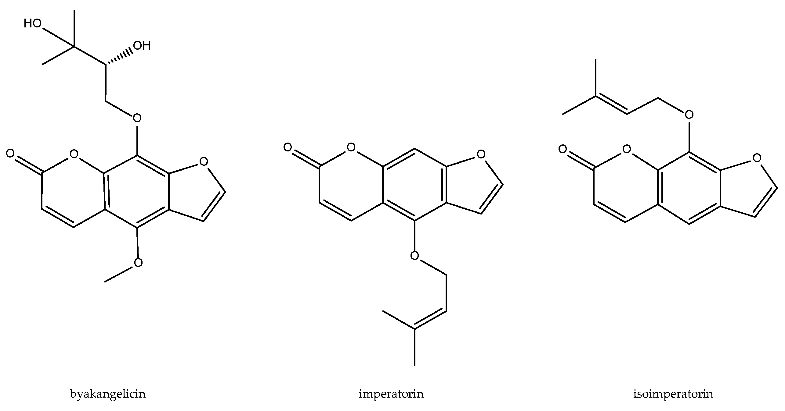
2.2. Angelica dahurica

Species of the genus Angelica (Apiaceae) are used in TCM as ingredients in many medicinal preparations. A. dahurica, A. pubescens and A. sinensis are characterized by containing compounds of the coumarin type.
A. dahurica did not attract much attention for its anti-inflammatory or antioxidant activity until Kimura and Okuda [36] mentioned its inhibitory action of histamine release in mice treated with the compound 40/80. The data from our combined studies show that this species was not able to inhibit 5-LOX activity in rat peritoneal PMNs, without being able to determine its action on human platelets due to co-elution problems. The only effect at the cellular level that could be observed was its ability to inhibit the release of elastase as well as its activity, with an IC50 of 129 g/µL. It was the only species whose extract was shown to be pro-oxidant in the enzymatic lipid peroxidation model in the CCl4/NADPH system, a trend that was repeated in the deoxyribose degradation model by the radical OH in the absence of ascorbate [32,35]. Further research unveiled that the activity of the plant extract is maximum in its Ethyl acetate fraction, which is endowed with inhibitory effects on LPS-induced TNF-alpha, NO and PGE2 production, and expression of iNOS and COX-2 in macrophage through blockade in the phosphorylation of MAPKs, following IκBα degradation and NF-κB activation [37].
A series of bioactive furanocoumarins with inhibitory effects on the arachidonic pathway, namely byakangelicin, imperatorin, and isoimperatorin (Figure 2), have been identified as the anti-inflammatory active principles. Imperatorin showed the most potent inhibitory activity on the LPS-induced PGE2 production and expression of COX-2 as well as microsomal prostaglandin E synthase (mPGES) [38]. Byakangelicol, inhibits IL-1β-induced COX-2 expression and PGE2 release in human pulmonary epithelial cell line (A549). It is a quite selective COX-2 inhibitor (10–50 µM) when compared to its IC50 > 200 µM for activity and expression of COX-1 in A549 cells; this inhibition may be mediated at least in part by the suppression of NF-kappaB activity [39]. Isoimperatorin exhibits a dual cyclooxygenase-1/2/5-lipoxygenase inhibitory activity measured as PGD2 and LTC4 biosynthesis in bone marrow-derived mast cells (IC50 = 10.7 and 5.7 µM, respectively). The above mentioned bioactive compounds are shown in Figure 2.

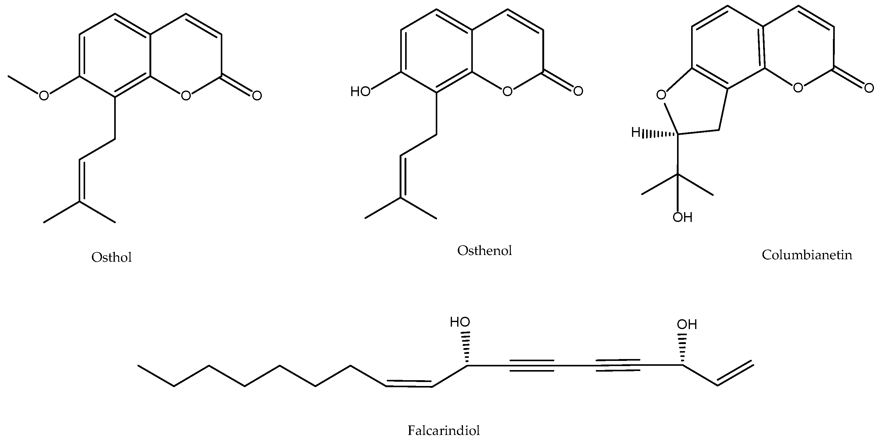
2.3. Angelica pubescens

Chen et al. reported the anti-inflammatory and analgesic effect of different extracts of A. pubescens in in vivo models of formalin- or carrageenan-induced edema [40]. Ko et al. further demonstrated that osthole on platelet aggregation is due to the inhibition of thromboxane formation and phosphoinositides breakdown [41] delayed the aggregation, release of ATP, thrombin and TXB2 in isolated and intact rabbit platelets by inhibition of both the TXs synthesis and the inositol pathway. Later, Liu et al. confirmed the inhibitory activity of the dichloromethane extract of Angelica pubescens f. biserrata on the production of 5-HETE, in intact porcine neutrophils, and PGE2, by microsomes of ram seminal vesicles, both from exogenous 3H-AA, isolating and identified the responsible principles as linoleic acid, osthol and osthenol in addition to the polyacetylenes falcarindiol and acetate of 11(S),16(R)-dihydroxyoctadeca-9Z,17-dieno-12,14-diino, all of them with CI50 of the order of 20–60 µM [42]. Our own work indicated that, although the ethanolic extract 70% of A. pubescens inhibits the production of 5-HETE by 49%, it does not significantly inhibit the total production of the 5-LOX pathway-in rat peritoneal PMNs, since it does not seem to affect the total production of the enzyme, compensating for an increase in LTB4 and their isomers. This fact could indicate that the overall effect of the extract lies in an inhibition of the conversion of 5-HPETE, the primary metabolite of 5-LOX, to 5-HETE. The 5-HPETE would more effectively become LTB4 than controls. Glutathione peroxidase is the enzyme most directly related to the formation of 5-HETE from 5-HPETE. One can think of a possible action of the extract at this level. Regarding the activity on the COX pathway, measured as production of 12-HHTrE, it can be affirmed that the inhibitory activity of the dichloromethene extract of A. pubescens in COX of seminal vesicle microsomes, finds correlation in intact human platelets at the total extract level, where this species is one of the most active (95% inhibition at 200 µg/mL). Since in the methods of antioxidant activity tested, A. pubescens was never shown to be active, a mechanism of nonspecific redox inhibition on the above enzymes might be ruled out [32,35]. A more recent paper supports such indirect activity coming from the heteropolysaccharide DF80-2 exhibited antioxidant activity by effectively scavenging hydroxyl radicals and chelating ferrous ions [43].
Works after 2011 substantiated the importance of columbianetin for the in vivo activities of this TCM drug in suppressing LPS-induced inflammation and apoptosis through the NOD1 pathway [44]. This coumarin is rapidly absorbed when administered orally and has quick clearance and good absolute bioavailability (54–81% for 5–20 mg/kg doses) [45]. The bioavailability of columbianetin is independent of the doses studied. Columbianetin showed dose proportionality over the dose range 5–20 mg/kg. After intestinal absorption this coumarin is likely metabolised by the liver into an array of derivatives as it happens with columbianadin, a closely related compound [46]. Regarding the above-mentioned active principle osthole, it is also active in murine models of neurogenic and inflammatory hyperalgesia by modulation of iNOS, COX-2, and inflammatory cytokines [47] as well as protecting against myocardial ischemia/reperfusion injury [48]. The above-mentioned bioactive compounds are shown in Figure 3.

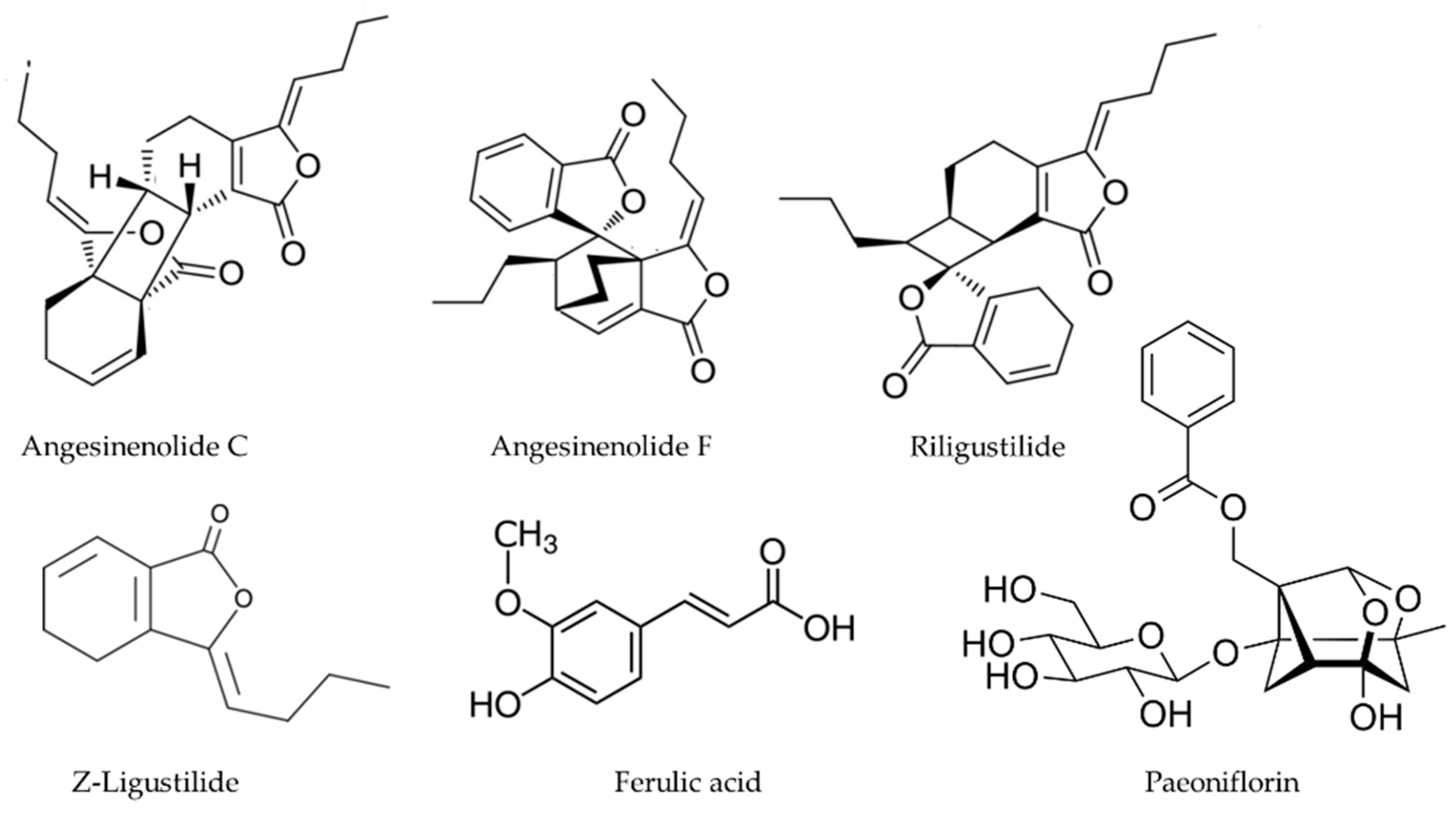
2.4. Angelica sinensis

Angelica sinensis is associated with Astragalus membranaceus, Cyperus rotundum, Ligusticum chuangxiong and Paeonia veitchii in a formulation called Danggui, whose function is to normalize blood rheological values and prevent thrombosis. Ethyl acetate fraction/extracts from this herbal drug have been described as potent anti-inflammatory substances due to the inhibition of pro-inflammatory mediators (NO and PGE2) in part via suppression of a signalling pathway such as NF-κB in macrophages [49] as well as rheumatoid synovial fibroblasts [50]. Wang et al. revealed its inhibitory activity of the production of TXA2 in porcine pulmonary microsomes [51], and our works demonstrated the total inhibition of 5-HETE production in rat peritoneal PMNs at the dose of 200 µg/mL [32] without any antioxidant activity in our models [35]. This may be in line with previous reports that revealed the effects on lipid peroxidation, the hypoxanthin/XOD system and the hydroxyl radical, after the processing of the drug radix Angelica sinensis, are highly variable [52]. Perhaps different polarity fractions have different anti-inflammatory and antioxidant profiles in view of report where a supercritical fluid CO2 extract attenuated d-galactose-induced liver and kidney impairment in mice by suppressing oxidative stress and Inflammation in terms of MDA levels, enhanced the activities and gene expressions of Cu, Zn-SOD, CAT, and GPx, reduction of iNOS, COX-2, IκBα, p-IκBα, and p65 expression in both hepatic and renal tissues [53]. The effect of this extract and other apolar fractions from A. sinensis may be due at least in part by the contribution of its volatile fraction [54] rich in alkylphthalides such as Z-ligustilide n-butylidenephthalide (Figure 4) [55].
Xiet et al. proposed that a polysaccharide is the main effective ingredient of A. sinensis and exerts anti-inflammatory effects via down-regulation of COX-1 on LPS-injured PC12 cells [56]. The reader interested on the structure of such class of compound/s can consult the paper by Hou and co-workers on the structure of the main polysaccharides present in this herbal drug [57]. Another class of anti-inflammatory compounds present in A. sinensis are dimeric phthalides, some inhibiting COX-2 activity with IC50 values as low as 30 μM [58]. A. sinensis also contains falcarindiol (Figure 3) [59] and similar polyacetylenes, as well as other well-known eicosanoid biosynthesis inhibitors such as paeoniflorin [60], and ferulic acid [61] (Figure 4).

2.5. Astragalus membranaceus

Although A. membranaceus has been long studied for its immunomodulatory properties and protective actions of cardiac function [34], there were no references to its anti-inflammatory action until Cuellar et al., demonstrated its activity in several in vivo models of acute edema induced by APD and AA in mouse ear, chronic by multiple applications of TPA, and delayed hypersensitivity induced by oxazolone, without being active in vitro on PLA2 of Naja naja [31]. The possibility of an inhibition of the lipoxygenase and cyclooxygenase pathways, although not phospholipase, was reinforced after our work: the same extract has been found to inhibit 5-LOX activity in rat peritoneal PMNs and COX-1 activity, although not 12-LOX, in human platelets [32]. According to Wang et al. (1993), A. membranaceus would be a better inhibitor of TXA2 production than of PGI2. The extracts reduced the inflammatory response induced by lipopolysaccharide from E. coli (LPS) plus interferon-γ (IFN), reducing COX-2 via NF-κB activation in the non-tumorigenic intestinal epithelial cell line (IEC-6) [62].
Despite having a reputation as an antioxidant [28], this plant extract was not active in any of our systems [35]. This discrepancy may be since the studies cited respectively used very high doses of total extract (even 2 mg/mL) or fractions enriched in flavonoids. The total extract was effective at reducing reactive oxygen species (ROS) release though [62].
It is accepted that the total flavonoids fraction from A. mebranaceus reduces both COX-2 mRNA and protein levels [63], Formononetin, a flavonoid present in this Chinese herb (Figure 5), is the main anti-inflammatory and antioxidative principle in different models [64].
The saponin fraction also offers with anti-inflammatory principles such as Astragaloside IV (Figure 5) that prevents UVB-induced oxidative damage in terms of reduced intracellular ROS level and lipid oxidation product malondialdehyde (MDA) content, as well as inflammation by inhibiting TLR4 expression and its downstream signalling molecules (NF-κB, iNOS and COX-2) [65]. Other active principles include bisphenol derivatives (Figure 5) with inhibitory effects on COX-2 mRNA expression at 50 μM [66].

2.6. Atratylodes macrocephala

The data from our previous work indicate the presence of COX-1 specific non-redox inhibitors, which are certainly not particularly active at the total extract level, as is often used in TCM [32]. It is possible that a fractionation results, as in the case of Resch et al. in obtaining sub-extracts with greater activity. These authors demonstrated the inhibitory activity of its hexanic extract at the level of 5-LOX and COX-1 (CI50 of 2.9 and 30.5 µg/mL, respectively). Its active ingredients were found to be atractylochromene (Figure 6) and 2-[(2E)-3,7-dimethyl-2,6-octadienyl]-6-methyl-2,5-cyclohexadien-1,4-dione, in addition to a moderate selective activity on 5-LOX of sesquiterpene atractylon (Figure 6) and the coumarin osthol (Figure 2). Preparations based on Atractylodes rhizomes are reputed as liver protectors in in vivo models of CCl4 toxicity [67]. This activity found no correlation in our works either in enzymatic microsomal lipid peroxidation induced by CCl4/NADPH or in the rest of the models tested [35].
Almost two decades after these seminal works, two groups have published new data on this TCM drug. Jeong and co-workers have isolated three polyacetylenes namely 2-[(2E)-3,7-dimethyl-2,6-octadienyl]-6-methyl-2, 5-cyclohexadiene-1, 4-dione; 1-acetoxy-tetradeca-6E,12E-diene-8, 10-diyne-3-ol and 1,3-diacetoxy-tetradeca-6E, 12E-diene-8, 10-diyne (Figure 6). They showed concentration-dependent inhibitory effects on production of NO and PGE2 in lipopolysaccharide (LPS)-activated RAW 264.7 macrophages by suppressing both the protein and mRNA levels via inhibition of nuclear translocation of NF-κB [68]. Wu and co-workers reported last year that the essential oil from this TCM drug containing atractylon (39.22%), β-eudesmol (27.70%), thymol (5.74%), hinesol (5.50%), and 11-isopropylidenetricyclo[4.3.1.12,5]undec-3-en-10-one (4.71%) exhibited strong antioxidant capacities and inhibited NO and PGE2 production as well as decreased the transcriptional levels of their originating enzymes in LPS-stimulated RAW264.7 cells [69].

2.7. Codonopsis pilosula

According to Wang et al. Codonopsis. pilosula is a preferential inhibitor of TXA 2 production over that of PGI2 and 6-ketoPGF [51,70]. In our own works its CI50 resulted higher than the limit of 200 µg/mL, although a tendency was observed to inhibit the 5-LOX and COX-1 pathway with the same efficiency [32]. It did not show any significant activity in peroxidation models or in superoxide radical production at concentrations up to 200 µg/mL either, although it behaved as a pro-oxidant in the deoxyribose degradation model when in the presence of ascorbate [35].
Efforts to find active small secondary metabolites such as the polyacetylene lobetyolin to antioxidant or anti-inflammatory activities have failed [71]. It was not until recently that more work started to show that the anti-inflammatory and antioxidant effects of the plant may rely on polysaccharides only. On the one hand, the CPP-1 and CTP-1 (Figure 7) can protect IPEC-J2 cells against the H2O2-induced oxidative stress by up-regulating nuclear factor-erythroid 2-related factor 2 and related genes in IPEC-J2 cells [72]. Furthermore, they increased the total antioxidant capacity, glutathione peroxidase, superoxide dismutase and catalase in the same cells, as well as reducing their levels of MDA [73]. On the other hand, the whole of Codonopsis pilosula polysaccharides (CPPS) protect RAW264.7 cells from hydrogen peroxide-induced injury via the Keap1-Nrf2/ARE pathway as well as inhibiting their proinflammatory activities [74,75].

2.8. Coptis chinensis

The rhizome of species of the genus Coptis is a reputed remedy in TCM for inflammatory processes. Coptis chinensis is usually associated with Astragalus membranaceus and Scutellaria baicalensis in the medicine called sanhuang to treat the so-called Qi Syndrome of venous stasis, showing this preparation inhibitory effects of platelet aggregation [76]. However, only C. japonica was studied for its eicosanoid inhibitory until our works [32,35]. Although due to co-elution problems it was not possible to determine the activity on 5-LOX, it was very active inhibiting the production of 12-HHTrE and 12-HETE in human platelets 89% and 70%, respectively, at 200 µg/mL. With these results, the possibility of an action at the PLA 2 level may be a possibility. At the same time Fukuda et al. reported that berberine (Figure 8), a bright yellow isoquinoline alkaloid present in plants of the genera Berberis and Coptis, effectively inhibits COX-2 transcriptional activity in colon cancer cells in a dose- and time-dependent manner at concentrations higher than 0.3 µM, so it is assumed that C. chinensis also acts by an indirect route on the production of eicosanoids [77].
In our hands, the extract exhibited a high inhibitory capacity (83% at 100 µg/mL, CI50 = 39 µM) in the CCl4/NADPH system of lipid peroxidation as well as 51% in the Fe3+-EDTA + H2O2 system in the presence of ascorbate, being the only active species in this test [35]. Liu and Ng also obtained positive results using the aqueous extract of this herbal drug in models of lipid peroxidation and production of superoxide and hydroxyl radical [78]. These effects are also shown in vivo as recently shown using a murine model of CCl4-induced liver injury [79].
Berberine alkaloids (Figure 8) are considered both phytomarkers and active principles of Coptis species (Ranunculaceae) but also of the phylogenetically unrelated Phellodendron amurense (Rutaceae). All berberine alkaloids suppress—in a variable extend—both the expression and the activity of LOX-5 and COX-2 simultaneously [80]. Berberine (Figure 8) also has anti-inflammatory properties related to its inhibition of NO, Fas, GM-CSF, LIF, LIX, RANTES, and MIP-2 in dsRNA-induced macrophages via the endoplasmic reticulum stress-related calcium-CHOP/STAT pathway [81].
The radical scavenging activity of berberine in the classic models of DPPH· and ABTS·+ stable free radical assays was very poor in our hands (IC50 > 1000 and 124 µg/mL, respectively) (Data not published). This poor activity is attributed to the lack of phenolic hydroxyl groups to quench the free radicals [82,83]. We also evaluated the ability of the berberine to inhibition non-enzymatic Lipid peroxidation induced by Fe2+/ascorbate and CCl4/NADPH (enzymatic) in rat liver microsomes, and the IC50 were 219 and 105 µg/mL respectively (Data not published). The inhibitory capacity of lipid peroxidation of berberine was also described by other authors [83,84], the increased inhibitory activity of lipid peroxidation in the enzyme system by berberine is attributed in part to the demonstrated inhibitory capacity of different isoforms of CYP450 [85].

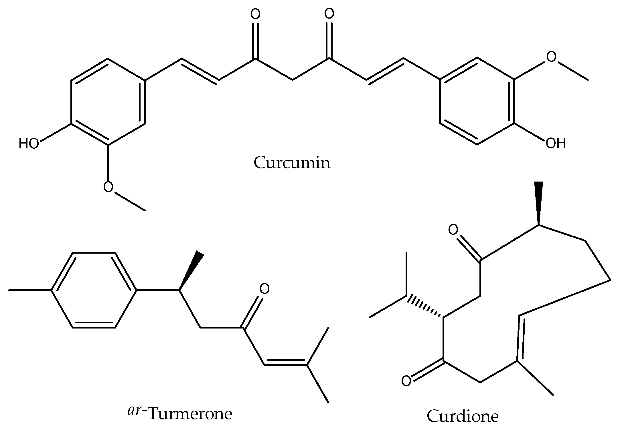
2.9. Curcuma aromatica

In TCM, tubers and dried rhizomes of C. aromatica are prescribed, among other things, as analgesics and the anti-inflammatory activity of its essential oil has been studied by Li (1985). However, until our works in the late 1990s few works are found regarding the eicosanoid inhibition properties. Ammon and co-workers described its active principle curcumin—a diarylheptanoid (Figure 9)—as an effective inhibitor of 5-LOX activities in rat peritoneal PMNs, as well as 12-LOX and COX in human platelets, in addition to having a powerful antioxidant effect in in vitro peroxidation models [86]. However, we could not find that the whole extract of the clinically prescribed TCM drug is able to show the same activities in the same models [32,35].
Hundreds of works have reported the topical anti-inflammatory and antioxidant activities of diarylheptanoids—and extracts enriched in these compounds—from Curcuma sp. extracts at both experimental and clinical levels [87,88,89,90] and how they regulate both COX and LOX [91] via transcription factors [92]. There is a controversy about the bioavailability of its components that contribute to a huge variability in therapeutic results [93]. The focus on curcuminoids is also shadowing the contribution of other phytochemical present in Curcuma sp., and curcumin-free extracts may be also active as recently reported [94].
However, there are only a few works specifically dealing with the effect of Curcuma aromatica extracts. Analyses of the data showed that C. aromatica consists of various classes of compounds, including alkaloids, flavonoids, curcuminoids, tannins, and terpenoids, that formed the bases of its pharmacological activities [95]. Its content in curcuminoids (curcumin, bis-demethoxycurcumin and demethoxycurcumin) are lower than in C. longa [96], thus explaining a lower contribution from this phytochemical class when researching the bioactivities of this herbal medicine. Still it was shown to be more effective reducing the TPA (12-O-tetradecanoylphorbol-13-acetate)-induced ear edema in BALB/c mice than ibuprofen, an effect that is accompanied by a significant reduction in COX-2 levels in ear tissues [97]. Other constituents that have been related with topical anti-inflammatory activity are those present in the volatile fraction of the tubers/rhizomes of the plant [98]. The sesquiterpenes curdione [99] and ar-turmerone (Figure 9) turned out as the major compounds [100] in the essential oils of C. aromatica growing in China. Both compounds have the ability to inhibit COX-2 in mouse macrophage RAW 264.7 cells (IC50 of 1.1 µM and 24 µM, respectively) [101,102]. This may occur via inhibition of NF-κB activation as shown in breast cancer cells [103]. It also attenuated inflammatory via cytokine expression by inactivating Hedgehog pathway in HaCaT cells [104]. This compound resulted more potent than aspirin at inhibiting platelet aggregation induced by collagen (IC50 = 14.4 μM) and arachidonic acid (IC50 = 43.6 μM), without any effect on platelet activating factor or thrombin-induced platelet aggregation, thus pointing to a potential direct or indirect inhibition of thromboxane synthesis [105], although COX-1 levels do not change upon ar-turmerone treatment in breast cancer cells [103]. Regarding its antioxidant activities, it was shown that extracts of C. aromatica effectively protect skin cells from UVA radiations by augmenting their antioxidant defenses [106].

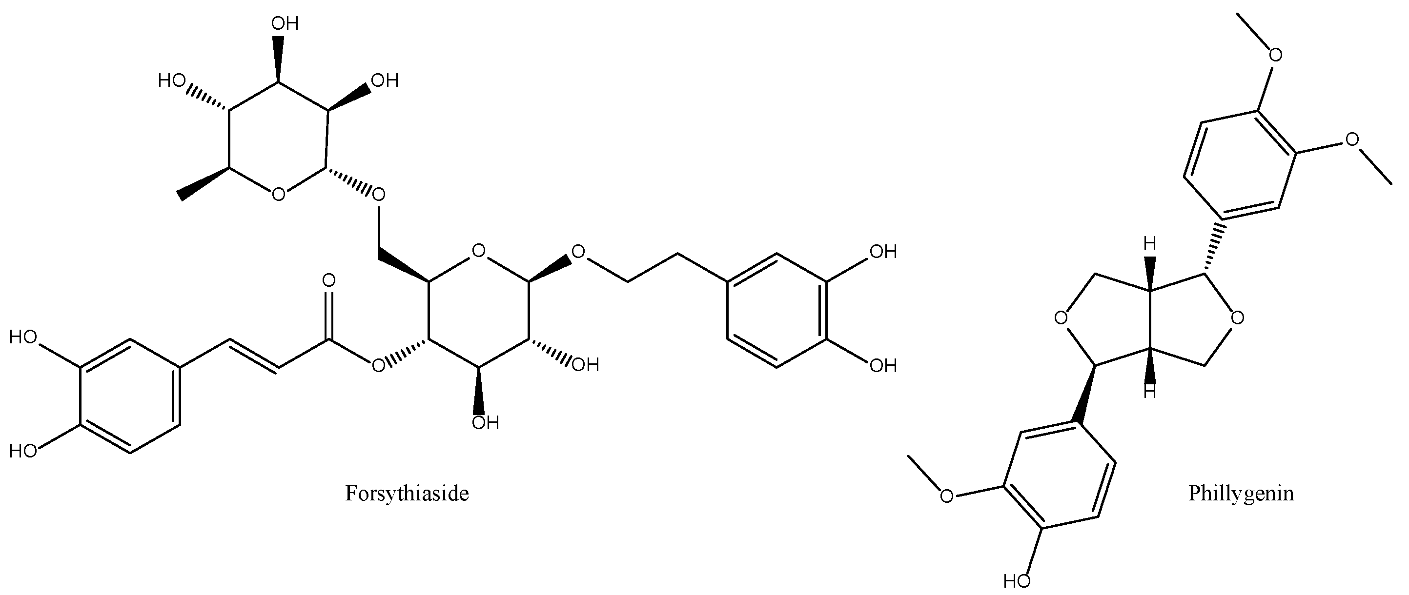
2.10. Forsythia suspensa

The fruits of F. suspensa are used in TCM as antipyretics and anti-inflammatories in the treatment of bacterial infections. Kimura and Okuda already described in 1987 that its caffeic acid glycosides are inhibitors of 5-HETE production in rat peritoneal PMNs [36]. Our results expanded on this by showing a concurrent inhibition to LTB4 and therefore to the total activity of the enzyme without affecting COX-1 synthesis of 12-HETE in human platelets, thus ruling out any action at the level of PLA2 [32]. We later showed that F. suspensa extract is a potent inhibitor of lipid peroxidation, both enzymatic and non-enzymatic (CI50 of 24 and 16.7 µg/mL, respectively) and of the action of the superoxide radical generated by the hypoxanthin/XOD system (CI50 = 11.3 µg/mL). However, it enhances the degradation of deoxyribose by the action of the hydroxyl radical generated in the absence of ascorbate [35]. All this pointed towards considering F. suspensa as a potentially useful herbal drug at the level of total extract since very marked effects are achieved without having to resort to its fractionation. However, its strong pro-oxidant character in the presence of the hydroxyl radical (system without ascorbate) requires a more careful assessment at the level of cell or organism. There is now some consensus [107,108] in that forsythosides (particularly its A form or forsythiaside) and phillygenin [109,110,111] are among its most important anti-inflammatory and antioxidant active principles (Figure 10).

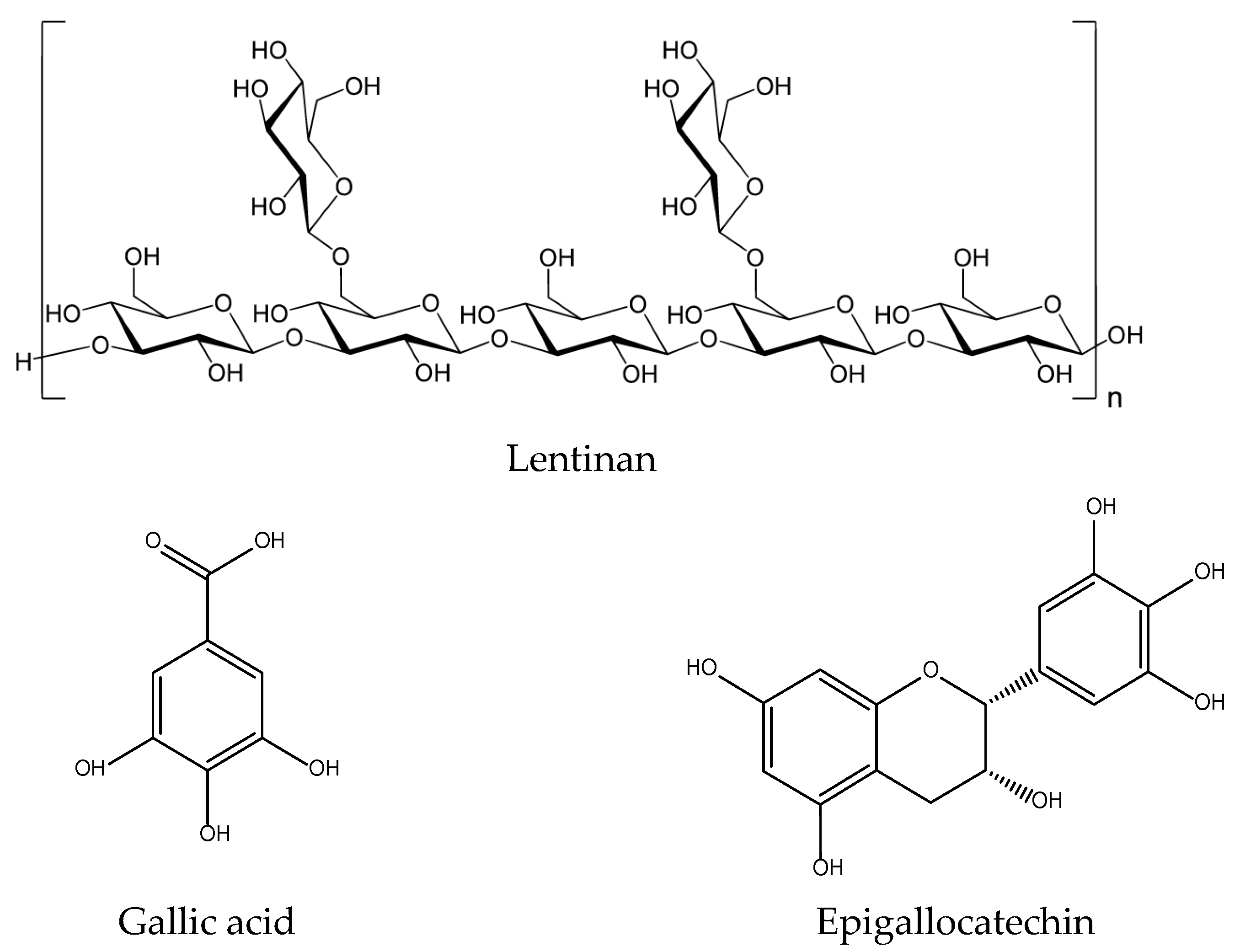
2.11. Lentinus edodes

In our experience, the aqueous extract of L. edodes had no activity in either of our eicosanoid pathway (COX-1, COX-2, 5-LOX, 12-LOX, 15-LOX) or antioxidant models [32,35]. Almost at the same time, Sia and Candlish demonstrated that the anti-inflammatory activity of the aqueous extract of this edible fungus lies on the inhibition of interleukin 1 production, without any effect on superoxide radical release in human neutrophils. These authors demonstrated that these effects are due to low molecular molecules rather than macromolecules such as the characteristics polysaccharides (lentinans) present in the mushroom [112].
Small phenolic secondary metabolites have been described in L. edodes including precursors of tannins such as gallic acid and epigallocatechin (>50 mg/kg) (Figure 11) and in lower quantity flavonoids such as isoquercetin, kaempferol and eriodictyol levels (<50 mg/kg) [113]. These compounds may justify potential anti-inflammatory and antioxidant activities in the models mentioned above only if concentrated at pharmacological-relevant levels [114].
Lentinan (Figure 11), extracted from its fruiting body, has clinically significant anticancer, antibacterial, antiviral, and anticoagulant effects. There is a report on its preventive effects on skin oxidative damage by H2O2, reduction MDA formation, and increased SOD activity in HaCat cells [115] as well as on the inhibition of the production of pro-inflammatory cytokines, including IL-1β, TNF-α, IL-8 and the secretion of PGE2 and NO, by reducing the expression of COX-2 and iNOS in AGE-challenged chondrocytes [116].
It could be said that the main clinical interest of this species in the treatment of pathologies that occur with inflammatory processes would derive from indirect effects at the level of the microbiota [117] or immunological level [118] and not to direct inhibitory actions on the LOX, COX or the production of free radicals in proinflammatory cells.

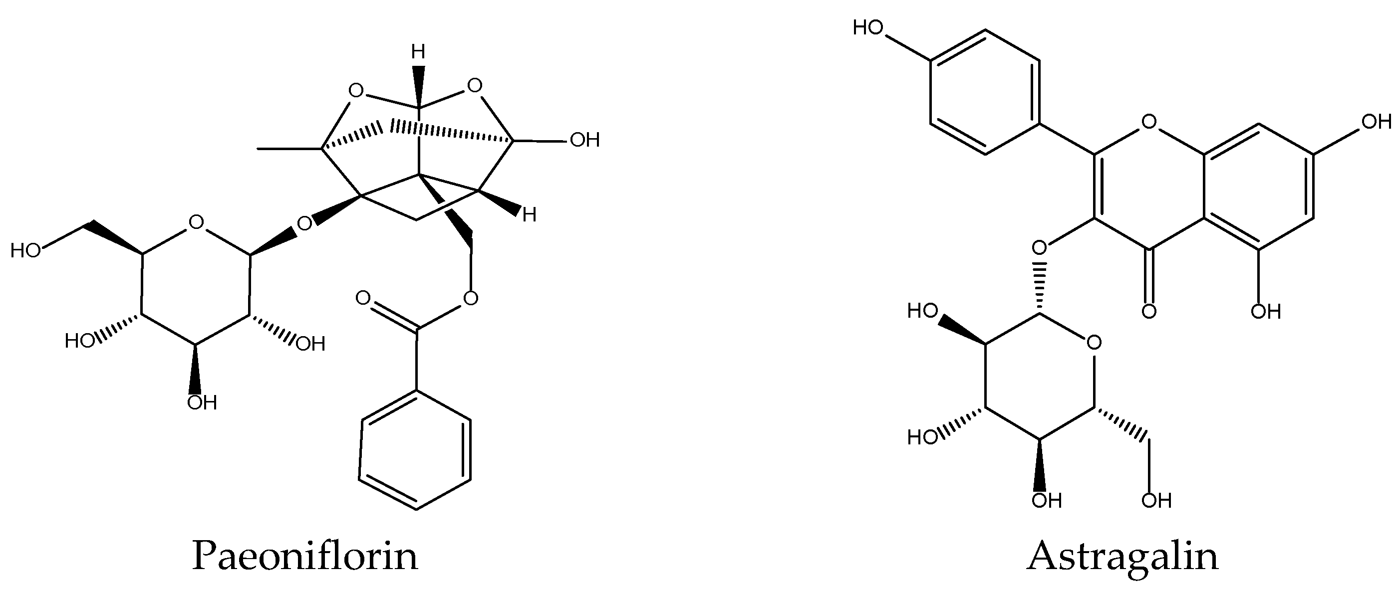
2.12. Paeonia lactiflora

P. lactiflora is a Ranunculaceae with a reputation for analgesic and bacteriostatic effects. Preliminary work demonstrated the inhibitory activity of one of its components, paeoniflorin, in platelet aggregation models [24]. Our results supported the existence of a specific action at the platelet level, since the total extract of this drug inhibited 70% the production of 12-HHTrE without altering the levels of 12-HETE, LTB4 or 5-HETE [32]. In the free radical generation tests, pro-oxidant was shown in the Fe3+-EDTA + H2O2 system without ascorbate [35]. These data would justify the use of the total extract as an analgesic, since it inhibits COX-1 activity in vitro, although in vivo this effect may also be due in part to the central nervous system depressant action of paeoniflorin (Figure 12) [24]. Little additional work has been done in this direction apart from the confirmation of anti-inflammatory effects of this compound in human dermal microvascular endothelial cells cancer cells by blocking nuclear factor-κB and ERK pathway [119]. Its antioxidant effects in UVA-induced damage in human dermal fibroblasts in terms of reduction of the ROS and MDA levels is due to the inhibition of the Nrf2/HO-1/NQ-O1 signalling pathway [120]. Astragalin (Figure 12) is another secondary metabolite present in this herbal drug that has been studied [121].

2.13. Phellodendron amurense
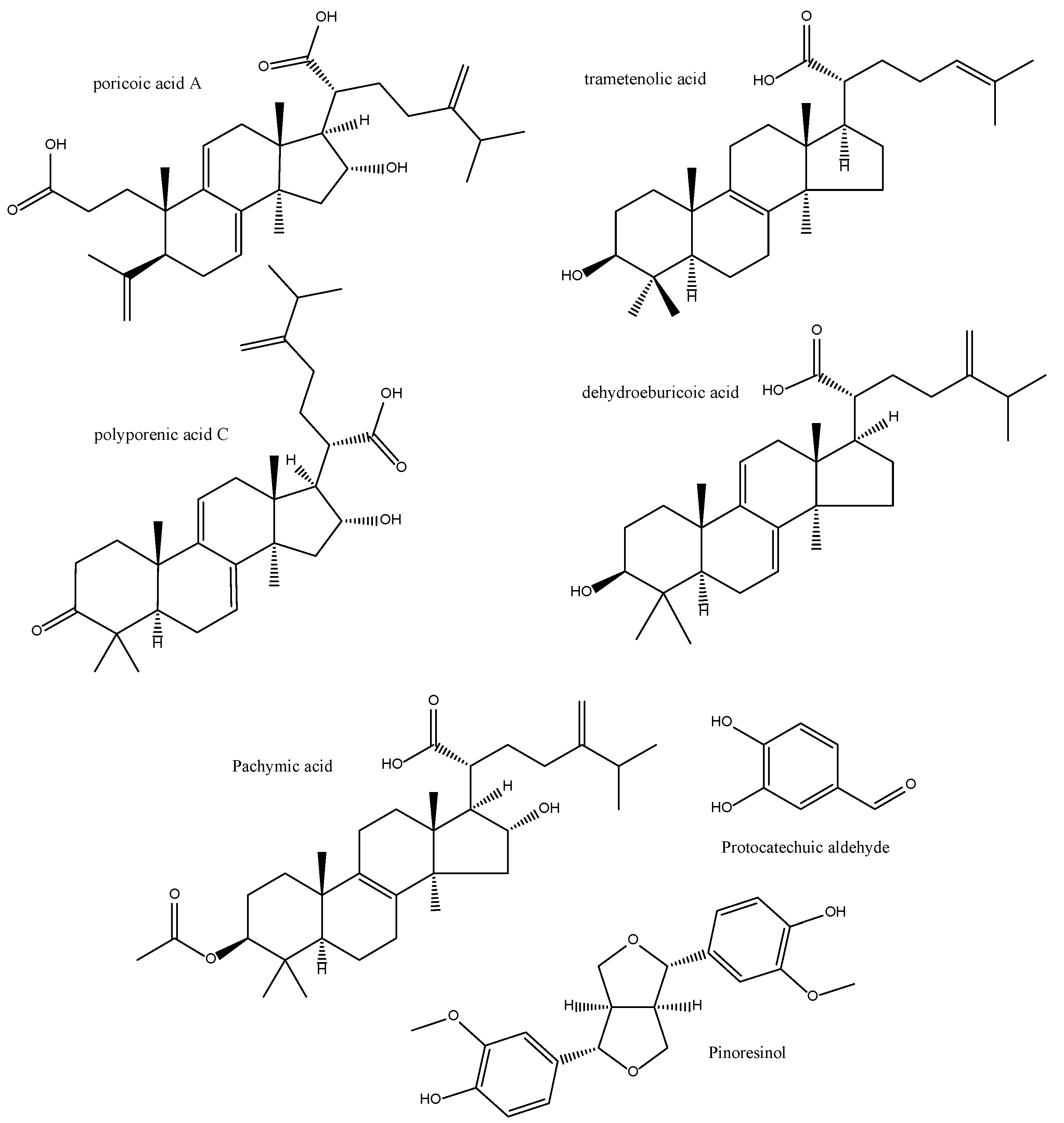
This rutaceae is, like Coptis chinensis, rich in alkaloids of the berberine type (berberine, palmatine, etc.) (Figure 8). Our works described the in vitro actions on the production of eicosanoids and free radicals by its total extract: although its action on LTB4 could not be quantified, a total inhibition of 5-HETE production was found, as well as an inhibition of 86% and 65% in the production of 12-HHTrE and 12-HETE, respectively at 200 µg/mL [32]. With these data, an effect at the level of PLA2 cannot be ruled out. In the enzymatic lipid peroxidation model, it obtained an IC50 = 21.6 µg/mL, not being particularly active in any of the other methods tested [35].
Müller and Ziereis could not demonstrate any significant activity of berberine on 5-LOX [122]. The bioactive alkaloids identified from this herbal drug (Figure 8) suppress the expression of LOX-5 and COX-2 simultaneously in rat cell and models of Bening Prostate Hyperplasia. In particular, protoberberine and demethyleneberberine were found to exhibit strong direct inhibitory activities against both LOX-5 and COX-2 enzymes, whilst palmatine and berberine showed moderate inhibitory activities only. Molecular docking analysis confirmed that demethyleneberberine could directly interact well with LOX-5/COX-2 [80].

2.14. Poria cocos

Poria cocos extracts inhibited PLA2-induced mouse paw edema by both the oral and parenteral routes [123]. Subsequent work led to the isolation of lanosthane-type anti-inflammatory principles with interesting PLA2 inhibitory activity from Naja naja in vitro and in vivo [29,30]. We described how this very same extract inhibits both the production of 5-LOX metabolites in rat peritoneal PMNs and 12-HHTrE and 12-HETE in human platelets. With very similar percentages of inhibition thus supporting an activity at PLA2 level, in line with works using by an in vitro polarographic, we also described for the first time the antioxidant activity of extracts from this fungus, which inhibited 43% the degradation of deoxyribose by the hydroxyl radical generated by the Fe3+-EDTA + H2O2 and ascorbate system. In this model, no other extract showed an effectiveness greater than 40%, except Coptis chinensis [35]. Little additional work has been done to unravel these activities. There is only one report on its anti-skin aging effects via activation of the Nrf2-antioxidant mechanism in human dermal fibroblasts [124].
The anti-inflammatory principles of the EtOH extract of the sclerotia of P. cocos after bioassay-guided fractionation using LPS-stimulated Raw264.7 cells, include triterpenoids (such as poricoic acid A, polyporenic acid C, trametenolic acid and dehydroeburicoic acid) as well as phenolics (pinoresinol and protocatechualdehyde) (Figure 13). They all have inhibitory effects on the production of NO, PGE2 and the expression of iNOS) and COX-2 [125]. Pachymic acid (Figure 13), another characteristic lanostane-type triterpenoid from Poria cocos, exerts anti-inflammatory and antioxidant effects in mice kidneys by increasing glutathione expression, decreasing MDA and COX-2 levels and increasing the expression levels of several NRF2 signaling pathway proteins [126]. Similarly, dehydrotrametenolic acid (Figure 13) can activate AP-1 and NF-κB transcriptional factors in human keratinocyte cell line HaCaT cells which may in turn modulate the arachidonate pathway [127]. The free radical scavenging activities of lanostanes are not very prominent, though [128].
As in the case of other higher fungi such as Lentinus edodes, polysaccharides are prominent in the chemical make-up of P. cocos aqueous extracts. These have been described as having in vitro antioxidant activities on the basis of DPPH radical, hydroxyl radical, reducing power and metal chelating ability [129]. Strikingly, such compounds have been reported to be pro-inflammatory as they can stimulate macrophages to express iNOS gene through the activation of NF-κB/Rel and interleukins, interferon and TNF through TLR4/TRAF6/NF-κB signalling both in vitro and in vivo [130]. Extracts containing lanostane triterpenoids also enhance non-specific (innate) immunity though activating natural killer cells and regulating interferon and interleukin synthesis in T-helper cells 1 and 2, respectively, thus modulating the cellular immune response [131]. Therefore, the balance of pro- and anti-inflammatory effects of P. cocos extracts may hugely vary depending on the polarity of the solvents and methods of extraction. The anti-inflammatory and antioxidant overall effects of truly whole extracts may be extremely difficult to predict and even turn out being pro-inflammatory.

2.15. Rehmannia glutinosa

Early works reported its in vivo effects on the platelet [132] and the action at the immunomodulatory level of its polysaccharides as shown by a pronounced anti-complementary activity [133]. In our hands the extract of R. glutinosa did not show any significant anti-inflammatory or antioxidant effects [32,35]. The presence of iridoids, especially catalpol, could suppose potential anti-inflammatory and antioxidant activities via modulation of pro-inflammatory cytokines, as seen in Caco-2 cells [134] and may contribute to skin healing due to the effect of acteoside, a phenylethanoid glycoside isolated from the leaves of this herbal drug, activating the expression of matrix metalloproteinases (MMPs) in normal human dermal fibroblasts [135].

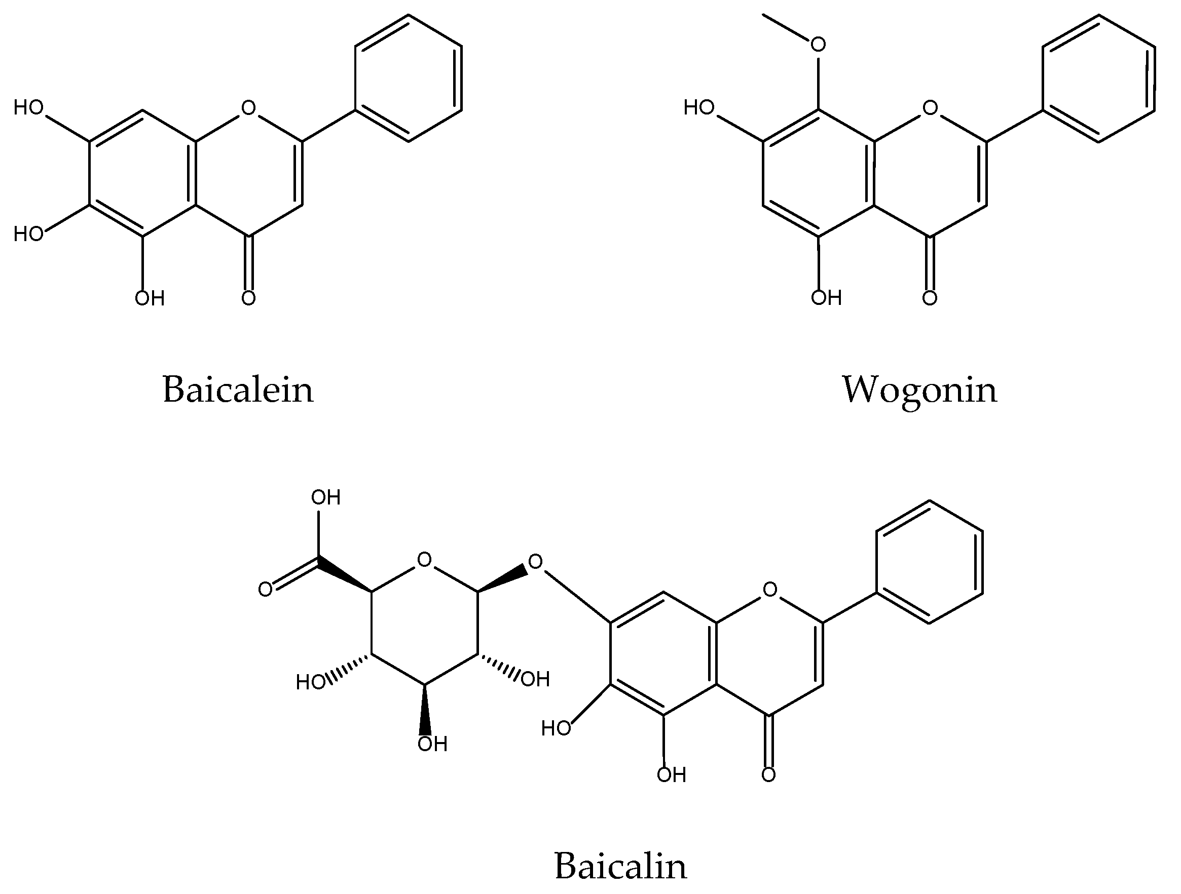
2.16. Scutellaria baicalensis

The pharmacological properties of the genus Scutellaria, and in particular S. baicalensis, have aroused great interest in the scientific community, an interest that is reflected in the large amount of earlier work done on its antioxidant and anti-inflammatory activities, especially of three of its main components: baicalein, baicalin, and wogonin (Figure 14) [136,137].
Although in our study S. baicalensis interfered with the quantification of arachidonic acid metabolites, an absence of 5-HETE production was observed. Although no previous work has been found reporting this effect, Butenko et al. described the inhibitory action of baicalin in the production of LTC4 in peritoneal macrophages of rats [136] and You et al. in 12-LOX without affecting COX activity [137].
Our works on the antioxidant activity of S. baicalensis extracts corroborate the interest of the total extract as an inhibitor of lipid peroxidation (CI50 of 5.3 µg/mL and 13.6 µg/mL in the enzymatic and non-enzymatic systems, respectively). It could not be tested in the hypoxanthin/XOD system since the total extract interferes with XOD activity, probably due to its content in baicalein, baicalin and wogonin, which according to Chang et al. are inhibitors of this activity [138]. In the hydroxyl radical generator system Fe3+-EDTA+H2O2 with and without ascorbate, the total extract of S. baicalensis was shown to be extremely pro-oxidizing, promoting the degradation of deoxyribose by 852% and 157%, respectively. This surprising fact, given that baicalein and baicalin are inhibitors of this effect [139] could only be explained by the fact that another characteristic flavonoid, escutelarin, is pro-oxidant [140], an effect that prevails in the total extract. This fact emphasizes the importance of testing the total extracts, as they are used ethnopharmacologically, instead of their components or fractions, since the observed effects can be radically different.
Later research strengthens baicalein as a protective agent for skin cells from the oxidative stress caused by H2O2 through activation of Nrf2 signalling pathway [141,142]. It also protects human keratinocytes from UV-induced ROS-mediated damage [143,144] thus explaining previously claims of the UV protection conferred by S. baicalensis crude and flavonoid-enriched extracts [145]. Effects of both baicalein and wogonin (Figure 14) in acute UVB-irradiated cells involve reduced levels of COX-2 [146], although it has been observed that high doses may slightly induce COX-1 mRNA, although eventually a decrease of PGE2 is always observed in wogonin-treated mice [147].

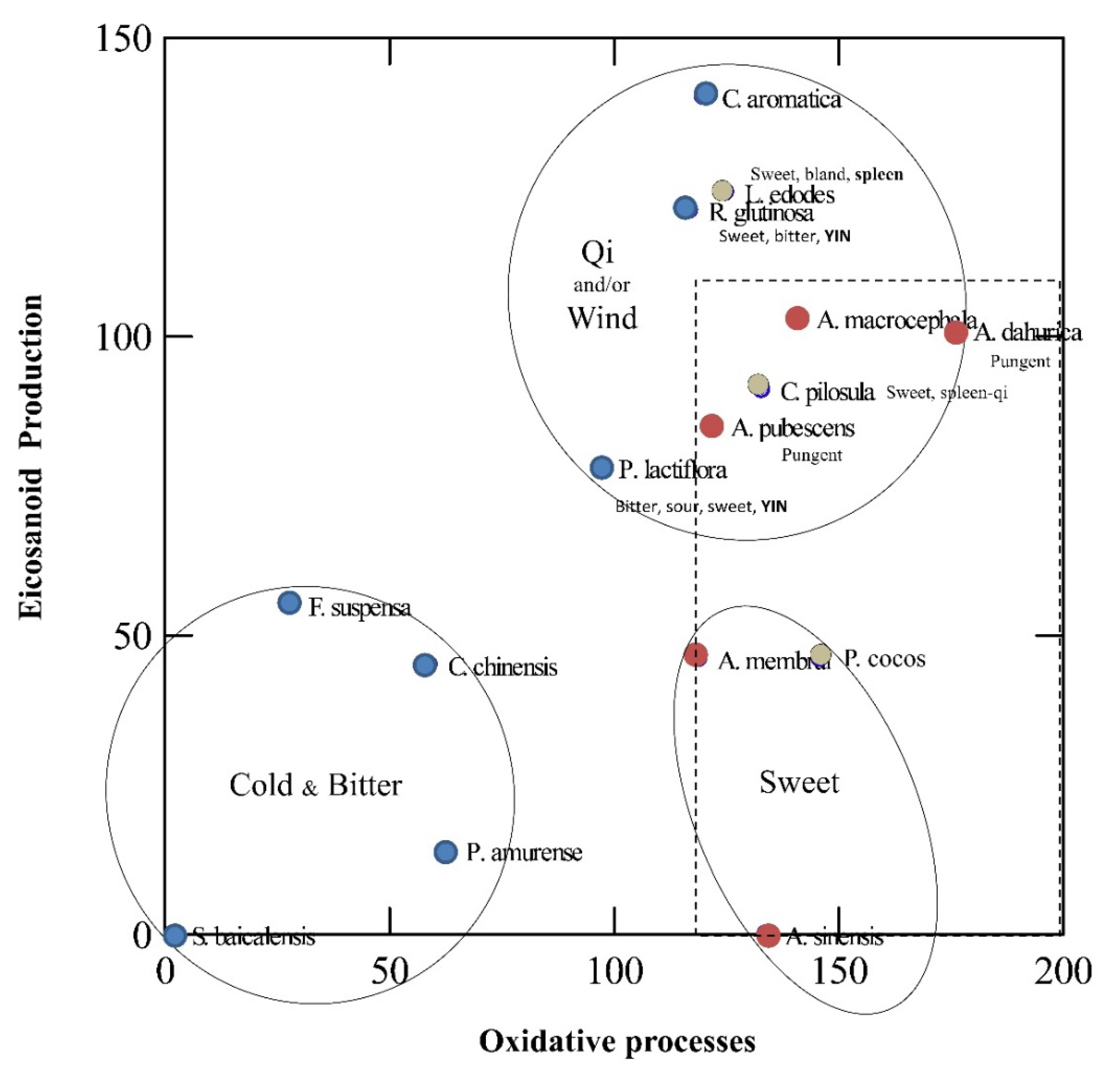
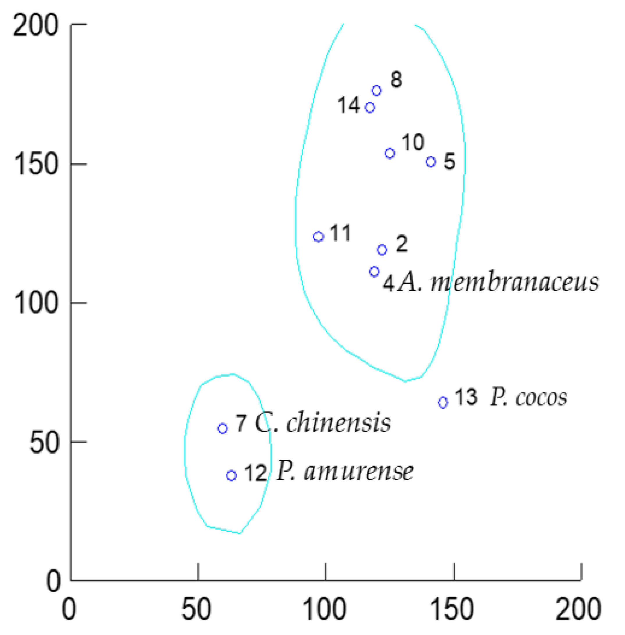
3. Unveiling Links between Traditional Chinese Plant Characters and Quantitative Antioxidant/Eicosanoid Inhibitory Activities of the Extracts

Our aim is to investigate now if the data point out to any link/s between the inhibitory properties of lipid peroxidation and eicosanoid biosynthesis in medicinal plants and the properties/characters that they are assigned to these herbal drugs by TCM doctors.

3.1. Data Sourcing

Most of the biochemical and pharmacological activities of the TCM drugs listed in Table 1 were published in two articles [32,35]. The origin of the plants and the extracts were maintained, thus ensuring that both sets of antioxidant and eicosanoid inhibition data are comparable. Where gaps existed, we filled with results from the literature using similar substances if available. All the raw data were normalised to percentage of inhibition of the biochemical or pharmacological endpoint (0% maximum inhibitory effect–100% minimum inhibitory effect, using MS Excel (Microsoft, Redmond, Washington). The data are summarised in Table 2. A three-dimensional vector positioned each herbal drug in an “antioxidant” space, defined by the values of the three antioxidant tests: (LNE) Lipid Non-Enzymatic Peroxidation; (LE) Lipid Enzymatic Peroxidation; (XO) Xanthine Oxidase. The magnitude of each 3D “antioxidant” vector was calculated (α). Another bi-dimensional vector positioned each herbal drug in an “anti-inflammatory” space, defined by the values of the 5-LOX and COX-1 eicosanoid biosynthesis tests. The magnitude of each 2D vector was calculated (β). Another three-dimensional vector positioned each herbal drug in a wider “anti-inflammatory” space, defined by the values of the 5-LOX, 12-LOX and COX-1 eicosanoid biosynthesis tests eicosanoid biosynthesis tests. The magnitude of each 3D vector was calculated (χ). The α and β values positioned each extract in a bidimensional space. These set was subject to cluster analyses by k-means. In a separate analysis, the α and χ values positioned each extract in a bidimensional space (antioxidant activity in X axis vs. eicosanoids inhibition in Y axis), and these sets were subject to cluster analyses by k-means. All k-means clustering was performed with SPSS 19 (IBM, Armonk, NY, USA).

3.2. Results and Discussion

When all selected TCM drugs were analysed for the relationship between their combined inhibitory properties on COX-1/2 and 5-LOX versus their combined antioxidant effects, three clusters were identified (Figure 15). The first one contains 4 plants characterised by high inhibitory activity of all the biochemical parameters. A second cluster was interpreted as plants with high inhibitory properties on eicosanoids release but mild/low antioxidant properties, and a third group of plants had mild/low activities on both parameters.
When overlapping TCM characters for each herbal drug, we could observe that the first cluster is coherent with some phytochemical traits as well as therapeutic uses in TCM (Table 1). Interestingly, all are considered “Bitter and cold”. Generally speaking, Chinese cleansing herbs are considered bitter herbs with a “cold property”. Phellodendron (Huang Bai), and Skullcap (Huang Qin) are the constituents of a popular herbal formulas used for a variety of skin disorders, the “Three Yellow Cleanser” (San Huang Xi Ji, where Huang means yellow), together with Rhubarb (Dai Huang) and Sophora (Ku Shen). Furthermore, Phellodendron and Coptis [25,26] share the same chemistry based on berberine alkaloids. The interchangeability of Phellodendron (“Drains Fire and relieves Fire toxicity”) and Coptis is known in both traditional and local medicinal systems in China as “Using different plants as the same herbal medicine” (使用不同的植物作为同一种药草). Features to identify Huang-lian are “yellow and bitter”, and chemically speaking this is strongly related to the presence of berberine type alkaloids. Theoretically, Scutellaria, Berberis, and Thalictrum species could be indistinctly used as Huang-lian (黄连) the common name for Rhizoma coptidis. However, in a study of the local medicine in NW Yunnan, the authors found that other herbs with different chemistry but overall same pharmacological features such as, Scutellaria spp (Huang-Qin, which also “Drains Fire and detoxifies”) were used as Huang-lian [151], thus supporting our results. The presence of F. suspensa in this cluster is surprising, as it is chemically very different but highly reputed for abscess and sores, sore throat, scrofula and subcutaneous nodules [24]. In the light of our review (see Section 2.10) not much “Western” science is available for this otherwise promising “anti-inflammatory and antioxidant” herbal drug.
The second cluster contains a mix of two hot herbs and one neutral fungi, all sweet in nature. They can strongly inhibit the synthesis of eicosanoids but show mild antioxidant activity. Therefore, these herbs may mimic NSAIDs or steroids activity. Indeed A. membranaceus and P.cocos contain steroidal-like compounds, namely astragalosides and lanostanes as reviewed in Section 2.5 and Section 2.14, respectively. A. dahurica is clearly separated from its two congeners, A. sinensis and A. pubescens. Interestingly, the “expel wind” action is almost exclusive of the two furanocoumarins containing Angelica species (A. dahurica, and A. pubescens) and differentiates them from A. sinensis, which “tonifies blood” but is not a significant source for such photodynamic compounds as per a recent review [152].
The third cluster is composed of a mix of warm, cold, and neutral drugs. However, the warm character seems to be confined to a particular region of the biochemical space (delimitated by a dashed red square) close to the second cluster. The differential trait here seems to be either tonifying/moving Qi and/or “expelling wind” actions as well as protecting the spleen and/or liver. The action on Qi seems to correlate with immunomodulation or protection of internal organs. The unprocessed R. glutinosa is a cortisol-like substance, which has the advantage of not suppressing, but rather enhancing, the immune system in many cases [23], and our review supports these immune effects via cytokines (see Section 2.15). The fungi L. edodes that acts as a “liver-enhancing” herbal medicine, thus protecting the liver from damage associated with autoimmunity, inflammation, oxidation, and infection [153], and C. aromatica that similarly cleans the liver and the blood (Table 1) seems to be linked to the Western “detoxification” concept. The activities of A. macrocephala and P. lactiflora target spleen and liver (Table 1), thus implying both detoxification and immunomodulatory effects.
When a reduced set of TCM drugs were analysed for the relationship between the combined inhibitory properties on COX-1, 5-LOX and 12-LOX vs. their combined antioxidant effects, three clusters were again identified (Figure 16).
The inclusion of the inhibition of 12-LOX inhibition—although restricting the dataset—points towards a “selective” influence of this eicosanoid pathway within the skin lipoperoxidation model. 12(S)-HETE is present in psoriatic scales [1]. Interestingly, human platelets produce 12(S)-HHTrE and 12(S)-HETE from the COX-1 and 12-LOX pathways, respectively, after stimulation with Ca++ and ionophore A23187. Therefore, its use as in vitro screening for anti-psoriatic drugs is relevant since in psoriatic epidermis only the platelet-type 12-LOX is detectable [154]. The lack of inhibition of 12-LOX also rules out any impairment of the release of endogenous arachidonic acid from the membranes by phospholipase A2 since endogenous arachidonate is available to 12-LOX. The two berberine alkaloid-containing herbal drugs, C. chinensis and P. amurense, remain in the Cold/Bitter cluster with very similar and relatively low IC50s for this enzyme, whilst F. suspensa -which fails to be active at this level- and S. baicalensis -for which no data on 12-LOX could be found- are now out of the picture. The Sweet-Hot/Warm cluster now contains the fungi P. cocos only.

4. Conclusions

We here present a thorough review of the eicosanoid inhibitory activities of important Chinese herbal drugs, as well as a biochemical explanation to some of the characters and actions of TCM drugs used—among other conditions—in skin diseases. Lipid peroxidation and eicosanoids production are intimately linked, and our cluster analysis unveiled how cleansing herbs of bitter and cold nature acting through removal of toxins—such as P. amurense, Coptis chinensis, S. baicalensis and F. suspensa—are highly correlated with strong inhibition of both lipid peroxidation and eicosanoids production. Sweet drugs—such as A. membranaceus, A. sinensis and P. cocos—act through a specific inhibition of the eicosanoids production. The therapeutic value of the remaining drugs with low antioxidant or anti-inflammatory activity—seems to be based on their actions on the Qi with the exception of furanocoumarin containing herbs—A. dahurica and A. pubescens—which “expel wind”.
A further observation from our results is that the drugs present in the highly active “Cleansing herbs” cluster are commonly used for skin conditions and may be bioequivalents (=interchangeable) thus supporting the special concept of Traditional Chinese Medicine called “Multisource” of herb (多基源). The inclusion of 12-LOX inhibition did not fundamentally change the clusters but pointed towards plants that may be more active in chronic skin conditions such as psoriasis.

Author Contributions

J.M.P. and G.R.S. conceived and contributed to the writing and editing of the manuscript. J.M.P. performed the calculations. All authors have read and agreed to the published version of the manuscript.

Funding

This research received no external funding.

Acknowledgments

This article is dedicated to Jose Luis Rios Canavate in the occassion of his retirement. His dedication to pharmacognosy and kindness inspired us both. We are grateful to Daniel Li for help with some Chinese–English translations.

Conflicts of Interest

The authors declare no conflict of interest.

References

  1. Simpson, E.L. Atopic dermatitis: A review of topical treatment options. Curr. Med. Res. Opin. 2010, 26, 633–640. [Google Scholar] [CrossRef] [PubMed]
  2. Borgia, F.; Giuffrida, R.; Caradonna, E.; Vaccaro, M.; Guarneri, F.; Cannavò, S.P. Early and Late Onset Side Effects of Photodynamic Therapy. Biomedicines 2018, 6, 12. [Google Scholar] [CrossRef] [PubMed] [Green Version]
  3. Speeckaert, R.; Dugardin, J.; Lambert, J.; Lapeere, H.; Verhaeghe, E.; Speeckaert, M.M.; van Geel, N. Critical appraisal of the oxidative stress pathway in vitiligo: A systematic review and meta-analysis. J. Eur. Acad. Dermatol. Venereol. 2018, 32, 1089–1098. [Google Scholar] [CrossRef] [PubMed]
  4. Furue, M.; Hashimoto-Hachiya, A.; Tsuji, G. Antioxidative Phytochemicals Accelerate Epidermal Terminal Differentiation via the AHR-OVOL1 Pathway: Implications for Atopic Dermatitis. Acta Derm. Venereol. 2018, 98, 918–923. [Google Scholar] [CrossRef] [Green Version]
  5. Umamaheswaran, S.; Dasari, S.K.; Yang, P.; Lutgendorf, S.K.; Sood, A.K. Stress, inflammation, and eicosanoids: An emerging perspective. Cancer Metastasis Rev. 2018, 37, 203–211. [Google Scholar] [CrossRef]
  6. Tretter, V.; Hochreiter, B.; Zach, M.L.; Krenn, K.; Klein, K.U. Understanding Cellular Redox Homeostasis: A Challenge for Precision Medicine. Int. J. Mol. Sci. 2021, 23, 106. [Google Scholar] [CrossRef]
  7. Briganti, S.; Picardo, M. Antioxidant activity, lipid peroxidation and skin diseases. What’s new. J. Eur. Acad. Dermatol. Venereol. 2003, 17, 663–669. [Google Scholar] [CrossRef]
  8. De Luca, C.; Valacchi, G. Surface lipids as multifunctional mediators of skin responses to environmental stimuli. Mediat. Inflamm. 2010, 2010, 321494. [Google Scholar] [CrossRef]
  9. Gutteridge, J.M. Lipid peroxidation and antioxidants as biomarkers of tissue damage. Clin. Chem. 1995, 41, 1819–1828. [Google Scholar] [CrossRef]
  10. Niki, E. Lipid oxidation in the skin. Free Radic. Res. 2015, 49, 827–834. [Google Scholar] [CrossRef]
  11. Catalá, A. Lipid peroxidation of membrane phospholipids generates hydroxy-alkenals and oxidized phospholipids active in physiological and/or pathological conditions. Chem. Phys. Lipids 2009, 157, 1–11. [Google Scholar] [CrossRef] [PubMed]
  12. Ayala, A.; Muñoz, M.F.; Argüelles, S. Lipid peroxidation: Production, metabolism, and signaling mechanisms of malondialdehyde and 4-hydroxy-2-nonenal. Oxid. Med. Cell Longev. 2014, 2014, 360438. [Google Scholar] [CrossRef] [PubMed]
  13. Nicolaou, A. Eicosanoids in skin inflammation. Prostaglandins Leukot. Essent. Fat. Acids 2013, 88, 131–138. [Google Scholar] [CrossRef] [PubMed]
  14. Serhan, C.N.; Yacoubian, S.; Yang, R. Anti-inflammatory and proresolving lipid mediators. Annu. Rev. Pathol. 2008, 3, 279–312. [Google Scholar] [CrossRef] [Green Version]
  15. Coras, R.; Kavanaugh, A.; Boyd, T.; Huynh, Q.; Pedersen, B.; Armando, A.M.; Dahlberg-Wright, S.; Marsal, S.; Jain, M.; Paravar, T.; et al. Pro- and anti-inflammatory eicosanoids in psoriatic arthritis. Metabolomics 2019, 15, 65. [Google Scholar] [CrossRef]
  16. Chiang, N.; Serhan, C.N. Specialized pro-resolving mediator network: An update on production and actions. Essays Biochem. 2020, 64, 443–462. [Google Scholar]
  17. Serhan, C.N.; Dalli, J.; Colas, R.A.; Winkler, J.W.; Chiang, N. Protectins and maresins: New pro-resolving families of mediators in acute inflammation and resolution bioactive metabolome. Biochim. Biophys. Acta 2015, 1851, 397–413. [Google Scholar] [CrossRef] [Green Version]
  18. Zou, W.; Gong, L.; Zhou, F.; Long, Y.; Li, Z.; Xiao, Z.; Ouyang, B.; Liu, M. Anti-inflammatory effect of traditional Chinese medicine preparation Penyanling on pelvic inflammatory disease. J. Ethnopharmacol. 2021, 266, 113405. [Google Scholar] [CrossRef]
  19. Forman, H.J.; Zhang, H. Targeting oxidative stress in disease: Promise and limitations of antioxidant therapy. Nat. Rev. Drug Discov. 2021, 20, 689–709. [Google Scholar] [CrossRef]
  20. Bensky, D. Chinese Herbal Medicine: Materia Medica, 3rd ed.; Eastland Press: Seattle, WA, USA, 2004. [Google Scholar]
  21. Li, S. Compendium of Materia Medica: Bencao Gangmu, 1st ed.; Foreign Languages Press: Beijing, China, 2003. [Google Scholar]
  22. Koo, J.; Desai, R. Traditional Chinese medicine in dermatology. Dermatol. Ther. 2003, 16, 98–105. [Google Scholar] [CrossRef]
  23. Amenta, R.; Camarda, L.; Di Stefano, V.; Lentini, F.; Venza, F. Traditional medicine as a source of new therapeutic agents against psoriasis. Fitoterapia 2000, 71 (Suppl. 1), S13–S20. [Google Scholar] [CrossRef]
  24. Tang, W.C.; Eisenbrand, G. Chinese Drugs of Plant Origin, Subtitle: Chemistry, Pharmacology and Use in Traditional and Modern Medicine; Springer: Berlin/Heidelberg, Germany, 1992. [Google Scholar]
  25. Pacific College of Medicine. Acupuncture for Eczema & Skin Disorders. 2022. Available online: https://www.pacificcollege.edu/news/blog/2015/01/16/acupuncture-eczema-skin-disorders (accessed on 8 March 2022).
  26. Darby, H.; Garcia, J.M.P. Development of an HPLC Method for the Quality Control of Chinese Herbal Medicinal Formulation: Three Yellow Cleanser (San Huang Xi Ji). Asian Basic Appl. Res. J. 2020, 2, 50–60. [Google Scholar]
  27. Keji, C.; Hao, X.U. The integration of traditional Chinese medicine and Western medicine. Eur. Rev. 2003, 11, 225–235. [Google Scholar] [CrossRef]
  28. Tang, W. Chinese Drugs of Plant Origin: Chemistry, Pharmacology, and Use in Traditional and Modern Medicine; Springer: Berlin, Germany; New York, NY, USA, 1992. [Google Scholar]
  29. Cuéllar, M.; Giner, R.; Recio, M.; Just, M.; Máñez, S.; Cerdá, S.; Rios, J.-L. Screening of antiinflammatory medicinal plants used in traditional medicine against skin diseases. Phytother. Res. 1998, 12, 18–23. [Google Scholar] [CrossRef]
  30. Cuellar, M.J.; Giner, R.M.; Recio, M.C.; Just, M.J.; Mañez, S.; Rios, J.L. Effect of the basidiomycete Poria cocos on experimental dermatitis and other inflammatory conditions. Chem. Pharm. Bull. 1997, 45, 492–494. [Google Scholar] [CrossRef] [Green Version]
  31. Cuéllar, M.J.; Giner, R.M.; Recio, M.C.; Máñez, S.; Ríos, J.L. Topical anti-inflammatory activity of some Asian medicinal plants used in dermatological disorders. Fitoterapia 2001, 72, 221–229. [Google Scholar] [CrossRef]
  32. Prieto, J.M.; Recio, M.C.; Giner, R.M.; Manez, S.; Giner-Larza, E.M.; Rios, J.L. Influence of traditional Chinese anti-inflammatory medicinal plants on leukocyte and platelet functions. J. Pharm. Pharmacol. 2003, 55, 1275–1282. [Google Scholar] [CrossRef]
  33. Recio, M.C.; Giner, R.M.; Máñez, S.; Ríos, J.L. Structural considerations on the iridoids as anti-inflammatory agents. Planta Med. 1994, 60, 232–234. [Google Scholar] [CrossRef]
  34. Rios, J.L.; Waterman, P.G. A review of the pharmacology and toxicology of Astragalus. Phytother. Res. 1997, 11, 411–418. [Google Scholar] [CrossRef]
  35. Schinella, G.R.; Tournier, H.A.; Prieto, J.M.; Mordujovich de Buschiazzo, P.; Rios, J.L. Antioxidant activity of anti-inflammatory plant extracts. Life Sci 2002, 70, 1023–1033. [Google Scholar] [CrossRef]
  36. Kimura, Y.; Okuda, H.; Nishibe, S.; Arichi, S. Effects of caffeoylglycosides on arachidonate metabolism in leukocytes. Planta Med. 1987, 53, 148–153. [Google Scholar] [CrossRef] [PubMed]
  37. Kang, O.H.; Lee, G.H.; Choi, H.J.; Park, P.S.; Chae, H.S.; Jeong, S.I.; Kim, Y.C.; Sohn, D.H.; Park, H.; Lee, J.H.; et al. Ethyl acetate extract from Angelica Dahuricae Radix inhibits lipopolysaccharide-induced production of nitric oxide, prostaglandin E2 and tumor necrosis factor-alphavia mitogen-activated protein kinases and nuclear factor-kappaB in macrophages. Pharmacol. Res. 2007, 55, 263–270. [Google Scholar] [CrossRef] [PubMed]
  38. Ban, H.S.; Lim, S.S.; Suzuki, K.; Jung, S.H.; Lee, S.; Lee, Y.S.; Shin, K.H.; Ohuchi, K. Inhibitory effects of furanocoumarins isolated from the roots of Angelica dahurica on prostaglandin E2 production. Planta Med. 2003, 69, 408–412. [Google Scholar] [PubMed]
  39. Lin, C.H.; Chang, C.W.; Wang, C.C.; Chang, M.S.; Yang, L.L. Byakangelicol, isolated from Angelica dahurica, inhibits both the activity and induction of cyclooxygenase-2 in human pulmonary epithelial cells. J. Pharm. Pharmacol. 2002, 54, 1271–1278. [Google Scholar] [CrossRef] [PubMed]
  40. Chen, Y.F.; Tsai, H.Y.; Wu, T.S. Anti-inflammatory and analgesic activities from roots of Angelica pubescens. Planta Med. 1995, 61, 2–8. [Google Scholar] [CrossRef] [PubMed]
  41. Ko, F.N.; Wu, T.S.; Liou, M.J.; Huang, T.F.; Teng, C.M. Inhibition of platelet thromboxane formation and phosphoinositides breakdown by osthole from Angelica pubescens. Thromb. Haemost. 1989, 62, 996–999. [Google Scholar] [CrossRef] [PubMed]
  42. Liu, J.H.; Zschocke, S.; Reininger, E.; Bauer, R. Inhibitory effects of Angelica pubescens f. biserrata on 5-lipoxygenase and cyclooxygenase. Planta Med. 1998, 64, 525–529. [Google Scholar] [CrossRef]
  43. Yuan, Q.; Zhang, J.; Xiao, C.; Harqin, C.; Ma, M.; Long, T.; Li, Z.; Yang, Y.; Liu, J.; Zhao, L. Structural characterization of a low-molecular-weight polysaccharide from Angelica pubescens Maxim. f. biserrata Shan et Yuan root and evaluation of its antioxidant activity. Carbohydr. Polym. 2020, 236, 116047. [Google Scholar] [CrossRef]
  44. Zhang, C.; Hsu, A.C.; Pan, H.; Gu, Y.; Zuo, X.; Dong, B.; Wang, Z.; Zheng, J.; Lu, J.; Zheng, R.; et al. Columbianadin Suppresses Lipopolysaccharide (LPS)-Induced Inflammation and Apoptosis through the NOD1 Pathway. Molecules 2019, 24, 549. [Google Scholar] [CrossRef] [Green Version]
  45. Luo, Q.; Wang, C.P.; Li, J.; Ma, W.F.; Bai, Y.; Ma, L.; Gao, X.M.; Zhang, B.L.; Chang, Y.X. The pharmacokinetics and oral bioavailability studies of columbianetin in rats after oral and intravenous administration. J. Ethnopharmacol. 2013, 150, 175–180. [Google Scholar] [CrossRef]
  46. Zhang, Y.B.; Li, W.; Yang, X.W. Biotransformation of columbianadin by rat hepatic microsomes and inhibition of biotransformation products on NO production in RAW 264.7 cells in vitro. Phytochemistry 2012, 81, 109–116. [Google Scholar] [CrossRef]
  47. Singh, G.; Bhatti, R.; Mannan, R.; Singh, D.; Kesavan, A.; Singh, P. Osthole ameliorates neurogenic and inflammatory hyperalgesia by modulation of iNOS, COX-2, and inflammatory cytokines in mice. Inflammopharmacology 2019, 27, 949–960. [Google Scholar] [CrossRef] [PubMed]
  48. Wang, X.Y.; Dong, W.P.; Bi, S.H.; Pan, Z.G.; Yu, H.; Wang, X.W.; Ma, T.; Wang, J.; Zhang, W.D. Protective effects of osthole against myocardial ischemia/reperfusion injury in rats. Int. J. Mol. Med. 2013, 32, 365–372. [Google Scholar] [CrossRef] [PubMed]
  49. Chao, W.-W.; Kuo, Y.-H.; Li, W.-C.; Lin, B.-F. The production of nitric oxide and prostaglandin E2 in peritoneal macrophages is inhibited by Andrographis paniculata, Angelica sinensis and Morus alba ethyl acetate fractions. J. Ethnopharmacol. 2009, 122, 68–75. [Google Scholar] [CrossRef] [PubMed]
  50. Lee, W.S.; Lim, J.H.; Sung, M.S.; Lee, E.G.; Oh, Y.J.; Yoo, W.H. Ethyl acetate fraction from Angelica sinensis inhibits IL-1β-induced rheumatoid synovial fibroblast proliferation and COX-2, PGE2, and MMPs production. Biol. Res. 2014, 47, 41. [Google Scholar] [CrossRef] [Green Version]
  51. Wang, S.R.; Guo, Z.Q.; Liao, J.Z. Experimental study on effects of 18 kinds of Chinese herbal medicine for synthesis of thromboxane A2 and PGI2. Zhongguo Zhong Xi Yi Jie He Za Zhi Zhongguo Zhongxiyi Jiehe Zazhi = Chin. J. Integr. Tradit. West. Med. 1993, 13, 167–170. [Google Scholar]
  52. Wu, H.; Kong, L.; Wu, M.; Xi, P. Effects of different processed products of radix Angelica sinensis on clearing out oxygen free radicals and anti-lipid peroxidation. Zhongguo Zhong Yao Za Zhi = Zhongguo Zhongyao Zazhi = China J. Chin. Mater. Med. 1996, 21, 599–601. [Google Scholar]
  53. Mo, Z.Z.; Lin, Z.X.; Su, Z.R.; Zheng, L.; Li, H.L.; Xie, J.H.; Xian, Y.F.; Yi, T.G.; Huang, S.Q.; Chen, J.P. Angelica sinensis Supercritical Fluid CO(2) Extract Attenuates D-Galactose-Induced Liver and Kidney Impairment in Mice by Suppressing Oxidative Stress and Inflammation. J. Med. Food 2018, 21, 887–898. [Google Scholar] [CrossRef]
  54. Li, J.; Hua, Y.; Ji, P.; Yao, W.; Zhao, H.; Zhong, L.; Wei, Y. Effects of volatile oils of Angelica sinensis on an acute inflammation rat model. Pharm. Biol. 2016, 54, 1881–1890. [Google Scholar] [CrossRef] [Green Version]
  55. Yeh, J.C.; Garrard, I.J.; Cho, C.W.; Annie Bligh, S.W.; Lu, G.H.; Fan, T.P.; Fisher, D. Bioactivity-guided fractionation of the volatile oil of Angelica sinensis radix designed to preserve the synergistic effects of the mixture followed by identification of the active principles. J. Chromatogr. A 2012, 1236, 132–138. [Google Scholar] [CrossRef]
  56. Xie, Y.; Zhang, H.; Zhang, Y.; Wang, C.; Duan, D.; Wang, Z. Chinese Angelica Polysaccharide (CAP) Alleviates LPS-Induced Inflammation and Apoptosis by Down-Regulating COX-1 in PC12 Cells. Cell. Physiol. Biochem. Int. J. Exp. Cell. Physiol. Biochem. Pharmacol. 2018, 49, 1380–1388. [Google Scholar] [CrossRef] [PubMed]
  57. Hou, C.; Yin, M.; Lan, P.; Wang, H.; Nie, H.; Ji, X. Recent progress in the research of Angelica sinensis (Oliv.) Diels polysaccharides: Extraction, purification, structure and bioactivities. Chem. Biol. Technol. Agric. 2021, 8, 13. [Google Scholar] [CrossRef]
  58. Lv, J.L.; Zhang, L.B.; Guo, L.M. Phthalide dimers from Angelica sinensis and their COX-2 inhibition activity. Fitoterapia 2018, 129, 102–107. [Google Scholar] [CrossRef] [PubMed]
  59. Bain, D. Pharmacological and Biochemical Action of Angelica sinensis (Dong Quai): Natural Product with Therapeutic Potential. Int. J. Recent Res. Life Sci. 2015, 2, 8–23. [Google Scholar]
  60. Jia, Z.; He, J. Paeoniflorin ameliorates rheumatoid arthritis in rat models through oxidative stress, inflammation and cyclooxygenase 2. Exp. Ther. Med. 2016, 11, 655–659. [Google Scholar] [CrossRef] [Green Version]
  61. Hirata, A.; Murakami, Y.; Atsumi, T.; Shoji, M.; Ogiwara, T.; Shibuya, K.; Ito, S.; Yokoe, I.; Fujisawa, S. Ferulic Acid Dimer Inhibits Lipopolysaccharide-stimulated Cyclooxygenase-2 Expression in Macrophages. In Vivo 2005, 19, 849–853. [Google Scholar]
  62. Adesso, S.; Russo, R.; Quaroni, A.; Autore, G.; Marzocco, S. Astragalus membranaceus Extract Attenuates Inflammation and Oxidative Stress in Intestinal Epithelial Cells via NF-κB Activation and Nrf2 Response. Int. J. Mol. Sci. 2018, 19, 800. [Google Scholar] [CrossRef] [Green Version]
  63. Li, J.; Xu, L.; Sang, R.; Yu, Y.; Ge, B.; Zhang, X. Immunomodulatory and anti-inflammatory effects of total flavonoids of Astragalus by regulating NF-ΚB and MAPK signalling pathways in RAW 264.7 macrophages. Pharmazie 2018, 73, 589–593. [Google Scholar]
  64. Zhang, B.; Hao, Z.; Zhou, W.; Zhang, S.; Sun, M.; Li, H.; Hou, N.; Jing, C.; Zhao, M. Formononetin protects against ox-LDL-induced endothelial dysfunction by activating PPAR-γ signaling based on network pharmacology and experimental validation. Bioengineered 2021, 12, 4887–4898. [Google Scholar] [CrossRef]
  65. Wang, J.; Ke, J.; Wu, X.; Yan, Y. Astragaloside prevents UV-induced keratinocyte injury by regulating TLR4/NF-κB pathway. J. Cosmet. Dermatol. 2022, 21, 1163–1170. [Google Scholar] [CrossRef]
  66. Chen, W.; Zhang, Y.Y.; Wang, Z.; Luo, X.H.; Sun, W.C.; Wang, H.B. Phenolic derivatives from Radix Astragali and their anti-inflammatory activities. Nat. Prod. Commun. 2014, 9, 1577–1580. [Google Scholar] [CrossRef] [PubMed] [Green Version]
  67. Kiso, Y.; Tohkin, M.; Hikino, H. Antihepatotoxic Principles of Atractylodes Rhizomes. J. Nat. Prod. 1983, 46, 651–654. [Google Scholar] [CrossRef] [PubMed]
  68. Jeong, D.; Dong, G.Z.; Lee, H.J.; Ryu, J.H. Anti-Inflammatory Compounds from Atractylodes macrocephala. Molecules 2019, 24, 1859. [Google Scholar] [CrossRef] [PubMed] [Green Version]
  69. Wu, Y.X.; Lu, W.W.; Geng, Y.C.; Yu, C.H.; Sun, H.J.; Kim, Y.J.; Zhang, G.; Kim, T. Antioxidant, Antimicrobial and Anti-Inflammatory Activities of Essential Oil Derived from the Wild Rhizome of Atractylodes macrocephala. Chem. Biodivers. 2020, 17, e2000268. [Google Scholar] [CrossRef] [PubMed]
  70. Wang, S.; Zhu, G. Effects of Codonopsis pilosulae on the synthesis of thromboxane A2 and prostacyclin. Zhong Xi Yi Jie He Za Zhi = Chin. J. Mod. Dev. Tradit. Med. 1990, 10, 391–394. [Google Scholar]
  71. Yoon, I.S.; Cho, S.S. Effects of lobetyolin on xanthine oxidase activity in vitro and in vivo: Weak and mixed inhibition. Nat. Prod. Res. 2021, 35, 1667–1670. [Google Scholar] [CrossRef]
  72. Zou, Y.F.; Zhang, Y.Y.; Paulsen, B.S.; Rise, F.; Chen, Z.L.; Jia, R.Y.; Li, L.X.; Song, X.; Feng, B.; Tang, H.Q.; et al. New pectic polysaccharides from Codonopsis pilosula and Codonopsis tangshen: Structural characterization and cellular antioxidant activities. J. Sci. Food Agric. 2021, 101, 6043–6052. [Google Scholar] [CrossRef]
  73. Zou, Y.F.; Zhang, Y.Y.; Zhu, Z.K.; Fu, Y.P.; Paulsen, B.S.; Huang, C.; Feng, B.; Li, L.X.; Chen, X.F.; Jia, R.Y.; et al. Characterization of inulin-type fructans from two species of Radix Codonopsis and their oxidative defense activation and prebiotic activities. J. Sci. Food Agric. 2021, 101, 2491–2499. [Google Scholar] [CrossRef]
  74. Qin, T.; Ren, Z.; Liu, X.; Luo, Y.; Long, Y.; Peng, S.; Chen, S.; Zhang, J.; Ma, Y.; Li, J.; et al. Study of the selenizing Codonopsis pilosula polysaccharides protects RAW264.7 cells from hydrogen peroxide-induced injury. Int. J. Biol. Macromol. 2019, 125, 534–543. [Google Scholar] [CrossRef]
  75. Qin, T.; Ren, Z.; Lin, D.; Song, Y.; Li, J.; Ma, Y.; Hou, X.; Huang, Y. Effects of Selenizing Codonopsis pilosula Polysaccharide on Macrophage Modulatory Activities. J. Microbiol. Biotechnol. 2016, 26, 1358–1366. [Google Scholar] [CrossRef] [Green Version]
  76. Huang, W.M.; Yan, J.; Xu, J. Clinical and experimental study on inhibitory effect of sanhuang mixture on platelet aggregation. Zhongguo Zhong Xi Yi Jie He Za Zhi Zhongguo Zhongxiyi Jiehe Zazhi = Chin. J. Integr. Tradit. West. Med. 1995, 15, 465–467. [Google Scholar]
  77. Fukuda, K.; Hibiya, Y.; Mutoh, M.; Koshiji, M.; Akao, S.; Fujiwara, H. Inhibition by berberine of cyclooxygenase-2 transcriptional activity in human colon cancer cells. J. Ethnopharmacol. 1999, 66, 227–233. [Google Scholar] [CrossRef]
  78. Liu, F.; Ng, T.B. Antioxidative and free radical scavenging activities of selected medicinal herbs. Life Sci. 2000, 66, 725–735. [Google Scholar] [CrossRef]
  79. Meng, X.; Tang, G.Y.; Liu, P.H.; Zhao, C.J.; Liu, Q.; Li, H.B. Antioxidant activity and hepatoprotective effect of 10 medicinal herbs on CCl(4)-induced liver injury in mice. World J. Gastroenterol. 2020, 26, 5629–5645. [Google Scholar] [CrossRef]
  80. Wang, S.; Lee, D.Y.; Shang, Y.; Liao, J.; Cao, X.; Xie, L.; Zhang, T.; Liu, J.; Dai, R. The bioactive alkaloids identified from Cortex Phellodendri ameliorate benign prostatic hyperplasia via LOX-5/COX-2 pathways. Phytomedicine Int. J. Phytother. Phytopharm. 2021, 93, 153813. [Google Scholar] [CrossRef]
  81. Kim, H.J.; Kim, Y.J.; Park, W. Berberine modulates hyper-inflammation in mouse macrophages stimulated with polyinosinic-polycytidylic acid via calcium-CHOP/STAT pathway. Sci. Rep. 2021, 11, 11298. [Google Scholar] [CrossRef]
  82. Pongkittiphan, V.; Chavasiri, W.; Supabphol, R. Antioxidant Effect of Berberine and its Phenolic Derivatives Against Human Fibrosarcoma Cells. Asian Pac. J. Cancer Prev. 2015, 16, 5371–5376. [Google Scholar] [CrossRef] [Green Version]
  83. Rajasekhar, K.; Samanta, S.; Bagoband, V.; Murugan, N.A.; Govindaraju, T. Antioxidant Berberine-Derivative Inhibits Multifaceted Amyloid Toxicity. iScience 2020, 23, 101005. [Google Scholar] [CrossRef]
  84. Misík, V.; Bezáková, L.; Máleková, L.; Kostálová, D. Lipoxygenase inhibition and antioxidant properties of protoberberine and aporphine alkaloids isolated from Mahonia aquifolium. Planta Med. 1995, 61, 372–373. [Google Scholar] [CrossRef]
  85. Guo, Y.; Li, F.; Ma, X.; Cheng, X.; Zhou, H.; Klaassen, C.D. CYP2D plays a major role in berberine metabolism in liver of mice and humans. Xenobiotica Fate Foreign Compd. Biol. Syst. 2011, 41, 996–1005. [Google Scholar] [CrossRef]
  86. Ammon, H.P.; Safayhi, H.; Mack, T.; Sabieraj, J. Mechanism of antiinflammatory actions of curcumine and boswellic acids. J. Ethnopharmacol. 1993, 38, 113–119. [Google Scholar] [CrossRef]
  87. Sohn, S.I.; Priya, A.; Balasubramaniam, B.; Muthuramalingam, P.; Sivasankar, C.; Selvaraj, A.; Valliammai, A.; Jothi, R.; Pandian, S. Biomedical Applications and Bioavailability of Curcumin-An Updated Overview. Pharmaceutics 2021, 13, 2102. [Google Scholar] [CrossRef] [PubMed]
  88. Vaughn, A.R.; Branum, A.; Sivamani, R.K. Effects of Turmeric (Curcuma longa) on Skin Health: A Systematic Review of the Clinical Evidence. Phytother. Res. 2016, 30, 1243–1264. [Google Scholar] [CrossRef]
  89. Pari, L.; Tewas, D.; Eckel, J. Role of curcumin in health and disease. Arch. Physiol. Biochem. 2008, 114, 127–149. [Google Scholar] [CrossRef] [PubMed]
  90. Menon, V.P.; Sudheer, A.R. Antioxidant and anti-inflammatory properties of curcumin. Adv. Exp. Med. Biol. 2007, 595, 105–125. [Google Scholar] [PubMed]
  91. Rao, C.V. Regulation of COX and LOX by curcumin. Adv. Exp. Med. Biol. 2007, 595, 213–226. [Google Scholar]
  92. Surh, Y.J.; Chun, K.S.; Cha, H.H.; Han, S.S.; Keum, Y.S.; Park, K.K.; Lee, S.S. Molecular mechanisms underlying chemopreventive activities of anti-inflammatory phytochemicals: Down-regulation of COX-2 and iNOS through suppression of NF-kappa B activation. Mutat. Res. 2001, 480–481, 243–268. [Google Scholar] [CrossRef]
  93. Kunnumakkara, A.B.; Harsha, C.; Banik, K.; Vikkurthi, R.; Sailo, B.L.; Bordoloi, D.; Gupta, S.C.; Aggarwal, B.B. Is curcumin bioavailability a problem in humans: Lessons from clinical trials. Expert Opin. Drug Metab. Toxicol. 2019, 15, 705–733. [Google Scholar] [CrossRef]
  94. Aggarwal, B.B.; Yuan, W.; Li, S.; Gupta, S.C. Curcumin-free turmeric exhibits anti-inflammatory and anticancer activities: Identification of novel components of turmeric. Mol. Nutr. Food Res. 2013, 57, 1529–1542. [Google Scholar] [CrossRef]
  95. Umar, N.M.; Parumasivam, T.; Aminu, N.; Toh, S.M. Phytochemical and pharmacological properties of Curcuma aromatica Salisb (wild turmeric). J. Appl. Pharm. Sci. 2020, 10, 180–194. [Google Scholar]
  96. Tohda, C.; Nakayama, N.; Hatanaka, F.; Komatsu, K. Comparison of Anti-inflammatory Activities of Six Curcuma Rhizomes: A Possible Curcuminoid-independent Pathway Mediated by Curcuma phaeocaulis Extract. Evid.-Based Complementary Altern. Med. 2006, 3, 785620. [Google Scholar] [CrossRef] [PubMed] [Green Version]
  97. Shi, Y.; Liang, X.; Chi, L.; Chen, Y.; Liang, L.; Zhao, J.; Luo, Y.; Zhang, W.; Cai, Q.; Wu, X.; et al. Ethanol extracts from twelve Curcuma species rhizomes in China: Antimicrobial, antioxidative and anti-inflammatory activities. S. Afr. J. Bot. 2021, 140, 167–172. [Google Scholar] [CrossRef]
  98. Xiang, H.; Zhang, L.; Yang, Z.; Chen, F.; Zheng, X.; Liu, X. Chemical compositions, antioxidative, antimicrobial, anti-inflammatory and antitumor activities of Curcuma aromatica Salisb. essential oils. Ind. Crops Prod. 2017, 108, 6–16. [Google Scholar] [CrossRef]
  99. Xiang, H.; Zhang, L.; Xi, L.; Yang, Y.; Wang, X.; Lei, D.; Zheng, X.; Liu, X. Phytochemical profiles and bioactivities of essential oils extracted from seven Curcuma herbs. Ind. Crops Prod. 2018, 111, 298–305. [Google Scholar] [CrossRef]
  100. Zhang, L.; Yang, Z.; Chen, D.; Huang, Z.; Li, Y.; Lan, X.; Su, P.; Pan, W.; Zhou, W.; Zheng, X.; et al. Variation on Composition and Bioactivity of Essential Oils of Four Common Curcuma Herbs. Chem. Biodivers. 2017, 14, e1700280. [Google Scholar] [CrossRef]
  101. Lee, S.K.; Hong, C.H.; Huh, S.K.; Kim, S.S.; Oh, O.J.; Min, H.Y.; Park, K.K.; Chung, W.Y.; Hwang, J.K. Suppressive effect of natural sesquiterpenoids on inducible cyclooxygenase (COX-2) and nitric oxide synthase (iNOS) activity in mouse macrophage cells. J. Environ. Pathol. Toxicol. Oncol. Off. Organ Int. Soc. Environ. Toxicol. Cancer 2002, 21, 141–148. [Google Scholar] [CrossRef]
  102. Oh, O.J.; Min, H.Y.; Lee, S.K. Inhibition of inducible prostaglandin E2 production and cyclooxygenase-2 expression by curdione from Curcuma zedoaria. Arch. Pharmacal. Res. 2007, 30, 1236–1239. [Google Scholar] [CrossRef]
  103. Park, S.Y.; Kim, Y.H.; Kim, Y.; Lee, S.J. Aromatic-turmerone attenuates invasion and expression of MMP-9 and COX-2 through inhibition of NF-κB activation in TPA-induced breast cancer cells. J. Cell. Biochem. 2012, 113, 3653–3662. [Google Scholar] [CrossRef]
  104. Yang, S.; Liu, J.; Jiao, J.; Jiao, L. Ar-Turmerone Exerts Anti-proliferative and Anti-inflammatory Activities in HaCaT Keratinocytes by Inactivating Hedgehog Pathway. Inflammation 2020, 43, 478–486. [Google Scholar] [CrossRef]
  105. Lee, H.S. Antiplatelet property of Curcuma longa L. rhizome-derived ar-turmerone. Bioresour. Technol. 2006, 97, 1372–1376. [Google Scholar] [CrossRef]
  106. Panich, U.; Kongtaphan, K.; Onkoksoong, T.; Jaemsak, K.; Phadungrakwittaya, R.; Thaworn, A.; Akarasereenont, P.; Wongkajornsilp, A. Modulation of antioxidant defense by Alpinia galanga and Curcuma aromatica extracts correlates with their inhibition of UVA-induced melanogenesis. Cell Biol. Toxicol. 2010, 26, 103–116. [Google Scholar] [CrossRef] [PubMed]
  107. Guo, Y.-P.; Lin, L.-G.; Wang, Y.-T. Chemistry and pharmacology of the herb pair Flos Lonicerae japonicae-Forsythiae fructus. Chin. Med. 2015, 10, 16. [Google Scholar] [CrossRef] [PubMed] [Green Version]
  108. Wang, Z.; Xia, Q.; Liu, X.; Liu, W.; Huang, W.; Mei, X.; Luo, J.; Shan, M.; Lin, R.; Zou, D.; et al. Phytochemistry, pharmacology, quality control and future research of Forsythia suspensa (Thunb.) Vahl: A review. J. Ethnopharmacol. 2018, 210, 318–339. [Google Scholar] [CrossRef] [PubMed]
  109. Wang, C.; Ma, C.; Fu, K.; Gong, L.-H.; Zhang, Y.-F.; Zhou, H.-L.; Li, Y.-X. Phillygenin Attenuates Carbon Tetrachloride-Induced Liver Fibrosis via Modulating Inflammation and Gut Microbiota. Front. Pharmacol. 2021, 12, 756924. [Google Scholar] [CrossRef]
  110. Lin, Y.; Yang, P. Phillygenin inhibits the inflammation and apoptosis of pulmonary epithelial cells by activating PPARγ signaling via downregulation of MMP8. Mol. Med. Rep. 2021, 24, 775. [Google Scholar] [CrossRef]
  111. Zhou, S.; Wen, H.; Han, X.; Li, H. Phillygenin protects against osteoarthritis by repressing inflammation via PI3K/Akt/NF-κB signaling: In vitro and vivo studies. J. Funct. Foods 2021, 80, 104456. [Google Scholar] [CrossRef]
  112. Sia, G.M.; Candlish, J.K. Effects of shiitake (Lentinus edodes) extract on human neutrophils and the U937 monocytic cell line. Phytother. Res. 1999, 13, 133–137. [Google Scholar] [CrossRef]
  113. Attarat, J.; Phermthai, T. Bioactive Compounds in Three Edible Lentinus Mushrooms. Walailak J. Sci. Technol. 2014, 12, 491–504. [Google Scholar]
  114. Zhang, H.; Tsao, R. Dietary polyphenols, oxidative stress and antioxidant and anti-inflammatory effects. Curr. Opin. Food Sci. 2016, 8, 33–42. [Google Scholar] [CrossRef]
  115. Zi, Y.; Zhang, B.; Jiang, B.; Yang, X.; Liang, Z.; Liu, W.; He, C.; Liu, L. Antioxidant action and protective and reparative effects of lentinan on oxidative damage in HaCaT cells. J. Cosmet. Dermatol. 2018, 17, 1108–1114. [Google Scholar] [CrossRef]
  116. Zhang, Z.; Zha, Z.; Zhao, Z.; Liu, W.; Li, W. Lentinan Inhibits AGE-Induced Inflammation and the Expression of Matrix-Degrading Enzymes in Human Chondrocytes. Drug Des. Dev. Ther. 2020, 14, 2819–2829. [Google Scholar] [CrossRef] [PubMed]
  117. Wang, M.; Chen, Y.; Wang, Y.; Li, Y.; Zheng, H.; Ma, F.; Ma, C.; Zhang, X.; Lu, B.; Xie, Z.; et al. The effect of probiotics and polysaccharides on the gut microbiota composition and function of weaned rats. Food Funct. 2018, 9, 1864–1877. [Google Scholar] [CrossRef] [PubMed]
  118. Wasser, S.P. Medicinal mushrooms as a source of antitumor and immunomodulating polysaccharides. Appl. Microbiol. Biotechnol. 2002, 60, 258–274. [Google Scholar]
  119. Chen, T.; Guo, Z.P.; Jiao, X.Y.; Jia, R.Z.; Zhang, Y.H.; Li, J.Y.; Huang, X.L.; Liu, H.J. Peoniflorin suppresses tumor necrosis factor-α induced chemokine production in human dermal microvascular endothelial cells by blocking nuclear factor-κB and ERK pathway. Arch. Dermatol. Res. 2011, 303, 351–360. [Google Scholar] [CrossRef] [PubMed]
  120. Lu, Y.S.; Jiang, Y.; Yuan, J.P.; Jiang, S.B.; Yang, Y.; Zhu, P.Y.; Sun, Y.Z.; Qi, R.Q.; Liu, T.; Wang, H.X.; et al. UVA Induced Oxidative Stress Was Inhibited by Paeoniflorin/Nrf2 Signaling or PLIN2. Front Pharm. 2020, 11, 736. [Google Scholar] [CrossRef]
  121. You, O.H.; Shin, E.A.; Lee, H.; Kim, J.H.; Sim, D.Y.; Kim, J.H.; Kim, Y.; Khil, J.H.; Baek, N.I.; Kim, S.H. Apoptotic Effect of Astragalin in Melanoma Skin Cancers via Activation of Caspases and Inhibition of Sry-related HMg-Box Gene 10. Phytother. Res. 2017, 31, 1614–1620. [Google Scholar] [CrossRef] [PubMed]
  122. Müller, K.; Ziereis, K. The antipsoriatic Mahonia aquifolium and its active constituents; I. Pro- and antioxidant properties and inhibition of 5-lipoxygenase. Planta Med. 1994, 60, 421–424. [Google Scholar] [CrossRef]
  123. Giner-Larza, E.M.; Máñez, S.; Giner-Pons, R.M.; Carmen Recio, M.; Ríos, J.L. On the anti-inflammatory and anti-phospholipase A(2) activity of extracts from lanostane-rich species. J. Ethnopharmacol. 2000, 73, 61–69. [Google Scholar] [CrossRef]
  124. Fang, C.L.; Paul, C.R.; Day, C.H.; Chang, R.L.; Kuo, C.H.; Ho, T.J.; Hsieh, D.J.; Viswanadha, V.P.; Kuo, W.W.; Huang, C.Y. Poria cocos (Fuling) targets TGFβ/Smad7 associated collagen accumulation and enhances Nrf2-antioxidant mechanism to exert anti-skin aging effects in human dermal fibroblasts. Environ. Toxicol. 2021, 36, 729–736. [Google Scholar] [CrossRef]
  125. Lee, S.R.; Lee, S.; Moon, E.; Park, H.J.; Park, H.B.; Kim, K.H. Bioactivity-guided isolation of anti-inflammatory triterpenoids from the sclerotia of Poria cocos using LPS-stimulated Raw264.7 cells. Bioorganic Chem. 2017, 70, 94–99. [Google Scholar] [CrossRef]
  126. Jiang, G.P.; Liao, Y.J.; Huang, L.L.; Zeng, X.J.; Liao, X.H. Effects and molecular mechanism of pachymic acid on ferroptosis in renal ischemia reperfusion injury. Mol. Med. Rep. 2021, 23, 63. [Google Scholar] [CrossRef]
  127. Choi, E.; Kang, Y.G.; Hwang, S.H.; Kim, J.K.; Hong, Y.D.; Park, W.S.; Kim, D.; Kim, E.; Cho, J.Y. In vitro Effects of Dehydrotrametenolic Acid on Skin Barrier Function. Molecules 2019, 24, 4583. [Google Scholar] [CrossRef] [PubMed] [Green Version]
  128. Zhou, L.; Zhang, Y.; Gapter, L.A.; Ling, H.; Agarwal, R.; Ng, K.Y. Cytotoxic and anti-oxidant activities of lanostane-type triterpenes isolated from Poria cocos. Chem. Pharm. Bull. 2008, 56, 1459–1462. [Google Scholar] [CrossRef] [PubMed] [Green Version]
  129. Wang, N.; Zhang, Y.; Wang, X.; Huang, X.; Fei, Y.; Yu, Y.; Shou, D. Antioxidant property of water-soluble polysaccharides from Poria cocos Wolf using different extraction methods. Int. J. Biol. Macromol. 2016, 83, 103–110. [Google Scholar] [CrossRef] [PubMed]
  130. Tian, H.; Liu, Z.; Pu, Y.; Bao, Y. Immunomodulatory effects exerted by Poria cocos polysaccharides via TLR4/TRAF6/NF-κB signaling in vitro and in vivo. Biomed. Pharmacother. 2019, 112, 108709. [Google Scholar] [CrossRef]
  131. Chao, C.L.; Huang, H.W.; Su, M.H.; Lin, H.C.; Wu, W.M. The Lanostane Triterpenoids in Poria cocos Play Beneficial Roles in Immunoregulatory Activity. Life 2021, 11, 111. [Google Scholar] [CrossRef]
  132. Kubo, M.; Asano, T.; Shiomoto, H.; Matsuda, H. Studies on rehmanniae radix. I. Effect of 50% ethanolic extract from steamed and dried rehmanniae radix on hemorheology in arthritic and thrombosic rats. Biol. Pharm. Bull. 1994, 17, 1282–1286. [Google Scholar] [CrossRef] [Green Version]
  133. Tomoda, M.; Miyamoto, H.; Shimizu, N. Structural features and anti-complementary activity of rehmannan SA, a polysaccharide from the root of Rehmannia glutinosa. Chem. Pharm. Bull. 1994, 42, 1666–1668. [Google Scholar] [CrossRef] [Green Version]
  134. Park, K.S. Catalpol reduces the production of inflammatory mediators via PPAR-γ activation in human intestinal Caco-2 cells. J. Nat. Med. 2016, 70, 620–626. [Google Scholar] [CrossRef]
  135. Si, N.; Kanazawa, H.; Okuyama, K.; Imada, K.; Wang, H.; Yang, J.; Zhao, H.; Bian, B.; Ito, A.; Sato, T. Involvement of Catechols in Acteoside in the Activation of Promatrix Metalloproteinase-2 and Membrane Type-1-Matrix Metalloproteinase Expression via a Phosphatidylinositol-3-Kinase Pathway in Human Dermal Fibroblasts. Biol. Pharm. Bull. 2018, 41, 1530–1536. [Google Scholar] [CrossRef] [Green Version]
  136. Butenko, I.G.; Gladtchenko, S.V.; Galushko, S.V. Anti-inflammatory properties and inhibition of leukotriene C4 biosynthesis in vitro by flavonoid baicalein from Scutellaria baicalensis georgy roots. Agents Actions 1993, 39, C49–C51. [Google Scholar] [CrossRef] [PubMed]
  137. You, K.M.; Jong, H.G.; Kim, H.P. Inhibition of cyclooxygenase/lipoxygenase from human platelets by polyhydroxylated/methoxylated flavonoids isolated from medicinal plants. Arch. Pharmacal Res. 1999, 22, 18–24. [Google Scholar] [CrossRef] [PubMed]
  138. Chang, W.S.; Lee, Y.J.; Lu, F.J.; Chiang, H.C. Inhibitory effects of flavonoids on xanthine oxidase. Anticancer Res. 1993, 13, 2165–2170. [Google Scholar] [PubMed]
  139. Gao, Z.; Huang, K.; Yang, X.; Xu, H. Free radical scavenging and antioxidant activities of flavonoids extracted from the radix of Scutellaria baicalensis Georgi. Biochim. Biophys. Acta 1999, 1472, 643–650. [Google Scholar] [CrossRef]
  140. Sanz, M.J.; Ferrandiz, M.L.; Cejudo, M.; Terencio, M.C.; Gil, B.; Bustos, G.; Ubeda, A.; Gunasegaran, R.; Alcaraz, M.J. Influence of a series of natural flavonoids on free radical generating systems and oxidative stress. Xenobiotica Fate Foreign Compd. Biol. Syst. 1994, 24, 689–699. [Google Scholar] [CrossRef]
  141. Ma, J.; Li, S.; Zhu, L.; Guo, S.; Yi, X.; Cui, T.; He, Y.; Chang, Y.; Liu, B.; Li, C.; et al. Baicalein protects human vitiligo melanocytes from oxidative stress through activation of NF-E2-related factor2 (Nrf2) signaling pathway. Free Radic. Biol. Med. 2018, 129, 492–503. [Google Scholar] [CrossRef]
  142. Yoon, J.J.; Jeong, J.W.; Choi, E.O.; Kim, M.J.; Hwang-Bo, H.; Kim, H.J.; Hong, S.H.; Park, C.; Lee, D.H.; Choi, Y.H. Protective effects of Scutellaria baicalensis Georgi against hydrogen peroxide-induced DNA damage and apoptosis in HaCaT human skin keratinocytes. EXCLI J. 2017, 16, 426–438. [Google Scholar]
  143. Chang, W.S.; Lin, E.Y.; Hsu, S.W.; Hu, P.S.; Chuang, C.L.; Liao, C.H.; Fu, C.K.; Su, C.H.; Gong, C.L.; Hsiao, C.L.; et al. Baicalin Scavenged Reactive Oxygen Species and Protected Human Keratinocytes Against UVB-induced Cytotoxicity. Vivo 2016, 30, 605–610. [Google Scholar]
  144. Wang, S.C.; Chen, S.F.; Lee, Y.M.; Chuang, C.L.; Bau, D.T.; Lin, S.S. Baicalin scavenges reactive oxygen species and protects human keratinocytes against UVC-induced cytotoxicity. In Vivo 2013, 27, 707–714. [Google Scholar]
  145. Seok, J.K.; Kwak, J.Y.; Choi, G.W.; An, S.M.; Kwak, J.H.; Seo, H.H.; Suh, H.J.; Boo, Y.C. Scutellaria radix Extract as a Natural UV Protectant for Human Skin. Phytother. Res. 2016, 30, 374–379. [Google Scholar] [CrossRef]
  146. Kimura, Y.; Sumiyoshi, M. Effects of baicalein and wogonin isolated from Scutellaria baicalensis roots on skin damage in acute UVB-irradiated hairless mice. Eur. J. Pharmacol. 2011, 661, 124–132. [Google Scholar] [CrossRef] [PubMed]
  147. Chi, Y.S.; Lim, H.; Park, H.; Kim, H.P. Effects of wogonin, a plant flavone from Scutellaria radix, on skin inflammation: In vivo regulation of inflammation-associated gene expression. Biochem. Pharmacol. 2003, 66, 1271–1278. [Google Scholar] [CrossRef]
  148. Hwang, S.H.; Lee, B.H.; Kim, H.J.; Cho, H.J.; Shin, H.C.; Im, K.S.; Choi, S.H.; Shin, T.J.; Lee, S.M.; Nam, S.W.; et al. Suppression of metastasis of intravenously-inoculated B16/F10 melanoma cells by the novel ginseng-derived ingredient, gintonin: Involvement of autotaxin inhibition. Int. J. Oncol. 2013, 42, 317–326. [Google Scholar] [CrossRef] [PubMed] [Green Version]
  149. Kim, M.J.; Im, K.R.; Yoon, K.-S. Anti-inflammatory effects of YeongyoSeungma-tang. J. Ethnopharmacol. 2009, 126, 377–381. [Google Scholar] [CrossRef] [PubMed]
  150. Ye, F.; Jiang, S.; Volshonok, H.; Wu, J.; Zhang, D.Y. Molecular Mechanism of Anti-Prostate Cancer Activity of Scutellaria baicalensis Extract. Nutr. Cancer 2007, 57, 100–110. [Google Scholar] [CrossRef]
  151. Zhang, Y.; Geng, Y.-F.; Zhang, L.-L.; Wang, L.; He, L.-J.; Wang, C.; Chai, Z.-Z.; Fan, R.-Y.; Li, S.; Wang, Y.-H. Finding new sources from “using different plants as the same herb”: A case study of Huang-lian in Northwest Yunnan, China. J. Ethnopharmacol. 2015, 169, 413–425. [Google Scholar] [CrossRef]
  152. Bruni, R.; Barreca, D.; Protti, M.; Brighenti, V.; Righetti, L.; Anceschi, L.; Mercolini, L.; Benvenuti, S.; Gattuso, G.; Pellati, F. Botanical Sources, Chemistry, Analysis, and Biological Activity of Furanocoumarins of Pharmaceutical Interest. Molecules 2019, 24, 2163. [Google Scholar] [CrossRef] [Green Version]
  153. Mizoguchi, Y.; Katoh, H.; Kobayashi, K.; Yamamoto, S.; Morisawa, S. Protection of liver cells against experimental damage by extract of cultured Lentinus edodes mycelia (LEM). Gastroenterol. Jpn. 1987, 22, 459–464. [Google Scholar] [CrossRef]
  154. Henke, D.; Danilowicz, R.; Eling, T. Arachidonic acid metabolism by isolated epidermal basal and differentiated keratinocytes from the hairless mouse. Biochim. Biophys. Acta 1986, 876, 271–279. [Google Scholar] [CrossRef]
Figure 1. Relationship between lipoperoxidation and skin inflammatory conditions.
Antioxidants 11 00611 g001
Figure 2. Bioactive anti-inflammatory (eicosanoid inhibition) and antioxidant principles from A. dahurica.
Antioxidants 11 00611 g002
Figure 3. Bioactive anti-inflammatory (eicosanoid inhibition) and antioxidant principles from A. pubescens.
Antioxidants 11 00611 g003
Figure 4. Bioactive anti-inflammatory (eicosanoid inhibition) and antioxidant principles from A. sinensis.
Antioxidants 11 00611 g004
Figure 5. Bioactive anti-inflammatory (eicosanoid inhibition) and antioxidant principles from A. sinensis.
Antioxidants 11 00611 g005
Figure 6. Bioactive anti-inflammatory (eicosanoid inhibition) and antioxidant principles from Atratylodes macrocephala.
Antioxidants 11 00611 g006
Figure 7. Bioactive anti-inflammatory (eicosanoid inhibition) and antioxidant principles from Atratylodes macrocephala. Note n1 < n2.
Antioxidants 11 00611 g007
Figure 8. Bioactive anti-inflammatory (eicosanoid inhibition) and antioxidant principles from Coptis chinensis and Phellodendron amurense.
Antioxidants 11 00611 g008
Figure 9. Bioactive anti-inflammatory (eicosanoid inhibition) and antioxidant principles from Curcuma aromatica.
Antioxidants 11 00611 g009
Figure 10. Bioactive anti-inflammatory (eicosanoid inhibition) and antioxidant principles from Forsythia suspensa.
Antioxidants 11 00611 g010
Figure 11. Bioactive anti-inflammatory (eicosanoid inhibition) and antioxidant principles from Lentinus edodes.
Antioxidants 11 00611 g011
Figure 12. Bioactive anti-inflammatory (eicosanoid inhibition) and antioxidant principles from Forsythia suspensa.
Antioxidants 11 00611 g012
Figure 13. Bioactive anti-inflammatory (eicosanoid inhibition) and antioxidant principles from Poria cocos.
Antioxidants 11 00611 g013
Figure 14. Bioactive anti-inflammatory (eicosanoid inhibition) and antioxidant principles from Scutellaria baicalensis.
Antioxidants 11 00611 g014
Figure 15. Scatterplot of the lipoperoxidative activity (X axis, α values) vs. COX-1/5-LOX inhibition (Y axis, β values). Blue spots: Cold Herbs; Red spots: Warm/Hot Herbs; Grey spots: Neutral Herbs.
Antioxidants 11 00611 g015
Figure 16. Scatterplot of the antioxidant activity (α values in X axis) vs. eicosanoids inhibition (χ values, Y axis) of the extracts. Numbers denote the species as per Table 2. Only key species for discussion are named here.
Antioxidants 11 00611 g016
Table 1. Scientific, pharmacopeial and Chinese names of the selected herbal drugs and their properties and actions according to the traditional Chinese medicinal system [20,21].
TCM Drug
Botanical Species
Chinese Name/Other Names
Properties
Meridians
Actions
Radix Angelica dahurica
Angelica dahurica Fisch. ex Hoffm.
Bai Zhi/Chinese angelica
Pungent and warm.
Lung, stomach and large intestine.
Expel wind and release exterior, alleviate pain, relieve stuffy nose, dry dampness, and stop leucorrhoea.
Radix Angelica pubescens
Angelica pubescens Franch.
Du Huo/Shishiudo
Pungent, bitter, slightly warm.
Liver, kidney, and lung.
Dispel wind-damp, alleviate pain, release exterior.
Radix Angelica sinensis
Angelica sinensis (Oliv.) Diels
Dang Gui/Female ginseng
Sweet, pungent, warm.
Heart and liver.
Tonify blood, activate blood, alleviate pain, regulate menstruation, and moisten intestines.
Radix Astragali
Astragalus membranaceus (Fisch.) Bunge
(Now A. propinquus Schischkin)
Huang Qi/Mongolian milkvetch
Sweet, warm.
Lung and spleen.
Tonify qi, raise yang, tonify defensive aspect to secure superficial, relieve edema through diuretic, dispel toxin to promote skin generation, nourish blood.
Atractylodis macrocephalae rhizome
Atractylodes macrocephala Koidz.
Bai Zhu
Sweet, bitter, warm.
spleen and stomach.
Tonify spleen qi, dry dampness, induce diuresis, arrest sweating and prevent abortion.
Radix Codonopsis
Codonopsis pilosula Franch.
Dang Shen
Sweet, neutral.
Lung and spleen.
Invigorate lung-qi and spleen-qi, nourish blood, and promote the generation of body fluid.
Rhizoma Coptidis
Coptis chinensis Franch.
Huang Lian
Bitter, cold.
Heart, stomach,
Large intestine and liver.
Clear heat and dry dampness, purge fire and relieve toxicity.
Radix Curcumae
Curcuma aromatica Salisb.
Yu Jin/Turmeric
Pungent, bitter, cold.
Liver, gallbladder and heart.
Activate blood and alleviate pain, move qi and relieve depression, clear heat and cool blood, promote excretion or bile and remove jaundice.
Fructus Forsythiae
Forsythia suspensa (Thunb.) Vahl
Lian Qiao/Weeping forsythia
Bitter,
slightly pungent, cold.
Lung, heart and small intestine.
Clear heat and remove toxicity, disperse wind-heat, clear heart-heat.
Lentinus edodes
Lentinus edodes (Berk.) Pegler
Xianggu/Oakwood mushroom
Sweet, neutral.
liver and stomach.
Tonify deficiency, strengthen the spleen, stimulate the appetite, expel wind, and promote eruption, resolve phlegm and regulate the flow of qi, remove toxicity and treat cancer.
Radix Paeoniae Alba
Paeonia lactiflora Pall.
Bai Shao (Chi Shao)/Chinese peony
Bitter, sour, sweet, slightly cold.
Spleen and liver.
Tonify blood, astringe yin to check sweating, emolliate liver to alleviate pain, calm and suppress liver yang.
Phellodendri Amurensis Cortex
Phellodendron amurense Rupr.
Huang Bo/Amur cork tree
Bitter, cold.
Liver, gallbladder, Large intestine, kidney and bladder.
Clear heat and dry dampness, purge fire and remove toxicity, subdue deficiency heat.
Poria
Poria cocos F.A.Wolf
Fu Ling/Poria
Sweet, bland, neutral.
Heart, spleen, and kidney.
Induce diuresis and drain dampness, invigorate spleen, and induce tranquilization.
Radix Rehmanniae
Rehmannia glutinosa (Gaertn.) DC.
Di Huang/Chinese Foxglove
Sweet, bitter, cold.
Heart, liver, stomach and kidney.
Clear heat and cool blood, stop bleeding, nourish yin
Radix Scutellariae
Scutellaria baicalensis Georgi
Huang Qin/Skullcap
Bitter, cold.
Lung, stomach, gallbladder, large intestine or bladder.
Clear heat and dry dampness, purge fire and relieve toxicity, cool blood, and stop bleeding.
Table 2. Biochemical activities (percentage of inhibition of the measured endpoint) of the selected TCM herbal drugs and magnitude of the resulting vectors. Warm colours denote high inhibitory activity whilst cold colours the opposite.
TCM DrugLipoperoxidation [35]Eicosanoids [32]
LNELEXOα5LOCOX1β12LOχ
1.A. dahurica94149741919825 a101--
2.A. pubescens8885731228558583119
3.A. sinensis95949013300 b0--
4.A. membranaceus8979116119372846101111
5.A. macrocephala91108891418656103110150
6.C. pilosula949499132626691--
7.C. chinensis571779604411453054
8.C. aromatica92775412086111140106176
9.F. suspensa32820282450 c55119131
10.L. edodes9285881251046712491154
11.P. lactiflora7463569772307896124
12.P. amurense60208563014143538
13.P. cocos94112751463925464464
14.R. glutinosa92737911710461121120170
15.S. baicalensis21-20 d0 e0--
(LNE) Lipid Non-Enzymatic Peroxidation; (LE) Lipid Enzymatic Peroxidation; (XO) Xanthine Oxidase; (5LO) 5-Lipoxigenase; (COX-1) Cycloxigenase-1; (12LO) 12-Lipoxigenase; (α) Magnitude of the vector defined by the inhibition of the antioxidant models; (β) Magnitude for the vector defined by the inhibition of the COX-1/2 and 5-LOX; (χ) Module for the vector defined by the inhibition of all LOX and COX activities; (a) COX-2 Inhibition value extracted from Hwang et al. [148]; (b) PGE2 inhibition value extracted from Chao et al. [49]; (c) PGE2 inhibition value extracted from et Kim et al. [149]; (d) Leukotriene inhibition value extrapolated from Kim et al. [149]; (e) PGE2 inhibition value extrapolated from Ye et al. [150].
Publisher’s Note: MDPI stays neutral with regard to jurisdictional claims in published maps and institutional affiliations.

