Article

The Effect Citrox BCL on Legionella pneumophila Mechanisms of Biofilm Formation, Oxidative Stress and Virulence

1
Bacteriology Branch, Veterinary Sciences Division, Agri-Food and Biosciences Institute, Belfast BT4 3SD, UK
2
Faculty of Bioengineering of Animal Resources, University of Life Sciences King Mihai I from Timisoara, 300645 Timisoara, Romania
3
Faculty of Agriculture, University of Life Sciences King Mihai I from Timisoara, 300645 Timisoara, Romania
4
Faculty of Management and Rural Development, University of Life Sciences King Mihai I from Timisoara, 300645 Timisoara, Romania
*
Authors to whom correspondence should be addressed.
Antioxidants 2022, 11(11), 2186; https://doi.org/10.3390/antiox11112186
Submission received: 16 October 2022 / Revised: 31 October 2022 / Accepted: 1 November 2022 / Published: 4 November 2022

Abstract

:
Legionella pneumophila is responsible for causing Legionnaires’ disease and Pontiac fever, also known as legionellosis. The aim of this study was to investigate the mechanistic effect of a mixture of natural antimicrobials (Citrox BCL) in preventing L. pneumophila biofilm formation and reducing its in vitro virulence. The minimum inhibitory concentrations were detected at 0.06%, and the MBC was established at 0.125%. Based on the growth curve profile, the sub-inhibitory concentration of 0.02% was further used to study the mechanistic implications in the absence of a cytotoxic effect on A549 cells. At 24 h post-infection, Citrox BCL reduced (p = 0.005) the intracellular growth of L. pneumophila when the A549 cells or the bacteria were pre-treated with 0.02% Citrox BCL. This result was replicated when Citrox BCL was added during the 24 h infection assay leading to a reduction in intracellular growth (p = 0.003). Herein we show that at the sub-inhibitory concentration of 0.02%, Citrox CBL lowers the ROS levels in infected A549 cells and causes a 45% reduction in L. pneumophila EPS production, a reduction associated with the decline in biofilm formation. Overall, our results corroborate the low c-di-GMP production with the decrease in biofilm formation and low EPS levels. The low EPS levels seemed to be caused by the downregulation of the tatB and tatC gene expressions. Moreover, inhibition of pvcA and pvcB gene expressions, leading to lower siderophore levels, suggests that Citrox BCL reduces the ability of L. pneumophila to sequester iron and reduce biofilm formation through iron starvation.

1. Introduction

The discovery of Legionnaires’ Disease began in 1968 in Pontiac, Michigan, USA, which was named Legionellosis in the aftermath [1]. Later in 1976, the first unprecedented outbreak of pneumonia was registered at the 58th Annual American Legion Convention from a hotel localised in the state of Philadelphia, where 221 people faced clinical criteria for acute respiratory syndrome, of which 34 died as a result [2,3]. Legionella species (spp.) are gram-negative bacillus pathogens included in the gammaproteobacterium class, a genus widely dispersed in nature, regularly identified soil, with the most frequent isolation rate in fresh surface waters and drinking water environments [1]. Currently, there are more than 50 Legionella spp. and above 60 different serogroups, whereas Legionella pneumophila serogroup 1 is reported as the most virulent species responsible for ≈85% of the documented cases, while Legionella longbeachae and Legionella bozemanii are responsible for 3.9% and 2.4% of cases [1,4].
A unique trait of this pathogen is to thrive in plumbing systems by forming biofilms by employing protozoa hosts to protect legionella against various disinfectants, biocides, and environmental stressors. This pathogen adopts an intracellular biphasic lifestyle and replicates inside the alveolar macrophages or in the interior of the multiple protozoa (Tetrahymena) hosts, including amoebae (e.g., Acanthamoeba, Vermamoeba, and Hartmanella) [5,6]. The lifestyle of being a member of parasitising free-living amoeba or nematodes and a part of multispecies biofilm are only a few biological factors that aid L. pneumophila biofilm formation [7]. L. pneumophila could overwhelm nutrient limitations from environmental settings by establishing multispecies biofilms. Consequently, L. pneumophila adheres to a pre-installed biofilm by other bacteria rather than adhering to the surfaces as the first colonising agent, which assists in increasing the pathogen survivability by incorporating it in the biofilm assembly [8]. The presence of the Lqs system in L. pneumophila is responsible for regulating biofilm formation. L. pneumophila in biofilms has increased resistance toward different biocidal agents, and there were cases when Legionellosis outbreaks were previously ascribed to established biofilms [7].
Over the last four decades, examining Legionella has opened new insights into its virulence strategies and mechanisms used by this bacterium to replicate between mammalian phagocytes and other differential environments that later trigger human infection. Legionella is reported to comprise the largest and most different effector repertoire among other intracellular pathogens [9]. Hence identification of novel strategies to prevent Legionella biofilm formation on various surfaces becomes of major importance for both animal and human health. Combinations of soluble bioflavonoids (Citrox) obtained from citrus fruits demonstrated antimicrobial activity against bacterial species and candidal species tested at a concentration of 1% (v/v) in both the broth and the biofilm assay [10]. The addition of 1–2% Citrox and 1% chitosan to meat samples lead to a 4-log reduction of C. jejuni; hence, combinations of antimicrobials show a great potential to increase meat safety and preservation [11]. Hence, searching for natural alternatives to disinfect water systems is attracting even more attention. Mixtures of antimicrobials, plant extracts, essential oils, organic acids and antimicrobial peptides have an important role in research for drug and antimicrobial substances development and are currently documented in many studies regarding safe biological effects and effectiveness against a wide range of pathogens [12,13,14,15]. There are multiple examples of natural antimicrobials with an anti-biofilm role, including the essential oil extracted from lemon (Citrus Limonum), which displayed increased biofilm inhibitory capability above 63% and biofilm eradication values by approximately 34.2% at the concentration of MIC/2 [15]. The same author also showed that sage (Salvia officinalis) and thyme (Thymus vulgaris), essential oils at their MICs, resulted in significant biofilm eradication and displayed values close to 39 and 31%, respectively. Other studies suggested that essential oils containing α, β-pinene and D-limonene are speculated to evoke efficiency for controlling L. pneumophila [16,17]. For example, α-pinene at 200 mg/mL concentration during disk diffusion assay produced huge zones of inhibition by representing zones of 70 mm against L. pneumophila (strain 130b) [17]. Similarly, at the same concentrations, essential oils from niaouli (Melaleuca quinquenervia), immortelle (Helichrysum italicum), rosemary (Rosmarinus officinalis) and juniper berry (Juniperus communis) produced notable inhibitory zones ranging from 33.3 to 55.5 mm.
Combinations of organic acids with citrus extracts have become more accessible as antimicrobial compounds (in mixture) due to the recently published data describing their biological mode of action both in vitro and in vivo [18,19]; however, little or no information is available regarding their effect on the virulence mechanisms of L. pneumophila. The aim of this study was to investigate the mechanistic effect of Citrox BCL in preventing not only in vitro infection of epithelial cells but also to describe its effect against biofilm formation.

2. Materials and Methods

2.1. Determination of Minimum Inhibitory and Minimum Bactericidal Concentration

The two-fold tube dilution method was used to determine the lowest concentration of Citrox BCL that inhibited bacterial growth (MIC), and the lowest concentration that induced bacterial death (MBC) was evaluated. Citrox BCL is trial formulation obtained from Citrox Biosciences UK. The technical data sheet provided detailed formulation (lactic acid 20%, caprylic acid 10%, citric acid 5% and 5% malic acid. The remaining 60% is citrus extract, glycerine, surfactants and water. Citrox BCL was diluted (8% to 0.015625% v/v) in Yeast Extract BYCE Broth and thoroughly vortexed. Overnight bacterial culture was harvested by centrifugation, washed twice in PBS, re-suspended in BYCE Broth, and diluted to 1 × 106 CFU/mL in BYCE Broth. Each tube was inoculated with 5 × 105 CFU/mL bacterial culture (final concentration). Non-inoculated bijou (5 mL) tubes containing the same growth medium were used as negative controls, whilst BYCE Broth tubes without Citrox BCL were inoculated with bacterial culture as positive controls. Tubes that did not show visible growth were above the MIC. The highest dilution of each antimicrobial with no microbial growth was considered as the MBC. The antimicrobial mixture was tested using concentrations from 0–0.08 to 0.0078% (v/v) in three independent replicates repeated three times for each strain. In order to determine the sub-inhibitory concentrations used, the pathogen was exposed to different concentrations of the antimicrobial mixture. The highest concentrations of antimicrobial that showed no effect on survivability and no growth inhibition (same growth kinetics as the control) were used in subsequent experiments.

2.2. Infection and Cytotoxicity Assay

The strain of L. pneumophila SG68703154 was grown in Yeast Extract BYCE Broth at 35 °C in a humidified incubator as previously described (laboratory isolate). The human alveolar epithelial cell line A549 (ATCC-CLL185) was maintained in DMEM media (Sigma-Aldrich, (Sigma-Aldrich, Gillingham, England, UK) containing 10% heat-inactivated fetal bovine serum (Sigma-Aldrich, Gillingham, England, UK), supplemented with 0.5% penicillin and streptomycin at 37 °C in humidified air under 5% CO2. The infection assay with A549 cells was performed in 24-well plates containing 105 cells/well subsequently infected with pre-treated (0.02% Citrox BCL) or untreated bacteria at a multiplicity of infection of 100 (MOI 100). The infected cells were centrifuged at 1500 rpm for 10 min. After incubation for 2 h, the extracellular fluid and bacteria were removed by washing 3 times with tissue culture medium, and the plates were further incubated for up to 3 days. At various times, the cultured cells were desquamated into the supernatant by gentle scratching with a pipette tip. The supernatant was finally harvested and diluted appropriately with sterile distilled water and subsequently cultured on BCYE-α agar. The cytotoxicity of Citrox BCL was determined as previously described [20] using the MTT assay (Sigma-Aldrich, Gillingham, England, UK).

2.3. c-di-GMP Assay

The cytosolic c-di-GMP measurement was performed as previously described [21]. Briefly, they were grown in 24-well plate, in the presence of 0.02% Citrox BCL, and collected for up to 24 h. Moreover, a co-culture experiment of L. pneumophila SG68703154 with A549 cells and 0.02% Citrox BCL was set up and supernatant bacteria were harvested by centrifugation at 13,000 g after 24 h. Samples were collected for analysis at 3, 9, 15 and 24 h. The centrifugation was performed at 13,000 g for 2 min and at 4 °C. Cell samples were used to determine total protein concentration using a Quick Start Bradford 1×dye reagent (Brennan, Ireland). The liquid supernatant was used to measure the intracellular c-di-GMP concentration1 using a Cyclic di-GMP ELISA Kit (Thermo-Fisher, Loughborough, UK). The intracellular c-di-GMP concentrations were converted to pmol/mg of protein. The experiment was performed in triplicate and on three separate occasions.

2.4. Biofilm Microtiter Plate Assay

Overnight cultured L. pneumophila SG68703154 (100 μL, OD600~0.2) was used to inoculate 24 well plates containing Yeast Extract BYCE Broth, followed by incubation at 35 °C in a humidified incubator in the presence of 0.02% Citrox BCL. The 24-well plates were incubated at 35 °C for 24 h without shaking. To quantify biofilm formation, wells were washed three times with PBS to remove all non-adherent cells. Cells were fixed with 200 µL of methanol for 20 min. Biofilms were stained with crystal violet (0.4% w.v) for 15 min and washed three times with PBS. The formed biofilm was then dissolved with 200 µL 96% ethanol for 30 min, and absorbance was measured (570 nm) in a microtiter plate reader. Cultures with no added antimicrobial served as positive controls.

2.5. RNA Extraction and qRT-PCR

This assay was designed to measure tetB and tetC virulence genes expression in L. pneumophila SG68703154 exposed or grown in the presence of Citrox BCL. Total RNA was isolated from bacteria exposed to 0.02% Citrox BCL for 24 h by using the RNeasy Plus Mini Kit (Qiagen, Manchester, UK). The RNA was reverse transcribed using Transcriptor First Strand cDNA Synthesis Kit (Roche, Dublin, Ireland) according to the manufacturer’s protocol. The mRNA levels were determined by quantitative RT-PCR using QuantiNovaSYBR® Green PCR Kit (Qiagen, Manchester, UK) on a LightCycler 96 (Roche, Dublin, Ireland). The primers used (Thermo-Fisher, Loughborough, UK) were 5′-tgacactcaaaaagacg-3′ and 5′-tgctaatattcaatcgg-3′ for tatB and 5′-taacccggcttgaattg-3′ and 5′-atgctgagttgccctgg-3′ for tatC [22]. For pvcA, the primers used were F: gtgtcccggaaaccattagt and R: cgggaaggcaggcaatataa with the probe FAM-aaagtgcgcttcacctcatttgcc-IBFQ, and for pvcB, the primers were F: agcgttacagtttgcctctt and R: ggtgataccgtcttgctgttat with the probe FAM-aggtgagtggtacctaccaaagga-IBFQ. For probe assay, the 20 μL PCR mix comprised 4 μL of 82 cDNA, 1× PrimeTime Gene Expression Mastermix (IDT), and 1 μL of 20× Probe assay mix (4 μM probe and 10 μM of each primer) [23]. The conditions for genes rRNA 16S consisted of incubating for 10 min at 95 °C followed by 45 cycles of 95 °C for 10 s, 55 °C for 30 s, and 72 °C for 10 s. A total of 5 μL of SYBR Green master mixture was used in each reaction along with 0.5 μL of 10 μM primer mixture, 3 μL of molecular grade water, and 1 μL of DNA sample. The 2–DDCT method was used to analyse the relative expression (fold changes), calculated relative to the control group. The expression of GAPDH gene was used as a control. Quantification of L. pneumophila in biofilm was performed as previously described [24]. Briefly, after biofilm formation in the presence or absence of 0.02% Citrox BCL, the adherent cells were removed, and DNA was extracted using a commercial DNA extraction kit (PureLink, Invitrogen, UK). For the quantitative PCR assays, specific primers for L. pneumophila, Mip A1 (5ʹ-gcattggtgccgatttgg-3ʹ) and Mip A2 (5ʹ-gytttgccatcaaatctttctgaa-3ʹ), were used. A positive genomic DNA control and water, as negative control, were included in all PCR assays. A standard curve was obtained with 10-fold serial dilutions of a known amount of L. pneumophila SG68703754 genomic DNA. All results are expressed in Genome Units (GU).

2.6. Exopolysaccharide (EPS) Measurement and Motility Assay

EPS measurement was performed as previously described [25]. Briefly, an L. pneumophila culture grown with and without 0.02% Citrox BCL was centrifuged, and the supernatant was filtered through a 0.22 µm filter. Three volumes of chilled 100% ethanol were added to the filtered supernatant and incubated overnight at 2 °C to precipitate the EPS, and after the ethanol wash, EPS was quantified using the colorimetric phenol-sulphuric acid method. To analyse the effect of Citrox BCL on L. pneumophila culture 10 μL of early stationary phase cultures of L. pneumophila culture grown for 24 h in the presence of 0.06% Citrox BCL were dropped onto fresh BCYE plates containing 1.6% agar [26]. As a control, 24 h bacterial growth in the absence of Citrox BCL was used. The inoculated plates were incubated at 30 °C, and growth was observed for the next 48 h. Motility was assessed as the measurement from the centre inoculation spot to the edge of growth circle and expressed in millimetres (mm). Experiments were performed in triplicates and on three separate occasions.

2.7. Intracellular ROS in L. pneumophila SG68703154 Infected A549 Cells

The production of intracellular ROS was measured using 2′,7′-dichlorofluorescein diacetate (DCFH-DA) as previously described [27]. Briefly, a 10 mM DCFH-DA stock solution (in methanol) was diluted 500-fold in PBS to obtain a 20 μM working solution. After L. pneumophila infection, with or without 0.02% Citrox-BCL, the cells in a 12-well plate were washed twice with PBS and then incubated in a 100-μL working solution of DCFH-DA at 37 °C for 30 min. Fluorescence was then determined with an excitation wavelength of 485 nm and an emission wavelength of 520 nm using a microplate reader (FLUOstar Omega from Premier Scientific, Belfast, UK). The ROS levels are expressed as fold change over the infected and treated control.

2.8. Siderophore and Intracellular Iron Measurement

Siderophore activity was measured in culture supernatants in the presence or absence of 0.02% Citrox BCL. The method used was previously described [28]. Briefly, bacteria were grown in BYE broth in the presence of 0.02 Citrox BCL for 24 h. After 24 h, 3 mL of supernatant was harvested and used to test for siderophore activity using the CAS assay [29], expressing the siderophore activity as equivalents of desferrioxamine (DFX) [30]. The intracellular iron concentration in L. pneumophila biofilms in the presence or absence of 0.02% Citrox BCL was measured as described [31,32]. Iron levels were normalised with the protein concentrations determined by the Bradford assay. All experiments were conducted in triplicate and at three separate times.

3. Results

3.1. Citrox BCL Effect on L. pneumophilia Growth, Minimum Inhibitory Concentration (MIC) and Minimum Bactericidal Concentration (MBC)

First, we had to identify the sub-inhibitory concentrations (MIC and MBC) of Citrox BCL and investigate the effect of Citrox BCL in modifying and attenuating the virulence of L. pneumophila. The minimum inhibitory concentration was established at 0.06%, and the MBC was determined at 0.125%. Following the analysis of L. pneumophila growth curves, complete inhibition of growth at MBC and MIC, as expected; however, the 1/2 MIC (0.25% v/v) concentrations of 0.04% caused a prolongation of the lag phase, whereas 0.02% showed a very similar growth curve to the control indicating no significant growth defect (Figure 1). Therefore, the sub-inhibitory concentration of 0.02% was selected to further study the effect on biofilm formation and in vitro virulence.

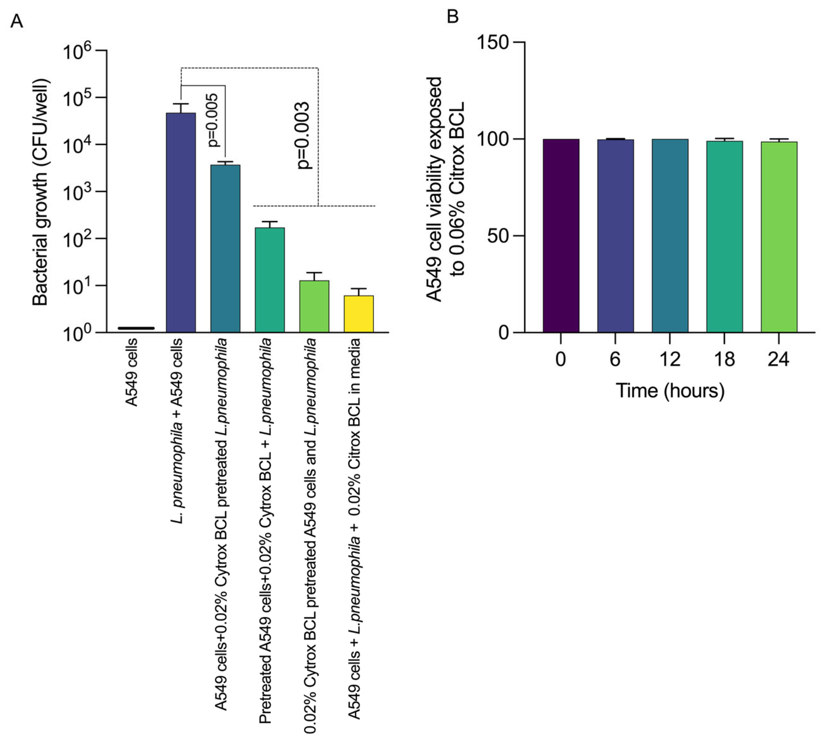
3.2. Citrox BCL Reduces L. pneumophila Infection of A549 Cells

Initially, we aimed to verify if the sub-inhibitory concentration of 0.02% Citrox BCL can inhibit the invasion of A549 cells by L. pneumophila. After 24 h of infection, we observed (Figure 2, panel A) that Citrox BCL was able to significantly reduce (p = 0.005) the intracellular presence of L. pneumophila when the A549 cells or the bacteria were pre-treated with 0.02% Citrox BCL. A similar significant result was observed when Citrox BCL was present in the media during the 24 h infection assay leading to a reduction in the intracellular presence (p = 0.003). To determine the cytotoxic effect of Citrox BCL in A549 cells, we measured LDH levels in the supernatants (Figure 2, panel B), indicating no cytotoxic effect. Taken together, these results show that Citrox BCL can reduce the invasion of L. pneumophila, an effect believed to be achieved by having a dual effect on the A549 cells and the bacterium itself.

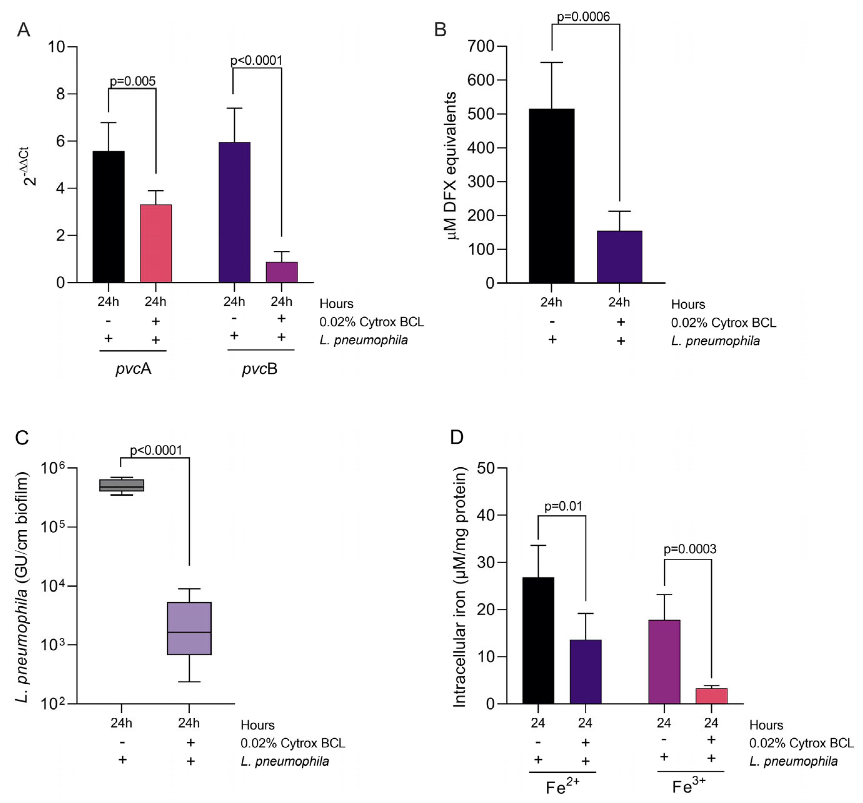
3.3. Citrox BCL Mediates Biofilm Formation and tetB and tetC Biofilm Related Gene Expression during Infection

We have next investigated the biological mechanisms involved by Citrox BCL to reduce the infection of A549 cells by L. pneumophila. Our hypothesis, suggesting that the levels of cyclic-imeric diguanylate (c-di-GMP) messenger in L. pneumophila are lowered in the presence of Citrox BCL, was confirmed (Figure 3, panel A) and restored in its absence at significant levels (p = 0.03). We have also measured the c-di-GMP levels in a co-culture scenario with A549 cells with similar results (Figure 3, panel A), suggesting that Citrox BCL directly acts upon L. pneumophila without A549 cell mediation (p = 0.04). Moreover, we show that the decreased levels of c-di-GMP levels were also accompanied by significantly lower (p < 0.0001) levels of biofilm formed after 24 h in the presence of 0.02% Citrox BCL (Figure 3, panel B). The high levels of bacterial c-di-GMP in the absence of Citrox-BCL were also associated with increased levels of ROS in infected A549 cells (Figure 3, panel C). We have also assumed that the above-observed reduction in virulence and biofilm formation is also accompanied by decreased expression of two of the major virulence genes, tatB and tatC. Our results show that the expression of both these genes was significantly reduced (p < 0.0001) in a time-dependent manner from 3–24 h (Figure 3, panels D and E).

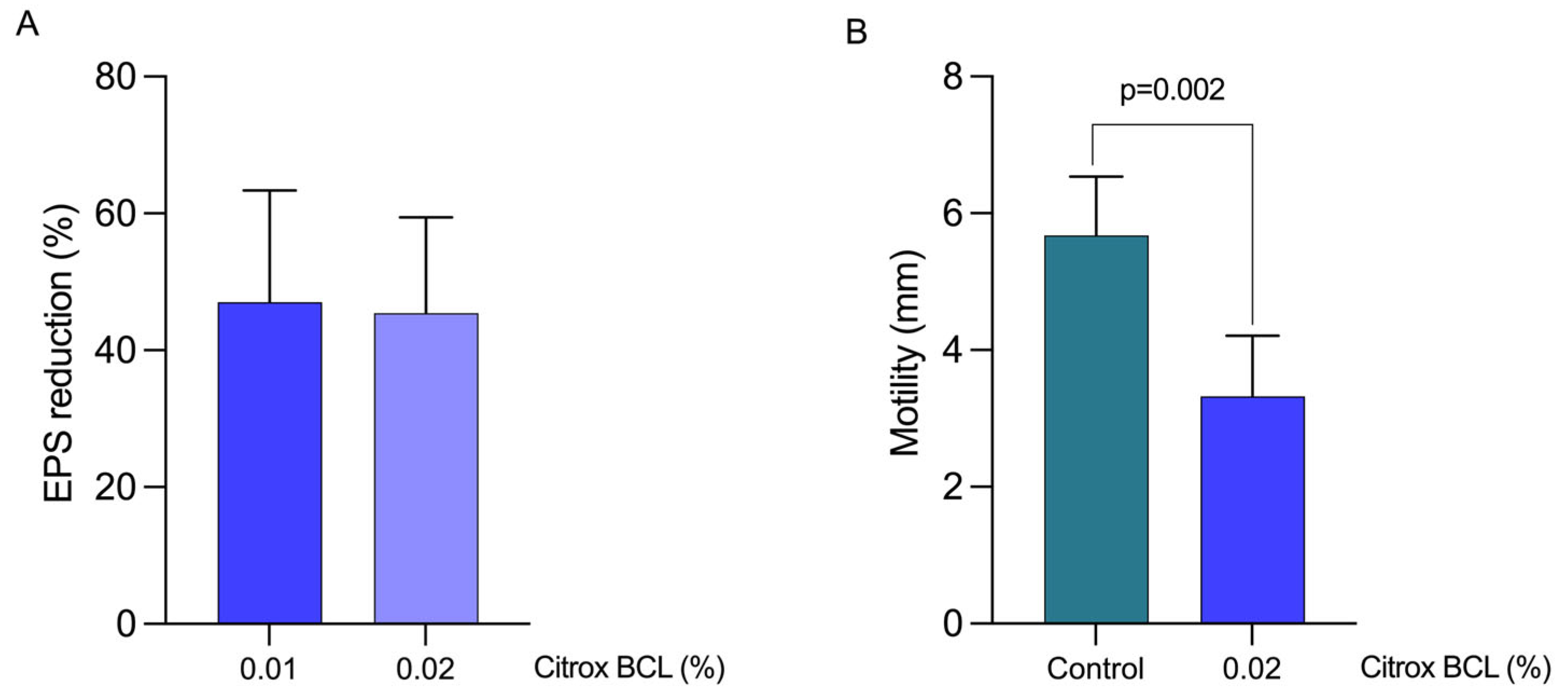
3.4. Impact on EPS Production and Bacterial Motility

The observed reduction in biofilm formation suggests that c-di-GMP production might regulate the EPS, hence inhibiting L. pneumophila biofilm. Our results show that EPS concentration decreased significantly during the growth of L. pneumophila in the presence of 0.02% Citrox BCL (Figure 4, panel A). We also show that the sub-inhibitory concentration of 0.02% induced a ~45% reduction in EPS production compared to the control and untreated L. pneumophila culture. The concentration of 0.01% was also used in this case to identify if further reductions are achieved; however, our data shows that below 0.02%, a plateau reduction is achieved. Further, we show that a significant reduction in motility was achieved by using the sub-inhibitory concentration of 0.02% (p = 0.002) (Figure 4, panel B). Taken together, these data suggest that the previously observed reduction in biofilm formation is also associated with reductions in two of the factors responsible for biofilm formation, EPS and motility, following exposure to 0.02% Citrox BCL.

3.5. Impact on Iron Sequestration and Siderophore Production

To further investigate the mechanisms involved in biofilm reduction, we have investigated the impact of Citrox BCL on iron sequestration genes pvcA and pvcB and siderophore activity. Our results show that the expression of pvcA and pvcB was significantly reduced (p = 0.005 and p < 0.0001) after 24 h in the presence of 0.02% Citrox BCL (Figure 5, panel A). The observed decrease in gene expression was also associated with a significant decrease in siderophore detection, in the culture supernatant, in the presence of 0.02% Citrox CBL (p = 0.0006) (Figure 5, panel B). The reduction in siderophore release was probably due to lower numbers of L. pneumophila cells (p < 0.0001) (Figure 5, panel C). This has caused decreased levels of Fe2+ and Iron 3+ detected in the formed biofilms in the presence of 0.02% Citrox BCL (Figure 5, panel D). These results suggest that Citrox CBL is involved in biofilm reduction by decreasing the ability of L. pheumophila to sequester iron required for growth and survival.

4. Discussion

Natural antimicrobials, such as essential oils, have been previously indicated as effective against L. pneumophila with great potential to be used as antimicrobials against this pathogen [33]. This study indicates that the spread of legionellosis in homes and resorts was successfully controlled with Cinnamomum osmophloeum leaf oil (cinnamaldehyde as a major constituent), having great potential to be used as an antibacterial agent in such circumstances. Their antimicrobial efficiency is improved when used as vapours in water, as it has been shown that citrus essential oil increased with 61% the presence of linalool. Therefore, establishing the proper delivery methods may dictate the level of efficiency [34].
Our study correlates the reduction in c-di-GMP production with the decrease in biofilm formation and significantly reduced EPS levels. Mixtures of organic acids have been previously shown to reduce EPS and biofilm production in E. coli O157:H7 [25]. EPS production is regulated by c-di-GMP, specifically of those involved in biofilm formation [35]. The down-regulation of the genetic factors, tatB and tatC, observed in our study is linked to reduced EPS levels detected, as they are part of the putative twin-arginine translocation pathway required for transmembrane protein translocation [36]. There is a clear connection between c-di-GMP and ROS tolerance [37]. Our data indeed shows that the ROS levels are reduced in infected and treated A549 cells, results which correlate with lower levels of c-di-GMP and lower infection rates. A link between iron regulation and c-di-GMP has been suggested as important in mediating biofilm formation in P. aeruginosa [38]. Biofilm formation in L. pneumophila is tightly controlled by iron as it is considered an essential nutrient during growth and replication [39] and mediates bacterial biofilm formation [31]. In L. pneumophila, iron concentration is controlled by the pvcA and pvcB genes which are involved in the production of siderophores and thus contribute to the uptake and sequestration of iron [8]. In our study, Citrox CBL was able to reduce siderophore levels and the expression of pvcA and pvcB genes, suggesting its involvement in bacterial iron starvation. Moreover, we show that the reduction in siderophore secretion, in the presence of Citrox CBL, is correlated with a decline in intracellular iron in biofilm-forming L. pneumophila cells. This observation clearly highlights and justifies the decrease in L. pneumophila virulence in A549 cells since iron bioavailability plays a fundamental role and regulates host-pathogen interactions [8].
Our results are in line with the current knowledge regarding biofilm regulation mechanisms in bacteria and novel regarding antimicrobial interference. For example, biofilm regulation factors are attributed to bacterial second messenger cyclic-dimeric diguanylate (c-di-GMP), where GGDEF/EAL-containing proteins were correlated with positive regulation of biofilm establishment by L. pneumophila [7]. For instance, L. pneumophila harbouring the absence of these proteins demonstrated a decreased biofilm formation, regardless of whether the expression of c-di-GMP was not different compared to the wild-type pathogen. Furthermore, the deletion of GGDEF/EAL proteins has been previously associated with the overproduction of biofilm, followed by a decrease in the expression of c-di-GMP [7]. Haem nitric oxide/Oxygen (H-NOX) is an additional binding domain of heme protein families involved in the c-di-GMP activity and biofilm regulation [7]. Compared to other prokaryotes, L. pneumophila is a unique prokaryote found to encode 2 H-NOX proteins in its genomes. The deletion of H-NOX-1 led to hyper biofilm formation without affecting the growth of the pathogen in murine macrophages, Acanthamoeba castellanii or proficient media [7]. Another cluster of genes orchestrated by ahpC2 and ahpD encrypts alkyl hydroperoxide reductases associated with playing an influential role in the defence against oxidative stress in the formed biofilm cells [7]. Similarly, FliA responds to L. pneumophila biofilm formation, allowing the pathogen to thrive in unfavourable conditions [40]. A recent in vitro study showed that the sessile form of L. pneumophila displayed a phenotypic heterogeneity and was able to form non-growing and growing bacterial populations [41]. Specifically, the non-growing sessile cells showed high metabolic activity and could lead to a dormant phase by promoting additional long-term survivability in the environmental niche [41]. Moreover, the authors indicated that LvbR is a pleiotropic regulator corresponding to shape the biofilm architecture, pathogen-host cell relations and competence for DNA uptake.

5. Conclusions

Our study shows that Citrox BCL inhibits a succession of biological events in L. pneumophila metabolism, which ultimately impact the bacterium’s ability to form biofilm and express virulence. First, it downregulates the pvcA and B gene expression leading to iron starvation in biofilms by lowering the production of siderophore molecules (Figure 6:1–6). Secondly, it causes a significant reduction in the production of c-di-GMP molecules (Figure 6:3) via the downregulation of the tatB and C genes which ultimately impact bacterial motility (Figure 6:4) and EPS production (Figure 6:5). All these events lead to reduced capacity to lower numbers of bacteria in biofilms and, therefore, reduced biofilm presence. Moreover, our study shows that the impact on all the above virulence factors of natural antimicrobials mixtures, such as Citrox BCL at sub-inhibitory concentrations (0.02%), will reduce host oxidative stress (Figure 6:7) and ultimately inhibit bacterial invasion of epithelial cells (Figure 6:8).

Author Contributions

All authors contributed equally. Conceptualization and data curation, N.C., E.B., I.B., N.-M.H., A.M., L.S., I.P., C.A.P. and T.I.; Funding acquisition, N.C.; writing—original draft, N.C., E.B., I.B. and L.S.; writing—review and editing, N.C., E.B., I.B., L.S., I.P., D.M., C.A.P. and T.I. All authors contributed equally. All authors have read and agreed to the published version of the manuscript.

Funding

We acknowledge Environtech, Dublin, Ireland, for funding the Ph.D. of Eugenia Butucel. Grant number 49650.

Institutional Review Board Statement

Not applicable.

Informed Consent Statement

Not applicable.

Data Availability Statement

Not applicable.

Conflicts of Interest

The authors declare no conflict of interest.

References

  1. Mraz, A.L.; Weir, M.H. Knowledge to Predict Pathogens: Legionella pneumophila Lifecycle Critical Review Part I Uptake into Host Cells. Water 2018, 10, 132. [Google Scholar] [CrossRef] [Green Version]
  2. Jomehzadeh, N.; Moosavian, M.; Saki, M.; Rashno, M. Legionella and legionnaires’ disease: An overview. J. Acute Dis. 2019, 8, 221. [Google Scholar]
  3. Boamah, D.K.; Zhou, G.; Ensminger, A.W.; O’Connor, T.J. From Many Hosts, One Accidental Pathogen: The Diverse Protozoan Hosts of Legionella. Front. Cell. Infect. Microbiol. 2017, 7, 477. [Google Scholar] [CrossRef] [PubMed] [Green Version]
  4. Palusińska-Szysz, M.; Jurak, M.; Gisch, N.; Waldow, F.; Zehethofer, N.; Nehls, C.; Schwudke, D.; Koper, P.; Mazur, A. The human LL-37 peptide exerts antimicrobial activity against Legionella micdadei interacting with membrane phospholipids. Biochim. Et Biophys. Acta (BBA)—Mol. Cell Biol. Lipids 2022, 1867, 159138. [Google Scholar] [CrossRef] [PubMed]
  5. Crépin, A.; Jégou, J.-F.; André, S.; Ecale, F.; Croitoru, A.; Cantereau, A.; Berjeaud, J.-M.; Ladram, A.; Verdon, J. In vitro and intracellular activities of frog skin temporins against Legionella pneumophila and its eukaryotic hosts. Sci. Rep. 2020, 10, 3978. [Google Scholar] [CrossRef] [Green Version]
  6. Falkinham, J.O. Living with Legionella and Other Waterborne Pathogens. Microorganisms 2020, 8, 2026. [Google Scholar] [CrossRef]
  7. Khweek, A.; Amer, A. Biofilm, a Cozy Structure for Legionella pneumophila Growth and Persistence in the Environment. In Bacterial Biofilms; IntechOpen: London, UK, 2019. [Google Scholar] [CrossRef] [Green Version]
  8. Abu Khweek, A.; Amer, A.O. Factors Mediating Environmental Biofilm Formation by Legionella pneumophila. Front. Cell. Infect. Microbiol. 2018, 8, 38. [Google Scholar] [CrossRef]
  9. Chauhan, D.; Shames, S.R. Pathogenicity and Virulence of Legionella: Intracellular replication and host response. Virulence 2021, 12, 1122–1144. [Google Scholar] [CrossRef]
  10. Hooper, S.J.; Lewis, M.A.O.; Wilson, M.J.; Williams, D.W. Antimicrobial activity of Citrox® bioflavonoid preparations against oral microorganisms. Br. Dent. J. 2011, 210, E22-E22. [Google Scholar] [CrossRef] [Green Version]
  11. Yehia, H.M.; Al-Masoud, A.H.; Elkhadragy, M.F.; Korany, S.M.; Nada, H.M.S.; Albaridi, N.A.; Alzahrani, A.A.; Al-Dagal, M.M. Improving the Quality and Safety of Fresh Camel Meat Contaminated with Campylobacter jejuni Using Citrox, Chitosan, and Vacuum Packaging to Extend Shelf Life. Animals 2021, 11, 1152. [Google Scholar] [CrossRef]
  12. Balta, I.; Linton, M.; Pinkerton, L.; Kelly, C.; Stef, L.; Pet, I.; Stef, D.; Criste, A.; Gundogdu, O.; Corcionivoschi, N. The effect of natural antimicrobials against Campylobacter spp. and its similarities to Salmonella spp, Listeria spp., Escherichia coli, Vibrio spp., Clostridium spp. and Staphylococcus spp. Food Control 2021, 121, 107745. [Google Scholar] [CrossRef]
  13. Balta, I.; Linton, M.; Pinkerton, L.; Kelly, C.; Ward, P.; Stef, L.; Pet, I.; Horablaga, A.; Gundogdu, O.; Corcionivoschi, N. The effect of natural antimicrobials on the Campylobacter coli T6SS+/− during in vitro infection assays and on their ability to adhere to chicken skin and carcasses. Int. J. Food Microbiol. 2021, 338, 108998. [Google Scholar] [CrossRef]
  14. Pinkerton, L.; Linton, M.; Kelly, C.; Ward, P.; Gradisteanu Pircalabioru, G.; Pet, I.; Stef, L.; Sima, F.; Adamov, T.; Gundogdu, O.; et al. Attenuation of Vibrio parahaemolyticus Virulence Factors by a Mixture of Natural Antimicrobials. Microorganisms 2019, 7, 679. [Google Scholar] [CrossRef] [Green Version]
  15. Ceylan, O.; Turasay, B. Removing Legionella pneumophila and biofilms from water supply systems using plant essential oils. J. Water Sanit. Hyg. Dev. 2017, 7, 67–73. [Google Scholar] [CrossRef] [Green Version]
  16. Gelmini, F.; Testa, C.; Angioletti, S.; Beretta, G. Essential Oils in the Environmental Bacteria Control: The Legionella Pneumophila case. J. Complement. Med. Altern. Healthc. 2017, 2. [Google Scholar] [CrossRef]
  17. Bićanić, L.; Mežnarić, S.; Gobin, I. Antimicrobial activity of the volatile phase of essential oils and their constituents on. Sanit. Inženirstvo Int. J. Sanit. Eng. Res. 2020, 14, 54–61. [Google Scholar] [CrossRef]
  18. Balta, I.; Marcu, A.; Linton, M.; Kelly, C.; Stef, L.; Pet, I.; Ward, P.; Pircalabioru, G.G.; Chifiriuc, C.; Gundogdu, O.; et al. The in vitro and in vivo anti-virulent effect of organic acid mixtures against Eimeria tenella and Eimeria bovis. Sci. Rep. 2021, 11, 16202. [Google Scholar] [CrossRef]
  19. Sima, F.; Stratakos, A.C.; Ward, P.; Linton, M.; Kelly, C.; Pinkerton, L.; Stef, L.; Gundogdu, O.; Lazar, V.; Corcionivoschi, N. A Novel Natural Antimicrobial Can Reduce the in vitro and in vivo Pathogenicity of T6SS Positive Campylobacter jejuni and Campylobacter coli Chicken Isolates. Front. Microbiol. 2018, 9, 2139. [Google Scholar] [CrossRef] [Green Version]
  20. Balta, I.; Stef, L.; Butucel, E.; Gradisteanu Pircalabioru, G.; Venig, A.; Ward, P.; Deshaies, M.; Pet, I.; Stef, D.; Koyun, O.Y.; et al. The Antioxidant Effect of Natural Antimicrobials in Shrimp Primary Intestinal Cells Infected with Nematopsis messor. Antioxidants (Basel) 2022, 11, 974. [Google Scholar] [CrossRef]
  21. Liu, C.; Sun, D.; Liu, J.; Chen, Y.; Zhou, X.; Ru, Y.; Zhu, J.; Liu, W. cAMP and c-di-GMP synergistically support biofilm maintenance through the direct interaction of their effectors. Nat. Commun. 2022, 13, 1493. [Google Scholar] [CrossRef]
  22. De Buck, E.; Lebeau, I.; Maes, L.; Geukens, N.; Meyen, E.; Van Mellaert, L.; Anné, J.; Lammertyn, E. A putative twin-arginine translocation pathway in Legionella pneumophila. Biochem. Biophys. Res. Commun. 2004, 317, 654–661. [Google Scholar] [CrossRef] [PubMed]
  23. Mou, Q.; Leung, P.H.M. Differential expression of virulence genes in Legionella pneumophila growing in Acanthamoeba and human monocytes. Virulence 2018, 9, 185–196. [Google Scholar] [CrossRef] [PubMed] [Green Version]
  24. Portier, E.; Bertaux, J.; Labanowski, J.; Hechard, Y. Iron Availability Modulates the Persistence of Legionella pneumophila in Complex Biofilms. Microbes Environ. 2016, 31, 387–394. [Google Scholar] [CrossRef] [PubMed] [Green Version]
  25. Stratakos, A.C.; Linton, M.; Ward, P.; Campbell, M.; Kelly, C.; Pinkerton, L. The antimicrobial effect of a commercial mixture of natural antimicrobials against Escherichia coli O157:H7. Foodborne Pathog. Dis. 2019, 16. [Google Scholar] [CrossRef] [Green Version]
  26. Hoppe, J.; Ünal, C.M.; Thiem, S.; Grimpe, L.; Goldmann, T.; Gaßler, N.; Richter, M.; Shevchuk, O.; Steinert, M. PilY1 Promotes Legionella pneumophila Infection of Human Lung Tissue Explants and Contributes to Bacterial Adhesion, Host Cell Invasion, and Twitching Motility. Front. Cell. Infect. Microbiol. 2017, 7, 63. [Google Scholar] [CrossRef] [Green Version]
  27. Sun, G.; Xu, X.; Wang, Y.; Shen, X.; Chen, Z.; Yang, J. Mycoplasma pneumoniae infection induces reactive oxygen species and DNA damage in A549 human lung carcinoma cells. Infect. Immun. 2008, 76, 4405–4413. [Google Scholar] [CrossRef] [Green Version]
  28. Starkenburg, S.R.; Casey, J.M.; Cianciotto, N.P. Siderophore activity among members of the Legionella genus. Curr. Microbiol. 2004, 49, 203–207. [Google Scholar] [CrossRef]
  29. Payne, S.M. Detection, isolation, and characterization of siderophores. Methods Enzymol. 1994, 235, 329–344. [Google Scholar] [CrossRef]
  30. Liles, M.R.; Scheel, T.A.; Cianciotto, N.P. Discovery of a nonclassical siderophore, legiobactin, produced by strains of Legionella pneumophila. J. Bacteriol. 2000, 182, 749–757. [Google Scholar] [CrossRef] [Green Version]
  31. Oh, E.; Andrews, K.J.; Jeon, B. Enhanced Biofilm Formation by Ferrous and Ferric Iron Through Oxidative Stress in Campylobacter jejuni. Front. Microbiol. 2018, 9, 1204. [Google Scholar] [CrossRef]
  32. Riemer, J.; Hoepken, H.H.; Czerwinska, H.; Robinson, S.R.; Dringen, R. Colorimetric ferrozine-based assay for the quantitation of iron in cultured cells. Anal. Biochem. 2004, 331, 370–375. [Google Scholar] [CrossRef]
  33. Chang, C.W.; Chang, W.L.; Chang, S.T.; Cheng, S.S. Antibacterial activities of plant essential oils against Legionella pneumophila. Water Res. 2008, 42, 278–286. [Google Scholar] [CrossRef]
  34. Laird, K.; Kurzbach, E.; Score, J.; Tejpal, J.; Chi Tangyie, G.; Phillips, C. Reduction of Legionella spp. in water and in soil by a citrus plant extract vapor. Appl. Environ. Microbiol. 2014, 80, 6031–6036. [Google Scholar] [CrossRef] [Green Version]
  35. Tamayo, R.; Pratt, J.T.; Camilli, A. Roles of cyclic diguanylate in the regulation of bacterial pathogenesis. Annu. Rev. Microbiol. 2007, 61, 131–148. [Google Scholar] [CrossRef] [Green Version]
  36. De Buck, E.; Maes, L.; Meyen, E.; Van Mellaert, L.; Geukens, N.; Anne, J.; Lammertyn, E. Legionella pneumophila Philadelphia-1 tatB and tatC affect intracellular replication and biofilm formation. Biochem. Biophys. Res. Commun. 2005, 331, 1413–1420. [Google Scholar] [CrossRef]
  37. Fernandez, N.L.; Waters, C.M. Cyclic di-GMP Increases Catalase Production and Hydrogen Peroxide Tolerance in Vibrio cholerae. Appl. Environ. Microbiol. 2019, 85, e01043-19. [Google Scholar] [CrossRef] [Green Version]
  38. Oglesby-Sherrouse, A.G.; Djapgne, L.; Nguyen, A.T.; Vasil, A.I.; Vasil, M.L. The complex interplay of iron, biofilm formation, and mucoidy affecting antimicrobial resistance of Pseudomonas aeruginosa. Pathog. Dis. 2014, 70, 307–320. [Google Scholar] [CrossRef] [Green Version]
  39. Mraz, A.L.; Weir, M.H. Knowledge to Predict Pathogens: Legionella pneumophila Lifecycle Systematic Review Part II Growth within and Egress from a Host Cell. Microorganisms 2022, 10, 141. [Google Scholar] [CrossRef]
  40. Appelt, S.; Heuner, K. The Flagellar Regulon of Legionella—A Review. Front. Cell. Infect. Microbiol. 2017, 7, 454. [Google Scholar] [CrossRef]
  41. Personnic, N.; Striednig, B.; Hilbi, H. Quorum sensing controls persistence, resuscitation, and virulence of Legionella subpopulations in biofilms. ISME J. 2021, 15, 196–210. [Google Scholar] [CrossRef]
Figure 1. The impact of Citrox CBL on growth and survival of L. pneumophila at concentrations of 0.02–0.06%. The MIC concentrations tested are indicated on the graphs in order to establish the sub-inhibitory concentration for further investigations. The experiments were performed in triplicate and on three separate occasions. In order to quantify the growth, the absorbance was measured at 600 nm every 3 h for 36 h.
Antioxidants 11 02186 g001
Figure 2. In vitro effect of 0.02% Citrox BCL on the internalisation of L. pneumophila in A549 cells Panel (A) and cytotoxicity Panel (B). Results are presented as CFU/well of three biological replicates, and the p values are indicated on graphs.
Antioxidants 11 02186 g002
Figure 3. Intracellular concentration of c-di-GMP levels, biofilm formation and virulence gene expression. Panel (A)—c-di-GMP levels; Panel (B)—biofilm biomass; Panel (C)—ROS levels in infected and un-treated A549 cells; Panels (D,E)—transcriptional analysis of tatB and tatC. Data are presented as the mean ± SD. Student’s t-test was used to analyse the statistical significance as indicated on the graphs. All experiments were performed in triplicate and on three separate occasions.
Antioxidants 11 02186 g003
Figure 4. Determination of EPS inhibition of L. pneumophila after exposure to 0.01–0.02% Citrox BCL for EPS (panel (A)) and 0.02% Citrox BCL for motility (panel (B)). Each point represents the mean ± standard deviation. The EPS results are expressed as percentage of the control.
Antioxidants 11 02186 g004
Figure 5. Expression of pvcA and pvcB genes (Panel (A)), CAS reactivity in supernatants (Panel (B)), number of L. pneumophila cells in formed biofilm (Panel (C)) and the levels of iron in formed biofilms (Panel (D)). The results are presented as means and standard deviations from triplicate samples and from three separate experiments. The supernatant’s CAS reactivity was tested in triplicates from three separate experiments (p < 0.05; Student’s t-test).
Antioxidants 11 02186 g005
Figure 6. Summary of conclusions. (A) In L. pneumophila and (B) in A549 cells. Citrox BCL downregulates the pvcA and B gene expression leading to iron starvation in biofilms by lowering the production of siderophore molecules (1,2,3–6). Causes significant reduction in the production of c-di-GMP molecules (3) and downregulates tatB and C genes expression, reducing bacterial motility (4) and EPS production (5). Citrox BCL at sub-inhibitory concentrations (0.02%) reduces host oxidative stress (7) and inhibits bacterial invasion of A549 epithelial cells (8). Created with BioRender.com.
Antioxidants 11 02186 g006
Publisher’s Note: MDPI stays neutral with regard to jurisdictional claims in published maps and institutional affiliations.

