Review

Endothelium as a Source and Target of H2S to Improve Its Trophism and Function

Department of Life Sciences, University of Siena, Via A. Moro 2, 53100 Siena, Italy
*
Author to whom correspondence should be addressed.
Antioxidants 2021, 10(3), 486; https://doi.org/10.3390/antiox10030486
Submission received: 16 February 2021 / Revised: 13 March 2021 / Accepted: 16 March 2021 / Published: 19 March 2021

Abstract

:
The vascular endothelium consists of a single layer of squamous endothelial cells (ECs) lining the inner surface of blood vessels. Nowadays, it is no longer considered as a simple barrier between the blood and vessel wall, but a central hub to control blood flow homeostasis and fulfill tissue metabolic demands by furnishing oxygen and nutrients. The endothelium regulates the proper functioning of vessels and microcirculation, in terms of tone control, blood fluidity, and fine tuning of inflammatory and redox reactions within the vessel wall and in surrounding tissues. This multiplicity of effects is due to the ability of ECs to produce, process, and release key modulators. Among these, gasotransmitters such as nitric oxide (NO) and hydrogen sulfide (H2S) are very active molecules constitutively produced by endotheliocytes for the maintenance and control of vascular physiological functions, while their impairment is responsible for endothelial dysfunction and cardiovascular disorders such as hypertension, atherosclerosis, and impaired wound healing and vascularization due to diabetes, infections, and ischemia. Upregulation of H2S producing enzymes and administration of H2S donors can be considered as innovative therapeutic approaches to improve EC biology and function, to revert endothelial dysfunction or to prevent cardiovascular disease progression. This review will focus on the beneficial autocrine/paracrine properties of H2S on ECs and the state of the art on H2S potentiating drugs and tools.

1. Vascular Endothelium

The vascular endothelium is the tissue that lines the inside of the circulatory system (blood vessels, lymphatic vessels and heart). The cells, arranged in a single layer oriented on the longitudinal axis of the vessel, assume a flattened shape and lay side by side with each other to form a complete monolayer. Structurally, the endothelial cell (EC) apical domain is in direct contact with blood or lymph, while the basolateral domain anchors to the basal lamina, which connects EC to the underlying tissues, such as the medial or muscular layer and the adventitia, rich in fibrous tissue.
The vascular endothelium acts as a selectively permeable barrier between extravascular and intravascular compartments and provides a non-thrombogenic surface for the cardiovascular system [1]. Nowadays, the endothelium can no longer be considered a passive barrier. Indeed, its anatomical position allows it to integrate the physical and neurohumoral signals from the blood and surrounding tissues for regulating vascular tone and permeability, cell adhesion, inflammation, smooth muscle phenotype and proliferation, as well as thromboresistance and blood fluidity [2,3].
The endothelial lining represents a wide area for the exchanges between blood and tissues (about 350 m2 in humans) [3]. Electron microscopy observations reveal the continuous nature of arterial endothelium, characterized by tight junctions among adjacent cells in order to limit macromolecule exchange, and by a complex micro-vesicular system involved in macromolecular transport. In spite of its apparent morphological lack of complexity, the endothelium is characterized by heterogeneity, with differences in permeability, reactivity, and biosynthesis in relation to the type of vascular district and organ considered [1,2,3].

2. Role of Endothelium in Physiology

The endothelium’s role as a semipermeable barrier is one of its fundamental and basic functions: it regulates macromolecule transport between the lumen and vascular smooth muscle tissue [4]. Several mechanisms control the passage of macromolecules across the endothelial barrier: (i) through ECs themselves (transcellular flux); (ii) through the cell–cell junctions (paracellular flux); and (iii) via vesicular transport.
Most biological transmitters consist of large molecules with anionic and hydrophilic features, unable to diffuse across the membrane bilayer. The majority of those transmitters are believed to move through intracellular junctions between cells, or via vesicular transport, thanks to the formation of transient channels resulting from vesicle fusion. The reorganization of the intercellular junctions, which involves actin and myosin reconfiguration or direct collapse of junctional connections, appears to be the main process by which ECs increase their permeability to solutes and water [1].
The function of the endothelium is not limited to the internal surface lining of vessels or to constitute the vascular wall in the microcirculation, but it produces and releases vasoactive factors such as nitric oxide (NO), prostacyclin (PGI2), hydrogen sulfide (H2S) and endothelin (ET) which, in the appropriate concentration and balance, maintain adequate vascular tone and blood fluidity, giving the endothelium itself an antithrombotic phenotype [5,6].
The synthesis of NO by ECs is constitutive, but it can be augmented by a wide variety of compounds, including acetylcholine, angiotensin II (AngII), bradykinin, histamine, arachidonic acid. NO has a very short half-life and is synthesized from l-arginine and oxygen by the NO synthase enzyme (NOS). The endothelial isoform of this enzyme, eNOS, constitutively expressed, appears to be Ca2+/calmodulin-dependent. Once synthesized, NO rapidly spreads to vascular smooth muscle cells where it stimulates soluble guanylate cyclase (sGC), with an increase in cGMP formation and consequent vascular relaxation, while its autocrine function is related to the control of EC trophism and angiogenesis [7].
NO is not the only endothelial-dependent vasodilator. The endothelium also constitutively generates PGI2, which relaxes the underlying smooth muscle cells by activating adenylate cyclase and increasing cAMP. PGI2 is released in high quantities following the binding of transmitters, such as thrombin, histamine, serotonin, on cell surface receptors. The endothelium also produces and releases a hyperpolarizing factor (EDHF) whose chemical nature is still debated. EDHF’s function is to hyperpolarize vascular smooth muscle cells, causing these cells to relax and allowing dilation of blood vessels [6].
In addition to NO and carbon monoxide (CO), H2S is an endogenous gasotransmitter involved in the regulation of the cardiovascular, nervous, gastrointestinal, and renal systems, with a great impact on inflammatory and immune responses [8]. Recently, it has been proposed as one of the EDHFs. H2S exerts a multitude of physiological effects on the wall of the vessels, acting in an autocrine/paracrine manner. It is produced by vascular cells and exhibits antioxidant, anti-apoptotic, anti-inflammatory and vasoactive properties. Indeed, it reduces arterial blood pressure, limits the formation of atheromatous plaques, and promotes the vascularization of ischemic/injured tissues [8].
In some pathophysiological circumstances including hypoxia, tissue hypoperfusion or arterial hypertension, some vasoconstrictor factors may be released from the endothelium as endothelins (ETs), thromboxane A2, and prostaglandin H2. Moreover, vascular endothelium participates at the regulation of vessel tone and trophism and blood flow through the processing of angiotensin, via the expression of angiotensin converting enzyme isoforms ACE-1 and ACE-2, responsible for the balance between AngII/AT-1 receptor and Ang(1–7)/Mas receptor [9].
ECs respond to the increase in blood flow through the release of NO and PGI2 by the same cells. Indeed, shear stress causes rapid activation of eNOS and increases its gene transcription; it also endorses ECs to release factors that inhibit coagulation, leukocyte migration, and smooth muscle cell proliferation, simultaneously promoting EC survival. Conversely, low shear stress and turbulent blood flow promote a pathological feature in the endothelium responsible for atherosclerosis ignition, documenting the pivotal role of ECs in finely controlling vascular functions [2,6].
A healthy functioning endothelium also provides protection against radical species of oxygen and nitrogen (ROS/RNS). It is now clear that increased levels of ROS and RNS are harmful to cells and tissues and are involved in a wide range of cardiovascular diseases having endothelial dysfunction as an underlying phenomenon. This boosted the concept of oxidation as synonymous with cell damage and senescence. The post-translational modifications involving RNS share a common ancestor—high NO concentrations mainly synthetized by inducible NOS (iNOS), upregulated in response to various endotoxin or cytokine signals. Several pathological states are linked to the deregulation of NO levels, indicating that aberrant production of NO and its products can have deleterious consequences on cells [10]. Again, one of the functions related to a healthy endothelium is the scavenging activity of reactive species through the production of antioxidant products as H2S or protective enzymatic pathways.
The intimate surface of a healthy endothelium is both anticoagulant and antithrombotic: ECs secrete a wide range of molecules relevant to the regulation of blood clotting and platelet functions, as PGI2 and NO. Damage to the vessels or exposure to certain cytokines and proinflammatory stimuli overturns the equilibrium towards a procoagulant and prothrombotic EC phenotype, through the exposure of basal membrane components and/or tissue factor, and reduced presence of glycosaminoglycans or tissue factor inhibitor [5].
Endothelial trophism is guaranteed by the response to vasoactive and growth factors produced by surrounding tissues or autocrinally by the same ECs. Among the various examples, we and others have contributed to characterizing the beneficial effects on vascular endothelium by NO derived from eNOS, bradykinin, substance P, vascular endothelial growth factor (VEGF), fibroblast growth factor-2 (FGF-2), prostaglandin E2, H2S [11,12,13,14,15]. The molecular mechanisms responsible for cell survival, proliferation, migration and functioning include eNOS/NO/cGMP/protein kinase G (PKG), PI-3K/Akt, MAPK/ERK1/2 and gene transcription of autocrine factors as FGF-2 [7].
Epigenetics is an emergent mechanism involved in the regulation of vascular biology and endothelial trophism. Through chromatin structure modification, epigenetics can modify endothelial functions with an impact on cardiovascular disease, being the regulatory functions of epigenetics also active on endothelial precursor cells and circulating angiogenic cells [16,17]. DNA methylation, variants, histone post-translational modifications, and recently discovered RNA-based mechanisms represent the major pathways involved in the molecular basis of epigenetics. VEGF-A and NOS are the key players in regulating and maintaining cardiovascular functions. Their expression can be controlled by epigenetic mechanisms. In particular, VEGF-A epigenetic control can occur mainly through changes in histone code by RNAs. VEGF-A acts through VEGFR2, which in turn is regulated by promoter DNA methylation [18]. Furthermore, accumulating evidence indicates that epigenetic pathways play an important role in eNOS gene regulation [19].
These findings suggest the importance to deeply understand the epigenetic mechanisms involved in the regulation of vascular functions both in physiology and in pathological conditions.

3. Endothelial Dysfunction

Endothelial dysfunction refers to a systemic condition in which the endothelium loses its physiological properties, including the control of vasodilation, fibrinolysis and platelet aggregation. Key features of the endothelial dysfunction are: (1) the reduced local production of NO due to impaired activity (uncoupling) of eNOS, and of other vasodilating mediators as H2S; (2) the decrease in anticoagulant factors such as heparin; (3) the increase in the secretion of reactive species, von Willebrand factor, and tissue factor; (4) the overexpression of adhesion molecules for leukocytes and platelets [20,21]. All these factors concur to compromise the physiological vascular homeostasis. Due to the impairment of the main protective transmitters NO and H2S, the resulting endothelial dysfunction is associated with increased ROS and RNS levels and vascular oxidative stress [22,23]. The perpetuation of this condition then leads to retraction and death of the endothelium with increased permeability and exposure of the components of the basement membrane, that further amplifies the picture of vascular inflammation [4,20,22].
Endothelial dysfunction risk factors are represented by pathological states such as hypertension, diabetes, and hyperlipidemia, and improper lifestyles such as high-fat diets, tobacco and alcohol consumption, and physical inactivity [24]. Most cardiovascular diseases share endothelial dysfunction as a hallmark: atherosclerosis, diabetes complications, thrombosis, and hypercoagulation [20]. Moreover, physiological ageing through the phenomenon of mild chronic inflammation (“inflammaging”) is accompanied by endothelial dysfunction [25]. Inflammatory factors such as tumor necrosis factor-α (TNF-α), inteleukin-6 (IL-6), intercellular adhesion molecule 1 (ICAM-1), and loss of the antioxidant mechanism are among the most influential promoters of vascular impairment [21].
Mounting evidence suggests that epigenetic mechanisms may contribute to vascular complications in many pathological conditions, such as diabetes or atherosclerosis, linked to altered endothelial trophism and functions [17,26]. The inflammatory phenotype in ECs induces the transcription of many cytokines and adhesion molecules, in a nuclear factor-kappa B (NF-κB)-dependent manner. Epigenetic modifications in the NF-κB promoter region produce an increased expression of p65 subunit of NF-κB, and a hyper-activation of the NF-κB pathway [27]. Another mechanism could involve histone deacetylase 2 (HDAC2), which interacts and deacetylates Nrf2. Oxidized low-density lipoproteins (ox-LDLs) are able to downregulate HDAC2 expression, resulting in increased production of eNOS-dependent reactive species [16]. Furthermore, shear stress represented by blood flow alteration modifies EC gene expression and function. Dunn et al. demonstrated that disturbed blood flow stimulates DNA methyltransferase-1 (DNMT-1) expression in endothelial cells with aberrant DNA methylation at the promoter of flow-inducible genes, contributing to atherosclerosis [28]. Finally, recent data showed the role of non-coding RNA in regulating the expression of endothelial adhesion molecule [29].
Acute or chronic infections both by bacteria and viruses have cardiovascular consequences for their direct or indirect effects on vascular endothelium, through bacterial products or cytokines released by tissue and immune system cells [30,31], and through epigenetic regulation [32]. The recent pandemic due to SARS-CoV-2 supports this concept [33,34].
Furthermore, our studies and those of others have revealed that endothelial dysfunction is associated to impaired EC survival and physiological angiogenic outcomes with subsequent rearrangement of the microcirculation that contributes to the emergence of various pathological conditions and healing disorders [7,35]. ECs play a key role in the adaptation of tissues to damage, revealing their plasticity. A change in endothelial functions following ischemia can induce the transition to a mesenchymal phenotype characterized by functional, metabolic and gene expression signatures. Indeed, the mesenchymal phenotype, with increased cell migration and clonal expansion, participates in regenerating a functioning vascular network [36].
Considering endothelial function as a “barometer for cardiovascular risk”, it is crucial to identify the molecular determinants underlying endothelial integrity and functionality. Seeing endothelium as an exchange regulator between the vascular wall and surrounding tissues, it is expected that dysfunctional ECs can determine damage to other tissues [37]. Indeed, a detailed assessment of the cellular and molecular mechanisms at the base of vascular function, and, particularly, of endothelial dysfunction, will help the diagnosis and treatment choice for a broad array of human disorders, including cardiovascular and neurodegenerative diseases [21,24,38,39,40].

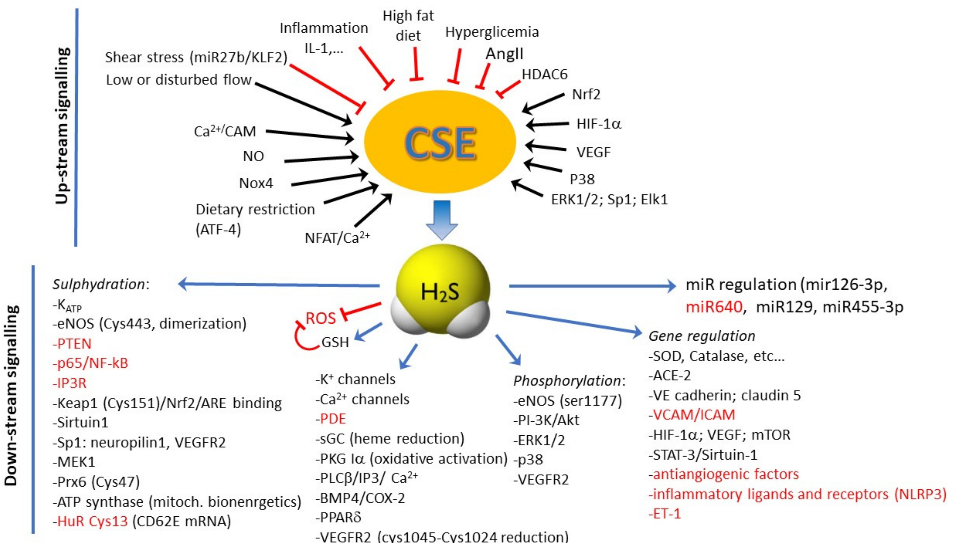
4. Biochemistry of H2S Production

H2S is a gas physiologically produced by tissue and vascular cells. The enzymes responsible for the synthesis of H2S are cystathionine β-synthase (CBS), cystathionine-γ-lyase (CSE), and 3-mercaptopyruvate sulfurtransferase (3-MST). The first two enzymes use l-cysteine as a substrate and are dependent on pyridoxal-5′-phosphate. 3-MST, on the other hand, works in association with cysteine aminotransferase which, starting from l-cysteine and α-ketoglutarate, produces 3-mercaptopyruvate. These enzymes are differentially expressed in the various tissues. In particular, the expression of CSE and 3-MST predominates in the cardiovascular system [8,41]. CSE is only present in the cytoplasm, while CBS and 3-MST both have a cytosolic and mitochondrial form, with the latter predominating. CSE is the principal enzyme responsible for H2S biosynthesis, located in vascular smooth muscle cells and found mainly in ECs [42,43]. This can explain why the concentration of H2S in the vascular tissues is around 100 times greater than in other tissues [44], suggesting a crucial role in vascular homeostasis, endothelial function, and trophism [45]. In addition, H2S can be also generated via a reduction in thiols and thiol-containing molecules, in a non-enzymatic manner [8]. Compared to CBS, knockout mice for CSE have no severe phenotype and normal lifespan. Their phenotype is mainly cardiovascular, with hypertension and endothelial dysfunction [46].
Information has become available about the regulation of the expression and activity of these enzymes (Figure 1). It has been reported that NADPHox4 derived ROS (via heme-regulated inhibitor kinase/eIF2/activating transcription factor 4 (ATF4) signaling) enhance the expression of CSE [47]. Blood flow has been reported to exert divergent effect of H2S producing enzymes, depending on the type of endothelium and flow stress. While laminar flow was initially demonstrated to enhance the expression of CSE and 3-MST in ECs [47,48], recent data are more complex. In particular, laminar flow (high shear stress) has been demonstrated to inhibit CSE expression via KLF2 regulated miRNA-27b [49], while turbulent flow (low shear stress) seems to upregulate CSE [50,51]. In rat aortic ECs, calcium-sensing receptors increase CSE expression in a phospho-calmodulin kinases II-dependent manner to inhibit platelet activation [52]. Transcription factors specifically controlling CSE expression are among the others NF-κB in lipopolysaccharide (LPS)-stimulated macrophages [53], specificity protein 1 (Sp1) in smooth muscle cells [54] and Elk1 in beta pancreatic cells [55]. Nuclear factor of activated T cell (NFAT) binding sites have been identified in the CSE promoter. Intermittent hypoxia exposure reduces Ca2+-dependent activation of calcineurin/NFAT to lower CSE expression and impair vasodilation, while NFAT activation lowers CSE expression at the cell and microvascular levels [56].
OxLDL in one of the major stimuli to cause the endothelial damage that leads to atherosclerosis. One of the epigenetic mechanisms underlying CSE reduced expression at both mRNA and protein levels by oxLDL is increased histone deacetylase 6 (HDAC6) [57]. Recently this finding has been extended to blood pressure control, by assessing the role of tubastain A in AngII induce hypertension, and documenting that upregulation of CSE and H2S through HDAC6 inhibition can be a valid therapeutic strategy [58].
Additional information on CSE gene and enzyme control is provided in the chapters below and is summarized in Figure 1.
H2S plasma levels are kept at appropriate concentrations by three elimination systems. The first pathway involves mitochondrial oxidative metabolism which converts H2S into thiosulfate, followed by further conversion into sulfate and then sulfite. The second metabolic pathway is cytosolic methylation to dimethyl sulfide via thiol S-methyltransferase. Finally, the binding of H2S to hemoglobin leads to the formation of sulfhemoglobin [45].

5. Molecular Signaling Activated by H2S into ECs

H2S in ECs performs a protective action on vessels in an autocrine/paracrine manner. It plays a role in the regulation of vasodilation, angiogenesis, inflammation, oxidative stress and apoptosis [59]. Three are the main mechanisms through which H2S exerts it biological effect: (i) reactive oxygen species/nitrogen species scavenging; (ii) interaction with metal centers; (iii) persulfidation (called also S-sulfhydration).
H2S acts through a post-translational modification—the S-sulfhydration of cysteine residues, which modifies the structure and activity of the target proteins [60]. The mechanism is persulfidation on reactive cysteine residues (-SH) of target proteins to form a persulfide group (-SSH). An example is the persulfidation of the ATP-dependent K channel (KATP) in ECs and smooth muscle cells, responsible for fast hyperpolarization and vasorelaxation [61]. Indeed, evidence has been provided in support of H2S function as an EDHF, exerting more remarkable vasorelaxation in the peripheral resistance arteries [62].
H2S also reduces ROS levels through their direct inactivation and by enhancing antioxidant defense mechanisms. One of the mechanisms underlying oxidative protection is given by the H2S regulation of the Keap1/Nrf2 pathway. Normally, the transcription factor Nrf2 is inhibited by its binding to Keap1 in the cytoplasm. In conditions of oxidative stress, H2S promotes the translocation of Nrf2 into the nucleus by means of the S-sulfhydration of the Keap1 inhibitor, causing the dissociation of the Keap1/Nrf2 complex. Nrf2 in the nucleus activates the antioxidant responsive element. Consequently, the transcription of many antioxidant genes such as superoxide dismutase, catalase, glutathione peroxidase and glutathione-S-transferase is induced, requiring hours or days to produce a biological effect [63].
Among other targets, H2S decreases inflammation by inhibiting transcription factors such as NF-κB through persulfidation [64], thus decreasing the expression of pro-inflammatory mediators. Most persulfidation reactions lead to target inhibition as phosphatase and tensin homolog (PTEN) [65], except for MEK1 activity, which in HUVEC leads to increased activity with DNA damage repair and senescence impairment [66]. Recently, Prx6 has been identified as a further target of sulfhydration on Cys47, which controls its decamerization and peroxidase activity [49], while the “S-sulphydrome” was identified among the many target proteins, and β3 integrin was identified as the key element of endothelial mechanotransduction [67].
The interaction of H2S with the NO/NOS pathway involves different modalities: as inhibition of PDE in smooth muscle cells, PI-3K/Akt-dependent phosphorylation of eNOS in Ser1177 [68] and stabilization of eNOS in the dimeric state through enzyme persulfidation [69]. Additionally, heme reduction in sGC enzyme with facilitated response to NO [70] and activation of protein kinase G Iα (PKG Iα) through disulfide bond formation [71] have been reported to potentiate the NO/cGMP pathway.
H2S also stimulates endothelial proliferation and migration, aiding the process of angiogenesis and wound repair. H2S acts at several levels on the mechanisms responsible for angiogenesis, including the control of VEGF expression, through upregulation of the transcription factor hypoxia inducible factor-1α (HIF-1α) or direct modulation of the PI-3K and Akt pathways in ECs (signaling pathways also activated by VEGF) [41,72,73]. In angiogenic ECs, H2S has also been reported to activate signal transducer and activator of transcription 3 (STAT3) [74], mammalian target of rapamycin (mTOR) and the VEGFR2 pathway [75]. It has been reported that CBS silencing in ECs reduces VEGF signaling through VEGFR2 and neuropilin-1 downregulation [76].
A schematic summary of the molecular mechanisms activated or inhibited by H2S in ECs is reported in Figure 1. As a note, the effects of H2S are context and tissue-dependent, sometimes producing divergent functional effects. It is plausible that this depends on endothelial heterogeneity, tissue microenvironment, and physio-pathological conditions where there is the influence of epigenetic mechanisms.

6. Cardiovascular Diseases Associated with Altered Levels of H2S

6.1. Hypertension

Altered levels of H2S have been reported in both experimental models and clinical studies on patients with severe hypertension, where lower plasma H2S levels are described along with reduced content of CBS and CSE (see [77,78] for recent reviews). A human cohort study demonstrated a reduced H2S plasma level in hypertensive patients, suggesting H2S as a potential therapeutic target and diagnostic marker [79].
The intimate relationship between H2S-associated endothelial dysfunction and hypertension comes from the observation that CSE KO mice develop hypertension particularly with impaired endothelium-dependent relaxation in resistance mesenteric arteries [46].
The levels of H2S-producing enzymes are reduced in the vessel wall of spontaneous or drug induced hypertensive animals [80,81,82]. Exposure of cultured endothelial cells to AngII and, similarly, to H2O2, downregulated the expression and activity of CSE with induction of endoplasmic reticulum stress [83]. In a mouse model of Ang II-induced hypertension, H2S reversed the aortic endothelial dysfunction and reduced NO bioavailability, while blockade of endogenous H2S exacerbated these alterations [84]. Other studies demonstrated that administration of H2S donors decreases blood pressure and reverses vascular remodeling through the suppression of smooth muscle cell proliferation and collagen deposition in the vessel wall [61,85,86,87,88,89]. H2S treatment noticeably reestablishes eNOS function and NO bioavailability in Nω-nitro-l-arginine methyl ester (L-NAME)-induced hypertensive rats [90]. From a mechanistic point of view, H2S improves endothelial function through the inhibition of oxidative stress, suppression of renin angiotensin system, downregulation of bone morphogenic protein 4/cyclooxygenase-2 (BMP4/COX-2) pathway, or activation of the PPARδ/PI-3K/Akt/AMPK/eNOS cascade, thus contributing to the antihypertensive mechanism of H2S in renovascular hypertensive rats [91,92,93]. In SHR, administration of H2S significantly decreases blood pressure and abrogates endothelial dysfunction through inactivation of NLRP3 inflammasome and oxidative stress [94]. In another disease model, lead-induced hypertension in rats, H2S treatment normalizes blood pressure and ameliorates endothelial dysfunction with an inhibition of oxidative stress [95].
Recently, it has been proposed that H2S can regulate EC pathological behavior through epigenetic mechanisms. H2S induces miR-129, which inhibits DNA methyltransferase-3 (DNMT3) and IL-17, found to be overexpressed in hypertension [96].
Summarizing all results, it can be concluded that the CSE/H2S signaling pathway may represent a potential therapeutic target for hypertension.

6.2. Diabetes

The relation between diabetes-induced endothelial dysfunction and H2S impairment is now well established (for review see [77,97]). H2S levels have been observed to be reduced in rats with diabetes induced by streptozotocin (STZ) and in subjects with type 2 diabetes mellitus [98,99,100,101,102]. In line with these findings, a high fat diet downregulates and dietary restriction induces (via ATF4) CSE expression [103,104]. Hyperglycemia lowers H2S levels due to the high H2S catabolism favored by the extremely oxidizing environment or the reduced gasotransmitter production due to a lower expression of the generating enzymes or their inhibition such as oxidative inactivation of the 3-MST at endothelial level [105]. The lack of H2S bioavailability supports the accumulation of intracellular ROS, which are not completely scavenged by H2S due to its consumption in high-glucose-treated ECs [106]. The consequent oxidative status favors mitochondrial dysfunction and mitophagy, cell damage, and apoptosis [107,108,109,110].
Endothelial dysfunction in diabetes correlates with angiogenesis impairment. CSE expression and H2S levels are strongly diminished in wound granulation tissues of obese diabetic mice [111], thus explaining the angiogenesis impairment described in wounds and critical limb ischemia in diabetes [112,113,114]. The availability of H2S donors or CSE upregulators could be an innovative therapeutic strategy to promote endothelial function and proper neovascularization of wounds. However, despite the protective effect of H2S on endothelial function and wellness, the stimulation of angiogenesis in atherosclerosis plaques by high CSE expression could have a negative outcome, resulting in plaque vulnerability and rupture [115]. The choice of the proper strategy and best control of H2S at tissue level is still a critical point to be resolved at the experimental and clinical level.

7. Molecular Mechanisms Regulated by H2S in Support of EC Function and Trophism

7.1. Antioxidant and Anti-Inflammatory Properties

Several studies document that H2S limits vascular permeability, directly or indirectly through antioxidant and anti-inflammatory actions. The multiplicity of the mechanism downstream H2S production is schematically reported in Figure 1. Vascular hyperpermeability was inhibited in mice undergoing cardiac arrest and blood–brain barrier disruption following H2S inhalation [116]. This protective effect was linked to reduced expression of VEGF and metalloproteinase-9 and increased angiopoietin-1. Another study documented scavenging of ROS and activation of Akt [117]. Data, however, are not all in the same direction, documenting that the final effect is context dependent.
The protective effect of H2S in conditions such as hypertension, atherosclerosis, and vascular diabetic complications may be related to multiple actions by the gasotransmitter: H2S inhibition of ROS production, blunting of ROS by direct scavenging, upregulation of glutathione, and antioxidant enzymes [101,118]. H2S reduces ROS levels in ECs exposed to high glucose, preventing their apoptosis and damage [108,119]. Gene transfer of CSE or administration of exogenous H2S in diabetes models reduced ROS levels and improved endothelial dependent vasorelaxation, while CSE KO was responsible for a greater impairment of endothelial function [106]. Many studies support the inhibitory effect of H2S on endothelial inflammation [120]. The autocrine/paracrine action of endothelial-derived H2S has been documented by endothelial specific deletion of CSE, which predisposes to vascular inflammation and atherosclerosis [50].
In ECs exposed to high glucose, a suppression of NF-κB activity and reduction in ICAM-1 levels were found upon NaHS pretreatment [121]. Moreover, stimulation of ECs with high glucose significantly promotes ET-1 secretion, which was reduced by administration of H2S [122].
Recently, inhibition of necroptosis together with ROS downregulation have been described in ECs exposed to hyperglycemia [123]. Inhibition of adhesion molecules such as ICAM-1 in ECs has been described in response to NaHS through NF-κB inhibition [124], while CSE inhibition increased leukocyte adherence to the endothelium [125]. The anti-inflammatory activity of H2S is not only related to the impairment of adhesion molecules as vascular cell adhesion molecule (VCAM) and ICAM, but also to the inhibition of inflammatory mediator production, such as IL1-β, TNF-α, IL-6 and monocyte chemoattractant protein-1 by ECs and monocytes/macrophages [126,127]. IL-1β, in turn, was found to be increased in atherosclerotic plaques, and induces the phosphorylation of Ser377 and inactivation of CSE [127].
In cultured ECs, the stability of eNOS regulated by miR-455-3p and NO production is induced by H2S. Moreover, H2S levels and miR-455-3p are incremented in human atherosclerosis plaques, implying that H2S could be involved in the miR-455-3p/eNOS/NO pathway controlling atherosclerosis development [128].
Sirtuin-1 has been reported to prevent premature senescence of ECs, protecting from dysfunction [129]. Exogenous H2S directly induces sirtuin-1 sulfhydration and stability, reducing aortic inflammation and formation of atherosclerosis plaques [130].
Further studies demonstrated that H2S reduces the severity of atherosclerosis in a mouse model of disturbed blood-flow, through the upregulation of ACE-2 and increase in Ang(1–7) levels [131]. At cellular level, in LPS-activated ECs, H2S promotes the upregulation of the beneficial side of the renin-angiotensin system [131], documenting a multitargeting effect of H2S.
An original mechanism of action of H2S in controlling endothelial dysfunction in atherosclerosis was recently proposed [50]. In both cultured ECs and in mice, endogenous CSE-derived H2S leads to sulfhydration and dimerization of the RNA-binding protein human antigen R (HuR), described to be inhibited in atherosclerosis [50]. The administration of SG1002, a slow polysulfide donor, in ECs isolated from CSE knockout mice, re-established HuR sulfhydration with subsequent inflammatory marker (CD62E) downregulation. Moreover, SG1002, administered to ApoE−/− CSE knockout mice exposed to partial carotid ligation, limited plaque formation, demonstrating an H2S-induced antiatherogenic effect [50].
Based on the above results, it appears that H2S donors may be a potential promise for the treatment of endothelial inflammation related disorders [132].

7.2. Proangiogenic Effect

Several studies report the effects of H2S, derived from endogenous biosynthesis or released by exogenous donors, on the process of angiogenesis and in the wound healing context, mainly at low micromolar concentration range, mimicking the physiological concentration of the gasotransmitter [15,68,105]. Additionally, CSE overexpression promotes in vitro angiogenesis [68,133], while CSE silencing, KO or pharmacological inhibition blocks in vitro and in vivo neovascularization responses [133,134]. In addition to the activation of the autocrine eNOS pathway [7], the exposure of ECs to VEGF produces an increase in CSE-dependent H2S [134].
Recently, a role of 3-MTS participation in angiogenesis occurrence has been demonstrated in vitro [135]. A connection between 3-MTS-derived H2S and EC metabolism has been demonstrated: 3-MTS downregulation decreased mitochondrial respiration and ATP production, increased glucose uptake, and perturbed the whole EC metabolome [135].
Pro-angiogenic effects of H2S are evident as increased EC proliferation, migration, and tube formation in vitro. Exogenous H2S has been shown also to promote in vivo angiogenesis in models of chicken chorioallantoic membrane and to induce neovascularization in mouse subcutaneous Matrigel plugs [15,134]. In a model of cutaneous burn injury and wound healing, topical administration of a H2S-saturated physiological solution has been demonstrated even to significantly increase the wound closure [134].
Therefore, various studies have investigated the cellular signaling pathways involved in the pro-angiogenic effect of H2S to discover its molecular targets (Figure 1). Hydrogen sulfide has been shown to activate multiple signaling pathways with a key role in the contribution of EC migration during angiogenesis. Exposure of ECs to H2S donors induced increased phosphorylation of Akt, ERK1/2, and p38 MAPK, resulting in their activation [15,134].
There is also evidence about the effect of H2S on the activity of eNOS, promoting its phosphorylation on Ser1177 and consequent NO production inside ECs [133,136,137]. Ultimately, H2S and NO emerged as being mutually dependent in inducing angiogenesis of ECs and vasorelaxation [68].
In addition, a reverse mechanism appeared to also be effective in controlling EC viability: NO, produced by eNOS, is able to induce CSE activation, resulting in further production of H2S in ECs [138].
However, the direct molecular target of H2S on angiogenesis remains to be elucidated. From studies of mass spectrometry and additional investigations, it emerged that a disulfide bond between Cys1045 and Cys1024 in the intracellular kinase core of VEGFR-2 serves as a molecular switch for H2S to regulate the function of VEGFR-2 [139]. In particular, data revealed that HS (in aqueous solution, H2S is a mixture of H2S and HS) breaks an inhibitory disulfide bond, bringing VEGFR-2 in an active conformation, probably promoting the activation of downstream signaling [139,140].
An alternative theory about H2S interaction with its target molecules is associated with S-sulfhydration, a post-translational modification of cysteine residues, induced by H2S on target proteins, involved in signaling pathways [141]. An example of this mechanism is S-sulfhydration of eNOS on Cys443 by NaHS, resulting in increased activity and stability of eNOS and promotion of its phosphorylation, with higher NO bioavailability in ECs to promote their survival and trophism [69].

7.3. Wound Healing Promotion

H2S has been reported to accelerate the healing of gastric ulcers and skin burn wounds [15,134,142]. Topical application of H2S improved recovery from burns in wild-type rats, while genetic ablation of CSE delayed healing in mice [134]. H2S improves angiogenesis and wound healing in db/db mice by promoting transcription of VEGF, epidermal growth factor (EGF), HIF-1α and eNOS, by upregulating VEGF and platelet-derived growth factor (PDGF) proteins and receptor phosphorylation [143,144]. H2S accelerates wound healing in STZ-induced diabetic mice with the formation of granulation tissue and increased levels of anti-inflammatory factors and VEGF [59]. Additionally, attenuation of inflammation has been attributed to H2S, thus improving diabetic wound healing in ob/ob mice [111]. Accordingly, H2S facilitates wound closure through the inhibition of neutrophil extracellular traps (NET) release-coupled neutrophil death (NETosis) in db/db mice [114]. Interestingly, H2S improved wound healing via restoration of endothelial progenitor cell functions and activation of angiopoietin-1 in db/db mice [113].
Recent epigenetic data document that treatment of ECs with H2S or upregulation of CSE rescued migration impairment due to high glucose, through a pathway involving miR126-3p upregulation and DNA methyl trasferase-1 downregulation [145].

8. Therapeutic Strategies to Improve H2S Concentration at Endothelial Level

The use of H2S donor compounds or gene therapy to increase the expression of enzymes responsible for the endogenous synthesis of H2S has the aim of restoring endothelial function and preventing the onset of pathologies associated with endothelial damage. Several efforts have been made to synthetize effective H2S donors showing different H2S releasing kinetics and site of action. The main objective has been to control blood pressure and correct endothelial dysfunction, vascular inflammation and redox state and to improve neovascularization and healing of wounds (Figure 2). Furthermore, H2S donor drugs have been evaluated as hypoglycemic agents in type 2 diabetes. H2S has been shown not only to protect cells from damage induced by hyperglycemia, but to prevent the onset of type 2 diabetes, preserving the functionality of β-pancreatic cells and regulating the sensitivity of target organs to insulin [146].
Due to the divergent responses induced by different concentrations of H2S, it is important for H2S donor drugs to maintain the plasma concentration of H2S at physiological levels, in the nanomolar order. Therefore, an ideal H2S donor should possess two qualities: slow and gradual production and intracellular release of H2S.
Indeed, NaHS, widely used for experimental purposes as H2S donor, is unsuitable for clinical use, due to its fast kinetics of H2S release, difficulties to titer the dosage, and its toxic effects [147,148].
A slow-releasing H2S donor was developed, GYY4137, demonstrating its vasodilating property in aortic, renal, and cardiac arteries in an L-NAME-induced hypertension model [85]. The antiatherogenic and endothelium-dependent vasodilating effects of GYY4137 were reported in ApoE−/− mice, through decreasing vascular inflammation (lower ICAM-1, IL-6 and TNF-α expression) and oxidative stress [149]. An additional antithrombotic action was demonstrated in mice [150]. Among the others, GYY4137 was proposed in post-ischemia remodeling. The beneficial effects on cardiac functions were correlated to greater vessel density in the infarcted area [151].
The mitochondria-targeted H2S donors AP123 and AP39 have been demonstrated to prevent hyperglycemia induced oxidative stress and metabolic alteration in microvascular ECs, suggesting their use in vascular complications of diabetes [119]. The slow releasing H2S donor AP39 remarkably reduced systemic blood pressure, heart rate and arterial stiffness in L-NAME treated rats [88].
The positive interaction and synergistic action between NO and H2S [152] lead to the development of H2S-NO hybrid donor as ZYZ-803, recently reported to promote angiogenesis with a crosstalk between STAT-3 and CAMKII [153]. The authors reported an increased blood flow and vascular density in the hind limbs of mice exposed to femoral artery ligation.
The orally available prodrug SG1002 is an inorganic mixture (sodium polysulthionate) which in vivo was demonstrated to increase both H2S and NO levels [154]. Its protective effects have been demonstrated in animal models of atherosclerosis and acute limb ischemia, and patients with heart failure [50,132,155,156].
Among the established drugs, ACE inhibitors bearing a SH group, such as captopril, can promote blood pressure reduction though the sulfhydryl moiety beside the primary pharmacological target. Zofenopril demonstrates vasorelaxant and proangiogenic properties in addition to its ACE inhibitory activity. Indeed, we have contributed to demonstrating that its active moiety, zofenoprilat, can be considered an H2S donor and an upregulator of CSE expression at the EC level [80,157,158,159].
A further therapeutic option is represented by H2S-releasing derivatives of a number of drugs, such as non-steroidal anti-inflammatory drugs [160]. Alongside its antithrombotic properties, H2S releasing-aspirin was recently demonstrated to exert pro-proliferative and anti-apoptotic actions on cultured ECs together with anti-inflammatory and anti-oxidative features [161].
The use of orally active compounds able to endogenously produce H2S, such as N-acetylcysteine (NAC) and taurine, has been proposed, but clinical trials unfortunately were not followed up with published data (Table 1). NAC, a well-tolerated compound, clinically employed to enhance cellular levels of glutathione, is rapidly cleaved in vivo to yield cysteine. On the other hand, in vivo and ex vivo studies demonstrate that the sulfur amino acid taurine markedly and dose-dependently increased the expression of both CSE and CBS, with a higher effect on CSE upregulation [162]. A reduction in blood pressure in patients with prehypertension has been described [163].
In the complex, only very few clinical studies on H2S donors or enhancers administered in endothelial dysfunction-related disorders are currently registered in NIH ClinicalTrials.gov as listed in Table 1, but no results have been posted or are available on PubMed.
In a manner similar to NAC, cysteine/cysteine-rich undenatured whey protein supplement improved pressure ulcer recovery in a small group of diabetic patients [164].
The development of natural compounds, present in the diet, as a H2S source is interesting, such as polysulfides. Diallyl trisulfide (DATS), diallyl disulfide (DADS) and diallyl sulfide (DAS) are the active principles of the Alliaceae family, such as garlic, which is recognized worldwide as a popular remedy of hypertension. These polysulfides have been demonstrated to exert vasodilating properties in relation to H2S release [165], behaving as anti-hypertensives in L-NAME-treated rats [166]. In animal experiments, DATS improved cardiac function in aortic constricted mice, via an upregulation of VEGF, reduced angiostatin and increased myocardial vascular density [167]. Systemic administration of DATS or local transplantation of DATS-treated or CSE-overexpressing bone marrow cells improved capillary density, cell survival and blood perfusion in ischemic hindlimb of db/db mice [168]. Administration of DATS improved neovascularization in STZ-induced diabetic mice through increased NO availability [169].
Erucin [4-(methylthio) butyl isothiocyanate] is a natural isothiocyanate particularly abundant in Eruca sativa Mill. (rocket salad), an edible cruciferous plant belonging to the family of Brassicaceae. Isothiocyanates (ITCs) in general represent a source of different beneficial biological effects on human health, and most are investigated in relation to their chemo-preventive and anti-cancer properties [170,171,172]. Numerous studies demonstrated a general anti-inflammatory and antioxidant activity [173], together with protective properties for the cardiovascular system, where ITCs exhibit vasorelaxing and antihypertensive activity and a protective effect against endothelial dysfunction [174,175,176]. Several biological effects of ITCs may be associated with their ability to release H2S inside cells in a slow and long-lasting manner, leading to the definition of “smart H2S-donors” [175,177]. H2S release from ITCs occurs in a specific manner, depending on the presence of thiols, and it is particularly facilitated in the cell cytosol, where high concentrations of organic thiols, glutathione (in 1–10 mM range), and cysteine (in 30–200 μM range) are present [175,176]. Natural isothiocyanates, including erucin, may therefore represent a possible exogenous source of H2S, which, if gradually released, could mimic the physiological concentrations of the endogenous gasotransmitters. On the other hand, they can be the base for the design of synthetic H2S donor hybrids with antioxidant property and interesting pharmacological development [178].
Considering the requirement of dressings able to protect ulcers with high exudate levels and to promote wound healing (i.e., in diabetic patients), medicated dressings have been designed and developed. In particular, a functional sodium alginate dressing with H2S-releasing properties (SA/JK-1) was fabricated incorporating a pH-dependent donor, JK-1 molecule, into a sodium alginate sponge [179]. The resulting construct provided a moist healing protection able to continuously release H2S under acidic pH and absorbing exudate at the wound interface. In vitro, the construct was demonstrated to be biocompatible and effective in improving fibroblast migration and proliferation. When tested in animal model of full thickness dermal defect, SA/JK-1 promoted granulation tissue formation, angiogenesis, collagen deposition, and re-epithelization [179]. Overlapping results were demonstrated by the same group with hyaluronic acid hydrogels doped with H2S which was shown to induce M2 macrophage polarization [180]. Another example is represented by silk fibroin porous scaffold loaded with GYY4137, reported to facilitate in vitro bone cell trophism and angiogenesis [181]. These data demonstrate that H2S-medicated wound dressing/biomaterial may represent promising strategies for non-healing wounds or bone healing and regeneration. Extensive animal and clinical studies are, however, necessary for assessing their safety and validation.

9. Concluding Remarks

H2S is nowadays considered an important transmitter able to maintain vascular homeostasis. Most of its activities are due to autocrine/paracrine actions by ECs, with a fine control of its plasma concentrations. The availability of H2S depends on the activity of endothelial and other cells that express the key enzymes involved in the gasotransmitter release, the reactivity of H2S and its inactivation by redox systems and the efficacy of elimination reactions. On top of these, the production of H2S by the gut microbiota and intestinal epithelium is important to consider, due to the increasing recognition of circulating molecules coming from this source and finely controlling the cardiovascular system performance [96,182].
There is still demand for the availability of safe and effective synthetic H2S donors or enhancers, and natural products or nutraceuticals are helping to fulfil this demand. Indeed, few clinical trials based on H2S exogenous sources have been interrupted or have not published their results for unknown reasons.
Since endothelial dysfunction and inflammation continue to be the main causes of morbidity and mortality all over the world, knowledge of the molecular and biochemical mechanisms underlying cardiovascular pathologies and their complications is still required, as well as the definition of new treatment options to prevent endothelial dysfunction or revert cardiovascular disorders [183,184]. The recent pandemic evidenced this unresolved medical need [185].
What it is expected from novel molecules in order to be druggable is the exhibition of H2S levels near the physiological ones, and many compounds are actually druggable H2S-donors, but it seems that no clinical trials are currently running against endothelial dysfunction (Table 1). Sulfur compounds with natural origin represent helpful pharmaceutical/nutraceutical tools to be used in therapy or as a template for the ideation of advanced H2S-donor molecules with improved pharmacodynamic and/or pharmacokinetic properties [132,178].
Although experimental data clearly document a protective effect of H2S donors against endothelial dysfunction, further clinical studies are needed. To the best of our knowledge, there are no clearly active clinical trials on patients affected by pathologies due to endothelial dysfunction and treated with H2S donors. Even a modest improvement in endothelial function and viability would be a therapeutic success due to the lack of drugs against this diffuse condition predisposing to cardiovascular pathologies.

Author Contributions

Conceptualization, methodology, investigation, resouces, writing-original draft preparation, writing—review and editing, V.C., S.G. and L.M.; supervision and funding acquisition, L.M. All authors have read and agreed to the published version of the manuscript.

Funding

This research was funded by MIUR-PRIN project, grant number 2017XP72RF.

Conflicts of Interest

The authors declare no conflict of interest.

References

  1. Cahill, P.A.; Redmond, E.M. Vascular endothelium—Gatekeeper of vessel health. Atherosclerosis 2016, 248, 97–109. [Google Scholar] [CrossRef] [PubMed] [Green Version]
  2. Boulanger, C.M. Endothelium. Arterioscler. Thromb. Vasc. Biol. 2016, 36, e26–e31. [Google Scholar] [CrossRef] [Green Version]
  3. Michiels, C. Endothelial Cell Functions. J. Cell Physiol. 2003, 196, 430–443. [Google Scholar] [CrossRef]
  4. Wagner, D.D.; Frenette, P.S. The vessel wall and its interactions. Blood 2008, 111, 5271–5281. [Google Scholar] [CrossRef]
  5. Boyce, S.; Lwaleed, B.; Kazmi, R. Homeostasis of hemostasis: The role of endothelium. Semin. Thromb. Hemost. 2015, 41, 549–555. [Google Scholar] [CrossRef]
  6. Good, S.; Shimokawa, H. Endothelial Functions. Arterioscler. Thromb. Vasc. Biol. 2017, 37, e108–e114. [Google Scholar] [CrossRef] [PubMed] [Green Version]
  7. Morbidelli, L.; Donnini, S.; Ziche, M. Therapeutic implications of nitric oxide pathway in angiogenesis of tumors and inflammatory related disorders. In Therapeutic Application of Nitric Oxide in Cancer and Inflammatory Disorders; Morbidelli, L., Bonavida, B., Eds.; Elsevier: Amsterdam, The Netherlands, 2019; pp. 65–91. [Google Scholar]
  8. Bełtowski, J. Synthesis, Metabolism, and Signaling Mechanisms of Hydrogen Sulfide: An Overview. Methods Mol. Biol. 2019, 2007, 1–8. [Google Scholar] [CrossRef]
  9. Kangussu, L.M.; Marzano, L.A.S.; Souza, C.F.; Dantas, C.C.; Miranda, A.S.; Simões E Silva, A.C. The Renin-Angiotensin System and the Cerebrovascular Diseases: Experimental and Clinical Evidence. Protein Pept. Lett. 2020, 27, 463–475. [Google Scholar] [CrossRef]
  10. Adams, L.; Franco, M.C.; Estevez, A.G. Reactive nitrogen species in cellular signaling. Exp. Biol. Med. 2015, 240, 711–717. [Google Scholar] [CrossRef] [Green Version]
  11. Ziche, M.; Morbidelli, L.; Masini, E.; Amerini, S.; Granger, H.J.; Maggi, C.; Geppetti, P.; Ledda, F. Nitric oxide mediates angiogenesis in vivo and endothelial cell growth and migration in vitro promoted by substance P. J. Clin. Investig. 1994, 94, 2036–2044. [Google Scholar] [CrossRef]
  12. Parenti, A.; Morbidelli, L.; Ledda, F.; Granger, H.J.; Ziche, M. The bradykinin/B1 receptor promotes angiogenesis by upregulation of endogenous FGF-2 in endothelium via the nitric oxide synthase pathway. FASEB J. 2001, 15, 1487–1489. [Google Scholar] [CrossRef]
  13. Finetti, F.; Solito, R.; Morbidelli, L.; Giachetti, A.; Ziche, M.; Donnini, S. Prostaglandin E2 regulates angiogenesis via activation of fibroblast growth factor receptor-1. J. Biol. Chem. 2008, 283, 2139–2146. [Google Scholar] [CrossRef] [Green Version]
  14. Ziche, M.; Morbidelli, L.; Choudhuri, R.; Zhang, H.-T.; Donnini, S.; Granger, H.J.; Bicknell, R. Nitric oxide synthase lies downstream from vascular endothelial growth factor-induced but not basic fibroblast growth factor-induced angiogenesis. J. Clin. Investig. 1997, 99, 2625–2664. [Google Scholar] [CrossRef] [Green Version]
  15. Cai, W.J.; Wang, M.J.; Moore, P.K.; Jin, H.M.; Yao, T.; Zhu, Y.C. The novel proangiogenic effect of hydrogen sulfide is dependent on Akt phosphorylation. Cardiovasc. Res. 2007, 76, 29–40. [Google Scholar] [CrossRef]
  16. Yan, M.S.; Marsden, P.A. Epigenetics in the Vascular Endothelium: Looking From a Different Perspective in the Epigenomics Era. Arterioscler. Thromb. Vasc. Biol. 2015, 35, 2297–2306. [Google Scholar] [CrossRef] [Green Version]
  17. Recchioni, R.; Marcheselli, F.; Antonicelli, R.; Mensà, E.; Lazzarini, R.; Procopio, A.D.; Olivieri, F. Epigenetic effects of physical activity in elderly patients with cardiovascular disease. Exp. Gerontol. 2017, 100, 17–27. [Google Scholar] [CrossRef]
  18. Turunen, M.P.; Ylä-Herttuala, S. Epigenetic regulation of key vascular genes and growth factors. Cardiovasc. Res. 2011, 90, 441–446. [Google Scholar] [CrossRef] [Green Version]
  19. Russell-Hallinan, A.; Watson, C.J.; O’Dwyer, D.; Grieve, D.J.; O’Neill, K.M. Epigenetic Regulation of Endothelial Cell Function by Nucleic Acid Methylation in Cardiac Homeostasis and Disease. Cardiovasc. Drugs Ther. 2020. [Google Scholar] [CrossRef]
  20. Rajendran, P.; Rengarajan, T.; Thangavel, J.; Nishigaki, Y.; Sakthisekaran, D.; Sethi, G.; Nishigaki, I. The vascular endothelium and human diseases. Int. J. Biol. Sci. 2013, 9, 1057–1069. [Google Scholar] [CrossRef] [Green Version]
  21. Haybar, H.; Shahrabi, S.; Rezaeeyan, H.; Shirzad, R.; Saki, N. Endothelial Cells: From Dysfunction Mechanism to Pharmacological Effect in Cardiovascular Disease. Cardiovasc. Toxicol. 2019, 19, 13–22. [Google Scholar] [CrossRef]
  22. Loscalzo, J. Oxidative stress in endothelial cell dysfunction and thrombosis. Pathophysiol. Haemost. Thromb. 2002, 32, 359–360. [Google Scholar] [CrossRef]
  23. Daiber, A.; Oelze, M.; Wenzel, P.; Wickramanayake, J.M.; Schuhmacher, S.; Jansen, T.; Lackner, K.J.; Torzewski, M.; Münzel, T. Nitrate tolerance as a model of vascular dysfunction: Roles for mitochondrial aldehyde dehydrogenase and mitochondrial oxidative stress. Pharmacol. Rep. 2009, 61, 33–48. [Google Scholar] [CrossRef]
  24. Donato, A.J.; Machin, D.R.; Lesniewski, L.A. Mechanisms of dysfunction in the ageing vasculature and role in age-related disease. Circ. Res. 2018, 123, 825–848. [Google Scholar] [CrossRef]
  25. Trott, D.W.; Fadel, P.J. Inflammation as a mediator of arterial ageing. Exp. Physiol. 2019, 104, 1455–1471. [Google Scholar] [CrossRef]
  26. Khyzha, N.; Alizada, A.; Wilson, M.D.; Fish, J.E. Epigenetics of Atherosclerosis: Emerging Mechanisms and Methods. Trends Mol. Med. 2017, 23, 332–347. [Google Scholar] [CrossRef]
  27. Coco, C.; Sgarra, L.; Potenza, M.A.; Nacci, C.; Pasculli, B.; Barbano, R.; Parrella, P.; Montagnani, M. Can Epigenetics of Endothelial Dysfunction Represent the Key to Precision Medicine in Type 2 Diabetes Mellitus? Int. J. Mol. Sci. 2019, 20, 2949. [Google Scholar] [CrossRef] [PubMed] [Green Version]
  28. Dunn, J.; Qiu, H.; Kim, S.; Jjingo, D.; Hoffman, R.; Kim, C.W.; Jang, I.; Son, D.J.; Kim, D.; Pan, C.; et al. Flow-dependent epigenetic DNA methylation regulates endothelial gene expression and atherosclerosis. J. Clin. Investig. 2014, 124, 3187–3199. [Google Scholar] [CrossRef] [Green Version]
  29. Mongelli, A.; Atlante, S.; Bachetti, T.; Martelli, F.; Farsetti, A.; Gaetano, C. Epigenetic Signaling and RNA Regulation in Cardiovascular Diseases. Int. J. Mol. Sci. 2020, 21, 509. [Google Scholar] [CrossRef] [PubMed] [Green Version]
  30. Armstrong, S.M.; Wang, C.; Tigdi, J.; Si, X.; Dumpit, C.; Charles, S.; Gamage, A.; Moraes, T.J.; Lee, W.L. Influenza infects lung microvascular endothelium leading to microvascular leak: Role of apoptosis and claudin-5. PLoS ONE 2012, 7, e47323. [Google Scholar] [CrossRef] [Green Version]
  31. Steinberg, B.E.; Goldenberg, N.M.; Lee, W.L. Do viral infections mimic bacterial sepsis? The role of microvascular permeability: A review of mechanisms and methods. Antivir. Res. 2012, 93, 2–15. [Google Scholar] [CrossRef]
  32. Cross, D.; Drury, R.; Hill, J.; Pollard, A.J. Epigenetics in Sepsis: Understanding Its Role in Endothelial Dysfunction, Immunosuppression, and Potential Therapeutics. Front. Immunol. 2019, 10, 1363. [Google Scholar] [CrossRef]
  33. Escher, R.; Breakey, N.; Lammle, B. Severe COVID-19 infection associated with endothelial activation. Thromb. Res. 2020, 190, 62. [Google Scholar] [CrossRef]
  34. Sardu, C.; Gambardella, J.; Morelli, M.B.; Wang, X.; Marfella, R.; Santulli, G. Hypertension, Thrombosis, Kidney Failure and Diabetes: Is COVID-19 an Endothelial Disease? A Comprehensive Evaluation of Clinical and Basic Evidence. J. Clin. Med. 2020, 9, 1417. [Google Scholar] [CrossRef] [PubMed]
  35. Ungvari, Z.; Tarantini, S.; Kiss, T.; Wren, J.; Giles, C.B.; Griffin, C.T.; Murfee, W.L.; Pacher, P.; Csiszar, A. Endothelial dysfunction and angiogenesis impairment in the ageing vasculature. Nat. Rev. Cardiol. 2018, 15, 555–565. [Google Scholar] [CrossRef]
  36. Tombor, L.S.; John, D.; Glaser, S.F.; Luxán, G.; Forte, E.; Furtado, M.; Rosenthal, N.; Baumgarten, N.; Schulz, M.H.; Wittig, J.; et al. Single cell sequencing reveals endothelial plasticity with transient mesenchymal activation after myocardial infarction. Nat. Commun. 2021, 12, 681. [Google Scholar] [CrossRef]
  37. Maniatis, N.A.; Orfanos, S.E. The endothelium in acute lung injury/acute respiratory distress syndrome. Curr. Opin. Crit. Care 2008, 14, 22–30. [Google Scholar] [CrossRef] [Green Version]
  38. Caruso, P.; Signori, R.; Moretti, R. Small vessel disease to subcortical dementia: A dynamic model, which interfaces ageing, cholinergic dysregulation and the neurovascular unit. Vasc. Health Risk Manag. 2019, 15, 259–281. [Google Scholar] [CrossRef] [PubMed]
  39. Nannelli, G.; Morbidelli, L.; Ziche, M.; Donnini, S. Endothelial aldehyde dehydrogenase 2 as a target to maintain vascular wellness and function in ageing. Biomedicines 2020, 8, 4. [Google Scholar] [CrossRef] [Green Version]
  40. Stromsnes, K.; Mas-Bargues, C.; Gambini, J.; Gimeno-Mallench, L. Protective Effects of Polyphenols Present in Mediterranean Diet on Endothelial Dysfunction. Oxid. Med. Cell. Longev. 2020, 2020, 2097096. [Google Scholar] [CrossRef]
  41. Szabo, C.; Papapetropoulos, A. International Union of Basic and Clinical Pharmacology. CII: Pharmacological Modulation of H2S Levels: H2S Donors and H2S Biosynthesis Inhibitors. Pharmacol. Rev. 2017, 69, 497–564. [Google Scholar] [CrossRef] [Green Version]
  42. Yang, G.; Wang, R. H2S and Blood Vessels: An Overview. Handb. Exp. Pharmacol. 2015, 230, 85–110. [Google Scholar]
  43. Kanagy, N.L.; Szabo, C.; Papapetropoulos, A. Vascular biology of hydrogen sulfide. Am. J. Physiol. Cell Physiol. 2017, 312, C537–C549. [Google Scholar] [CrossRef]
  44. Levitt, M.D.; Abdel-Rehim, M.S.; Furne, J. Free and acid-labile hydrogen sulfide concentrations in mouse tissues: Anomalously high free hydrogen sulfide in aortic tissue. Antioxid. Redox Signal. 2011, 15, 373–378. [Google Scholar] [CrossRef]
  45. Yu, X.H.; Cui, L.B.; Wu, K.; Zheng, X.L.; Cayabyab, F.S.; Chen, Z.W.; Tang, C.K. Hydrogen sulfide as a potent cardiovascular protective agent. Clin. Chim. Acta 2014, 437, 78–87. [Google Scholar] [CrossRef]
  46. Yang, G.; Wu, L.; Jiang, B.; Yang, W.; Qi, J.; Cao, K.; Meng, Q.; Mustafa, A.K.; Mu, W.; Zhang, S.; et al. H2S as a physiologic vasorelaxant: Hypertension in mice with deletion of cystathionine gamma-lyase. Science 2008, 322, 587–590. [Google Scholar] [CrossRef] [Green Version]
  47. Mistry, R.K.; Murray, T.V.A.; Prysyazhna, O.; Martin, D.; Burgoyne, J.R.; Santos, C.; Eaton, P.; Shah, A.M.; Brewer, A.C. Transcriptional regulation of cystathionine-γ-lyase in endothelial cells by NADPH oxidase 4-dependent signaling. J. Biol. Chem. 2016, 291, 1774–1788. [Google Scholar] [CrossRef] [Green Version]
  48. Huang, P.; Chen, S.; Wang, Y.; Liu, J.; Yao, Q.; Huang, Y.; Li, H.; Zhu, M.; Wang, S.; Li, L.; et al. Down-regulated CBS/H2S pathway is involved in high-salt-induced hypertension in Dahl rats. Nitric Oxide 2015, 46, 192–203. [Google Scholar] [CrossRef]
  49. Bibli, S.I.; Hu, J.; Leisegang, M.S.; Wittig, J.; Zukunft, S.; Kapasakalidi, A.; Fisslthaler, B.; Tsilimigras, D.; Zografos, G.; Filis, K.; et al. Shear stress regulates cystathionine γ lyase expression to preserve endothelial redox balance and reduce membrane lipid peroxidation. Redox Biol. 2020, 28, 101379. [Google Scholar] [CrossRef]
  50. Bibli, S.I.; Hu, J.; Sigala, F.; Wittig, I.; Heidler, J.; Zukunft, S.; Tsilimigras, D.I.; Randriamboavonjy, V.; Wittig, J.; Kojonazarov, B.; et al. Cystathionine gamma lyase sulfhydrates the RNA binding protein human antigen r to preserve endothelial cell function and delay atherogenesis. Circulation 2019, 139, 101–114. [Google Scholar] [CrossRef]
  51. Yuan, S.; Yurdagul, A., Jr.; Peretik, J.M.; Alfaidi, M.; Al Yafeai, Z.; Pardue, S.; Kevil, C.G.; Orr, A.W. Cystathionine γ-Lyase Modulates Flow-Dependent Vascular Remodeling. Arterioscler. Thromb. Vasc. Biol. 2018, 38, 2126–2136. [Google Scholar] [CrossRef] [Green Version]
  52. Wang, Y.; Zhao, Z.; Shi, S.; Gao, F.; Wu, J.; Dong, S.; Zhang, W.; Liu, Y.; Zhong, X. Calcium sensing receptor initiating cystathionine-gamma-lyase/hydrogen sulfide pathway to inhibit platelet activation in hyperhomocysteinemia rat. Exp. Cell Res. 2017, 358, 171–181. [Google Scholar] [CrossRef]
  53. Badiei, A.; Gieseg, S.; Davies, S.; Izani Othman, M.; Bhatia, M. LPS up-regulates cystathionine g-lyase gene expression in primary human macrophages via NF-κB/ERK pathway. Inflamm. Allergy Drug Targets 2015, 14, 99–104. [Google Scholar] [CrossRef]
  54. Yang, G.; Pei, Y.; Teng, H.; Cao, Q.; Wang, R. Specificity protein-1 as a critical regulator of human cystathionine g-lyase in smooth muscle cells. J. Biol. Chem. 2011, 286, 26450–26460. [Google Scholar] [CrossRef] [Green Version]
  55. Taniguchi, S.; Kimura, T.; Umeki, T.; Kimura, Y.; Kimura, H.; Ishii, I.; Itoh, N.; Naito, Y.; Yamamoto, H.; Niki, I. Protein phosphorylation involved in the gene expression of the hydrogen sulphide producing enzyme cystathionine γ-lyase in the pancreatic β-cell. Mol. Cell Endocrinol. 2012, 350, 31–38. [Google Scholar] [CrossRef]
  56. Gonzalez Bosc, L.V.; Osmond, J.M.; Giermakowska, W.K.; Pace, C.E.; Riggs, J.L.; Jackson-Weaver, O.; Kanagy, N.L. NFAT regulation of cystathionine γ-lyase expression in endothelial cells is impaired in rats exposed to intermittent hypoxia. Am. J. Physiol. Heart Circ Physiol. 2017, 312, H791–H799. [Google Scholar] [CrossRef]
  57. Leucker, T.M.; Nomura, Y.; Kim, J.H.; Bhatta, A.; Wang, V.; Wecker, A.; Jandu, S.; Santhanam, L.; Berkowitz, D.; Romer, L.; et al. Cystathionine gamma-lyase protects vascular endothelium: A role for inhibition of histone deacetylase 6. Am. J. Physiol. Heart Circ. Physiol. 2017, 312, H711–H720. [Google Scholar] [CrossRef]
  58. Chi, Z.; Byeon, H.E.; Seo, E.; Nguyen, Q.T.; Lee, W.; Jeong, Y.; Choi, J.; Pandey, D.; Berkowitz, D.E.; Kim, J.H.; et al. Histone deacetylase 6 inhibitor tubastatin A attenuates angiotensin II-induced hypertension by preventing cystathionine γ-lyase protein degradation. Pharmacol. Res. 2019, 146, 104281. [Google Scholar] [CrossRef]
  59. Wang, G.G.; Chen, Q.Y.; Li, W.; Lu, X.H.; Zhao, X. Ginkgolide B increases hydrogen sulfide and protects against endothelial dysfunction in diabetic rats. Croat. Med. J. 2015, 56, 4–13. [Google Scholar] [CrossRef] [Green Version]
  60. Paul, B.D.; Snyder, S.H. H2S: A novel gasotransmitter that signals by sulfhydration. Trends Biochem. Sci. 2015, 40, 687–700. [Google Scholar] [CrossRef] [Green Version]
  61. Zhao, W.; Zhang, J.; Lu, Y.; Wang, R. The vasorelaxant effect of H(2)S as a novel endogenous gaseous K(ATP) channel opener. EMBO J. 2001, 20, 6008–6016. [Google Scholar] [CrossRef] [Green Version]
  62. Wang, R. Hydrogen sulfide: A new EDRF. Kidney Int. 2009, 76, 700–704. [Google Scholar] [CrossRef] [Green Version]
  63. Yang, G.; Zhao, K.; Ju, Y.; Mani, S.; Cao, Q.; Puukila, S.; Khaper, N.; Wu, L.; Wang, R. Hydrogen sulfide protects against cellular senescence via S-sulfhydration of Keap1 and activation of Nrf2. Antioxid. Redox Signal. 2013, 18, 1906–1919. [Google Scholar] [CrossRef]
  64. Sen, N.; Paul, B.D.; Gadalla, M.M.; Mustafa, A.K.; Sen, T.; Xu, R.; Kim, S.; Snyder, S.H. Hydrogen sulfide-linked sulfhydration of NF-κB mediates its antiapoptotic actions. Mol. Cell 2012, 45, 13–24. [Google Scholar] [CrossRef] [Green Version]
  65. Ohno, K.; Okuda, K.; Uehara, T. Endogenous S-sulfhydration of PTEN helps protect against modification by nitric oxide. Biochem. Biophys. Res. Commun. 2015, 456, 245–249. [Google Scholar] [CrossRef]
  66. Zhao, K.; Ju, Y.; Li, S.; Altaany, Z.; Wang, R.; Yang, G. S-sulfhydration of MEK1 leads to PARP-1 activation and DNA damage repair. EMBO Rep. 2014, 15, 792–800. [Google Scholar] [CrossRef] [Green Version]
  67. Bibli, S.I.; Hu, J.; Looso, M.; Weigert, A.; Ratiu, C.; Wittig, J.; Drekolia, M.K.; Tombor, L.; Randriamboavonjy, V.; Leisegang, M.S.; et al. Mapping the Endothelial Cell S-Sulfhydrome Highlights the Crucial Role of Integrin Sulfhydration in Vascular Function. Circulation 2021, 143, 935–948. [Google Scholar] [CrossRef]
  68. Coletta, C.; Papapetropoulos, A.; Erdelyi, K.; Olah, G.; Módis, K.; Panopoulos, P.; Asimakopoulou, A.; Gerö, D.; Sharina, I.; Martin, E.; et al. Hydrogen sulfide and nitric oxide are mutually dependent in the regulation of angiogenesis and endothelium-dependent vasorelaxation. Proc. Natl. Acad. Sci. USA 2012, 109, 9161–9166. [Google Scholar] [CrossRef] [Green Version]
  69. Altaany, Z.; Ju, Y.; Yang, G.; Wang, R. The coordination of S-sulfhydration, S-nitrosylation, and phosphorylation of endothelial nitric oxide synthase by hydrogen sulfide. Sci. Signal. 2014, 7, ra87. [Google Scholar] [CrossRef]
  70. Zhou, Z.; Martin, E.; Sharina, I.; Esposito, I.; Szabo, C.; Bucci, M.; Cirino, G.; Papapetropoulos, A. Regulation of soluble guanylyl cyclase redox state by hydrogen sulfide. Pharmacol. Res. 2016, 111, 556–562. [Google Scholar] [CrossRef] [Green Version]
  71. Stubbert, D.; Prysyazhna, O.; Rudyk, O.; Scotcher, J.; Burgoyne, J.R.; Eaton, P. Protein kinase G Iα oxidation paradoxically underlies blood pressure lowering by the reductant hydrogen sulfide. Hypertension 2014, 64, 1344–1351. [Google Scholar] [CrossRef] [Green Version]
  72. Liu, X.; Pan, L.; Zhuo, Y.; Gong, Q.; Rose, P.; Zhu, Y. Hypoxia-inducible factor-1alpha is involved in the pro-angiogenic effect of hydrogen sulfide under hypoxic stress. Biol. Pharm. Bull. 2010, 33, 1550–1554. [Google Scholar] [CrossRef] [PubMed] [Green Version]
  73. Szabo, C.; Papapetropoulos, A. Hydrogen sulphide and angiogenesis: Mechanisms and applications. Br. J. Pharmacol. 2011, 164, 853–865. [Google Scholar] [CrossRef] [Green Version]
  74. Kan, J.; Guo, W.; Huang, C.; Bao, G.; Zhu, Y.; Zhu, Y.Z. S-propargylcysteine, a novel water-soluble modulator of endogenous hydrogen sulfide, promotes angiogenesis through activation of signal transducer and activator of transcription 3. Antioxid. Redox Signal. 2014, 20, 2303–2316. [Google Scholar] [CrossRef] [PubMed] [Green Version]
  75. Zhou, Y.; Li, X.H.; Zhang, C.C.; Wang, M.J.; Xue, W.L.; Wu, D.D.; Ma, F.F.; Li, W.W.; Tao, B.B.; Zhu, Y.C. Hydrogen sulfide promotes angiogenesis by downregulating miR-640 via the VEGFR2/mTOR pathway. Am. J. Physiol. Cell Physiol. 2016, 31, C305–C317. [Google Scholar] [CrossRef] [PubMed] [Green Version]
  76. Saha, S.; Chakraborty, P.K.; Xiong, X.; Dwivedi, S.K.D.; Mustafi, S.B.; Leigh, N.R.; Ramchandran, R.; Mukherjee, P.; Bhattacharya, R. Cystathionine g_-synthase regulates endothelial function via protein S-sulfhydration. FASEB J. 2016, 30, 441–456. [Google Scholar] [CrossRef] [Green Version]
  77. Citi, V.; Martelli, A.; Gorica, E.; Brogi, S.; Testai, L.; Calderone, V. Role of hydrogen sulfide in endothelial dysfunction: Pathophysiology and therapeutic approaches. J. Adv. Res. 2020, 27, 99–113. [Google Scholar] [CrossRef] [PubMed]
  78. Sun, H.J.; Wu, Z.Y.; Nie, X.W.; Bian, J.S. Role of Endothelial Dysfunction in Cardiovascular Diseases: The Link Between Inflammation and Hydrogen Sulfide. Front. Pharmacol. 2020, 10, 1568. [Google Scholar] [CrossRef] [Green Version]
  79. Kutz, J.L.; Greaney, J.L.; Santhanam, L.; Alexander, L.M. Evidence for a functional vasodilatatory role for hydrogen sulphide in the human cutaneous microvasculature. J. Physiol. 2015, 593, 2121–2129. [Google Scholar] [CrossRef] [PubMed] [Green Version]
  80. Bucci, M.; Vellecco, V.; Cantalupo, A.; Brancaleone, V.; Zhou, Z.; Evangelista, S.; Calderone, V.; Papapetropoulos, A.; Cirino, G. Hydrogen sulfide accounts for the peripheral vascular effects of zofenopril independently of ACE inhibition. Cardiovasc. Res. 2014, 102, 138–147. [Google Scholar] [CrossRef] [PubMed] [Green Version]
  81. D’Emmanuele di Villa Bianca, R.; Mitidieri, E.; Donnarumma, E.; Tramontano, T.; Brancaleone, V.; Cirino, G.; Bucci, M.; Sorrentino, R. Hydrogen sulfide is involved in dexamethasone-induced hypertension in rat. Nitric Oxide 2015, 46, 80–86. [Google Scholar] [CrossRef]
  82. Huang, B.; Chen, C.-T.; Chen, C.-S.; Wang, Y.-M.; Hsieh, H.-J.; Wang, D.L. Laminar shear flow increases hydrogen sulfide and activates a nitric oxide producing signaling cascade in endothelial cells. Biochem. Biophys. Res. Commun. 2015, 464, 1254–1259. [Google Scholar] [CrossRef]
  83. Hu, H.J.; Jiang, Z.S.; Qiu, J.; Zhou, S.H.; Liu, Q.M. Protective effects of hydrogen sulfide against angiotensin II-induced endoplasmic reticulum stress in HUVECs. Mol. Med. Rep. 2017, 15, 2213–2222. [Google Scholar] [CrossRef]
  84. Al-Magableh, M.R.; Kemp-Harper, B.K.; Hart, J.L. Hydrogen sulfide treatment reduces blood pressure and oxidative stress in angiotensin II-induced hypertensive mice. Hypertens. Res. 2015, 38, 13–20. [Google Scholar] [CrossRef]
  85. Li, L.; Whiteman, M.; Guan, Y.Y.; Neo, K.L.; Cheng, Y.; Lee, S.W.; Zhao, Y.; Baskar, R.; Tan, C.H.; Moore, P.K. Characterization of a novel, watersoluble hydrogen sulfide-releasing molecule (GYY4137): New insights into the biology of hydrogen sulfide. Circulation 2008, 117, 2351–2360. [Google Scholar] [CrossRef] [PubMed] [Green Version]
  86. Wang, R. Physiological implications of hydrogen sulfide: A whiff exploration that blossomed. Physiol. Rev. 2012, 92, 791–896. [Google Scholar] [CrossRef] [Green Version]
  87. Meng, G.; Ma, Y.; Xie, L.; Ferro, A.; Ji, Y. Emerging role of hydrogen sulfide in hypertension and related cardiovascular diseases. Br. J. Pharmacol. 2015, 172, 5501–5511. [Google Scholar] [CrossRef] [PubMed]
  88. Tomasova, L.; Pavlovicova, M.; Malekova, L.; Misak, A.; Kristek, F.; Grman, M.; Cacanyiova, S.; Tomasek, M.; Tomaskova, Z.; Perry, A.; et al. Effects of AP39, a novel triphenylphosphonium derivatized anethole dithiolethione hydrogen sulfide donor, on rat haemodynamic parameters and chloride and calcium Cav3 and RyR2 channels. Nitric Oxide 2015, 46, 131–144. [Google Scholar] [CrossRef]
  89. Van Goor, H.; van den Born, J.C.; Hillebrands, J.-L.; Joles, J.A. Hydrogen sulfide in hypertension. Curr. Opin. Nephrol. Hypertens. 2016, 25, 107–113. [Google Scholar] [CrossRef] [PubMed]
  90. Ji, W.; Liu, S.; Dai, J.; Yang, T.; Jiang, X.; Duan, X.; Wu, Y. Hydrogen sulfide defends against the cardiovascular risk of Nw-nitro-L-argininemethyl ester induced hypertension in rats via the nitric oxide/endothelial nitric oxide synthase pathway. Chin. Med. J. 2014, 127, 3751–3757. [Google Scholar] [CrossRef]
  91. Xue, H.; Zhou, S.; Xiao, L.; Guo, Q.; Liu, S.; Wu, Y. Hydrogen sulfide improves the endothelial dysfunction in renovascular hypertensive rats. Physiol. Res. 2015, 64, 663–672. [Google Scholar] [CrossRef] [PubMed]
  92. Xiao, L.; Dong, J.H. Hydrogen sulfide improves endothelial dysfunction via downregulating BMP4/COX-2 pathway in rats with hypertension. Oxid. Med. Cell. Longev. 2016, 8128957. [Google Scholar] [CrossRef] [PubMed] [Green Version]
  93. Xiao, L.; Dong, J.H.; Teng, X.; Jin, S.; Xue, H.M.; Liu, S.Y.; Guo, Q.; Shen, W.; Ni, X.C.; Wu, Y.M. Hydrogen sulfide improves endothelial dysfunction in hypertension by activating peroxisome proliferator-activated receptor delta/endothelial nitric oxide synthase signaling. J. Hypertens. 2018, 36, 651–665. [Google Scholar] [CrossRef] [PubMed]
  94. Li, J.; Teng, X.; Jin, S.; Dong, J.; Guo, Q.; Tian, D.; Wu, Y. Hydrogen sulfide improves endothelial dysfunction by inhibiting the vicious cycle of NLRP3 inflammasome and oxidative stress in spontaneously hypertensive rats. J. Hypertens. 2019, 37, 1633–1643. [Google Scholar] [CrossRef] [PubMed]
  95. Possomato-Vieira, J.S.; Goncalves-Rizzi, V.H. Clinical and experimental evidences of hydrogen sulfide involvement in lead-induced hypertension. BioMed Res. Int. 2018, 2018, 4627391. [Google Scholar] [CrossRef] [PubMed]
  96. Weber, G.J.; Pushpakumar, S.; Tyagi, S.C.; Sen, U. Homocysteine and hydrogen sulfide in epigenetic, metabolic and microbiota related renovascular hypertension. Pharmacol. Res. 2016, 113, 300–312. [Google Scholar] [CrossRef] [Green Version]
  97. Cheng, Z.; Kishore, R. Potential role of hydrogen sulfide in diabetes-impaired angiogenesis and ischemic tissue repair. Redox Biol. 2020, 101704. [Google Scholar] [CrossRef]
  98. Brancaleone, V.; Roviezzo, F.; Vellecco, V.; De Gruttola, L.; Bucci, M.; Cirino, G. Biosynthesis of H2S is impaired in non-obese diabetic (NOD) mice. Br. J. Pharmacol. 2008, 155, 673–680. [Google Scholar] [CrossRef] [Green Version]
  99. Jain, S.K.; Bull, R.; Rains, J.L.; Bass, P.F.; Levine, S.N.; Reddy, S.; McVie, R.; Bocchini, J.A., Jr. Low levels of hydrogen sulfide in the blood of diabetes patients and streptozotocin-treated rats causes vascular inflammation? Antioxid. Redox Signal. 2010, 12, 1333–1337. [Google Scholar] [CrossRef]
  100. Jin, S.; Pu, S.X.; Hou, C.L.; Ma, F.F.; Li, N.; Li, X.H.; Tan, B.; Tao, B.B.; Wang, M.J.; Zhu, Y.C. Cardiac H2S generation is reduced in ageing diabetic mice. Oxid. Med. Cell. Longev. 2015, 758358, 2015. [Google Scholar] [CrossRef] [PubMed] [Green Version]
  101. Szabo, C. Roles of hydrogen sulfide in the pathogenesis of diabetes mellitus and its complications. Antioxid. Redox Signal. 2012, 17, 68–80. [Google Scholar] [CrossRef]
  102. Suzuki, K.; Sagara, M.; Aoki, C.; Tanaka, S.; Aso, Y. Clinical implication of plasma hydrogen sulfide levels in Japanese patients with type 2 diabetes. Intern. Med. 2017, 56, 17–21. [Google Scholar] [CrossRef] [Green Version]
  103. Geng, B.; Cai, B.; Liao, F.; Zheng, Y.; Zeng, Q.; Fan, X.; Gong, Y.; Yang, J.; Cui, Q.H.; Tang, C.; et al. Increase or decrease hydrogen sulfide exert opposite lipolysis, but reduce global insulin resistance in high fatty diet induced obese mice. PLoS ONE 2013, 8, e73892. [Google Scholar] [CrossRef] [PubMed] [Green Version]
  104. Predmore, B.L.; Alendy, M.J.; Ahmed, K.I.; Leeuwenburgh, C.; Julian, D. The hydrogen sulfide signaling system: Changes during aging and the benefits of caloric restriction. Age 2010, 32, 467–481. [Google Scholar] [CrossRef] [PubMed] [Green Version]
  105. Coletta, C.; Módis, K.; Szczesny, B.; Brunyánszki, A.; Oláh, G.; Rios, E.C.S.; Yanagi, K.; Ahmad, A.; Papapetropoulos, A.; Szabo, C. Regulation of vascular tone, angiogenesis and cellular bioenergetics by the 3-mercaptopyruvate sulfurtransferase/H2S pathway: Functional impairment by hyperglycemia and restoration by DL-lipoic acid. Mol. Med. 2015, 21, 1–14. [Google Scholar] [CrossRef]
  106. Suzuki, K.; Olah, G.; Modis, K.; Coletta, C.; Kulp, G.; Gerö, D.; Szoleczky, P.; Chang, T.; Zhou, Z.; Wu, L.; et al. Hydrogen sulfide replacement therapy protects the vascular endothelium in hyperglycemia by preserving mitochondrial function. Proc. Natl. Acad. Sci. USA 2011, 108, 13829–13834. [Google Scholar] [CrossRef] [PubMed] [Green Version]
  107. Wen, Y.D.; Wang, H.; Kho, S.H.; Rinkiko, S.; Sheng, X.; Shen, H.M.; Zhu, Y.Z. Hydrogen sulfide protects HUVECs against hydrogen peroxide induced mitochondrial dysfunction and oxidative stress. PLoS ONE. 2013, 8, e53147. [Google Scholar] [CrossRef] [PubMed] [Green Version]
  108. Guan, Q.; Zhang, Y.; Yu, C.; Liu, Y.; Gao, L.; Zhao, J. Hydrogen sulfide protects against high-glucose-induced apoptosis in endothelial cells. J. Cardiovasc. Pharmacol. 2012, 59, 188–193. [Google Scholar] [CrossRef] [PubMed]
  109. Liu, J.; Wu, J.; Sun, A.; Sun, Y.; Yu, X.; Liu, N.; Dong, S.; Yang, F.; Zhang, L.; Zhong, X.; et al. Hydrogen sulfide decreases high glucose/palmitate-induced autophagy in endothelial cells by the Nrf2-ROS-AMPK signaling pathway. Cell Biosci. 2016, 6, 33. [Google Scholar] [CrossRef] [Green Version]
  110. Liu, N.; Wu, J.; Zhang, L.; Gao, Z.; Sun, Y.; Yu, M.; Zhao, Y.; Dong, S.; Lu, F.; Zhang, W. Hydrogen Sulphide modulating mitochondrial morphology to promote mitophagy in endothelial cells under high-glucose and high-palmitate. J. Cell Mol. Med. 2017, 21, 3190–3203. [Google Scholar] [CrossRef]
  111. Zhao, H.; Lu, S.; Chai, J.; Zhang, Y.; Ma, X.; Chen, J.; Guan, Q.; Wan, M.; Liu, Y. Hydrogen sulfide improves diabetic wound healing in ob/ob mice via attenuating inflammation. J. Diabetes Complicat. 2017, 31, 1363–1369. [Google Scholar] [CrossRef] [PubMed]
  112. Costa, P.Z.; Soares, R. Neovascularization in diabetes and its complications. Unraveling the angiogenic paradox. Life Sci. 2013, 92, 1037–1045. [Google Scholar] [CrossRef]
  113. Liu, F.; Chen, D.D.; Sun, X.; Xie, H.H.; Yuan, H.; Jia, W.; Chen, A.F. Hydrogen sulfide improves wound healing via restoration of endothelial progenitor cell functions and activation of angiopoietin-1 in type 2 diabetes. Diabetes 2014, 63, 1763–1778. [Google Scholar] [CrossRef] [Green Version]
  114. Yang, C.T.; Chen, L.; Chen, W.L.; Li, N.; Chen, M.J.; Li, X.; Zheng, X.; Zhao, Y.Z.; Wu, Y.X.; Xian, M.; et al. Hydrogen sulfide primes diabetic wound to close through inhibition of NETosis. Mol. Cell. Endocrinol. 2019, 480, 74–82. [Google Scholar] [CrossRef] [PubMed]
  115. Van den Born, J.C.; Mencke, R.; Conroy, S.; Zeebregts, C.J.; van Goor, H.; Hillebrands, J.L. Cystathionine γ-lyase is expressed in human atherosclerotic plaque microvessels and is involved in micro-angiogenesis. Sci. Rep. 2016, 6, 34608. [Google Scholar] [CrossRef] [PubMed] [Green Version]
  116. Geng, Y.; Li, E.; Mu, Q.; Zhang, Y.; Wei, X.; Li, H.; Cheng, L.; Zhang, B. Hydrogen sulfide inhalation decreases early blood-brain barrier permeability and brain edema induced by cardiac arrest and resuscitation. J. Cereb. Blood Flow Metab. 2015, 35, 494–500. [Google Scholar] [CrossRef] [Green Version]
  117. Wang, T.; Wang, L.; Zaidi, S.R.; Sammani, S.; Siegler, J.; Moreno-Vinasco, L.; Mathew, B.; Natarajan, V.; Garcia, J.G.N. Hydrogen sulfide attenuates particulate matter-induced human lung endothelial barrier disruption via combined reactive oxygen species scavenging and Akt activation. Am. J. Respir. Cell Mol. Biol. 2012, 47, 491–496. [Google Scholar] [CrossRef] [Green Version]
  118. Xie, Z.Z.; Liu, Y.; Bian, J.S. Hydrogen sulfide and cellular redox homeostasis. Oxid. Med. Cell Longev. 2016, 6043038. [Google Scholar] [CrossRef] [Green Version]
  119. Gerö, D.; Torregrossa, R.; Perry, A.; Waters, A.; Le-Trionnaire, S.; Whatmore, J.L.; Wood, M.; Whiteman, M. The novel mitochondria-targeted hydrogen sulfide (H2S) donors AP123 and AP39 protect against hyperglycemic injury in microvascular endothelial cells in vitro. Pharmacol. Res. 2016, 113, 186–198. [Google Scholar] [CrossRef] [PubMed]
  120. Wen, Y.D.; Wang, H.; Zhu, Y.Z. The drug developments of hydrogen sulfide on cardiovascular disease. Oxid. Med. Cell Longev. 2018, 2018, 4010395. [Google Scholar] [CrossRef] [PubMed] [Green Version]
  121. Guan, Q.; Wang, X.; Gao, L.; Chen, J.; Liu, Y.; Yu, C.; Zhang, N.; Zhang, X.; Zhao, J. Hydrogen sulfide suppresses high glucose-induced expression of intercellular adhesion molecule-1 in endothelial cells. J. Cardiovasc. Pharmacol. 2013, 62, 278–284. [Google Scholar] [CrossRef] [PubMed]
  122. Guan, Q.; Liu, W.; Liu, Y.; Fan, Y.; Wang, X.; Yu, C.; Zhang, Y.; Wang, S.; Liu, J.; Zhao, J.; et al. High glucose induces the release of endothelin-1 through the inhibition of hydrogen sulfide production in HUVECs. Int. J. Mol. Med. 2015, 35, 810–814. [Google Scholar] [CrossRef] [PubMed]
  123. Lin, J.; Chen, M.; Liu, D.; Guo, R.; Lin, K.; Deng, H.; Zhi, X.; Zhang, W.; Feng, J.; Wu, W. Exogenous hydrogen sulfide protects human umbilical vein endothelial cells against high glucose induced injury by inhibiting the necroptosis pathway. Int. J. Mol. Med. 2018, 41, 1477–1486. [Google Scholar] [CrossRef] [PubMed]
  124. Pan, L.L.; Liu, X.H.; Gong, Q.H.; Wu, D.; Zhu, Y.Z. Hydrogen sulfide attenuated tumor necrosis factor-α-induced inflammatory signaling and dysfunction in vascular endothelial cells. PLoS ONE 2011, 6, e19766. [Google Scholar] [CrossRef] [PubMed] [Green Version]
  125. Zanardo, R.C.O.; Brancaleone, V.; Distrutti, E.; Fiorucci, S.; Cirino, G.; Wallace, J.L. Hydrogen sulfide is an endogenous modulator of leukocyte mediated inflammation. FASEB J. 2006, 20, 2118–2120. [Google Scholar] [CrossRef]
  126. Perna, A.F.; Sepe, I.; Lanza, D.; Capasso, R.; Zappavigna, S.; Capasso, G.; Caraglia, M.; Ingrosso, D. Hydrogen sulfide reduces cell adhesion and relevant inflammatory triggering by preventing ADAM17-dependent TNF-alpha activation. J. Cell Biochem. 2013, 114, 1536–1548. [Google Scholar] [CrossRef]
  127. Mani, S.; Untereiner, A.; Wu, L.; Wang, R. Hydrogen sulfide and the pathogenesis of atherosclerosis. Antioxid. Redox Signal. 2014, 20, 805–817. [Google Scholar] [CrossRef]
  128. Li, X.H.; Xue, W.L.; Wang, M.J.; Zhou, Y.; Zhang, C.C.; Sun, C.; Zhu, L.; Liang, K.; Chen, Y.; Tao, B.B.; et al. H2S regulates endothelial nitric oxide synthase protein stability by promoting microRNA-455-3p expression. Sci. Rep. 2017, 7, 44807. [Google Scholar] [CrossRef] [Green Version]
  129. Guo, Y.; Xu, A.; Wang, Y. SIRT1 in Endothelial Cells as a Novel Target for the Prevention of Early Vascular Aging. J. Cardiovasc. Pharmacol. 2016, 67, 465–473. [Google Scholar] [CrossRef]
  130. Du, C.; Lin, X.; Xu, W.; Zheng, F.; Cai, J.; Yang, J.; Cui, Q.; Tang, C.; Cai, J.; Xu, G.; et al. Sulfhydrated Sirtuin-1 increasing its deacetylation activity is an essential epigenetics mechanism of anti-atherogenesis by hydrogen sulfide. Antioxid. Redox Signal. 2019, 30, 184–197. [Google Scholar] [CrossRef]
  131. Lin, Y.; Zeng, H.; Gao, L.; Gu, T.; Wang, C.; Zhang, H. Hydrogen sulfide attenuates atherosclerosis in a partially ligated carotid artery mouse model via regulating angiotensin converting enzyme 2 expression. Front. Physiol. 2017, 8, 782. [Google Scholar] [CrossRef] [Green Version]
  132. Zhang, H.; Bai, Z.; Zhu, L.; Liang, Y.; Fan, X.; Li, J.; Wen, H.; Shi, T.; Zhao, Q.; Wang, Z. Hydrogen sulfide donors: Therapeutic potential in anti-atherosclerosis. Eur. J. Med. Chem. 2020, 205, 112665. [Google Scholar] [CrossRef] [PubMed]
  133. Altaany, Z.; Yang, G.; Wang, R. Crosstalk between hydrogen sulfide and nitric oxide in endothelial cells. J. Cell. Mol. Med. 2013, 17, 879–888. [Google Scholar] [CrossRef] [PubMed]
  134. Papapetropoulos, A.; Pyriochou, A.; Altaany, Z.; Yang, G.; Marazioti, A.; Zhou, Z.; Jeschke, M.G.; Branski, L.K.; Herndon, D.N.; Wang, R.; et al. Hydrogen sulfide is an endogenous stimulator of angiogenesis. Proc. Natl. Acad. Sci. USA. 2009, 106, 21972–21977. [Google Scholar] [CrossRef] [Green Version]
  135. Abdollahi Govar, A.; Törő, G.; Szaniszlo, P.; Pavlidou, A.; Bibli, S.I.; Thanki, K.; Resto, V.A.; Chao, C.; Hellmich, M.R.; Szabo, C.; et al. 3-Mercaptopyruvate sulfurtransferase supports endothelial cell angiogenesis and bioenergetics. Br. J. Pharmacol. 2020, 177, 866–883. [Google Scholar] [CrossRef] [Green Version]
  136. Predmore, B.L.; Julian, D.; Cardounel, A.J. Hydrogen sulfide increases nitric oxide production from endothelial cells by an akt-dependent mechanism. Front. Physiol. 2011, 2, 104. [Google Scholar] [CrossRef] [Green Version]
  137. Szabo, C. Hydrogen sulfide, an enhancer of vascular nitric oxide signaling: Mechanisms and implications. Am. J. Physiol. Cell Physiol. 2017, 312, C3–C15. [Google Scholar] [CrossRef]
  138. Monti, M.; Hyseni, I.; Pacini, A.; Monzani, E.; Casella, L.; Morbidelli, L. Cross-talk between endogenous H2S and NO accounts for vascular protective activity of the metal-nonoate Zn(PipNONO)Cl. Biochem. Pharmacol. 2018, 152, 143–152. [Google Scholar] [CrossRef] [PubMed]
  139. Tao, B.B.; Liu, S.Y.; Zhang, C.C.; Fu, W.; Cai, W.J.; Wang, Y.; Shen, Q.; Wang, M.J.; Chen, Y.; Zhang, L.J.; et al. VEGFR2 functions as an H2S-targeting receptor protein kinase with its novel Cys1045-Cys1024 disulfide bond serving as a specific molecular switch for hydrogen sulfide actions in vascular endothelial cells. Antioxid. Redox Signal. 2013, 19, 448–464. [Google Scholar] [CrossRef] [Green Version]
  140. Tao, B.B.; Cai, W.J.; Zhu, Y.C. H2S Is a Promoter of Angiogenesis: Identification of H2S “Receptors” and Its Molecular Switches in Vascular Endothelial Cells. Handb. Exp. Pharmacol. 2015, 230, 137–152. [Google Scholar] [PubMed]
  141. Meng, G.; Zhao, S.; Xie, L.; Han, Y.; Ji, Y. Protein S-sulfhydration by hydrogen sulfide in cardiovascular system. Br. J. Pharmacol. 2018, 175, 1146–1156. [Google Scholar] [CrossRef] [PubMed]
  142. Wallace, J.L.; Dicay, M.; McKnight, W.; Martin, G.R. Hydrogen sulfide enhances ulcer healing in rats. FASEB J. 2007, 21, 4070–4076. [Google Scholar] [CrossRef] [PubMed]
  143. Wang, G.G.; Li, W. Hydrogen sulfide improves vessel formation of the ischemic adductor muscle and wound healing in diabetic db/db mice. Iran. J. Basic Med. Sci. 2019, 22, 1192–1197. [Google Scholar] [PubMed]
  144. Xu, M.; Hua, Y.; Qi, Y.; Meng, G.; Yang, S. Exogenous hydrogen sulphide supplement accelerates skin wound healing via oxidative stress inhibition and vascular endothelial growth factor enhancement. Exp. Dermatol. 2019, 28, 776–785. [Google Scholar] [CrossRef]
  145. Xue, W.L.; Chen, R.Q.; Zhang, Q.Q.; Li, X.H.; Cao, L.; Li, M.Y.; Li, Y.; Lin, G.; Chen, Y.; Wang, M.J.; et al. Hydrogen sulfide rescues high glucose-induced migration dysfunction in HUVECs by upregulating miR-126-3p. Am. J. Physiol. Cell Physiol. 2020, 318, C857–C869. [Google Scholar] [CrossRef] [PubMed]
  146. Piragine, E.; Calderone, V. Pharmacological modulation of the hydrogen sulfide (H2S) system by dietary H2 S-donors: A novel promising strategy in the prevention and treatment of type 2 diabetes mellitus. Phytother. Res. 2020. [Google Scholar] [CrossRef]
  147. Calderone, V.; Martelli, A.; Testai, L.; Citi, V.; Breschi, M.C. Using hydrogen sulfide to design and develop drugs. Expert Opin. Drug Discov. 2016, 11, 163–175. [Google Scholar] [CrossRef]
  148. Hartle, M.D.; Pluth, M.D. A practical guide to working with H2S at the interface of chemistry and biology. Chem. Soc. Rev. 2016, 45, 6108–6117. [Google Scholar] [CrossRef] [Green Version]
  149. Liu, Z.; Han, Y.; Li, L.; Lu, H.; Meng, G.; Li, X.; Shirhan, M.; Peh, M.T.; Xie, L.; Zhou, S.; et al. The hydrogen sulfide donor, GYY4137, exhibits anti-atherosclerotic activity in high fat fed apolipoprotein E(−/−) mice. Br. J. Pharmacol. 2013, 169, 1795–1809. [Google Scholar] [CrossRef] [Green Version]
  150. Grambow, E.; Mueller-Graf, F.; Delyagina, E.; Frank, M.; Kuhla, A.; Vollmar, B. Effect of the hydrogen sulfide donor GYY4137 on platelet activation and microvascular thrombus formation in mice. Platelets 2014, 25, 166–174. [Google Scholar] [CrossRef]
  151. Lilyanna, S.; Peh, M.T.; Liew, O.W.; Wang, P.; Moore, P.K.; Richards, A.M.; Martinez, E.C. GYY4137 attenuates remodeling, preserves cardiac function and modulates the natriuretic peptide response to ischemia. J. Mol. Cell. Cardiol. 2015, 87, 27–37. [Google Scholar] [CrossRef]
  152. Kuschman, H.P.; Palczewski, M.B.; Thomas, D.D. Nitric oxide and hydrogen sulfide: Sibling rivalry in the family of epigenetic regulators. Free Radic. Biol. Med. 2021. [Google Scholar] [CrossRef] [PubMed]
  153. Xiong, Y.; Chang, L.L.; Tran, B.; Dai, T.; Zhong, R.; Mao, Y.C.; Zhu, Y.Z. ZYZ-803, a novel hydrogen sulfide-nitric oxide conjugated donor, promotes angiogenesis via cross- talk between STAT3 and CaMKII. Acta Pharmacol. Sin 2020, 41, 218–228. [Google Scholar] [CrossRef] [PubMed]
  154. Kondo, K.; Bhushan, S.; King, A.L.; Prabhu, S.D.; Hamid, T.; Koenig, S.; Murohara, T.; Predmore, B.L.; Gojon, G., Sr.; Gojon, G.; et al. H2S protects against pressure overload-induced heart failure via upregulation of endothelial nitric oxide synthase. Circulation 2013, 127, 1116–1127. [Google Scholar] [CrossRef] [Green Version]
  155. Polhemus, D.J.; Li, Z.; Pattillo, C.B.; Gojon, G., Sr.; Gojon, G., Jr.; Giordano, T.; Krum, H. A novel hydrogen sulfide prodrug, SG1002, promotes hydrogen sulfide and nitric oxide bioavailability in heart failure patients. Cardiovasc. Ther. 2015, 33, 216–226. [Google Scholar] [CrossRef]
  156. Rushing, A.M.; Donnarumma, E.; Polhemus, D.J.; Au, K.R.; Victoria, S.E.; Schumacher, J.D.; Li, Z.; Jenkins, J.S.; Lefer, D.J.; Goodchild, T.T. Effects of a novel hydrogen sulfide prodrug in a porcine model of acute limb ischemia. J. Vasc. Surg. 2019, 69, 1924–1935. [Google Scholar] [CrossRef]
  157. Monti, M.; Terzuoli, E.; Ziche, M.; Morbidelli, L. The sulphydryl containing ACE inhibitor Zofenoprilat protects coronary endothelium from Doxorubicin-induced apoptosis. Pharmacol. Res. 2013, 76, 171–181. [Google Scholar] [CrossRef]
  158. Terzuoli, E.; Monti, M.; Vellecco, V.; Bucci, M.; Cirino, G.; Ziche, M.; Morbidelli, L. Characterization of zofenoprilat as an inducer of functional angiogenesis through increased H2 S availability. Br. J. Pharmacol. 2015, 172, 2961–2973. [Google Scholar] [CrossRef] [Green Version]
  159. Monti, M.; Terzuoli, E.; Ziche, M.; Morbidelli, L. H2S dependent and independent anti-inflammatory activity of zofenoprilat in cells of the vascular wall. Pharmacol. Res. 2016, 113, 426–437. [Google Scholar] [CrossRef] [PubMed]
  160. Wallace, J.L.; Wang, R. Hydrogen sulfide-based therapeutics: Exploiting a unique but ubiquitous gasotransmitter. Nat. Rev. Drug Discov. 2015, 14, 329–345. [Google Scholar] [CrossRef]
  161. Zhao, A.S.; Zou, D.; Wang, H.H.; Han, X.; Yang, P.; Huang, N. Hydrogen sulphide-releasing aspirin enhances cell capabilities of anti-oxidative lesions and anti- inflammation. Med. Gas Res. 2019, 9, 145–152. [Google Scholar] [CrossRef]
  162. DiNicolantonio, J.J.; OKeefe, J.H.; McCarty, M.F. Boosting endogenous production of vasoprotective hydrogen sulfide via supplementation with taurine and N-acetylcysteine: A novel way to promote cardiovascular health. Open Heart 2017, 4, e000600. [Google Scholar] [CrossRef]
  163. Sun, Q.; Wang, B.; Li, Y.; Sun, F.; Li, P.; Xia, W.; Zhou, X.; Li, Q.; Wang, X.; Chen, J.; et al. Taurine Supplementation Lowers Blood Pressure and Improves Vascular Function in Prehypertension: Randomized, Double-Blind, Placebo-Controlled Study. Hypertension 2016, 67, 541–549. [Google Scholar] [CrossRef] [Green Version]
  164. Gutman, J.B.L.; Kongshavn, P.A.L. Cysteine/cystine-rich undenatured whey protein supplement in patients’ pressure ulcers outcomes: An open label study. J. Wound Care. 2019, 28 (Suppl. S7), S16–S23. [Google Scholar] [CrossRef] [PubMed]
  165. Benavides, G.A.; Squadrito, G.L.; Mills, R.W.; Patel, H.D.; Isbell, T.S.; Patel, R.P.; Darley-Usmar, V.M.; Doeller, J.E.; Kraus, D.W. Hydrogen sulfide mediates the vasoactivity of garlic. Proc. Natl. Acad. Sci. USA 2007, 104, 17977–17982. [Google Scholar] [CrossRef] [Green Version]
  166. Sharma, D.K.; Manral, A.; Saini, V.; Singh, A.; Srinivasan, B.P.; Tiwari, M. Novel diallyldisulfide analogs ameliorate cardiovascular remodeling in rats with L-NAME-induced hypertension. Eur. J. Pharmacol. 2012, 691, 198–208. [Google Scholar] [CrossRef] [PubMed]
  167. Polhemus, D.J.; Kondo, K.; Bhushan, S.; Bir, S.C.; Kevil, C.G.; Murohara, T.; Lefer, D.J.; Calvert, J.W. Hydrogen sulfide attenuates cardiac dysfunction after heart failure via induction of angiogenesis. Circ. Heart Fail. 2013, 6, 1077–1086. [Google Scholar] [CrossRef] [PubMed] [Green Version]
  168. Cheng, Z.; Garikipati, V.N.; Nickoloff, E.; Wang, C.; Polhemus, D.J.; Zhou, J.; Benedict, C.; Khan, M.; Verma, S.K.; Rabinowitz, J.E.; et al. Restoration of hydrogen sulfide production in diabetic mice improves reparative function of bone marrow cells. Circulation 2016, 134, 1467–1483. [Google Scholar] [CrossRef]
  169. Yang, H.B.; Liu, H.M.; Yan, J.C.; Lu, Z.Y. Effect of diallyl trisulfide on ischemic tissue injury and revascularization in a diabetic mouse model. J. Cardiovasc. Pharmacol. 2018, 71, 367–374. [Google Scholar] [CrossRef] [PubMed]
  170. Citi, V.; Piragine, E.; Pagnotta, E.; Ugolini, L.; Di Cesare Mannelli, L.; Testai, L.; Ghelardini, C.; Lazzeri, L.; Calderone, V.; Martelli, A. Anticancer properties of erucin, an H2S-releasing isothiocyanate, on human pancreatic adenocarcinoma cells (AsPC-1). Phytother. Res. 2019, 33, 845–855. [Google Scholar] [CrossRef] [PubMed] [Green Version]
  171. Dinkova-Kostova, A.T.; Kostov, R.V. Glucosinolates and isothiocyanates in health and disease. Trends Mol. Med. 2012, 18, 337–347. [Google Scholar] [CrossRef]
  172. Miao, X.; Bai, Y.; Sun, W.; Cui, W.; Xin, Y.; Wang, Y.; Tan, Y.; Miao, L.; Fu, Y.; Su, G.; et al. Sulforaphane prevention of diabetes-induced aortic damage was associated with the up-regulation of Nrf2 and its down-stream antioxidants. Nutr. Metab. 2012, 9, 84. [Google Scholar] [CrossRef] [PubMed] [Green Version]
  173. Wagner, A.E.; Terschluesen, A.M.; Rimbach, G. Health promoting effects of brassica-derived phytochemicals: From chemopreventive and anti-inflammatory activities to epigenetic regulation. Oxid. Med. Cell. Longev. 2013, 964539. [Google Scholar] [CrossRef] [PubMed]
  174. Huang, C.S.; Lin, A.H.; Liu, C.T.; Tsai, C.W.; Chang, I.S.; Chen, H.W.; Lii, C.K. Isothiocyanates protect against oxidized LDL-induced endothelial dysfunction by upregulating Nrf2-dependent antioxidation and suppressing NFκB activation. Mol. Nutr. Food Res. 2013, 57, 1918–1930. [Google Scholar] [CrossRef]
  175. Martelli, A.; Citi, V.; Testai, L.; Brogi, S.; Calderone, V. Organic Isothiocyanates as Hydrogen Sulfide Donors. Antioxid. Redox Signal. 2020, 32, 110–144. [Google Scholar] [CrossRef] [PubMed]
  176. Martelli, A.; Piragine, E.; Citi, V.; Testai, L.; Pagnotta, E.; Ugolini, L.; Lazzeri, L.; Di Cesare Mannelli, L.; Manzo, O.L.; Bucci, M.; et al. Erucin exhibits vasorelaxing effects and antihypertensive activity by H2S-releasing properties. Br. J. Pharmacol. 2020, 177, 824–835. [Google Scholar] [CrossRef] [Green Version]
  177. Citi, V.; Martelli, A.; Testai, L.; Marino, A.; Breschi, M.C.; Calderone, V. Hydrogen sulfide releasing capacity of natural isothiocyanates: Is it a reliable explanation for the multiple biological effects of Brassicaceae? Planta Med. 2014, 80, 610–613. [Google Scholar] [CrossRef]
  178. Sestito, S.; Pruccoli, L.; Runfola, M.; Citi, V.; Martelli, A.; Saccomanni, G.; Calderone, V.; Tarozzi, A.; Rapposelli, S. Design and synthesis of H2S-donor hybrids: A new treatment for Alzheimer’s disease? Eur. J. Med. Chem. 2019, 184, 111745. [Google Scholar] [CrossRef]
  179. Zhao, X.; Liu, L.; An, T.; Xian, M.; Luckanagul, J.A.; Su, Z.; Lin, Y.; Wang, Q. A hydrogen sulfide-releasing alginate dressing for effective wound healing. Acta Biomater. 2020, 104, 85–94. [Google Scholar] [CrossRef]
  180. Wu, J.; Chen, A.; Zhou, Y.; Zheng, S.; Yang, Y.; An, Y.; Xu, K.; He, H.; Kang, J.; Luckanagul, J.A.; et al. Novel H2S-Releasing hydrogel for wound repair via in situ polarization of M2 macrophages. Biomaterials 2019, 222, 119398. [Google Scholar] [CrossRef]
  181. Gambari, L.; Amore, E.; Raggio, R.; Bonani, W.; Barone, M.; Lisignoli, G.; Grigolo, B.; Motta, A.; Grassi, F. Hydrogen sulfide-releasing silk fibroin scaffold for bone tissue engineering. Mater. Sci. Eng. C Mater. Biol. Appl. 2019, 102, 471–482. [Google Scholar] [CrossRef]
  182. Amedei, A.; Morbidelli, L. Circulating Metabolites Originating from Gut Microbiota Control Endothelial Cell Function. Molecules 2019, 24, 3992. [Google Scholar] [CrossRef] [PubMed] [Green Version]
  183. Morbidelli, L.; Donnini, S.; Ziche, M. Targeting endothelial cell metabolism for cardio-protection from the toxicity of antitumor agents. Cardio-Oncology 2016, 2, 3. [Google Scholar] [CrossRef] [PubMed] [Green Version]
  184. Veith, A.P.; Henderson, K.; Spencer, A.; Sligar, A.D.; Baker, A.B. Therapeutic strategies for enhancing angiogenesis in wound healing. Adv. Drug Deliv. Rev. 2019, 146, 97–125. [Google Scholar] [CrossRef]
  185. Citi, V.; Martelli, A.; Brancaleone, V.; Brogi, S.; Gojon, G.; Montanaro, R.; Morales, G.; Testai, L.; Calderone, V. Anti-inflammatory and antiviral roles of hydrogen sulfide: Rationale for considering H2S donors in COVID-19 therapy. Br. J. Pharmacol. 2020, 177, 4931–4941. [Google Scholar] [CrossRef] [PubMed]
Figure 1. Summary of the molecular mechanisms controlling CSE expression and function in endothelial cells (up-stream signaling) and of the multiple downstream signaling activated or inhibited by H2S in ECs. Red target or lines means inhibition. Note that some signals are both up- and downstream, strengthening the central role of CSE/H2S in controlling vascular trophism and functions. ACE-2, angiotensin converting enzyme 2; Akt, protein kinase B; AngII, angiotensin II; ARE, antioxidant responsive elements; ATF4, activating transcription factor 4; BMP4, bone morphogenetic protein 4; cGMP, cyclic guanosine monophosphate; COX-2, cyclooxygenase-2; CSE, cystathionine γ-lyase; Elk1, ETS Like-1 protein; eNOS, endothelial NO synthase; ERK1/2, extracellular signaling regulated kinase ½; ET-1, endothelin-1; HIF-1a, hypoxia inducible factor-1a; HuR, human antigen R; ICAM, intercellular adhesion molecule; IL-1, interleukin 1; IP3, inositol-3-phosphate; IP3R, inositol-3-phosphate receptor; KATP, ATP-sensitive K+ channels; Keap1, Kelch-like ECH associated protein 1, KLF2, Krüppel-like Factor 2; MAPK, mitogen-activated protein kinase; MEK1, MAP kinase kinase 1; NFAT, nuclear factor of activated T-cells; NF-κB, nuclear factor-kappa B; NLRP3, nucleotide-binding oligomerization domain, leucine rich repeat, and pyrin domain containing protein 3; NO, nitric oxide; Nox4, NADPH oxidase 4; Nrf2, nuclear factor erythroid 2-related factor 2; p38, p38 mitogen-activated protein kinases; PDE, phosphodiesterase; PI-3K, phosphoinositide 3-kinase; PKG, protein kinase G; PLCβ, phospholipase Cβ; PPARδ, peroxisome proliferators-activated receptor δ; Prx6, thioredoxin-dependent peroxiredoxin; PTEN, phosphatase and tensin homolog; sGC, soluble guanylate cyclase; SOD, superoxide dismutase; Sp1, specificity protein 1 transcription factor; STAT3, signal transducer and activator of transcription 3; VCAM, vascular cell adhesion molecule; VEGF, vascular endothelial growth factor; VEGFR2, vascular endothelial growth factor receptor 2.
Antioxidants 10 00486 g001
Figure 2. H2S exerts autocrine/paracrine actions in vascular endothelial cells in order to maintain their trophism and physiological functions.
Antioxidants 10 00486 g002
Table 1. Clinical trials on endothelial dysfunction-related diseases with H2S donors/enhancers.
Identifier
Year
(Location)
Condition or DiseaseDrugPhaseStatus
Results
NCT01232257 2011
(The Netharlands)
HYPERTENSION
(Chronic Kidney Disease,
Chronic Kidney Failure,
End Stage Kidney Disease,
End Stage Renal Disease)
N-acetylcysteine (NAC)Phase 3Completed
No results posted
NCT03179163 2020
(USA)
HYPERTENSIONCaptoprilPhase 1/2Recruiting
NCT03410537 2018
(China)
DIABETES TYPE 2
(Lower Extremity Artery Disease)
Taurine vs. PlaceboNDRecruiting
No results posted
NoneATHEROSCLEROSIS/
THROMBOSIS
---
NoneANGIOGENESIS/
WOUND HEALING
---
The search of clinical trials listed in ClinicalTrials.gov was performed combining the keywords: H2S, hydrogen sulfide, endothelial dysfunction and the disease listed in the second column. Other not recent studies performed on patients are reported in the text (see [154]).
Publisher’s Note: MDPI stays neutral with regard to jurisdictional claims in published maps and institutional affiliations.

