Review

Mitochondria, Oxidative Stress, cAMP Signalling and Apoptosis: A Crossroads in Lymphocytes of Multiple Sclerosis, a Possible Role of Nutraceutics

1
Department of Basic Medical Sciences, Neurosciences and Sense Organs, University of Bari Aldo Moro, 70124 Bari, Italy
2
CNR-Institute of Biomembranes, Bioenergetics and Molecular Biotechnologies, 70126 Bari, Italy
*
Authors to whom correspondence should be addressed.
Antioxidants 2021, 10(1), 21; https://doi.org/10.3390/antiox10010021
Submission received: 10 November 2020 / Revised: 13 December 2020 / Accepted: 15 December 2020 / Published: 28 December 2020

Abstract

:
Multiple sclerosis (MS) is a complex inflammatory and neurodegenerative chronic disease that involves the immune and central nervous systems (CNS). The pathogenesis involves the loss of blood–brain barrier integrity, resulting in the invasion of lymphocytes into the CNS with consequent tissue damage. The MS etiology is probably a combination of immunological, genetic, and environmental factors. It has been proposed that T lymphocytes have a main role in the onset and propagation of MS, leading to the inflammation of white matter and myelin sheath destruction. Cyclic AMP (cAMP), mitochondrial dysfunction, and oxidative stress exert a role in the alteration of T lymphocytes homeostasis and are involved in the apoptosis resistance of immune cells with the consequent development of autoimmune diseases. The defective apoptosis of autoreactive lymphocytes in patients with MS, allows these cells to perpetuate, within the CNS, a continuous cycle of inflammation. In this review, we discuss the involvement in MS of cAMP pathway, mitochondria, reactive oxygen species (ROS), apoptosis, and their interaction in the alteration of T lymphocytes homeostasis. In addition, we discuss a series of nutraceutical compounds that could influence these aspects.

Graphical Abstract

1. Introduction

Multiple sclerosis (MS) is an inflammatory and neurodegenerative chronic disease that involves immune and central nervous system (CNS) [1,2]. MS presents different clinical phenotypes: relapsing-remitting (RR-MS), which is the prevalent form and it characterized by various attacks with neurologic symptoms, such as altered sensation, weakness, impairment of visual acuity, or double vision, balance disturbance, followed by periods of partial or complete recovery. The majority of untreated RR-MS patients do eventually progress into secondary progressive (SP-MS) course caractherized by a history of a gradual disability progression, independent of relapses; primary progressive (PP-MS) course, characterized by worsening of neurological functions from the onset of the disease [3]. As few as 5% of MS patients present progressive neurological deficits with acute attacks with or without recovery, named progressive-relapsing MS (PR-MS) [4,5]. In 2013, these originally established MS subtypes were revised to review potential imaging and biological correlates and to reflect further MS clinical aspects. Thus, new phenotypes, radiologically isolated syndrome, and clinically isolated syndrome were considered in addition with two new subtypes: activity and progression applied to RR and progressive MS phenotypes [6].
The pathogenesis of MS involves the loss of blood-brain barrier (BBB) integrity resulting in the invasion of lymphocytes into CNS, with the consequent tissue damage [6]. Despite immunology, cell biology, and genetic knowledge [1,2,3,6], the etiology of MS remains unknown, but it is probably an interaction among autoimmune, genetic, and environmental factors [7,8,9,10,11] which can affect a disturbed immune response. T lymphocytes play a key role in the initiation and propagation of MS, in fact, these cells, once infiltrated in the CNS, release a large amount of pro-inflammatory cytokines that activate macrophages, leading to the inflammation [6,7,8,9,10,11,12], and consequently to myelin sheath destruction [6] in both white and grey matter regions [12]. Furthermore, the stimulation of T helper (Th) lymphocytes (presumably Th1) and their lymphokines synthesis, such as interferon-γ (IFN-γ) and interleukin-2 (IL-2), induces B lymphocytes to transform into plasma cells. B cells represent almost 40% of all CNS infiltrating lymphocytes and they can costimulate activated T cells, producing autoantibodies against components of neurons, i.e., axon and myelin. The response of immune system is regulated by subpopulations of T lymphocytes, including Th (CD4+), cytotoxic T cells (CD8+), regulatory T cells (Tregs) (CD4+/CD25+), and natural killer cells (CD16+/CD56+) [13].
Many cellular processes participate in the activation of lymphocytes, among others cyclic AMP (cAMP) [14], mitochondrial dysfunction [15] and oxidative stress (OS) [16] play a crucial role and have a further regulatory effect on many disease aspects [14,15,16,17,18]. cAMP is the most studied second messenger in the context of T lymphocyte activation and proliferation [19]. It has been reported that the increase of cAMP level attenuates the T lymphocyte-mediated production of pro-inflammatory cytokines such as IFN-γ and interleukin (IL)-1b (IL-1b) suggesting that a decrease of cAMP level is required for T cell activation [14,20,21]. The activation of immune cells, especially T cells, is also related to OS [15] and the peripheral blood mononuclear cells (PBMCs) of MS patients show an impaired redox status associated with a metabolic reprogramming that includes mitochondrial alterations [22,23,24,25].
Several mechanisms participate in the maintenance of the immune homeostasis preventing autoimmune disease developments. In this regard, the apoptosis is an important process that removes potentially pathogenic autoreactive lymphocytes, limiting the tissue damage caused by immune response [26,27]. Although the OS is an activator of apoptotic process, the lymphocytes in MS appear to be resistant to apoptotic stimuli. The defective apoptosis of autoreactive lymphocytes in patients with MS, allows these cells to propagate, within the CNS, a continuous cycle of inflammation [28,29]. In particular, in CD4+ T lymphocytes of MS patients, the impairment of mitochondria-mediated apoptosis and a reduction of mitochondrial respiration are reported [30,31].
Mitochondria are important organelles for both cell death and life, and they are a major source of reactive oxygen species (ROS) production. At the same time, mitochondria are responsive to OS and other cellular signalling and are critical in modulating apoptosis. In this review, we will discuss the involvement of mitochondria, ROS production, apoptosis, and cAMP pathway, and how they can mutually modulate each other in the alteration of T lymphocytes homeostasis in MS. In addition, we discuss a series of nutraceutical compounds that could influence all these aspects.

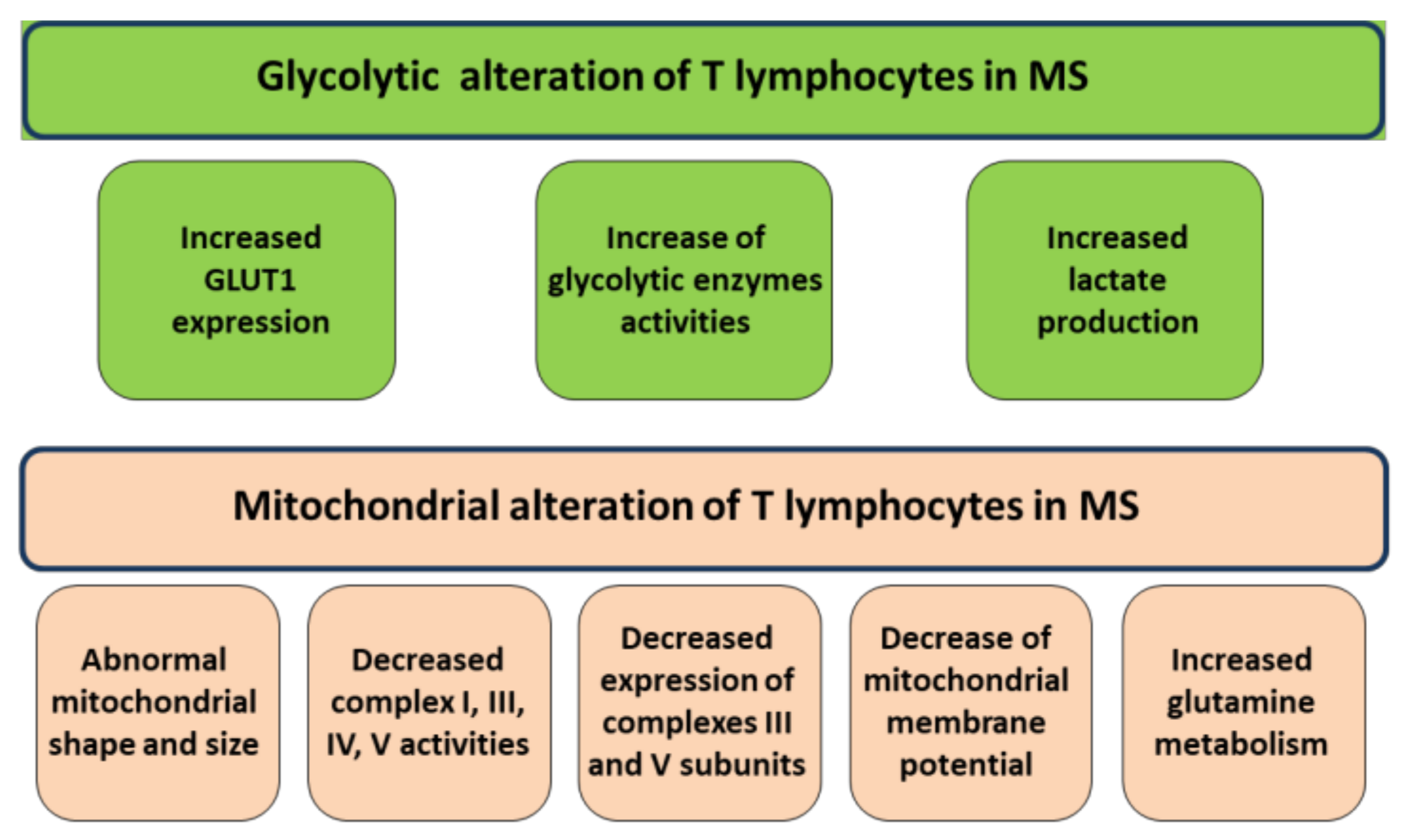
2. Mitochondrial Alterations of T Lymphocytes in MS

A central part of the T cell activation is the change of metabolism in the cell. Proliferating T cells need more ATP for enhanced activity and acquire anabolic capacity to sustain the growth. Also, the most dramatic change in T cell metabolism, upon activation, is a marked increase in glucose metabolism through the increase localization of glucose transporter 1 (GLUT1) to the plasma membrane and the enhance of glycolytic enzyme activities [20,32]. Activated T cells take up large amounts of glucose associated with lactate production. Indeed, activated T cells presented primarily glycolytic despite oxygen supply (aerobic glycolysis) [33,34]. However, while glycolysis is a rapid source of ATP and it can lead the pentose phosphate pathway to generate NADPH and nucleotides, it could be insufficient to completely sustain cell proliferation. Interestingly, upon activation, T cells also augment glutaminolysis process, that producing α-ketoglutarate, supplies the mitochondrial tricarboxylic acid (TCA) [35,36]. This is important in proliferating cells in which the intermediates of TCA cycle are continually depleted for their use in biosynthetic pathways [36,37]. Thus, mitochondrial metabolism plays a crucial role in T cell activation. Mitochondrial metabolism is a critical component of T cell activation also through mitochondrial-dependent ROS production [34,38] that, in turn, is also involved in the activation of a biosynthetic pathway of nucleic acid and T-cell proliferation [33].
One of first evidence of mitochondrial alteration in T lymphocytes of MS comes from the electron microscopy observation of abnormal mitochondria in shape and size and very often with thickened cristae [39]. Change in mitochondrial structure are associated, indeed, with changes in mitochondrial metabolism and mitochondrial apoptosis. Nowadays, decreased complex I and complex IV activities have been reported in lymphocytes of subjects affected by RR-MS with respect to those of control subjects associated with a decrease of their constituent subunits [40]. The reduced activity and protein expression of constituent subunits is also reported for complexes III and V of oxidative phosphorylation system (OXPHOS) in PBMC of RR-MS patients [24]. Also, a reduction of mitochondrial membrane potential, according to decrease of respiratory electron flux, has been found in lymphocytes of a cohort of 65 MS patients [41]. Very interesting, the decrease of mitochondrial respiratory chain activity and mitochondrial membrane potential are associated with disease severity [24,41]. Moreover, mitochondrial membrane potential reduction can be ascribed to a mitochondrial decoupling. In fact, studies reported a decrease of the ratio of mitochondrial ATP over glycolytic ATP in lymphocytes of MS subjects depending on both mitochondrial uncoupling, as shown by reduction of oxygen consumption in the presence of ADP, and increase activity of key enzymes of glycolysis such as hexokinase and phosphofructo kinase I [40]. According to the increase of glycolytic flux, an increase of GLUT1 expression has been found as well as an increase of secreted lactate [40]. In agreement with these results, another work reported a decrease of mitochondrial respiration activity but also a decrease of glycolytic flux [29].
So far, a reduced activity of mitochondrial enzymes and bioenergetics decoupling of mitochondria in term of efficiency to produce ATP has been observed and confirmed, in MS patients’ lymphocytes (Figure 1), by using different experimental approaches and conditions [24,40,41]. More reports show an increase of glycolytic flux in MS lymphocytes. However, another work observed a reduced glycolysis associated with reduced expression of GLUT1 [31] (Figure 1).

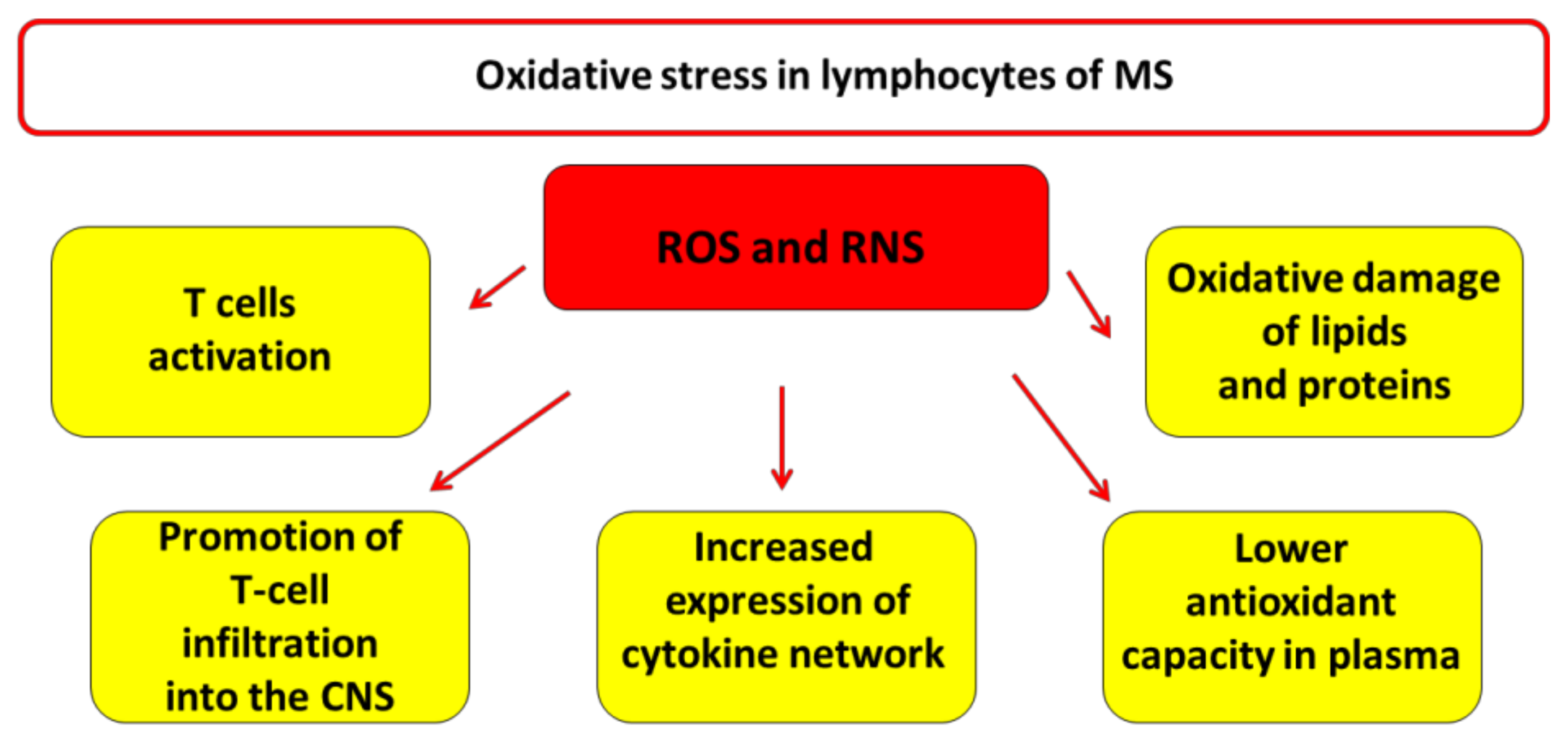
3. Oxidative Stress in Lymphocytes of MS

Oxidative stress condition is characterized by the imbalance between the production of free radicals and their counteraction by antioxidant defenses [42]. An increasing number of studies has shown that OS plays an important role in the pathogenesis of MS [5,6,22,23,43,44]. OS, associated with inflammation and neuronal damage, results in the oxidation of cellular components, such as proteins, lipids, and nucleic acids potentiates leading to a vicious cycle that can accelerate the progression of the disease [5,44,45,46,47]. OS is involved in several characteristic process of MS such as the activation of immune cells, especially T cells [15], the loss of BBB selectivity [48], promotion of T-cell migration and infiltration into the CNS [49] and the increased expression of the cytokine network [50] (Figure 2). The major radical species are represented by ROS and reactive nitrogen species (RNS) that are produced by specific brain cells such as astrocytes, activated macrophages, and microglia in the CNS of MS patients.
ROS are highly reactive molecules due to the presence of unpaired electrons and they can generate new free radicals reacting with other biological molecules. Many different forms of ROS exist, anion superoxide (O2), hydroxyl radical (OH∙) and hydrogen peroxide (H2O2). Anion superoxide can be produced by NAD(P)H oxidase and as a by-product of oxidative phosphorylation. Anion superoxide has a short life span, and it can be rapidly converted by superoxide dismutases (SODs) into hydrogen peroxide or into other ROS [51]. Hydrogen peroxide is relatively stable and permeates cellular membranes generating hydroxyl radicals by Fenton reaction. Furthermore, ROS are produced in large amount by immune cells to kill pathogens and facilitate phagocytosis [52]. However, in several neurological diseases, including Huntington’s disease, Parkinson’s disease, Alzheimer’s disease, and MS, the production of ROS overwhelms the cellular antioxidant capacity contributing to cellular injury [53,54].
In macrophages and microglial cells, the inflammatory cytokines such as tumor necrosis α (TNF-α) and IFN-γ inducing nitric oxide synthase (NOS), are responsible for the production of nitric oxide (NO). There are three major NOS isoforms: neuronal (nNOS), endothelial (eNOS), and inducible (iNOS). In an experimental autoimmune encephalomyelitis (EAE) animal model, an increase expression of iNOS mRNA and protein has been associated with the severity of clinical symptoms [55,56,57]. In addition, acting as a vasodilator, NO increases BBB permeability allowing inflammatory cells to infiltrate the CNS [58]. NO can react with superoxide anions to generate RNS such as peroxynitrite (ONOO−), which is a very dangerous oxidant associated with neuronal loss and is considered as pathogenic agent in MS [59]. Although the literature suggests that OS may be involved in the pathogenesis of MS in which activated microglia and macrophages are major cell source of ROS, few evidences are reported on OS in peripheral T lymphocytes.
Mitochondria are one of the major sources of ROS. They normally produce O2− at point of electron leakage in the electron transfer chain essentially at complex I and complex III. Mitochondria dysfunctions can result in an excessive O2 production that induces overexpression of the mitochondrial manganese superoxide dismutase (Mn-SOD), which increases production of H2O2 [60]. H2O2 is reduced to water by glutathione peroxidase in both mitochondrial matrix and cytosol. H2O2, which diffuses to peroxisomes, is converted to water and oxygen by catalase [60]. Data reported that the plasma of MS patients show a lower antioxidant capacity [61] and higher levels of oxidative damage to lipid and proteins [61,62]. Our study conducted in PBMCs from 15 MS patients and 15 healthy subjects showed increased level of H2O2 in MS with respect to healthy subjects [25] detected spectrofluorimetrically by dichlorodihydrofluorescin (DCFDA) probe. Interesting, the increased level of ROS production correlated with deregulation of some proteins involved in the mitochondrial dynamics and apoptosis [25]. Another report, carried out in PBMC of 34 MS and 24 healthy subjects, showed a higher concentration of anion superoxide in MS evaluated by flow cytometry, associated with a decrease in the protein content of the mitochondrial OXPHOS (complexes I–V) [24].
Mechanistically, the increase of ROS production can come from two hypotheses. The first is the stimulation of TCA cycle in glycolytic lymphocytes, resulting in an increase of mitochondrial NADH to a level greater than that needed to sustain mitochondrial respiration, that associated with the reduced expression of mitochondrial respiratory chain subunits [24,33] saturates the electron transport chain with consequent increased ROS production. The second is the decoupling of mitochondrial respiratory chain as already reported in lymphocytes of MS patients [40] that can result in an increased production of anion superoxide. These findings support the mitochondrial involvement in lymphocyte ROS production in MS. However, the OS in peripheral lymphocytes and its contribution to the disease remain to be analyzed more in depth.

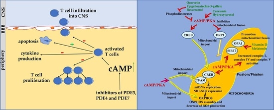
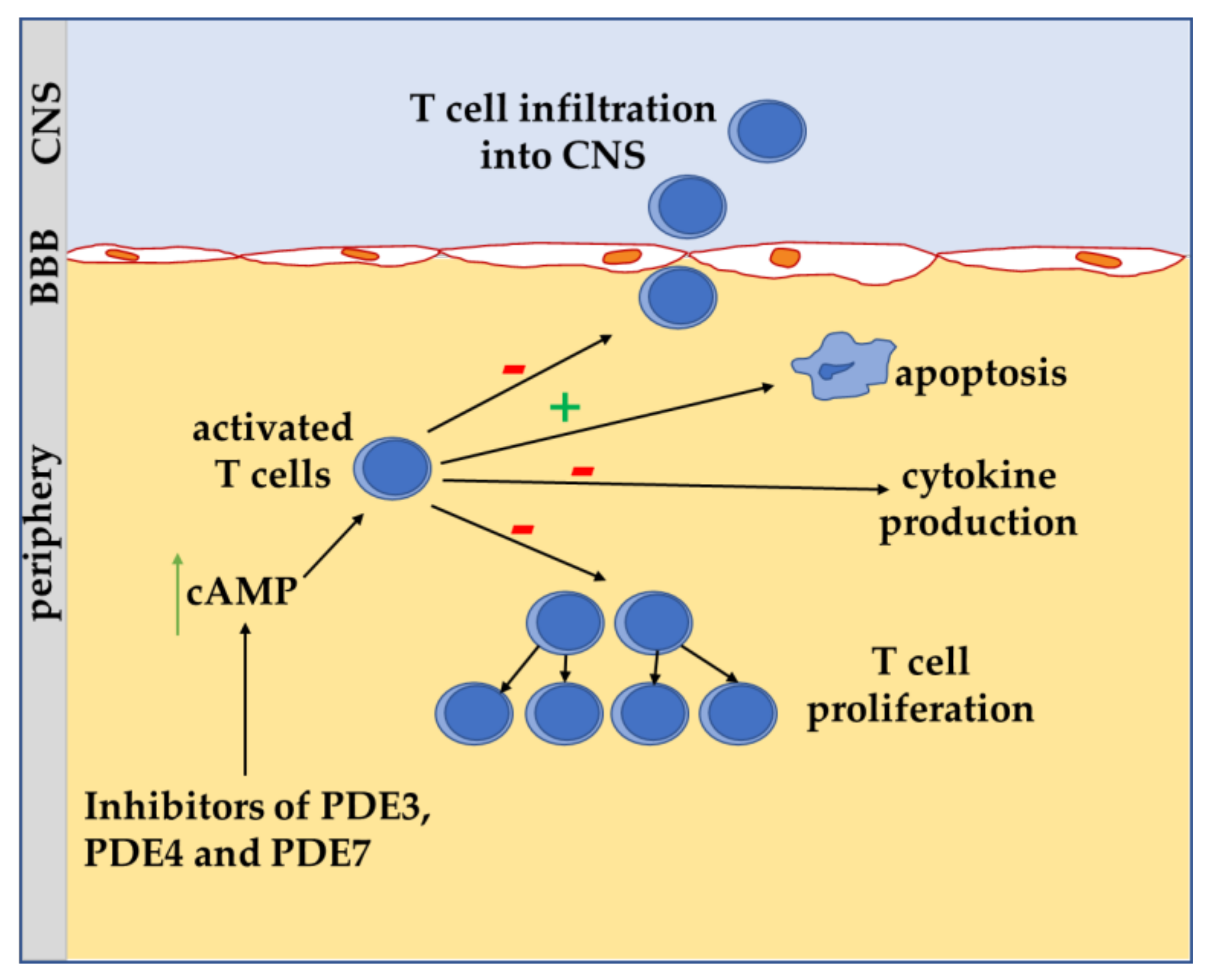
4. Role of cAMP Signaling in T Lymphocyte of MS

In the context of T lymphocyte differentiation, proliferation and activation, cAMP is the most studied second messenger and it has well recognized as potent negative regulators of immune function of T cells [14,20,21]. In mammalian cells, cAMP is produced by a family of transmembrane adenylyl cyclases (tmACs) and by the soluble adenylyl cyclase (sAC), that generates cAMP pools in various intracellular compartments, including mitochondria. tmAC is activated by β-adrenergic receptor stimulation, while sAC is modulated by intracellular stimuli, such as calcium, bicarbonate, and ATP [63]. cAMP level is also controlled by a super family of phosphodiesterases (PDEs) enzymes that hydrolyze and inactivate cAMP signalling. cAMP can act through different effectors, protein chinase A (PKA) (isoform I and II, PKA I and PKA II, respectively) and/or exchange protein directly activated by cAMP (EPAC). cAMP/PKA signalling regulates T cell function at the level of transcription factors, members of mitogen-activated protein kinase (MAPK) pathway, phospholipases 8PLs, ras homolog (RhoA) and proteins implicated in the control of cell cycle progression [64]. In the T cells, 80% of the total PKA activity is associated with PKA I, whereas approximatively 10–20% is accounted for by PKA II [19]. Specific involvement of the isoforms I and II of PKA can give a different role of cAMP in cell fate. For example, specific cAMP analogues for the isoform I inhibit natural killer cell-mediated cytotoxicity [65].
Many studies report the contribution of elevation of cAMP in the immunosuppression. Indeed, increased cAMP level in response to prostaglandin E2 (PGE2) and other agents has been reported to cause immunosuppression [19]. Interestingly, activation of the cAMP/PKA pathway has been implicated in the T cell dysfunction associated with human immunodeficiency virus (HIV) infection and a subset of common variable immunodeficiency [66]. Conversely, the inhibition of PKA I is reduced in patients with the autoimmune disease systemic lupus erythematosus [67]. It is reported that lipoic acid attenuates inflammation with reduction of IL-6 via cAMP and protein kinase signaling [68]. Data from EAE models indicated that lipoic acid may be effective as a treatment for MS by stimulating the production of cAMP in T-cell enriched peripheral blood mononuclear cells [69]. Moreover, the total cAMP level in PBMC was found reduced in RR-MS with respect to healthy subjects [69]. This is consistent with other reports in which the use of the cAMP elevating agents, such as forskolin, dibutyryl cyclic AMP (dbcAMP), and cholera toxin, reduces Il-2 level in murine T cells [70]. Moreover, in experimental models of EAE, dbcAMP inhibits the progression of disease, improves spatial memory retention in male rats, attenuates demyelination and enhances endogenous neural stem cell recruitment, [71,72,73].
Thus, the general observation on T cells is that increasing cAMP levels, reducing the production of pro-inflammatory cytokines (e.g., IFN-γ, TNF-α, and IL-1β), T cell proliferation and T cell activation attenuates the T lymphocyte mediated immune response [66]. Conversely, consistent with these results, a decrease of cAMP primes T cell activation [69]. Furthermore, increased cAMP level is important for the development of Tregs to maintain immunological homeostasis by suppressing the innate immune responses [66]. Evidence indicates an important role of PDEs in the modulating cAMP level in MS and thus T cell activation and proliferation. It has been reported that anti CD3/CD28 stimulation to activate naïve CD4+ T cells resulted in increased enzymatic activity of PDE7 [74]. Accordingly, in EAE mice, the use of the PDE7 inhibitor, named TC3.6, increased the mRNA level of forkhead box P3 (FOXP3) protein, a master regulator of Treg formation, the production of anti-inflammatory cytokine IL-10, and decreased the level of pro-inflammatory cytokine IL-17, T cell proliferation and T cell infiltration into CNS [74]. However, PDE7A knockout mice failed to alter T cell activation and/or cytokine production [75]. In the context of pro-inflammatory processes, PDE4 is the most studied cAMP-specific PDE. As observed for PDE7, in EAE mice, the inhibition with rolipram of PDE4 decreased T cell proliferation and reduced the production of TNF-α and IL-17 while increasing the production of IL-10 [21,74]. In addition, rolipram decreased the number of perivascular inflammatory infiltrates associated with a reduction of clinical symptoms [21,74]. Furthermore, in activated human CD4+ T cells, knockdown by siRNA of PDE4D reduced their proliferation rate and reduced the secretion of IFN-γ [76]. In EAE mice, mRNA level of the PDE4B2 isoform has been found increased in infiltrating T cells in the CNS [77]. The increased PDE4B2 positively correlated with FOXP3 and transforming growth factor beta (TGF-β) mRNA levels, supporting a regulatory role for PDE4B2 in Treg modulation [77,78]. As for PDE7 and PDE4, the dual substrate PDE3 inhibitor, cilostazol, has been shown to ameliorate T cell response in EAE mice by reducing IFN-γ production and lymphocytic proliferation in the CNS [79]. Summarizing, increasing cAMP by inhibiting specific PDEs could be considered as a potential therapeutic strategy to reduce the activation of T cell and the production of proinflammatory cytokines [21,74] (Figure 3). It should be noted that inhibitors of PDE3, 4, and 7 induce apoptosis in leukemic cells [74,80].

5. Apoptosis and Lymphocytes in MS

Programmed cell death (apoptosis) is a key anti-autoimmune mechanism that removes pathogenic autoreactive lymphocytes from the circulation and tissues, and controls the immune-mediated tissue damage [81]. More evidence suggests that apoptotic deletion of autoreactive lymphocytes is defective in patients with MS, and the relevance of this process in MS pathogenesis is confirmed considering that some disease modifying therapies, that is known to have beneficial effects in the disease, act by pushing T lymphocytes towards apoptosis [28,29,35,82]. Both peripheral and intrathecal lymphocytes from MS patients show reduced susceptibility to extrinsic and intrinsic cell death [29,83]. Indeed, the apoptosis is mediated through two main pathways: (i) extrinsic or death receptor-induced mechanism, which includes the surface receptor Fas (CD95) and other members of the tumor necrosis factor receptor family; and (ii) intrinsic or mitochondria-mediated pathway. The cause of lymphocyte resistance to cell death in MS is not fully understood but it appears to involve apoptotic defects at multiple cellular levels, as, for instance, the elevated expression of antiapoptotic factors [84].
Involvement of impairment of mitochondria-mediated apoptotic deletion has been reported in CD4+ T lymphocytes of PP-MS patients [30]. Evidence has been presented for a role of T CD4+ cells expressing the chemokine receptors CCR5 and CXCR3 in the trafficking of activated memory T cells of Th1 phenotype into the CNS of MS patients [85]. It has been observed that CD4+CCR5+ T cells from MS patients were more resistant to Fas-induced apoptosis compared with healthy subjects [30]. In this study, it has been shown that defects in apoptosis were more significant in patients with PPMS which also presented a reduction of mitochondrial membrane potential rising the implication of the intrinsic mitochondrial apoptotic pathway in the Fas-mediated apoptosis of CD4+CCR5+T cells [30]. A cross-talk between the extrinsic Fas-mediated and intrinsic pathways exists, indeed, through the Bcl-2 family member Bid, which, after cleavage by caspase-8, translocates to mitochondria where it promotes the cytochrome c release [86].
Mitochondria-mediated mechanism of apoptosis involves Bcl-2 proteins, that have anti-apoptotic function (Bcl-2 like), Bax proteins (Bax-like) and other proteins such as Bad, Bak and Bim, that have pro-apoptotic function [87]. The Bcl-2 gene has an anti-apoptotic activity in different cell types, including lymphocytes [88], in fact, upregulation of Bcl-2 proteins may cause systemic autoimmune disease as result of activated T cells accumulation [89]. Histopathologic studies have observed in MS plaques high level of lymphocytes expressing Bcl-2 [90]. However, other studies failed to find a significant Bcl-2 upregulation in intrathecal or peripheral lymphocytes from MS patients [91,92,93]. Other mitochondria parameters, such as fusion and fission, and mitochondria cristae architecture are involved in mitochondrial apoptosis [94,95,96]. Several mitochondrial proteins have been found to play a key role in mitochondria-mediated apoptosis in MS lymphocytes (see Table 1). Among others, optic atrophy 1 (OPA1) protein is a mitochondrial dynamin like GTPase that regulates mitochondrial fusion, the mitochondrial respiratory chain complex stability, the maintenance of mitochondrial cristae architecture and pro-apoptotic cytochrome c release [97]. Interestingly, it is observed that OPA1 gene mutations, resulting in autosomal dominant optic atrophy (ADOA), are correlated with multiple sclerosis-like disorders [98]. OPA1 undergoes constitutive proteolytic processing resulting in un-cleaved long OPA1 (L-OPA) and in a cleaved short OPA1 (S-OPA) forms. The balance between L-OPA1 and S-OPA1 influences the apoptosis susceptibility. Many stress conditions, such as apoptotic stimuli, trigger L-OPA1 conversion into S-OPA1 [95]. Recently it has been shown in PBMC of MS patients a different proteolytic processing of OPA1 compared with healthy controls [25]. The activity and processing of OPA1 are controlled by mitochondrial proteases, OMA1 and YMEL1 and regulated by post-translational modification such as sirtuin 3 (SIRT3)-dependent acetylation status [95,99] and OS [95,100]. OMA1-dependent degradation of OPA1 is a cellular response to OS, in fact, although OMA1 is constitutively active, it shows an enhanced activity in response to OS [100]. However, PBMCs of MS patients present an increase of ROS level that does not lead to the increase of the stress-induced active form of OMA1, but rather to the accumulation of the inactive form of OMA1 [25]. SIRT3 has a crucial role in many OS-mediated cellular responses [100], in the controlling of bioenergetics and antioxidant defense of mitochondria under OS [101]. OS regulates the level of SIRT3 protein that, in turn, modulates OPA1 acetylation/processing evolving in the regulation of mitochondrial apoptosis [95]. In particular, sustained SIRT3 protein level supports apoptosis resistance, while a reduced level promotes cell death [95]. Data showed that, despite the augmented ROS levels in MS, no change has been detected in the level of SIRT3 protein in MS lymphocytes compared to healthy control samples [25]. Although the level of SIRT3 protein was unaffected, this could be interpreted as a failure of reaction to OS of lymphocytes of MS patients. The data on OMA1 and SIRT3 suggest a deregulated stress response mechanism in PBMCs of MS patients that, in turn, alters OPA1 proteolytic processing. Furthermore, OPA1 stability also is under the control of prohibitin 2 (PHB2), a chaperone like protein, localized in nucleus, plasma membrane and mitochondria [102]. In inner membrane of mitochondria, PHB2 forms with prohibitin 1 (PHB1) a large membrane-bound complex [103,104] that is required for OPA1 stability [103,104]. Deletion of PHB2 results in decreased cellular proliferation, aberrant morphogenesis of mitochondrial cristae, and apoptosis, while PHB2 over-expression is shown to protect from apoptosis [104,105]. Evidence indicates that mitochondrial PHB2 is over-expressed in lymphocytes of MS patients [25,106,107] thus representing another element of resistance to apoptosis. Moreover, PHB2 is involved in the mechanism of mitophagy (selective autophagy) by functioning as an autophagosome formation receptor [108]. Factors involved in the process of autophagy have been implicated in neurodegenerative diseases and molecules involved in autophagy have also been found to impinge with T cells homeostasis [109]. In particular, autophagy-related gene-5 (Atg5)-deficient T lymphocytes display a reduced number in vivo, increased T cell apoptosis, and an incapability to undergo T cell receptor-induced proliferation [110]. Other studies have shown that Atg5 post-translational cleavage can also induce T cells apoptosis, on the contrary, an important change in Atg5 T cells, may be associated with the increase of T cell survival and/or the promotion of T cell proliferation during active disease [111]. It has reported that the upregulation and post-translational modification of Atg5 in autoreactive T cells of MS patients and in the mouse model of EAE correlated with viability of T cells and may contribute to inflammatory demyelination in MS [112].

6. The cAMP Signalling in the Regulation of Mitochondrial Activity, ROS Balance and T Cell Proliferation: Possible Implications of Some Nutraceutics

Summarizing, reduced mitochondrial respiration and mitochondrial membrane potential, increased ROS production and defected mitochondrial-mediated apoptosis have been observed in T cells of MS patients. Moreover, reduction of T lymphocyte mediated immune response, T cell proliferation and T cell activation in response to elevation of cAMP levels [19,21,60] as well as a reduction of cAMP level in RR-MS lymphocytes [69] have been observed. Several studies have reported that an antioxidant therapy has beneficial effects in vivo and in vitro animal models for MS. In the context of antioxidant therapy, due to their own antioxidant properties, the use of nutraceutics in neurodegenerative, inflammatory, and metabolic diseases is in constant expansion in order to ameliorate or prevent aging-related diseases [113]. These molecules are easily extracted from plants or derived from fermentation in bioreactors [113]. Among people with MS, nutraceutics are commonly used, often concomitantly with conventional treatment [114]. We would like to highlight that cAMP strongly affects both mitochondrial functions, ROS production, and mitochondria-mediated apoptosis and that some nutraceutical compounds, beyond the own antioxidant effect and inhibition of inflammation, can act by targeting mitochondria, apoptosis, and cAMP signaling (Figure 4).

6.1. cAMP, Mitochondria and ROS Balance

Defects of complex I and IV activities and their protein expression have been observed in MS lymphocytes. The complex I of mitochondrial respiratory chain is the major source of ROS and it is strongly regulated by cAMP pathway (Figure 4). Several studies have shown that cAMP/PKA signaling has a positive regulatory effect on ROS balance and complex I. In fact, the induced increase of cAMP rescues the activity of complex I and the increase of ROS levels occuring in G0 phase in murine and human cells and in oxidatively damaged cells [118,127,128]. Many reports showed that the increase of cAMP level, by stimulation of tmAC in mammalian cell cultures, increases the activity of complex I and decreases the complex I-dependent ROS production by promoting the PKA-dependent phosphorylation, mitochondrial import, and assembly in complex I of NDUFS4 subunits [118,119,120]. Other works reported the involvement of cAMP/PKA pathway in the regulation of complex IV activity [129]. Moreover, the cAMP affects the mitochondrial biogenesis via cAMP response element binding (CREB) protein. Upon phosphorylation by PKA, the CREB protein activates the expression of peroxisome proliferator-activated receptor gamma coactivator 1-alpha (PGC-1α), which in turn induces the expression of downstream nuclear and mitochondrial transcription factors, such as TFAM [115] (Figure 4).
Beyond to reside in the nucleus, CREB has also been reported to reside in mitochondria, where it, by binding to the D-loop of mitochondrial DNA [116], induces the expression of OXPHOS subunits, in particular the seven genes of complex I [117]. Mitochondrial pool of cAMP, resulting from mitochondrially localized sAC, increases the activity of cytochrome—c oxidase [121], the ATP production [122], the complex I activity and the turnover of its nuclear-encoded subunits [123] and controls the structural organization and functional activity of FoF1 ATP synthase [124].
As for mitochondrial respiratory chain activity, mitochondrial size, shape (dynamics) and cristae structure, and mitochondrial shaping protein (OPA1) have been found altered in MS lymphocytes. The mitochondrial dynamics and structure depend essentially on dynamin-related protein 1 (DRP1) proteins and OPA1, influencing several processes of the cell such as apoptosis [125]. Mitochondria fission is mainly controlled by the dynamin-like GTPase, DRP1, which is recruited to the surface of mitochondria and assembled around the constriction points of the dividing mitochondria into a multimeric ring-like structure [126]. PKA-dependent phosphorylation of DRP1 blocks its translocation to the surface of mitochondria, resulting in inhibition of fission and so in a mitochondrial elongation, which promotes cell survival. DRP1 dephosphorylation facilitates its translocation to mitochondrial surface promoting fission, apoptosis, and autophagy [126,130]. In addition, mitochondrially produced cAMP regulates OPA1 stability. In particular, mitochondrial cAMP reduction causes OPA1 proteolytic processing by inhibiting SIRT3-dependent protein deacetylation, resulting in an anti-fusion event and apoptosis [95]. On the contrary, increase in mitochondrial cAMP enhances SIRT3 protein level and promotes fusion events that protect myoblasts from ROS-dependent apoptosis [95]. In addition to DRP1, PKA phosphorylates mitofusin 2 (Mfn2) [131] (Figure 4).
In the context of mitochondrial cAMP, it is worth mentioning that the treatment with cysteamine of the EAE mice model ameliorated disease severity. Cysteamine has been also found to increase specifically mitochondrial cAMP resulting in increase of mitochondrial respiration, membrane potential, cristae structure and fusion process [132,133].
cAMP, acting through different effectors, PKA and/or EPAC, could determine a pro-apoptotic or anti-apoptotic response [134]. In addition, a specific involvement of one of the two isoforms of PKA (I and II) can impinge with the role of cAMP in cell death. For example, analogues of cAMP specific for the isoform I inhibit natural killer cell-mediated cytotoxicity [65]. Instead, expression of PKA isoform II can modulate apoptosis of fibroblasts [135].

6.2. Nutraceutics

Various nutraceutics, such as curcumin, flavonoids, melatonin, hydroxytirosol (HT), and vitamin D, have been trialed for their efficacy as adjunctive treatments in MS [136]. Beyond their own antioxidant effect, they can have molecular targets implicated in MS pathology, such as inflammation, oxidative stress, mitochondrial dysfunction, and cAMP signaling (Figure 5).

6.2.1. Curcumin

Curcumin is a natural antioxidant polyphenolic compound with anti-inflammatory and immunomodulatory properties, which can influence the immune cell activation, including T cells. Furthermore, curcumin can inhibit the pro-inflammatory cytokines and chemokines expressions by suppressing the nuclear factor kappa-light-chain-enhancer of activated B cells (NF-κB) signaling pathway [137]. In an EAE rat model, the daily treatment with 50 or 100 µg curcumin decreased the pathological and clinical disease severity [138]. In addition, curcumin inhibits cytokine production and phytohemagglutinin-induced lymphocyte proliferation by inhibiting lipopolysaccharide-induced NF-kB target genes [139]. Curcumin exerts various other pharmacological effects, including proapoptotic and anti-proliferative effects [140]. In HT-29 cells, the curcumin-dependent apoptosis includes the activation of caspase and mitochondrial dysfunction primed by enhanced Bax activation [139,141]. In addition, it is reported the involvement of endoplasmic reticulum-stress in curcumin-dependent apoptosis of HL-60 cells [142] and the induction of unfolded protein response (UPR) components promoting apoptosis after the curcumin treatment in activated human CD4+ T cells promoting apoptosis [140]. It is also observed that, with the treatment of HEK-293T cells at 10 µM concentration, curcumin upregulated cAMP signaling [143].

6.2.2. Flavonoids

Flavonoids play an important role in the management of MS [144,145]. Flavonoids such as quercetin and epigallocatechin-3-gallate (EGCG) can decrease the production of pro-inflammatory cytokines, including IL-1β, IL-6, and TNF-α [146]. The mechanism of flavonoid action includes the free radical scavenging accompanied with the silencing the pro-oxidative enzymes associated with free radical generation, such as protein kinase C, xanthine oxidase, cyclooxygenase, lipoxygenase, and NADPH oxidase and chelating metals [147]. EGCG is one of the best studied flavonoids in EAE and MS is. It is an active ingredient of green tea and it has been shown to inhibit the differentiation of immature CD4+ T cell into Th1 and Th17 effector subsets influencing the immune and inflammation profiles of CNS and lymphoid tissues. This results in a reduction of pro-inflammatory cytokine production, autoreactive T-cell proliferation and Th1 and Th17 subpopulations, and in the increase of regulatory T cell populations [148]. Moreover, in EAE oral-applied, EGCG decreased inflammation by inhibiting TNF-α synthesis in T cells. These results suggest a potential role for EGCG in the prevention and treatment of T-cell-mediated autoimmune diseases [148]. ECGC rat treatment has been found to promote an increase of mitochondrial membrane potential in lymphocytes [149], and mitochondrial DNA content as well as nuclear encoded proteins involved in mitochondrial biogenesis in lymphoblastoide cell cultures [150]. It is also known that EGCG can increase the cAMP cellular level [151,152] by inhibiting PDEs [147] and/or activating adenylyl cyclase [153].
Quercetin is a flavonoid phytoestrogen showing anticancer and anti-inflammatory activities. It is reported that quercetin in EAE model, inhibited IL-12 production and neural antigen-specific Th1 differentiation [154]. Moreover, in jurkat cells, it has been found that quercetin induced apoptosis by activating Bak dependent mitochondrial pathway [155]. It should be noted that quercetin is also an inhibitor of PDE promoting cAMP accumulation [156].

6.2.3. Resveratrol

Resveratrol is a natural polyphenol that is found in more than 70 plant species such as peanuts, cranberry and red grapes. Resveratrol has been shown to display anti-inflammatory effects through a series of mechanisms, including inhibition of the synthesis and release of pro-inflammatory molecules and promotion of apoptosis in T cells [157]. At molecular level, it has been suggested that resveratrol acts by inhibiting PDE [158] and thus increasing the cAMP level culminating in an increase of sirtuin 1 (SIRT1) and PGC1α protein activities which prevents axonal degeneration [159]. Moreover, resveratrol has been observed to induce the amelioration of mitochondrial activity in some neurodegenerative diseases [160,161].

6.2.4. Hydroxytyrosol

HT is one of polyphenolic compounds present in extra virgin olive oil (EVOO). EVOO represents a functional food for prevention of immune and inflammatory disease [162]. This can be due to the capacity of HT to cross the BBB explaining thus its neuroprotective role [163]. It has been proposed that the biological activity of HT can come from its metabolites suggesting that hydroxytyrosol-3-O-glucuronide exhibits a stronger antioxidant activity than hydroxytyrosol itself [164]. The plasma concentration of HT does not only come from the absorption of olive oil polyphenols, but HT also is a by-product of dopamine oxidative metabolism [165]. HT is one of the polyphenols of EVOO with the most powerful anti-inflammatory properties such as reduced secretion of pro-inflammatory cytokines (interleukin (IL)-1α, IL-1β, IL-6, IL-12), tumor necrosis factor-α (TNF-α), and chemokines, inhibition of PGE2 and NO production, and decreased gene expression of iNOS [166]. The beneficial effect of EVOO, and in particular of two of its components, HT and oleic acid, on the improvement of OS in the blood and CNS of EAE rats has been observed [167]. The study reported in the CNS and blood of EAE rats a reduction of lipid peroxidation and protein carbonylation [167]. Interestingly, it is reported that high doses of hydroxytyrosol can induce apoptosis by increasing the expression of caspase 3 gene and the BAX/BCL2 ratio [168]. In different cell lines, in the range of concentration of 0.1–10 µM, HT promotes the activity and expression of PGC1α, which, in turn, promotes the expression and activity of mitochondrial respiratory chain complexes I, II, III, IV, and complex V thus ameliorating mitochondrial function [169,170]. In addition, in serum-starved fibroblast cultures, HT prevents the decline in the expression of the PGC-1α transcription cascade and this effect appears to be associated with the PKA activation and, thus, CREB phosphorylation [171]. In addition to the role of HT on mitochondrial function and biogenesis, studies performed in vivo and in vitro show that HT can control the expression of mitochondrial shaping proteins [172].

6.2.5. Vitamin D

Vitamin D is a steroidal compound metabolized in the liver, skin, epithelial cells, immune cells, and kidneys [173]. Two forms of vitamin D exist, vitamin D2 (ergocalciferol) and vitamin D3 (cholecalciferol). The vitamin D active form is 1,25-dihydroxycholecalciferol (1,25-(OH)2D3) derived by two-step transformation process catalyzed by 25-hydroxylase-CYP2R1 and 1-alpha-hydroxylase. The principal organs involved in vitamin D metabolism are liver and kidney, however, other tissues such as immune cells, parathyroid gland and epithelial cells perform the metabolic conversion in vitamin D active form [173]. Vitamin D3 is primarily found in fatty animal-sourced foods, such as egg yolk and fish oil, while vitamin D2 in plant-sourced foods, for instance fortified foods and UV light-grown mushrooms [174]. The vitamin D receptors are present in most cells, like activated monocytes, B and T cells, and microglia and tissues like skin and intestine. Very interestingly, vitamin D supplementation as well as a higher circulating vitamin D level are associated with a reduced risk of MS [175]. Vitamin D has also been observed to have immunomodulatory effects, increasing Th2 and Tregs activity and decreasing Th1 activity, and thus it may play a role in the MS etiology [175]. The active vitamin D regulates the expression of iNOS and the production of NO in different cells, such as astrocytes, microglial cells, and macrophages [176]. The protective mechanism against EAE involves a decreased accumulation of T cells and macrophages in the CNS [177,178], an increased apoptosis of inflammatory cells, and enhanced survival of CNS cells. However, the activated inflammatory cells are able to produce 1,25-(OH)2D3, that subsequently exerts anti-inflammatory activities against these cells [179]. In vivo molecular experiments show that 1,25-(OH)2D3 directly acts on pathogenic CD4+ T cells and inhibits EAE via receptors of vitamin D in T lymphocytes [180]. It should be noted that 1,25-(OH)2D3 can suppress lymphocyte mitogenesis by acting synergistically with agents that augment cellular cAMP level [180]. Interestingly, vitamin D supplementation ameliorates mitochondrial cristae shape by modulating mitofusin-1/2 (Mfn1/2), OPA1 and DRP1 expression [181].

6.2.6. Melatonin

Melatonin is produced by the pineal gland in response to the darkness during the night. Melatonin is also found in several foods, such as fruits, vegetables, seeds, and herbs [182]. The level of melatonin has been found impaired in MS patients, and exogenous melatonin administration improves the disease by modulating the Th1/Th17/Treg responses in MS animal models [183] and ameliorates several symptoms and quality of life in MS patients [184]. In addition, melatonin has a cytoprotective actions by controlling OS, mitochondrial homeostasis, and apoptosis [185]. It exerts its actions by binding to G-protein-coupled receptors at the membrane and by interacting with intracellular targets to control signal transduction pathways, redox processes, and free radicals scavenging [186]. Melatonin stimulates the activities of glutathione peroxidase and SOD but inhibits the prooxidant enzyme NOS, thus it may be a potent antioxidant molecule acting also at mitochondrial level. Indeed, in several cell types, melatonin has been found to increase the activities of respiratory chain complexes I and IV, stimulate oxidative phosphorylation, and promote a rise in ATP level [187]. Furthermore, melatonin improves mitochondrial dynamics by reducing DRP1 expression and increasing that of fusion proteins, Mfn2 and OPA1 [188].

7. Conclusions

In this review we have highlighted the involvement of mitochondria, ROS production, cAMP pathway, apoptosis, and their interconnection in alteration of T lymphocytes homeostasis in MS. So far, in lymphocytes of MS patients, a reduced activity of mitochondrial enzymes and a bioenergetic decoupling of mitochondria in term of efficiency of ATP production associated with increased production of anion superoxide have been observed. At the same time, the increase of cAMP, that reduced T cell proliferation, strongly modulates mitochondrial activity in terms of mitochondrial respiration, coupling, dynamics, structure, biogenesis, and mitochondria-mediated apoptosis (Figure 5). Nutritional status in MS patients and dietary supplementation have been proposed as potential critical factors that can affect risk and progression of the disease [189] in fact, several dietary supplements might reduce inflammation and fatigue and increase autoimmunity tolerance in patients with MS, even if it should be noted that dietary supplementation is not an effective clinical indication as complementary treatment for MS patients. Further research on nutraceutics and their mechanisms and molecular pathways involved should be performed in order to verify the link among inflammation, cAMP, and mitochondria. So, an increasing cAMP level, by inhibiting specific PDEs and/or by using nutraceutics, can be considered as a possible strategy to limit the activation of T cells.

Author Contributions

Conceptualization, writing—original draft preparation, figure preparation, A.S. and D.D.R.; writing—review and editing, A.F., M.R., D.P., M.T. and P.L. All authors have read and agreed to the published version of the manuscript.

Funding

During the preparation of the manuscript A.S. and D.D.R. were supported by University of Bari to A.S., project title “Regolazione dei sistemi redox e bioenergetici cellulari in condizioni fisio-patologiche”.

Acknowledgments

We thank Annarita Armenise (IBIOM-CNR) for kind assistance with manuscript preparation.

Conflicts of Interest

The authors declare no conflict of interest.

References

  1. Lassmann, H.; Brück, W.; Lucchinetti, C.F. The immunopathology of multiple sclerosis: An overview. Brain Pathol. 2007, 17, 210–218. [Google Scholar] [CrossRef] [PubMed]
  2. Tobore, T.O. Towards a comprehensive etiopathogenetic and pathophysiological theory of multiple sclerosis. Int. J. Neurosci. 2020, 130, 279–300. [Google Scholar] [CrossRef] [PubMed]
  3. Lublin, F.D.; Reingold, S.C. Defining the clinical course of multiple sclerosis: Results of an international survey. Neurology 1996, 46, 907–911. [Google Scholar] [CrossRef] [PubMed]
  4. Klineova, S.; Lublin, F.D. Clinical Course of Multiple Sclerosis. Cold Spring Harb. Perspect. Med. 2018, 8, a028928. [Google Scholar] [CrossRef] [PubMed]
  5. Tanaka, M.; Vécsei, L. Monitoring the Redox Status in Multiple Sclerosis. Biomedicines 2020, 8, 406. [Google Scholar] [CrossRef] [PubMed]
  6. Sospedra, M.; Martin, R. Immunology of multiple sclerosis. Annu. Rev. Immunol. 2005, 23, 683–747. [Google Scholar] [CrossRef] [PubMed] [Green Version]
  7. Sadovnick, A.D.; Armstrong, H.; Rice, G.P.; Bulman, D.; Hashimoto, L.; Party, D.W.; Hashimoto, S.A.; Warren, S.; Hader, W.; Murrary, T.J.; et al. A population-based study of multiple sclerosis in twins: Update. Ann. Neurol. 1993, 33, 281–285. [Google Scholar] [CrossRef]
  8. Dyment, D.A.; Ebers, G.C.; Sadovnick, A.D. Genetics of multiple sclerosis. Lancet Neurol. 2004, 3, 104–110. [Google Scholar] [CrossRef] [Green Version]
  9. Hewer, S.; Lucas, R.; van der Mei, I.; Taylor, B.V. Vitamin D and multiple sclerosis. J. Clin. Neurosci. 2013, 20, 634–641. [Google Scholar] [CrossRef]
  10. Pender, M.P.; Burrows, S.R. Epstein-Barr virus and multiple sclerosis: Potential opportunities for immunotherapy. Clin. Transl. Immunol. 2014, 3, e27. [Google Scholar] [CrossRef]
  11. Tanaka, M.; Toldi, J.; Vécsei, L. Exploring the Etiological Links behind Neurodegenerative Diseases: Inflammatory Cytokines and Bioactive Kynurenines. J. Mol. Sci. 2020, 21, 2431. [Google Scholar] [CrossRef] [PubMed] [Green Version]
  12. Lassmann, H. Pathogenic Mechanisms Associated with Different Clinical Courses of Multiple Sclerosis. Front. Immunol. 2019, 9, 3116. [Google Scholar] [CrossRef] [PubMed]
  13. Van Langelaar, J.; Rijvers, L.; Smolders, J.; van Luijn, M.M. B and T Cells Driving Multiple Sclerosis: Identity, Mechanisms and Potential Triggers. Front. Immunol. 2020, 11, 760. [Google Scholar] [CrossRef]
  14. Kammer, G.M. The adenylate cyclase-cAMP-protein kinase A pathway and regulation of the immune response. Immunol. Today 1988, 9, 222–229. [Google Scholar] [CrossRef]
  15. Geltink, R.I.K.; Kyle, R.L.; Pearce, E.L. Unraveling the Complex Interplay Between T Cell Metabolism andFunction. Annu. Rev. Immunol. 2018, 36, 461–488. [Google Scholar] [CrossRef]
  16. Corthay, A. A three-cell model for activation of naïve T helper cells. Scand. J. Immunol. 2006, 64, 93–96. [Google Scholar] [CrossRef] [PubMed]
  17. Lee, J.Y.; Taghian, K.; Petratos, S. Axonal degeneration in multiple sclerosis: Can we predict and prevent permanent disability? Acta Neuropathol. Commun. 2014, 2, 97. [Google Scholar] [CrossRef]
  18. Bargiela, D.; Chinnery, P.F. Mitochondria in neuroinflammation - Multiple sclerosis (MS), leber hereditary optic neuropathy (LHON) and LHON-MS. Neurosci. Lett. 2019, 710, 132932. [Google Scholar] [CrossRef]
  19. Wehbi, V.L.; Taskén, K. Molecular Mechanisms for cAMP-Mediated Immunoregulation in T cells - Role of Anchored Protein Kinase A Signaling Units. Front. Immunol. 2016, 7, 222. [Google Scholar] [CrossRef] [Green Version]
  20. Frauwirth, K.A.; Thompson, C.B. Activation and inhibition of lymphocytes by costimulation. J. Clin. Investig. 2002, 109, 295–299. [Google Scholar] [CrossRef]
  21. Schepers, M.; Tiane, A.; Paes, D.; Sanchez, S.; Rombaut, B.; Piccart, E.; Rutten, B.P.F.; Brône, B.; Hellings, N.; Prickaerts, J.; et al. Targeting Phosphodiesterases-Towards a Tailor-Made Approach in Multiple Sclerosis Treatment. Front. Immunol. 2019, 10, 1727. [Google Scholar] [CrossRef] [PubMed]
  22. Gilgun-Sherki, Y.; Melamed, E.; Offen, D. The role of oxidative stress in the pathogenesis of multiple sclerosis: The need for effective antioxidant therapy. J. Neurol. 2004, 251, 261–268. [Google Scholar] [PubMed]
  23. Ohl, K.; Tenbrock, K.; Kipp, M. Oxidative stress in multiple sclerosis: Central and peripheral mode of action. Exp. Neurol. 2016, 277, 58–67. [Google Scholar] [CrossRef] [PubMed]
  24. Gonzalo, H.; Nogueras, L.; Gil-Sánchez, A.; Hervás, J.V.; Valcheva, P.; González-Mingot, C.; Martin-Gari, M.; Canudes, M.; Peralta, S.; Solana, M.J.; et al. Impairment of Mitochondrial Redox Status in Peripheral Lymphocytes of Multiple Sclerosis Patients. Front. Neurosci. 2019, 13, 938. [Google Scholar] [CrossRef] [PubMed]
  25. De Rasmo, D.; Ferretta, A.; Russo, S.; Ruggieri, M.; Lasorella, P.; Paolicelli, D.; Trojano, M.; Signorile, A. PBMC of Multiple Sclerosis Patients Show Deregulation of OPA1 Processing Associated with Increased ROS and PHB2 Protein Levels. Biomedicines 2020, 8, 85. [Google Scholar] [CrossRef] [PubMed] [Green Version]
  26. McFarland, H.F.; Martin, R. Multiple sclerosis: A complicated picture of autoimmunity. Nat. Immunol. 2007, 8, 913–919. [Google Scholar] [CrossRef]
  27. Comabella, M.; Khoury, S.J. Immunopathogenesis of multiple sclerosis. Clin. Immunol. 2012, 142, 2–8. [Google Scholar] [CrossRef]
  28. Segal, B.M.; Cross, A.H. Fas(t) track to apoptosis in MS: TNF receptors may suppress or potentiate CNS demyelination. Neurology 2000, 55, 906–907. [Google Scholar] [CrossRef]
  29. Ruggieri, M.; Avolio, C.; Scacco, S.; Pica, C.; Lia, A.; Zimatore, G.B.; Papa, S.; Livrea, P.; Trojano, M. Glatiramer acetate induces pro-apoptotic mechanisms involving Bcl-2, Bax and Cyt-c in peripheral lymphocytes from multiple sclerosis patients. J. Neurol. 2006, 253, 231–236. [Google Scholar] [CrossRef]
  30. Julià, E.; Edo, M.C.; Horga, A.; Montalban, X.; Comabella, M. Differential susceptibility to apoptosis of CD4+T cells expressing CCR5 and CXCR3 in patients with MS. Clin. Immunol. 2009, 133, 364–374. [Google Scholar] [CrossRef]
  31. La Rocca, C.; Carbone, F.; De Rosa, V.; Colamatteo, A.; Galgani, M.; Perna, F.; Lanzillo, R.; Brescia Morra, V.; Orefice, G.; Cerillo, I.; et al. Immunometabolic profiling of T cells from patients with relapsing-remitting multiple sclerosis reveals an impairment in glycolysis and mitochondrial respiration. Metabolism 2017, 77, 39–46. [Google Scholar] [CrossRef] [PubMed] [Green Version]
  32. Shyer, J.A.; Flavell, R.A.; Bailis, W. Metabolic signaling in T cells. Cell Res. 2020, 30, 649–659. [Google Scholar] [CrossRef] [PubMed]
  33. Jones, R.G.; Thompson, C.B. Revving the engine: Signal transduction fuels T cell activation. Immunity 2007, 27, 173–178. [Google Scholar] [CrossRef] [PubMed] [Green Version]
  34. Previte, D.M.; O’Connor, E.C.; Novak, E.A.; Martins, C.P.; Mollen, K.P.; Piganelli, J.D. Reactive oxygen species are required for driving efficient and sustained aerobic glycolysis during CD4+ T cell activation. PLoS ONE 2017, 12, e0175549. [Google Scholar] [CrossRef]
  35. Johnson, M.O.; Wolf, M.M.; Madden, M.Z.; Andrejeva, G.; Sugiura, A.; Contreras, D.C.; Maseda, D.; Liberti, M.V.; Paz, K.; Kishton, R.J.; et al. Distinct regulation of Th17 and Th1 cell differentiation by glutaminase-dependent metabolism. Cell 2018, 175, 1780–1795. [Google Scholar] [CrossRef] [Green Version]
  36. DeBerardinis, R.J.; Mancuso, A.; Daikhin, E.; Nissim, I.; Yudkoff, M.; Wehrli, S.; Thompson, C.B. Beyond aerobic glycolysis: Transformed cells can engage in glutamine metabolism that exceeds the requirement for protein and nucleotide synthesis. Proc. Natl. Acad. Sci. USA 2007, 104, 19345–19350. [Google Scholar] [CrossRef] [Green Version]
  37. Seyfried, T.N.; Arismendi-Morillo, G.; Mukherjee, P.; Chinopoulos, C. On the Origin of ATP Synthesis in Cancer. iScience 2020, 23, 101761. [Google Scholar] [CrossRef]
  38. Sena, L.A.; Li, S.; Jairaman, A.; Prakriya, M.; Ezponda, T.; Hildeman, D.A.; Wang, C.R.; Schumacker, P.T.; Licht, J.D.; Perlman, H.; et al. Mitochondria are required for antigen-specific T cell activation through reactive oxygen species signaling. Immunity 2013, 38, 225–236. [Google Scholar] [CrossRef] [Green Version]
  39. Djaldetti, R.; Achiron, A.; Ziv, I.; Djaldetti, M. Lymphocyte ultrastructure in patients with multiple sclerosis. Biomed. Pharmacother. 1995, 49, 300–303. [Google Scholar] [CrossRef]
  40. De Riccardis, L.; Rizzello, A.; Ferramosca, A.; Urso, E.; De Robertis, F.; Danieli, A.; Giudetti, A.M.; Trianni, G.; Zara, V.; Maffia, M. Bioenergetics profile of CD4+T cells in relapsing remittingmultiplesclerosissubjects. J. Biotechnol. 2015, 202, 31–39. [Google Scholar] [CrossRef]
  41. Armon-Omer, A.; Neuman, H.; Sharabi-Nov, A.; Shahien, R. Mitochondrial activity is impaired in lymphocytes of MS patients in correlation with disease severity. Mult. Scler. Relat. Disord. 2020, 41, 102025. [Google Scholar] [CrossRef] [PubMed]
  42. Salim, S. Oxidative stress and the central nervous system. J. Pharmacol. Exp. Ther. 2017, 360, 201–205. [Google Scholar] [CrossRef] [PubMed]
  43. Miljković, D.; Spasojević, I. Multiple sclerosis: Molecular mechanisms and therapeutic opportunities. Antioxid. Redox Signal. 2013, 19, 2286–2334. [Google Scholar] [CrossRef] [PubMed]
  44. Török, N.; Tanaka, M.; Vécsei, L. Searching for Peripheral Biomarkers in Neurodegenerative Diseases: The Tryptophan-Kynurenine Metabolic Pathway. Int. J. Mol. Sci. 2020, 21, 9338. [Google Scholar] [CrossRef] [PubMed]
  45. Miller, E.; Walczak, A.; Saluk, J.; Ponczek, M.B.; Majsterek, I. Oxidative modification of patient’s plasma proteins andits role in pathogenesis of multiple sclerosis. Clin. Biochem. 2012, 45, 26–30. [Google Scholar] [CrossRef] [PubMed]
  46. Padureanu, R.; Albu, C.V.; Mititelu, R.R.; Bacanoiu, M.V.; Docea, A.O.; Calina, D.; Padureanu, V.; Olaru, G.; Sandu, R.E.; Malin, R.D.; et al. Oxidative Stress and Inflammation Interdependence in Multiple Sclerosis. J. Clin. Med. 2019, 8, 1815. [Google Scholar] [CrossRef] [Green Version]
  47. Siotto, M.; Filippi, M.M.; Simonelli, I.; Landi, D.; Ghazaryan, A.; Vollaro, S.; Ventriglia, M.; Pasqualetti, P.; Rongioletti, M.C.A.; Squitti, R.; et al. Oxidative Stress Related to Iron Metabolism in Relapsing Remitting Multiple Sclerosis Patients with Low Disability. Front. Neurosci. 2019, 13, 86. [Google Scholar] [CrossRef] [Green Version]
  48. Wu, M.; Tsirka, S.E. Endothelial NOS-deficient mice reveal dual roles for nitric oxide during experimental autoimmune encephalomyelitis. Glia 2009, 57, 1204–1215. [Google Scholar] [CrossRef] [Green Version]
  49. Marui, N.; Offermann, M.K.; Swerlick, R.; Kunsch, C.; Rosen, C.A.; Ahmad, M.; Alexander, R.W.; Medford, R.M. Vascular cell adhesion molecule-1 (VCAM-1) gene transcription and expression are regulated through an antioxidant-sensitive mechanism in human vascular endothelial cells. J. Clin. Investig. 1993, 92, 1866–1874. [Google Scholar] [CrossRef]
  50. Jäger, A.; Dardalhon, V.; Sobel, R.A.; Bettelli, E.; Kuchroo, V.K. Th1, Th17, and Th9 effector cells induce experimental autoimmune encephalomyelitis with different pathological phenotypes. J. Immunol. 2009, 183, 7169–7177. [Google Scholar]
  51. Munro, D.; Treberg, J.R. A radical shift in perspective: Mitochondria as regulators of reactive oxygen species. J. Exp. Biol. 2017, 220, 1170–1180. [Google Scholar] [CrossRef] [PubMed] [Green Version]
  52. Hampton, M.B.; Kettle, A.J.; Winterbourn, C.C. Inside the neutrophil phagosome: Oxidants, myeloperoxidase, and bacterial killing. Blood 1998, 92, 3007–3017. [Google Scholar] [CrossRef] [PubMed]
  53. Pacelli, C.; De Rasmo, D.; Signorile, A.; Grattagliano, I.; di Tullio, G.; D’Orazio, A.; Nico, B.; Comi, G.P.; Ronchi, D.; Ferranini, E.; et al. Mitochondrial defect and PGC-1α dysfunction in parkin-associated familial Parkinson’s disease. Biochim. Biophys. Acta 2011, 1812, 1041–1053. [Google Scholar] [CrossRef] [PubMed] [Green Version]
  54. Tarafdar, A.; Pula, G. The Role of NADPH Oxidases and Oxidative Stress in Neurodegenerative Disorders. Int. J. Mol. Sci. 2018, 19, 3824. [Google Scholar] [CrossRef] [PubMed] [Green Version]
  55. Kahl, K.G.; Zielasek, J.; Uttenthal, L.O.; Rodrigo, J.; Toyka, K.V.; Schmidt, H.H. Protective role of the cytokine-inducible isoform of nitric oxide synthase induction and nitrosative stress in experimental autoimmune encephalomyelitis of the DA rat. J. Neurosci. Res. 2003, 73, 198–205. [Google Scholar] [CrossRef]
  56. Zehntner, S.; Bourbonniere, L.; Hassan-Zahraee, M.; Tran, E.; Owens, T. Bone marrow-derived versus parenchymal sources of inducible nitric oxide synthase in experimental autoimmune encephalomyelitis. J. Neuroimmunol. 2004, 150, 70–79. [Google Scholar] [CrossRef]
  57. Dalton, D.; Wittmer, S. Nitric-oxide-dependent and independent mechanisms of protection from CNS inflammation during Th1-mediated autoimmunity: Evidence from EAE in iNOS KO mice. J. Neuroimmunol. 2005, 160, 110–121. [Google Scholar] [CrossRef]
  58. Thiel, V.E.; Audus, K.L. Nitric oxide and blood-brain barrier integrity. Antioxid. Redox Signal. 2001, 3, 273–278. [Google Scholar] [CrossRef]
  59. Lassmann, H.; van Horssen, J. Oxidative stress and its impact on neurons and glia in multiple sclerosis lesions. Biochim. Biophys. Acta 2016, 1862, 506–510. [Google Scholar] [CrossRef]
  60. Wang, Y.; Branicky, R.; Noë, A.; Hekimi, S. Superoxide dismutases: Dual roles in controlling ROS damage and regulating ROS signaling. J. Cell Biol. 2018, 217, 1915–1928. [Google Scholar] [CrossRef]
  61. Karlík, M.; Valkovič, P.; Hančinová, V.; Krížová, L.; Tóthová, L.; Celec, P. Markers of oxidative stress in plasma and saliva in patients with multiple sclerosis. Clin. Biochem. 2015, 48, 24–28. [Google Scholar]
  62. Morel, A.; Bijak, M.; Niwald, M.; Miller, E.; Saluk, J. Markers of oxidative/nitrative damage of plasma proteins correlated with EDSS and BDI scores in patients with secondary progressive multiple sclerosis. Redox Rep. 2017, 22, 547–555. [Google Scholar] [CrossRef] [PubMed] [Green Version]
  63. Di Benedetto, G.; Gerbino, A.; Lefkimmiatis, K. Shaping mitochondrial dynamics: The role of cAMP signalling. Biochem. Biophys. Res. Commun. 2018, 27, 65–74. [Google Scholar]
  64. Mosenden, R.; Taskén, K. Cyclic AMP-mediated immune regulation--overview of mechanisms of action in T cells. Cell. Signal. 2011, 23, 1009–1016. [Google Scholar] [CrossRef] [PubMed]
  65. Raskovalova, T.; Lokshin, A.; Huang, X.; Jackson, E.K.; Gorelik, E. Adenosine-mediated inhibition of cytotoxic activity and cytokine production by IL-2/NKp46-activated NK cells: Involvement of protein kinase A isozyme I (PKA I). Immunol. Res. 2006, 36, 91–99. [Google Scholar] [CrossRef]
  66. Raker, V.K.; Becker, C.; Steinbrink, K. The cAMP Pathway as Therapeutic Target in Autoimmune and Inflammatory Diseases. Front. Immunol. 2016, 7, 123. [Google Scholar] [CrossRef] [Green Version]
  67. Kammer, G.M. High prevalence of T cell type I protein kinase A deficiency in systemic lupus erythematosus. Arthritis Rheum. 1999, 42, 1458–1465. [Google Scholar] [CrossRef]
  68. Salinthone, S.; Yadav, V.; Schillace, R.V.; Bourdette, D.N.; Carr, D.W. Lipoic acid attenuates inflammation via cAMP and protein kinase A signaling. PLoS ONE 2010, 5, e13058. [Google Scholar] [CrossRef] [Green Version]
  69. Fiedler, S.E.; Yadav, V.; Kerns, A.R.; Tsang, C.; Markwardt, S.; Kim, E.; Spain, R.; Bourdette, D.; Salinthone, S. Lipoic Acid Stimulates cAMP Production in Healthy Control and Secondary Progressive MS Subjects. Mol. Neurobiol. 2018, 55, 6037–6049. [Google Scholar] [CrossRef]
  70. Lacour, M.; Arrighi, J.F.; Müller, K.M.; Carlberg, C.; Saurat, J.H.; Hauser, C. cAMP up-regulates IL-4 and IL-5 production from activated CD4+ T cells while decreasing IL-2 release and NF-AT induction. Int. Immunol. 1994, 6, 1333–1343. [Google Scholar] [CrossRef] [Green Version]
  71. Sharifzadeh, M.; Zamanian, A.R.; Gholizadeh, S.; Tabrizian, K.; Etminani, M.; Khalaj, S.; Zarrindast, M.R.; Roghani, A. Post-training intrahippocampal infusion of nicotine-bucladesine combination causes a synergistic enhancement effect on spatial memory retention in rats. Eur. J. Pharmacol. 2007, 562, 212–220. [Google Scholar] [CrossRef] [PubMed]
  72. Khezri, S.; Javan, M.; Goudarzvand, M.; Semnanian, S.; Baharvand, H. Dibutyryl cyclic AMP inhibits the progression of experimental autoimmune encephalomyelitis and potentiates recruitment of endogenous neural stem cells. J. Mol. Neurosci. 2013, 51, 298–306. [Google Scholar] [CrossRef] [PubMed]
  73. Vakilzadeh, G.; Khodagholi, F.; Ghadiri, T.; Darvishi, M.; Ghaemi, A.; Noorbakhsh, F.; Gorji, A.; Sharifzadeh, M. Protective Effect of a cAMP Analogue on Behavioral Deficits and Neuropathological Changes in Cuprizone Model of Demyelination. Mol. Neurobiol. 2015, 52, 130–141. [Google Scholar] [CrossRef] [PubMed]
  74. Szczypka, M. Role of Phosphodiesterase 7 (PDE7) in T Cell Activity: Effects of Selective PDE7 Inhibitors and Dual PDE4/7 Inhibitors on T Cell Functions. Int. J. Mol. Sci. 2020, 21, 6118. [Google Scholar] [CrossRef] [PubMed]
  75. Yang, G.; McIntyre, K.W.; Townsend, R.M.; Shen, H.H.; Pitts, W.J.; Dodd, J.H.; Nadler, S.G.; McKinnon, M.; Watson, A.J. Phosphodiesterase 7A-deficient mice have functional T cells. J. Immunol. 2003, 171, 6414–6420. [Google Scholar] [CrossRef] [Green Version]
  76. Peter, D.; Jin, S.L.; Conti, M.; Hatzelmann, A.; Zitt, C. Differential expression and function of phosphodiesterase 4 (PDE4) subtypes in human primary CD4+ T cells: Predominant role of PDE4D. J. Immunol. 2007, 178, 4820–4831. [Google Scholar] [CrossRef] [Green Version]
  77. Sanabra, C.; Johansson, E.M.; Mengod, G. Critical role for PDE4 subfamilies in the development of experimental autoimmune encephalomyelitis. J. Chem. Neuroanat. 2013, 47, 96–105. [Google Scholar] [CrossRef] [Green Version]
  78. Reyes-Irisarri, E.; Sanchez, A.J.; Garcia-Merino, J.A.; Mengod, G. Selective induction of cAMP phosphodiesterase PDE4B2 expression in experimental autoimmune encephalomyelitis. J. Neuropathol. Exp. Neurol. 2007, 66, 923–931. [Google Scholar] [CrossRef] [Green Version]
  79. Kureshiro, J.; Miyamoto, K.; Tanaka, N.; Kusunoki, S. Selective phosphodiesterase-3 inhibitor cilostazol ameliorates experimental autoimmune encephalomyelitis. NeuroReport 2009, 20, 718–722. [Google Scholar] [CrossRef]
  80. De Medeiros, A.S.; Wyman, A.R.; Alaamery, M.A.; Allain, C.; Ivey, F.D.; Wang, L.; Le, H.; Morken, J.P.; Habara, A.; Le, C.; et al. Identification and characterization of a potent and biologically-active PDE4/7 inhibitor via fission yeast-based assays. Cell. Signal. 2017, 40, 73–80. [Google Scholar] [CrossRef]
  81. Ebrahimiyan, H.; Aslani, S.; Rezaei, N.; Jamshidi, A.; Mahmoudi, M. Survivin and autoimmunity; the ins and outs. Immunol. Lett. 2018, 193, 14–24. [Google Scholar] [CrossRef] [PubMed]
  82. Rommer, P.S.; Milo, R.; Han, M.H.; Satyanarayan, S.; Sellner, J.; Hauer, L.; Illes, Z.; Warnke, C.; Laurent, S.; Weber, M.S.; et al. Immunological Aspects of Approved MS Therapeutics. Front. Immunol. 2019, 10, 1564. [Google Scholar] [CrossRef] [PubMed]
  83. Volpe, E.; Sambucci, M.; Battistini, L.; Borsellino, G. Fas-Fas Ligand: Checkpoint of T Cell Functions in Multiple Sclerosis. Front. Immunol. 2016, 7, 382. [Google Scholar] [CrossRef] [PubMed] [Green Version]
  84. De Oliveira, G.L.; Ferreira, A.F.; Gasparotto, E.P.; Kashima, S.; Covas, D.T.; Guerreiro, C.T.; Brum, D.G.; Barreira, A.A.; Voltarelli, J.C.; Simões, B.P.; et al. Defective expression of apoptosis-related molecules in multiple sclerosis patients is normalized early after autologous haematopoietic stem cell transplantation. Clin. Exp. Immunol. 2017, 187, 383–398. [Google Scholar] [CrossRef] [PubMed] [Green Version]
  85. Balashov, K.E.; Rottman, J.B.; Weiner, H.L.; Hancock, W.W. CCR5(+) and CXCR3(+) T cells are increased in multiple sclerosis and their ligands MIP-1alpha and IP-10 are expressed in demyelinating brain lesions. Proc. Natl. Acad. Sci. USA 1999, 96, 6873–6878. [Google Scholar] [CrossRef] [Green Version]
  86. Fricker, M.; Tolkovsky, A.M.; Borutaite, V.; Coleman, M.; Brown, G.C. Neuronal Cell Death. Physiol. Rev. 2018, 98, 813–880. [Google Scholar] [CrossRef]
  87. Naim, S.; Kaufmann, T. The Multifaceted Roles of the BCL-2 Family Member BOK. Front. Cell Dev. Biol. 2020, 8, 574338. [Google Scholar] [CrossRef]
  88. Martinou, J.C.; Green, D.R. Breaking the mitochondrial barrier. Nat. Rev. Mol. Cell Biol. 2001, 2, 63–67. [Google Scholar] [CrossRef]
  89. Vaux, D.L.; Flavell, R.A. Apoptosis genes and autoimmunity. Curr. Opin. Immunol. 2000, 12, 719–724. [Google Scholar] [CrossRef]
  90. Zettl, U.K.; Kuhlmann, T.; Brück, W. Bcl-2 expressing T lymphocytes in multiple sclerosis lesions. Neuropathol. Appl. Neurobiol. 1998, 24, 202–208. [Google Scholar] [CrossRef]
  91. Schmidt, J.; Gold, R.; Schonrock, L.; Zettl, U.K.; Hartung, H.P.; Toyka, K. T-cell apoptosis in situ inexperimental autoimmune encephalomyelitis following methylprednisolone pulse therapy. Brain 2000, 123, 1431–1441. [Google Scholar] [CrossRef] [PubMed] [Green Version]
  92. Sharief, M.K. Increased cellular expression of the caspase inhibitor FLIP in intrathecal lymphocytes from patients with multiple sclerosis. J. Neuroimmunol. 2000, 111, 203–209. [Google Scholar] [CrossRef]
  93. Semra, Y.K.; Seidi, O.A.; Sharief, M.K. Overexpression of the apoptosis inhibitor FLIP in T cells correlates with disease activity in multiple sclerosis. J. Neuroimmunol. 2001, 113, 268–274. [Google Scholar] [CrossRef]
  94. Wai, T.; Langer, T. Mitochondrial Dynamics and Metabolic Regulation. Trends Endocrinol. Metab. 2016, 27, 105–117. [Google Scholar] [CrossRef]
  95. Signorile, A.; Santeramo, A.; Tamma, G.; Pellegrino, T.; D’Oria, S.; Lattanzio, P.; De Rasmo, D. Mitochondrial cAMP prevents apoptosis modulating Sirt3 protein level and OPA1 processing in cardiac myoblast cells. Biochim. Biophys. Acta Mol. Cell. Res. 2017, 1864, 355–366. [Google Scholar] [CrossRef]
  96. Kalkavan, H.; Green, D.R. MOMP, cell suicide as a BCL-2 family business. Cell Death Differ. 2018, 25, 46–55. [Google Scholar] [CrossRef]
  97. Kondadi, A.K.; Anand, R.; Reichert, A.S. Cristae Membrane Dynamics—A Paradigm Change. Trends Cell. Biol. 2020, 30, 923–936. [Google Scholar] [CrossRef]
  98. Yu-Wai-Man, P.; Spyropoulos, A.; Duncan, H.J.; Guadagno, J.V.; Chinnery, P.F. A multiple sclerosis-like disorder in patients with OPA1 mutations. Ann. Clin. Trans. Neurol. 2016, 3, 723–729. [Google Scholar] [CrossRef]
  99. MacVicar, T.; Langer, T. OPA1 processing in cell death and disease - the long and short of it. J. Cell Sci. 2016, 129, 2297–2306. [Google Scholar] [CrossRef] [Green Version]
  100. Rainbolt, T.K.; Lebeau, J.; Puchades, C.; Wiseman, R.L. Reciprocal Degradation of YME1L and OMA1Adapts Mitochondrial Proteolytic Activity during Stress. Cell Rep. 2016, 14, 2041–2049. [Google Scholar] [CrossRef] [Green Version]
  101. Wang, C.H.; Wei, Y.H. Roles of Mitochondrial Sirtuins in Mitochondrial Function, Redox Homeostasis, Insulin Resistance and Type 2 Diabetes. Int. J. Mol. Sci. 2020, 21, 5266. [Google Scholar] [CrossRef] [PubMed]
  102. Li, L.; Martin-Levilain, J.; Jiménez-Sánchez, C.; Karaca, M.; Foti, M.; Martinou, J.C.; Maechler, P. In vivo stabilization of OPA1 in hepatocytes potentiates mitochondrial respiration and gluconeogenesis in a prohibitin-dependent way. J. Biol. Chem. 2019, 294, 12581–12598. [Google Scholar] [CrossRef] [PubMed] [Green Version]
  103. Hernando-Rodríguez, B.; Artal-Sanz, M. Mitochondrial Quality Control Mechanisms and the PHB (Prohibitin) Complex. Cells 2018, 7, 238. [Google Scholar] [CrossRef] [PubMed] [Green Version]
  104. Signorile, A.; Sgaramella, G.; Bellomo, F.; De Rasmo, D. Prohibitins: A Critical Role in Mitochondrial Functions and Implication in Diseases. Cells 2019, 8, 71. [Google Scholar] [CrossRef] [PubMed] [Green Version]
  105. Muraguchi, T.; Kawawa, A.; Kubota, S. Prohibitin protects against hypoxia-induced H9c2 cardiomyocyte cell death. Biomed. Res. 2010, 31, 113–122. [Google Scholar] [CrossRef] [Green Version]
  106. Kumar, M.K.S.; Nair, S.; Mony, U.; Kalingavarman, S.; Venkat, R.; Sivanarayanan, T.B.; Unni, A.K.K.; Rajeshkannan, R.; Anandakuttan, A.; Radhakrishnan, S.; et al. Significance of elevated Prohibitin 1 levels in Multiple Sclerosis patients lymphocytes towards the assessment of subclinical disease activity and its role in the central nervous system pathology of disease. Int. J. Biol. Macromol. 2018, 110, 573–581. [Google Scholar] [CrossRef]
  107. Kozin, M.S.; Kulakova, O.G.; Favorova, O.O. Involvement of Mitochondria in Neurodegeneration in Multiple Sclerosis. Biochemistry 2018, 83, 813–830. [Google Scholar] [CrossRef]
  108. Wei, Y.; Chiang, W.C.; Sumpter, R., Jr.; Mishra, P.; Levine, B. Prohibitin 2 Is an Inner Mitochondrial Membrane. Mitophagy Recept. Cell 2017, 168, 224–238. [Google Scholar]
  109. Plaza-Zabala, A.; Sierra-Torre, V.; Sierra, A. Autophagy and Microglia: Novel Partners in Neurodegeneration and Aging. Int. J. Mol. Sci. 2017, 18, 598. [Google Scholar] [CrossRef] [Green Version]
  110. Pua, H.H.; He, Y.W. Maintaining T lymphocyte homeostasis: Another duty of autophagy. Autophagy 2007, 3, 266–267. [Google Scholar] [CrossRef] [Green Version]
  111. Yousefi, S.; Perozzo, R.; Schmid, I.; Ziemiecki, A.; Schaffner, T.; Scapozza, L.; Brunner, T.; Simon, H.U. Calpain-mediated cleavage of Atg5 switches autophagy to apoptosis. Nat. Cell Biol 2006, 8, 1124–1132. [Google Scholar] [CrossRef] [PubMed]
  112. Alirezaei, M.; Fox, H.S.; Flynn, C.T.; Moore, C.S.; Hebb, A.L.; Frausto, R.F.; Bhan, V.; Kiosses, W.B.; Whitton, J.L.; Robertson, G.S.; et al. Elevated ATG5 expression in autoimmune demyelination and multiple sclerosis. Autophagy 2009, 5, 152–158. [Google Scholar] [CrossRef] [PubMed] [Green Version]
  113. López-Gambero, A.J.; Sanjuan, C.; Serrano-Castro, P.J.; Suárez, J.; de Fonseca, F.R. The Biomedical Uses of Inositols: A Nutraceutical Approach to Metabolic Dysfunction in Aging and Neurodegenerative Diseases. Biomedicines 2020, 8, 295. [Google Scholar]
  114. Bergien, S.O.; Petersen, C.M.; Lynning, M.; Kristiansen, M.; Skovgaard, L. Use of natural medicine and dietary supplements concomitant with conventional medicine among people with Multiple Sclerosis. Mult. Scler. Relat. Disord. 2020, 44, 102197. [Google Scholar] [CrossRef] [PubMed]
  115. Papa, S.; Scacco, S.; De Rasmo, D.; Signorile, A.; Papa, F.; Panelli, D.; Nicastro, A.; Scaringi, R.; Santeramo, A.; Roca, E.; et al. cAMP-dependent protein kinase regulates post-translational processing and expression of complex I subunits in mammalian cells. Biochim. Biophys. Acta 2010, 1797, 649–658. [Google Scholar] [CrossRef] [Green Version]
  116. Ryu, H.; Lee, J.; Impey, S.; Ratan, R.R.; Ferrante, R.J. Antioxidants modulate mitochondrial PKA and increase CREB binding to D-loop DNA of the mitochondrial genome in neurons. Proc. Natl. Acad. Sci. USA 2005, 102, 13915–13920. [Google Scholar] [CrossRef] [Green Version]
  117. De Rasmo, D.; Signorile, A.; Roca, E.; Papa, S. cAMP response element-binding protein (CREB) is imported into mitochondria and promotes protein synthesis. FEBS J. 2009, 276, 4325–4333. [Google Scholar] [CrossRef]
  118. De Rasmo, D.; Signorile, A.; Larizza, M.; Pacelli, C.; Cocco, T.; Papa, S. Activation of the cAMP cascade in human fibroblast cultures rescues the activity of oxidatively damaged complex I. Free Radic. Biol. Med. 2012, 52, 757–764. [Google Scholar] [CrossRef]
  119. De Rasmo, D.; Palmisano, G.; Scacco, S.; Technikova-Dobrova, Z.; Panelli, D.; Cocco, T.; Sardanelli, A.M.; Gnoni, A.; Micelli, L.; Trani, A.; et al. Phosphorylation pattern of the NDUFS4 subunit of complex I of the mammalian respiratory chain. Mitochondrion 2010, 10, 464–471. [Google Scholar] [CrossRef]
  120. De Rasmo, D.; Panelli, D.; Sardanelli, A.M.; Papa, S. cAMP-dependent protein kinase regulates the mitochondrial import of the nuclear encoded NDUFS4 subunit of complex I. Cell. Signal. 2008, 20, 989–997. [Google Scholar] [CrossRef]
  121. Acin-Perez, R.; Salazar, E.; Brosel, S.; Yang, H.; Schon, E.A.; Manfredi, G. Modulation of mitochondrial protein phosphorylation by soluble adenylyl cyclase ameliorates cytochrome oxidase defects. EMBO Mol. Med. 2009, 1, 392–406. [Google Scholar] [CrossRef] [PubMed]
  122. Acin-Perez, R.; Salazar, E.; Kamenetsky, M.; Buck, J.; Levin, L.R.; Manfredi, G. Cyclic AMP produced inside mitochondria regulates oxidative phosphorylation. Cell. Metab. 2009, 9, 265–276. [Google Scholar] [CrossRef] [PubMed] [Green Version]
  123. De Rasmo, D.; Signorile, A.; Santeramo, A.; Larizza, M.; Lattanzio, P.; Capitanio, G.; Papa, S. Intramitochondrial adenylyl cyclase controls the turnover of nuclear-encoded subunits and activity of mammalian complex I of the respiratory chain. Biochim. Biophys. Acta 2015, 1853, 183–191. [Google Scholar] [CrossRef] [PubMed] [Green Version]
  124. De Rasmo, D.; Micelli, L.; Santeramo, A.; Signorile, A.; Lattanzio, P.; Papa, S. cAMP regulates the functional activity, coupling efficiency and structural organization of mammalian FOF1 ATP synthase. Biochim. Biophys. Acta 2016, 1857, 350–358. [Google Scholar] [CrossRef]
  125. Trotta, A.P.; Chipuk, J.E. Mitochondrial Dynamics as Regulators of Cancer Biology. Cell. Mol. Life Sci. 2017, 74, 1999–2017. [Google Scholar] [CrossRef]
  126. Qi, Z.; Huang, Z.; Xie, F.; Chen, L. Dynamin-related protein 1: A critical protein in the pathogenesis of neural system dysfunctions and neurodegenerative diseases. J. Cell. Physiol. 2019, 234, 10032–10046. [Google Scholar] [CrossRef]
  127. Piccoli, C.; Scacco, S.; Bellomo, F.; Signorile, A.; Iuso, A.; Boffoli, D.; Scrima, R.; Capitanio, N.; Papa, S. cAMP controls oxygen metabolism in mammalian cells. FEBS Lett. 2006, 580, 4539–4543. [Google Scholar] [CrossRef] [Green Version]
  128. De Rasmo, D.; Gattoni, G.; Papa, F.; Santeramo, A.; Pacelli, C.; Cocco, T.; Micelli, L.; Sardaro, N.; Larizza, M.; Scivetti, M.; et al. The β-adrenoceptor agonist isoproterenol promotes the activity of respiratory chain complex I and lowers cellular reactive oxygen species in fibroblasts and heart myoblasts. Eur. J. Pharmacol. 2011, 652, 15–22. [Google Scholar] [CrossRef]
  129. Ramzan, R.; Rhiel, A.; Weber, P.; Kadenbach, B.; Vogt, S. Reversible dimerization of cytochrome c oxidase regulates mitochondrial respiration. Mitochondrion 2019, 49, 149–155. [Google Scholar] [CrossRef]
  130. Smirnova, E.; Griparic, L.; Shurland, D.L.; van der Bliek, A.M. Dynamin-related protein Drp1 is required for mitochondrial division in mammalian cells. Mol. Biol. Cell 2001, 12, 2245–2256. [Google Scholar] [CrossRef] [Green Version]
  131. Li, Y.; Dong, W.; Shan, X.; Hong, H.; Liu, Y.; Liu, X.; Zhang, X.; Zhang, J. The anti-tumor effects of Mfn2 in breast cancer are dependent on promoter DNA methylation, the P21(Ras) motif and PKA phosphorylation site. Oncol. Lett. 2018, 15, 8011–8018. [Google Scholar] [PubMed] [Green Version]
  132. Bellomo, F.; Signorile, A.; Tamma, G.; Ranieri, M.; Emma, F.; De Rasmo, D. Impact of atypical mitochondrial cyclic AMP level in nephropathiccystinosis. Cell. Mol. Life Sci. 2018, 75, 3411–3422. [Google Scholar] [CrossRef] [PubMed]
  133. De Rasmo, D.; Signorile, A.; De Leo, E.; Polishchuk, E.V.; Ferretta, A.; Raso, R.; Russo, S.; Polishchuk, R.; Emma, F.; Bellomo, F. Mitochondrial Dynamics of Proximal Tubular Epithelial Cells in Nephropathic Cystinosis. Int. J. Mol. Sci. 2019, 21, 192. [Google Scholar] [CrossRef] [PubMed] [Green Version]
  134. Insel, P.A.; Zhang, L.; Murray, F.; Yokouchi, H.; Zambon, A.C. Cyclic AMP is both a pro-apoptotic and anti-apoptotic second messenger. Acta Physiol. 2012, 204, 277–287. [Google Scholar] [CrossRef] [PubMed]
  135. Porcellini, A.; Messina, S.; De Gregorio, G.; Feliciello, A.; Carlucci, A.; Barone, M.; Picascia, A.; De Blasi, A.; Avvedimento, E.V. The expression of the thyroid-stimulating hormone (TSH) receptor and the cAMP-dependent protein kinase RII beta regulatory subunit confers TSH-cAMP-dependent growth to mouse fibroblasts. J. Biol. Chem. 2003, 278, 40621–40630. [Google Scholar] [CrossRef] [Green Version]
  136. Miller, E.D.; Dziedzic, A.; Saluk-Bijak, J.; Bijak, M. A Review of Various Antioxidant Compounds and their Potential Utility as Complementary Therapy in Multiple Sclerosis. Nutrients 2019, 11, 1528. [Google Scholar] [CrossRef] [Green Version]
  137. Bisht, A.; Dickens, M.; Rutherfurd-Markwick, K.; Thota, R.; Mutukumira, A.N.; Singh, H. Chlorogenic Acid Potentiates the Anti-Inflammatory Activity of Curcumin in LPS-Stimulated THP-1 Cells. Nutrients 2020, 12, 2706. [Google Scholar] [CrossRef]
  138. Liu, J.Q.; Yan, Y.Q.; Liu, J.T.; Wang, Y.R.; Wang, X. Curcumin prevents experimental autoimmune encephalomyelitis by inhibiting proliferation and effector CD4+ T cell activation. Eur. Rev. Med. Pharmacol. Sci. 2019, 23, 9108–9116. [Google Scholar]
  139. Zhang, Y.; Li, L.; Zhang, J. Curcumin in antidepressant treatments: An overview of potential mechanisms, pre-clinical/clinical trials and ongoing challenges. Basic Clin. Pharmacol. Toxicol. 2020, 127, 243–253. [Google Scholar] [CrossRef]
  140. Zheng, M.; Zhang, Q.; Joe, Y.; Lee, B.H.; Ryu, D.G.; Kwon, K.B.; Ryter, S.W.; Chung, H.T. Curcumin induces apoptotic cell death of activated human CD4+ T cells via increasing endoplasmic reticulum stress and mitochondrial dysfunction. Int. Immunopharmacol. 2013, 15, 517–523. [Google Scholar] [CrossRef]
  141. Pfeffer, C.M.; Singh, A.T.K. Apoptosis: A Target for Anticancer Therapy. Int. J. Mol. Sci. 2018, 19, 448176–448178. [Google Scholar]
  142. Pae, H.O.; Jeong, S.O.; Jeong, G.S.; Kim, K.M.; Kim, H.S.; Kim, S.A.; Kim, Y.C.; Kang, S.D.; Kim, B.N.; Chung, H.T. Curcumin induces pro-apoptotic endoplasmic reticulum stress in human leukemia HL-60 cells. Biochem. Biophys. Res. Commun. 2007, 353, 1040–1045. [Google Scholar] [CrossRef] [PubMed]
  143. Safavifar, F.; Saadat, F.; Jalali, S.Z.; Khoramizadeh, M.R. Augmented cAMP Signaling by Co-Administration of Resveratrol and Curcumin: A Cellular Biosensor Kinetic Assessment. Iran. J. Public. Health 2019, 48, 1310–1316. [Google Scholar] [PubMed]
  144. Maher, P. The Potential of Flavonoids for the Treatment of Neurodegenerative Diseases. Int. J. Mol. Sci. 2019, 20, 3056. [Google Scholar] [CrossRef] [PubMed] [Green Version]
  145. Khan, A.; Ikram, M.; Hahm, J.R.; Kim, M.O. Antioxidant and Anti-Inflammatory Effects of Citrus FlavonoidHesperetin: Special Focus on Neurological Disorders. Antioxidants 2020, 9, 609. [Google Scholar] [CrossRef]
  146. Karak, P. Biological activities of flavonoids: An overview. Int. J. Pharm. Sci. Res. 2019, 3, 1567–1574. [Google Scholar]
  147. Maleki, S.J.; Crespo, J.F.; Cabanillas, B. Anti-inflammatory effects of flavonoids. Food Chem. 2019, 299, 125124. [Google Scholar] [CrossRef]
  148. Wu, D. Green tea EGCG, T-cell function, and T-cell-mediated autoimmune encephalomyelitis. J. Investig. Med. 2016, 64, 1213–1219. [Google Scholar] [CrossRef]
  149. Meng, Q.; Velalar, C.N.; Ruan, R. Regulating the age-related oxidative damage, mitochondrial integrity, and antioxidative enzyme activity in Fischer 344 rats by supplementation of the antioxidant epigallocatechin-3-gallate. Rejuvenation Res. 2008, 3, 649–660. [Google Scholar] [CrossRef]
  150. Valenti, D.; De Bari, L.; De Rasmo, D.; Signorile, A.; Henrion-Caude, A.; Contestabile, A.; Vacca, R.A. The polyphenols resveratrol and epigallocatechin-3-gallate restore the severe impairment of mitochondria in hippocampal progenitor cells from a Down syndrome mouse model. Biochim. Biophys. Acta 2016, 1862, 1093–1104. [Google Scholar] [CrossRef]
  151. Valenti, D.; De Rasmo, D.; Signorile, A.; Rossi, L.; De Bari, L.; Scala, I.; Granese, B.; Papa, S.; Vacca, R.A. Epigallocatechin-3-gallate prevents oxidative phosphorylation deficit and promotes mitochondrial biogenesis in human cells from subjects with Down’s syndrome. Biochim. Biophys. Acta 2013, 1832, 542–552. [Google Scholar] [CrossRef] [PubMed] [Green Version]
  152. Valenti, D.; Braidy, N.; De Rasmo, D.; Signorile, A.; Rossi, L.; Atanasov, A.G.; Volpicella, M.; Henrion-Caude, A.; Nabavi, S.M.; Vacca, R.A. Mitochondria as pharmacological targets in Down syndrome. Free Radic. Biol. Med. 2018, 114, 69–83. [Google Scholar] [CrossRef] [PubMed]
  153. Ok, W.J.; Cho, H.J.; Kim, H.H.; Lee, D.H.; Kang, H.Y.; Kwon, H.W.; Rhee, M.H.; Kim, M.; Park, H.J. Epigallocatechin-3-gallate has an anti-platelet effect in a cyclic AMP-dependent manner. J. Atheroscler. Thromb. 2012, 19, 337–348. [Google Scholar] [CrossRef] [PubMed] [Green Version]
  154. Muthian, G.; Bright, J.J. Quercetin, a flavonoid phytoestrogen, ameliorates experimental allergic encephalomyelitis by blocking IL-12 signaling through JAK-STAT pathway in T lymphocyte. J. Clin. Immunol. 2004, 24, 542–545. [Google Scholar] [CrossRef] [PubMed]
  155. Ha, E.J.; Kim, K.Y.; Kim, C.E.; Jun, D.Y.; Kim, Y.H. Enhancement of Quercetin-Induced Apoptosis by Cotreatment with Autophagy Inhibitor Is Associated with Augmentation of BAK-Dependent Mitochondrial Pathway in Jurkat T Cells. Oxid. Med. Cell. Longev. 2019, 15, 7989276. [Google Scholar] [CrossRef] [PubMed] [Green Version]
  156. Ko, W.C.; Shih, C.M.; Chen, M.C.; Lai, Y.H.; Chen, J.H.; Chen, C.M.; Lin, C.N. Suppressive effects of 3-O-methylquercetin on ovalbumin-induced airway hyperresponsiveness. Planta Med. 2004, 70, 1123–1127. [Google Scholar] [CrossRef]
  157. Malaguarnera, L. Influence of Resveratrol on the Immune Response. Nutrients 2019, 11, 946. [Google Scholar] [CrossRef] [Green Version]
  158. Park, S.J.; Ahmad, F.; Philp, A.; Baar, K.; Williams, T.; Luo, H.; Ke, H.; Rehmann, H.; Taussig, R.; Brown, A.L.; et al. Resveratrol ameliorates aging-related metabolic phenotypes by inhibiting cAMP phosphodiesterases. Cell 2012, 148, 421–433. [Google Scholar] [CrossRef] [Green Version]
  159. Zhao, Q.; Tian, Z.; Zhou, G.; Niu, Q.; Chen, J.; Li, P.; Dong, L.; Xia, T.; Zhang, S.; Wang, A. SIRT1-dependent mitochondrial biogenesis supports therapeutic effects of resveratrol against neurodevelopment damage by fluoride. Theranostics 2020, 10, 4822–4838. [Google Scholar] [CrossRef]
  160. Vergara, D.; Gaballo, A.; Signorile, A.; Ferretta, A.; Tanzarella, P.; Pacelli, C.; Di Paola, M.; Cocco, T.; Maffia, M. Resveratrol Modulation of ProteinExpression in parkin-Mutant Human Skin Fibroblasts: A Proteomic Approach. Oxid. Med. Cell. Longev. 2017, 2017, 2198243. [Google Scholar] [CrossRef] [Green Version]
  161. Stacchiotti, A.; Corsetti, G. Natural Compounds and Autophagy: Allies Against Neurodegeneration. Front. Cell Dev. Biol. 2020, 8, 555409. [Google Scholar] [CrossRef] [PubMed]
  162. Aparicio-Soto, M.; Sánchez-Hidalgo, M.; Rosillo, M.A.; Castejón, M.L.; Alarcón-de-la-Lastra, L. Extra virgin olive oil: A key functional food for prevention of immune-inflammatory diseases. Food Funct. 2016, 7, 4492–4505. [Google Scholar] [CrossRef] [PubMed]
  163. Gallardo-Fernández, M.; Hornedo-Ortega, R.; Cerezo, A.B.; Troncoso, A.M.; García-Parrilla, M.C. Melatonin, protocatechuic acid and hydroxytyrosol effects on vitagenes system against alpha-synuclein toxicity. Food Chem. Toxicol. 2019, 134, 110817. [Google Scholar] [CrossRef] [PubMed]
  164. Tuck, K.L.; Hayball, P.J.; Stupans, I. Structural characterization of the metabolites of hydroxytyrosol, the principal phenolic component in olive oil, in rats. J. Agric. Food Chem. 2002, 50, 2404–2409. [Google Scholar] [CrossRef]
  165. De la Torre, R. Bioavailability of olive oil phenolic compounds in humans. Inflammopharmacology 2008, 16, 245–247. [Google Scholar] [CrossRef]
  166. Richard, N.; Arnold, S.; Kilpert, C.; Wertz, K. Hydroxytyrosol is the major anti-inflammatory compound in aqueous olive extracts and impairs cytokine and chemokine production in macrophages. Planta Med. 2011, 77, 1890–1897. [Google Scholar] [CrossRef] [Green Version]
  167. Conde, C.; Escribano, B.M.; Luque, E.; Feijóo, M.; Caballero-Villarraso, J.; Valdelvira, M.E.; Ochoa-Sepúlveda, J.J.; Lillo, R.; Paz, E.; Santamaría, A.; et al. Extra-Virgin Olive Oil Modifies the Changes Induced in Non-Nervous Organs and Tissues by Experimental Autoimmune Encephalomyelitis Models. Nutrients 2019, 11, 2448. [Google Scholar] [CrossRef] [Green Version]
  168. Toteda, G.; Lupinacci, S.; Vizza, D.; Bonofiglio, R.; Perri, E.; Bonofiglio, M.; Lofaro, D.; La Russa, A.; Leone, F.; Gigliotti, P.; et al. High doses of hydroxytyrosol induce apoptosis in papillary and follicular thyroid cancer cells. J. Endocrinol. Investig. 2017, 40, 153–162. [Google Scholar] [CrossRef]
  169. Echeverría, F.; Ortiz, M.; Valenzuela, R.; Videla, L.A. Hydroxytyrosol and Cytoprotection: A Projection for Clinical Interventions. Int. J. Mol. Sci. 2017, 18, 930. [Google Scholar]
  170. Hao, J.; Shen, W.; Yu, G.; Jia, H.; Li, X.; Feng, Z.; Wang, Y.; Weber, P.; Wertz, K.; Sharman, E.; et al. Hydroxytyrosol promotes mitochondrial biogenesis and mitochondrial function in 3T3-L1 adipocytes. J. Nutr. Biochem. 2010, 21, 634–644. [Google Scholar] [CrossRef]
  171. Signorile, A.; Micelli, L.; De Rasmo, D.; Santeramo, A.; Papa, F.; Ficarella, R.; Gattoni, G.; Scacco, S.; Papa, S. Regulation of the biogenesis of OXPHOS complexes in cell transition from replicating to quiescent state: Involvement of PKA and effect of hydroxytyrosol. Biochim. Biophys. Acta 2014, 1843, 675–684. [Google Scholar] [CrossRef] [PubMed] [Green Version]
  172. Cai, W.J.; Chen, Y.; Shi, L.X.; Cheng, H.R.; Banda, I.; Ji, Y.H.; Wang, Y.T.; Li, X.M.; Mao, Y.X.; Zhang, D.F.; et al. AKT-GSK3 β Signaling Pathway Regulates Mitochondrial Dysfunction-Associated OPA1 Cleavage Contributing to Osteoblast Apoptosis: Preventative Effects of Hydroxytyrosol. Oxid. Med. Cell. Longev. 2019, 2, 4101738. [Google Scholar]
  173. Bikle, D.D.; Patzek, S.; Wang, Y. Physiologica and pathophysiologic roles of extra renal CYP27b1: Case report. Bone Rep. 2018, 8, 255–267. [Google Scholar] [CrossRef] [PubMed]
  174. Ložnjak, P.; Jakobsen, J. Stability of vitamin D3 and vitamin D2 in oil, fish and mushrooms after household cooking. Food Chem. 2018, 254, 144–149. [Google Scholar] [CrossRef] [PubMed] [Green Version]
  175. Munger, K.L.; Levin, L.I.; Hollis, B.W.; Howard, N.S.; Ascherio, A. Serum 25-hydroxyvitamin D levels and risk of multiple sclerosis. JAMA 2006, 296, 2832–2838. [Google Scholar] [CrossRef] [PubMed] [Green Version]
  176. Oliveira, S.R.; Simão, A.N.C.; Alfieri, D.F.; Flauzino, T.; Kallaur, A.P.; Mezzaroba, L.; Lozovoy, M.A.B.; Sabino, B.S.; Ferreira, K.P.Z.; Pereira, W.L.; et al. Vitamin D deficiency is associated with disability and disease progression in multiple sclerosis patients independently of oxidative and nitrosative stress. J. Neurol. Sci. 2017, 381, 213–219. [Google Scholar] [CrossRef]
  177. Nashold, F.E.; Hoag, K.A.; Goverman, J.; Hayes, C.E. Rag-1-dependent cells are necessary for 1,25-dihydroxyvitamin D3 prevention of experimental autoimmune encephalomyelitis. J. Neuroimmunol. 2001, 119, 16–29. [Google Scholar] [CrossRef]
  178. Nashold, F.E.; Miller, D.J.; Hayes, C.E. 1,25-dihydroxyvitamin D3 treatment decreases macrophage accumulation in the CNS of mice with experimental autoimmune encephalomyelitis. J. Neuroimmunol. 2000, 103, 171–179. [Google Scholar] [CrossRef]
  179. Spach, K.M.; Pedersen, L.B.; Nashold, F.E.; Kayo, T.; Yandell, B.S.; Prolla, T.A.; Hayes, C.E. Gene expressionstimulating inflammatory cell apoptosis. Physiol. Genom. 2004, 18, 141–151. [Google Scholar] [CrossRef] [Green Version]
  180. Ravid, A.; Koran, R.; Narinsky, R.; Rotem, C.; Novogrodsky, A.; Liberman, U.A. 1,25-Dihydroxyvitamin D3 and agents that increase intracellular adenosine3’,5’-monophosphate synergistically inhibit the mitogenic stimulation of human lymphocytes. J. Clin. Endocrinol. Metab. 1990, 70, 1687–1692. [Google Scholar] [CrossRef]
  181. Ren, L.; Xuan, L.; Han, F.; Zhang, J.; Gong, L.; Lv, Y.; Zhang, W.; Yang, S.; Xu, B.; Yan, Y.; et al. Vitamin D supplementation rescues simvastatin induced myopathy in mice via improving mitochondrial cristae shape. Toxicol. Appl. Pharmacol. 2020, 401, 115076. [Google Scholar] [CrossRef] [PubMed]
  182. Hardeland, R.; Poeggeler, B. Non-vertebrate melatonin. J. Pineal Res. 2003, 34, 233–241. [Google Scholar] [CrossRef] [PubMed]
  183. Alvarez-Sanchez, N.; Cruz-Chamorro, I.; Lopez-Gonzalez, A.; Utrilla, J.C.; Fernández-Santos, J.M.; Martínez-López, A.; Lardone, P.J.; Guerrero, J.M.; Carrillo-Vico, A. Melatonin controls experimental autoimmune encephalomyelitis by altering the T effector/regulatory balance. Brain Behav. Immun. 2015, 50, 101–114. [Google Scholar] [CrossRef] [PubMed]
  184. Emamgholipour, S.; Hossein-Nezhad, A.; Sahraian, M.A.; Askarisadr, A.; Ansari, M. Evidence for possible role of melatonin in reducing oxidative stress in multiple sclerosis through its effect on SIRT1 and anti-oxidant enzymes. Life Sci. 2016, 145, 34–40. [Google Scholar] [CrossRef] [PubMed]
  185. Hardeland, R.; Cardinali, D.P.; Srinivasan, V.; Spence, D.W.; Brown, G.M.; Pandi-Perumal, S.R. Melatonin—A pleiotropic, orchestrating regulator molecule. Prog. Neurobiol. 2011, 93, 350–384. [Google Scholar] [CrossRef] [PubMed] [Green Version]
  186. Hardeland, R.; Madrid, J.A.; Tan, D.X.; Reiter, R.J. Melatonin, the circadian multioscillator system and health: The need for detailed analyses of peripheral melatonin signaling. J. Pineal Res. 2012, 52, 139–166. [Google Scholar] [CrossRef] [PubMed]
  187. Reiter, R.J.; Rosales-Corral, S.; Tan, D.; Jou, M.J.; Galano, A.; Xu, B. Melatonin as a mitochondria-targeted antioxidant: One of evolution’s best ideas. Cell. Mol. Life Sci. 2017, 74, 3863–3881. [Google Scholar] [CrossRef]
  188. Agil, A.; Chayah, M.; Visiedo, L.; Navarro-Alarcon, M.; Rodríguez Ferrer, J.M.; Tassi, M.; Reiter, R.J.; Fernández-Vázquez, G. Melatonin Improves Mitochondrial Dynamics and Function in the Kidney of Zücker Diabetic Fatty Rats. J. Clin. Med. 2020, 9, 2916. [Google Scholar] [CrossRef]
  189. Tryfonos, C.; Mantzorou, M.; Fotiou, D.; Vrizas, M.; Vadikolias, K.; Pavlidou, E.; Giaginis, C. Dietary Supplements on Controlling Multiple Sclerosis Symptoms and Relapses: Current Clinical Evidence and Future Perspectives. Medicines 2019, 6, 95. [Google Scholar] [CrossRef] [Green Version]
Figure 1. Metabolic alterations of T lymphocytes in multiple sclerosis (MS). Activated T cells metabolize large amounts of glucose associated with lactate production by increasing glucose transporter 1 (GLUT1) expression at the plasma membrane [40]. In addition, to sustain cell proliferation, T cells also augments glutamine metabolism [35]. Mitochondria of T lymphocytes of MS patients show abnormal structure, decrease of oxidative phosphorylation system (OXPHOS) in term of subunit expression and complex activities and decrease of mitochondrial membrane potential [24,39,40,41].
Antioxidants 10 00021 g001
Figure 2. Impact of oxidative stress in T lymphocytes of multiple sclerosis (MS). Oxidative stress (OS) plays an important role in the pathogenesis of MS. The major radical species are represented by reactive oxygen species (ROS) and reactive nitrogen species (RNS). OS is involved in several characteristic process of MS such as the activation of immune cells [15], especially T cells, promotion of T-cell migration and infiltration into the central nervous system (CNS) [47] and the increased expression of the cytokine network [50]. The plasma of MS patients shows a lower antioxidant capacity and higher levels of lipids and proteins oxidative damage [44,45,46,47].
Antioxidants 10 00021 g002
Figure 3. Schematic view of impact of cAMP on T cells in multiple sclerosis. Inhibition of phosphodiesterase 3 (PDE3), 4 (PDE4) and 7 (PDE7) results in an increase of cAMP level that can lead to augmented T cell apoptosis, reduction of infiltration by crossing the blood-brain barrier (BBB) into central nervous system (CNS), decrease of cytokine production and T cell proliferation [19,66,68,70,74,75,79,80].
Antioxidants 10 00021 g003
Figure 4. Control by the cAMP pathway of mitochondria. PKA-dependent phosphorylation of CREB activates the PGC-1 alpha transcription cascade promoting the expression of nuclear genes coding for OXPHOS subunits and mitochondrial transcription factor (TFAM) [115]. In addition to the nucleus, CREB resides inside mitochondria, where it binds to the D-loop of mitochondrial DNA inducing expression of structural OXPHOS components [116,117]. PKA-dependent phosphorylation of the NDUFS4 subunit increases its import into mitochondria and assembly in complex I [118,119,120]. sAC-dependent cAMP production also promotes the complex I, IV and V activities [121,122,123,124]. PKA-dependent phosphorylation of DRP1, inhibits its pro-fission activity, thus promoting mitochondrial fusion and respiration [125,126]. sAC-dependent cAMP stabilizes SIRT3 expression and OPA1 proteolytic processing promoting mitochondrial fusion and resistance to apoptosis [95]. The nutraceutics are shown in green, closed to the target molecules, for details see the text. The cAMP/PKA pathway is shown in red, the nuclear protein transport in bluette and the mitochondrial protein import in yellow.
Antioxidants 10 00021 g004
Figure 5. cAMP, the possible crossroad between lymphocytes and mitochondrial dysfunctions in multiple sclerosis (MS). The figure shows the alterations of lymphocytes (blue semicircle) and mitochondrial lymphocytes (red semicircle) observed in MS. cyclic AMP (cAMP) level has been found decrease in peripheral blood mononuclear cells of relapsing remitting (RR) MS patients [84]. The general observation on T cells is that increasing cAMP levels, by phosphodiesyterase inhibitors (PDEi) [74,78,79,80] or by nutraceutics interventions [113], attenuates the T lymphocyte mediated immune response by decreasing the pro-inflammatory cytokines production (e.g., interferon-γ (IFN-γ), tumor necrosis α (TNF-α), and interleukin-1β), T cell proliferation, activation, and migration in central nervous system (CNS) as well as increases their apoptosis. Mitochondrial alterations have been observed in lymphocytes of MS in terms of mitochondrial respiration, coupling, dynamics, structure, biogenesis, and mitochondria-mediated apoptosis. Although cAMP is a master regulator of mitochondrial metabolism and structure [116], no information is available on mitochondrial rescues after increasing cAMP level in MS lymphocytes.
Antioxidants 10 00021 g005
Table 1. Proteins involved in apoptosis resistance of lymphocytes in multiple sclerosis.
ProteinDeregulation in MSReference
Bcl2Increased expression[29,89,90]
OPA1Altered proteolytic processing[25,98]
PHB2Increased expression[25,106,107]
SIRT3No response to oxidative stress[25]
OMA1No response to oxidative stress[25]
ATG5Upregulation and post-translational modification[112]
Publisher’s Note: MDPI stays neutral with regard to jurisdictional claims in published maps and institutional affiliations.

