Evolving Landscape of Glioblastoma Research: Integrating Therapeutic Advances and Diagnostic Frontiers

 , , , ,
  , , and
Abstract
1. Introduction
1.1. Standard of Care and Current Clinical Outcomes
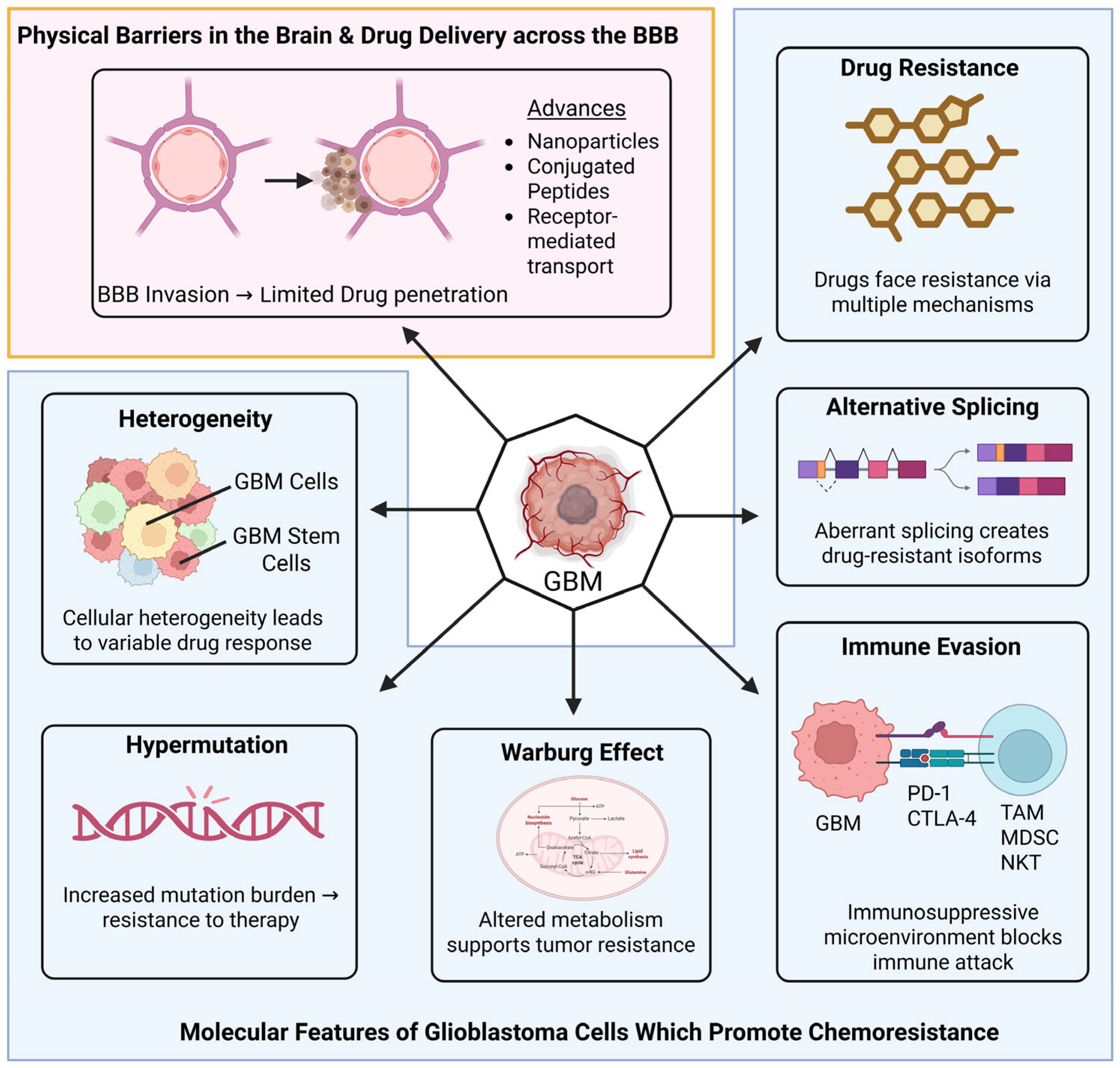
1.2. Key Barriers in Therapeutic Efficacy
1.2.1. The Blood–Brain Barrier (BBB)—Biological Constraints and Integrated Bypass Strategies
1.2.2. Molecular and Cellular Drivers of Drug Resistance
1.2.3. Gene Tumor Heterogeneity
1.2.4. Immunosuppressive Microenvironment
1.3. From Neuroimaging to Liquid Biopsy: Enhancing Diagnostic Precision
2. Recent Advances in Molecular Biology and/or GB Biomarkers
2.1. New Frontiers in Classification: Differentiating GB from Histological Mimics
- A.
- The “IDH-Mutant GBM” term is no longer recognized: Tumors that were previously called “IDH-mutant glioblastomas” are now classified as Astrocytoma, IDH-mutant, CNS WHO grade 4.
- B.
- Molecular vs. Histological Diagnosis: A tumor can be diagnosed as GB even if it lacks “classic” histological features like necrosis or microvascular proliferation, provided it carries specific molecular markers.
- C.
- The Essential Diagnostic Criteria: For a diffuse astrocytic glioma in an adult to be classified as GB, IDH-wildtype (Grade 4), it must be IDH-wildtype and H3-wildtype and meet at least one of the following criteria: molecular TERT-promoter mutations, EGFR gene amplification, or +7/−10 chromosome copy number alterations, allowing for a definitive Grade 4 diagnosis even in the absence of microvascular proliferation or necrosis.
- D.
- Key Molecular and Genetic Changes: The 6th Edition places higher emphasis on refined molecular profiling to distinguish GBM from “mimics” that may look identical under a microscope but have different clinical trajectories.
- E.
- Integrated “Layered” Diagnosis: The WHO 2026 format requires a “layered” reporting structure for glioblastoma to ensure all data are captured.
2.2. Prognostic Biomarkers and Emerging Molecular Targets in Glioblastoma
- Emerging Transcriptional Profiles: Recent studies have identified specific gene expression patterns in TMZ-resistant tumors, such as the upregulation of growth differentiation factor (GDF15), serum amyloid (SA) A1/2, downregulation of tyrosine kinase with immunoglobulin and epidermal growth factor homology domains 1 (TIE1), calcium voltage-gated channel auxiliary subunit α2Δ1 (CACNA2D1), calpain 6 (CAPN6) and a disintegrin and metalloproteinase with thrombospondin motifs 6 (ADAMTS6), which may serve as novel therapeutic targets [36].
2.3. Therapeutic Evasion: Investigating PI3K/AKT/mTOR and Beyond
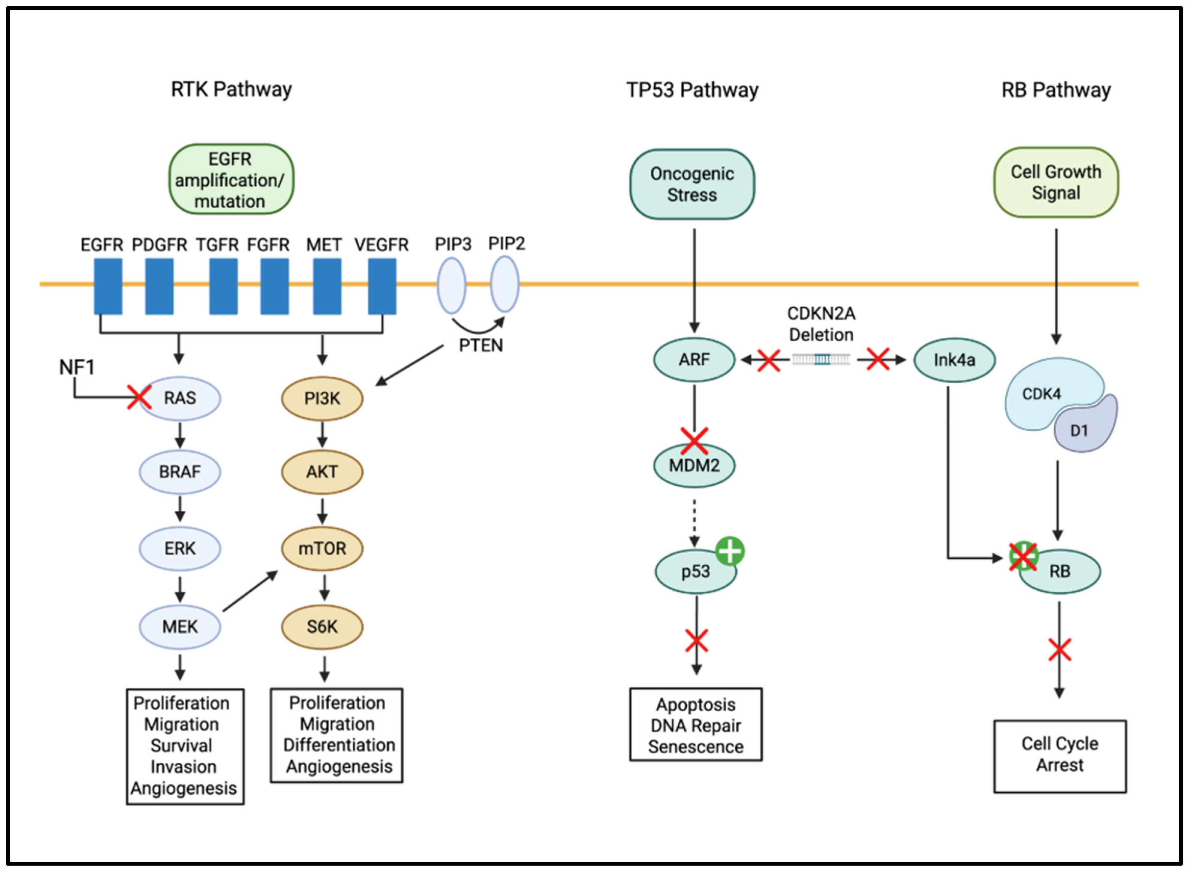
- RTK/RAS/PI3K Pathway: This is the most frequently altered pathway in GBM. It is often triggered by EGFR amplification or mutations, leading to the activation of PI3K and AKT. These molecules signal the cell to increase proliferation and resist programmed cell death [39,40]. p53 Signaling Pathway: Mutations in TP53 or alterations in its regulators (like mouse double minute (MDM2)) disrupt the cell’s ability to repair DNA damage or undergo apoptosis. This allows damaged cells to continue dividing, leading to rapid tumor growth [41].
2.4. The Hippo Signaling Pathway: A Hub for Multidrug Resistance and Therapeutic Targeting
2.5. MicroRNAs as Post-Transcriptional Regulators and Therapeutic Targets
3. Targeted Therapy Using Biomimetic Nano-Formulations (BNFs)
3.1. Rationale and Key Design Principles
3.2. Mechanisms of Action and Targeting Strategies of BNFs
- Homotypic targeting: tumor cell membrane coatings present adhesion molecules that preferentially bind parental tumor cells, improving accumulation in GB tissue [70].
- Immune camouflage and prolonged circulation: erythrocyte or platelet membranes provide “self” markers (e.g., CD47) to reduce phagocytosis and extend half-life [71].
- BBB transcytosis: surface display of ligands for BBB transporters (transferrin receptor, low-density lipoprotein receptor (LDLR)-related proteins) or use of cell carriers (macrophage-membrane, neutrophil-mimics) promotes receptor-mediated transcytosis or “Trojan-horse” migration into the brain [72].
- Multimodal payloads: BNFs are platforms for chemotherapy (TMZ, paclitaxel), siRNA/CRISPR payloads, metabolic inhibitors, photosensitizers or radionuclides, enabling combination therapy and theranostics [73].
3.3. Lipoprotein-Based Nanoparticles
3.4. Translational Status and Clinical Considerations
3.5. The Translational Gap: Biological Hurdles and Model Limitations
4. Advances in Cancer Stem Cells (CSCs) for GB Therapeutics
4.1. Harnessing Stem Cell Tropism for Targeted GB Therapy
4.2. Technological Advances Improving Delivery and Safety of GSCs
- (a)
- Genetic engineering of stem cells to express oncolytic viruses (or to carry replication-competent viral payloads) or suicide enzymes that convert systemically administered prodrugs into cytotoxins locally, thereby increasing intratumoral drug concentration while reducing systemic toxicity [101,102,103,104,105];
- (b)
- Locoregional administration (intra-tumoral, resection-cavity, intraventricular) to maximize contact with infiltrative cells; and
- (c)
4.3. Biomimetic Delivery via MSC Platforms: Opportunities and Safety Considerations
4.4. A Related and Fast-Moving Area Is Engineered Immune–Stem Cell Hybrids and Cell Therapies
4.5. Deconstructing GB Heterogeneity Through High-Resolution Mapping
4.6. Outlook and Challenges
5. Advances in Immunotherapy for GB
5.1. Immune Checkpoint Inhibitors (ICIs)
5.2. Chimeric Antigen Receptors (CAR)
5.3. Tumor Vaccines
5.4. Oncolytic Virotherapy (OV)
6. Bioinformatics and Machine Learning in GB Therapy
6.1. High-Throughput Omics Technologies
6.2. Machine Learning (ML) in GB Survival Prediction: Performance vs. Generalizability
6.3. Computational Frontiers in GB: Leveraging AI for Precision Oncology
7. Radiotherapeutics Advances in GB
7.1. Proton and Heavy Ion Therapy:
7.2. Cold Atmospheric Plasma (CAP)
7.3. Tumor-Treating Fields + Radiotherapy
7.4. Boron Neutron Capture Therapy (BNCT)
7.5. Radioimmunotherapy (RIT)
8. Advances in Imaging and Diagnostic Techniques in Neuro-Oncology
8.1. Radiomics and Radiogenomics:
8.2. Enhanced Neuroimaging: Indicators of BBB Integrity and Treatment Response
8.3. Diffusion Tensor Imaging (DTI) and Neurite Imaging: Mapping Microstructural Heterogeneity
8.4. Next-Generation Contrast Agents: Enhancing Signal Augmentation in GB
8.5. Liquid Biopsy Frontiers
9. Local Surgical Interventions and Cytoreductive Advancements
9.1. Fluorescence-Guided Resection
9.2. Stereotactic Radiosurgery (SRS) and Hypofractionation
9.3. Laser Interstitial Thermal Therapy (LITT)
10. Other Emerging/Investigational Adjuvant Therapeutic Approaches
10.1. The Ketogenic Diet (KD)
10.2. Focused Ultrasound (FUS)
10.3. Nose-to-Brain Delivery
10.4. NG101m
10.5. Neoadjuvant and Adjuvant Dual Checkpoint Blockade
10.6. Non-Coding RNA-Targeted Therapy and Drug Repurposing
10.7. Optimization of Treatment Scheduling
11. Comparative Synthesis of the Glioblastoma Translational Landscape
- Evidence Quality:
- ➢
- High: Supported by randomized controlled trials (RCTs) or large-scale multicenter prospective studies.
- ➢
- Moderate: Supported by Phase I/II trials, retrospective cohorts, or well-validated pilot studies.
- ➢
- Low: Primarily supported by in vitro or in vivo animal models.
- Typical Effect Size: Refers to the relative impact on overall survival (OS), progression-free survival (PFS), or diagnostic accuracy as reported in the primary literature.
- Clinical Readiness: Ranges from standard of care (widely implemented) to pre-clinical (not yet approved for human use).
- Translational Barrier: The primary technical or biological hurdle preventing the modality from achieving higher efficacy or broader clinical adoption.
12. Ongoing Clinical Trials on GB
12.1. Landmark Clinical Trials and Therapeutic Breakthroughs
- ▪
- The INDIGO trial NCT04164901, a Phase 3 study spearheaded by the Dana-Farber Cancer Institute, is investigating the efficacy of vorasidenib in patients with Grade 2 gliomas exhibiting IDH1 or IDH2 mutations [262]. The Phase I trial (NCT02481154) evaluated vorasidenib, a brain-penetrant dual IDH1/2 inhibitor, in patients with recurrent or progressive mutant IDH glioma. The drug was well tolerated, with reversible liver enzyme elevations as the main toxicity. In non-enhancing gliomas, vorasidenib achieved an 18% response rate and a median progression-free survival of 36.8 months, showing promising antitumor activity [263].
- ▪
- In another significant venture, UNC Health is conducting a Phase 2b clinical study (IGV-001) that examines a combination immunotherapy approach in newly diagnosed GB patients. This multicenter trial, aiming to enroll 93 participants, is poised to assess both the safety and efficacy of this novel therapeutic strategy. The trial underscores the increasing relevance of immunotherapy in the GB treatment paradigm [264].
- ▪
- The University of California, San Diego (UCSD) is actively engaged in a diverse array of clinical trials targeting GB. These trials encompass a range of strategies, including the investigation of drug-resistant immunotherapy that combines activated gene-modified T cells with temozolomide, as well as studies evaluating the efficacy of agents like berubicin and enzastaurin in conjunction with temozolomide. This portfolio of trials at UCSD illustrates the multifaceted nature of current research efforts in GB treatment [265].
- ▪
- Oncolytic virus therapy Teserpaturev (G47∆, “Delytact”) was approved in Japan for malignant glioma, including recurrent/residual GB and used via intratumoral injection. In clinical trials it showed an 84.2% one-year survival in a certain group, with a median overall survival of ~20.2 months [266].
- ▪
- Furthermore, the role of immunotherapy in GB is being explored through various trials, with a significant emphasis on nivolumab, an anti-PD-1 antibody. Trials such as CheckMate 143 and NCT02550249 have explored its application in both recurrent and newly diagnosed GB. Despite some trials not achieving their primary endpoints, they highlight the intricacies of GB treatment and the necessity for continued research in this arena [267]. Table 3 exhibits the scenario of current worldwide trials of GB.
12.2. Lessons from the Frontier: Analyzing Landmark Trial Failures

| Trial Name | NCT ID | Phase | Primary Endpoint(s) | Recruitment Status (as of Q1 2026) Reference | Est. Completion |
|---|---|---|---|---|---|
| GBM AGILE (Platform Trial) | NCT03970447 | II/III | Overall Survival (OS) | Active/Recruiting | December 2026 |
| IL13Rα2-CAR T cells ± Checkpoint | NCT04003649 | I | Safety/Dose-Limiting Toxicities (DLT) | Active/Recruiting | June 2027 |
| Chlorotoxin-domain CAR T (CTX-CAR) | NCT04214392 | I | Safety/MTD | Active/Recruiting | June 2026 |
| CART-EGFR-IL13Rα2 (Bivalent CAR) | NCT05168423 | I | Safety/Feasibility | Active/Recruiting | October 2025 * |
| [177Lu]Lu-DOTA-TATE in GBM | NCT05109728 | I | Safety/MTD | Active/Recruiting | July 2027 |
| Verteporfin Photodynamic Therapy | NCT04590664 | I/II | Safety/MTD | Active/Recruiting | August 2025 * |
| Lerapolturev (PVSRIPO) | NCT02986178 | II | OS at 24 months | Completed | Completed February 2023 |
| VBI-1901 (Dendritic Cell Vaccine) | NCT03382977 | I/II | Safety/Immunogenicity | Active/Recruiting | December 2026 |
| ReSPECT-GBM (186Re Obisbemeda) | NCT01906385 | I/II | Safety/MTD/PFS | Active/Recruiting | December 2025 * |
| IL13Rα2 CAR (New Structure) | NCT06355908 | I | Safety/MTD/RP2D | Suspended | May 2027 ** |
13. The Future of GB Therapy: A Clinician’s Perspective
Concluding Remarks
Author Contributions
Funding
Data Availability Statement
Acknowledgments
Conflicts of Interest
Abbreviations
| ACVR1 | Activin A Receptor Type I |
| ADAMTS6 | A Disintegrin And Metalloproteinase with Thrombospondin Motifs 6 |
| AKT | Protein Kinase B (PKB)—often referred to by its gene name AKT |
| ALK | Anaplastic Lymphoma Kinase |
| ATRX | Alpha Thalassemia/Mental Retardation Syndrome X-Linked |
| AUC | Area Under the Curve |
| BBB | Blood–Brain Barrier |
| BTB | Blood–Tumor Barrier |
| BNFs | Biomimetic Nano-Formulations |
| BNCT | Boron Neutron Capture Therapy |
| BRAF | B-Raf Proto-Oncogene |
| CACNA2D1 | Calcium Voltage-Gated Channel Auxiliary Subunit α2Δ1 |
| CAPN6 | Calpain 6 (Calcium-Dependent Cysteine Protease 6) |
| CAP | Cold Atmospheric Plasma |
| CARs | Chimeric Antigen Receptors |
| CD47 | Cluster of Differentiation 47 |
| CDKN2A/B | Cyclin-Dependent Kinase Inhibitor 2A and 2B |
| CSF | Cerebro Spinal Fluid |
| CIC | Capicua Transcriptional Repressor (downstream effector in RTK/MAPK signaling) |
| CIRT | Carbon Ion Radiotherapy |
| CNS | Central Nervous System |
| COX-2 | Cyclooxygenase-2 |
| CRISPR | Clustered Regularly Interspaced Short Palindromic Repeats |
| CSCs | Cancer Stem Cells |
| CSF | Cerebrospinal Fluid |
| CT | Computed Tomography |
| cfDM | Cell-Free DNA Methylation |
| ctDNA | Circulating DNA |
| CTCs | Circulating Tumor Cells |
| ctRNA | Circulating Tumor RNA |
| CTLA-4 | Cytotoxic T-Lymphocyte-Associated Protein 4 |
| ctDNA | Circulating Tumor DNA |
| DCs | Dendritic Cells |
| DTI | Diffusion Tensor Imaging |
| ECM | Extracellular Matrix |
| EphA2 | Ephrin Type-A Receptor 2 |
| EGFR | Epidermal Growth Factor Receptor |
| EOR | Extent of Resection |
| ER | Extracellular Signal-Regulated Kinase |
| EVs | Extracellular Vesicles |
| EZHIP | Enhancer of Zeste Homologs Inhibitory Protein |
| FGFR | Fibroblast Growth Factor Receptor |
| fMRI | Functional Magnetic Resonance Imaging |
| FUBP | Far Upstream Element (FUSE) Binding Protein |
| FUS | Focused Ultrasound |
| GB | Glioblastoma |
| GBM | Glioblastoma Multiforme |
| GD2 | Ganglioside 2 |
| GDF15 | Growth Differentiation Factor 15 |
| GLP | Good Laboratory Practice |
| GP130 | Glycoprotein 130 |
| GSCs | Glioma Stem Cells |
| HDL | High-Density Lipoprotein |
| HER2 | Human Epidermal Growth Factor Receptor 1 |
| HLA | Human Leukocyte Antigen |
| ICIs | Immune Checkpoint Inhibitors |
| IDH | Isocitrate Dehydrogenase |
| IDHwt | Isocitrate Dehydrogenase Wild Type |
| IKBKE | Inhibitor of Nuclear Factor Kappa-B Kinase Subunit Epsilon |
| IL-10 | Interleukin-10 |
| IL-13R⍺2 | Interleukin 13 receptor, Alpha 1 |
| KIAA1549 | KIAA1549 Gene |
| KD | Ketogenic Diet |
| LAG3 | Lymphocyte Activation Gene 3 |
| LATS1/2 | Large Tumor Suppressor Kinase 1 and 2 |
| LDL | Low-Density Lipoprotein |
| LDLR | Low-Density Lipoprotein Receptor |
| ML | Machine Learning |
| MAML2 | Mastermind-Like Transcriptional Coactivator 2 |
| MAPK | Mitogen-Activated Protein Kinase |
| MEK | Mitogen-Activated Protein Kinase (also known as MAP2K) |
| MCL-1 | Myeloid Cell Leukemia Protein 1 |
| MDSCs | Myeloid-Derived Suppressor Cells |
| MDM2 | Mouse Double Minute 2 |
| MET | MET Proto-Oncogene |
| MN1 | Meningioma 1 Gene |
| MGMT | O6-methylguanine-DNA Methyltransferase |
| miRNAs | Micro Ribonucleic Acid |
| MPS | Mononuclear Phagocyte System |
| MRI | Magnetic Resonance Imaging |
| MSCs | Mesenchymal Stem/Stromal Cells |
| mTOR | Mammalian (or mechanistic) Target of Rapamycin |
| MYB | Myeloblastosis |
| MYCN | v-Myc Avian Myelocytomatosis |
| NCCN | National Comprehensive Cancer Network |
| NODDI | Neurite Orientation Dispersion and Density Imaging |
| NF1 | Neurofibromin 1 |
| NK cell | Natural Killer Cells |
| NFKB | Nuclear Factor Kappa-Light-Chain-Enhancer of Activated B Cells |
| NKT | Natural Killer T cells |
| NOTCH | Notch Receptor Family |
| NP | Nanoparticle |
| NSCs | Neural Stem Cells |
| NTRK family | Neurotrophic Tyrosine Receptor Kinase Family |
| OS | Overall Survival |
| OV | Oncolytic Virotherapy |
| p53 | Tumor Protein p53 |
| PCSK9 | Proprotein Convertase Subtil-isin/kexin Type 9 |
| PD-1 | Programmed Cell Death Protein 1 |
| PDCD4 | Programmed Cell Death 4 |
| PD-L1 | Programmed Death-Ligand 1 |
| PDGFRA | Platelet-Derived Growth Factor Receptor Alpha |
| PGE2 | Prostaglandin E2 |
| PI3K | Phosphoinositide 3-kinase |
| PLGA | Poly(lactic-co-glycolic) Acid |
| PT | Proton Therapy |
| PTEN | Phosphatase and Tensin Homolog |
| RAF/MEK/ERK | The MAPK Cascade |
| RAF | Rapidly Accelerated Fibrosarcoma |
| RB | Retinoblastoma |
| RAS | Rat Sarcoma Virus |
| RELA | v-Rel Avian Reticuloendotheliosis Viral Oncogene Homolog A |
| ReSPOND | Report, Evaluate, Stabilize, Preserve, Organize, Normalize, Document/Debrief |
| rHDL | Reconstituted High-Density Lipoprotein |
| RIT | Radioimmunotherapy |
| ROS | Ras Oncogene from Esteosarcoma |
| ROC | Receiver Operating Characteristic curve |
| RT | Radiotherapy |
| RTK | Receptor Tyrosine Kinase |
| SAA1 | Serum Amyloid A1 |
| SAA2 | Serum Amyloid A2 |
| scRNA-seq | Single-Cell RNA Sequencing |
| SR-B1 | Scavenger Receptor Class B type 1 |
| siRNAs | Small Interfering RNAs |
| SHH | Sonic Hedgehog |
| SOC | Standard of Care |
| SRS | Stereotactic Radiosurgery |
| STING | Stimulator of Interferon Genes |
| TAAs | Tumor-Associated Antigens |
| TAMs | Tumor-Associated Macrophages |
| TAZ | Transcriptional Co-Activator with PDZ-binding Motif |
| TCIA | The Cancer Imaging Archive |
| TEPs | Tumor-Educated Platelets |
| TERT | Telomerase Reverse Transcriptase |
| TGF-ß | Transforming Growth Factor-Beta |
| TIE1 | Tyrosine Kinase with Immunoglobulin-Like and EGF-Like Domains 1 |
| TIGIT | T Cell Immunoreceptor with Ig and ITIM Domains |
| TMZ | Temozolomide |
| TP53 | Tumor Protein p53 |
| Tregs | Regulatory T Cells |
| TSAs | Tumor-Specific Antigens |
| TSC1/2 | Tuberous Sclerosis Complex 1 and 2 |
| TTFields | Tumor-Treating Fields |
| USP26 | Ubiquitin Specific Peptidase 26 |
| US FDA | United States Food and Drug Administration |
| WNT | Wingless/Integrated Signaling Pathway |
| WHO | World Health Organization |
| YAP1 | Yes-Associated Protein 1 |
| ZFTA | Zinc Finger Transcription Factor AT-Hook 1 |
References
- Satish, S.; Athavale, M.; Kharkar, P.S. Targeted therapies for Glioblastoma multiforme (GBM): State-of-the-art and future prospects. Drug Dev. Res. 2024, 85, e22261. [Google Scholar] [CrossRef] [Scilit] [PubMed]
- Kumari, S.; Gupta, R.; Ambasta, R.K.; Kumar, P. Multiple therapeutic approaches of glioblastoma multiforme: From terminal to therapy. Biochim. Biophys. Acta Rev. Cancer 2023, 1878, 188913. [Google Scholar] [CrossRef] [Scilit]
- Pouyan, A.; Ghorbanlo, M.; Eslami, M.; Jahanshahi, M.; Ziaei, E.; Salami, A.; Mokhtari, K.; Shahpasand, K.; Farahani, N.; Meybodi, T.E.; et al. Glioblastoma multiforme: Insights into pathogenesis, key signaling pathways, and therapeutic strategies. Mol. Cancer 2025, 24, 58. [Google Scholar] [CrossRef] [Scilit] [PubMed]
- Eisenbarth, D.; Wang, Y.A. Glioblastoma heterogeneity at single cell resolution. Oncogene 2023, 42, 2155–2165. [Google Scholar] [CrossRef] [Scilit] [PubMed]
- Stupp, R.; Mason, W.P.; van den Bent, M.J.; Weller, M.; Fisher, B.; Taphoorn, M.J.B.; Belanger, K.; Brandes, A.A.; Marosi, C.; Bogdahn, U.; et al. Radiotherapy plus concomitant and adjuvant temozolomide for glioblastoma. N. Engl. J. Med. 2005, 352, 987–996. [Google Scholar] [CrossRef] [Scilit]
- McKinnon, C.; Nandhabalan, M.; Murray, S.A.; Plaha, P. Glioblastoma: Clinical presentation, diagnosis, and management. BMJ 2021, 374, n1560. [Google Scholar] [CrossRef] [Scilit] [PubMed]
- Stupp, R.; Taillibert, S.; Kanner, A.; Read, W.; Steinberg, S.; Lhermitte, B.; Toms, S.; Idbaih, A.; Ahluwalia, M.S.; Fink, K.; et al. Effect of tumor-treating fields plus maintenance temozolomide vs maintenance temozolomide alone on survival in patients with glioblastoma: A randomized clinical trial. JAMA 2017, 318, 2306–2316. [Google Scholar] [CrossRef] [Scilit] [PubMed]
- Lassman, A.B.; Joanta-Gomez, A.E.; Pan, P.C.; Wick, W. Current usage of tumor treating fields for glioblastoma. Neuro-Oncol. Adv. 2020, 2, vdaa069. [Google Scholar] [CrossRef] [Scilit]
- Fabian, D.; Guillermo Prieto Eibl, M.D.P.; Alnahhas, I.; Sebastian, N.; Giglio, P.; Puduvalli, V.; Gonzalez, J.; Palmer, J.D. Treatment of glioblastoma (GBM) with the addition of tumor-treating fields (TTF): A review. Cancers 2019, 11, 174. [Google Scholar] [CrossRef] [Scilit]
- Santhi, J.L.A.; Ali, A.; Vegivinti, C.T.R.; Bodrova, E.; Mehta, M.; Varghese, R.M.; Sriram, V.; Thirumaran, R. EPR25-138: Glioblastoma Multiforme: A SEER STAT Analysis of Epidemiological, Laterality, and Anatomical Prognostic Indicators. J. Natl. Compr. Cancer Netw. 2025, 23, EPR25-138. [Google Scholar] [CrossRef] [Scilit]
- Nozhat, Z.; Heydarzadeh, S.; Shahriari-Khalaji, M.; Wang, S.; Iqbal, M.Z.; Kong, X. Advanced biomaterials for human glioblastoma multiforme (GBM) drug delivery. Biomater. Sci. 2023, 11, 4094–4131. [Google Scholar] [CrossRef] [Scilit] [PubMed]
- Ahmed, M.H.; Canney, M.; Carpentier, A.; Idbaih, A. Overcoming the blood brain barrier in glioblastoma: Status and future perspective. Rev. Neurol. 2023, 179, 430–436. [Google Scholar] [CrossRef] [Scilit] [PubMed]
- Digiovanni, S.; Lorenzati, M.; Bianciotto, O.T.; Godel, M.; Fontana, S.; Akman, M.; Costamagna, C.; Couraud, P.O.; Buffo, A.; Kopecka, J.; et al. Blood-brain barrier permeability increases with the differentiation of glioblastoma cells in vitro. Fluids Barriers CNS 2024, 21, 89. [Google Scholar] [CrossRef] [Scilit]
- Zhang, Y.; Xi, K.; Zhang, Y.; Fang, Z.; Zhang, Y.; Zhao, K.; Feng, F.; Shen, J.; Wang, M.; Zhang, R.; et al. Blood-Brain Barrier Penetrating Nanovehicles for Interfering with Mitochondrial Electron Flow in Glioblastoma. ACS Nano 2024, 18, 9511–9524. [Google Scholar] [CrossRef] [Scilit]
- Li, Y.; Bao, K.; Huan, R.; Wang, T.; Wang, Y.; Wu, S.; Chen, S.; Huang, J.; Zhu, L.; Li, J.; et al. RAP-peptide functionalized biomimetic nanoformulations with pathological ROS/pH-responsive drug release for target immunotherapy in glioma. J. Nanobiotechnol. 2026, 24, 225. [Google Scholar] [CrossRef] [Scilit]
- Xu, J.; Zhang, J.; Chen, W.; Ni, X. The tumor-associated fibrotic reactions in microenvironment aggravate glioma chemoresistance. Front. Oncol. 2024, 14, 1388700. [Google Scholar] [CrossRef] [Scilit] [PubMed]
- Sherman, J.H.; Bobak, A.; Arsiwala, T.; Lockman, P.; Aulakh, S. Targeting drug resistance in glioblastoma (Review). Int. J. Oncol. 2024, 65, 80. [Google Scholar] [CrossRef] [Scilit] [PubMed]
- Onishi, S.; Kajiwara, Y.; Takayasu, T.; Kolakshyapati, M.; Ishifuro, M.; Amatya, V.J.; Takeshima, Y.; Sugiyama, K.; Kurisu, K.; Yamasaki, F. Perfusion Computed Tomography Parameters Are Useful for Differentiating Glioblastoma, Lymphoma, and Metastasis. World Neurosurg. 2018, 119, e890–e897. [Google Scholar] [CrossRef] [Scilit] [PubMed]
- Chen, L.; Chen, R.; Li, T.; Huang, L.; Tang, C.; Li, Y.; Zeng, Z. MRI radiomics model for predicting TERT promoter mutation status in glioblastoma. Brain Behav. 2023, 13, e3324. [Google Scholar] [CrossRef] [Scilit] [PubMed]
- Lan, Z.; Li, X.; Zhang, X. Glioblastoma: An Update in Pathology, Molecular Mechanisms and Biomarkers. Int. J. Mol. Sci. 2024, 25, 3040. [Google Scholar] [CrossRef] [Scilit] [PubMed]
- Cheung, E.Y.W.; Wu, R.W.K.; Li, A.S.M.; Chu, E.S.M. AI Deployment on GBM Diagnosis: A Novel Approach to Analyze Histopathological Images Using Image Feature-Based Analysis. Cancers 2023, 15, 5063. [Google Scholar] [CrossRef] [Scilit] [PubMed]
- Bonosi, L.; Ferini, G.; Giammalva, G.R.; Benigno, U.E.; Porzio, M.; Giovannini, E.A.; Musso, S.; Gerardi, R.M.; Brunasso, L.; Costanzo, R.; et al. Liquid Biopsy in Diagnosis and Prognosis of High-Grade Gliomas; State-of-the-Art and Literature Review. Life 2022, 2, 407. [Google Scholar] [CrossRef] [Scilit] [PubMed]
- Ronvaux, L.; Riva, M.; Coosemans, A.; Herzog, M.; Rommelaere, G.; Donis, N.; D’Hondt, L.; Douxfils, J. Liquid Biopsy in Glioblastoma. Cancers 2022, 14, 3394. [Google Scholar] [CrossRef] [Scilit] [PubMed]
- Seyhan, A.A. Circulating Liquid Biopsy Biomarkers in Glioblastoma: Advances and Challenges. Int. J. Mol. Sci. 2024, 25, 7974. [Google Scholar] [CrossRef] [Scilit] [PubMed]
- Gatto, L.; Franceschi, E.; Di Nunno, V.; Tosoni, A.; Lodi, R.; Brandes, A.A. Liquid Biopsy in Glioblastoma Management: From Current Research to Future Perspectives. Oncologist 2021, 26, 865–878. [Google Scholar] [CrossRef] [Scilit] [PubMed]
- Louis, D.N.; Perry, A.; Wesseling, P.; Brat, D.J.; Cree, I.A.; Figarella-Branger, D.; Hawkins, C.; Ng, H.K.; Pfiste, S.M.; Reifenberger, G.; et al. The 2021 WHO Classification of Tumors of the Central Nervous System: A summary. Neuro-Oncology 2021, 23, 1231–1251. [Google Scholar] [CrossRef] [Scilit]
- Torp, S.H.; Solheim, O.; Skjulsvik, A.J. The WHO 2021 Classification of Central Nervous System tumours: A practical update on what neurosurgeons need to know—A minireview. Acta Neurochir. 2022, 164, 2453–2464. [Google Scholar] [CrossRef] [Scilit]
- World Health Organization. Central Nervous System Tumours, 6th ed.; WHO Classification of Tumours; International Agency for Research on Cancer: Lyon, France, 2026; Volume 1. [Google Scholar]
- Verhaak, R.G.; Hoadley, K.A.; Purdom, E.; Wang, V.; Qi, Y.; Wilkerson, M.D.; Miller, C.R.; Ding, L.; Golub, T.; Mesirov, J.P.; et al. Integrated Genomic Analysis Identifies Clinically Relevant Subtypes of Glioblastoma Characterized by Abnormalities in PDGFRA, IDH1, EGFR, and NF1. Cancer Cell 2010, 17, 98–110. [Google Scholar] [CrossRef] [Scilit]
- Wang, Q.; Hu, B.; Hu, X.; Kim, H.; Squatrito, M.; Scarpace, L.; deCarvalho, A.C.; Lyu, S.; Li, P.; Li, Y.; et al. Tumor Evolution of Glioma-Intrinsic Gene Expression Subtypes Associates with Immunological Changes in the Microenvironment. Cancer Cell 2017, 32, 42–56.e6. [Google Scholar] [CrossRef] [Scilit] [PubMed]
- Bikfalvi, A.; da Costa, C.A.; Avril, T.; Barnier, J.V.; Bauchet, L.; Brisson, L.; Cartron, P.F.; Castel, H.; Chevet, E.; Chneiweiss, H.; et al. Challenges in glioblastoma research: Focus on the tumor microenvironment. Trends Cancer 2023, 9, 9–27. [Google Scholar] [CrossRef] [Scilit] [PubMed]
- Ben Dor, N.; Friso, F.; Ziv, G.; Bortolotti, C.; Martinoni, M.; Badaloni, F.; Corazzelli, G.; Rustici, A.; Balestrini, D.; Scibilia, A.; et al. Machine Learning-Enhanced Prognostic Modeling in Elderly Glioblastoma Isocitrate Dehydrogenase-Wildtype: A Multidimensional Single-Center Cohort Study. World Neurosurg. 2025, 206, 124754. [Google Scholar] [CrossRef] [Scilit] [PubMed]
- Dovek, L.; Nguyen, N.T.; Ozer, B.H.; Li, N.; Elashoff, R.M.; Green, R.M.; Liau, L.; Nghiemphu, P.L.; Cloughesy, T.F.; Lai, A. Correlation of commercially available quantitative MGMT (O-6-methylguanine-DNA methyltransferase) promoter methylation scores and GBM patient survival. Neuro-Oncol. Pract. 2019, 6, 194–202. [Google Scholar] [PubMed]
- Zhang, P.; Liu, D.; Yu, T.; Zhang, Y.; Zhong, L.; Ouyang, X.; Xia, Q.; Dong, L. Integrated pathway analysis identifies prognostically relevant subtypes of glioblastoma characterized by abnormalities in multi-omics. Clin. Transl. Med. 2025, 15, e70517. [Google Scholar] [CrossRef] [Scilit] [PubMed]
- Frattini, V.; Trifonov, V.; Chan, J.M.; Castano, A.; Lia, M.; Abate, F.; Keir, S.T.; Ji, A.X.; Zoppoli, P.; Niola, F.; et al. The integrated landscape of driver genomic alterations in glioblastoma. Nat. Genet. 2013, 45, 1141–1149. [Google Scholar] [CrossRef] [Scilit]
- Chen, W.-F.; Chuang, J.M.-J.; Yang, S.-N.; Chen, N.-F.; Bhattacharya, M.; Liu, H.-T.; Dhama, K.; Chakraborty, C.; Wen, Z.-F. Gene expression profiling and the isocitrate dehydrogenase mutational landscape of temozolomide-resistant glioblastoma. Oncol. Lett. 2024, 28, 378. [Google Scholar] [CrossRef] [Scilit]
- Hasan, H.; Afzal, M.; Castresana, J.S.; Shahi, M.H. A Comprehensive Review of miRNAs and Their Epigenetic Effects in Glioblastoma. Cells 2023, 12, 1578. [Google Scholar] [CrossRef] [Scilit]
- Behrooz, A.B.; Latifi-Navid, H.; Nezhadi, A.; Świat, M.; Los, M.; Jamalpoor, Z.; Ghavami, S. Molecular mechanisms of microRNAs in glioblastoma pathogenesis. Biochim. Biophys. Acta Mol. Cell Res. 2023, 870, 119482. [Google Scholar] [CrossRef] [Scilit]
- Dymova, M.Y.; Kuligina, E.V.; Richter, V.A. Molecular Mechanisms of Drug Resistance in Glioblastoma. Int. J. Mol. Sci. 2021, 22, 6385. [Google Scholar] [CrossRef] [Scilit]
- Oronsky, B.; Reid, T.R.; Oronsky, A.; Sandhu, N.; Knox, S.J. A Review of Newly Diagnosed Glioblastoma. Front. Oncol. 2021, 10, 574012. [Google Scholar] [CrossRef] [Scilit]
- Berezovsky, A.; Nuga, O.; Datta, I.; Bergman, K.; Sabedot, T.; Gurdziel, K.; Irtenkauf, S.; Hasselbach, L.; Meng, Y.; Mueller, C.; et al. Impact of developmental state, p53 status, and interferon signaling on glioblastoma cell response to radiation and temozolomide treatment. PLoS ONE 2025, 20, e0315171. [Google Scholar] [CrossRef] [Scilit]
- Chkheidze, R.; Raisanen, J.; Gagan, J.; Richardson, T.E.; Pinho, M.C.; Raj, K.; Achilleos, M.; Slepicka, C.; White, C.L.; Evers, B.M.; et al. Alterations in the RB pathway with inactivation of characterize Glioblastomas with a primitive neuronal component. J. Neuropathol. Exp. Neurol. 2021, 80, 1092–1098. [Google Scholar] [CrossRef] [Scilit]
- Carpenter, A.B.; Sacknovitz, A.; Hanft, S.; Gandhi, C.D.; Jhanwar-Uniyal, M. Complex role of mTOR signaling pathway in glioblastoma and its stem cells. Adv. Biol. Regul. 2025, 10, 101143. [Google Scholar] [CrossRef] [Scilit]
- Liu, H.; Qiu, W.; Sun, T.; Wang, L.; Du, L.; Hu, Y.; Liu, W.; Feng, F.; Chen, Y.; Sun, H. Therapeutic strategies of glioblastoma (GBM): The current advances in the molecular targets and bioactive small molecule compounds. Acta Pharm. Sin. B 2022, 12, 1781–1804. [Google Scholar] [CrossRef] [Scilit]
- Hashemi, M.; Etemad, S.; Rezaei, S.; Ziaolhagh, S.; Rajabi, R.; Rahmanian, P.; Abdi, S.; Koohpar, Z.K.; Rafiei, R.; Raei, B.; et al. Progress in targeting PTEN/PI3K/Akt axis in glioblastoma therapy: Revisiting molecular interactions. Biomed. Pharmacother. 2023, 158, 114204. [Google Scholar] [CrossRef] [Scilit] [PubMed]
- Casati, G.; Giunti, L.; Iorio, A.L.; Marturano, A.; Galli, L.; Sardi, I. Hippo Pathway in Regulating Drug Resistance of Glioblastoma. Int. J. Mol. Sci. 2021, 22, 13431. [Google Scholar] [CrossRef] [Scilit] [PubMed]
- Masliantsev, K.; Desette, A.; Gonzalez, A.-A.; Garrouche, I.; Noblanc, A.; Soulard, M.; Triquard, M.; Milin, S.; Wager, M.; Karayan-Tapon, L.; et al. Hippo pathway effectors are associated with glioma patient survival, control cell proliferation and sterol metabolism through TEAD3. Brain Pathol. 2025, 35, e70021. [Google Scholar] [CrossRef] [Scilit]
- Wang, H.; Xu, T.; Jiang, Y.; Yan, Y.; Qin, R.; Chen, J. MicroRNAs in human glioblastoma: From bench to beside. Front. Biosci. (Landmark Ed.) 2015, 20, 105–118. [Google Scholar] [CrossRef] [Scilit] [PubMed]
- Makowska, M.; Smolarz, B.; Romanowicz, H. microRNAs (miRNAs) in Glioblastoma Multiforme (GBM)-Recent Literature Review. Int. J. Mol. Sci. 2023, 24, 3521. [Google Scholar] [CrossRef] [Scilit]
- Epistolio, S.; Spina, P.; Zaed, I.; Cardia, A.; Marchi, F.; Frattini, M. The clinical role of miRNAs in the development and treatment of Glioblastoma. Int. J. Mol. Sci. 2025, 26, 2723. [Google Scholar] [CrossRef] [Scilit] [PubMed]
- Elshaer, S.S.; Abulsoud, A.I.; Fathi, D.; Abdelmaksoud, N.M.; Zaki, M.B.; El-Mahdy, H.A.; Ismail, A.; Elsakka, E.G.E.; Abd-Elmawla, M.A.; Abulsoud, L.A.; et al. miRNAs role in glioblastoma pathogenesis and targeted therapy: Signaling pathways interplay. Pathol. Res. Pract. 2023, 246, 154511. [Google Scholar] [CrossRef] [Scilit] [PubMed]
- Bassot, A.; Dragic, H.; Haddad, S.A.; Moindrot, L.; Odouard, S.; Corlazzoli, F.; Marinari, E.; Bomane, A.; Brassens, A.; Marteyn, A.; et al. Identification of a miRNA multi-targeting therapeutic strategy in glioblastoma. Cell Death Dis. 2023, 14, 630. [Google Scholar] [CrossRef] [Scilit] [PubMed]
- Sales, A.H.A.; Beck, J.; Schnell, O.; Fung, C.; Meyer, B.; Gempt, J. Surgical Treatment of Glioblastoma: State-of-the-Art and Future Trends. J. Clin. Med. 2022, 11, 5354. [Google Scholar] [CrossRef] [Scilit]
- Wu, W.; Klockow, J.L.; Zhang, M.; Lafortune, F.; Chang, E.; Jin, L.; Wu, Y.; Daldrup-Link, H.E. Glioblastoma Multiforme (GBM): An Overview of Current Therapies and Mechanisms of Resistance. Pharmacol. Res. 2021, 171, 105780. [Google Scholar] [CrossRef] [Scilit]
- Pandey, N.; Anastasiadis, P.; Carney, C.P.; Kanvinde, P.P.; Woodworth, G.F.; Winkles, J.A.; Kim, A.J. Nanotherapeutic Treatment of the Invasive Glioblastoma Tumor Microenvironment. Adv. Drug Deliv. Rev. 2022, 188, 114415. [Google Scholar] [CrossRef] [Scilit]
- Silver, A.; Feier, D.; Ghosh, T.; Rahman, M.; Huang, J.; Sarkisian, M.R.; Deleyrolle, L.P. Heterogeneity of Glioblastoma Stem Cells in the Context of the Immune Microenvironment and Geospatial Organization. Front. Oncol. 2022, 12, 1022716. [Google Scholar] [CrossRef] [Scilit] [PubMed]
- Pandit, R.; Chen, L.; Götz, J. The Blood-Brain Barrier: Physiology and Strategies for Drug Delivery. Adv. Drug Deliv. Rev. 2020, 165–166, 1–14. [Google Scholar] [CrossRef] [Scilit] [PubMed]
- Wu, D.; Chen, Q.; Chen, X.; Han, F.; Chen, Z.; Wang, Y. The Blood-Brain Barrier: Structure, Regulation, and Drug Delivery. Signal Transduct. Target. Ther. 2023, 8, 217. [Google Scholar] [CrossRef] [Scilit]
- Zhao, Y.; Yue, P.; Peng, Y.; Sun, Y.; Chen, X.; Zhao, Z.; Han, B. Recent Advances in Drug Delivery Systems for Targeting Brain Tumors. Drug Deliv. 2023, 30, 1–18. [Google Scholar] [CrossRef] [Scilit]
- Barar, J.; Rafi, M.A.; Pourseif, M.M.; Omidi, Y. Blood-brain barrier transport machineries and targeted therapy of brain diseases. BioImpacts 2016, 6, 225–248. [Google Scholar] [CrossRef] [Scilit]
- Hawkins, R.A.; O’kane, R.L.; Simpson, I.A.; Vina, J.R. Structure of the blood-brain barrier and its role in the transport of amino acids. J. Nutr. 2006, 136, 218S–226S. [Google Scholar] [CrossRef] [Scilit]
- Li, J.; Guo, Y.; Kuang, Y.; An, S.; Ma, H.; Jiang, C. Choline transporter-targeting and co-delivery system for glioma therapy. Biomaterials 2013, 34, 9142–9148. [Google Scholar] [CrossRef] [Scilit]
- Straehla, J.P.; Hajal, C.; Safford, H.C.; Offeddu, G.S.; Boehnke, N.; Dacoba, T.G.; Wyckoff, J.; Kamm, R.D.; Hammond, P.D. A predictive microfluidic model of human glioblastoma to assess trafficking of blood-brain barrier-penetrant nanoparticles. Proc. Natl. Acad. Sci. USA 2022, 119, e2118697119. [Google Scholar] [CrossRef] [Scilit]
- Ganipineni, L.P.; Danhier, F.; Préat, V. Drug Delivery Challenges and Future of Chemotherapeutic Nanomedicine for Glioblastoma Treatment. J. Control. Release 2018, 281, 42–57. [Google Scholar] [CrossRef] [Scilit]
- Xing, Y.; Yasinjan, F.; Yang, M.; Du, Y.; Geng, H.; He, M.; Wang, Y.; Sun, J.; Jiang, W.; Zhang, L.; et al. A Scientometric Analysis and Up-to-Date Review of Nano-Based Drug Delivery Systems in Glioblastoma Treatment. Nano Today 2023, 52, 101961. [Google Scholar] [CrossRef] [Scilit]
- Quader, S.; Kataoka, K.; Cabral, H. Nanomedicine for Brain Cancer. Adv. Drug Deliv. Rev. 2022, 182, 114115. [Google Scholar] [CrossRef] [Scilit]
- Mendes, M.; Sousa, J.J.; Pais, A.; Vitorino, C. Targeted Theranostic Nanoparticles for Brain Tumor Treatment. Pharmaceutics 2018, 10, 181. [Google Scholar] [CrossRef] [Scilit]
- Lu, G.; Wang, X.; Li, F.; Wang, S.; Zhao, J.; Wang, J.; Liu, J.; Lyu, C.; Ye, P.; Tan, H.; et al. Engineered biomimetic nanoparticles achieve targeted delivery and efficient metabolism-based synergistic therapy against glioblastoma. Nat. Commun. 2022, 13, 4214. [Google Scholar] [CrossRef] [Scilit]
- Duan, M.; Cao, R.; Yang, Y.; Chen, X.; Liu, L.; Ren, B.; Wang, L.; Goh, B.-N. Blood–Brain Barrier Conquest in Glioblastoma Nanomedicine: Strategies, Clinical Advances, and Emerging Challenges. Cancers 2024, 16, 3300. [Google Scholar] [CrossRef] [Scilit]
- Allami, P.; Heidari, A.; Rezaei, N. The role of cell membrane-coated nanoparticles as a novel treatment approach in glioblastoma. Front. Mol. Biosci. 2023, 9, 1083645. [Google Scholar] [CrossRef] [Scilit]
- Yang, J.; Li, X.; Li, T.; Mei, J.; Chen, Y. Recent advances in biomimetic nanodelivery systems for cancer Immunotherapy. Mater. Today Bio 2025, 32, 101726. [Google Scholar] [CrossRef] [Scilit]
- Fernández-Borbolla, A.; García-Hevia, L.; Fanarraga, M.L. Cell Membrane-Coated Nanoparticles for Precision Medicine: A Comprehensive Review of Coating Techniques for Tissue-Specific Therapeutics. Int. J. Mol. Sci. 2024, 25, 2071. [Google Scholar] [CrossRef] [Scilit]
- Liu, P.; Lan, S.; Gao, D.; Hu, D.; Chen, Z.; Li, Z.; Jiang, G.; Sheng, Z. Targeted blood-brain barrier penetration and precise imaging of infiltrative glioblastoma margins using hybrid cell membrane-coated ICG liposomes. J. Nanobiotechnol. 2024, 22, 603. [Google Scholar] [CrossRef] [Scilit]
- Huang, J.L.; Jiang, G.; Song, Q.X.; Gu, X.; Song, H.H.; Wang, X.L.; Jiang, D.; Kang, T.; Feng, X.Y.; Jiang, X.G.; et al. High-density lipoprotein-biomimetic nanocarriers for glioblastoma-targeting delivery: The effect of shape. Pharmazie 2016, 71, 709–714. [Google Scholar] [CrossRef] [Scilit] [PubMed]
- Wang, R.; Zhang, X.; Feng, K.; Zeng, W.; Wu, J.; Sun, D.; Lu, Z.; Feng, H.; Di, L. Nanotechnologies meeting natural sources: Engineered lipoproteins for precise brain disease theranostics. Asian J. Pharm. Sci. 2023, 18, 100857. [Google Scholar] [CrossRef] [Scilit]
- Raut, S.; Mooberry, L.; Sabnis, N.; Garud, A.; Dossou, A.S.; Lacko, A. Reconstituted HDL: Drug Delivery Platform for Overcoming Biological Barriers to Cancer Therapy. Front. Pharmacol. 2018, 9, 1154. [Google Scholar] [CrossRef] [Scilit]
- Nikanjam, M.; Gibbs, A.R.; Hunt, C.A.; Budinger, T.F.; Forte, T.M. Synthetic nano-LDL with paclitaxel oleate as a targeted drug delivery vehicle for glioblastoma multiforme. J. Control. Release 2007, 124, 163–171. [Google Scholar] [CrossRef] [Scilit]
- Kadiyala, P.; Li, D.; Nuñez, F.M.; Altshuler, D.; Doherty, R.; Kuai, R.; Yu, M.; Kamran, N.; Edwards, M.; Moon, J.J.; et al. High-Density Lipoprotein-Mimicking Nanodiscs for Chemo-immunotherapy against Glioblastoma Multiforme. ACS Nano 2019, 13, 1365–1384. [Google Scholar] [CrossRef] [Scilit]
- Kapic, A.; Sabnis, N.; Dossou, A.S.; Chavez, J.; Ceresa, L.; Gryczynski, Z.; Fudala, R.; Dickerman, R.; Bunnell, B.A.; Lacko, A.G. Photophysical Characterization and In Vitro Evaluation of α-Mangostin-Loaded HDL Mimetic Nano-Complex in LN-229 Glioblastoma Spheroid Model. Int. J. Mol. Sci. 2024, 25, 7378. [Google Scholar] [CrossRef] [Scilit]
- Huang, J.L.; Jiang, G.; Song, Q.X.; Gu, X.; Hu, M.; Wang, X.L.; Song, H.H.; Chen, L.P.; Lin, Y.Y.; Jiang, D.; et al. Lipoprotein-biomimetic nanostructure enables efficient targeting delivery of siRNA to Ras-activated glioblastoma cells via macropinocytosis. Nat. Commun. 2017, 8, 15144. [Google Scholar] [CrossRef] [Scilit] [PubMed]
- Ihnatsyeu-Kachan, A.; Saichuk, A.; Sharko, O.; Zhogla, V.; Abashkin, V.; Le, G.W.; Shmanai, V.; Shcharbin, D.; Guillas, I.; Kim, S. Biomimetic high-density lipoprotein nanoparticles for the delivery of nucleic acid-based therapeutics. Biotechnol. Adv. 2025, 83, 108606. [Google Scholar] [CrossRef] [Scilit] [PubMed]
- Delk, S.C.; Chattopadhyay, A.; Escola-Gil, J.C.; Fogelman, A.M.; Reddy, S.T. Apolipoprotein mimetics in cancer. Semin. Cancer Biol. 2021, 73, 158–168. [Google Scholar] [CrossRef] [Scilit]
- Tang, C.; Wang, Y.; Wu, M.; Wang, Z.; Zhou, Y.; Lin, Y.; Wang, Y.; Xu, H. Cancer cell membrane-camouflaged biomimetic nanoparticles for enhancing chemo-radiation therapy efficacy in glioma. J. Biomed. Res. 2024, 39, 87–102. [Google Scholar] [PubMed]
- Zou, Y.; Wang, Y.; Xu, S.; Liu, Y.; Yin, J.; Lovejoy, D.B.; Zheng, M.; Liang, X.J.; Park, J.B.; Efremov, Y.M.; et al. Brain Co-Delivery of Temozolomide and Cisplatin for Combinatorial Glioblastoma Chemotherapy. Adv. Mater. 2022, 34, e2203958. [Google Scholar] [CrossRef] [Scilit]
- Ismail, M.; Yang, W.; Li, Y.; Wang, Y.; He, W.; Wang, J.; Muhammad, P.; Chaston, T.B.; Rehman, F.U.; Zheng, M.; et al. Biomimetic Dp44mT-nanoparticles selectively induce apoptosis in Cu-loaded glioblastoma resulting in potent growth inhibition. Biomaterials 2022, 289, 121760. [Google Scholar] [CrossRef] [Scilit] [PubMed]
- Shan, T.; Wang, W.; Fan, M.; Bi, J.; He, T.; Sun, Y.; Zheng, M.; Yan, D. Effective glioblastoma immune sonodynamic treatment mediated by macrophage cell membrane cloaked biomimetic nanomedicines. J. Control. Release 2024, 370, 866–878. [Google Scholar] [CrossRef] [Scilit]
- Zhang, D.; Tian, S.; Liu, Y.; Zheng, M.; Yang, X.; Zou, Y.; Shi, B.; Luo, L. Near infrared-activatable biomimetic nanogels enabling deep tumor drug penetration inhibit orthotopic glioblastoma. Nat. Commun. 2022, 13, 6835. [Google Scholar] [CrossRef] [Scilit]
- Li, J.; Xu, X.; Zhao, Y.; Wu, X.; Chen, S.; Xu, J.; Xiao, R.; Tang, J.; Hong, X.; Chen, S.; et al. Microglial Membrane-Coated Biomimetic Nanoplatform for Enhanced Blood-Brain Barrier Penetration and Targeted Photodynamic Therapy in Orthotopic Glioblastoma. Adv. Healthc. Mater. 2026, 15, e02808. [Google Scholar] [CrossRef] [Scilit] [PubMed]
- Branco, F.; Cunha, J.; Mendes, M.; Vitorino, C.; Sousa, J.J. Peptide-Hitchhiking for the Development of Nanosystems in Glioblastoma. ACS Nano 2024, 18, 16359–16394. [Google Scholar] [CrossRef] [Scilit]
- Rahman, M.A.; Jalouli, M.; Yadab, M.K.; Al-Zharani, M. Progress in Drug Delivery Systems Based on Nanoparticles for Improved Glioblastoma Therapy: Addressing Challenges and Investigating Opportunities. Cancers 2025, 17, 701. [Google Scholar] [CrossRef] [Scilit]
- Bardhan, M.; Muneer, M.A.; Khare, A.; Shah, R.M.; Anmol, K.; Sonit, S.P.; Suresh, V.; Podder, V.; Ahluwalia, M.; Odia, Y.; et al. Advances in stem cell-based therapeutic transfers for glioblastoma (systematic review). Expert Rev. Neurother. 2025, 25, 699–715. [Google Scholar] [CrossRef] [Scilit]
- Agosti, E.; Antonietti, S.; Ius, T.; Fontanella, M.M.; Zeppieri, M.; Panciani, P.P. A Systematic Review of Mesenchymal Stem Cell-Derived Extracellular Vesicles: A Potential Treatment for Glioblastoma. Brain Sci. 2024, 14, 1058. [Google Scholar] [CrossRef] [Scilit] [PubMed]
- Mahdi, A.; Aittaleb, M.; Tissir, F. Targeting Glioma Stem Cells: Therapeutic Opportunities and Challenges. Cells 2025, 14, 675. [Google Scholar] [CrossRef] [Scilit]
- Biserova, K.; Jakovlevs, A.; Uljanovs, R.; Strumfa, I. Cancer Stem Cells: Significance in Origin, Pathogenesis and Treatment of Glioblastoma. Cell 2021, 10, 621. [Google Scholar] [CrossRef] [Scilit] [PubMed]
- Khedri, A.; Guo, S.; Ramar, V.; Hudson, B.; Liu, M. FOSL1’s Oncogene Roles in Glioma/Glioma Stem Cells and Tumorigenesis: A Comprehensive Review. Int. J. Mol. Sci. 2024, 25, 5362. [Google Scholar] [CrossRef] [Scilit] [PubMed]
- Fares, J.; Ahmed, A.U.; Ulasov, I.V.; Sonabend, A.M.; Miska, J.; Lee-Chang, C.; Balyasnikova, I.V.; Chandler, J.P.; Portnow, J.; Tate, M.C.; et al. Neural stem cell delivery of an oncolytic adenovirus in newly diagnosed malignant glioma: A first-in-human, phase 1, dose-escalation trial. Lancet Oncol. 2021, 22, 1103–1114. [Google Scholar] [CrossRef] [Scilit]
- Ramos-Zúñiga, R.; Guerrero-Cázares, H.; Gómez-Pinedo, U.; Matias-Guiu, J. Editorial: The Use of Biomaterials with Stem and Precursor Cells in Diseases of the Central Nervous System; A Step to Clinical Trials. Front. Neurol. 2021, 12, 654890. [Google Scholar] [CrossRef] [Scilit]
- Verdugo, E.; Puerto, I.; Medina, M.Á. An update on the molecular biology of glioblastoma, with clinical implications and progress in its treatment. Cancer Commun. 2022, 42, 1083–1111. [Google Scholar] [CrossRef] [Scilit] [PubMed]
- Buruiana, A.; Florian, S.I.; Florian, A.I.; Soritau, O.; Susman, S. Endothelial transdifferentiation of glioma stem cells: A literature review. Acta Neuropathol. Commun. 2025, 13, 181. [Google Scholar] [CrossRef] [Scilit] [PubMed]
- Kang, Q.M.; Wang, J.; Chen, S.M.; Song, S.R.; Yu, S.C. Glioma-associated mesenchymal stem cells. Brain 2024, 147, 755–765. [Google Scholar] [CrossRef] [Scilit] [PubMed]
- Sabu, A.; Liu, T.I.; Ng, S.S.; Doong, R.A.; Huang, Y.F.; Chiu, H.C. Nanomedicines Targeting Glioma Stem Cells. ACS Appl. Mater. Interfaces 2023, 15, 158–181. [Google Scholar] [CrossRef] [Scilit] [PubMed]
- Hawly, J.; Murcar, M.G.; Schcolnik-Cabrera, A.; Issa, M.E. Glioblastoma stem cell metabolism and immunity. Cancer Metastasis Rev. 2024, 43, 1015–1035. [Google Scholar] [CrossRef] [Scilit] [PubMed]
- Oishi, T.; Koizumi, S.; Kurozumi, K. Mesenchymal stem cells as therapeutic vehicles for glioma. Cancer Gene Ther. 2024, 31, 1306–1314. [Google Scholar] [CrossRef] [Scilit] [PubMed]
- Dey, M.; Yu, D.; Kanojia, D.; Li, G.; Sukhanova, M.; Spencer, D.A.; Pituch, K.C.; Zhang, L.; Han, Y.; Ahmed, A.U.; et al. Intranasal Oncolytic Virotherapy with CXCR4-Enhanced Stem Cells Extends Survival in Mouse Model of Glioma. Stem Cell Rep. 2016, 7, 471–482. [Google Scholar] [CrossRef] [Scilit]
- Zhang, J.; Chen, H.; Chen, C.; Liu, H.; He, Y.; Zhao, J.; Yang, P.; Mao, Q.; Xia, H. Systemic administration of mesenchymal stem cells loaded with a novel oncolytic adenovirus carrying IL-24/endostatin enhances glioma therapy. Cancer Lett. 2021, 509, 26–38. [Google Scholar] [CrossRef] [Scilit] [PubMed]
- Li, Y.; Fang, B. Neural stem cell-derived extracellular vesicles: The light of central nervous system diseases. Biomed. Pharmacother. 2023, 165, 115092. [Google Scholar] [CrossRef] [Scilit]
- Geng, L.; Xu, J.; Zhu, Y.; Hu, X.; Liu, Y.; Yang, K.; Xiao, H.; Zou, Y.; Liu, H.; Ji, J.; et al. Targeting miR-9 in Glioma Stem Cell-Derived Extracellular Vesicles: A Novel Diagnostic and Therapeutic Biomarker. Transl. Oncol. 2022, 22, 101451. [Google Scholar] [CrossRef] [Scilit]
- Nag, S.; Bhattacharya, B.; Dutta, S.; Mandal, D.; Mukherjee, S.; Anand, K.; Eswaramoorthy, R.; Thorat, N.; Jha, S.K.; Gorai, S. Clinical Theranostics Trademark of Exosome in Glioblastoma Metastasis. ACS Biomater. Sci. Eng. 2023, 9, 5205–5221. [Google Scholar] [CrossRef] [Scilit]
- Wang, G.; Zhang, Z.; Zhong, K.; Wang, Z.; Yang, N.; Tang, X.; Li, H.; Lu, Q.; Wu, Z.; Yuan, B.; et al. CXCL11-armed oncolytic adenoviruses enhance CAR-T cell therapeutic efficacy and reprogram tumor microenvironment in glioblastoma. Mol. Ther. 2023, 31, 134–153. [Google Scholar] [CrossRef] [Scilit]
- Look, T.; Sankowski, R.; Bouzereau, M.; Fazio, S.; Sun, M.; Buck, A.; Binder, N.; Mastall, M.; Prisco, F.; Seehusen, F.; et al. CAR T cells, CAR NK cells, and CAR macrophages exhibit distinct traits in glioma models but are similarly enhanced when combined with cytokines. Cell Rep. Med. 2025, 6, 101931. [Google Scholar] [CrossRef] [Scilit]
- Kong, D.; Kwon, D.; Moon, B.; Kim, D.H.; Kim, M.J.; Choi, J.; Kang, K.S. CD19 CAR-expressing iPSC-derived NK cells effectively enhance migration and cytotoxicity into glioblastoma by targeting to the pericytes in tumor microenvironment. Biomed. Pharmacother. 2024, 174, 116436. [Google Scholar] [CrossRef] [Scilit]
- Kumar, S.; Tsai, M.C.; Solomon, D.D.; Ngadio, J.L.; Xuan, D.T.M.; Ko, C.C.; Lin, H.Y.; Yen, M.C.; Yeh, I.J.; Lee, Y.K.; et al. Single-cell transcriptomics reveal PRRC1 as a malignant cell enriched driver of DNA repair and therapy resistance in glioblastoma. DNA Repair 2026, 161, 103934. [Google Scholar] [CrossRef] [Scilit]
- Wang, J.; Li, J.; Zhang, C.; Tang, C.; Miao, C.; Chen, Z.; Luo, Q. Single-Cell and Bulk Transcriptomic Integration Reveals a Stemness-Related Astrocyte Subpopulation for Prognostic Risk Stratification in Glioblastoma. Curr. Med. Chem. 2026, in press. [Google Scholar] [CrossRef] [Scilit] [PubMed]
- Smyth, M.J.; Teng, M.W. Nobel Prize in physiology or medicine. Clin. Transl. Immunol. 2018, 7, e1041. [Google Scholar] [CrossRef] [Scilit] [PubMed]
- Leach, D.R.; Krummel, M.F.; Allison, J.P. Enhancement of Antitumor Immunity by CTLA-4 Blockade. Science 1996, 271, 1734–1736. [Google Scholar] [CrossRef] [Scilit]
- Ishida, Y.; Agata, Y.; Shibahara, K.; Honjo, T. Induced expression of PD-1, a novel member of the immunoglobulin gene superfamily, upon programmed cell death. EMBO J. 1992, 11, 3887–3895. [Google Scholar] [CrossRef] [Scilit] [PubMed]
- Nishimura, H. Immunological studies on PD-1 deficient mice: Implication of PD-1 as a negative regulator for B cell responses. Int. Immunol. 1998, 10, 1563–1572. [Google Scholar] [CrossRef] [Scilit]
- Nishimura, H.; Nose, M.; Hiai, H.; Minato, N.; Honjo, T. Development of Lupus-like Autoimmune Diseases by Disruption k of the PD-1 Gene Encoding an ITIM Motif-Carrying Immunoreceptor. Immunity 1999, 11, 141–151. [Google Scholar] [CrossRef] [Scilit]
- Sharma, P.; Wagner, K.; Wolchok, J.D.; Allison, J.P. Novel cancer immunotherapy agents with survival benefit: Recent successes and next steps. Nat. Rev. Cancer 2011, 11, 805–812. [Google Scholar] [CrossRef] [Scilit]
- Porter, D.L.; Levine, B.L.; Kalos, M.; Bagg, A.; June, C.H. Chimeric Antigen Receptor–Modified T Cells in Chronic Lymphoid Leukemia. N. Engl. J. Med. 2011, 365, 725–733. [Google Scholar] [CrossRef] [Scilit]
- Schwartzentruber, D.J.; Lawson, D.H.; Richards, J.M.; Conry, R.M.; Miller, D.M.; Treisman, J.; Gailani, F.; Riley, L.; Conlon, K.; Pockaj, B.; et al. gp100 Peptide Vaccine and Interleukin-2 in Patients with Advanced Melanoma. N. Engl. J. Med. 2011, 364, 2119–2127. [Google Scholar] [CrossRef] [Scilit]
- Majc, B.; Novak, M.; Kopitar-Jerala, N.; Jewett, A.; Breznik, B. Immunotherapy of Glioblastoma: Current Strategies and Challenges in Tumor Model Development. Cells 2021, 10, 265. [Google Scholar] [CrossRef] [Scilit]
- Ren, X.; Guo, S.; Guan, X.; Kang, Y.; Liu, J.; Yang, X. Immunological Classification of Tumor Types and Advances in Precision Combination Immunotherapy. Front. Immunol. 2022, 13, 790113. [Google Scholar] [CrossRef] [Scilit] [PubMed]
- Haddad, A.F.; Chen, J.-S.; Taemin Oh, T.; Pereira, M.P.; Joshi, R.S.; Aghi, M.K. Higher cytolytic score correlates with an immunosuppressive tumor microenvironment and reduced survival in glioblastoma. Sci. Rep. 2020, 10, 17580. [Google Scholar] [CrossRef] [Scilit]
- Shah, P.; Arrieta, V.; Lee-Chang, C.; Sonabend, A. Cancer Immunoediting in Gliomas: Recent Advances and Implications for Immunotherapy. J. Cell. Immunol. 2020, 2, 352–358. [Google Scholar] [CrossRef] [Scilit] [PubMed]
- Yu, M.W.; Quail, D.F. Immunotherapy for Glioblastoma: Current Progress and Challenges. Front. Immunol. 2021, 12, 676301. [Google Scholar] [CrossRef] [Scilit] [PubMed]
- Khan, F.; Pang, L.; Dunterman, M.; Lesniak, M.S.; Heimberger, A.B.; Chen, P. Macrophages and microglia in glioblastoma: Heterogeneity, plasticity, and therapy. J. Clin. Investig. 2023, 133, e163446. [Google Scholar] [CrossRef] [Scilit]
- Weenink, B.; French, P.J.; Sillevis Smitt, P.A.E.; Debets, R.; Geurts, M. Immunotherapy in Glioblastoma: Current Shortcomings and Future Perspectives. Cancers 2020, 12, 751. [Google Scholar] [CrossRef] [Scilit]
- Zhang, B.; Li, X.; Tao, Y.; Qin, Y.; Chen, Y.; Ma, Q.; Shu, P.; Wang, Y. Neurotoxicity of Tumor Immunotherapy: The Emergence of Clinical Attention. J. Oncol. 2022, 2022, 4259205. [Google Scholar] [CrossRef] [Scilit]
- Platten, M.; Ochs, K.; Lemke, D.; Opitz, C.; Wick, W. Microenvironmental Clues for Glioma Immunotherapy. Curr. Neurol. Neurosci. Rep. 2014, 14, 440. [Google Scholar] [CrossRef] [Scilit]
- Johnson, A.L.; Laterra, J.; Lopez-Bertoni, H. Exploring glioblastoma stem cell heterogeneity: Immune microenvironment modulation and therapeutic opportunities. Front. Oncol. 2022, 12, 995498. [Google Scholar] [CrossRef] [Scilit]
- Guo, Z.; Zhang, R.; Yang, A.-G.; Zheng, G. Diversity of immune checkpoints in cancer immunotherapy. Front. Immunol. 2023, 14, 1121285. [Google Scholar] [CrossRef] [Scilit]
- Khan, M.; Arooj, S.; Wang, H. NK Cell-Based Immune Checkpoint Inhibition. Front. Immunol. 2020, 11, 167. [Google Scholar] [CrossRef] [Scilit] [PubMed]
- Wu, J.; Wang, N. Current progress of anti-PD-1/PDL1 immunotherapy for glioblastoma (Review). Mol. Med. Rep. 2024, 30, 221. [Google Scholar] [CrossRef] [Scilit] [PubMed Central]
- Zhou, Y.; Shi, F.; Zhu, J.; Yuan, Y. An update on the clinical trial research of immunotherapy for glioblastoma. Front. Immunol. 2025, 6, 1582296. [Google Scholar] [CrossRef] [Scilit] [PubMed Central]
- Omuro, A. Immune-checkpoint inhibitors for glioblastoma: What have we learned? Arq. Neuro-Psiquiatr. 2022, 80, 266–269. [Google Scholar] [CrossRef] [Scilit]
- Baskaran, A.B.; Kozel, O.A.; Venkatesh, O.; Wainwright, D.A.; Sonabend, A.M.; Heimberger, A.B.; Lukas, R.V. Immune Checkpoint Inhibitors in Glioblastoma IDHwt Treatment: A Systematic Review. Cancers 2024, 16, 4148. [Google Scholar] [CrossRef] [Scilit]
- Ser, M.H.; Webb, M.J.; Sener, U.; Campian, J.L. Immune Checkpoint Inhibitors and Glioblastoma: A Review on Current State and Future Directions. J. Immunother. Precis. Oncol. 2024, 7, 97–110. [Google Scholar] [CrossRef] [Scilit]
- Liu, X.; Liu, X.; Luo, X.; Zhu, M.; Liu, N.; Li, J.; Zhang, Q.; Zou, C.; Wu, Y.; Cao, Z.; et al. Synergistic strategies for glioblastoma treatment: CRISPR-based multigene editing combined with immune checkpoint blockade. J. Nanobiotechnol. 2025, 23, 94. [Google Scholar] [CrossRef] [Scilit] [PubMed] [PubMed Central]
- Cooksey, L.C.; Hoteit, T.; Mathew, E.; Sabnis, N.A.; Dickerman, R.D.; Chaudhary, P.; Mathew, P.A. Cell-Surface PCNA Is Co-Expressed with Biomarkers of Stemness and Immunosuppression in Glioblastoma. Cancers 2025, 17, 3903. [Google Scholar] [CrossRef] [Scilit]
- Ahmad, U.; Khan, Z.; Ualiyeva, D.; Amissah, O.B.; Noor, Z.; Khan, A.; Zaman, N.; Khan, M.; Khan, A.; Ali, B. Chimeric antigen receptor T cell structure, its manufacturing, and related toxicities; A comprehensive review. Adv. Cancer Biol. Metastasis 2022, 4, 100035. [Google Scholar] [CrossRef] [Scilit]
- Gong, Y.; Klein Wolterink, R.G.J.; Wang, J.; Bos, G.M.J.; Germeraad, W.T.V. Chimeric antigen receptor natural killer (CAR-NK) cell design and engineering for cancer therapy. J. Hematol. Oncol. 2021, 14, 73. [Google Scholar] [CrossRef] [Scilit] [PubMed]
- Xie, G.; Dong, H.; Liang, Y.; Ham, J.D.; Rizwan, R.; Chen, J. CAR-NK cells: A promising cellular immunotherapy for cancer. eBioMedicine 2020, 59, 102975. [Google Scholar] [CrossRef] [Scilit]
- Siegler, E.L.; Zhu, Y.; Wang, P.; Yang, L. Off-the-Shelf CAR-NK Cells for Cancer Immunotherapy. Cell Stem Cell 2020, 23, 160–161. [Google Scholar] [CrossRef] [Scilit]
- Guzman, G.; Reed, M.R.; Bielamowicz, K.; Koss, B.; Rodriguez, A. CAR-T Therapies in Solid Tumors: Opportunities and Challenges. Curr. Oncol. Rep. 2023, 25, 479–489. [Google Scholar] [CrossRef] [Scilit]
- Luksik, A.S.; Yazigi, E.; Shah, P.; Jackson, C.M. CAR T Cell Therapy in Glioblastoma: Overcoming Challenges Related to Antigen Expression. Cancers 2023, 15, 1414. [Google Scholar] [CrossRef] [Scilit]
- Abbott, R.C.; Iliopoulos, M.; Watson, K.A.; Arcucci, V.; Go, M.; Hughes-Parry, H.E.; Smith, P.; Call, M.J.; Cross, R.S.; Jenkins, M.R. Human EGFRvIII chimeric antigen receptor T cells demonstrate favorable safety profile and curative responses in orthotopic glioblastoma. Clin. Transl. Immunol. 2023, 12, e1440. [Google Scholar] [CrossRef] [Scilit]
- Ahmed, N.; Brawley, V.; Hegde, M.; Bielamowicz, K.; Kalra, M.; Landi, D.; Robertson, C.; Gray, T.L.; Diouf, O.; Wakefield, A.; et al. HER2-Specific Chimeric Antigen Receptor–Modified Virus-Specific T Cells for Progressive Glioblastoma: A Phase 1 Dose-Escalation Trial. JAMA Oncol. 2017, 3, 1094. [Google Scholar] [CrossRef] [Scilit]
- Lin, Q.; Ba, T.; Ho, J.; Chen, D.; Cheng, Y.; Wang, L.; Xu, G.; Xu, X.; Zhou, Y.; Wei, Y.; et al. First-in-Human Trial of EphA2-Redirected CAR T-Cells in Patients with Recurrent Glioblastoma: A Preliminary Report of Three Cases at the Starting Dose. Front. Oncol. 2021, 11, 694941. [Google Scholar] [CrossRef] [Scilit] [PubMed]
- Choi, B.D.; Gerstner, E.R.; Frigault, M.J.; Leick, M.B.; Mount, C.W.; Balaj, L.; Nikiforow, S.; Carter, B.S.; Curry, W.T.; Gallagher, K.; et al. Intraventricular CARv3-TEAM-E T Cells in Recurrent Glioblastoma. N. Engl. J. Med. 2024, 390, 1290–1298. [Google Scholar] [CrossRef] [Scilit] [PubMed]
- Lin, M.J.; Svensson-Arvelund, J.; Lubitz, G.S.; Marabelle, A.; Melero, I.; Brown, B.D.; Brody, J.D. Cancer vaccines: The next immunotherapy frontier. Nat. Cancer 2022, 3, 911–926. [Google Scholar] [CrossRef] [Scilit]
- Liu, D.; Che, X.; Wang, X.; Ma, C.; Wu, G. Tumor Vaccines: Unleashing the Power of the Immune System to Fight Cancer. Pharmaceuticals 2023, 16, 1384. [Google Scholar] [CrossRef] [Scilit]
- Xiong, Z.; Raphael, I.; Olin, M.; Okada, H.; Li, X.; Kohanbash, G. Glioblastoma vaccines: Past, present, and opportunities. eBioMedicine 2024, 100, 104963. [Google Scholar] [CrossRef] [Scilit]
- Buonaguro, L.; Tagliamonte, M. Selecting Target Antigens for Cancer Vaccine Development. Vaccines 2020, 8, 615. [Google Scholar] [CrossRef] [Scilit]
- Kiel, K.; Piranlioglu, R.; Godlewski, J.; Bronisz, A. Harnessing immunotherapy: Cancer vaccines as novel therapeutic strategies for brain tumor. Front. Immunol. 2025, 16, 1588081. [Google Scholar] [CrossRef] [Scilit] [PubMed Central]
- Yang, T.; Shi, Y.; Liang, T.; Xing, H.; Ma, W.; Li, M.Y.; Wang, Y. Peptide vaccine against glioblastoma: From bench to bedside. Holist. Integr. Oncol. 2022, 1, 21. [Google Scholar] [CrossRef] [Scilit]
- Swartz, A.M.; Li, Q.-J.; Sampson, J.H. Rindopepimut: A promising immunotherapeutic for the treatment of glioblastoma multiforme. Immunotherapy 2014, 6, 679–690. [Google Scholar] [CrossRef] [Scilit]
- Fenstermaker, R.A.; Ciesielski, M.J.; Qiu, J.; Yang, N.; Frank, C.L.; Lee, K.P.; Mechtler, L.R.; Belal, A.; Ahluwalia, M.S.; Hutson, A.D. Clinical study of a survivin long peptide vaccine (SurVaxM) in patients with recurrent malignant glioma. Cancer Immunol. Immunother. 2016, 65, 1339–1352. [Google Scholar]
- Ahluwalia, M.S.; Reardon, D.A.; Abad, A.P.; Curry, W.T.; Wong, E.T.; Figel, S.A.; Mechtler, L.L.; Peereboom, D.M.; Hutson, A.D.; Withers, H.G.; et al. Phase IIa Study of SurVaxM Plus Adjuvant Temozolomide for Newly Diagnosed Glioblastoma. J. Clin. Oncol. 2023, 41, 1453–1465. [Google Scholar] [PubMed]
- Bloch, O.; Crane, C.A.; Fuks, Y.; Kaur, R.; Aghi, M.K.; Berger, M.S.; Butowski, N.A.; Chang, S.M.; Clarke, J.L.; McDermott, M.W.; et al. Heat-shock protein peptide complex–96 vaccination for recurrent glioblastoma: A phase II, single-arm trial. Neuro-Oncology 2014, 16, 274–279. [Google Scholar] [CrossRef] [Scilit] [PubMed]
- Weller, M.; Butowski, N.; Tran, D.D.; Recht, L.D.; Lim, M.; Hirte, H.; Ashby, L.; Mechtler, L.; Goldlust, S.A.; Iwamoto, F.; et al. Rindopepimut with temozolomide for patients with newly diagnosed, EGFRvIII-expressing glioblastoma (ACT IV): A randomised, double-blind, international phase 3 trial. Lancet Oncol. 2017, 18, 1373–1385. [Google Scholar] [CrossRef] [Scilit] [PubMed]
- Barati, S.; Ghoflchi, S.; Hosseinzadeh, P.; Pouramini, S.; Hosseini, H.; Jalili-Nik, M. Promising Cancer Vaccine for Glioblastoma Therapy: A Focus on mRNA Vaccine. Cancer Med. 2025, 14, e71187. [Google Scholar] [CrossRef] [Scilit]
- Liu, Y.; Zhou, F.; Ali, H.; Lathia, J.D.; Chen, P. Immunotherapy for glioblastoma: Current state, challenges, and future perspectives. Cell. Mol. Immunol. 2024, 21, 1354–1375. [Google Scholar] [CrossRef] [Scilit]
- Desjardins, A.; Gromeier, M.; Herndon, J.E., II; Beaubier, N.; Bolognesi, D.P.; Friedman, A.H.; Friedman, H.S.; Bigner, D.D. Recurrent Glioblastoma Treated with Recombinant Poliovirus. N. Engl. J. Med. 2018, 379, 150–161. [Google Scholar] [CrossRef] [Scilit] [PubMed]
- Bommareddy, P.K.; Wakimoto, H.; Martuza, R.L.; Kaufman, H.L.; Rabkin, S.D.; Saha, D. Oncolytic herpes simplex virus expressing IL-2 controls glioblastoma growth and improves survival. J. Immunother. Cancer 2024, 12, e008880. [Google Scholar] [CrossRef] [Scilit]
- Shoaf, M.L.; Desjardins, A. Oncolytic Viral Therapy for Malignant Glioma and Their Application in Clinical Practice. Neurotherapeutics 2022, 19, 1818–1831. [Google Scholar] [CrossRef] [Scilit]
- Ageenko, A.; Vasileva, N.; Richter, V.; Kuligina, E. Combination of Oncolytic Virotherapy with Different Antitumor Approaches against Glioblastoma. Int. J. Mol. Sci. 2024, 25, 2042. [Google Scholar] [CrossRef] [Scilit] [PubMed] [PubMed Central]
- Hamad, A.; Yusubalieva, G.M.; Baklaushev, V.P.; Chumakov, P.M.; Lipatova, A.V. Recent Developments in Glioblastoma Therapy: Oncolytic Viruses and Emerging Future Strategies. Viruses 2023, 15, 547. [Google Scholar] [CrossRef] [Scilit] [PubMed Central]
- Onciul, R.; Brehar, F.M.; Dumitru, A.V.; Crivoi, C.; Covache-Busuioc, R.-A.; Serban, M.; Radoi, P.M.; Toader, C. Predicting overall survival in glioblastoma patients using machine learning: An analysis of treatment efficacy and patient prognosis. Front. Oncol. 2025, 15, 1539845. [Google Scholar] [CrossRef] [Scilit]
- Zhang, N.; Zhang, H.; Wu, W.; Zhou, R.; Li, S.; Wang, Z.; Dai, Z.; Zhang, L.; Liu, F.; Liu, Z.; et al. Machine learning-based identification of tumor-infiltrating immune cell-associated lncRNAs for improving outcomes and immunotherapy responses in patients with low-grade glioma. Theranostics 2022, 12, 5931–5948. [Google Scholar] [CrossRef] [Scilit] [PubMed]
- Luo, J.; Pan, M.; Mo, K.; Mao, Y.; Zou, D. Emerging role of artificial intelligence in diagnosis, classification and clinical management of glioma. Semin. Cancer Biol. 2023, 91, 110–123. [Google Scholar] [CrossRef] [Scilit]
- Fernandez, E.G.; Mai, W.X.; Song, K.; Bayley, N.A.; Kim, J.; Zhu, H.; Pioso, M.; Young, P.; Andrasz, C.L.; Cadet, D.; et al. Integrated molecular and functional characterization of the intrinsic apoptotic machinery identifies therapeutic vulnerabilities in glioma. Nat. Commun. 2024, 5, 10089. [Google Scholar] [CrossRef] [Scilit] [PubMed]
- Ahanger, A.B.; Aalam, S.W.; Masoodi, T.A.; Shah, A.; Khan, M.A.; Bhat, A.A.; Assad, A.; Macha, M.A.; Bhat, M.R. Radiogenomics and machine learning predict oncogenic signaling pathways in glioblastoma. J. Transl. Med. 2025, 23, 121. [Google Scholar] [CrossRef] [Scilit] [PubMed]
- Migliozzi, S.; Oh, Y.T.; Hasanain, M.; Garofano, L.; D’Angelo, F.; Najac, R.D.; Picca, A.; Bielle, F.; Di Stefano, A.L.; Lerond, J.; et al. Integrative multi-omics networks identify PKCδ and DNA-PK as master kinases of glioblastoma subtypes and guide targeted cancer therapy. Nat. Cancer 2023, 4, 181–202. [Google Scholar] [CrossRef] [Scilit]
- Xu, C.; Yang, J.; Xiong, H.; Cui, X.; Zhang, Y.; Gao, M.; He, L.; Fang, Q.; Han, C.; Liu, W.; et al. Machine learning and multi-omics analysis reveal key regulators of proneural-mesenchymal transition in glioblastoma. Sci. Rep. 2025, 15, 19731. [Google Scholar] [CrossRef] [Scilit] [PubMed]
- Ding, Y.; Xu, Z.; Hu, W.; Deng, P.; Ma, M.; Wu, J. Comprehensive multi-omics and machine learning framework for glioma subtyping and precision therapeutics. Sci. Rep. 2025, 15, 24874. [Google Scholar] [CrossRef] [Scilit] [PubMed]
- Li, L.; Xiao, F.; Wang, S.; Kuang, S.; Li, Z.; Zhong, Y.; Xu, D.; Cai, Y.; Li, S.; Chen, J.; et al. Preoperative prediction of MGMT promoter methylation in glioblastoma based on multiregional and multi-sequence MRI radiomics analysis. Sci. Rep. 2024, 14, 16031. [Google Scholar] [CrossRef] [Scilit]
- Akbari, H.; Bakas, S.; Sako, C.; Kazerooni, A.F.; Villanueva-Meyer, J.; Garcia, J.A.; Mamourian, E.; Liu, F.; Cao, Q.; Shinohara, R.T.; et al. Machine learning-based prognostic subgrouping of glioblastoma: A multicenter study. Neuro Oncol. 2025, 27, 1102–1115. [Google Scholar] [CrossRef] [Scilit]
- Comas, S.; Luguera, E.; Molero, J.; Balaña, C.; Estival, A.; Castañer, S.; Carrato, C.; Hostalot, C.; Teixidor, P.; Villà, S.; et al. Influence of glioblastoma contact with the subventricular zone on survival and recurrence patterns. Clin. Transl. Oncol. 2021, 23, 554–564. [Google Scholar] [CrossRef] [Scilit]
- Castro, M.; Pampana, A.; Alam, A.; Parashar, R.; Rajagopalan, S.; Lala, D.A.; Roy, K.G.G.; Basu, S.; Prakash, A.; Nair, P.; et al. Combination chemotherapy versus temozolomide for patients with methylated MGMT (m-MGMT) glioblastoma: Results of computational biological modeling to predict the magnitude of treatment benefit. J. Neurooncol. 2021, 153, 393–402. [Google Scholar] [CrossRef] [Scilit] [PubMed Central]
- Poe, J.; Kim, C.; Coleman, C.; Nguyen, H.; Velazhahan, V.; Bergsneider, B.; Sanker, V.; Kim, S.; Chen, Y.; Abikenari, M.; et al. Precision Immunotherapeutics for Glioblastoma: Current Approaches and Emerging Strategies in 2026. Cells 2026, 15, 561. [Google Scholar] [CrossRef] [Scilit]
- Zerbib, C.; Robinet, L.; Ken, S.; Cavillon, A.; Roques, M.; Larrieu, D.; Siegfried, A.; Roux, F.E.; Berjaoui, A.; Cohen-Jonathan Moyal, E. Clinical outcome and deep learning imaging characteristics of patients treated by radio-chemotherapy for a “molecular” glioblastoma. Oncologist 2025, 30, oyaf127. [Google Scholar] [CrossRef] [Scilit]
- Taha, B.; Boley, D.; Sun, J.; Chen, C.C. State of Radiomics in Glioblastoma. Neurosurgery 2021, 89, 177–184. [Google Scholar] [CrossRef] [Scilit]
- Goff, K.M.; Zheng, C.; Alonso-Basanta, M. Proton radiotherapy for glioma and glioblastoma. Chin. Clin. Oncol. 2022, 11, 46. [Google Scholar] [CrossRef] [Scilit] [PubMed]
- Press, R.H.; Chhabra, A.M.; Choi, J.I.; Hasan, S.; Garg, M.; Sharma, S.; Lazarev, S.; Yang, T.J.; Yamada, J.; Simone, C.B., 2nd. Proton therapy for newly diagnosed glioblastoma: More room for investigation. Neuro Oncol. 2021, 23, 1980–1981. [Google Scholar] [CrossRef] [Scilit]
- Favaudon, V.; Caplier, L.; Monceau, V.; Pouzoulet, F.; Sayarath, M.; Fouillade, C.; Poupon, M.F.; Brito, I.; Hupé, P.; Bourhis, J.; et al. Ultrahigh dose-rate FLASH irradiation increases the differential response between normal and tumor tissue in mice. Sci. Transl. Med. 2014, 6, 245ra93. [Google Scholar] [CrossRef] [Scilit]
- Vozenin, M.C.; De Fornel, P.; Petersson, K.; Favaudon, V.; Jaccard, M.; Germond, J.F.; Petit, B.; Burki, M.; Ferrand, G.; Patin, D.; et al. The Advantage of FLASH Radiotherapy Confirmed in Mini-pig and Cat-cancer Patients. Clin. Cancer Res. 2019, 25, 35–42. [Google Scholar]
- Hughes, J.R.; Parsons, J.L. FLASH Radiotherapy: Current Knowledge and Future Insights Using Proton-Beam Therapy. Int. J. Mol. Sci. 2020, 21, 6492. [Google Scholar] [CrossRef] [Scilit]
- Montay-Gruel, P.; Petersson, K.; Jaccard, M.; Boivin, G.; Germond, J.F.; Petit, B.; Doenlen, R.; Favaudon, V.; Bochud, F.; Bailat, C.; et al. Irradiation in a flash: Unique sparing of memory in mice after whole brain irradiation with dose rates above 100Gy/s. Radiother. Oncol. 2017, 124, 365–369. [Google Scholar] [CrossRef] [Scilit] [PubMed]
- Lautenschlaeger, F.S.; Dumke, R.; Schymalla, M.; Hauswald, H.; Carl, B.; Stein, M.; Keber, U.; Jensen, A.; Engenhart-Cabillic, R.; Eberle, F. Comparison of carbon ion and photon reirradiation for recurrent glioblastoma. Strahlenther. Onkol. 2022, 198, 427–435. [Google Scholar] [CrossRef] [Scilit]
- Guo, Y.; Li, P.; Zhang, J.; Hao, S.; Zhou, X.; Di, C.; Long, F.; Zhang, H.; Si, J. Carbon ion irradiation conquers the radioresistance by inducing complex DNA damage and apoptosis in U251 human glioblastomas cells. Med. Oncol. 2025, 42, 64. [Google Scholar] [CrossRef] [Scilit] [PubMed]
- Soni, V.; Simonyan, H.; Adhikari, E.R.; Arjunan, K.P.; Shrivastav, S.; Chawla, S.; De-Jesus-Inojosa, M.; Cohen, P.; Sherman, J.H.; Trink, B.; et al. In Vitro and In Vivo Enhancement of Temozolomide Effect in Human Glioblastoma by Non-Invasive Application of Cold Atmospheric Plasma. Cancers 2021, 13, 4485. [Google Scholar] [CrossRef] [Scilit] [PubMed]
- Bakhtiyari-Ramezani, M.; Nohekhan, M.; Akbari, M.E.; Abbasvandi, F.; Bayat, M.; Akbari, A.; Nasiri, M. Comparative assessment of direct and indirect cold atmospheric plasma effects, based on helium and argon, on human glioblastoma: An in vitro and in vivo study. Sci. Rep. 2024, 14, 3578. [Google Scholar] [CrossRef] [Scilit]
- Gjika, E.; Pal-Ghosh, S.; Tang, A.; Kirschner, M.; Tadvalkar, G.; Canady, J.; Stepp, M.A.; Keidar, M. Adaptation of Operational Parameters of Cold Atmospheric Plasma for in Vitro Treatment of Cancer Cells. ACS Appl. Mater. Interfaces 2018, 10, 9269–9279. [Google Scholar] [CrossRef] [Scilit]
- Peeters, S.; Kim, W.J.; Armellini, A.; Haist, B.; Ge, W.; Chen, Z.; Obenchain, R.; Sabiston, G.; Wu, P.; Ayad, G.I.; et al. The activity of cold atmospheric plasma against high-grade glioma: A review. J. Neuro-Oncol. 2025, 176, 52. [Google Scholar] [CrossRef] [Scilit]
- Ballo, M.T.; Conlon, P.; Lavy-Shahaf, G.; Kinzel, A.; Vymazal, J.; Rulseh, A.M. Association of Tumor Treating Fields (TTFields) therapy with survival in newly diagnosed glioblastoma: A systematic review and meta-analysis. J. Neuro-Oncol. 2023, 164, 1–9. [Google Scholar] [CrossRef] [Scilit]
- Khagi, S.; Kotecha, R.; Gatson, N.T.N.; Jeyapalan, S.; Abdullah, H.I.; Avgeropoulos, N.G.; Batzianouli, E.T.; Giladi, M.; Lustgarten, L.; Goldlust, S.A. Recent advances in Tumor Treating Fields (TTFields) therapy for glioblastoma. Oncologist 2025, 30, oyae227. [Google Scholar] [CrossRef] [Scilit] [PubMed]
- Crompton, D.J.; Kern, S.; Bogan, A.; Do, B.; Vora, S.; Quinones-Hinojosa, A.; Burns, T.; Sherman, W.; Sener, U.T.; Porter, A.B.; et al. Tumor treating fields utilization and efficacy for glioblastoma at a large multicenter academic practice. J. Clin. Neurosci. 2025, 138, 111395. [Google Scholar] [CrossRef] [Scilit]
- Rominiyi, O.; Vanderlinden, A.; Clenton, S.J.; Bridgewater, C.; Al-Tamimi, Y.; Collis, S.J. Tumour treating fields therapy for glioblastoma: Current advances and future directions. Br. J. Cancer 2021, 124, 697–709. [Google Scholar] [PubMed]
- Chen, D.; Le, S.B.; Ghiaseddin, A.P.; Manektalia, H.; Li, M.; O'Dell, A.; Rahman, M.; Tran, D.D. Efficacy and safety of adjuvant TTFields plus pembrolizumab and temozolomide in newly diagnosed glioblastoma: A phase 2 study. Med 2025, 6, 100708. [Google Scholar] [CrossRef] [Scilit]
- Zhang, S.; Wei, Y.; Gu, W.; Li, S.; Liu, T.; Shuai, L.; Tang, Y.; Jiang, Y.; Zhou, X.; Wei, Y.; et al. Uptakes of boronophenylalanine in the in vitro and in situ glioblastoma and the potential efficacy of boron neutron capture therapy. IBRO Neurosci. Rep. 2025, 19, 192–197. [Google Scholar] [CrossRef] [Scilit] [PubMed]
- Marcaccio, B.; Portu, A.M.; Santa Cruz, G.A.; Fiore, L.; Gadan, M.A.; Olivera, M.S.; Policastro, L.; Pozzi, E.C.; Thorp, S.I.; Curotto, P.; et al. Unraveling the role of boron microdistribution in BNCT dosimetry of glioblastoma multiforme: Combined theoretical and experimental insights. Phys. Med. Biol. 2026, 71, 085004. [Google Scholar] [CrossRef] [Scilit] [PubMed]
- Kutuk, T.; Atak, E.; Harrell, M.; Raval, R.R.; Fekrmandi, F.; Zhu, S.; Beyer, S.; Singh, P.K.; Giglio, P.; Mohtashami, H.; et al. Boron Neutron Capture Therapy for High-Grade CNS Tumors: Mechanisms, Carriers, and Clinical Progress: A Narrative Review. Int. J. Mol. Sci. 2026, 27, 2765. [Google Scholar] [CrossRef] [Scilit] [PubMed]
- Zheng, D.; Han, G.; Lemus, O.D.M.; Podgorsak, A.; Webster, M.; Li, F.; Zhou, Y.; Jung, H.; Yoon, J. Boron Neutron Capture Therapy: A Technology-Driven Renaissance. Cancers 2026, 18, 498. [Google Scholar] [CrossRef] [Scilit]
- Sun, Y.; Wang, Y.; Mu, S.; Wu, X.; Yu, S.; Wang, Z. Combining immunotherapy with radiotherapy for glioblastoma: Recent advances and challenges. Front. Oncol. 2025, 15, 1523675. [Google Scholar] [CrossRef] [Scilit]
- Li, Y.; Marcu, L.G.; Hull, A.; Bezak, E. Radioimmunotherapy of glioblastoma multiforme—Current status and future prospects. Crit. Rev. Oncol. Hematol. 2021, 163, 103395. [Google Scholar] [CrossRef] [Scilit]
- Martucci, M.; Russo, R.; Giordano, C.; Schiarelli, C.; D’Apolito, G.; Tuzza, L.; Lisi, F.; Ferrara, G.; Schimperna, F.; Vassalli, S.; et al. Advanced Magnetic Resonance Imaging in the Evaluation of Treated Glioblastoma: A Pictorial Essay. Cancers 2023, 15, 3790. [Google Scholar] [CrossRef] [Scilit] [PubMed]
- Fülöp, A.C.; Opriș, Z.; Fülöp, E.F.; Ghilință, I.-G.; Szodorai, R.; Kövecsi, A. Development of a low-grade glioma in an ischemic brain territory that evolved into a glioblastoma. A case report and brief literature review. Acta Marisiensis-Ser. Medica 2022, 68, 125–128. [Google Scholar] [CrossRef] [Scilit]
- Wang, W.; Steward, C.E.; Desmond, P.M. Diffusion tensor imaging in glioblastoma multiforme and brain metastases: The role of p, q, L, and fractional anisotropy. AJNR Am. J. Neuroradiol. 2009, 30, 203–208. [Google Scholar] [CrossRef] [Scilit]
- Gui, Y.; Lou, J.; Guo, Y.; Ren, Y.; Gong, B.; Yang, Y.; Li, Y.; Zhu, D.; Yang, L. MRI semantic features as prognostic indicators and biological mechanism insights in glioblastoma multiforme. Neurosurg. Rev. 2026, 49, 344. [Google Scholar] [CrossRef] [Scilit]
- Gatto, L.; Franceschi, E.; Tosoni, A.; Di Nunno, V.; Tonon, C.; Lodi, R.; Agati, R.; Bartolini, S.; Brandes, A.A. Beyond Imaging and Genetic Signature in Glioblastoma: Radiogenomic Holistic Approach in Neuro-Oncology. Biomedicines 2022, 10, 3205. [Google Scholar] [CrossRef] [Scilit] [PubMed]
- Politi, L.S.; Levi, R. Editorial comment: Radiogenomics of glioblastoma: Shifting the focus from tumor cells to immune microenvironment. Eur. Radiol. 2023, 33, 207–208. [Google Scholar] [CrossRef] [Scilit]
- Buzdugan, S.; Mazher, M.; Puig, D. Radiogenomics for Glioblastoma Survival Prediction: Integrating Radiomics, Clinical, and Genomic Features Using Artificial Intelligence. J. Imaging Inform. Med. 2025. Online ahead of print. [CrossRef] [Scilit]
- Singh, G.; Manjila, S.; Sakla, N.; True, A.; Wardeh, A.H.; Beig, N.; Vaysberg, A.; Matthews, J.; Prasanna, P.; Spektor, V. Radiomics and radiogenomics in gliomas: A contemporary update. Br. J. Cancer 2021, 125, 641–657. [Google Scholar] [CrossRef] [Scilit]
- Kwiatkowska-Miernik, A.; Mruk, B.; Sklinda, K.; Zaczyński, A.; Walecki, J. Radiomics in the diagnosis of glioblastoma. Pol. J. Radiol. 2023, 88, e461–e466. [Google Scholar] [CrossRef] [Scilit] [PubMed]
- Aftab, K.; Aamir, F.B.; Mallick, S.; Mubarak, F.; Pope, W.B.; Mikkelsen, T.; Rock, J.P.; Enam, S.A. Radiomics for precision medicine in glioblastoma. J. Neurooncol. 2022, 156, 217–231. [Google Scholar] [CrossRef] [Scilit]
- Martin, P.; Holloway, L.; Metcalfe, P.; Koh, E.S.; Brighi, C. Challenges in Glioblastoma Radiomics and the Path to Clinical Implementation. Cancers 2022, 14, 3897. [Google Scholar] [CrossRef] [Scilit] [PubMed]
- Fan, H.; Luo, Y.; Gu, F.; Tian, B.; Xiong, Y.; Wu, G.; Nie, X.; Yu, J.; Tong, J.; Liao, X. Artificial intelligence-based MRI radiomics and radiogenomics in glioma. Cancer Imaging 2024, 24, 36. [Google Scholar] [CrossRef] [Scilit]
- Park, B.; Yee, C.; Lee, K.M. The effect of radiation on the immune response to cancers. Int. J. Mol. Sci. 2014, 15, 927–943. [Google Scholar] [CrossRef] [Scilit] [PubMed]
- Montagne, A.; Toga, A.W.; Zlokovic, B.V. Blood-Brain Barrier Permeability and Gadolinium: Benefits and Potential Pitfalls in Research. JAMA Neurol. 2016, 73, 13–14. [Google Scholar]
- Basser, P.J.; Jones, D.K. Diffusion-tensor MRI: Theory, experimental design and data analysis—A technical review. NMR Biomed. 2002, 15, 456–467. [Google Scholar] [CrossRef] [Scilit]
- Ghaderi, S.; Mohammadi, S.; Fatehi, F. Diffusion Tensor Imaging (DTI) Biomarker Alterations in Brain Metastases and Comparable Tumors: A Systematic Review of DTI and Tractography Findings. World Neurosurg. 2024, 190, 113–129. [Google Scholar] [CrossRef] [Scilit]
- Cozzi, F.M.; Mayrand, R.C.; Wan, Y.; Price, S.J. Predicting glioblastoma progression using MR diffusion tensor imaging: A systematic review. J. Neuroimaging 2025, 35, e13251. [Google Scholar] [CrossRef] [Scilit]
- Zhang, H.; Schneider, T.; Wheeler-Kingshott, C.A.; Alexander, D.C. NODDI: Practical in vivo neurite orientation dispersion and density imaging of the human brain. Neuroimage 2012, 61, 1000–1016. [Google Scholar] [CrossRef] [Scilit]
- Bai, J.; He, M.; Gao, E.; Yang, G.; Zhang, C.; Yang, X.; Dong, J.; Ma, X.; Gao, Y.; Zhang, H.; et al. High-performance presurgical differentiation of glioblastoma and metastasis by means of multiparametric neurite orientation dispersion and density imaging (NODDI) radiomics. Eur. Radiol. 2024, 34, 6616–6628. [Google Scholar] [CrossRef] [Scilit]
- Dhar, D.; Ghosh, S.; Das, S.; Chatterji, J. A review of recent advances in magnetic nanoparticle-based theranostics of glioblastoma. Nanomedicine 2022, 17, 107–132. [Google Scholar] [CrossRef] [Scilit]
- Yang, L.; Peng, X.H.; Wang, Y.A.; Wang, X.; Cao, Z.; Ni, C.; Karna, P.; Zhang, X.; Wood, W.C.; Gao, X.; et al. Receptor-targeted nanoparticles for in vivo imaging of breast cancer. Clin. Cancer Res. 2009, 15, 4722–4732. [Google Scholar] [CrossRef] [Scilit]
- Chiang, M.-C.; Yang, Y.-P.; Nicol, C.J.B. Magnetic Field-Guided Magnetic Nanoparticles as Neurotherapeutics for Neurological Disorders and Glioblastoma. Life 2026, 16, 293. [Google Scholar] [CrossRef] [Scilit] [PubMed]
- Alobaidi, A.M.A.; Kumeiko, V.V. Superparamagnetic Nanoparticles Targeting Brain Cancer: Innovations in Carbohydrate-Based Coatings and Magnetic Field Guidance. Cancers 2026, 18, 419. [Google Scholar] [CrossRef] [Scilit] [PubMed]
- Pérez-Alfayate, R.; Paz-Cabezas, M.; Pérez-Segura, P.; Sanchez del Hoyo, R.; Cabezas-Camarero, S. Cerebrospinal fluid ctDNA as a diagnostic and prognostic tool in gliomas: A systematic review and meta-analysis. Front. Oncol. 2025, 15, 1714287. [Google Scholar] [CrossRef] [Scilit]
- Wolfson, E. The Transformative Potential of Liquid Biopsies and Circulating Tumor DNA (ctDNA) in Modern Oncology. Diagnostics 2026, 16, 523. [Google Scholar] [CrossRef] [Scilit]
- Schupper, A.J.; Roa, J.A.; Hadjipanayis, C.G. Contemporary intraoperative visualization for GBM with use of exoscope, 5-ALA fluorescence-guided surgery and tractography. Neurosurg. Focus Video 2022, 6, V5. [Google Scholar] [CrossRef] [Scilit] [PubMed]
- Umana, G.E.; Passanisi, M.; Fricia, M.; Scalia, G.; Graziano, F.; Ponzo, G.; Giuffrida, M.; Raudino, G.; Chaurasia, B.; Cicero, S.; et al. Contralateral Transfalcine Approach for Recurrent Glioblastoma: A Technical Note on Multimodal Planning, Fluorescence-Guided Resection, and Reirradiation Strategy. World Neurosurg. 2025, 204, 124579. [Google Scholar] [CrossRef] [Scilit] [PubMed]
- Kong, L.; Gao, J.; Hu, J.; Lu, R.; Yang, J.; Qiu, X.; Hu, W.; Lu, J.J. Carbon ion radiotherapy boost in the treatment of glioblastoma: A randomized phase I/III clinical trial. Cancer Commun. 2019, 39, 5. [Google Scholar] [CrossRef] [Scilit] [PubMed]
- Milano, M.T.; Usuki, K.Y.; Walter, K.A.; Clark, D.; Schell, M.C. Stereotactic radiosurgery and hypofractionated stereotactic radiotherapy: Normal tissue dose constraints of the central nervous system. Cancer Treat. Rev. 2011, 37, 567–578. [Google Scholar] [CrossRef] [Scilit] [PubMed]
- Lehrer, E.J.; Breen, W.G.; Singh, R.; Palmer, J.D.; Brown, P.D.; Trifiletti, D.M.; Sheehan, J.P. Hypofractionated Stereotactic Radiosurgery in the Management of Brain Metastases. Neurosurgery 2024, 95, 253–258. [Google Scholar] [CrossRef] [Scilit]
- Jablonska, P.A.; Diez-Valle, R.; Pérez-Larraya, J.G.; Moreno-Jiménez, M.; Idoate, M.A.; Arbea, L.; Tejada, S.; de Eulate, M.R.G.; Ramos, L.; Arbizu, H. Hypofractionated radiation therapy and temozolomide in patients with glioblastoma and poor prognostic factors. A prospective, single-institution experience. PLoS ONE 2019, 14, e0217881. [Google Scholar]
- Minniti, G.; Lanzetta, G.; Scaringi, C.; Caporello, P.; Salvati, M.; Arcella, A.; De Sanctis, V.; Giangaspero, F.; Enrici, R.M. Phase II study of short-course radiotherapy plus concomitant and adjuvant temozolomide in elderly patients with glioblastoma. Int. J. Radiat. Oncol. Biol. Phys. 2012, 83, 93–99. [Google Scholar] [CrossRef] [Scilit]
- Vargas, L.O.; Salucci, M.; Marenco-Hillembrand, L.; Chaichana, K.L. Modulating the glioma microenvironment with laser interstitial thermal therapy: Mechanisms and therapeutic implications. J. Neuro-Oncol. 2025, 168, 99. [Google Scholar] [CrossRef] [Scilit] [PubMed]
- Ribeiro, A.; Almeida, R.; Pires, A.S.; Abrantes, A.M.; Botelho, M.F. Glioblastoma: From Pathophysiology to Novel Therapeutic Approaches. Biomedicines 2025, 13, 45. [Google Scholar] [CrossRef] [Scilit]
- Berke, C.N.; Azzam, D.; Ivan, M.E.; Komotar, R.J. Hybrid Therapy for Newly Diagnosed and Recurrent Glioblastoma: Staged Procedure Integrating Open Surgical Resection with Laser Interstitial Thermal Therapy. Oper. Neurosurg. 2026, 30, 112–120. [Google Scholar] [CrossRef] [Scilit]
- Suryadevara, C.M.; Gigliotti, B.P.; Choi, B.D.; Fecci, P.E. Neoadjuvant PD1 blockade with laser interstitial thermal therapy for recurrent high-grade glioma. J. Clin. Neurosci. 2026, 124, 88–95. [Google Scholar] [CrossRef] [Scilit]
- Tao, R.; Zhang, J.; Wang, L.; Liu, X. A comprehensive single-cell analysis reveals the impact of laser interstitial thermal therapy on the tumor microenvironment in glioblastoma. J. Immunol. 2026, 216, 2045–2058. [Google Scholar] [CrossRef] [Scilit]
- Duraj, T.; Kalamian, M.; Zuccoli, G.; Maroon, J.C.; D’Agostino, D.P.; Scheck, A.C.; Poff, A.; Winter, S.F.; Hu, J.; Klement, R.J.; et al. Clinical research framework proposal for ketogenic metabolic therapy in glioblastoma. BMC Med. 2024, 22, 578. [Google Scholar] [CrossRef] [Scilit]
- Thomas, J.G.; Veznedaroglu, E. Ketogenic Diet for Malignant Gliomas: A Review. Curr. Nutr. Rep. 2020, 9, 258–263. [Google Scholar] [CrossRef] [Scilit] [PubMed]
- Gujaran, T.V.; Easwaran, V.B.; Sankhe, R.; Bakthavatchalam, P.; Dsouza, H.S.; Pai, K.S.R. Ketogenic diet with oxyresveratrol and zinc inhibits glioblastoma and restores memory function and motor coordination. Oncol. Res. 2025, 33, 381–395. [Google Scholar] [CrossRef] [Scilit]
- Bonosi, L.; Marino, S.; Benigno, U.E.; Musso, S.; Buscemi, F.; Giardina, K.; Gerardi, R.; Brunasso, L.; Costanzo, R.; Iacopino, D.G.; et al. Sonodynamic therapy and magnetic resonance-guided focused ultrasound: New therapeutic strategy in glioblastoma. J. Neurooncol. 2023, 163, 219–238. [Google Scholar] [CrossRef] [Scilit] [PubMed]
- Yang, Q.; Zhou, Y.; Chen, J.; Huang, N.; Wang, Z.; Cheng, Y. Gene Therapy for Drug-Resistant Glioblastoma via Lipid-Polymer Hybrid Nanoparticles Combined with Focused Ultrasound. Int. J. Nanomed. 2021, 16, 185–199. [Google Scholar] [CrossRef] [Scilit]
- He, C.; Wu, Z.; Zhuang, M.; Li, X.; Xue, S.; Xu, S.; Xu, J.; Wu, Z.; Lu, M. Focused ultrasound-mediated blood-brain barrier opening combined with magnetic targeting cytomembrane based biomimetic microbubbles for glioblastoma therapy. J. Nanobiotechnol. 2023, 21, 297. [Google Scholar] [CrossRef] [Scilit]
- Koo, J.; Lim, C.; Oh, K.T. Recent Advances in Intranasal Administration for Brain-Targeting Delivery: A Comprehensive Review of Lipid-Based Nanoparticles and Stimuli-Responsive Gel Formulations. Int. J. Nanomed. 2024, 19, 1767–1807. [Google Scholar] [CrossRef] [Scilit] [PubMed Central]
- Ferreira, N.N.; Leite, C.M.; Moreno, N.S.; Miranda, R.R.; Lins, P.M.; Rodero, C.F.; de Oliveira Junior, E.; Lima, E.M.; Reis, R.M.; Zucolotto, V. Nose-to-Brain Delivery of Biomimetic Nanoparticles for Glioblastoma Targeted Therapy. ACS Appl. Mater. Interfaces 2024, 17, 484–499. [Google Scholar] [CrossRef] [Scilit] [PubMed Central]
- Won, S.; An, J.; Song, H.; Im, S.; You, G.; Lee, S.; Koo, K.I.; Hwang, C.H. Transnasal targeted delivery of therapeutics in central nervous system diseases: A narrative review. Front. Neurosci. 2023, 17, 1137096. [Google Scholar] [CrossRef] [Scilit] [PubMed] [PubMed Central]
- Study Details. NCT04373785. NG101m Adjuvant Therapy in Glioblastoma Patients. Available online: https://ichgcp.net/clinical-trials-registry/NCT04373785 (accessed on 10 July 2025).
- National Brain Tumor Society. Enhanced Adjuvant Therapy for Newly Diagnosed GBM with Partial Surgical Resection or Short-Term Progression. Available online: https://biotechhunter.com/trials/NCT06936046 (accessed on 10 July 2025).
- Liu, M.; Wang, Y.; Chen, X.; Zeng, Y.; Huang, W.; Yang, J.; Ai, H.; Cheng, L.; Mauro, C.; Cheung, K.C.P. A novel approach to enhance glioblastoma multiforme treatment efficacy: Non-coding RNA targeted therapy and adjuvant approaches. Clin. Epigenet. 2025, 17, 108. [Google Scholar] [CrossRef] [Scilit]
- Costantini, S.; Romeo, I.; Fanelli, G.; Roca, M.S.; Gualtieri, G.; Bagnara, P.; Polo, A.; Di Gennaro, E.; Ortuso, F.; Alcaro, S.; et al. Integrated screening for small molecules interfering with PKM2: A drug repurposing strategy against glioblastoma. Transl. Med. 2025, 23, 1388. [Google Scholar] [CrossRef] [Scilit]
- Liu, Z.; Zhang, J.; Hong, L.; Nie, Q.; Sun, X. Multiscale mathematical model-informed reinforcement learning optimizes combination treatment scheduling in Glioblastoma evolution. Sci. Adv. 2025, 11, eadv3316. [Google Scholar] [CrossRef] [Scilit] [PubMed]
- McDonald, T.O.; Cheng, Y.-C.; Graser, C.; Nicol, P.B.; Temko, D.; Michor, F. Computational approaches to modelling and optimizing cancer treatment. Nat. Rev. Bioeng. 2023, 1, 695–711. [Google Scholar] [CrossRef] [Scilit]
- Van Gool, S.W.; Makalowski, J.; Fiore, S.; Sprenger, T.; Prix, L.; Schirrmacher, V.; Stuecker, W. Randomized clinical trials for GBM challenged. Cancers 2020, 13, 32. [Google Scholar] [CrossRef] [Scilit]
- Perry, J.R.; Laperriere, N.; O’Callaghan, C.J.; Brandes, A.A.; Menten, J.; Phillips, C.; Fay, M.; Nishikawa, R.; Cairncross, J.G.; Roa, W.; et al. Short-course radiation plus temozolomide in elderly patients with glioblastoma. N. Engl. J. Med. 2017, 376, 1027–1037. [Google Scholar] [CrossRef] [Scilit] [PubMed]
- Smits, A.; Lysiak, M.; Magnusson, A.; Rosell, J.; Söderkvis, P.; Malmström, A. Sex Disparities in MGMT Promoter Methylation and Survival in Glioblastoma: Further Evidence from Clinical Cohorts. J. Clin. Med. 2021, 10, 556. [Google Scholar] [CrossRef] [Scilit]
- Mellinghoff, I.K.; van den Bent, M.J.; Blumenthal, D.T.; Touat, M.; Peters, K.B.; Clarke, J.; Yust-Katz, S.; Welsh, L.; Mason, W.P.; Ducray, F. Vorasidenib in IDH1- or IDH2-Mutant Low-Grade Glioma. N. Engl. J. Med. 2023, 389, 589–601. [Google Scholar] [CrossRef] [Scilit]
- Mellinghoff, I.K.; Penas-Prado, M.; Peters, K.B.; Burris, H.A., 3rd; Maher, E.A.; Janku, F.; Cote, G.M.; de la Fuente, M.I.; Clarke, J.L.; Ellingson, B.M.; et al. Vorasidenib, a Dual Inhibitor of Mutant IDH1/2, in Recurrent or Progressive Glioma; Results of a First-in-Human Phase I Trial. Clin. Cancer Res. 2021, 27, 4491–4499. [Google Scholar] [CrossRef] [Scilit]
- Wen, P.Y.; Weller, M.; Lee, E.Q.; Alexander, B.M.; Barnholtz-Sloan, J.S.; Barthel, F.P.; Batchelor, T.T.; Bindra, R.S.; Chang, S.M.; Chiocca, E.A.; et al. Glioblastoma in adults: A Society for Neuro-Oncology (SNO) and European Society of Neuro-Oncology (EANO) consensus review on current management and future directions. Neuro-Oncology 2020, 22, 1073–1113. [Google Scholar] [CrossRef] [Scilit]
- Glioblastoma Clinical Trials at UCSD. Available online: https://clinicaltrials.ucsd.edu/glioblastoma (accessed on 20 November 2025).
- Maruyama, Y.; Sakurai, A.; Noda, S.; Fujiwara, Y.; Okura, N.; Takagi, T.; Asano, J.; Honda, F. Regulatory Issues: PMDA—Review of Sakigake Designation Products: Oncolytic Virus Therapy with Delytact Injection (Teserpaturev) for Malignant Glioma. Oncologist 2023, 28, 664–670. [Google Scholar] [CrossRef] [Scilit] [PubMed Central]
- Schalper, K.A.; Rodriguez-Ruiz, M.E.; Diez-Valle, R.; López-Janeiro, A.; Porciuncula, A.; Idoate, M.A.; Carlos de Andrea, S.I.; López-Diaz de Cerio, A.; Tejada, S.; Berraondo, P.; et al. Neoadjuvant nivolumab modifies the tumor immune microenvironment in resectable glioblastoma. Nat. Med. 2019, 25, 470–476. [Google Scholar] [CrossRef] [Scilit]
- Reardon, D.A.; Brandes, A.A.; Omuro, A.; Mulholland, P.; Lim, M.; Wick, A.; Baehring, J.; Ahluwalia, M.S.; Roth, P.; Bähr, O.; et al. Effect of Nivolumab vs Bevacizumab in Patients with Recurrent Glioblastoma: The CheckMate 143 Phase 3 Randomized Clinical Trial. JAMA Oncol. 2020, 6, 1003–1010. [Google Scholar] [CrossRef] [Scilit] [PubMed]
- Gilbert, M.R.; Dignam, J.J.; Armstrong, T.S.; Wefel, J.S.; Blumenthal, D.T.; Vogelbaum, M.A.; Colman, H.; Chakravarti, A.; Jeraj, R.; Janss, A.J.; et al. A randomized trial of bevacizumab for newly diagnosed glioblastoma. N. Engl. J. Med. 2014, 370, 699–708. [Google Scholar] [CrossRef] [Scilit]
- A Trial to Evaluate Multiple Regimens in Newly Diagnosed and Recurrent Glioblastoma (GBM AGILE). Sponsor Global Coalition for Adaptive Research. Available online: https://clinicaltrials.gov/study/nct03970447 (accessed on 10 July 2025).
- IL13Ra2-CAR T Cells with or Without Nivolumab and Ipilimumab in Treating Patients with GBM. Sponsor City of Hope Medical Center. Available online: https://clinicaltrials.gov/study/nct04003649 (accessed on 10 July 2025).
- Chimeric Antigen Receptor (CAR) T Cells with a Chlorotoxin Tumor-Targeting Domain for the Treatment of MMP2+ Recurrent or Progressive Glioblastoma. Sponsor City of Hope Medical Center. Available online: https://clinicaltrials.gov/study/nct04214392 (accessed on 10 July 2025).
- CART-EGFR-IL13Ra2 in EGFR Amplified Recurrent GBM. Sponsor University of Pennsylvania. Available online: https://clinicaltrials.gov/study/nct05168423 (accessed on 10 July 2025).
- A Dose Finding Study of [177Lu]Lu-DOTA-TATE in Newly Diagnosed Glioblastoma in Combination with Standard of Care and in Recurrent Glioblastoma as a Single Agent. Sponsor Novartis Pharmaceuticals. Available online: https://clinicaltrials.gov/study/nct05109728 (accessed on 10 July 2025).
- Verteporfin for the Treatment of Recurrent High-Grade EGFR-Mutated Glioblastoma. Sponsor Emory University. Available online: https://clinicaltrials.gov/study/nct04590664 (accessed on 10 July 2025).
- Lerapolturev in Recurrent Malignant Glioma. Sponsor Istari Oncology, Inc. Available online: https://clinicaltrials.gov/study/nct02986178 (accessed on 10 July 2025).
- Study to Evaluate Safety, Tolerability, and Optimal Dose of Candidate GBM Vaccine VBI-1901 in Recurrent GBM Subjects. Sponsor VBI Vaccines Inc. Available online: http://clinicaltrials.gov/study/nct03382977 (accessed on 10 July 2025).
- Maximum Tolerated Dose, Safety, and Efficacy of Rhenium Nanoliposomes in Recurrent Glioma (ReSPECT). Sponsor Plus Therapeutics. Available online: https://clinicaltrials.gov/study/nct01906385 (accessed on 10 July 2025).
- IL13Rα2 CAR-T for Patients with r/r Glioma (ENHANCING). Sponsor—Yang Zhang. Available online: https://clinicaltrials.gov/study/nct06355908 (accessed on 10 July 2025).
- Yamamoto, J.; Kakeda, S.; Yoneda, T.; Ogura, S.-I.; Shimajiri, S.; Tanaka, T.; Korogi, Y.; Nishizawa, S. Improving contrast enhancement in magnetic resonance imaging using 5-aminolevulinic acid-induced protoporphyrin IX for high-grade gliomas. Oncol. Lett. 2016, 13, 1269–1275. [Google Scholar] [CrossRef] [Scilit]
- Xu, P.; Liu, D.; Hu, H.; Liu, T.; Liang, C.; Wen, Z.; Yuan, K.; Yin, J.; Orlandini, L.C.; Lang, J. Magnetic resonance spectroscopy guided radiotherapy boost for patients with glioblastoma. Sci. Rep. 2025, 15, 13371. [Google Scholar] [CrossRef] [Scilit]
- Molotkov, A.; Doubrovin, M.; Bhatt, N.; Hsu, F.C.; Beserra, A.; Chopra, R.; Mintz, A. 3D optical/CT as a preclinical companion imaging platform for glioblastoma drug development. Drug Deliv. 2020, 27, 1686–1694. [Google Scholar] [CrossRef] [Scilit] [PubMed]
- Hodge, J.; Netto, G.; Rekhi, B.; Cooper, W.A.; Eden, M.; Field, A.S.; Goh, V.; Kench, J.G.; Khoury, J.D.; Leite, K.R.M.; et al. WHO Classification of Tumours: Evolution of a Global Resource in the Molecular Era. Lancet Oncol. 2025, 26, 410–413. [Google Scholar] [CrossRef] [Scilit] [PubMed]
- Maddison, K.; Bowden, N.A.; Graves, M.C.; Tooney, P.A. Characteristics of vasculogenic mimicry and tumour to endothelial transdifferentiation in human glioblastoma: A systematic review. BMC Cancer 2023, 23, 185. [Google Scholar] [CrossRef] [Scilit] [PubMed]
- Koriyama, S.; Matsui, Y.; Shioyama, T.; Onodera, M.; Tamura, M.; Kobayashi, T.; Ro, B.; Masui, K.; Komori, T.; Muragaki, Y.; et al. High-precision intraoperative diagnosis of gliomas: Integrating imaging and intraoperative flow cytometry with machine learning. Front. Neurol. 2025, 16, 1647009. [Google Scholar] [CrossRef] [Scilit]
- Geng, F.; Cheng, X.; Wu, X.; Yoo, J.Y.; Cheng, C.; Guo, J.Y.; Mo, X.; Ru, P.; Hurwitz, B.; Kim, S.H.; et al. Inhibition of SOAT1 Suppresses Glioblastoma Growth via Blocking SREBP-1-Mediated Lipogenesis. Clin. Cancer Res. 2016, 22, 5337–5348. [Google Scholar] [CrossRef] [Scilit] [PubMed]
- Wang, M.; Chao, C.; Luo, G.; Wang, B.; Zhan, X.; Di, D.; Qian, Y.; Zhang, X. Prognostic and clinicopathological significance of SR-B1 in solid tumors: A meta-analysis. Medicine 2021, 100, e25691. [Google Scholar] [CrossRef] [Scilit] [PubMed]
- Berney, E.; Sabnis, N.; Panchoo, M.; Raut, S.; Dickerman, R.; Lacko, A.G. The SR-B1 Receptor as a Potential Target for Treating Glioblastoma. J. Oncol. 2019, 2019, 1805841. [Google Scholar] [CrossRef] [Scilit]
- Bagley, S.J.; Kothari, S.; Rahman, R.; Lee, E.Q.; Dunn, G.P.; Galanis, E.; Chang, S.M.; Nabors, L.B.; Ahluwalia, M.S.; Stupp, R.; et al. Glioblastoma Clinical Trials: Current Landscape and Opportunities for Improvement. Clin. Cancer Res. 2022, 28, 594–602. [Google Scholar] [CrossRef] [Scilit] [PubMed]


| Tumor Type | Altered Molecular Profiles/Genes | CNS WHO Grade | Key Clinical/Diagnostic Change (WHO 2026, 6th Ed. [28]) |
|---|---|---|---|
| Adult-type diffuse gliomas | |||
| Glioblastoma, IDH-wildtype | IDH-wild type, TERT promoter, chromosomes 7/10, EGFR | 4 | Diagnosis no longer requires histological necrosis if molecular markers are present. |
| Astrocytoma, IDH-mutant | IDH1, IDH2, CDKN2A/B | 2,3,4 | Presence of CDKN2A/B homozygous deletion now mandates Grade 4 status. |
| Oligodendroglioma, IDH-mutant, and 1p/19q-codeleted | IDH1, IDH2, 1p/19q, TERT promoter, CIC, FUBP1, NOTCH1 | 2,3 | “Anaplastic” nomenclature fully retired in. Graded based on mitotic activity and necrosis. |
| Pediatric-type diffuse low grade gliomas | |||
| Diffuse low-grade glioma, MAPK pathway-altered | FGFR1, BRAF | N/A | |
| Diffuse astrocytoma, MYB- or MYBL1-altered | MYB, MYBL1 | 1 | |
| Angiocentric glioma | MYB | 1 | |
| Polymorphous low-grade neuroepithelial tumor of the young | BRAF, FGFR family | 1 | |
| Pediatric-type diffuse high grade gliomas | |||
| Diffuse midline glioma, H3 K27-altered | H3 K27, TP53, ACVR1, PDGFRA, EGFR, EZHIP | 4 | Expanded to include EZHIP overexpression as a defining molecular feature. |
| Diffuse hemispheric glioma, H3 G34-mutant | H3 G34, TP53, ATRX | 4 | |
| Diffuse pediatric-type high-grade glioma, H3-wildtype, and IDH-wildtype | IDH-wildtype, H3-wildtype, PDGFRA, MYCN, EGFR (methylome) | 4 | |
| Infant-type hemispheric glioma | NTRK family, ALK, ROS, MET | N/A | |
| Circumscribed astrocytic gliomas | |||
| Pilocytic astrocytoma | KIAA1549-BRAF, BRAF, NF1 | 1 | |
| High-grade astrocytoma with piloid features | BRAF, NF1, ATRX, CDKN2A/B (methylome) | 4 | HGAP is now a formally recognized molecular entity (Grade 4). |
| Pleomorphic xanthoastrocytoma | BRAF, CDKN2A/B | 2,3 | |
| Subependymal giant cell astrocytoma | TSC1, TSC2 | 1 | |
| Chordoid glioma | PRKCA | 2 | |
| Astroblastoma, MN1-altered | MN1 | N/A | |
| Ependymal glioma tumors | |||
| Supratentorial ependymomas | ZFTA, RELA, YAP1, MAML2 | 1,2,3 | The nomenclature has shifted fully from “RELA-fusion” to ZFTA-fusion. |
| Posterior fossa ependymomas | H3 K27me3, EZHIP (methylome) | 1,2,3 | |
| Spinal ependymomas | NF2, MYCN | 1,2,3 | |
| Spinal ependymoma (SP-EPN) | |||
| Spinal ependymoma with MYCN mutation (SP-MYCN) | MYCN | ||
| Myxopapillary ependymoma (MPE) | Chromosomal abnormalities, DNA methylation | 2 | Now classified as Grade 2 due to their clinical recurrence rates (changed from the older Grade 1 designation). |
| Subependymoma (SE) | DNA methylation, TERT promotor mutation, loss of chromosome 6 | 1,2 | |
| spinal ependymoma NEC/NOS | No distinct biomarker yet |
| Modality | Evidence Quality | Typical Effect Size | Clinical Readiness | Translational Barrier |
|---|---|---|---|---|
| Surgical and Radiotherapies | High | Moderate to High | Standard of Care | Infiltrative recurrence |
| High-Resolution Mapping (fMRI/DTI) | High | Moderate (Safety) | Routine Clinical | Intraoperative brain shift |
| TTFields (Optune) | High | Moderate (OS Benefit) | Standard of Care | Patient compliance/cost |
| Biomarker Targeting (MGMT) | High | High (Prognostic) | Routine Clinical | Tumor heterogeneity |
| Advanced MRI (Perfusion/Spec) | Moderate/High | Moderate (Diagnostic) | Routine Clinical | Complex post-processing |
| Focused Ultrasound (FUS) | Moderate | High (Targeting) | Clinical Trials | Scalability/skull density |
| Bioinformatics/Machine Learning | Moderate | High (Accuracy) | Early Clinical | Model interpretability |
| Immunotherapies | Moderate | Low to Variable | Clinical Trials | Immunosuppressive tumor microenvironment (TME) |
| Radiomics/Radiogenomics | Moderate | Moderate | Research/Pilot | Lack of standardization |
| Hippo Pathway Targeting | Low | High (Mechanistic) | Pre-Clinical | Target specificity |
| Liquid Biopsy | Low/Moderate | Low (Sensitivity) | Experimental | Blood–brain barrier (BBB) |
| Biomimetic Nanoformulations | Low | High (In Vivo) | Pre-Clinical | Regulatory hurdles |
| Stem Cells/Extracellular Vesicles (EVs) | Low | Moderate | Pre-Clinical | Delivery and persistence |
Disclaimer/Publisher’s Note: The statements, opinions and data contained in all publications are solely those of the individual author(s) and contributor(s) and not of MDPI and/or the editor(s). MDPI and/or the editor(s) disclaim responsibility for any injury to people or property resulting from any ideas, methods, instructions or products referred to in the content. |
© 2026 by the authors. Licensee MDPI, Basel, Switzerland. This article is an open access article distributed under the terms and conditions of the Creative Commons Attribution (CC BY) license.
Share and Cite
Sabnis, N.A.; Cooksey, L.C.; Jayakumar, H.; Moguel Mendez, M.; Mathew, E.; Petty, R.M.; Ranjan, A.; Colon-Perez, L.; Dickerman, R.; Mathew, P.A.; et al. Evolving Landscape of Glioblastoma Research: Integrating Therapeutic Advances and Diagnostic Frontiers. Brain Sci. 2026, 16, 487. https://doi.org/10.3390/brainsci16050487
Sabnis NA, Cooksey LC, Jayakumar H, Moguel Mendez M, Mathew E, Petty RM, Ranjan A, Colon-Perez L, Dickerman R, Mathew PA, et al. Evolving Landscape of Glioblastoma Research: Integrating Therapeutic Advances and Diagnostic Frontiers. Brain Sciences. 2026; 16(5):487. https://doi.org/10.3390/brainsci16050487
Chicago/Turabian StyleSabnis, Nirupama A., Luke C. Cooksey, Hareesh Jayakumar, Mariana Moguel Mendez, Ezek Mathew, Roland Max Petty, Amalendu Ranjan, Luis Colon-Perez, Rob Dickerman, Porunelloor A. Mathew, and et al. 2026. "Evolving Landscape of Glioblastoma Research: Integrating Therapeutic Advances and Diagnostic Frontiers" Brain Sciences 16, no. 5: 487. https://doi.org/10.3390/brainsci16050487
APA StyleSabnis, N. A., Cooksey, L. C., Jayakumar, H., Moguel Mendez, M., Mathew, E., Petty, R. M., Ranjan, A., Colon-Perez, L., Dickerman, R., Mathew, P. A., & Bunnell, B. A. (2026). Evolving Landscape of Glioblastoma Research: Integrating Therapeutic Advances and Diagnostic Frontiers. Brain Sciences, 16(5), 487. https://doi.org/10.3390/brainsci16050487